Luteolysis in the cow: a novel concept of vasoactive · PDF fileLuteolysis in the cow: a novel...
13
Anim. Reprod., v.6, n.1, p.47-59, Jan./Mar. 2009 _________________________________________ 1 Corresponding author: [email protected] Fax: +81(155)49-5593 Luteolysis in the cow: a novel concept of vasoactive molecules A. Miyamoto, K. Shirasuna Graduate School of Animal and Food Hygiene, Obihiro University of Agriculture and Veterinary Medicine, Obihiro, 080-8555, Japan. Abstract The corpus luteum (CL) undergoes drastic changes in its function and structure during the estrous cycle. To secrete a sufficient amount of progesterone (P4) to ensure the occurrence of pregnancy in a cow with a body weight greater than 500 kg, the bovine CL weighs 5-8 g which is 2-3 thousand times heavier than rat CL. If pregnancy does not occur successfully, rapid luteolysis is caused by prostaglandin F 2α (PGF 2α ) that is released from the endometrium around days 17-19 of the estrous cycle in the cow. Thus, it is clear that the bovine CL lifespan is controlled by well-coordinated mechanisms. As the CL matures, the steroidogenic cells establish contact with many capillary vessels, so that the CL is composed of a large number of vascular endothelial cells that can account for up to 50% of all cells in the bovine CL. Also, luteal endothelial cells secrete several vasoactive substances such as PGF 2α , nitric oxide, endothelin-1 and angiotensin II that regulate blood flow as well as P4 secretion in an autocrine/paracrine manner within the CL. Therefore, blood vessels and endothelial cells within the CL have an essential role in luteal function in the cow, suggesting that the study of vasoactive molecules from the CL is of great importance to give an insight into systems which regulate luteolysis locally. In the present review, we describe novel concepts on the luteolytic mechanisms in the cow, with emphasis on luteal blood flow and vasoactive molecules. Keywords: endothelin-1, luteal blood flow, luteolysis, nitric oxide, prostaglandin F 2α . Introduction In mammalian species, the ovary plays essential roles, both as the site of oocyte production and as an endocrine gland. In the ovary, the corpus luteum (CL) derived from the ovulated follicle is a unique hormone-regulated, transient reproductive gland that produces and secretes progesterone (P4). In cattle and other species, the main function of the CL is to produce P4 that is a prerequisite for implantation and maintenance of pregnancy (Rodgers et al., 1988). The bovine CL develops rapidly within 2-3 days after ovulation, supported by active angiogenesis and vascularization, and is functional for 17-18 days in the non-pregnant cow. If pregnancy does not occur successfully, the CL must regress within a few days to allow the opportunity of a new ovulation. In non- pregnant cows, luteolysis is caused by pulses of prostaglandin F 2α (PGF 2α ) that are secreted by the endometrium around days 17-19 of the estrous cycle. PGF 2α induces a decrease in P4 release from the CL as well as a decrease in the CL volume and blood flow to the CL (Niswender et al., 1976; Acosta et al., 2002). The bovine CL is composed of heterogeneous cell types that consist of not only steroidogenic cells (small and large luteal cells) but also non-steroidogenic cells (endothelial cells, smooth muscle cells, pericytes, fibrocytes and immune cells; Farin et al., 1986; Penny, 2000). The steroidogenic cells, particularly large luteal cells, produce and secrete a large amount of P4 during the estrous cycle in the cow ( Rodgers et al., 1988; Meidan et al., 1990; Girsh et al., 1995). Vascular endothelial cells represent more than 50% of the total number of cells in the CL (O'Shea et al., 1989; Lei et al., 1991) and luteal endothelial cells secrete various vasoactive substances, such as nitric oxide (NO), endothelin-1 (EDN1), angiotensin II (Ang II) and prostaglandins (PGs), that directly regulate P4 secretion within the CL (Miyamoto et al., 1993, 1997; Girsh et al., 1996a, b; Hayashi et al., 2000; Skarzynski et al., 2000). Therefore, blood vessels and endothelial cells within the CL have an essential role in luteal function in the cow. Thus, the study of vasoactive molecules from the CL is of great importance to provide an insight into a local regulatory system for luteolysis. The site-restricted action of PGF 2α : the earliest physiological sign of luteolytic cascade in the cow Blood flow has a crucial role in the physiology of reproductive organs. During the past 30 years, it has been proposed that a rapid decrease in luteal blood flow is one of the essential impacts of exogenous and endogenous (uterine) PGF 2α (Nett et al., 1976). In general, the administration of PGF 2α during the mid luteal phase (Days 8-12 of the estrous cycle; mid CL) drastically reduces plasma P4 concentrations and the volume of the CL. However, PGF 2α does not induce luteolysis during the early luteal phase (up to Day 5 of the estrous cycle; Henricks et al., 1974; Schallenberger et al., 1984). We reported previously that treatment of mature CL (Day 10 of the estrous cycle, referred to as mid CL), but not early CL, with a luteolytic dose of PGF 2α induced an acute increase (from 30 min to 2 h) in blood flow at
Transcript of Luteolysis in the cow: a novel concept of vasoactive · PDF fileLuteolysis in the cow: a novel...
Anim. Reprod., v.6, n.1, p.47-59, Jan./Mar. 2009
_________________________________________ 1Corresponding author: [email protected] Fax: +81(155)49-5593
Luteolysis in the cow: a novel concept of vasoactive molecules
A. Miyamoto, K. Shirasuna
Graduate School of Animal and Food Hygiene, Obihiro University of Agriculture and Veterinary Medicine, Obihiro, 080-8555, Japan.
Abstract
The corpus luteum (CL) undergoes drastic changes in its function and structure during the estrous cycle. To secrete a sufficient amount of progesterone (P4) to ensure the occurrence of pregnancy in a cow with a body weight greater than 500 kg, the bovine CL weighs 5-8 g which is 2-3 thousand times heavier than rat CL. If pregnancy does not occur successfully, rapid luteolysis is caused by prostaglandin F2α (PGF2α) that is released from the endometrium around days 17-19 of the estrous cycle in the cow. Thus, it is clear that the bovine CL lifespan is controlled by well-coordinated mechanisms. As the CL matures, the steroidogenic cells establish contact with many capillary vessels, so that the CL is composed of a large number of vascular endothelial cells that can account for up to 50% of all cells in the bovine CL. Also, luteal endothelial cells secrete several vasoactive substances such as PGF2α, nitric oxide, endothelin-1 and angiotensin II that regulate blood flow as well as P4 secretion in an autocrine/paracrine manner within the CL. Therefore, blood vessels and endothelial cells within the CL have an essential role in luteal function in the cow, suggesting that the study of vasoactive molecules from the CL is of great importance to give an insight into systems which regulate luteolysis locally. In the present review, we describe novel concepts on the luteolytic mechanisms in the cow, with emphasis on luteal blood flow and vasoactive molecules. Keywords: endothelin-1, luteal blood flow, luteolysis, nitric oxide, prostaglandin F2α.
Introduction
In mammalian species, the ovary plays essential roles, both as the site of oocyte production and as an endocrine gland. In the ovary, the corpus luteum (CL) derived from the ovulated follicle is a unique hormone-regulated, transient reproductive gland that produces and secretes progesterone (P4). In cattle and other species, the main function of the CL is to produce P4 that is a prerequisite for implantation and maintenance of pregnancy (Rodgers et al., 1988). The bovine CL develops rapidly within 2-3 days after ovulation, supported by active angiogenesis and vascularization, and is functional for 17-18 days in the non-pregnant cow. If pregnancy does not occur
successfully, the CL must regress within a few days to allow the opportunity of a new ovulation. In non-pregnant cows, luteolysis is caused by pulses of prostaglandin F2α (PGF2α) that are secreted by the endometrium around days 17-19 of the estrous cycle. PGF2α induces a decrease in P4 release from the CL as well as a decrease in the CL volume and blood flow to the CL (Niswender et al., 1976; Acosta et al., 2002).
The bovine CL is composed of heterogeneous cell types that consist of not only steroidogenic cells (small and large luteal cells) but also non-steroidogenic cells (endothelial cells, smooth muscle cells, pericytes, fibrocytes and immune cells; Farin et al., 1986; Penny, 2000). The steroidogenic cells, particularly large luteal cells, produce and secrete a large amount of P4 during the estrous cycle in the cow ( Rodgers et al., 1988; Meidan et al., 1990; Girsh et al., 1995). Vascular endothelial cells represent more than 50% of the total number of cells in the CL (O'Shea et al., 1989; Lei et al., 1991) and luteal endothelial cells secrete various vasoactive substances, such as nitric oxide (NO), endothelin-1 (EDN1), angiotensin II (Ang II) and prostaglandins (PGs), that directly regulate P4 secretion within the CL (Miyamoto et al., 1993, 1997; Girsh et al., 1996a, b; Hayashi et al., 2000; Skarzynski et al., 2000). Therefore, blood vessels and endothelial cells within the CL have an essential role in luteal function in the cow. Thus, the study of vasoactive molecules from the CL is of great importance to provide an insight into a local regulatory system for luteolysis.
The site-restricted action of PGF2α: the earliest physiological sign of luteolytic cascade in the cow
Blood flow has a crucial role in the physiology
of reproductive organs. During the past 30 years, it has been proposed that a rapid decrease in luteal blood flow is one of the essential impacts of exogenous and endogenous (uterine) PGF2α (Nett et al., 1976). In general, the administration of PGF2α during the mid luteal phase (Days 8-12 of the estrous cycle; mid CL) drastically reduces plasma P4 concentrations and the volume of the CL. However, PGF2α does not induce luteolysis during the early luteal phase (up to Day 5 of the estrous cycle; Henricks et al., 1974; Schallenberger et al., 1984). We reported previously that treatment of mature CL (Day 10 of the estrous cycle, referred to as mid CL), but not early CL, with a luteolytic dose of PGF2α induced an acute increase (from 30 min to 2 h) in blood flow at
Miyamoto and Shirasuna. A novel concept of luteolysis in the cow.
Anim. Reprod., v.6, n.1, p.47-59, Jan./Mar. 2009 48
the periphery of the CL, which was followed by a gradual decrease in luteal blood flow (Acosta et al., 2002). Moreover, during spontaneous CL regression in the cow, an increase in luteal blood flow in the periphery of the CL on Day 17-18 was associated to peak levels of plasma 13,14 dihydro 15 keto PGF2α (PGFM; a product of the metabolism of the PGF2α), just prior to the decline in luteal P4 secretion (Miyamoto et al., 2005; Shirasuna et al., 2008c). A recent report by Ginther et al. (2007) provided additional evidence showing that CL blood flow increased with each PGFM pulse during spontaneous luteolysis in the heifer. Thus, we demonstrated that PGF2α-induced increases in luteal blood flow are one of the earliest physiological events observed during the luteolytic cascade in the cow.
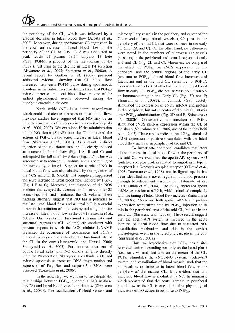
Nitric oxide (NO) is a potent vasorelaxant which could mediate the increases in luteal blood flow. Previous studies have suggested that NO may be an important mediator of luteolysis in the cow (Skarzynski et al., 2000, 2003). We examined if the administration of the NO donor (SNAP) into the CL mimicked the actions of PGF2α on the acute increase in luteal blood flow (Shirasuna et al., 2008b). As a result, a direct injection of the NO donor into the CL clearly induced an increase in blood flow (Fig. 1-A, B and C) and anticipated the fall in P4 by 3 days (Fig. 1-D). This was associated with reduced CL volume and a shortening of the estrous cycle length. Support for a role of NO in luteal blood flow was also obtained by the injection of the NOS inhibitor (L-NAME) that completely suppressed the acute increase in luteal blood flow induced by PGF2α (Fig. 1-E to G). Moreover, administration of the NOS inhibitor also delayed the decreases in P4 secretion for 23 hours (Fig. 1-H) and maintained the CL volume. These findings strongly suggest that NO has a potential to regulate luteal blood flow and a luteal NO is a crucial factor in the initiation of luteolysis by inducing a drastic increase of luteal blood flow in the cow (Shirasuna et al., 2008b). Our results on functional (plasma P4) and structural regression (CL volume) are consistent with previous reports in which the NOS inhibitor L-NAME prevented the occurrence of spontaneous and PGF2α-induced luteolysis and extended the functional life of the CL in the cow (Jaroszewski and Hansel, 2000; Skarzynski et al., 2003). Furthermore, treatment of bovine luteal cells with NO donors in vitro directly inhibited P4 secretion (Skarzynski and Okuda, 2000) and induced apoptosis as increased DNA fragmentation and expression of Fas, Bax and caspase-3 mRNA were observed (Korzekwa et al., 2006).
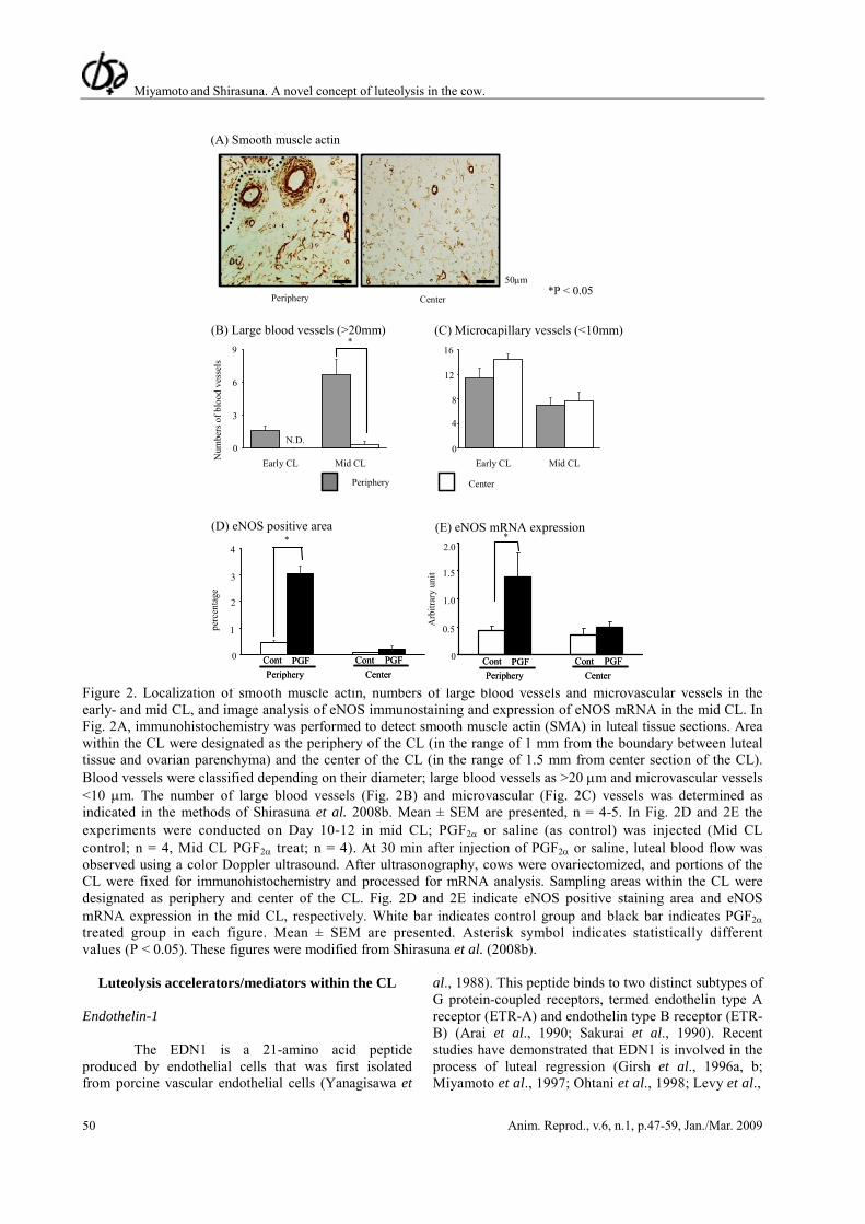
In the next step, we went on to investigate the relationships between PGF2α, endothelial NO synthase (eNOS) and luteal blood vessels in the cow (Shirasuna et al., 2008b). The localization of blood vessels and
microcapillary vessels in the periphery and center of the CL revealed large blood vessels (>20 μm) in the periphery of the mid CL that were not seen in the early CL (Fig. 2A and C). On the other hand, no differences were noted in the numbers of microvascular vessels (<10 μm) in the peripheral and central regions of early and mid CL (Fig. 2B and C). Moreover, we compared the effect of PGF2α on eNOS expression in the peripheral and the central regions of the early CL (resistant to PGF2α-induced blood flow increases and luteolysis) and in the mid CL (sensitive to PGF2α). Consistent with a lack of effect of PGF2α on luteal blood flow in early CL, PGF2α did not increase eNOS mRNA or immunostaining in the Early CL (Fig. 2D and E; Shirasuna et al., 2008b). In contrast, PGF2α acutely stimulated the expression of eNOS mRNA and protein in the periphery, but not in center of the mid CL 30 min after PGF2α administration (Fig. 2D and E; Shirasuna et al., 2008b). Consistently, an injection of PGF2α stimulated eNOS mRNA expression within the CL of the sheep (Vonnahme et al., 2006) and of the rabbit (Boiti et al., 2003). These results indicate that PGF2α-stimulated eNOS expression is positively correlated with the luteal blood flow increase in periphery of the mid CL.
To investigate additional candidate regulators of the increase in luteal blood flow in the periphery of the mid CL, we examined the apelin-APJ system. APJ (putative receptor protein related to angiotensin type 1 receptor) is a G-protein-coupled receptor (O'Dowd et al., 1993; Tatemoto et al., 1998), and its ligand, apelin, has been identified as a novel regulator of blood pressure through NO-dependent vasodilation (Tatemoto et al., 2001; Ishida et al., 2004). The PGF2α increased apelin mRNA expression at 0.5-2 h, which coincided completely with the timing of luteal blood flow increase (Shirasuna et al., 2008a). Moreover, both apelin mRNA and protein expression were stimulated by PGF2α injection at 30 min in the peripheral area of the mid CL, but not in the early CL (Shirasuna et al., 2008a). These results suggest that the apelin-APJ system is involved in the acute increase of luteal blood flow due to regulated NO-vasodilation mechanism and this is the earliest physiological event in the luteolytic cascade in the cow (Shirasuna et al., 2008a).
Thus, we hypothesize that PGF2α has a site-restricted action depending not only on the luteal phase (i.e., early vs. mid) but also on the region of the CL. PGF2α stimulates the eNOS-NO system, apelin-APJ system, and vasodilation of blood vessels, such that the net result is an increase in luteal blood flow in the periphery of the mature CL. It is evident that this increased blood flow is mediated by NO. In summary, we demonstrated that the acute increase in peripheral blood flow to the CL is one of the first physiological indicators of NO action in response to PGF2α.
Miyamoto and Shirasuna. A novel concept of luteolysis in the cow.
Anim. Reprod., v.6, n.1, p.47-59, Jan./Mar. 2009 49
Time after intraluteal injection (hour)
Time after intraluteal injection (hour)
Figure 1. Effect of NO donor (SNAP) or control (DMSO) treatment on luteal blood flow area and plasma P4 (Fig. 1A-D) and effect of the NOS inhibitor (L-NAME) or control (saline) treatment during PGF2α–induced luteolysis in the cow on luteal blood flow area and plasma P4 (Fig. 1E-H). Fig. 1A-D: the experiments were started on Day 14 of the estrous cycle in the cow. The NO donor (S-nitroso-N-acetylpenicillamine, SNAP; 10 mg/ml; 500 μl, n = 5) or dimethylsulfoxide (DMSO) as control (n = 4) was injected directly into the CL twice at 0 h (first injection) and 4 h. Fig. 1A and Fig. 1B show typical image of luteal blood flow treated by NO donor (SNAP) and DMSO, respectively. Fig. 1C shows change of luteal blood flow area, and Fig. 1D shows change of plasma P4 concentration. A direct injection of the NO donor into the CL clearly induced blood flow increase (Fig. 1-A and C) only after the first injection, and shortened the start of decrease of P4 for 3 days (Fig. 1-D) resulting in a shortening of the estrous cycle. Fig. 1E-H: NOS inhibitor (L-NAME; 50 mg/ml; 1 ml, n = 5) was directly injected into the CL four times at -0.5 h, 0 h (PGF2α administration), 2 h and 4 h together with PGF2α administration on Day 14 of the estrous cycle in the cow (saline was directly injected within the CL of cows in the control group, n = 5). Fig. 1E and Fig. 1F show typical image of luteal blood flow treated by NOS inhibitor (L-NAME) and saline as control, respectively. Fig. 1G shows change of luteal blood flow area. Change of plasma P4 concentration is shown in Fig. 1H. The injection of the NOS inhibitor (L-NAME) completely suppressed the acute increase in luteal blood flow induced by PGF2∝ (Fig. 1-E and G). Moreover, administration of the NOS inhibitor delayed the decreases in P4 secretion for 23 hours (Fig. 1-H). In luteal blood flow, the mean values of 0 h (Fig. 1C and D) or -0.5 h (Fig. 1G and H) were used to calculate the baseline for each measurement (defined as 100%) and all values were expressed as a percentage of the corresponding baseline. Mean ± SEM are presented. Asterisk and pound key symbols indicate statistically different values (P < 0.05) from baseline for control and treated groups, respectively. These figures were modified from Shirasuna et al., (2008b).
(A) NO donor (SNAP) treatment
(B) Control (DMSO)0 h 1 h 4 h 12 h
0 h 1 h 4 h 12 h
(E) NOS inhibitor (L-NAME) treatment
(F) Control (saline)
-0.5 h 0 h 2 h 24 h
-0.5 h 0 h 2 h 24 h
(G)
300
100
0
200
-0.5 720 1 2 4 6 8 24 48 96
*
*
*
**
* * *# # # #
#
# # #
Control group
NOS inhibitor treated group
(H)
4
6
2
0
* **
* *
* * * *
#
# # #
-0.5 720 1 2 4 6 8 24 48 96
23 hours
(G)
300
100
0
200
-0.5 720 1 2 4 6 8 24 48 96-0.5 720 1 2 4 6 8 24 48 96
*
*
*
**
* * *# # # #
#
# # #
Control group
NOS inhibitor treated group
Control group
NOS inhibitor treated group
(H)
4
6
2
0
* **
* *
* * * *
#
# # #
-0.5 720 1 2 4 6 8 24 48 96-0.5 720 1 2 4 6 8 24 48 96
23 hours
Blo
od fl
ow a
rea
(%)
Pla
sma
P4 (n
g/m
l)
Blo
od fl
ow a
rea
(%)
Plas
ma
P4 (n
g/m
l)
1441241 24 48 72 96 120 168 192
100
50
0
150
0
* *
**
****
####
#
#
#
##
200(C) Control group
NO donor treated group
4
6
2
0
8
**
*
*
**
# # ## # # # #
(D)
1441241 24 48 72 96 120 168 1920
3 days
1441241 24 48 72 96 120 168 1921441241 24 48 72 96 120 168 192
100
50
0
150
0
* *
**
****
####
#
#
#
##
200(C) Control group
NO donor treated group
4
6
2
0
8
**
*
*
**
# # ## # # # #
(D)
1441241 24 48 72 96 120 168 1920 1441241 24 48 72 96 120 168 1921441241 24 48 72 96 120 168 1920
3 days
Miyamoto and Shirasuna. A novel concept of luteolysis in the cow.
Anim. Reprod., v.6, n.1, p.47-59, Jan./Mar. 2009 50
Figure 2. Localization of smooth muscle actin, numbers of large blood vessels and microvascular vessels in the early- and mid CL, and image analysis of eNOS immunostaining and expression of eNOS mRNA in the mid CL. In Fig. 2A, immunohistochemistry was performed to detect smooth muscle actin (SMA) in luteal tissue sections. Area within the CL were designated as the periphery of the CL (in the range of 1 mm from the boundary between luteal tissue and ovarian parenchyma) and the center of the CL (in the range of 1.5 mm from center section of the CL). Blood vessels were classified depending on their diameter; large blood vessels as >20 μm and microvascular vessels <10 μm. The number of large blood vessels (Fig. 2B) and microvascular (Fig. 2C) vessels was determined as indicated in the methods of Shirasuna et al. 2008b. Mean ± SEM are presented, n = 4-5. In Fig. 2D and 2E the experiments were conducted on Day 10-12 in mid CL; PGF2α or saline (as control) was injected (Mid CL control; n = 4, Mid CL PGF2α treat; n = 4). At 30 min after injection of PGF2α or saline, luteal blood flow was observed using a color Doppler ultrasound. After ultrasonography, cows were ovariectomized, and portions of the CL were fixed for immunohistochemistry and processed for mRNA analysis. Sampling areas within the CL were designated as periphery and center of the CL. Fig. 2D and 2E indicate eNOS positive staining area and eNOS mRNA expression in the mid CL, respectively. White bar indicates control group and black bar indicates PGF2α treated group in each figure. Mean ± SEM are presented. Asterisk symbol indicates statistically different values (P < 0.05). These figures were modified from Shirasuna et al. (2008b).
Luteolysis accelerators/mediators within the CL Endothelin-1
The EDN1 is a 21-amino acid peptide
produced by endothelial cells that was first isolated from porcine vascular endothelial cells (Yanagisawa et
al., 1988). This peptide binds to two distinct subtypes of G protein-coupled receptors, termed endothelin type A receptor (ETR-A) and endothelin type B receptor (ETR-B) (Arai et al., 1990; Sakurai et al., 1990). Recent studies have demonstrated that EDN1 is involved in the process of luteal regression (Girsh et al., 1996a, b; Miyamoto et al., 1997; Ohtani et al., 1998; Levy et al.,
Periphery Center
(A) Smooth muscle actin
Num
bers
of b
lood
ves
sels
9
Periphery
6
3
0
Center
*
* ; P < 0.05
Mid CL Mid CLEarly CL Early CL
N.D.
12
8
4
0
16
(B) Large blood vessels (>20mm) (C) Microcapillary vessels (<10mm)
50μm
perc
enta
ge
(E) eNOS mRNA expression
Arb
itrar
y un
it
* *(D) eNOS positive area
4
3
1
0
2
1.5
0.5
0
1.0
2.0
Periphery CenterCont PGF Cont PGFPeriphery Center
Cont PGF Cont PGFPeriphery Center
Cont PGF Cont PGFPeriphery Center
Cont PGF Cont PGF
*P < 0.05
Miyamoto and Shirasuna. A novel concept of luteolysis in the cow.
Anim. Reprod., v.6, n.1, p.47-59, Jan./Mar. 2009 51
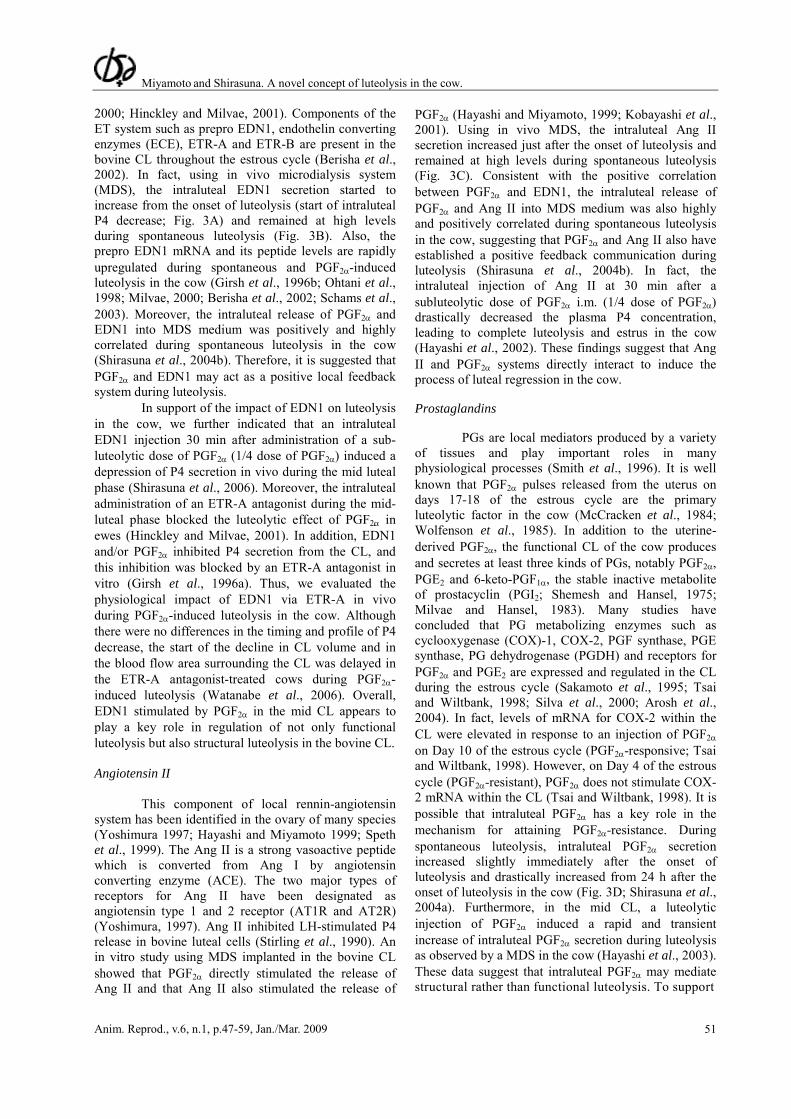
2000; Hinckley and Milvae, 2001). Components of the ET system such as prepro EDN1, endothelin converting enzymes (ECE), ETR-A and ETR-B are present in the bovine CL throughout the estrous cycle (Berisha et al., 2002). In fact, using in vivo microdialysis system (MDS), the intraluteal EDN1 secretion started to increase from the onset of luteolysis (start of intraluteal P4 decrease; Fig. 3A) and remained at high levels during spontaneous luteolysis (Fig. 3B). Also, the prepro EDN1 mRNA and its peptide levels are rapidly upregulated during spontaneous and PGF2α-induced luteolysis in the cow (Girsh et al., 1996b; Ohtani et al., 1998; Milvae, 2000; Berisha et al., 2002; Schams et al., 2003). Moreover, the intraluteal release of PGF2α and EDN1 into MDS medium was positively and highly correlated during spontaneous luteolysis in the cow (Shirasuna et al., 2004b). Therefore, it is suggested that PGF2α and EDN1 may act as a positive local feedback system during luteolysis.
In support of the impact of EDN1 on luteolysis in the cow, we further indicated that an intraluteal EDN1 injection 30 min after administration of a sub-luteolytic dose of PGF2α (1/4 dose of PGF2α) induced a depression of P4 secretion in vivo during the mid luteal phase (Shirasuna et al., 2006). Moreover, the intraluteal administration of an ETR-A antagonist during the mid-luteal phase blocked the luteolytic effect of PGF2α in ewes (Hinckley and Milvae, 2001). In addition, EDN1 and/or PGF2α inhibited P4 secretion from the CL, and this inhibition was blocked by an ETR-A antagonist in vitro (Girsh et al., 1996a). Thus, we evaluated the physiological impact of EDN1 via ETR-A in vivo during PGF2α-induced luteolysis in the cow. Although there were no differences in the timing and profile of P4 decrease, the start of the decline in CL volume and in the blood flow area surrounding the CL was delayed in the ETR-A antagonist-treated cows during PGF2α-induced luteolysis (Watanabe et al., 2006). Overall, EDN1 stimulated by PGF2α in the mid CL appears to play a key role in regulation of not only functional luteolysis but also structural luteolysis in the bovine CL. Angiotensin II
This component of local rennin-angiotensin system has been identified in the ovary of many species (Yoshimura 1997; Hayashi and Miyamoto 1999; Speth et al., 1999). The Ang II is a strong vasoactive peptide which is converted from Ang I by angiotensin converting enzyme (ACE). The two major types of receptors for Ang II have been designated as angiotensin type 1 and 2 receptor (AT1R and AT2R) (Yoshimura, 1997). Ang II inhibited LH-stimulated P4 release in bovine luteal cells (Stirling et al., 1990). An in vitro study using MDS implanted in the bovine CL showed that PGF2α directly stimulated the release of Ang II and that Ang II also stimulated the release of
PGF2α (Hayashi and Miyamoto, 1999; Kobayashi et al., 2001). Using in vivo MDS, the intraluteal Ang II secretion increased just after the onset of luteolysis and remained at high levels during spontaneous luteolysis (Fig. 3C). Consistent with the positive correlation between PGF2α and EDN1, the intraluteal release of PGF2α and Ang II into MDS medium was also highly and positively correlated during spontaneous luteolysis in the cow, suggesting that PGF2α and Ang II also have established a positive feedback communication during luteolysis (Shirasuna et al., 2004b). In fact, the intraluteal injection of Ang II at 30 min after a subluteolytic dose of PGF2α i.m. (1/4 dose of PGF2α) drastically decreased the plasma P4 concentration, leading to complete luteolysis and estrus in the cow (Hayashi et al., 2002). These findings suggest that Ang II and PGF2α systems directly interact to induce the process of luteal regression in the cow. Prostaglandins
PGs are local mediators produced by a variety
of tissues and play important roles in many physiological processes (Smith et al., 1996). It is well known that PGF2α pulses released from the uterus on days 17-18 of the estrous cycle are the primary luteolytic factor in the cow (McCracken et al., 1984; Wolfenson et al., 1985). In addition to the uterine-derived PGF2α, the functional CL of the cow produces and secretes at least three kinds of PGs, notably PGF2α, PGE2 and 6-keto-PGF1α, the stable inactive metabolite of prostacyclin (PGI2; Shemesh and Hansel, 1975; Milvae and Hansel, 1983). Many studies have concluded that PG metabolizing enzymes such as cyclooxygenase (COX)-1, COX-2, PGF synthase, PGE synthase, PG dehydrogenase (PGDH) and receptors for PGF2α and PGE2 are expressed and regulated in the CL during the estrous cycle (Sakamoto et al., 1995; Tsai and Wiltbank, 1998; Silva et al., 2000; Arosh et al., 2004). In fact, levels of mRNA for COX-2 within the CL were elevated in response to an injection of PGF2α on Day 10 of the estrous cycle (PGF2α-responsive; Tsai and Wiltbank, 1998). However, on Day 4 of the estrous cycle (PGF2α-resistant), PGF2α does not stimulate COX-2 mRNA within the CL (Tsai and Wiltbank, 1998). It is possible that intraluteal PGF2α has a key role in the mechanism for attaining PGF2α-resistance. During spontaneous luteolysis, intraluteal PGF2α secretion increased slightly immediately after the onset of luteolysis and drastically increased from 24 h after the onset of luteolysis in the cow (Fig. 3D; Shirasuna et al., 2004a). Furthermore, in the mid CL, a luteolytic injection of PGF2α induced a rapid and transient increase of intraluteal PGF2α secretion during luteolysis as observed by a MDS in the cow (Hayashi et al., 2003). These data suggest that intraluteal PGF2α may mediate structural rather than functional luteolysis. To support
Miyamoto and Shirasuna. A novel concept of luteolysis in the cow.
Anim. Reprod., v.6, n.1, p.47-59, Jan./Mar. 2009 52
PGFM
Ang II
this concept, intraluteal implants of indomethacin, a potent PG synthase inhibitor, on Day 11 of the estrous cycle in ewes resulted in heavier CL on Day 18 than that in untreated control ewes (Griffeth et al., 2002),
suggesting that intraluteal production of PGF2α is required for structural luteolysis. Furthermore, the systemic administration of PG synthesis inhibitors delayed structural luteolysis in rats (Kurusu et al., 2001).
Time from onset of luteolysis (h) Figure 3. Local release within the corpus luteum of P4 (A), EDN1 (B), Ang II (C), PGF2α (D), PGFM (E) and OXT (F) into MDS (bars; 18 lines from 6 cows) during spontaneous luteolysis in the cow. For statistical analysis, the experimental period was divided into 12 stages. Each stage contains measurements from samples collected within a 12 h period (3 fractions). The MDS data are expressed as a percentage of basal release (baseline) for first 24 h. Mean ± SEM are presented. Asterisk symbol indicates statistically different values (P < 0.05) from baseline (-24-0 h). These figures were modified from Shirasuna et al., (2004a, b, 2007a). Oxytocin
In the bovine CL, the expression of oxytocin (OXT) mRNA is high during the early luteal phase (Ivell et al., 1985; Furuya et al., 1990; Wathes and Denning-Kendall, 1992), and the OXT peptide is expressed at a higher level during the mid luteal phase (Parkinson et al., 1992; Wathes and Denning-Kendall, 1992). PGF2α stimulates OXT secretion from the CL
(Flint and Sheldrick, 1982), and OXT in turn stimulates uterine secretion of PGF2α (Sharma and Fitzpatrick, 1974; Roberts and McCracken, 1976). Thus, endometrial PGF2α and luteal OXT comprise a positive feedback mechanism which acts between the uterus and the CL to induce luteal regression (Schallenberger et al., 1984). Although the mean release of intraluteal OXT was maintained at the same levels during spontaneous luteolysis in the cow (Fig. 3F), the pulsatile release of
P4 PGF2α
EDN1
OXT
Perc
enta
ge fr
om b
asel
ine
*
*
*
*
(B)
0
50
100
150
200
250
0
100
300
500
0
80
100
(A) (D)
60
40
20
120
400
200
(C)
600
100
200
300
150
250
0
50
100
150
200
0
50
100
150
200
(E)
(F)250
**
* * * **
*
**
*
*
*
*
*
*
*
**
*
*
**
**
*** *
*
*
-48 0 24 48 72 96-24 24 48 72 96-24 0-480
50
*
*
*
*
(B)
0
50
100
150
200
250
0
100
300
500
0
80
100
(A) (D)
60
40
20
120
400
200
(C)
600
100
200
300
150
250
0
50
100
150
200
0
50
100
150
200
(E)
(F)250
**
* * * **
*
**
*
*
*
*
*
*
*
**
*
*
**
**
*** *
*
*
-48 0 24 48 72 96-24-48 0 24 48 72 96-24 24 48 72 96-24 0-48 24 48 72 96-24 0-480
50
Miyamoto and Shirasuna. A novel concept of luteolysis in the cow.
Anim. Reprod., v.6, n.1, p.47-59, Jan./Mar. 2009 53
OXT within CL was highly positively associated with intraluteal PGF2α and EDN1, but not with intraluteal Ang II (Shirasuna et al., 2007a). In fact, OXT stimulates the release of PGF2α in luteal cells (Grazul et al., 1989) and EDN1 in luteal endothelial cells (Girsh et al.,
1996b). Thus, we propose that luteal OXT, PGF2α and EDN1 may establish a local positive feedback loop within the microenvironment, and OXT may amplify the frequency of vasoactive substance secretion after the onset of luteal regression within the CL (Fig. 4).
Luteal CellsEndothelial Cells
CL
ProgesteroneBlood Flow
OXT
Ang II Luteal PGF2α
Uterine PGF2α
EDN1
Luteal CellsEndothelial Cells
CL
ProgesteroneBlood Flow
OXT
Ang II Luteal PGF2α
Uterine PGF2α
EDN1
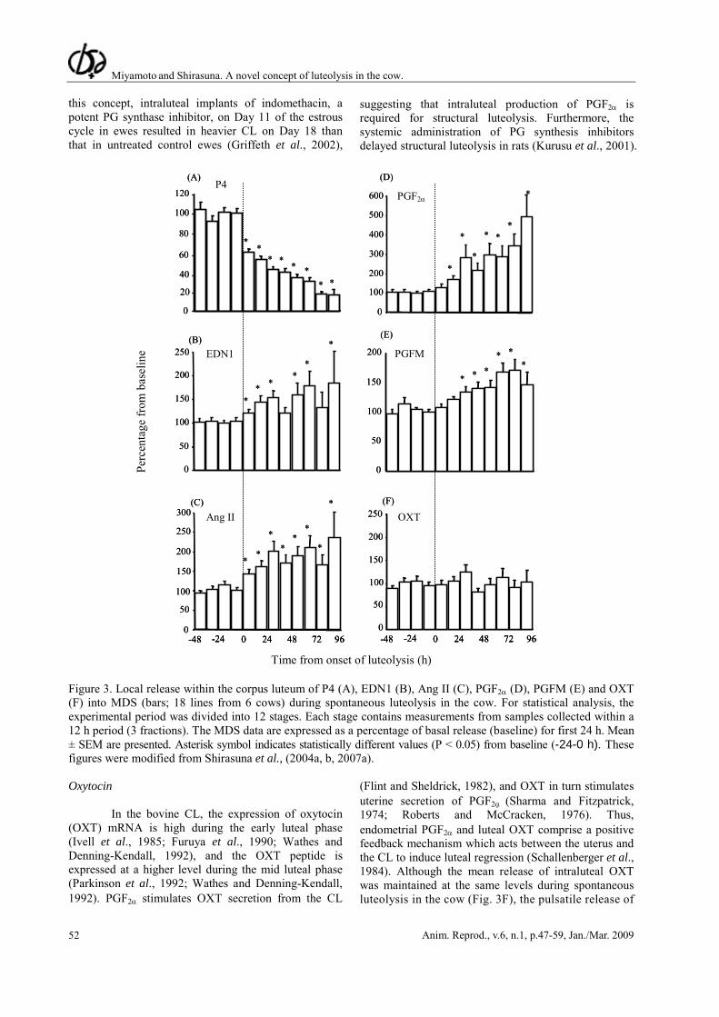
Figure 4. Proposed model for local positive feedback system among vasoactive molecules established during luteolysis in the cow. In the CL, following an increase in luteal blood flow, PGF2α directly stimulates the production of EDN1 and Ang II from luteal endothelial cells and luteal PGF2α from luteal cells. It is suggested that PGF2α, EDN1 and Ang II may act as a positive local feedback system during luteolysis. Moreover, luteal OXT, PGF2α and EDN1 may establish a local positive feedback loop within the microenvironment, and OXT may amplify the frequency of vasoactive substance secretion after the onset of luteal regression within the CL. Overall, these mechanisms in the CL appear to play a key role in regulation of not only functional luteolysis (P4 decrease) but also the decrease of luteal blood flow in the bovine CL. Involvement of cell adhesion systems within the CL
It is likely that cell-to-cell interactions are
important for the maintenance and regulation of CL integrity and physiological function. Therefore, it has been suggested that there are intercellular communications via a contact-dependent pathway among various cell types within the CL.
Gap junction are formed with tunnel-like structures that enable regulatory molecules, nutrients and ions of less than about 1 kDa (e.g., calcium ions, cAMP and inositol 1,4,5-triphosphate) to be exchanged between adjacent cells (Yamasaki and Naus, 1996). Gap junctions are formed by connexin (Cx) proteins such as Cx43 (Beyer et al., 1990), and these have been suggested to be predominantly important for regulation of growth, differentiation and regression of the CL (Grazul-Bilska et al., 1997). On the other hand, the adherence junction is another cell adhesion type, and cadherins have key roles in these junctions. The cadherin family, includes vascular endothelial cell cadherin (VE-cadherin), epithelial cadherin (E-cadherin) and neuronal cadherin (N-cadherin). Cadherins act as major factors in tissue development and in the differentiation of many organs, including the CL of humans (Khan-Dawood et al., 1996), baboons (Khan-Dawood et al., 1996), mice (Nakhuda et al., 2005) and rats (Trolice et al., 1997; Sundfeldt et al., 2000).
In the bovine CL, Cx43 mRNA expression was detected in luteal endothelial cells and in luteinized granulosa cells (GCs). Moreover, Cx43 mRNA expression tended to increase 24 h after PGF2α administration and thereafter it was significantly low during the regressing luteal stage. In fact, expression of Cx43 mRNA remained at a relatively high level during PGF2α-induced luteolysis in sheep (Borowczyk et al., 2006), and PGF2α enhanced gap junctional intercellular communication of bovine luteal cells from the late luteal phase (Grazul-Bilska et al., 1997). In addition, the mRNA expressions of VE-cadherin and E-cadherin increased at 24 h during PGF2α-induced luteolysis. These observations suggest that maintenance of Cx43, VE-cadherin and E-cadherin mRNA expression during regression may contribute to ensure the cell adhesion and gap junction necessary for transferring the luteolytic signal between luteal cells and endothelial cells (Grazul-Bilska et al., 1997; Borowczyk et al., 2006).
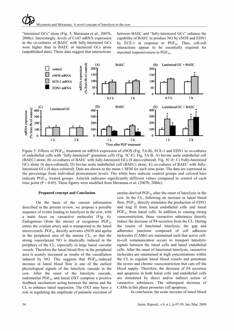
To test the hypothesis that cell adhesion is important for luteolysis, we recently established a co-culture system using bovine aorta endothelial cells (BAEC) and “fully-luteinized” GCs, and investigated the effect of PGF2α on the expression of eNOS mRNA and EDN1 system (Shirasuna et al., 2007b, 2008c). PGF2α stimulated the expression of eNOS, EDN1 and ECE-1 mRNA only in the co-cultures of endothelial cells with “fully-luteinized GCs”, but not in BAEC or
Miyamoto and Shirasuna. A novel concept of luteolysis in the cow.
Anim. Reprod., v.6, n.1, p.47-59, Jan./Mar. 2009 54
“luteinized GCs” alone (Fig. 5; Shirasuna et al., 2007b, 2008c). Interestingly, levels of Cx43 mRNA expression in the co-cultures of BAEC with fully-luteinized GCs were higher than in BAEC or luteinized GCs alone (unpublished data). These data suggest that interactions
between BAEC and “fully-luteinized GCs” enhance the capability of BAEC to produce NO by eNOS and EDN1 by ECE-1 in response to PGF2α. Thus, cell-cell interactions appear to be essentially required for maximal responsiveness to PGF2α.
control
eNOS mRNAm
RN
A e
xpre
ssio
n(%
from
0h
of c
ontro
l)
150
100
50
0
200
150
100
50
0
200
300
200
100
0
400
300
200
100
0
400
300
200
100
0
400
mR
NA
exp
ress
ion
(% fr
om 0
h of
con
trol)
ECE-1 mRNA
EDN1 mRNA
(B)(A)
(D)(C) (E)
BAEC
BAEC
Luteinized GC + BAEC
Luteinized GC + BAECLuteinized GC
1 h 2 h 1 h 2 h 1 h 2 h
0.5 h 1 h 0.5 h 1 h
*
*
* **
Time after PGF treatment
control
eNOS mRNAm
RN
A e
xpre
ssio
n(%
from
0h
of c
ontro
l)
150
100
50
0
200
150
100
50
0
200
300
200
100
0
400
300
200
100
0
400
300
200
100
0
400
mR
NA
exp
ress
ion
(% fr
om 0
h of
con
trol)
ECE-1 mRNA
EDN1 mRNA
(B)(A)
(D)(C) (E)
BAEC
BAEC
Luteinized GC + BAEC
Luteinized GC + BAECLuteinized GC
1 h 2 h 1 h 2 h 1 h 2 h
0.5 h 1 h 0.5 h 1 h
*
*
* **
Time after PGF treatment Figure 5. Effects of PGF2α treatment on mRNA expression of eNOS (Fig. 5A-B), ECE-1 and EDN1 in co-cultures of endothelial cells with “fully-luteinized” granulosa cells (Fig. 5C-E). Fig. 5A-B: A) bovine aorta endothelial cell (BAEC) alone; B) co-cultures of BAEC with fully-luteinized GCs (8 days-cultured). Fig. 5C-E: C) Fully-luteinized GCs alone (8 days-cultured); D) bovine aorta endothelial cell (BAEC) alone; E) co-cultures of BAEC with fully-luteinized GCs (8 days-cultured). Data are shown as the mean ± SEM for each time point. The data are expressed as the percentage from individual pretreatment levels. The white bars indicate control groups and colored bars indicate PGF2α treated groups. Asterisk indicates significantly different values compared to control of each time point (P < 0.05). These figures were modified from Shirasuna et al. (2007b, 2008c).
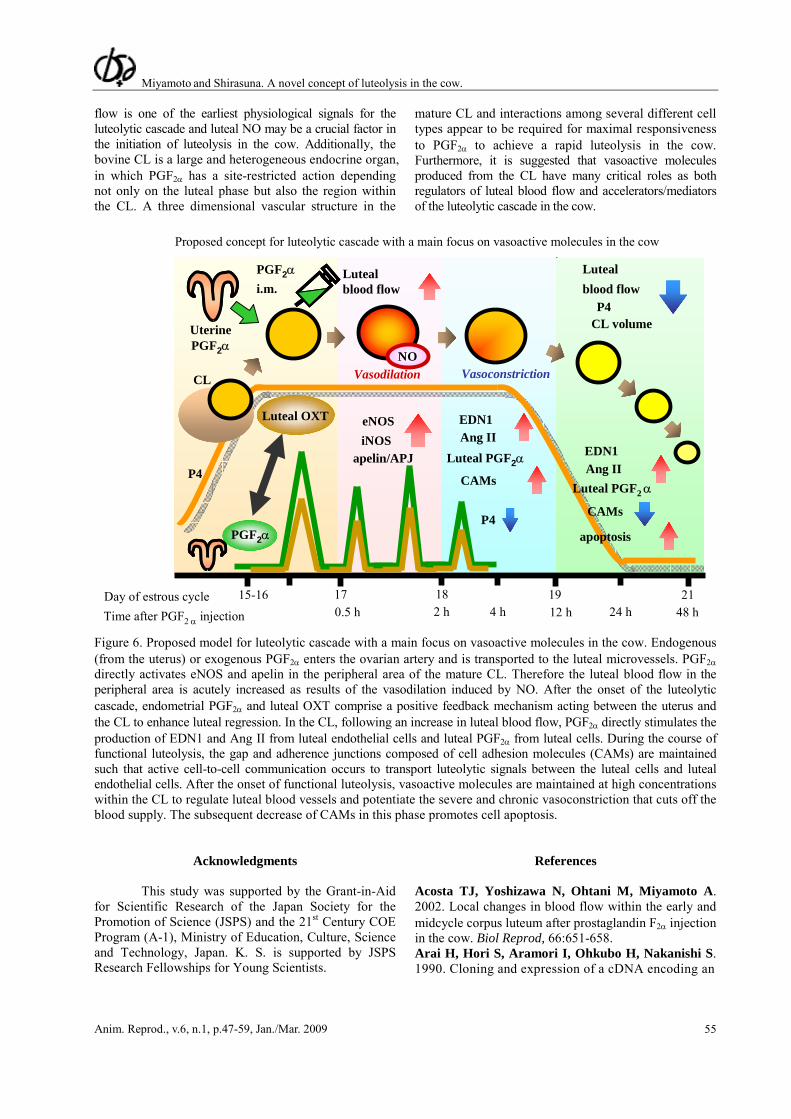
Proposed concept and Conclusion
On the basis of the current information described in the present review, we propose a possible sequence of events leading to luteolysis in the cow, with a main focus on vasoactive molecules (Fig. 6). Endogenous (from the uterus) or exogenous PGF2α enters the ovarian artery and is transported to the luteal microvessels. PGF2α directly activates eNOS and apelin in the peripheral area of the mature CL, so that the strong vasorelaxant NO is drastically induced in the periphery of the CL, especially in large luteal vascular vessels. Therefore the luteal blood flow in the peripheral area is acutely increased as results of the vasodilation induced by NO. This suggests that PGF2α-induced increase in luteal blood flow is one of the earliest physiological signals of the luteolytic cascade in the cow. After the onset of the luteolytic cascade, endometrial PGF2α and luteal OXT comprise a positive feedback mechanism acting between the uterus and the CL to enhance luteal regression. The OXT may have a role in regulating the amplitude of pulsatile secretion of
uterine-derived PGF2α after the onset of luteolysis in the cow. In the CL, following an increase in luteal blood flow, PGF2α directly stimulates the production of EDN1 and Ang II from luteal endothelial cells and luteal PGF2α from luteal cells. In addition to causing strong vasoconstriction, these vasoactive substances directly induce the decrease of P4 secretion from the CL. During the course of functional luteolysis, the gap and adherence junctions composed of cell adhesion molecules (CAMs) are maintained such that active cell-to-cell communication occurs to transport luteolytic signals between the luteal cells and luteal endothelial cells. After the onset of functional luteolysis, vasoactive molecules are maintained at high concentrations within the CL to regulate luteal blood vessels and potentiate the severe and chronic vasoconstriction that cuts off the blood supply. Therefore, the decrease of P4 secretion and apoptosis in both luteal cells and endothelial cells are stimulated by direct and/or indirect action of vasoactive substances. The subsequent decrease of CAMs in this phase promotes cell apoptosis.
In conclusion, the acute increase of luteal blood
Miyamoto and Shirasuna. A novel concept of luteolysis in the cow.
Anim. Reprod., v.6, n.1, p.47-59, Jan./Mar. 2009 55
flow is one of the earliest physiological signals for the luteolytic cascade and luteal NO may be a crucial factor in the initiation of luteolysis in the cow. Additionally, the bovine CL is a large and heterogeneous endocrine organ, in which PGF2α has a site-restricted action depending not only on the luteal phase but also the region within the CL. A three dimensional vascular structure in the
mature CL and interactions among several different cell types appear to be required for maximal responsiveness to PGF2α to achieve a rapid luteolysis in the cow. Furthermore, it is suggested that vasoactive molecules produced from the CL have many critical roles as both regulators of luteal blood flow and accelerators/mediators of the luteolytic cascade in the cow.
CL
PGF2αi.m.
Time after PGF2 α injection
15-16 17 18 19 21
EDN1
Lutealblood flow
UterinePGF2α
Luteal OXT
PGF2α
eNOSiNOS
apelin/APJ
Lutealblood flow
CL volume
Luteal PGF2α
Ang II
P4
CAMs
P4
CAMs
apoptosis
EDN1
Luteal PGF2 αAng II
0.5 h 2 h 12 h 48 h4 h 24 hDay of estrous cycle
NOVasodilation Vasoconstriction
Proposed concept for luteolytic cascade with a main focus on vasoactive molecules in the cow
P4
Figure 6. Proposed model for luteolytic cascade with a main focus on vasoactive molecules in the cow. Endogenous (from the uterus) or exogenous PGF2α enters the ovarian artery and is transported to the luteal microvessels. PGF2α directly activates eNOS and apelin in the peripheral area of the mature CL. Therefore the luteal blood flow in the peripheral area is acutely increased as results of the vasodilation induced by NO. After the onset of the luteolytic cascade, endometrial PGF2α and luteal OXT comprise a positive feedback mechanism acting between the uterus and the CL to enhance luteal regression. In the CL, following an increase in luteal blood flow, PGF2α directly stimulates the production of EDN1 and Ang II from luteal endothelial cells and luteal PGF2α from luteal cells. During the course of functional luteolysis, the gap and adherence junctions composed of cell adhesion molecules (CAMs) are maintained such that active cell-to-cell communication occurs to transport luteolytic signals between the luteal cells and luteal endothelial cells. After the onset of functional luteolysis, vasoactive molecules are maintained at high concentrations within the CL to regulate luteal blood vessels and potentiate the severe and chronic vasoconstriction that cuts off the blood supply. The subsequent decrease of CAMs in this phase promotes cell apoptosis.
Acknowledgments
This study was supported by the Grant-in-Aid for Scientific Research of the Japan Society for the Promotion of Science (JSPS) and the 21st Century COE Program (A-1), Ministry of Education, Culture, Science and Technology, Japan. K. S. is supported by JSPS Research Fellowships for Young Scientists.
References Acosta TJ, Yoshizawa N, Ohtani M, Miyamoto A. 2002. Local changes in blood flow within the early and midcycle corpus luteum after prostaglandin F2α injection in the cow. Biol Reprod, 66:651-658. Arai H, Hori S, Aramori I, Ohkubo H, Nakanishi S. 1990. Cloning and expression of a cDNA encoding an
Miyamoto and Shirasuna. A novel concept of luteolysis in the cow.
Anim. Reprod., v.6, n.1, p.47-59, Jan./Mar. 2009 56
endothelin receptor. Nature, 348:730-732. Arosh JA, Banu SK, Chapdelaine P, Madore E, Sirois J, Fortier MA. 2004. Prostaglandin biosynthesis, transport, and signaling in corpus luteum: a basis for autoregulation of luteal function. Endocrinology, 145:2551-2560. Berisha B, Schams D, Miyamoto A. 2002. The expression of angiotensin and endothelin system members in bovine corpus luteum during estrous cycle and pregnancy. Endocrine, 19:305-312. Beyer EC, Paul DL, Goodenough DA. 1990. Connexin family of gap junction proteins. J Membr Biol, 116:187-194. Boiti C, Guelfi G, Zampini D, Brecchia G, Gobbetti A, Zerani M. 2003. Regulation of nitric oxide synthase isoforms and role of nitric oxide during prostaglandin F2α-induced luteolysis in rabbits. Reproduction, 125:807-816. Borowczyk E, Johnson ML, Bilski JJ, Borowicz PP, Redmer DA, Reynolds LP, Grazul-Bilska AT. 2006. Expression of gap junctional connexins 26, 32, and 43 mRNA in ovarian preovulatory follicles and corpora lutea in sheep. Can J Physiol Pharmacol, 84:1011-1020. Farin CE, Moeller CL, Sawyer HR, Gamboni F, Niswender GD. 1986. Morphometric analysis of cell types in the ovine corpus luteum throughout the estrous cycle. Biol Reprod, 35:1299-1308. Flint AP, Sheldrick EL. 1982. Ovarian secretion of oxytocin is stimulated by prostaglandin. Nature, 297:587-588. Furuya K, McArdle CA, Ivell R. 1990. The regulation of oxytocin gene expression in early bovine luteal cells. Mol Cell Endocrinol, 70:81-88. Ginther OJ, Silva LA, Araujo RR, Beg MA. 2007. Temporal associations among pulses of 13,14-dihydro-15-keto-PGF2α, luteal blood flow, and luteolysis in cattle. Biol Reprod, 76:506-513. Girsh E, Greber Y, Meidan R. 1995. Luteotrophic and luteolytic interactions between bovine small and large luteal-like cells and endothelial cells. Biol Reprod, 52:954-962. Girsh E, Milvae RA, Wang W, Meidan R. 1996a. Effect of endothelin-1 on bovine luteal cell function: role in prostaglandin F2α-induced antisteroidogenic action. Endocrinology, 137:1306-1312. Girsh E, Wang W, Mamluk R, Arditi F, Friedman A, Milvae RA, Meidan R. 1996b. Regulation of endothelin-1 expression in the bovine corpus luteum: elevation by prostaglandin F2α. Endocrinology, 137:5191-5196. Grazul AT, Kirsch JD, Slanger WD, Marchello MJ, Redmer DA. 1989. Prostaglandin F2α, oxytocin and progesterone secretion by bovine luteal cells at several stages of luteal development: effects of oxytocin, luteinizing hormone, prostaglandin F2α and estradiol-17β. Prostaglandins, 38:307-318. Grazul-Bilska AT, Redmer DA, Reynolds LP. 1997.
Cellular interactions in the corpus luteum. Semin Reprod Endocrinol, 15:383-393. Griffeth R, Nett T, Burns P, Escudero J, Inskeep E, Niswender G. 2002. Is luteal production of PGF2α required for luteolysis? Biol Reprod Suppl, 66:465. Hayashi K, Miyamoto A. 1999. Angiotensin II interacts with prostaglandin F2α and endothelin-1 as a local luteolytic factor in the bovine corpus luteum in vitro. Biol Reprod, 60:1104-1109. Hayashi K, Miyamoto A, Berisha B, Kosmann MR, Okuda K, Schams D. 2000. Regulation of angiotensin II production and angiotensin receptors in microvascular endothelial cells from bovine corpus luteum. Biol Reprod, 62:162-167. Hayashi K, Tanaka J, Hayashi KG, Hayashi M, Ohtani M, Miyamoto A. 2002. The cooperative action of angiotensin II with subluteolytic administration of PGF2α in inducing luteolysis and oestrus in the cow. Reproduction, 124:311-315. Hayashi K, Acosta TJ, Berisha B, Kobayashi S, Ohtani M, Schams D, Miyamoto A. 2003. Changes in prostaglandin secretion by the regressing bovine corpus luteum. Prostaglandins Other Lipid Mediat, 70:339-349. Henricks DM, Long JT, Hill JR, Dickey JF. 1974. The effect of prostaglandin F2α during various stages of the oestrous cycle of beef heifers. J Reprod Fertil, 41:113-120. Hinckley ST, Milvae RA. 2001. Endothelin-1 mediates prostaglandin F2α-induced luteal regression in the ewe. Biol Reprod, 64:1619-1623. Ishida J, Hashimoto T, Hashimoto Y, Nishiwaki S, Iguchi T, Harada S, Sugaya T, Matsuzaki H, Yamamoto R, Shiota N, Okunishi H, Kihara M, Umemura S, Sugiyama F, Yagami K, Kasuya Y, Mochizuki N, Fukamizu A. 2004. Regulatory roles for APJ, a seven-transmembrane receptor related to angiotensin-type 1 receptor in blood pressure in vivo. J Biol Chem, 279:26274-26279. Ivell R, Brackett KH, Fields MJ, Richter D. 1985. Ovulation triggers oxytocin gene expression in the bovine ovary. FEBS Lett, 190:263-267. Jaroszewski JJ, Hansel W. 2000. Intraluteal administration of a nitric oxide synthase blocker stimulates progesterone and oxytocin secretion and prolongs the life span of the bovine corpus luteum. Proc Soc Exp Biol Med, 224:50-55. Khan-Dawood FS, Yang J, Dawood MY. 1996. Expression of gap junction protein connexin-43 in the human and baboon (Papio anubis) corpus luteum. J Clin Endocrinol Metab, 81:835-842. Kobayashi S, Berisha B, Amselgruber WM, Schams D, Miyamoto A. 2001. Production and localisation of angiotensin II in the bovine early corpus luteum: a possible interaction with luteal angiogenic factors and prostaglandin F2α. J Endocrinol, 170:369-380. Korzekwa AJ, Okuda K, Woclawek-Potocka I, Murakami S, Skarzynski DJ. 2006. Nitric oxide
Miyamoto and Shirasuna. A novel concept of luteolysis in the cow.
Anim. Reprod., v.6, n.1, p.47-59, Jan./Mar. 2009 57
induces apoptosis in bovine luteal cells. J Reprod Dev, 52:353-361. Kurusu S, Sakaguchi S, Kawaminami M, Hashimoto I. 2001. Dexamethasone and indomethacin inhibition of structural luteolysis in rats: an intraluteal mechanism involving prolonged activation of phospholipase A2 activity and prostaglandin synthesis may facilitate the luteolytic process. J Reprod Dev, 47:383-391. Lei ZM, Chegini N, Rao CV. 1991. Quantitative cell composition of human and bovine corpora lutea from various reproductive states. Biol Reprod, 44:1148-1156. Levy N, Kobayashi S, Roth Z, Wolfenson D, Miyamoto A, Meidan R. 2000. Administration of prostaglandin F2α during the early bovine luteal phase does not alter the expression of ET-1 and of its type A receptor: a possible cause for corpus luteum refractoriness. Biol Reprod, 63:377-382. McCracken JA, Schramm W, Okulicz WC. 1984. Hormone receptor control of pulsatile secretion of PGF2α from the ovine uterus during luteolysis and its abrogation in early pregnancy. Anim Reprod Sci, 7:31-55. Meidan R, Girsh E, Blum O, Aberdam E. 1990. In vitro differentiation of bovine theca and granulosa cells into small and large luteal-like cells: morphological and functional characteristics. Biol Reprod, 43:913-921. Milvae RA, Hansel W. 1983. Prostacyclin, prostaglandin F2α and progesterone production by bovine luteal cells during the estrous cycle. Biol Reprod, 29:1063-1068. Milvae RA. 2000. Inter-relationships between endothelin and prostaglandin F2α in corpus luteum function. Rev Reprod, 5:1-5. Miyamoto A, von Lutzow H, Schams D. 1993. Acute actions of prostaglandin F2α, E2, and I2 in microdialyzed bovine corpus luteum in vitro. Biol Reprod, 49:423-430. Miyamoto A, Kobayashi S, Arata S, Ohtani M, Fukui Y, Schams D. 1997. Prostaglandin F2α promotes the inhibitory action of endothelin-1 on the bovine luteal function in vitro. J Endocrinol, 152:R7-11. Miyamoto A, Shirasuna K, Wijayagunawardane MP, Watanabe S, Hayashi M, Yamamoto D, Matsui M, Acosta TJ. 2005. Blood flow: a key regulatory component of corpus luteum function in the cow. Domest Anim Endocrinol, 29:329-339. Nakhuda GS, Zimmermann RC, Bohlen P, Liao F, Sauer MV, Kitajewski J. 2005. Inhibition of the vascular endothelial cell (VE)-specific adhesion molecule VE-cadherin blocks gonadotropin-dependent folliculogenesis and corpus luteum formation and angiogenesis. Endocrinology 146:1053-1059. Nett TM, McClellan MC, Niswender GD. 1976. Effects of prostaglandins on the ovine corpus luteum: blood flow, secretion of progesterone and morphology. Biol Reprod 15:66-78. Niswender GD, Reimers TJ, Diekman MA, Nett TM. 1976. Blood flow: a mediator of ovarian function. Biol Reprod 14:64-81. O'Dowd BF, Heiber M, Chan A, Heng HH, Tsui LC,
Kennedy JL, Shi X, Petronis A, George, SR, Nguyen T. 1993. A human gene that shows identity with the gene encoding the angiotensin receptor is located on chromosome 11. Gene:136:355-360. Ohtani M, Kobayashi S, Miyamoto A, Hayashi K, Fukui Y. 1998. Real-time relationships between intraluteal and plasma concentrations of endothelin, oxytocin, and progesterone during prostaglandin F2α-induced luteolysis in the cow. Biol Reprod, 58:103-108. O'Shea JD, Rodgers RJ, D'Occhio MJ. 1989. Cellular composition of the cyclic corpus luteum of the cow. J Reprod Fertil, 85:483-487. Parkinson TJ, Wathes DC, Jenner LJ, Lamming GE. 1992. Plasma and luteal concentrations of oxytocin in cyclic and early-pregnant cattle. J Reprod Fertil, 94:161-167. Penny LA. 2000. Monocyte chemoattractant protein 1 in luteolysis. Rev Reprod, 5:63-66. Roberts JS, McCracken JA. 1976. Does prostaglandin F2α released from the uterus by oxytocin mediate the oxytocic action of oxytocin? Biol Reprod, 15:457-463. Rodgers RJ, Mitchell MD, Simpson ER. 1988. Secretion of progesterone and prostaglandins by cells of bovine corpora lutea from three stages of the luteal phase. J Endocrinol, 118:121-126. Sakamoto K, Miwa K, Ezashi T, Okuda-Ashitaka E, Okuda K, Houtani T, Sugimoto T, Ito S, Hayaishi O. 1995. Expression of mRNA encoding the prostaglandin F2α receptor in bovine corpora lutea throughout the oestrous cycle and pregnancy. J Reprod Fertil,103:99-105. Sakurai T, Yanagisawa M, Takuwa Y, Miyazaki H, Kimura S, Goto K, Masaki T. 1990. Cloning of a cDNA encoding a non-isopeptide-selective subtype of the endothelin receptor. Nature 348:732-735. Schallenberger E, Schams D, Bullermann B, Walters DL. 1984. Pulsatile secretion of gonadotrophins, ovarian steroids and ovarian oxytocin during prostaglandin-induced regression of the corpus luteum in the cow. J Reprod Fertil, 71:493-501. Schams D, Berisha B, Neuvians T, Amselgruber W, Kraetzl WD. 2003. Real-time changes of the local vasoactive peptide systems (angiotensin, endothelin) in the bovine corpus luteum after induced luteal regression. Mol Reprod Dev, 65:57-66. Sharma SC, Fitzpatrick RJ. 1974. Effect of oestradiol-17beta and oxytocin treatment on prostaglandin F2α release in the anoestrous ewe. Prostaglandins, 6:97-105. Shemesh M, Hansel W. 1975. Stimulation of prostaglandin synthesis in bovine ovarian tissues by arachidonic acid and luteinizing hormone. Biol Reprod 13:448-452. Shirasuna K, Asaoka H, Acosta TJ, Wijayagunawardane MP, Ohtani M, Hayashi KG, Matsui M, Miyamoto A. 2004a. Real-time dynamics of prostaglandin F2α release from uterus and corpus luteum during spontaneous luteolysis in the cow. Reproduction,
Miyamoto and Shirasuna. A novel concept of luteolysis in the cow.
Anim. Reprod., v.6, n.1, p.47-59, Jan./Mar. 2009 58
128:189-195. Shirasuna K, Asaoka H, Acosta TJ, Wijayagunawardane MP, Ohtani M, Hayashi M, Matsui M, Miyamoto A. 2004b. Real-time relationships in intraluteal release among prostaglandin F2α, endothelin-1, and angiotensin II during spontaneous luteolysis in the cow. Biol Reprod, 71:1706-1711. Shirasuna K, Watanabe S, Oki N, Wijayagunawardane MP, Matsui M, Ohtani M, Miyamoto A. 2006. A cooperative action of endothelin-1 with prostaglandin F2α on luteal function in the cow. Domest Anim Endocrinol, 31:186-196. Shirasuna K, Shimizu T, Hayashi KG, Nagai K, Matsui M, Miyamoto A. 2007a. Positive association, in local release, of luteal oxytocin with endothelin 1 and prostaglandin F2α during spontaneous luteolysis in the cow: a possible intermediatory role for luteolytic cascade within the corpus luteum. Biol Reprod, 76:965-970. Shirasuna K, Watanabe S, Yamamoto D, Hasyashi M, Nagai K, Miyamoto A. 2007b. Bovine endothelial cells interact with fully-luteinized, but not luteinizing, granulosa cells in the mRNA expression of endothelin-1 system in response to prostaglandin F2α. Reprod Dom Anim, 42:637-642. Shirasuna K, Shimizu T, Sayama K, Asahi T, Sasaki M, Berisha B, Schams D, Miyamoto A. 2008a. Expression and localization of apelin and its receptor APJ in the bovine corpus luteum during the estrous cycle and prostaglandin F2α-induced luteolysis. Reproduction, 135:519-525. Shirasuna K, Watanabe S, Asahi T, Wijagunawardane MPB, Sasahara K, Jiang C, Matsui M, Sasaki M, Shimizu T, Davis JS, Miyamoto A. 2008b. Prostaglandin F2α increases endothelial nitric oxide synthase in the periphery of the bovine corpus luteum: the possible regulation of blood flow at an early stage of luteolysis. Reproduction, 135:527-539. Shirasuna K, Yamamoto D, Morota K, Shimizu T, Matui M, Miyamoto A. 2008c. PGF2α stimulates endothelial nitric oxide synthase depending on the existence of bovine granulosa cells: analysis by co-culture system of endothelial cells, smooth muscle cells and granulosa cells. Reprod Dom Anim. 43: 592-598. Silva PJ, Juengel JL, Rollyson MK, Niswender GD. 2000. Prostaglandin metabolism in the ovine corpus luteum: catabolism of prostaglandin F2α (PGF2α) coincides with resistance of the corpus luteum to PGF2α. Biol Reprod, 63:1229-1236. Skarzynski DJ, Kobayashi S, Okuda K. 2000. Influence of nitric oxide and noradrenaline on prostaglandin F2α-induced oxytocin secretion and intracellular calcium mobilization in cultured bovine luteal cells. Biol Reprod, 63:1000-1005. Skarzynski DJ, Okuda K. 2000. Different actions of noradrenaline and nitric oxide on the output of prostaglandins and progesterone in cultured bovine luteal cells. Prostaglandins Other Lipid Mediat, 60:35-47.
Skarzynski DJ, Jaroszewski JJ, Bah MM, Deptula KM, Barszczewska B, Gawronska B, Hansel W. 2003. Administration of a nitric oxide synthase inhibitor counteracts prostaglandin F2α-induced luteolysis in cattle. Biol Reprod, 68:1674-1681. Smith WL, Garavito RM, DeWitt DL. 1996. Prostaglandin endoperoxide H synthases (cyclooxygenases)-1 and -2. J Biol Chem, 271:33157-33160. Speth RC, Daubert DL, Grove KL. 1999. Angiotensin II: a reproductive hormone too? Regul Pept, 79:25-40. Stirling D, Magness RR, Stone R, Waterman MR, Simpson ER. 1990. Angiotensin II inhibits luteinizing hormone-stimulated cholesterol side chain cleavage expression and stimulates basic fibroblast growth factor expression in bovine luteal cells in primary culture. J Biol Chem, 265:5-8. Sundfeldt K, Piontkewitz Y, Billig H, Hedin L. 2000. E-cadherin-catenin complex in the rat ovary: cell-specific expression during folliculogenesis and luteal formation. J Reprod Fertil, 118:375-385. Tatemoto K, Hosoya M, Habata Y, Fujii R, Kakegawa T, Zou MX, Kawamata Y, Fukusumi S, Hinuma S, Kitada C, Kurokawa T, Onda H, Fujino M. 1998. Isolation and characterization of a novel endogenous peptide ligand for the human APJ receptor. Biochem Biophys Res Commun, 251:471-476. Tatemoto K, Takayama K, Zou MX, Kumaki I, Zhang W, Kumano K, Fujimiya M. 2001. The novel peptide apelin lowers blood pressure via a nitric oxide-dependent mechanism. Regul Pept, 99:87-92. Trolice MP, Pappalardo A, Peluso, JJ. 1997. Basic fibroblast growth factor and N-cadherin maintain rat granulosa cell and ovarian surface epithelial cell viability by stimulating the tyrosine phosphorylation of the fibroblast growth factor receptors. Endocrinology, 138:107-113. Tsai SJ, Wiltbank MC. 1998. Prostaglandin F2α regulates distinct physiological changes in early and mid-cycle bovine corpora lutea. Biol Reprod, 58:346-352. Vonnahme KA, Redmer DA, Borowczyk E, Bilski JJ, Luther JS, Johnson ML, Reynolds LP, Grazul-Bilska AT. 2006. Vascular composition, apoptosis, and expression of angiogenic factors in the corpus luteum during prostaglandin F2α-induced regression in sheep. Reproduction, 131:1115-1126. Watanabe S, Shirasuna K, Matsui M, Yamamoto D, Berisha B, Schams D, Miyamoto A. 2006. Effect of intraluteal injection of endothelin type A receptor antagonist on PGF2α-induced luteolysis in the cow. J Reprod Dev, 52:551-559. Wathes DC, Denning-Kendall, PA. 1992. Control of synthesis and secretion of ovarian oxytocin in ruminants. J Reprod Fertil Suppl, 45:39-52. Wolfenson D, Thatcher WW, Drost M, Caton D, Foster DB, LeBlanc MM. 1985. Characteristics of prostaglandin F measurements in the ovarian circulation during the oestrous cycle and early pregnancy in the cow. J Reprod Fertil, 75:491-499.
Miyamoto and Shirasuna. A novel concept of luteolysis in the cow.
Anim. Reprod., v.6, n.1, p.47-59, Jan./Mar. 2009 59
Yamasaki H, Naus CC. 1996. Role of connexin genes in growth control. Carcinogenesis, 17:1199-213. Yanagisawa M, Kurihara H, Kimura S, Tomobe Y, Kobayashi M, Mitsui Y, Yazaki Y, Goto K, Masaki T. 1988. A novel potent vasoconstrictor peptide
produced by vascular endothelial cells. Nature, 332:411-415. Yoshimura Y. 1997. The ovarian renin-angiotensin system in reproductive physiology. Front Neuroendocrinol, 18:247-291.