Loss of the cylindromatosis tumour suppressor inhibits apoptosis by activating NF-κB
5
.............................................................. Loss of the cylindromatosis tumour suppressor inhibits apoptosis by activating NF-kB Thijn R. Brummelkamp*, Sebastian M. B. Nijman*, Annette M. G. Dirac* & Rene ´ Bernards Division of Molecular Carcinogenesis and Center for Biomedical Genetics, The Netherlands Cancer Institute, Plesmanlaan 121, 1066 CX Amsterdam, The Netherlands * These authors contributed equally to this work ............................................................................................................................................................................. Protein modification by the conjugation of ubiquitin moieties— ubiquitination—plays a major part in many biological processes, including cell cycle and apoptosis 1 . The enzymes that mediate ubiquitin-conjugation have been well-studied, but much less is known about the ubiquitin-specific proteases that mediate de- ubiquitination of cellular substrates 2,3 . To study this gene family, we designed a collection of RNA interference vectors to suppress 50 human de-ubiquitinating enzymes, and used these vectors to identify de-ubiquitinating enzymes in cancer-relevant pathways. We report here that inhibition of one of these enzymes, the familial cylindromatosis tumour suppressor gene (CYLD) 4 , having no known function, enhances activation of the transcrip- tion factor NF-kB. We show that CYLD binds to the NEMO (also known as IKKg) component of the IkB kinase (IKK) complex, and appears to regulate its activity through de-ubiquitination of TRAF2, as TRAF2 ubiquitination can be modulated by CYLD. Inhibition of CYLD increases resistance to apoptosis, suggesting a mechanism through which loss of CYLD contributes to onco- genesis. We show that this effect can be relieved by aspirin derivatives that inhibit NF-kB activity 5 , which suggests a thera- peutic intervention strategy to restore growth control in patients suffering from familial cylindromatosis. The family of de-ubiquitinating enzymes (DUBs) consists of ubiquitin carboxy-terminal hydrolases and ubiquitin-specific pro- cessing proteases 1–3 . A role for DUB genes in cancer is suggested by the fact that this family contains both oncogenes 6,7 and tumour suppressor genes 4 . The strategy we pursued to study the function of the family of DUB enzymes was to inhibit expression of individual family members through RNA interference and search for pheno- types. A total of 50 genes containing the conserved catalytic domain of DUB enzymes could be identified in sequence databases (Fig. 1a, and Supplementary Information). We retrieved the complementary DNA sequences, and selected four unique 19-mer sequences from each transcript for cloning into pSUPER, a vector that mediates suppression of gene expression through synthesis of short hairpin RNAs having small interfering RNA (siRNA)-like properties 8 . We chose to make four knockdown vectors against each DUB to increase the chance that a significant inhibition of DUB expression would be obtained. In total, we made 200 knockdown vectors, which were subsequently pooled into 50 sets of 4 vectors, where each set of vectors was designed to target a single DUB transcript (Fig. 1a). To ask how effective sets of four knockdown vectors inhibited DUB gene expression, we fused the open reading frame of four of the DUBs to green fluorescent protein (GFP) and determined the levels of GFP–DUB fusion protein expression in the absence and presence of co-expression of the DUB knockdown vectors. A significant reduction in protein levels was induced by all four DUB knockdown vector pools, whereas control p21–red fluorescent protein (RFP) fusion protein was unaffected (Fig. 1b). We conclude that this strategy allows efficient inhibition of DUB expression. To study members of the DUB family, we asked if suppression of DUBs affected the activity of NF-kB, a transcription factor involved in inflammation, immune responses and protection against apop- tosis 9 . We transfected an NF-kB-luciferase reporter gene, together with each of the 50 sets of 4 DUB knockdown vectors into human U2-OS cells, and measured the effect of DUB knockdown on tumour necrosis factor-a (TNF-a)-activation of NF-kB. Only one of the sets of DUB knockdown vectors (no. 36) enhanced TNF-a-activation of NF-kB (Fig. 1c). Importantly, the DUB no. 36 set of knockdown vectors targets the cylindromatosis tumour suppressor gene CYLD 4 (see Supplementary Information). Levels of haemagglutinin (HA)-CYLD protein levels were significantly reduced by the most active of the four knockdown vectors (pSU- PER-CYLD) (Fig. 1d). CYLD knockdown did not enhance basal level of NF-kB activity, but caused a further increase in NF-kB activation by phorbol 12-myristate 13-acetate (PMA) (Fig. 1e). This indicates that CYLD loss activates NF-kB in response to diverse stimuli. Together, these data suggest that CYLD is an NF-kB regulator. NF-kB is inactivated by IkB proteins. Signalling through the IkB kinase (IKK) complex, containing the IkB kinases IKKa and b and the structural component NEMO (or IKKg), causes phosphoryl- ation and subsequent degradation of IkB, allowing activation of NF-kB 9,10 . The observed effect of CYLD on NF-kB activity could result from an effect of CYLD on the TNF-a receptor, the TNF- receptor-associated factor (TRAF) proteins, a downstream effect on the IKK complex or on the IkB/NF-kB complex. To address this, we asked if CYLD associates with members of the NF-kB signalling machinery. We found that CYLD co-immunoprecipitated with NEMO (Fig. 2a). Since CYLD interacts with NEMO in a yeast two hybrid assay (see accompanying manuscript 11 ), this is likely to be direct. However, owing to the lower expression of IkBa and IKKb (Fig. 2a), we cannot exclude the possibility that CYLD also interacts with other components of the IKK complex. To study the effect of CYLD on IKK activity, we measured IKKb activity in an in vitro kinase assay. In the absence of TNF-a no IKKb kinase activity towards IkBa could be detected (Fig. 2b). As expected, TNF-a treatment stimulated IKKb kinase activity. Impor- tantly, this activity was enhanced when cells were co-transfected with pSUPER-CYLD (compare Fig. 2b, lanes 5, 6). No effects were seen of CYLD knockdown on IKKb protein levels (Fig. 2b, lower panel), suggesting that CYLD does not regulate IKKb abundance. Consistent with increased IKKb kinase activity by pSUPER-CYLD, we observed that CYLD knockdown resulted in a reduction in IkBa levels, an endogenous substrate of IKKb kinase (Fig. 2c). This decrease in IkBa level occurs without TNF-a treatment, indicating that there might be some IKK activity in U2-OS cells under the tissue culture conditions used, or that CYLD is required to maintain shut-off of IKK signalling in the absence of activating signals. These data indicate that CYLD acts as an antagonist of the IKK complex that binds to the NEMO component, and that a reduction in CYLD stimulates IKK-activity. A prediction from these data is that expression of a catalytically inactive mutant of CYLD will also cause a reduction of the IkBa levels through competition with endogenous CYLD. Figure 2d shows that transfection of cells with a point mutant of CYLD in which the active site cysteine is mutated 7 causes a similar reduction in IkBa levels as seen with pSUPER- CYLD (compare Fig. 2d, lanes 2 and 4). We conclude that the de-ubiquitinating enzyme activity of CYLD is required to act on NF-kB. TNF receptor-associated factors (TRAFs) are involved in cyto- kine-mediated activation of NF-kB 12,13 . NF-kB-inducing activity of TRAF2 and TRAF6 requires their ubiquitin ligase activity and non-destructive lysine-63 poly-ubiquitination 14,15 . Their ubiquiti- nation during TNF signalling makes these proteins candidate substrates for CYLD. We co-transfected TRAF2 and HA-ubiquitin expression vectors, and monitored levels of TRAF2 ubiquitination in the presence of both wild-type CYLD and an active site mutant letters to nature NATURE | VOL 424 | 14 AUGUST 2003 | www.nature.com/nature 797 © 2003 Nature Publishing Group
Transcript of Loss of the cylindromatosis tumour suppressor inhibits apoptosis by activating NF-κB
..............................................................
Loss of the cylindromatosistumour suppressor inhibitsapoptosis by activating NF-kBThijn R. Brummelkamp*, Sebastian M. B. Nijman*, Annette M. G. Dirac*& Rene Bernards
Division of Molecular Carcinogenesis and Center for Biomedical Genetics,The Netherlands Cancer Institute, Plesmanlaan 121, 1066 CX Amsterdam,The Netherlands
* These authors contributed equally to this work
.............................................................................................................................................................................
Protein modification by the conjugation of ubiquitin moieties—ubiquitination—plays a major part in many biological processes,including cell cycle and apoptosis1. The enzymes that mediateubiquitin-conjugation have been well-studied, but much less isknown about the ubiquitin-specific proteases that mediate de-ubiquitination of cellular substrates2,3. To study this gene family,we designed a collection of RNA interference vectors to suppress50 human de-ubiquitinating enzymes, and used these vectors toidentify de-ubiquitinating enzymes in cancer-relevant pathways.We report here that inhibition of one of these enzymes, thefamilial cylindromatosis tumour suppressor gene (CYLD)4,having no known function, enhances activation of the transcrip-tion factor NF-kB. We show that CYLD binds to the NEMO (alsoknown as IKKg) component of the IkB kinase (IKK) complex,and appears to regulate its activity through de-ubiquitination ofTRAF2, as TRAF2 ubiquitination can be modulated by CYLD.Inhibition of CYLD increases resistance to apoptosis, suggestinga mechanism through which loss of CYLD contributes to onco-genesis. We show that this effect can be relieved by aspirinderivatives that inhibit NF-kB activity5, which suggests a thera-peutic intervention strategy to restore growth control in patientssuffering from familial cylindromatosis.
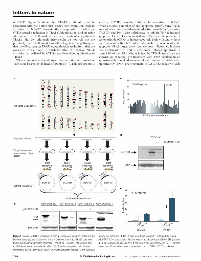
The family of de-ubiquitinating enzymes (DUBs) consists ofubiquitin carboxy-terminal hydrolases and ubiquitin-specific pro-cessing proteases1–3. A role for DUB genes in cancer is suggested bythe fact that this family contains both oncogenes6,7 and tumoursuppressor genes4. The strategy we pursued to study the function ofthe family of DUB enzymes was to inhibit expression of individualfamily members through RNA interference and search for pheno-types. A total of 50 genes containing the conserved catalytic domainof DUB enzymes could be identified in sequence databases (Fig. 1a,and Supplementary Information). We retrieved the complementaryDNA sequences, and selected four unique 19-mer sequences fromeach transcript for cloning into pSUPER, a vector that mediatessuppression of gene expression through synthesis of short hairpinRNAs having small interfering RNA (siRNA)-like properties8. Wechose to make four knockdown vectors against each DUB toincrease the chance that a significant inhibition of DUB expressionwould be obtained. In total, we made 200 knockdown vectors,which were subsequently pooled into 50 sets of 4 vectors, whereeach set of vectors was designed to target a single DUB transcript(Fig. 1a).
To ask how effective sets of four knockdown vectors inhibitedDUB gene expression, we fused the open reading frame of four ofthe DUBs to green fluorescent protein (GFP) and determined thelevels of GFP–DUB fusion protein expression in the absence andpresence of co-expression of the DUB knockdown vectors. Asignificant reduction in protein levels was induced by all fourDUB knockdown vector pools, whereas control p21–red fluorescentprotein (RFP) fusion protein was unaffected (Fig. 1b). We concludethat this strategy allows efficient inhibition of DUB expression.
To study members of the DUB family, we asked if suppression of
DUBs affected the activity of NF-kB, a transcription factor involvedin inflammation, immune responses and protection against apop-tosis9. We transfected an NF-kB-luciferase reporter gene, togetherwith each of the 50 sets of 4 DUB knockdown vectors into humanU2-OS cells, and measured the effect of DUB knockdown ontumour necrosis factor-a (TNF-a)-activation of NF-kB. Onlyone of the sets of DUB knockdown vectors (no. 36) enhancedTNF-a-activation of NF-kB (Fig. 1c). Importantly, the DUB no. 36set of knockdown vectors targets the cylindromatosis tumoursuppressor gene CYLD4 (see Supplementary Information). Levelsof haemagglutinin (HA)-CYLD protein levels were significantlyreduced by the most active of the four knockdown vectors (pSU-PER-CYLD) (Fig. 1d). CYLD knockdown did not enhance basallevel of NF-kB activity, but caused a further increase in NF-kBactivation by phorbol 12-myristate 13-acetate (PMA) (Fig. 1e).This indicates that CYLD loss activates NF-kB in response todiverse stimuli. Together, these data suggest that CYLD is anNF-kB regulator.
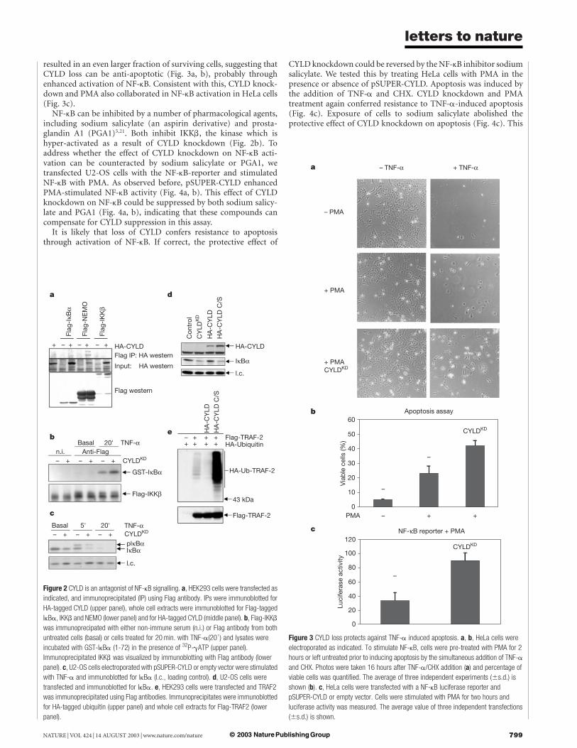
NF-kB is inactivated by IkB proteins. Signalling through the IkBkinase (IKK) complex, containing the IkB kinases IKKa and b andthe structural component NEMO (or IKKg), causes phosphoryl-ation and subsequent degradation of IkB, allowing activation ofNF-kB9,10. The observed effect of CYLD on NF-kB activity couldresult from an effect of CYLD on the TNF-a receptor, the TNF-receptor-associated factor (TRAF) proteins, a downstream effect onthe IKK complex or on the IkB/NF-kB complex. To address this, weasked if CYLD associates with members of the NF-kB signallingmachinery. We found that CYLD co-immunoprecipitated withNEMO (Fig. 2a). Since CYLD interacts with NEMO in a yeasttwo hybrid assay (see accompanying manuscript11), this is likely tobe direct. However, owing to the lower expression of IkBa and IKKb(Fig. 2a), we cannot exclude the possibility that CYLD also interactswith other components of the IKK complex.
To study the effect of CYLD on IKK activity, we measured IKKbactivity in an in vitro kinase assay. In the absence of TNF-a no IKKbkinase activity towards IkBa could be detected (Fig. 2b). Asexpected, TNF-a treatment stimulated IKKb kinase activity. Impor-tantly, this activity was enhanced when cells were co-transfectedwith pSUPER-CYLD (compare Fig. 2b, lanes 5, 6). No effects wereseen of CYLD knockdown on IKKb protein levels (Fig. 2b, lowerpanel), suggesting that CYLD does not regulate IKKb abundance.Consistent with increased IKKb kinase activity by pSUPER-CYLD,we observed that CYLD knockdown resulted in a reduction in IkBalevels, an endogenous substrate of IKKb kinase (Fig. 2c). Thisdecrease in IkBa level occurs without TNF-a treatment, indicatingthat there might be some IKK activity in U2-OS cells under thetissue culture conditions used, or that CYLD is required to maintainshut-off of IKK signalling in the absence of activating signals. Thesedata indicate that CYLD acts as an antagonist of the IKK complexthat binds to the NEMO component, and that a reduction in CYLDstimulates IKK-activity. A prediction from these data is thatexpression of a catalytically inactive mutant of CYLD will alsocause a reduction of the IkBa levels through competition withendogenous CYLD. Figure 2d shows that transfection of cells with apoint mutant of CYLD in which the active site cysteine is mutated7
causes a similar reduction in IkBa levels as seen with pSUPER-CYLD (compare Fig. 2d, lanes 2 and 4). We conclude that thede-ubiquitinating enzyme activity of CYLD is required to act onNF-kB.
TNF receptor-associated factors (TRAFs) are involved in cyto-kine-mediated activation of NF-kB12,13. NF-kB-inducing activity ofTRAF2 and TRAF6 requires their ubiquitin ligase activity andnon-destructive lysine-63 poly-ubiquitination14,15. Their ubiquiti-nation during TNF signalling makes these proteins candidatesubstrates for CYLD. We co-transfected TRAF2 and HA-ubiquitinexpression vectors, and monitored levels of TRAF2 ubiquitinationin the presence of both wild-type CYLD and an active site mutant
letters to nature
NATURE | VOL 424 | 14 AUGUST 2003 | www.nature.com/nature 797© 2003 Nature Publishing Group
of CYLD. Figure 2e shows that TRAF2 is ubiquitinated, inagreement with the notion that TRAF2 over-expression leads toactivation of NF-kB11. Importantly, co-expression of wild-typeCYLD caused a reduction in TRAF2 ubiquitination, and an activesite mutant of CYLD markedly increased levels of ubiquitinatedTRAF2 (Fig. 2e). Although these results do not rule out thepossibility that CYLD could have other targets in the pathway, orthat the effects seen on TRAF2 ubiquitination are indirect, they areconsistent with a model in which the effect of CYLD on NF-kBactivation is mediated by CYLD-dependent de-ubiquitination ofTRAF2.
When combined with inhibitors of transcription or translation,TNF-a can be a potent inducer of apoptosis16–19. This pro-apoptotic
activity of TNF-a can be inhibited by activation of NF-kB,which activates a number of anti-apoptotic genes20. Since CYLDknockdown stimulates PMA-induced activation of NF-kB, we askedif CYLD and PMA also collaborate to inhibit TNF-a-inducedapoptosis. HeLa cells were treated with TNF-a in the presence ofcycloheximide (CHX) to induce apoptosis both with and withoutpre-treatment with PMA, which stimulates expression of anti-apoptotic NF-kB target genes (see Methods). Figure 3a, b showsthat treatment with TNF-a efficiently induced apoptosis insome 95% of the HeLa cells, as judged by TUNEL assay (data notshown). As expected, pre-treatment with PMA resulted in anapproximately four-fold increase of the number of viable cells.Significantly, PMA pre-treatment in CYLD knockdown cells
5′ 5′3′
5′3′
5′3′
Figure 1 Genome-wide DUB knockdown screen. a, Overview of identified DUBs using the
Ensembl database, and construction of the knockdown library. b, HEK293 cells were
transfected and immunoblotted against GFP or p21–RFP (control). WB, western blot.
c, U2-OS cells were co-transfected with a NF-kB luciferase reporter and individual
members of the DUB knockdown library. Cells were stimulated with TNF-a and luciferase
activity was measured. d, U2-OS cells were transfected with HA-tagged CYLD and
pSUPER-CYLD or empty vector. Extracts were immunoblotted against HA or GFP (control).
e, U2-OS cells were transfected as in c, and were stimulated with PMA or TNF-a. Average
values are of three independent transfections (^s.d.). CYLDKD, CYLD knockdown.
letters to nature
NATURE | VOL 424 | 14 AUGUST 2003 | www.nature.com/nature798 © 2003 Nature Publishing Group
resulted in an even larger fraction of surviving cells, suggesting thatCYLD loss can be anti-apoptotic (Fig. 3a, b), probably throughenhanced activation of NF-kB. Consistent with this, CYLD knock-down and PMA also collaborated in NF-kB activation in HeLa cells(Fig. 3c).
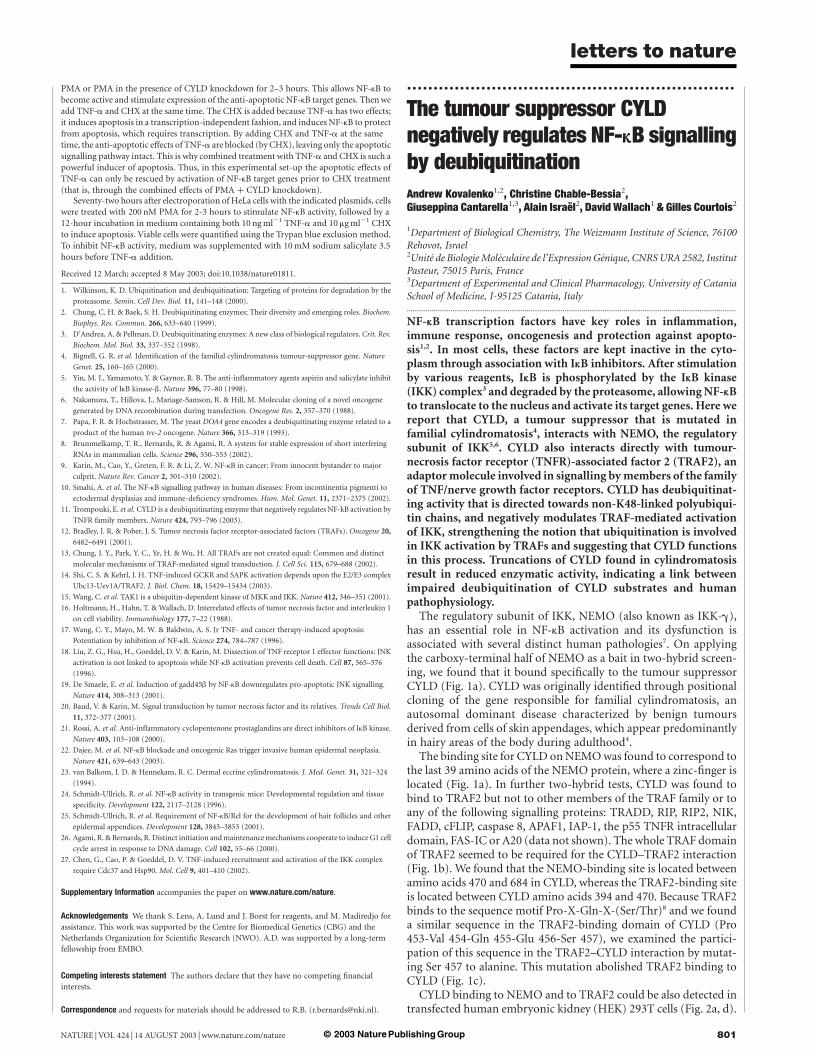
NF-kB can be inhibited by a number of pharmacological agents,including sodium salicylate (an aspirin derivative) and prosta-glandin A1 (PGA1)5,21. Both inhibit IKKb, the kinase which ishyper-activated as a result of CYLD knockdown (Fig. 2b). Toaddress whether the effect of CYLD knockdown on NF-kB acti-vation can be counteracted by sodium salicylate or PGA1, wetransfected U2-OS cells with the NF-kB-reporter and stimulatedNF-kB with PMA. As observed before, pSUPER-CYLD enhancedPMA-stimulated NF-kB activity (Fig. 4a, b). This effect of CYLDknockdown on NF-kB could be suppressed by both sodium salicy-late and PGA1 (Fig. 4a, b), indicating that these compounds cancompensate for CYLD suppression in this assay.
It is likely that loss of CYLD confers resistance to apoptosisthrough activation of NF-kB. If correct, the protective effect of
CYLD knockdown could be reversed by the NF-kB inhibitor sodiumsalicylate. We tested this by treating HeLa cells with PMA in thepresence or absence of pSUPER-CYLD. Apoptosis was induced bythe addition of TNF-a and CHX. CYLD knockdown and PMAtreatment again conferred resistance to TNF-a-induced apoptosis(Fig. 4c). Exposure of cells to sodium salicylate abolished theprotective effect of CYLD knockdown on apoptosis (Fig. 4c). This
Figure 2 CYLD is an antagonist of NF-kB signalling. a, HEK293 cells were transfected as
indicated, and immunoprecipitated (IP) using Flag antibody. IPs were immunoblotted for
HA-tagged CYLD (upper panel), whole cell extracts were immunoblotted for Flag-tagged
IkBa, IKKb and NEMO (lower panel) and for HA-tagged CYLD (middle panel). b, Flag-IKKb
was immunoprecipated with either non-immune serum (n.i.) or Flag antibody from both
untreated cells (basal) or cells treated for 20 min. with TNF-a(200) and lysates were
incubated with GST-IkBa (1-72) in the presence of 32P-gATP (upper panel).
Immunoprecipitated IKKb was visualized by immunoblotting with Flag antibody (lower
panel). c, U2-OS cells electroporated with pSUPER-CYLD or empty vector were stimulated
with TNF-a and immunoblotted for IkBa (l.c., loading control). d, U2-OS cells were
transfected and immunoblotted for IkBa. e, HEK293 cells were transfected and TRAF2
was immunoprecipitated using Flag antibodies. Immunoprecipitates were immunoblotted
for HA-tagged ubiquitin (upper panel) and whole cell extracts for Flag-TRAF2 (lower
panel).
Figure 3 CYLD loss protects against TNF-a induced apoptosis. a, b, HeLa cells were
electroporated as indicated. To stimulate NF-kB, cells were pre-treated with PMA for 2
hours or left untreated prior to inducing apoptosis by the simultaneous addition of TNF-a
and CHX. Photos were taken 16 hours after TNF-a/CHX addition (a) and percentage of
viable cells was quantified. The average of three independent experiments (^s.d.) is
shown (b). c, HeLa cells were transfected with a NF-kB luciferase reporter and
pSUPER-CYLD or empty vector. Cells were stimulated with PMA for two hours and
luciferase activity was measured. The average value of three independent transfections
(^s.d.) is shown.
letters to nature
NATURE | VOL 424 | 14 AUGUST 2003 | www.nature.com/nature 799© 2003 Nature Publishing Group
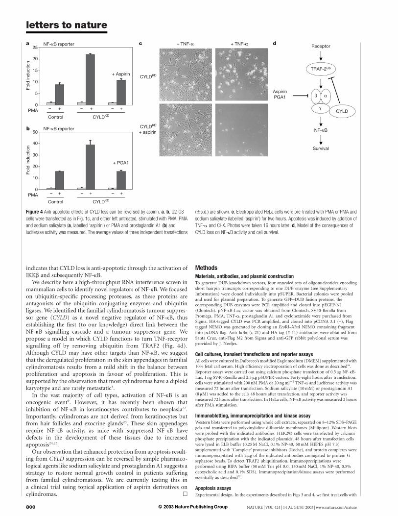
indicates that CYLD loss is anti-apoptotic through the activation ofIKKb and subsequently NF-kB.
We describe here a high-throughput RNA interference screen inmammalian cells to identify novel regulators of NF-kB. We focusedon ubiquitin-specific processing proteases, as these proteins areantagonists of the ubiquitin conjugating enzymes and ubiquitinligases. We identified the familial cylindromatosis tumour suppres-sor gene (CYLD) as a novel negative regulator of NF-kB, thusestablishing the first (to our knowledge) direct link between theNF-kB signalling cascade and a tumour suppressor gene. Wepropose a model in which CYLD functions to turn TNF-receptorsignalling off by removing ubiquitin from TRAF2 (Fig. 4d).Although CYLD may have other targets than NF-kB, we suggestthat the deregulated proliferation in the skin appendages in familialcylindromatosis results from a mild shift in the balance betweenproliferation and apoptosis in favour of proliferation. This issupported by the observation that most cylindromas have a diploidkaryotype and are rarely metastatic4.
In the vast majority of cell types, activation of NF-kB is anoncogenic event9. However, it has recently been shown thatinhibition of NF-kB in keratinocytes contributes to neoplasia22.Importantly, cylindromas are not derived from keratinocytes butfrom hair follicles and exocrine glands23. These skin appendagesrequire NF-kB activity, as mice with suppressed NF-kB havedefects in the development of these tissues due to increasedapoptosis24,25.
Our observation that enhanced protection from apoptosis result-ing from CYLD suppression can be reversed by simple pharmaco-logical agents like sodium salicylate and prostaglandin A1 suggests astrategy to restore normal growth control in patients sufferingfrom familial cylindromatosis. We are currently testing this ina clinical trial using topical application of aspirin derivatives oncylindromas. A
MethodsMaterials, antibodies, and plasmid constructionTo generate DUB knockdown vectors, four annealed sets of oligonucleotides encodingshort hairpin transcripts corresponding to one DUB enzyme (see SupplementaryInformation) were cloned individually into pSUPER. Bacterial colonies were pooledand used for plasmid preparation. To generate GFP–DUB fusion proteins, thecorresponding DUB enzymes were PCR amplified and cloned into pEGFP-N1(Clontech). pNF-kB-Luc vector was obtained from Clontech, SV40-Renilla fromPromega. PMA, TNF-a, prostaglandin A1 and cycloheximide were purchased fromSigma. HA-tagged CYLD was PCR amplified, and cloned into pCDNA 3.1 (–), Flagtagged NEMO was generated by cloning an EcoRI–XbaI NEMO containing fragmentinto pcDNA-flag. Anti-IkBa (c-21) and HA tag (Y-11) antibodies were obtained fromSanta Cruz, anti-Flag M2 from Sigma and anti-GFP rabbit polyclonal serum wasprovided by J. Neefjes.
Cell cultures, transient transfections and reporter assaysAll cells were cultured in Dulbecco’s modified Eagle medium (DMEM) supplemented with10% fetal calf serum. High efficiency electroporation of cells was done as described26.Reporter assays were carried out using calcium phosphate transfection of 0.5 mg NF-kB-Luc, 1 ng SV40-Renilla and 2.5 mg pSUPER vectors. Forty-eight hours after transfection,cells were stimulated with 200 nM PMA or 20 ng ml21 TNF-a and luciferase activity wasmeasured 72 hours after transfection. Sodium salicylate (10 mM) or prostaglandin A1(8 mM) was added to the cells 48 hours after transfection, and reporter activity wasmeasured 72 hours after transfection. In HeLa cells, NF-kB activity was measured 2 hoursafter PMA stimulation.
Immunoblotting, immunoprecipitation and kinase assayWestern blots were performed using whole cell extracts, separated on 8–12% SDS–PAGEgels and transferred to polyvinylidine difluoride membranes (Millipore). Western blotswere probed with the indicated antibodies. HEK293 cells were transfected by calciumphosphate precipitation with the indicated plasmids; 48 hours after transfection cellswere lysed in ELB buffer (0.25 M NaCl, 0.1% NP-40, 50 mM HEPES pH 7.3)supplemented with ‘Complete’ protease inhibitors (Roche), and protein complexes wereimmunoprecipitated with 2 mg of the indicated antibodies conjugated to protein Gsepharose beads. To detect TRAF2 ubiquitination, immunoprecipitations wereperformed using RIPA buffer (50 mM Tris pH 8.0, 150 mM NaCl, 1% NP-40, 0.5%deoxycholic acid and 0.1% SDS). Immunoprecipitation/kinase assays were performedessentially as described27.
Apoptosis assaysExperimental design. In the experiments described in Figs 3 and 4, we first treat cells with
Figure 4 Anti-apoptotic effects of CYLD loss can be reversed by aspirin. a, b, U2-OS
cells were transfected as in Fig. 1c, and either left untreated, stimulated with PMA, PMA
and sodium salicylate (a, labelled ‘aspirin’) or PMA and prostaglandin A1 (b) and
luciferase activity was measured. The average values of three independent transfections
(^s.d.) are shown. c, Electroporated HeLa cells were pre-treated with PMA or PMA and
sodium salicylate (labelled ‘aspirin’) for two hours. Apoptosis was induced by addition of
TNF-a and CHX. Photos were taken 16 hours later. d, Model of the consequences of
CYLD loss on NF-kB activity and cell survival.
letters to nature
NATURE | VOL 424 | 14 AUGUST 2003 | www.nature.com/nature800 © 2003 Nature Publishing Group
PMA or PMA in the presence of CYLD knockdown for 2–3 hours. This allows NF-kB tobecome active and stimulate expression of the anti-apoptotic NF-kB target genes. Then weadd TNF-a and CHX at the same time. The CHX is added because TNF-a has two effects;it induces apoptosis in a transcription-independent fashion, and induces NF-kB to protectfrom apoptosis, which requires transcription. By adding CHX and TNF-a at the sametime, the anti-apoptotic effects of TNF-a are blocked (by CHX), leaving only the apoptoticsignalling pathway intact. This is why combined treatment with TNF-a and CHX is such apowerful inducer of apoptosis. Thus, in this experimental set-up the apoptotic effects ofTNF-a can only be rescued by activation of NF-kB target genes prior to CHX treatment(that is, through the combined effects of PMA þ CYLD knockdown).
Seventy-two hours after electroporation of HeLa cells with the indicated plasmids, cellswere treated with 200 nM PMA for 2-3 hours to stimulate NF-kB activity, followed by a12-hour incubation in medium containing both 10 ng ml21 TNF-a and 10 mg ml21 CHXto induce apoptosis. Viable cells were quantified using the Trypan blue exclusion method.To inhibit NF-kB activity, medium was supplemented with 10 mM sodium salicylate 3.5hours before TNF-a addition.
Received 12 March; accepted 8 May 2003; doi:10.1038/nature01811.
1. Wilkinson, K. D. Ubiquitination and deubiquitination: Targeting of proteins for degradation by the
proteasome. Semin. Cell Dev. Biol. 11, 141–148 (2000).
2. Chung, C. H. & Baek, S. H. Deubiquitinating enzymes: Their diversity and emerging roles. Biochem.
Biophys. Res. Commun. 266, 633–640 (1999).
3. D’Andrea, A. & Pellman, D. Deubiquitinating enzymes: A new class of biological regulators. Crit. Rev.
Biochem. Mol. Biol. 33, 337–352 (1998).
4. Bignell, G. R. et al. Identification of the familial cylindromatosis tumour-suppressor gene. Nature
Genet. 25, 160–165 (2000).
5. Yin, M. J., Yamamoto, Y. & Gaynor, R. B. The anti-inflammatory agents aspirin and salicylate inhibit
the activity of IkB kinase-b. Nature 396, 77–80 (1998).
6. Nakamura, T., Hillova, J., Mariage-Samson, R. & Hill, M. Molecular cloning of a novel oncogene
generated by DNA recombination during transfection. Oncogene Res. 2, 357–370 (1988).
7. Papa, F. R. & Hochstrasser, M. The yeast DOA4 gene encodes a deubiquitinating enzyme related to a
product of the human tre-2 oncogene. Nature 366, 313–319 (1993).
8. Brummelkamp, T. R., Bernards, R. & Agami, R. A system for stable expression of short interfering
RNAs in mammalian cells. Science 296, 550–553 (2002).
9. Karin, M., Cao, Y., Greten, F. R. & Li, Z. W. NF-kB in cancer: From innocent bystander to major
culprit. Nature Rev. Cancer 2, 301–310 (2002).
10. Smahi, A. et al. The NF-kB signalling pathway in human diseases: From incontinentia pigmenti to
ectodermal dysplasias and immune-deficiency syndromes. Hum. Mol. Genet. 11, 2371–2375 (2002).
11. Trompouki, E. et al. CYLD is a deubiquitinating enzyme that negatively regulates NF-kB activation by
TNFR family members. Nature 424, 793–796 (2003).
12. Bradley, J. R. & Pober, J. S. Tumor necrosis factor receptor-associated factors (TRAFs). Oncogene 20,
6482–6491 (2001).
13. Chung, J. Y., Park, Y. C., Ye, H. & Wu, H. All TRAFs are not created equal: Common and distinct
molecular mechanisms of TRAF-mediated signal transduction. J. Cell Sci. 115, 679–688 (2002).
14. Shi, C. S. & Kehrl, J. H. TNF-induced GCKR and SAPK activation depends upon the E2/E3 complex
Ubc13-Uev1A/TRAF2. J. Biol. Chem. 18, 15429–15434 (2003).
15. Wang, C. et al. TAK1 is a ubiquitin-dependent kinase of MKK and IKK. Nature 412, 346–351 (2001).
16. Holtmann, H., Hahn, T. & Wallach, D. Interrelated effects of tumor necrosis factor and interleukin 1
on cell viability. Immunobiology 177, 7–22 (1988).
17. Wang, C. Y., Mayo, M. W. & Baldwin, A. S. Jr TNF- and cancer therapy-induced apoptosis:
Potentiation by inhibition of NF-kB. Science 274, 784–787 (1996).
18. Liu, Z. G., Hsu, H., Goeddel, D. V. & Karin, M. Dissection of TNF receptor 1 effector functions: JNK
activation is not linked to apoptosis while NF-kB activation prevents cell death. Cell 87, 565–576
(1996).
19. De Smaele, E. et al. Induction of gadd45b by NF-kB downregulates pro-apoptotic JNK signalling.
Nature 414, 308–313 (2001).
20. Baud, V. & Karin, M. Signal transduction by tumor necrosis factor and its relatives. Trends Cell Biol.
11, 372–377 (2001).
21. Rossi, A. et al. Anti-inflammatory cyclopentenone prostaglandins are direct inhibitors of IkB kinase.
Nature 403, 103–108 (2000).
22. Dajee, M. et al. NF-kB blockade and oncogenic Ras trigger invasive human epidermal neoplasia.
Nature 421, 639–643 (2003).
23. van Balkom, I. D. & Hennekam, R. C. Dermal eccrine cylindromatosis. J. Med. Genet. 31, 321–324
(1994).
24. Schmidt-Ullrich, R. et al. NF-kB activity in transgenic mice: Developmental regulation and tissue
specificity. Development 122, 2117–2128 (1996).
25. Schmidt-Ullrich, R. et al. Requirement of NF-kB/Rel for the development of hair follicles and other
epidermal appendices. Development 128, 3843–3853 (2001).
26. Agami, R. & Bernards, R. Distinct initiation and maintenance mechanisms cooperate to induce G1 cell
cycle arrest in response to DNA damage. Cell 102, 55–66 (2000).
27. Chen, G., Cao, P. & Goeddel, D. V. TNF-induced recruitment and activation of the IKK complex
require Cdc37 and Hsp90. Mol. Cell 9, 401–410 (2002).
Supplementary Information accompanies the paper on www.nature.com/nature.
Acknowledgements We thank S. Lens, A. Lund and J. Borst for reagents, and M. Madiredjo for
assistance. This work was supported by the Centre for Biomedical Genetics (CBG) and the
Netherlands Organization for Scientific Research (NWO). A.D. was supported by a long-term
fellowship from EMBO.
Competing interests statement The authors declare that they have no competing financial
interests.
Correspondence and requests for materials should be addressed to R.B. ([email protected]).
..............................................................
The tumour suppressor CYLDnegatively regulates NF-kB signallingby deubiquitinationAndrew Kovalenko1,2, Christine Chable-Bessia2,Giuseppina Cantarella1,3, Alain Israel2, David Wallach1 & Gilles Courtois2
1Department of Biological Chemistry, The Weizmann Institute of Science, 76100Rehovot, Israel2Unite de Biologie Moleculaire de l’Expression Genique, CNRS URA 2582, InstitutPasteur, 75015 Paris, France3Department of Experimental and Clinical Pharmacology, University of CataniaSchool of Medicine, I-95125 Catania, Italy.............................................................................................................................................................................
NF-kB transcription factors have key roles in inflammation,immune response, oncogenesis and protection against apopto-sis1,2. In most cells, these factors are kept inactive in the cyto-plasm through association with IkB inhibitors. After stimulationby various reagents, IkB is phosphorylated by the IkB kinase(IKK) complex3 and degraded by the proteasome, allowing NF-kBto translocate to the nucleus and activate its target genes. Here wereport that CYLD, a tumour suppressor that is mutated infamilial cylindromatosis4, interacts with NEMO, the regulatorysubunit of IKK5,6. CYLD also interacts directly with tumour-necrosis factor receptor (TNFR)-associated factor 2 (TRAF2), anadaptor molecule involved in signalling by members of the familyof TNF/nerve growth factor receptors. CYLD has deubiquitinat-ing activity that is directed towards non-K48-linked polyubiqui-tin chains, and negatively modulates TRAF-mediated activationof IKK, strengthening the notion that ubiquitination is involvedin IKK activation by TRAFs and suggesting that CYLD functionsin this process. Truncations of CYLD found in cylindromatosisresult in reduced enzymatic activity, indicating a link betweenimpaired deubiquitination of CYLD substrates and humanpathophysiology.
The regulatory subunit of IKK, NEMO (also known as IKK-g),has an essential role in NF-kB activation and its dysfunction isassociated with several distinct human pathologies7. On applyingthe carboxy-terminal half of NEMO as a bait in two-hybrid screen-ing, we found that it bound specifically to the tumour suppressorCYLD (Fig. 1a). CYLD was originally identified through positionalcloning of the gene responsible for familial cylindromatosis, anautosomal dominant disease characterized by benign tumoursderived from cells of skin appendages, which appear predominantlyin hairy areas of the body during adulthood4.
The binding site for CYLD on NEMO was found to correspond tothe last 39 amino acids of the NEMO protein, where a zinc-finger islocated (Fig. 1a). In further two-hybrid tests, CYLD was found tobind to TRAF2 but not to other members of the TRAF family or toany of the following signalling proteins: TRADD, RIP, RIP2, NIK,FADD, cFLIP, caspase 8, APAF1, IAP-1, the p55 TNFR intracellulardomain, FAS-IC or A20 (data not shown). The whole TRAF domainof TRAF2 seemed to be required for the CYLD–TRAF2 interaction(Fig. 1b). We found that the NEMO-binding site is located betweenamino acids 470 and 684 in CYLD, whereas the TRAF2-binding siteis located between CYLD amino acids 394 and 470. Because TRAF2binds to the sequence motif Pro-X-Gln-X-(Ser/Thr)8 and we founda similar sequence in the TRAF2-binding domain of CYLD (Pro453-Val 454-Gln 455-Glu 456-Ser 457), we examined the partici-pation of this sequence in the TRAF2–CYLD interaction by mutat-ing Ser 457 to alanine. This mutation abolished TRAF2 binding toCYLD (Fig. 1c).
CYLD binding to NEMO and to TRAF2 could be also detected intransfected human embryonic kidney (HEK) 293T cells (Fig. 2a, d).
letters to nature
NATURE | VOL 424 | 14 AUGUST 2003 | www.nature.com/nature 801© 2003 Nature Publishing Group
![Soluble αKlotho downregulates Orai1-mediated store ......Klotho is an aging-suppressor gene that encodes type 1 transmembrane glycoprotein called αKlotho [22, 23]. Klotho-deficient](https://static.fdocument.org/doc/165x107/613b4592f8f21c0c8268e811/soluble-klotho-downregulates-orai1-mediated-store-klotho-is-an-aging-suppressor.jpg)