ERK2 drives tumour cell migration in 3D microenvironments...
37
Von Thun et al. ERK2 suppresses Rab17 and Liprin- 2 to drive cell migration 1 ERK2 drives tumour cell migration in 3D microenvironments by suppressing expression of Rab17 and Liprin- 2 Anne von Thun 1 , Marc Birtwistle 2 , Gabriela Kalna 1 , Joan Grindlay 1 , David Strachan 1 , Walter Kolch 2 and Alexander von Kriegsheim 2 *, Jim C. Norman 1 * 1 Beatson Institute for Cancer Research, Glasgow, United Kingdom G61 1BD 2 Systems Biology Ireland, University College Dublin, Dublin, Ireland * Correspondence: [email protected] [email protected] Word count: 5,696 (excluding references and supplementary material) Running title: ERK2 suppresses Rab17 and Liprin- 2 to drive cell migration Key words: ERK2, cell migration, invasion, cancer, Rab17, Liprin- 2 Journal of Cell Science Accepted manuscript © 2012. Published by The Company of Biologists Ltd. JCS ePress online publication date 10 February 2012
Transcript of ERK2 drives tumour cell migration in 3D microenvironments...
Von Thun et al. ERK2 suppresses Rab17 and Liprin-β2 to drive cell migration
1
ERK2 drives tumour cell migration in 3D microenvironments by
suppressing expression of Rab17 and Liprin-β2
Anne von Thun1, Marc Birtwistle2, Gabriela Kalna1, Joan Grindlay1, David Strachan1,
Walter Kolch2 and Alexander von Kriegsheim2*, Jim C. Norman1*
1Beatson Institute for Cancer Research, Glasgow, United Kingdom G61 1BD
2Systems Biology Ireland, University College Dublin, Dublin, Ireland
* Correspondence:
Word count: 5,696 (excluding references and supplementary material)
Running title: ERK2 suppresses Rab17 and Liprin-β2 to drive cell migration
Key words: ERK2, cell migration, invasion, cancer, Rab17, Liprin-β2
Jour
nal o
f Cel
l Sci
ence
Acc
epte
d m
anus
crip
t© 2012. Published by The Company of Biologists Ltd. JCS ePress online publication date 10 February 2012
Von Thun et al. ERK2 suppresses Rab17 and Liprin-β2 to drive cell migration
2
ABSTRACT (190 words)
Upregulation of the extracellular signal-regulated kinase (ERK) pathway has been shown
to contribute to tumour invasion and progression. Since the two predominant ERK
isoforms (ERK1 and ERK2) are highly homologous and have indistinguishable kinase
activities in vitro, both enzymes were believed to be redundant and interchangeable. To
challenge this view, here we show that ERK2 silencing inhibits invasive migration of
MDA-MB-231 cells, and re-expression of ERK2 but not ERK1 restores the normal
invasive phenotype. A detailed quantitative analysis of cell movement on 3D matrices
indicates that ERK2 knockdown impairs cellular motility by decreasing the migration
velocity as well as increasing the time that cells spend not moving. We used gene
expression arrays to identify rab17 and liprin-β2 as genes whose expression was
increased by knockdown of ERK2 and restored to normal levels following re-expression
of ERK2, but not ERK1. Both Rab17 and Liprin-β2 play inhibitory roles in the invasive
behaviour of three independent cancer cell lines. Importantly, knockdown of either
Rab17 or Liprin-β2 restores invasiveness of ERK2-depleted cells, indicating that ERK2
drives invasion of MDA-MB-231 cells by suppressing expression of these genes.
Jour
nal o
f Cel
l Sci
ence
Acc
epte
d m
anus
crip
t
Von Thun et al. ERK2 suppresses Rab17 and Liprin-β2 to drive cell migration
3
INTRODUCTION
The extracellular signal-regulated kinase (ERK) pathway is one of the most
intensively studied mammalian MAPK pathways and is deregulated in approximately
one-third of all human cancers (Reddy et al., 2003). Aberrant ERK signalling is prevalent
in most cancers as a consequence of increased expression and/or mutations of
upstream components of the cascade. The canonical role of this abnormal signalling is
its positive influence on cellular survival and proliferation. It is becoming clear, however,
that the ERK pathway also controls tumour cell migration, invasion and progression
(Klemke et al., 1997; Ochieng et al., 1991; Reddy et al., 2003; Rodier et al., 1995; Ueoka
et al., 2000). ERK1 and ERK2 display 85% sequence identity and have indistinguishable
kinase activities in vitro (Boulton et al., 1991; Lefloch et al., 2008; Pouyssegur et al.,
2002). Therefore, both isoforms were believed to be redundant and interchangeable.
However, accumulating evidence, in particular marked discrepancies of the ERK-/-
phenotypes, challenges this view. Whereas ERK1-/- mice are viable, fertile, and of
normal size (Pages et al., 1999), ERK2-/- mice are embryonic lethal (Hatano et al., 2003;
Saba-El-Leil et al., 2003; Yao et al., 2003), suggesting specific roles for these two
isoforms during embryogenesis. However, as ERK2 is the predominant isoform in most
tissues, this apparent functional specificity may simply reflect differences in ERK
expression or activity levels (Lefloch et al., 2008). Indeed, a recent study has shown that
loss of either ERK1 or ERK2 slows proliferation of fibroblasts to an extent that reflects
the expression level of the individual kinase (Voisin et al.). Moreover, proliferation is
reduced following conditional inactivation of the (more abundant) ERK2 isoform in
neuronal precursor cells, but cell growth resumes when ERK1 activity is upregulated to
compensate for loss of ERK2 (Samuels et al., 2008). Thus, although disruption of ERK1
and ERK2 can clearly produce different phenotypes in a range of biological contexts, it is
Jour
nal o
f Cel
l Sci
ence
Acc
epte
d m
anus
crip
t
Von Thun et al. ERK2 suppresses Rab17 and Liprin-β2 to drive cell migration
4
unclear to what extent this reflects true functional differences between the kinases or
other factors, such as gene dosage.
This study aims to investigate the role of the two ERK isoforms in the
invasiveness of tumour cell lines, and to determine whether ERK isoform differences
with regards to cell migration are true functional disparities, or rather are the result of
different expression levels of the two kinases. We provide evidence that ERK2 (but not
ERK1) is particularly important in the invasiveness of cancer cells. Furthermore, we
report that ERK2 drives invasive migration in 3D microenvironments by reducing the
expression of two novel motility suppressor genes; rab17 and liprin-β2.
Jour
nal o
f Cel
l Sci
ence
Acc
epte
d m
anus
crip
t
Von Thun et al. ERK2 suppresses Rab17 and Liprin-β2 to drive cell migration
5
MATERIALS AND METHODS
Cell culture. MDA-MB-231 and BE cells were cultured in Dulbecco’s Modified Eagle
Medium (DMEM) supplemented with glutamine (100µg/ml) and 10% FCS. A2780-Rab25
cells were grown in RPMI-1640 media containing glutamine (100µg/ml) and 10% FCS.
All cells were maintained at 37ºC under a humidified 5% CO2 atmosphere.
Plasmid constructs.
ERK1 and ERK2 genes were amplified by PCR from a cDNA library using the following
primers: ERK1 attB1 5’-GGGGACAAGTTTGTACAAAAAAGCAGGCTTCGCGGCG
GCGGCGGCTCAGGG-3’, ERK1 attB2 5’-GGGGACCACTTTGTACAAGAAAGCTG
GGTTTTACTAGGGGGCCTCCAGCACTCC-3’, ERK2 attB1 5’-GGGGACAAGTTTG
TACAAAAAAGCAGGCTTCGCGGCGGCGGCGGCGGCGG-3’, ERK2 attB2
5’-GGGGACCACTTTGTACAAGAAAGCTGGGTTTTATTAAGATCTGTATCCTGGCTG-3
’. ERK1 and ERK2 were recombined with the SF-TAP Gateway expression vector
(Gloeckner et al., 2007). siRNA-resistant ERK2 expression vectors were generated by
site-directed mutagenesis (QuikChange Multi Site-Directed Mutagenesis Kit from
Stratagene) using the following primer:
5’-GCCTACGGCATGGTGTGTAGTGCTTATGATAATGTCAACAAAGTTCG-3’. ERK
chimeras were generated as follows; For E2>E1 an internal HindIII restriction site was
removed using the following primer 5’-GGGCCAAGCTCTTCCCCAAGTCAGACTCC-3’,
while a novel HindIII site was introduced by site-directed mutatgenesis using the primer
5’-CTGACCTGTACAAGCTTCTGAAAAGCCAGCAGCTGAGCAATGACC-3’. For E1>E2
a novel HindIII site was introduced using the following primer 5’-
CAGGACCTCATGGAAACAGATCTTTACAAGCTTTTGAAGACACAACACC-3’. Next,
both expression plasmids were digested with HindIII, which cut within the SF-tag, and
Jour
nal o
f Cel
l Sci
ence
Acc
epte
d m
anus
crip
t
Von Thun et al. ERK2 suppresses Rab17 and Liprin-β2 to drive cell migration
6
the ERK sequences (LYKLLK, restriction site in underlined amino acids), and the
respective domains were swapped.
EGFP-C1-Rab17 was a gift from Jeremy Simpson (University College Ireland). pCMV6-
GFP-Liprin-β2 expression vector was obtained from Origene.
Western Blot.
The following antibodies were used for immunoblotting: Tubulin (Insight), ERK1/2
(Sigma), pERK (Sigma), PARP (BD), Vinculin (F9, a gift from Victor Koteliansky), FLAG
(Sigma)
Transfection and RNA interference.
Transfection of siRNA duplexes into MDA-MB-231 and BE cells was performed using
HiPerFect (Qiagen) according to the manufacturer’s instructions. Expression vectors
were introduced using the Amaxa Nucleofector System according to the manufacturer’s
protocol. siRNA oligos and respective vectors were transfected into A2780-Rab25 cells
using Amaxa. Assays were set up 48 hours (MDA-MB-231 and BE) and 24 hours
(A2780-Rab25) after transfection. siRNA oligos were: ERK1, 5’-
GACCGGAUGUUAACCUUUA-3’ (#1), 5’-CCUGCGACCUUAAGAUUUG-3’ (#2); ERK2,
5’-CACCAACCAUCGAGCAAAU-3’ (#1), 5’-GGUGUGCUCUGCUUAUGAU -3’ (#2);
Rab17 (SMARTpool, Dharmacon) and 5’-GAAGUGGCUCCGUGGGUAA-3’ (#1), 5’-
ACGCUGCGCUUCUGGUGUA-3’ (#2); Liprin-β2 (SMARTpool, Dharmacon) and 5’-
AGAUAAGGACCGUCGGAUA-3’ (#1), 5’-UGUUAAACCAGUACCGGAA-3’ (#2).
Inverted invasion assay. Inverted invasion assays (Hennigan et al., 1994), were
performed as described previously (Caswell et al., 2007). Cells were allowed to invade
towards a gradient of EGF (30nM) and 10% serum for 2 days.
Migration studies. Cell-derived matrix (CDM) was generated as described previously
(Bass et al., 2007; Cukierman et al., 2001). 70,000 cells were plated onto CDM four
hours prior to time-lapse microscopy. For scratch wound assays, knockdown cells were
Jour
nal o
f Cel
l Sci
ence
Acc
epte
d m
anus
crip
t
Von Thun et al. ERK2 suppresses Rab17 and Liprin-β2 to drive cell migration
7
plated in 6-well dishes so that they reached confluence 48 hours post transfection, at
which point they were wounded with a plastic pipette tip. Migration was monitored with a
10x objective using a Nikon time-lapse microscope and migration characteristics were
analyzed using the Manual Tracking tool of ImageJ, a customised ImageJ macro and
MATLAB script.
Gene expression array. Isolated RNA was labelled using a TotalPrep RNA Labeling Kit
(Ambion). Comparative whole-genome expression profiling was performed using two
Illumina HumanHT-12 v4 Expression BeadChips. Gene signal profiles of 24 samples
were normalised and analysed in Partek® Genomics Suite Software, version 6.5.
Quantile normalisation and log2 transformation of the data was followed by removal of
batch effects between three groups of replicates. Outliers were removed and remaining
19 samples re-normalised. Differentiated genes were identified by Anova and post-hoc
linear contrasts performed between all pairs of experimental conditions. Multiple test
corrections were performed for all calculated p-values. Genes, which showed significant
changes in expression level when comparing ERK2 knockdown versus control (step-up
p-value < 0.05) and inverse changes when comparing ERK2 knockdown versus
re-expression of ERK2 (step-up p-value < 0.05, fold change > ±1.3) were identified.
ERK2-specific genes also had to meet the criteria of a step-up p-values > 0.5 when
comparing ERK2 knockdown cells to ERK1 re-expression.
qRT-PCR. The RNeasy kit (Qiagen) was used to isolate total RNA from relevant cell
lines plated onto cell-derived matrix. cDNA was prepared using an Improm II Reverse
Transcription kit (Promega). qRT-PCR reactions were prepared using the SYBR Green
qRT-PCR kit (Finnzymes). Primers for respective genes were obtained from Qiagen.
Amplified products were analysed by a Chromo4 Continuous Fluorescence Detector
(BioRad) and Opticon Monitor3 software. ΔΔC(t) was determined as previously
Jour
nal o
f Cel
l Sci
ence
Acc
epte
d m
anus
crip
t
Von Thun et al. ERK2 suppresses Rab17 and Liprin-β2 to drive cell migration
8
described (Livak and Schmittgen, 2001) using GAPDH as a reference. Control
transfected transcript levels were assigned the arbitrary value of 1.
Proliferation Assay. MDA-MB-231 cells were transfected with non-targeting siRNAs
(NT), or those targeting ERK1 or ERK2. One day after transfection 40,000 cells were
seeded into a 6-well dish. Cells were counted the following 5 days using a Casey
Counter. As a negative control, cells were treated with the MEK inhibitor, U0126
(10 µM).
Statistical analysis. All experiments were performed in triplicate. Comparisons of
invasion, momentary velocities and qRT-PCR were assessed using nonparametric
Mann-Whitney U tests. P values of less than 0.05 were considered significant.
Jour
nal o
f Cel
l Sci
ence
Acc
epte
d m
anus
crip
t
Von Thun et al. ERK2 suppresses Rab17 and Liprin-β2 to drive cell migration
9
RESULTS
Knockdown of ERK2 impairs invasion into Matrigel and migration on cell-derived
matrix
We used the MDA-MB-231 breast cancer cell line to investigate the respective
roles of ERK1 and ERK2 in invasive migration. We transiently knocked down either
ERK1 or ERK2 (Fig.1A) with two independent siRNAs for each isoform, and plated the
cells into inverted invasion assays. Although the knockdown of ERK1 was efficient and
persistent for up to 96 hr following transfection (Fig. 1A), its suppression had no
significant effect on invasive migration into fibronectin-containing Matrigel (Fig 1B). By
contrast, invasion into Matrigel was clearly reduced when ERK2 was silenced (Fig. 1B).
Addition of the MEK inhibitor, U0126, showed a slightly greater effect on migration than
the ERK2 knockdown (Fig. 1B). As impaired cell viability may adversely effect cell
invasion and migration, we investigated whether ERK knockdown affected cell growth
and apoptosis. We found that knocking down either ERK isoform had no effect on
proliferation (Fig. S1A) or apoptosis (Fig. S1B) indicating that our invasion and motility
results were not influenced by effects of ERK signalling on cell viability.
As the invasive process is difficult to visualise in Matrigel plugs, we monitored
cell movement on plates coated with cell-derived matrix (CDM); a relatively thick, pliable
matrix composed mainly of fibrillar collagen and fibronectin which recapitulates key
aspects of the type of matrix found in connective tissues (Bass et al., 2007; Cukierman
et al., 2001). We transfected MDA-MB-231 cells with ERK1 or ERK2 siRNAs and
recorded cell movement on CDM over 16 hours. We noticed that ERK2 knockdown cells
had a tendency to remain stationary for extended periods of time. To quantify this we
defined a cell that moved less than 2 µm within 90 minutes as one that was engaged in
‘cellular resting’. ERK2 knockdown or addition of U0126 markedly increased the
Jour
nal o
f Cel
l Sci
ence
Acc
epte
d m
anus
crip
t
Von Thun et al. ERK2 suppresses Rab17 and Liprin-β2 to drive cell migration
10
proportion of cells that were resting, whereas siRNA of ERK1 was ineffective in this
regard (Fig. 1C). We also determined whether siRNA of ERK2 influenced cell movement
during the period in which cells were not resting. To do this we calculated frame-to-frame
migration speeds, which we have termed the momentary velocity and compared values
greater than zero. We found the momentary velocity to be significantly reduced following
ERK2 knockdown or addition of U0126, but was it was unaffected by siRNA of ERK1
(Fig. 1C). Taken together, these data indicate that knockdown of ERK2 decreases cell
invasiveness, and that this is due to a combination of reduced momentary velocity and
an increased tendency of ERK2 knockdown cells to remain immobile or rest for
extended periods.
Ectopic expression of ERK2 (but not ERK1) restores the migratory characteristics
of MDA-MB-231 cells after ERK2 knockdown
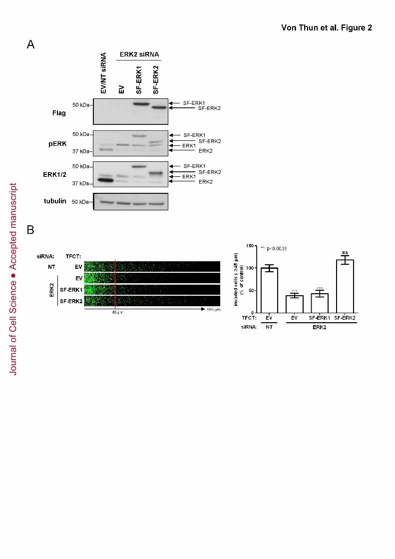
MDA-MB-231 cells predominantly express the ERK2 isoform. Therefore,
observations that knockdown of ERK2 affects invasion (whereas siRNA of ERK1 does
not) do not necessarily reflect functional differences between the two kinases, but may
be attributable to differences in their expression level (Lefloch et al., 2008; Voisin et al.).
To address this, we expressed recombinant ERK1 and siRNA-resistant ERK2 to equal
levels after knocking down ERK2 (Fig. 2A & 3A). The expressed levels of SF-ERK1 and
SF-ERK2 isoforms were similar to one another (Fig. 2A & 3A), their expression was
maintained for up to 3 days following transfection (thus allowing sufficient time to carry
out Matrigel invasion assays) (Fig. S2A), and both recombinant kinases were equally
phosphorylated by MEK in MDA-MB-231 cells (Fig. 2A & 3A). Moreover, expression of
the recombinant ERKs compromised neither the extent, nor the persistence of ERK2
siRNA suppression (Fig. 2A, 3A & S2A). Expression of recombinant ERK1 did not
rescue the inhibitory effect of ERK2 knockdown on invasion, whereas siRNA-resistant
Jour
nal o
f Cel
l Sci
ence
Acc
epte
d m
anus
crip
t
Von Thun et al. ERK2 suppresses Rab17 and Liprin-β2 to drive cell migration
11
ERK2 expression completely restored the invasive phenotype of MDA-MB-231 cells (Fig.
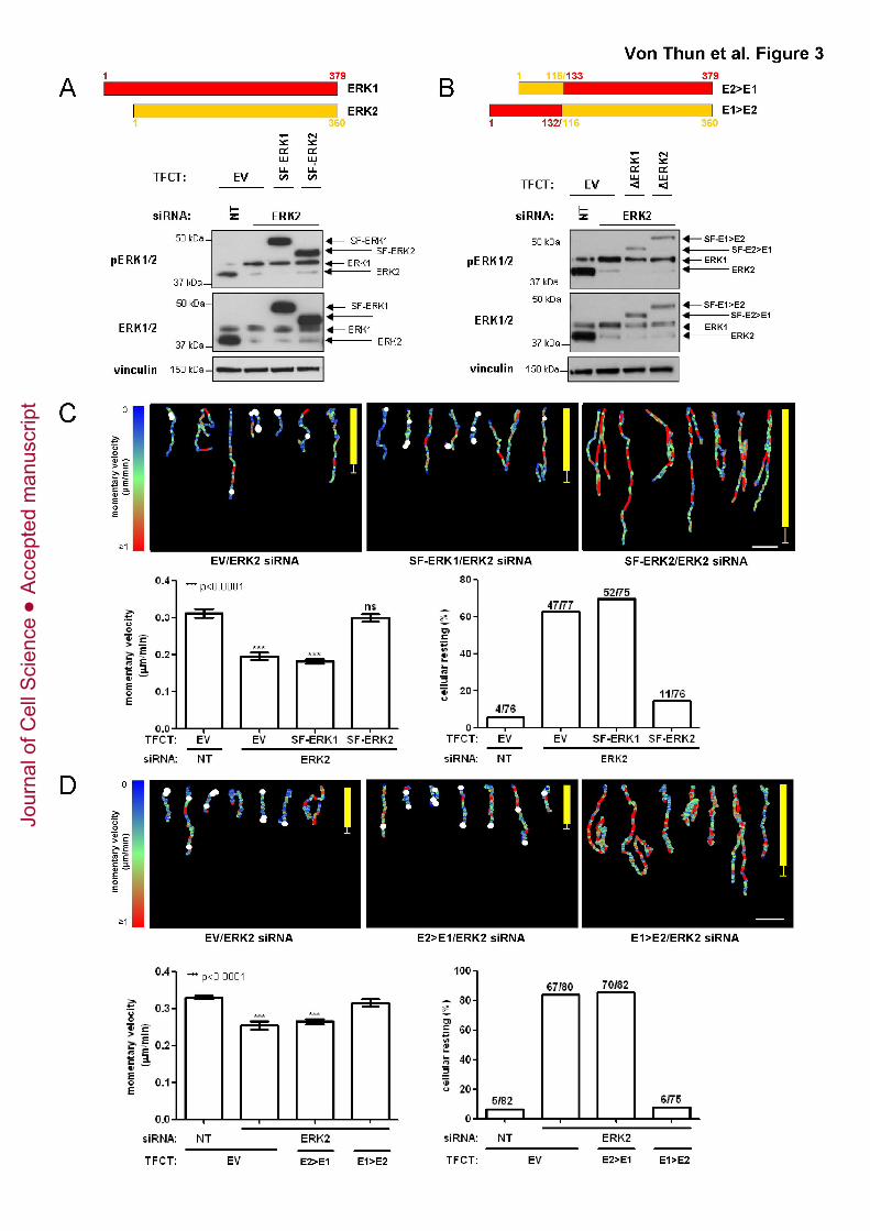
2B). Moreover, when cell migration within CDM was analysed, re-expression of
siRNA-resistant ERK2 increased the momentary velocity and reduced the tendency of
cells to pause (cellular resting) relative to that of control cells, whereas re-expression of
ERK1 did not rescue the migratory defects of ERK2 knockdown cells (Fig. 3C).
The primary sequences of ERK1 and ERK2 diverge most from one another in
their N-terminal portions. A recent study has found that ERK1 shuttles into the nucleus
more slowly than ERK2, and domain swap experiments indicated that this property was
conferred by a region in ERK1’s longer N-terminal domain (Marchi et al., 2008). To
address whether the information responsible for driving cell migration resides within the
N-terminal portion of ERK2, we generated two chimeras; one containing the N-terminal
region of ERK2 fused to the C-terminal portion of ERK1 (E2>E1) and another
corresponding chimera of the N-terminal region of ERK1 fused to the C-terminal portion
of ERK2 (E1>E2) (Fig. 3B). Although expressed at lower levels than the native kinases,
E2>E1 and E1>E2 were present at similar levels to one another (on an ERK2
knockdown background) and both chimeras were similarly phosphorylated at the TEY
motif (Fig. 3B), suggesting an intact tertiary structure. Analysis of cell migration within
CDM indicated that although expression of E1>E2 restored the migratory defects caused
by ERK2 knockdown, E2>E1 was ineffective in this regard (Fig. 3D). These data indicate
that ERK2’s capacity to drive migration is not located within the divergent N-terminal
portion of the kinase.
Identifying an ERK2-dependent gene expression signature
To determine whether isoform-specific regulation of gene expression was
responsible for ERK2’s influence over cell migration and invasion, we expressed ERK1
or ERK2 in ERK2 knockdown cells (Fig. S2B), plated them onto CDM for 16hr and
Jour
nal o
f Cel
l Sci
ence
Acc
epte
d m
anus
crip
t
Von Thun et al. ERK2 suppresses Rab17 and Liprin-β2 to drive cell migration
12
performed an Illumina gene expression array. These gene expression screens were
performed with three independent biological replicates (i.e. separate experiments), and
the reproducibility of the ERK knockdowns and re-expression of siRNA-resistant ERK2
and ERK1 across these three separate experiments is shown in Fig. S2B. Knockdown
of ERK2 altered the expression of a large number of genes, and in most cases
expression of these was restored to normal levels by ectopic expression of either ERK1
or -2. For instance, egr1 fell into this category, and regulation of egr1 gene expression
by both ERKs has previously been demonstrated (Lefloch et al., 2008). However, we
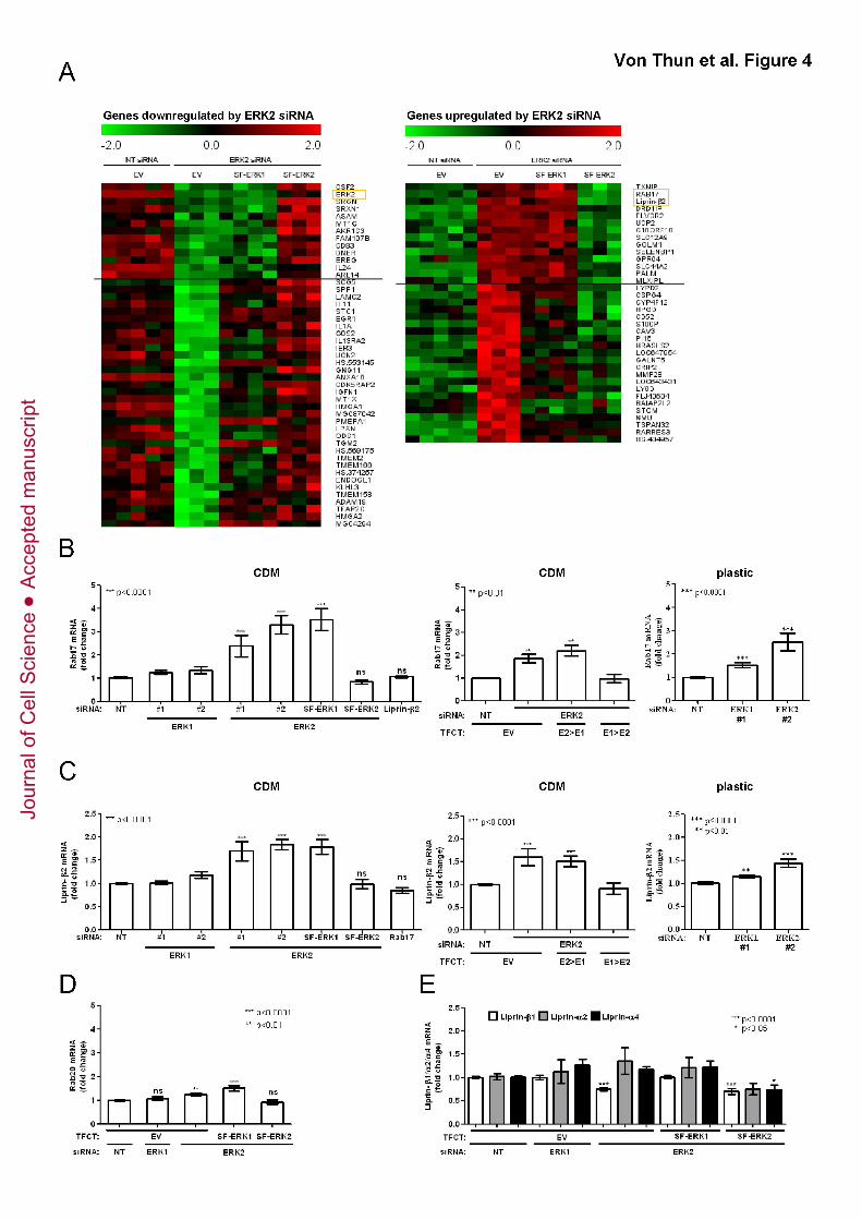
identified a subset of 27 genes whose expression was altered by knockdown of ERK2,
but were restored to normal levels by re-expression of siRNA-resistant ERK2 but not
ERK1. Prominent amongst these were Ras-related protein 17 (Rab17) and Liprin-β2,
whose mRNA levels were strongly increased by knockdown of ERK2, and normalised by
re-expression of ERK2 (but not ERK1) (Fig. 4A). Rab17 is a small GTPase which has
been shown to regulate intracellular transport of proteins and lipids (Zacchi et al., 1998).
Rab17 expression is thought to be restricted to epithelial cells and there are indications
that it is involved in the maintenance of epithelial polarity (McMurtrie et al., 1997).
Liprin-β2 belongs to the family of LAR-interacting proteins (Liprins) and has been shown
predominantly to localise to membrane structures (Serra-Pages et al., 1998).
We used quantitative real-time-PCR (qRT-PCR) to confirm that levels of Rab17 and
Liprin-β2 mRNA were significantly increased following knockdown of ERK2 (with two
independent RNA duplexes), whereas ERK1 silencing did not elevate either Rab17 or
Liprin-β2 levels (Fig. 4B and C). Furthermore, re-expression of siRNA-resistant ERK2 or
the E1>E2 chimera restored Rab17 and Liprin-β2 to levels displayed by control cells,
while expression of ERK1 or E2>E1 was ineffective in this regard (Fig. 4B and C).
We wished to evaluate the extent to which the ECM environment is responsible for these
ERK2-specific changes in gene expression. We, therefore, knocked down ERK1 or
Jour
nal o
f Cel
l Sci
ence
Acc
epte
d m
anus
crip
t
Von Thun et al. ERK2 suppresses Rab17 and Liprin-β2 to drive cell migration
13
ERK2, plated the cells onto plastic dishes (instead of CDM) and looked at the expression
levels of Rab17 and Liprin-β2. Clearly, siRNA of ERK2 increased expression of Rab17
and Liprin-β2 when cells were plated onto plastic surfaces, and knockdown of ERK1 also
drove Rab17 expression, but to a lesser extent (Fig. 4B, C). These data indicate that the
presence of a 3D microenvironment is not a prerequisite for ERK2’s ability to suppress
Rab17 and Liprin-β2 expression, but that ERK1 may also acquire some capacity to
control Rab17 when cells are plated onto plastic. Unfortunately, we were unable to
assess to what extent these changes in mRNA levels relate to changes in protein levels,
as all of the available antibodies that we have tested are not capable of recognising
Rab17 or Liprin-β2 protein in cell lysates.
To determine whether Liprin-β2 and Rab17 influenced one another’s expression,
we knocked down either Liprin-β2 or Rab17 and measured their respective mRNA levels
by qRT-PCR. We found that siRNA of Liprin-β2 did not alter levels of Rab17 mRNA, and
neither did Rab17 knockdown affect Liprin-β2 expression (Fig.4B & C). Next, we
determined whether related members of the Rab or Liprin family were regulated by
ERK2. Expression of Rab20, a Rab GTPase that exhibits close homology to Rab17, was
minimally affected by manipulation of ERK2 expression (Fig. 4D), and other members of
the Liprin family (Liprins β1, α2, and α4), were also minimally affected by knockdown or
re-expression of ERKs but not in the same fashion as was Liprin-β2 (Fig. 4E).
ERK2 drives invasion by suppressing Rab17 and Liprin-β2 expression
To determine whether Rab17 and Liprin-β2 influence cell motility, we used siRNA
to reduce their expression (Fig. S3) and performed invasion assays. At this stage, we
also looked at the behaviour of A2780 ovarian carcinoma cells stably transfected with
the Rab11 GTPase, Rab25 (A2780-Rab25 cells) and BE colon carcinoma cells as, like
MDA-MB-231’s, these cells invade into fibronectin-containing Matrigel with high
Jour
nal o
f Cel
l Sci
ence
Acc
epte
d m
anus
crip
t
Von Thun et al. ERK2 suppresses Rab17 and Liprin-β2 to drive cell migration
14
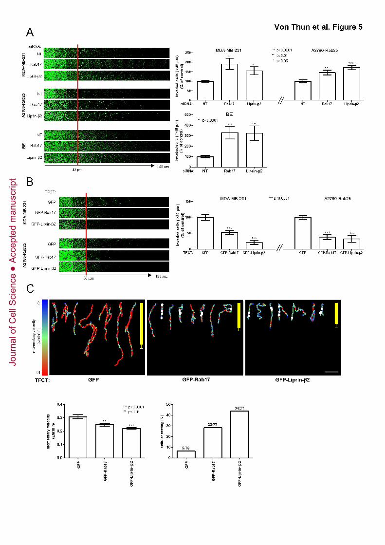
efficiency (Caswell et al., 2007; Moran-Jones et al., 2009). Interestingly, knockdown of
either Rab17 or Liprin-β2 promoted invasion of all three cell lines into Matrigel (Fig. 5A).
Consistent with this, we found that overexpression of either GFP-Rab17 or GFP-Liprin-
β2 suppressed invasion of MDA-MB-231 and A2780-Rab25 cells (by comparison with
expression of GFP alone) (Fig. 5B). Moreover, overexpression of GFP-Rab17 or GFP-
Liprin-β2 reduced the momentary velocity and increased the tendency of MDA-MB-231
cells to pause (cellular resting) whilst migrating within CDM in much the same way as did
knockdown of ERK2, indicating that Rab17 and Liprin-β2 act to restrict carcinoma cell
migration in 3D microenvironments (Fig. 5C).
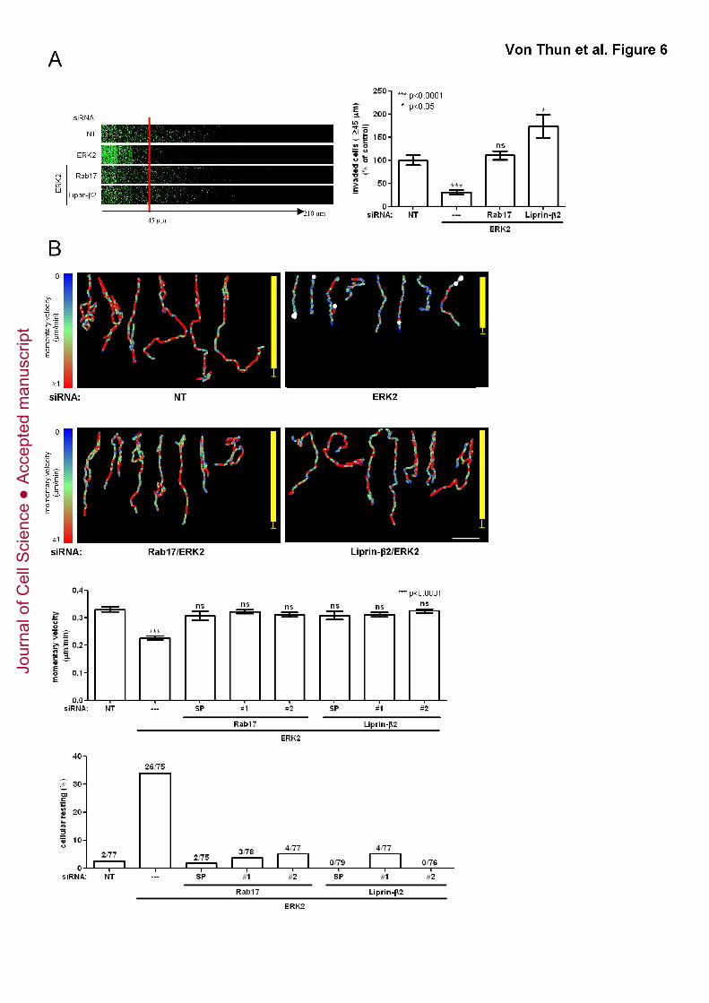
We wished to determine to what extent ERK2’s suppression of Rab17 and Liprin-
β2 was responsible for the kinase’s ability to drive invasion. To do this, we knocked
down ERK2 in combination with knockdown of either Rab17 or Liprin-β2. Interestingly,
siRNA of either Rab17 or Liprin-β2 restored invasion of ERK2 knockdown cells to levels
comparable to control cells (Fig. 6A). Moreover, siRNA of Rab17 or Liprin-β2 (with either
a SMARTPool or two individual RNA duplexes) increased momentary velocity and
reduced the tendency of ERK2 knockdown cells to pause (cellular resting) whilst
migrating within CDM. Indeed, cells that had ERK2 knocked down in combination with
either Rab17 or Liprin-β2 migrated in a way that was indistinguishable from control cells
(Fig. 6B). Taken together these data indicate that rab17 or liprin-β2 are novel motility
suppressor genes, and in order to drive invasion and migration of carcinoma cells in 3D
microenvironments, ERK2 must reduce the expression of at least one of these genes.
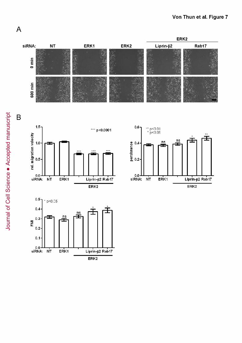
ERK2 drives migration on plastic surfaces, but not through Rab17 or Liprin-β2
We wished to investigate the requirement for suppression of Rab17 and Liprin-β2
in ERK2-dependent cell migration into scratch wounds. Knockdown of ERK2 (but not
ERK1) delayed wound closure (Fig. 7A), and cell tracking analyses indicated that this
Jour
nal o
f Cel
l Sci
ence
Acc
epte
d m
anus
crip
t
Von Thun et al. ERK2 suppresses Rab17 and Liprin-β2 to drive cell migration
15
was owing to reduced migration speed, whilst the migratory persistence and the forward
migration index (FMI; a quantitative measurement of persistent migration perpendicular
to the wound’s edge) remained unaltered following ERK2 knockdown (Fig. 7B). Thus,
ERK2 (but not ERK1) likely plays a role in cell migration in both 3D (Matrigel plugs and
CDM) and 2D (scratch wound assays) microenvironments. However, by contrast with
the situation in 3D microenvironments, siRNA of Rab17 and Liprin-β2 was completely
unable to restore the migration velocity of ERK2 knockdown cells (Fig7B), although the
persistence of Rab17 and Liprin-β2 knockdown cells was marginally increased (Fig. 7B).
Taken together, these data indicate that although ERK2 controls cell movement in 3D
microenvironments by suppressing Rab17 and Liprin-β2, the pathways by which ERK2
controls cell migration on plastic do not involve these two novel ERK2 effectors.
Jour
nal o
f Cel
l Sci
ence
Acc
epte
d m
anus
crip
t
Von Thun et al. ERK2 suppresses Rab17 and Liprin-β2 to drive cell migration
16
DISCUSSION
In recent years, much evidence supporting ERK isoform-specific functions has
accumulated. Firstly, striking discrepancies between the ERK1-/- and ERK2-/- phenotypes
in mice argued for distinct roles of these kinases in embryogenesis (Bost et al., 2005;
Hatano et al., 2003; Mazzucchelli et al., 2002; Pages et al., 1999; Saba-El-Leil et al.,
2003; Yao et al., 2003). Moreover, some studies have suggested specific roles for ERK2
in learning and memory, proliferation, and cell cycle progression (Fremin et al., 2007;
Satoh et al., 2007; Vantaggiato et al., 2006). However, this view has been challenged by
Lefloch et al. in 2008, who proposed erk gene dosage as the reason for the differences
in the ERK-/- phenotypes (Lefloch et al., 2008). More recently, genetic evidence indicates
that ablation of either ERK 1 or ERK2 had no effect on K-Ras-driven lung carcinogenesis
owing to the ability of these kinases to compensate for one another (Blasco et al.).
However, this study looked primarily at primary tumour formation and did not consider
potential isoform specific roles for ERKs 1 & 2 in invasion and metastasis. By
developing a system in which we can knockdown ERK2 and then ectopically express
either ERK1 or ERK2 to similar levels, we have shown that ERK2 is the main driver of
cell migration and invasion in 3D microenvironments in a way that is not influenced by
gene dosage.
The role of the ERKs in cell migration has been studied extensively and it is clear
that these kinases play a key role in tumour progression by regulating cell invasiveness
(Klemke et al., 1997; Ochieng et al., 1991; Reddy et al., 2003; Rodier et al., 1995; Ueoka
et al., 2000). However, little is known about the respective contributions of ERK1 and
ERK2 to cell migration. Our finding that it is ERK2, and not ERK1, that contributes to cell
migration in 3D microenvironments is certainly in agreement with observations in vivo,
where ERK2+/- mice show a delay in wound healing after partial-thickness burn in
Jour
nal o
f Cel
l Sci
ence
Acc
epte
d m
anus
crip
t
Von Thun et al. ERK2 suppresses Rab17 and Liprin-β2 to drive cell migration
17
comparison to ERK2+/+ mice (Satoh et al., 2009). Studies in Zebrafish further support this
view as ERK2 but not ERK1 morphants show defects in cytoskeletal reorganisation
processes which lead to anterior-to-posterior migration retardations (Krens et al., 2008).
More recently, by using a retroviral system to express ERK 1 and -2 to similar levels, a
study has demonstrated specific role for ERK2 in cell migration in MCF-10A cells which
was due to the participation of this ERK isoform in Ras-induced
epithelial-to-mesenchymal transformation (EMT) (Shin et al.).
Both ERKs have indistinguishable kinase activities in vitro (Lefloch et al., 2008)
and so far no isoform-specific protein interaction domains have been identified, thus
leaving the question of how isoform-specificity can be achieved unanswered. However,
Marchi et al. have shown isoform-specific nuclear shuttling rates caused by differences
in the N-terminal amino acid sequence (Marchi et al., 2008), suggesting that ERK1 and
ERK2 may perform different roles within the nucleus. We find that, although the
expression of most ERK target genes is controlled equally by ERK1 and ERK2, a small
subset of ERK targets (including Rab17 and Liprin-β2) are specific to ERK2. At this
stage we do not know how ERK2 suppresses Rab17 and Liprin-β2, but the rate at which
they are upregulated following ERK inhibition (it takes 24hr before the mRNA for Rab17
is increased following addition of U0126 (not shown)), suggests that the rab17 and liprin-
β2 genes are targeted indirectly by ERK2. Here we report that the ability of ERK2 to
control expression of Rab17 and Liprin-β2 (and thereby cell migration and invasion) is
not conferred by sequences within the divergent N-terminal portion of the kinase. This
region of the ERKs is known to be responsible for the nuclear-cytoplasmic shuttling of
ERK1 and ERK2 indicating that the capacity to selectively control Rab17 and Liprin-β2
expression is unlikely to be linked to the nuclear import/export rates for these kinases.
However, despite having identified ERK2-specific events that play a clear role in
its ability to drive invasion, at this stage we may only speculate on how specificity is
Jour
nal o
f Cel
l Sci
ence
Acc
epte
d m
anus
crip
t
Von Thun et al. ERK2 suppresses Rab17 and Liprin-β2 to drive cell migration
18
accomplished. Isoform-specific scaffolds, although none identified to date, may localise
the kinases differently and thereby provide specificity. It is also possible that ERK1 and
ERK2 possess distinct protein interactomes or varying affinities for the same substrate.
Shin et al. demonstrated that ERK2 induced EMT via DEF motif-dependent signalling
(Shin et al.). As both ERK isoforms bind to DEF motifs (Turjanski et al., 2007), this
observation argues for distinct substrate affinities in vivo. Moreover, ERK2 has recently
been identified as an unconventional DNA-binding protein, which specifically binds to
G/CAAG/C consensus motif (Hu et al., 2009). Although DNA-binding was shown to be
independent of ERK2’s kinase activity (Hu et al., 2009), it is possible that active ERK
bound to the promoter region can influence transcription by phosphorylating
neighbouring transcription factors. Interestingly, both ERK isoforms harbour the critical
residues involved in DNA-binding, but amino acids flanking these residues are only
partially conserved between the two kinases. Thus, differing DNA binding grooves
between the two kinases may exist and may account for differences in regulation of
gene expression.
Rab17 has been shown to influence receptor-mediated transcytosis and
recycling of receptors to the apical membrane in non-transformed epithelial cells
(Hunziker and Peters, 1998; Zacchi et al., 1998). Recently, a study by Singh et al. found
that Rab17 was one of many genes to be upregulated in cancer cell lines exhibiting a
more epithelial morphology, whereas Rab17 was downregulated in cells displaying a
more mesenchymal morphology (Singh et al., 2009). This indicates that Rab17 is
associated with the maintenance of a polarised epithelial morphology and this may
explain why Rab17 levels must be reduced in order for metastatic tumour cells to
migrate with mesenchymal characteristics. Given that Rab17 would be expected to
control membrane trafficking, it is interesting to speculate as to how it might suppress
cell migration. Recycling of α5β1 integrin is known to be key to tumour cell migration and
Jour
nal o
f Cel
l Sci
ence
Acc
epte
d m
anus
crip
t
Von Thun et al. ERK2 suppresses Rab17 and Liprin-β2 to drive cell migration
19
invasion (Caswell et al., 2008; Caswell et al., 2007; Muller et al., 2009) and we have
preliminary data suggesting that overexpression of Rab17 leads to the accumulation of
β1-integrin within large endosomes to which Rab17 itself is also localised (data not
shown). Taken together with reports suggesting that Rab17 opposes the return of
receptors to the plasma membrane (Hunziker and Peters, 1998; Zacchi et al., 1998), this
moots that Rab17 may be an integrin recycling suppressor whose expression must be
reduced for cells to migrate efficiently, and future work will explore this further.
The role of Liprin family members, in particular the α-liprins, has been well
established in synaptic transmission. It is clear from reverse genetic studies in
vertebrates, Drosophila and C. elegans that Liprin mutants have defects in synaptic
vesicle transport, in which synaptic vesicles accumulate in axons (Stryker and Johnson,
2007). Thus Liprins control vesicular transport, and this is due to their ability to act as
scaffolds to recruit and stabilise a number of different proteins to the sites of exocytosis
(Stryker and Johnson, 2007). There is a report that Liprin-α1 may regulate integrin
trafficking (Asperti et al., 2009), consistent with a role for these proteins contributing to
cell migration by controlling trafficking events, and more recently Liprin-α1 has been
shown to contribute to the invasiveness of MDA-MB-231 cells and their ability to migrate
persistently on 2D surfaces; both of which are integrin-regulated events (Astro et al.).
Although it is thought that they function by heterodimerising with α-Liprins, there is little
data available concerning the function of β-Liprins. We find that Liprin-β2 is a novel
suppressor of cell migration and invasion downstream of ERK2 and so it is likely that it
functions either by promoting the trafficking of anti-migratory receptors or by inhibiting
the recycling of pro-invasive ones. Notably, Liprins have been identified as inhibitors of
cell invasion in head and neck squamous carcinoma cells (Tan et al., 2008), however
further work will be needed to establish how Liprin-β2 acts to oppose tumour cell
invasion and whether this involves recycling of integrins or other receptors involved in
Jour
nal o
f Cel
l Sci
ence
Acc
epte
d m
anus
crip
t
Von Thun et al. ERK2 suppresses Rab17 and Liprin-β2 to drive cell migration
20
cell adhesion and migration. It is unclear from our studies how Rab17 and Liprin-β2
interact functionally, but our observations that their silencing yields indistinguishable
migratory phenotypes suggest that both proteins operate in series on the same pathway.
In conclusion, this study has identified Rab17 and Liprin-β2 as novel inhibitors of
cell motility whose expression is regulated by ERK2 in MDA-MB-231 cells. Moreover, we
have demonstrated that suppression of either Rab17 or Liprin-β2 can completely
compensate for loss of ERK2. Thus, we propose one way for ERK2 to drive
invasiveness is by suppressing Rab17 and/or Liprin-β2. The potential for inhibitors of the
MAPK pathway to be used as anticancer agents is now being assessed in the clinic, and
our finding that ERK2-mediated suppression of Rab17 and Liprin-β2 drives cancer
invasiveness will be important to the interpretation of data from these studies.
Jour
nal o
f Cel
l Sci
ence
Acc
epte
d m
anus
crip
t
Von Thun et al. ERK2 suppresses Rab17 and Liprin-β2 to drive cell migration
21
ACKNOWLEDGEMENTS
This work was funded by Cancer Research UK. MRB acknowledges a Marie Curie
International Incoming Fellowship (236758) and an EMBO long-term fellowship (ALTF
815-2010). We would like to thank Jeremy Simpson (University College Dublin) for
providing the EGFP-Rab17 plasmid.
Jour
nal o
f Cel
l Sci
ence
Acc
epte
d m
anus
crip
t
Von Thun et al. ERK2 suppresses Rab17 and Liprin-β2 to drive cell migration
22
REFERENCES
Asperti, C., Astro, V., Totaro, A., Paris, S. and de Curtis, I. (2009). Liprin-alpha1 promotes cell spreading on the extracellular matrix by affecting the distribution of activated integrins. J Cell Sci 122, 3225-32. Astro, V., Asperti, C., Cangi, M. G., Doglioni, C. and de Curtis, I. Liprin-alpha1 regulates breast cancer cell invasion by affecting cell motility, invadopodia and extracellular matrix degradation. Oncogene 30, 1841-9. Bass, M. D., Roach, K. A., Morgan, M. R., Mostafavi-Pour, Z., Schoen, T., Muramatsu, T., Mayer, U., Ballestrem, C., Spatz, J. P. and Humphries, M. J. (2007). Syndecan-4-dependent Rac1 regulation determines directional migration in response to the extracellular matrix. J Cell Biol 177, 527-38. Blasco, R. B., Francoz, S., Santamaria, D., Canamero, M., Dubus, P., Charron, J., Baccarini, M. and Barbacid, M. c-Raf, but not B-Raf, is essential for development of K-Ras oncogene-driven non-small cell lung carcinoma. Cancer Cell 19, 652-63. Bost, F., Aouadi, M., Caron, L., Even, P., Belmonte, N., Prot, M., Dani, C., Hofman, P., Pages, G., Pouyssegur, J. et al. (2005). The extracellular signal-regulated kinase isoform ERK1 is specifically required for in vitro and in vivo adipogenesis. Diabetes 54, 402-11. Boulton, T. G., Nye, S. H., Robbins, D. J., Ip, N. Y., Radziejewska, E., Morgenbesser, S. D., DePinho, R. A., Panayotatos, N., Cobb, M. H. and Yancopoulos, G. D. (1991). ERKs: a family of protein-serine/threonine kinases that are activated and tyrosine phosphorylated in response to insulin and NGF. Cell 65, 663-75. Caswell, P. T., Chan, M., Lindsay, A. J., McCaffrey, M. W., Boettiger, D. and Norman, J. C. (2008). Rab-coupling protein coordinates recycling of alpha5beta1 integrin and EGFR1 to promote cell migration in 3D microenvironments. J Cell Biol 183, 143-55. Caswell, P. T., Spence, H. J., Parsons, M., White, D. P., Clark, K., Cheng, K. W., Mills, G. B., Humphries, M. J., Messent, A. J., Anderson, K. I. et al. (2007). Rab25 associates with alpha5beta1 integrin to promote invasive migration in 3D microenvironments. Dev Cell 13, 496-510. Cukierman, E., Pankov, R., Stevens, D. R. and Yamada, K. M. (2001). Taking cell-matrix adhesions to the third dimension. Science 294, 1708-12. Fremin, C., Ezan, F., Boisselier, P., Bessard, A., Pages, G., Pouyssegur, J. and Baffet, G. (2007). ERK2 but not ERK1 plays a key role in hepatocyte replication: an RNAi-mediated ERK2 knockdown approach in wild-type and ERK1 null hepatocytes. Hepatology 45, 1035-45. Gloeckner, C. J., Boldt, K., Schumacher, A., Roepman, R. and Ueffing, M. (2007). A novel tandem affinity purification strategy for the efficient isolation and characterisation of native protein complexes. Proteomics 7, 4228-34. Hatano, N., Mori, Y., Oh-hora, M., Kosugi, A., Fujikawa, T., Nakai, N., Niwa, H., Miyazaki, J., Hamaoka, T. and Ogata, M. (2003). Essential role for
Jour
nal o
f Cel
l Sci
ence
Acc
epte
d m
anus
crip
t
Von Thun et al. ERK2 suppresses Rab17 and Liprin-β2 to drive cell migration
23
ERK2 mitogen-activated protein kinase in placental development. Genes Cells 8, 847-56. Hennigan, R. F., Hawker, K. L. and Ozanne, B. W. (1994). Fos-transformation activates genes associated with invasion. Oncogene 9, 3591-600. Hu, S., Xie, Z., Onishi, A., Yu, X., Jiang, L., Lin, J., Rho, H. S., Woodard, C., Wang, H., Jeong, J. S. et al. (2009). Profiling the human protein-DNA interactome reveals ERK2 as a transcriptional repressor of interferon signaling. Cell 139, 610-22. Hunziker, W. and Peters, P. J. (1998). Rab17 localizes to recycling endosomes and regulates receptor-mediated transcytosis in epithelial cells. J Biol Chem 273, 15734-41. Klemke, R. L., Cai, S., Giannini, A. L., Gallagher, P. J., de Lanerolle, P. and Cheresh, D. A. (1997). Regulation of cell motility by mitogen-activated protein kinase. J Cell Biol 137, 481-92. Krens, S. F., He, S., Lamers, G. E., Meijer, A. H., Bakkers, J., Schmidt, T., Spaink, H. P. and Snaar-Jagalska, B. E. (2008). Distinct functions for ERK1 and ERK2 in cell migration processes during zebrafish gastrulation. Dev Biol 319, 370-83. Lefloch, R., Pouyssegur, J. and Lenormand, P. (2008). Single and combined silencing of ERK1 and ERK2 reveals their positive contribution to growth signaling depending on their expression levels. Mol Cell Biol 28, 511-27. Livak, K. J. and Schmittgen, T. D. (2001). Analysis of relative gene expression data using real-time quantitative PCR and the 2(-Delta Delta C(T)) Method. Methods 25, 402-8. Marchi, M., D'Antoni, A., Formentini, I., Parra, R., Brambilla, R., Ratto, G. M. and Costa, M. (2008). The N-terminal domain of ERK1 accounts for the functional differences with ERK2. PLoS One 3, e3873. Mazzucchelli, C., Vantaggiato, C., Ciamei, A., Fasano, S., Pakhotin, P., Krezel, W., Welzl, H., Wolfer, D. P., Pages, G., Valverde, O. et al. (2002). Knockout of ERK1 MAP kinase enhances synaptic plasticity in the striatum and facilitates striatal-mediated learning and memory. Neuron 34, 807-20. McMurtrie, E. B., Barbosa, M. D., Zerial, M. and Kingsmore, S. F. (1997). Rab17 and rab18, small GTPases with specificity for polarized epithelial cells: genetic mapping in the mouse. Genomics 45, 623-5. Moran-Jones, K., Grindlay, J., Jones, M., Smith, R. and Norman, J. C. (2009). hnRNP A2 regulates alternative mRNA splicing of TP53INP2 to control invasive cell migration. Cancer Res 69, 9219-27. Muller, P. A., Caswell, P. T., Doyle, B., Iwanicki, M. P., Tan, E. H., Karim, S., Lukashchuk, N., Gillespie, D. A., Ludwig, R. L., Gosselin, P. et al. (2009). Mutant p53 drives invasion by promoting integrin recycling. Cell 139, 1327-41. Ochieng, J., Basolo, F., Albini, A., Melchiori, A., Watanabe, H., Elliott, J., Raz, A., Parodi, S. and Russo, J. (1991). Increased invasive, chemotactic and locomotive abilities of c-Ha-ras-transformed human breast epithelial cells. Invasion Metastasis 11, 38-47.
Jour
nal o
f Cel
l Sci
ence
Acc
epte
d m
anus
crip
t
Von Thun et al. ERK2 suppresses Rab17 and Liprin-β2 to drive cell migration
24
Pages, G., Guerin, S., Grall, D., Bonino, F., Smith, A., Anjuere, F., Auberger, P. and Pouyssegur, J. (1999). Defective thymocyte maturation in p44 MAP kinase (Erk 1) knockout mice. Science 286, 1374-7. Pouyssegur, J., Volmat, V. and Lenormand, P. (2002). Fidelity and spatio-temporal control in MAP kinase (ERKs) signalling. Biochem Pharmacol 64, 755-63. Reddy, K. B., Nabha, S. M. and Atanaskova, N. (2003). Role of MAP kinase in tumor progression and invasion. Cancer Metastasis Rev 22, 395-403. Rodier, J. M., Valles, A. M., Denoyelle, M., Thiery, J. P. and Boyer, B. (1995). pp60c-src is a positive regulator of growth factor-induced cell scattering in a rat bladder carcinoma cell line. J Cell Biol 131, 761-73. Saba-El-Leil, M. K., Vella, F. D., Vernay, B., Voisin, L., Chen, L., Labrecque, N., Ang, S. L. and Meloche, S. (2003). An essential function of the mitogen-activated protein kinase Erk2 in mouse trophoblast development. EMBO Rep 4, 964-8. Samuels, I. S., Karlo, J. C., Faruzzi, A. N., Pickering, K., Herrup, K., Sweatt, J. D., Saitta, S. C. and Landreth, G. E. (2008). Deletion of ERK2 mitogen-activated protein kinase identifies its key roles in cortical neurogenesis and cognitive function. J Neurosci 28, 6983-95. Satoh, Y., Endo, S., Ikeda, T., Yamada, K., Ito, M., Kuroki, M., Hiramoto, T., Imamura, O., Kobayashi, Y., Watanabe, Y. et al. (2007). Extracellular signal-regulated kinase 2 (ERK2) knockdown mice show deficits in long-term memory; ERK2 has a specific function in learning and memory. J Neurosci 27, 10765-76. Satoh, Y., Saitoh, D., Takeuchi, A., Ojima, K., Kouzu, K., Kawakami, S., Ito, M., Ishihara, M., Sato, S. and Takishima, K. (2009). ERK2 dependent signaling contributes to wound healing after a partial-thickness burn. Biochem Biophys Res Commun 381, 118-22. Serra-Pages, C., Medley, Q. G., Tang, M., Hart, A. and Streuli, M. (1998). Liprins, a family of LAR transmembrane protein-tyrosine phosphatase-interacting proteins. J Biol Chem 273, 15611-20. Shin, S., Dimitri, C. A., Yoon, S. O., Dowdle, W. and Blenis, J. ERK2 but not ERK1 induces epithelial-to-mesenchymal transformation via DEF motif-dependent signaling events. Mol Cell 38, 114-27. Singh, A., Greninger, P., Rhodes, D., Koopman, L., Violette, S., Bardeesy, N. and Settleman, J. (2009). A gene expression signature associated with "K-Ras addiction" reveals regulators of EMT and tumor cell survival. Cancer Cell 15, 489-500. Stryker, E. and Johnson, K. G. (2007). LAR, liprin alpha and the regulation of active zone morphogenesis. J Cell Sci 120, 3723-8. Tan, K. D., Zhu, Y., Tan, H. K., Rajasegaran, V., Aggarwal, A., Wu, J., Wu, H. Y., Hwang, J., Lim, D. T., Soo, K. C. et al. (2008). Amplification and overexpression of PPFIA1, a putative 11q13 invasion suppressor gene, in head and neck squamous cell carcinoma. Genes Chromosomes Cancer 47, 353-62. Turjanski, A. G., Vaque, J. P. and Gutkind, J. S. (2007). MAP kinases and the control of nuclear events. Oncogene 26, 3240-53.
Jour
nal o
f Cel
l Sci
ence
Acc
epte
d m
anus
crip
t
Von Thun et al. ERK2 suppresses Rab17 and Liprin-β2 to drive cell migration
25
Ueoka, Y., Kato, K., Kuriaki, Y., Horiuchi, S., Terao, Y., Nishida, J., Ueno, H. and Wake, N. (2000). Hepatocyte growth factor modulates motility and invasiveness of ovarian carcinomas via Ras-mediated pathway. Br J Cancer 82, 891-9. Vantaggiato, C., Formentini, I., Bondanza, A., Bonini, C., Naldini, L. and Brambilla, R. (2006). ERK1 and ERK2 mitogen-activated protein kinases affect Ras-dependent cell signaling differentially. J Biol 5, 14. Voisin, L., Saba-El-Leil, M. K., Julien, C., Fremin, C. and Meloche, S. Genetic demonstration of a redundant role of extracellular signal-regulated kinase 1 (ERK1) and ERK2 mitogen-activated protein kinases in promoting fibroblast proliferation. Mol Cell Biol 30, 2918-32. Yao, Y., Li, W., Wu, J., Germann, U. A., Su, M. S., Kuida, K. and Boucher, D. M. (2003). Extracellular signal-regulated kinase 2 is necessary for mesoderm differentiation. Proc Natl Acad Sci U S A 100, 12759-64. Zacchi, P., Stenmark, H., Parton, R. G., Orioli, D., Lim, F., Giner, A., Mellman, I., Zerial, M. and Murphy, C. (1998). Rab17 regulates membrane trafficking through apical recycling endosomes in polarized epithelial cells. J Cell Biol 140, 1039-53.
Jour
nal o
f Cel
l Sci
ence
Acc
epte
d m
anus
crip
t
Von Thun et al. ERK2 suppresses Rab17 and Liprin-β2 to drive cell migration
26
FIGURE LEGENDS
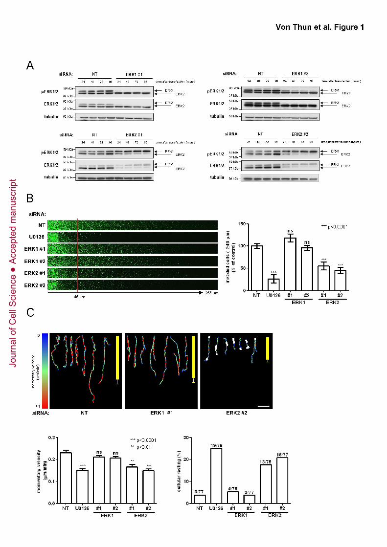
Figure 1. Knockdown of ERK2 opposes invasion into Matrigel and migration of
MDA-MB-231 cells on CDM
(A) MDA-MB-231 cells were transfected with non-targeting siRNAs (NT), or those
targeting ERK1 or ERK2. The effectiveness of the ERK knockdown was assessed by
Western blot over the course of four days following transfection.
(B) MDA-MB-231 cells were transfected with non-targeting siRNAs (NT), or those
targeting ERK1 or ERK2 and plated onto plugs of Matrigel supplemented with fibronectin
(25 μg/ml). The MEK inhibitor, U0126 (10µM) was included as indicated. 36 hr following
this, invading cells were visualized by Calcein-AM staining. Serial optical sections were
captured every 15 µm and are presented as a sequence in which the depth increases
from left to right (left panel). Invasive migration was quantified by measuring the
fluorescence intensity of cells penetrating the Matrigel plug to depths of ≥ 45 µm and
expressed relative to cells transfected with non-targeting (NT) siRNA. Values are means
± standard error of the mean (SEM) of 18 replicates from three independent experiments
(right panel).
(C) MDA-MB-231 cells were transfected with non-targeting siRNAs (NT), or those
targeting ERK1 or ERK2 and plated onto cell-derived matrix. Images were captured
every 10 minutes over a 16 hr period and movies generated from these (Movies S1 and
S2). Cell movement was followed using cell-tracking software. Representative migration
tracks are displayed in the upper panels and the migration speed is denoted by a colour
code, the scale of which is indicated on the left side of the panels. The points at which
cells moved less than 2 μm in 90 min (cellular resting) are indicated by white dots. Scale
bar (white) is 100 μm. The average vectorial (Euclidian) distance migrated by the cells
under each experimental condition is depicted by the vertical yellow bar (mean ± SEM).
Jour
nal o
f Cel
l Sci
ence
Acc
epte
d m
anus
crip
t
Von Thun et al. ERK2 suppresses Rab17 and Liprin-β2 to drive cell migration
27
Momentary migration velocities were calculated for each timeframe of the time-lapse
experiment (lower left panel).Values are means ± standard error of the mean (SEM) of
15 fields from three independent experiments. 5 cells were tracked for each field.
Percentage of resting cells is displayed in the lower right panel with absolute numbers
for each condition above the column.
Figure 2. Ectopic expression of ERK2 but not ERK1 restores invasion of ERK2
knockdown cells
(A) MDA-MB-231 cells were transfected with non-targeting siRNAs (NT), or siRNA
targeting ERK2 in combination with expression plasmids for SF-ERK1, SF-ERK2 or an
empty vector control (EV). Cells were harvested two days after transfection and ERK
expression levels were determined by Western blotting.
(B) MDA-MB-231 cells were transfected with non-targeting siRNAs (NT), or siRNA
targeting ERK2 in combination with expression plasmids for SF-ERK1, SF-ERK2 or an
empty vector control (EV). Cells were plated onto plugs of fibronectin-supplemented
Matrigel and invasion determined as for Fig. 1B.
Figure 3. Expression of recombinant ERK2 or the E2>E1 chimera restores
migration characteristics of ERK2-depleted cells
(A) Cells were transfected and as for Fig. 2A, harvested two days after transfection and
ERK expression levels were determined by Western blotting.
(B) MDA-MB-231 cells were transfected with non-targeting siRNAs (NT), or siRNA
targeting ERK2 in combination with expression plasmids for SF-tagged E2>E1 or E1>E2
chimeras or an empty vector control (EV). The coordinates of the ERK chimeras are
indicated with the portions of the primary sequence (with amino acid numbers in the
appropriate colour) that derive from ERK1 and ERK2 being denoted in red and yellow
Jour
nal o
f Cel
l Sci
ence
Acc
epte
d m
anus
crip
t
Von Thun et al. ERK2 suppresses Rab17 and Liprin-β2 to drive cell migration
28
respectively. Cells were harvested two days after transfection and ERK expression
levels were determined by Western blotting.
(C, D) Cell transfected as for (A & B) were plated onto CDM and their migration
characteristics were analysed and presented as for Fig. 1C.
Figure 4. Rab17 and Liprin-β2 transcription is regulated in an ERK2 dependent
manner
(A) MDA-MB-231 cells were transfected with non-targeting siRNAs (NT), or an siRNA
targeting ERK2 in combination with expression plasmids for SF-ERK1, SF-ERK2 or an
empty vector control (EV) and plated onto CDM. RNA was extracted, labelled and
comparative whole-genome expression profiling was performed using Illumina HT-12 v4
expression chips. The heat maps display genes which are down- (left panel) or
upregulated (right panel) in an ERK2-dependent manner and these are ranked as
described in Methods and Materials.
(B, C) MDA-MB-231 cells were transfected with the indicated siRNAs and ERK
expression plasmids, and plated onto CDM or plastic dishes as indicated. qRT-PCR
was performed to validate the differential regulation of Rab17 (B) or Liprin-β2 (C). Data
was normalised to GAPDH and values relative to the control transfected are expressed
as means ± standard error of the mean (SEM) of 18 replicates from 6 independent
experiments.
(D, E) qRT-PCR was performed to assess ERK2-dependent expression of Rab20 and
Liprin-β1/α2/α4. Data was analysed as in (B)
Jour
nal o
f Cel
l Sci
ence
Acc
epte
d m
anus
crip
t
Von Thun et al. ERK2 suppresses Rab17 and Liprin-β2 to drive cell migration
29
Figure 5. Rab17 and Liprin-β2 are inhibitors of cell migration and invasion
(A) MDA-MB-231, A2780-Rab25 or BE cells were transfected with non-targeting siRNAs
(NT), siRNAs targeting Rab17 or Liprin-β2. Cells were plated into plugs of fibronectin-
supplemented Matrigel and invasion determined as for Fig. 1B.
(B) MDA-MB-231 or A2780 cells were transfected with expression plasmids for GFP-
Rab17, GFP-Liprin-β2 or GFP alone. Cells were plated into plugs of fibronectin-
supplemented Matrigel and invasion determined as for Fig. 1B.
(C) Transfected cells from (B) were seeded onto CDM and their migration characteristics
were analysed and presented as for Fig. 1C.
Figure 6. Knockdown of Rab17 or Liprin-β2 restores the invasiveness and
migratory characteristics of ERK2 knockdown cells
MDA-MB-231 cells were transfected with non-targeting siRNAs (NT) or siRNAs targeting
ERK2 in combination with individual or SMARTPool (SP) oligos targeting Rab17 or
Liprin-β2. Cells were plated onto plugs of fibronectin-supplemented Matrigel and
invasion determined as for Fig. 1B (A) or onto CDM and their migration characteristics
were analysed and presented as for Fig. 1C (B).
Figure 7. ERK2 impairs cell migration on plastic surfaces through downstream
effectors other than Rab17 and Liprin-β2
MDA-MB-231 cells were transfected with non-targeting siRNAs (NT), or siRNAs
targeting ERK1 or ERK2 in combination with those targeting Rab17 or Liprin-β2 and
plated onto 6-well dishes so that they reached confluence 48 hr after transfection. A
wound was introduced by scratching and wound closure monitored using time-lapse
microscopy. Representative images from two time points are shown in (A).
Jour
nal o
f Cel
l Sci
ence
Acc
epte
d m
anus
crip
t
Von Thun et al. ERK2 suppresses Rab17 and Liprin-β2 to drive cell migration
30
(B) Cell movement was followed using cell-tracking software. Migration velocity,
persistence and forward migration index (FMI) were extracted from the trackplots.
Values are means ± standard error of the mean (SEM) of 15 fields from three
independent experiments. 5 cells or more were tracked for each field.
Jour
nal o
f Cel
l Sci
ence
Acc
epte
d m
anus
crip
t
Jour
nal o
f Cel
l Sci
ence
Acc
epte
d m
anus
crip
t
Jour
nal o
f Cel
l Sci
ence
Acc
epte
d m
anus
crip
t
Jour
nal o
f Cel
l Sci
ence
Acc
epte
d m
anus
crip
t
Jour
nal o
f Cel
l Sci
ence
Acc
epte
d m
anus
crip
t
Jour
nal o
f Cel
l Sci
ence
Acc
epte
d m
anus
crip
t
Jour
nal o
f Cel
l Sci
ence
Acc
epte
d m
anus
crip
t
Jour
nal o
f Cel
l Sci
ence
Acc
epte
d m
anus
crip
t