Loss of β-adrenergic–stimulated phosphorylation of …²-adrenergic stimulation of Ca V1.2...
10
Loss of β-adrenergic–stimulated phosphorylation of Ca V 1.2 channels on Ser1700 leads to heart failure Linghai Yang a , Dao-Fu Dai b , Can Yuan a , Ruth E. Westenbroek a , Haijie Yu a , Nastassya West c , Horacio O. de la Iglesia c , and William A. Catterall a,1 a Department of Pharmacology, University of Washington, Seattle, WA 98195; b Department of Pathology, University of Washington, Seattle, WA 98195; and c Department of Biology, University of Washington, Seattle, WA 98195 Contributed by William A. Catterall, October 14, 2016 (sent for review September 12, 2016; reviewed by Joël Nargeot and Joerg Striessnig) L-type Ca 2+ currents conducted by voltage-gated calcium channel 1.2 (Ca V 1.2) initiate excitation–contraction coupling in the heart, and altered expression of Ca V 1.2 causes heart failure in mice. Here we show unexpectedly that reducing β-adrenergic regulation of Ca V 1.2 channels by mutation of a single PKA site, Ser1700, in the proximal C-terminal domain causes reduced contractile function, cardiac hypertrophy, and heart failure without changes in expres- sion, localization, or function of the Ca V 1.2 protein in the mutant mice (SA mice). These deficits were aggravated with aging. Dual mutation of Ser1700 and a nearby casein-kinase II site (Thr1704) caused accelerated hypertrophy, heart failure, and death in mice with these mutations (STAA mice). Cardiac hypertrophy was in- creased by voluntary exercise and by persistent β-adrenergic stim- ulation. PKA expression was increased, and PKA sites Ser2808 in ryanodine receptor type-2, Ser16 in phospholamban, and Ser23/24 in troponin-I were hyperphosphorylated in SA mice, whereas phosphorylation of substrates for calcium/calmodulin-dependent protein kinase II was unchanged. The Ca 2+ pool in the sarcoplasmic reticulum was increased, the activity of calcineurin was elevated, and calcineurin inhibitors improved contractility and ameliorated cardiac hypertrophy. Cardio-specific expression of the SA mutation also caused reduced contractility and hypertrophy. These results suggest engagement of compensatory mechanisms, which initially may enhance the contractility of individual myocytes but eventually contribute to an increased sensitivity to cardiovascular stress and to heart failure in vivo. Our results demonstrate that normal regulation of Ca V 1.2 channels by phosphorylation of Ser1700 in cardiomyo- cytes is required for cardiovascular homeostasis and normal physi- ological regulation in vivo. calcium channel | heart failure | excitation–contraction coupling | PKA | casein kinase II E xcitation–contraction (EC) coupling in the heart is initiated by L-type calcium (Ca 2+ ) currents conducted by voltage- gated calcium 1.2 (Ca V 1.2) channels that are activated by membrane depolarization (1). Ca 2+ influx via Ca V 1.2 channels activates Ca 2+ release from the sarcoplasmic reticulum (SR), lead- ing to rapid and forceful contraction of myofilaments. Ryanodine receptor type-2 (RyR2) is the major channel for this release of Ca 2+ from cardiac SR (2, 3). Mobilized Ca 2+ is transported back into the SR by the SR Ca 2+ ATPase (SERCA) during muscle relaxation (4). SERCA activity is controlled by the inhibitor protein phospho- lamban (PLB), which is phosphorylated by PKA to release its in- hibition of SERCA activity (4). The force and rate of cardiac contractions are critically dependent on the amplitude and kinetics of the Ca 2+ signal generated by Ca V 1.2 channels and RyR2 (1). In the fight-or-flight response, a marked increase in the beating rate and force of contraction of the heart is triggered by activation of the sympathetic nervous system and stimulation of the β-adrenergic signaling pathway. As a result, PKA is activated and phosphor- ylates Ca V 1.2, RyR2, and PLB to enhance Ca 2+ influx, Ca 2+ mobilization, and reuptake into the SR in cardiomyocytes, leading directly to increased contractility and contributing to the acceleration of the heart rate (3–7). PKA-mediated phosphory- lation of Ca V 1.2 channels causes a three- to fourfold increase in the peak amplitude of Ca 2+ currents in mammalian car- diomyocytes (5–7). Despite much study, the exact PKA phos- phorylation sites on Ca V 1.2 that mediate increased channel activity in response to β-adrenergic regulation remain only par- tially resolved (7–9). Cardiac Ca V 1.2 channels are composed of pore-forming α 1 1.2 subunits and associated β and α 2 δ subunits (10, 11). In skeletal muscle, an additional γ subunit is associated with the closely related Ca V 1.1 channels (11, 12). Multiple PKA sites have been identified by phosphorylation of purified preparations of α 1 1.2 (Ser1928) and β (Ser459, Ser478, and Ser479) subunits; however, none of these sites is required for β-adrenergic regulation of Ca V 1.2 channels in cardiac myocytes in vivo or in transfected mammalian cells (7, 8). In addition to channel phosphorylation, the formation of an autoinhibitory signaling complex is also es- sential for β-adrenergic regulation of Ca V 1.2 channel activity (7, 13, 14). In skeletal and cardiac myocytes (15), as well as in neurons (16, 17), the α 1 1.1 and α 1 1.2 subunits are proteolytically processed at Ala1800 (or the equivalent), and the cleaved distal C terminus (dCT) is noncovalently associated with the pro- teolytically processed channels (13, 18). In this noncovalent complex, the dCT acts as a potent inhibitor of Ca V 1.2 channel activity (13). Truncated Ca V 1.2 channels with dCT deletion ex- hibit increased Ca 2+ channel activity in transfected nonmuscle cells (13, 19), and they have reduced expression and impaired β-adrenergic regulation in vivo (20). Coexpression of the dCT with truncated Ca V 1.2 channels in nonmuscle cells greatly in- hibits channel activity through interaction with the proximal C Significance Calcium entry initiates contraction in cardiac myocytes, and altered expression of voltage-gated calcium channel 1.2 (Ca V 1.2) causes heart failure in mice. Here we show that re- ducing β-adrenergic regulation of Ca V 1.2 by mutation of a PKA site in the C-terminal domain causes age-related heart failure. Dual mutation of a nearby casein-kinase II phosphorylation site accelerated heart failure. The PKA level was increased; PKA- mediated phosphorylation of ryanodine receptor type-2, phospholamban, and troponin-I was increased; the calcium pool in the sarcoplasmic reticulum was increased; and the activity of the calcium-dependent phosphoprotein phospha- tase calcineurin was persistently elevated. These changes in mice with a mutation at the PKA site Ser1700 (SA mice) suggest that compensatory mechanisms may initially enhance contrac- tility but eventually cause increased sensitivity to cardiovascular stress and heart failure. Author contributions: L.Y., R.E.W., H.O.d.l.I., and W.A.C. designed research; L.Y., D.-F. D., C.Y., H.Y., and N.W. performed research; L.Y., D.-F.D., C.Y., R.E.W., H.Y., H.O.d.l.I., and W.A.C. analyzed data; and L.Y. and W.A.C. wrote the paper. Reviewers: J.N., University of Montpellier; and J.S., University of Innsbruck. The authors declare no conflict of interest. 1 To whom correspondence should be addressed. Email: [email protected]. E7976–E7985 | PNAS | Published online November 18, 2016 www.pnas.org/cgi/doi/10.1073/pnas.1617116113
Transcript of Loss of β-adrenergic–stimulated phosphorylation of …²-adrenergic stimulation of Ca V1.2...
Loss of β-adrenergic–stimulated phosphorylation ofCaV1.2 channels on Ser1700 leads to heart failureLinghai Yanga, Dao-Fu Daib, Can Yuana, Ruth E. Westenbroeka, Haijie Yua, Nastassya Westc, Horacio O. de la Iglesiac,and William A. Catteralla,1
aDepartment of Pharmacology, University of Washington, Seattle, WA 98195; bDepartment of Pathology, University of Washington, Seattle, WA 98195;and cDepartment of Biology, University of Washington, Seattle, WA 98195
Contributed by William A. Catterall, October 14, 2016 (sent for review September 12, 2016; reviewed by Joël Nargeot and Joerg Striessnig)
L-type Ca2+ currents conducted by voltage-gated calcium channel1.2 (CaV1.2) initiate excitation–contraction coupling in the heart,and altered expression of CaV1.2 causes heart failure in mice. Herewe show unexpectedly that reducing β-adrenergic regulation ofCaV1.2 channels by mutation of a single PKA site, Ser1700, in theproximal C-terminal domain causes reduced contractile function,cardiac hypertrophy, and heart failure without changes in expres-sion, localization, or function of the CaV1.2 protein in the mutantmice (SA mice). These deficits were aggravated with aging. Dualmutation of Ser1700 and a nearby casein-kinase II site (Thr1704)caused accelerated hypertrophy, heart failure, and death in micewith these mutations (STAA mice). Cardiac hypertrophy was in-creased by voluntary exercise and by persistent β-adrenergic stim-ulation. PKA expression was increased, and PKA sites Ser2808 inryanodine receptor type-2, Ser16 in phospholamban, and Ser23/24in troponin-I were hyperphosphorylated in SA mice, whereasphosphorylation of substrates for calcium/calmodulin-dependentprotein kinase II was unchanged. The Ca2+ pool in the sarcoplasmicreticulum was increased, the activity of calcineurin was elevated,and calcineurin inhibitors improved contractility and amelioratedcardiac hypertrophy. Cardio-specific expression of the SA mutationalso caused reduced contractility and hypertrophy. These resultssuggest engagement of compensatory mechanisms, which initiallymay enhance the contractility of individual myocytes but eventuallycontribute to an increased sensitivity to cardiovascular stress and toheart failure in vivo. Our results demonstrate that normal regulationof CaV1.2 channels by phosphorylation of Ser1700 in cardiomyo-cytes is required for cardiovascular homeostasis and normal physi-ological regulation in vivo.
calcium channel | heart failure | excitation–contraction coupling |PKA | casein kinase II
Excitation–contraction (EC) coupling in the heart is initiatedby L-type calcium (Ca2+) currents conducted by voltage-
gated calcium 1.2 (CaV1.2) channels that are activated bymembrane depolarization (1). Ca2+ influx via CaV1.2 channelsactivates Ca2+ release from the sarcoplasmic reticulum (SR), lead-ing to rapid and forceful contraction of myofilaments. Ryanodinereceptor type-2 (RyR2) is the major channel for this release of Ca2+
from cardiac SR (2, 3). Mobilized Ca2+ is transported back into theSR by the SR Ca2+ ATPase (SERCA) during muscle relaxation (4).SERCA activity is controlled by the inhibitor protein phospho-lamban (PLB), which is phosphorylated by PKA to release its in-hibition of SERCA activity (4). The force and rate of cardiaccontractions are critically dependent on the amplitude and kineticsof the Ca2+ signal generated by CaV1.2 channels and RyR2 (1). Inthe fight-or-flight response, a marked increase in the beating rateand force of contraction of the heart is triggered by activation ofthe sympathetic nervous system and stimulation of the β-adrenergicsignaling pathway. As a result, PKA is activated and phosphor-ylates CaV1.2, RyR2, and PLB to enhance Ca2+ influx, Ca2+
mobilization, and reuptake into the SR in cardiomyocytes,leading directly to increased contractility and contributing to theacceleration of the heart rate (3–7). PKA-mediated phosphory-
lation of CaV1.2 channels causes a three- to fourfold increasein the peak amplitude of Ca2+ currents in mammalian car-diomyocytes (5–7). Despite much study, the exact PKA phos-phorylation sites on CaV1.2 that mediate increased channelactivity in response to β-adrenergic regulation remain only par-tially resolved (7–9).Cardiac CaV1.2 channels are composed of pore-forming α11.2
subunits and associated β and α2δ subunits (10, 11). In skeletalmuscle, an additional γ subunit is associated with the closelyrelated CaV1.1 channels (11, 12). Multiple PKA sites have beenidentified by phosphorylation of purified preparations of α11.2(Ser1928) and β (Ser459, Ser478, and Ser479) subunits; however,none of these sites is required for β-adrenergic regulation ofCaV1.2 channels in cardiac myocytes in vivo or in transfectedmammalian cells (7, 8). In addition to channel phosphorylation,the formation of an autoinhibitory signaling complex is also es-sential for β-adrenergic regulation of CaV1.2 channel activity (7,13, 14). In skeletal and cardiac myocytes (15), as well as inneurons (16, 17), the α11.1 and α11.2 subunits are proteolyticallyprocessed at Ala1800 (or the equivalent), and the cleaved distalC terminus (dCT) is noncovalently associated with the pro-teolytically processed channels (13, 18). In this noncovalentcomplex, the dCT acts as a potent inhibitor of CaV1.2 channelactivity (13). Truncated CaV1.2 channels with dCT deletion ex-hibit increased Ca2+ channel activity in transfected nonmusclecells (13, 19), and they have reduced expression and impairedβ-adrenergic regulation in vivo (20). Coexpression of the dCTwith truncated CaV1.2 channels in nonmuscle cells greatly in-hibits channel activity through interaction with the proximal C
Significance
Calcium entry initiates contraction in cardiac myocytes, andaltered expression of voltage-gated calcium channel 1.2(CaV1.2) causes heart failure in mice. Here we show that re-ducing β-adrenergic regulation of CaV1.2 by mutation of a PKAsite in the C-terminal domain causes age-related heart failure.Dual mutation of a nearby casein-kinase II phosphorylation siteaccelerated heart failure. The PKA level was increased; PKA-mediated phosphorylation of ryanodine receptor type-2,phospholamban, and troponin-I was increased; the calciumpool in the sarcoplasmic reticulum was increased; and theactivity of the calcium-dependent phosphoprotein phospha-tase calcineurin was persistently elevated. These changes inmice with a mutation at the PKA site Ser1700 (SA mice) suggestthat compensatory mechanisms may initially enhance contrac-tility but eventually cause increased sensitivity to cardiovascularstress and heart failure.
Author contributions: L.Y., R.E.W., H.O.d.l.I., and W.A.C. designed research; L.Y., D.-F. D.,C.Y., H.Y., and N.W. performed research; L.Y., D.-F.D., C.Y., R.E.W., H.Y., H.O.d.l.I., andW.A.C. analyzed data; and L.Y. and W.A.C. wrote the paper.
Reviewers: J.N., University of Montpellier; and J.S., University of Innsbruck.
The authors declare no conflict of interest.1To whom correspondence should be addressed. Email: [email protected].
E7976–E7985 | PNAS | Published online November 18, 2016 www.pnas.org/cgi/doi/10.1073/pnas.1617116113
terminus (13). The dCT also contains a leucine zipper motif thatinteracts with A kinase anchoring protein 15 (AKAP15) andother AKAPs to mediate PKA targeting and is required forβ-adrenergic regulation of CaV1.1 and CaV1.2 channels (21, 22).This mode of regulation has been reconstituted successfully intransfected nonmuscle cells that coexpress truncated CaV1.2Δ1800,dCT, AKAP15, CaVβ, and α2δ subunits (14). PKA activation byforskolin increased Ca2+ currents up to 3.6-fold compared withthose observed in the presence of protein kinase inhibitors (14). Incontrast, full-length CaV1.2 is not regulated by PKA activation inthe same reconstituted system (14). These results suggest thatβ-adrenergic stimulation of CaV1.2 channel activity is mediatedthrough disinhibition of the inhibitory effect of the dCT on pro-teolytically processed CaV1.2 channels.Proteomic studies identified Ser1700 (or its equivalent) in the
proximal C-terminus of CaV1.1 and CaV1.2 as a PKA phos-phorylation site in skeletal and cardiac myocytes. This site isphosphorylated in vivo in response to β-adrenergic stimulation inrabbit skeletal muscle and mouse heart (23, 24). A nearbythreonine (Thr1704 in CaV1.2) in a consensus sequence for ca-sein kinase 2 (CKII) was also phosphorylated (23). In recon-stituted nonmuscle cells, Ser1700 and Thr1704 are required fornormal basal and PKA-regulated CaV1.2 channel activity (14).Either the CaV1.2/S1700A (SA) mutation or the CaV1.2/S1700A/T1704A (STAA) mutation (25, 26) reduced basal CaV1.2 chan-nel activity in cardiomyocytes, decreased β-adrenergic stimula-tion of channel activity and cellular contractility, and impairedexercise capacity (25, 26). Here we report that the SA and STAAmutations lead to dramatic hypertrophy with increasing age andto 100% lethal heart failure, accompanied by compensatory in-creases in PKA expression, hyperphosphorylation of RyR2PLB, and troponin-I at PKA sites, increased Ca2+ load in the SR,and activation of a calcineurin-dependent hypertrophic pathway.
Similar effects are observed in cardiomyocyte-specific mutants.These dramatic physiological and pathophysiological conse-quences of the removal of a single hydroxyl group from Ser1700highlight the importance of this regulatory site in cardiac ho-meostasis and physiological regulation.
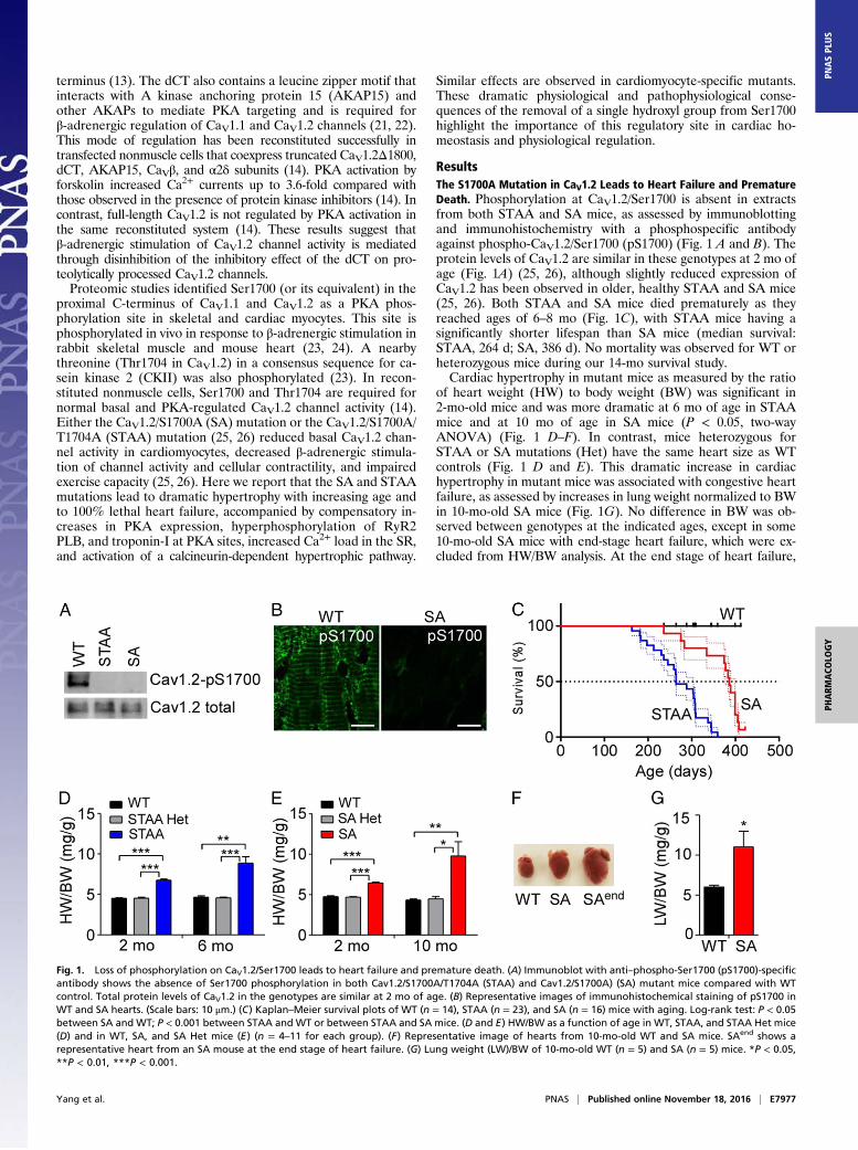
ResultsThe S1700A Mutation in CaV1.2 Leads to Heart Failure and PrematureDeath. Phosphorylation at CaV1.2/Ser1700 is absent in extractsfrom both STAA and SA mice, as assessed by immunoblottingand immunohistochemistry with a phosphospecific antibodyagainst phospho-CaV1.2/Ser1700 (pS1700) (Fig. 1 A and B). Theprotein levels of CaV1.2 are similar in these genotypes at 2 mo ofage (Fig. 1A) (25, 26), although slightly reduced expression ofCaV1.2 has been observed in older, healthy STAA and SA mice(25, 26). Both STAA and SA mice died prematurely as theyreached ages of 6–8 mo (Fig. 1C), with STAA mice having asignificantly shorter lifespan than SA mice (median survival:STAA, 264 d; SA, 386 d). No mortality was observed for WT orheterozygous mice during our 14-mo survival study.Cardiac hypertrophy in mutant mice as measured by the ratio
of heart weight (HW) to body weight (BW) was significant in2-mo-old mice and was more dramatic at 6 mo of age in STAAmice and at 10 mo of age in SA mice (P < 0.05, two-wayANOVA) (Fig. 1 D–F). In contrast, mice heterozygous forSTAA or SA mutations (Het) have the same heart size as WTcontrols (Fig. 1 D and E). This dramatic increase in cardiachypertrophy in mutant mice was associated with congestive heartfailure, as assessed by increases in lung weight normalized to BWin 10-mo-old SA mice (Fig. 1G). No difference in BW was ob-served between genotypes at the indicated ages, except in some10-mo-old SA mice with end-stage heart failure, which were ex-cluded from HW/BW analysis. At the end stage of heart failure,
Fig. 1. Loss of phosphorylation on CaV1.2/Ser1700 leads to heart failure and premature death. (A) Immunoblot with anti–phospho-Ser1700 (pS1700)-specificantibody shows the absence of Ser1700 phosphorylation in both Cav1.2/S1700A/T1704A (STAA) and Cav1.2/S1700A) (SA) mutant mice compared with WTcontrol. Total protein levels of CaV1.2 in the genotypes are similar at 2 mo of age. (B) Representative images of immunohistochemical staining of pS1700 inWT and SA hearts. (Scale bars: 10 μm.) (C) Kaplan–Meier survival plots of WT (n = 14), STAA (n = 23), and SA (n = 16) mice with aging. Log-rank test: P < 0.05between SA and WT; P < 0.001 between STAA and WT or between STAA and SA mice. (D and E) HW/BW as a function of age in WT, STAA, and STAA Het mice(D) and in WT, SA, and SA Het mice (E) (n = 4–11 for each group). (F) Representative image of hearts from 10-mo-old WT and SA mice. SAend shows arepresentative heart from an SA mouse at the end stage of heart failure. (G) Lung weight (LW)/BW of 10-mo-old WT (n = 5) and SA (n = 5) mice. *P < 0.05,**P < 0.01, ***P < 0.001.
Yang et al. PNAS | Published online November 18, 2016 | E7977
PHARM
ACO
LOGY
PNASPL
US
both STAA and SA mice had dramatically enlarged hearts(HW:WT: 0.14 ± 0.01 g; STAA: 0.54 ± 0.05 g; SA: 0.52 ± 0.03 g,P < 0.01) (Fig. 1 D–F) and reduced BW resulting from illness.These results show that loss of Ser1700 phosphorylation onCaV1.2 causes spontaneous cardiac hypertrophy and heart fail-ure, without changes in the expression, localization, or function ofCaV1.2 (25, 26) and further indicate that loss of Thr1704 phos-phorylation plays a synergistic role. In further studies, we focusedprimarily on the healthier SA mice, in which only PKA phos-phorylation of Ser1700 is prevented.
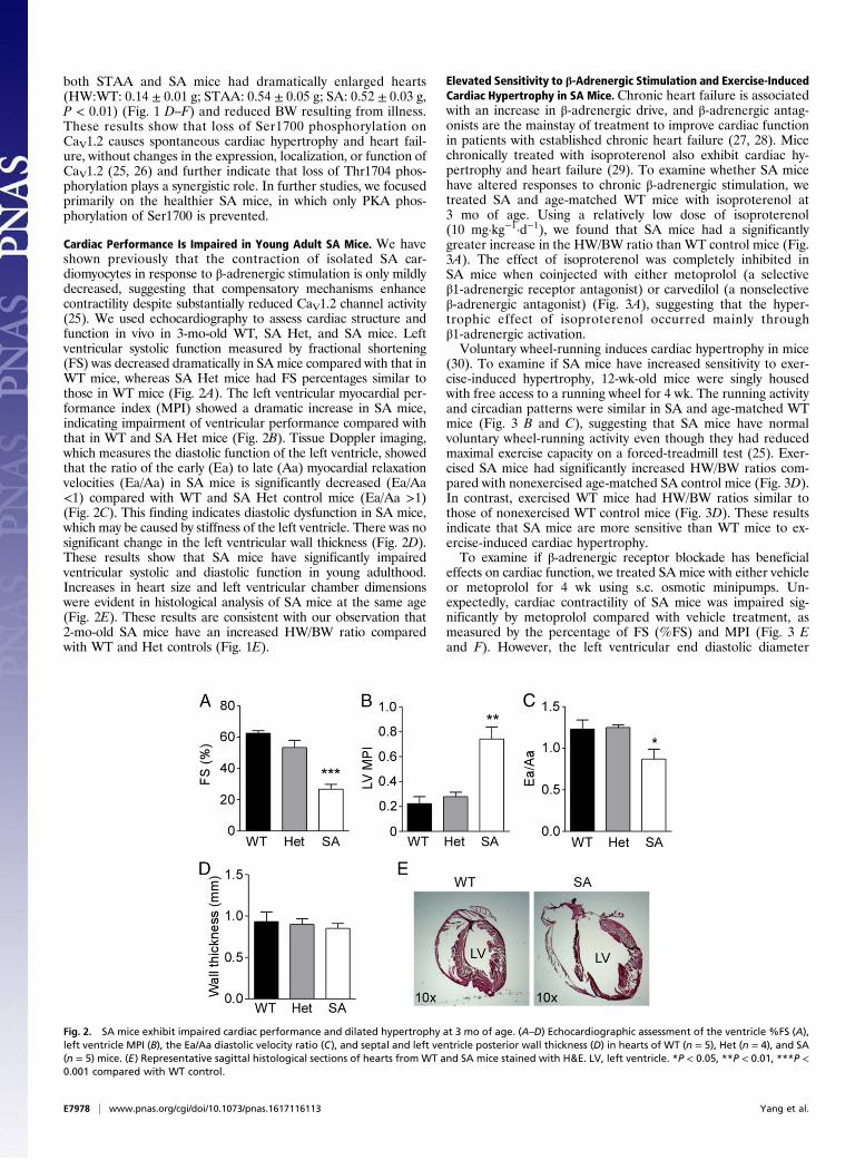
Cardiac Performance Is Impaired in Young Adult SA Mice. We haveshown previously that the contraction of isolated SA car-diomyocytes in response to β-adrenergic stimulation is only mildlydecreased, suggesting that compensatory mechanisms enhancecontractility despite substantially reduced CaV1.2 channel activity(25). We used echocardiography to assess cardiac structure andfunction in vivo in 3-mo-old WT, SA Het, and SA mice. Leftventricular systolic function measured by fractional shortening(FS) was decreased dramatically in SA mice compared with that inWT mice, whereas SA Het mice had FS percentages similar tothose in WT mice (Fig. 2A). The left ventricular myocardial per-formance index (MPI) showed a dramatic increase in SA mice,indicating impairment of ventricular performance compared withthat in WT and SA Het mice (Fig. 2B). Tissue Doppler imaging,which measures the diastolic function of the left ventricle, showedthat the ratio of the early (Ea) to late (Aa) myocardial relaxationvelocities (Ea/Aa) in SA mice is significantly decreased (Ea/Aa<1) compared with WT and SA Het control mice (Ea/Aa >1)(Fig. 2C). This finding indicates diastolic dysfunction in SA mice,which may be caused by stiffness of the left ventricle. There was nosignificant change in the left ventricular wall thickness (Fig. 2D).These results show that SA mice have significantly impairedventricular systolic and diastolic function in young adulthood.Increases in heart size and left ventricular chamber dimensionswere evident in histological analysis of SA mice at the same age(Fig. 2E). These results are consistent with our observation that2-mo-old SA mice have an increased HW/BW ratio comparedwith WT and Het controls (Fig. 1E).
Elevated Sensitivity to β-Adrenergic Stimulation and Exercise-InducedCardiac Hypertrophy in SA Mice. Chronic heart failure is associatedwith an increase in β-adrenergic drive, and β-adrenergic antag-onists are the mainstay of treatment to improve cardiac functionin patients with established chronic heart failure (27, 28). Micechronically treated with isoproterenol also exhibit cardiac hy-pertrophy and heart failure (29). To examine whether SA micehave altered responses to chronic β-adrenergic stimulation, wetreated SA and age-matched WT mice with isoproterenol at3 mo of age. Using a relatively low dose of isoproterenol(10 mg·kg−1·d−1), we found that SA mice had a significantlygreater increase in the HW/BW ratio than WT control mice (Fig.3A). The effect of isoproterenol was completely inhibited inSA mice when coinjected with either metoprolol (a selectiveβ1-adrenergic receptor antagonist) or carvedilol (a nonselectiveβ-adrenergic antagonist) (Fig. 3A), suggesting that the hyper-trophic effect of isoproterenol occurred mainly throughβ1-adrenergic activation.Voluntary wheel-running induces cardiac hypertrophy in mice
(30). To examine if SA mice have increased sensitivity to exer-cise-induced hypertrophy, 12-wk-old mice were singly housedwith free access to a running wheel for 4 wk. The running activityand circadian patterns were similar in SA and age-matched WTmice (Fig. 3 B and C), suggesting that SA mice have normalvoluntary wheel-running activity even though they had reducedmaximal exercise capacity on a forced-treadmill test (25). Exer-cised SA mice had significantly increased HW/BW ratios com-pared with nonexercised age-matched SA control mice (Fig. 3D).In contrast, exercised WT mice had HW/BW ratios similar tothose of nonexercised WT control mice (Fig. 3D). These resultsindicate that SA mice are more sensitive than WT mice to ex-ercise-induced cardiac hypertrophy.To examine if β-adrenergic receptor blockade has beneficial
effects on cardiac function, we treated SA mice with either vehicleor metoprolol for 4 wk using s.c. osmotic minipumps. Un-expectedly, cardiac contractility of SA mice was impaired sig-nificantly by metoprolol compared with vehicle treatment, asmeasured by the percentage of FS (%FS) and MPI (Fig. 3 Eand F). However, the left ventricular end diastolic diameter
Fig. 2. SA mice exhibit impaired cardiac performance and dilated hypertrophy at 3 mo of age. (A–D) Echocardiographic assessment of the ventricle %FS (A),left ventricle MPI (B), the Ea/Aa diastolic velocity ratio (C), and septal and left ventricle posterior wall thickness (D) in hearts of WT (n = 5), Het (n = 4), and SA(n = 5) mice. (E) Representative sagittal histological sections of hearts from WT and SA mice stained with H&E. LV, left ventricle. *P < 0.05, **P < 0.01, ***P <0.001 compared with WT control.
E7978 | www.pnas.org/cgi/doi/10.1073/pnas.1617116113 Yang et al.
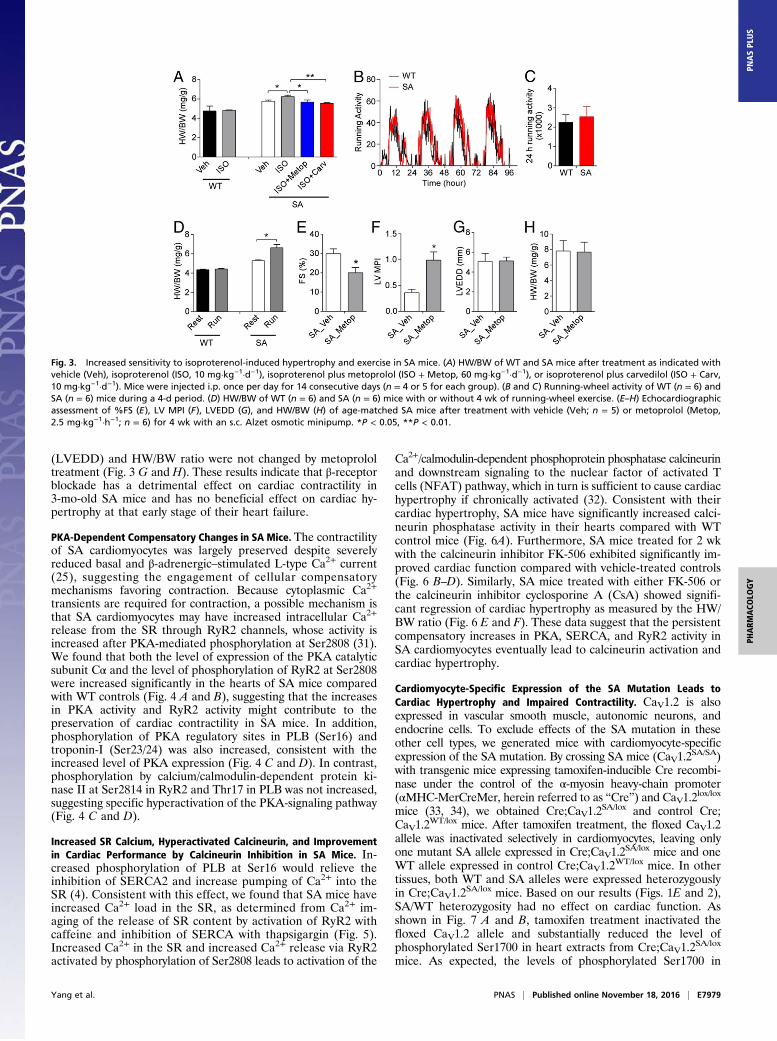
(LVEDD) and HW/BW ratio were not changed by metoprololtreatment (Fig. 3 G and H). These results indicate that β-receptorblockade has a detrimental effect on cardiac contractility in3-mo-old SA mice and has no beneficial effect on cardiac hy-pertrophy at that early stage of their heart failure.
PKA-Dependent Compensatory Changes in SA Mice. The contractilityof SA cardiomyocytes was largely preserved despite severelyreduced basal and β-adrenergic–stimulated L-type Ca2+ current(25), suggesting the engagement of cellular compensatorymechanisms favoring contraction. Because cytoplasmic Ca2+
transients are required for contraction, a possible mechanism isthat SA cardiomyocytes may have increased intracellular Ca2+
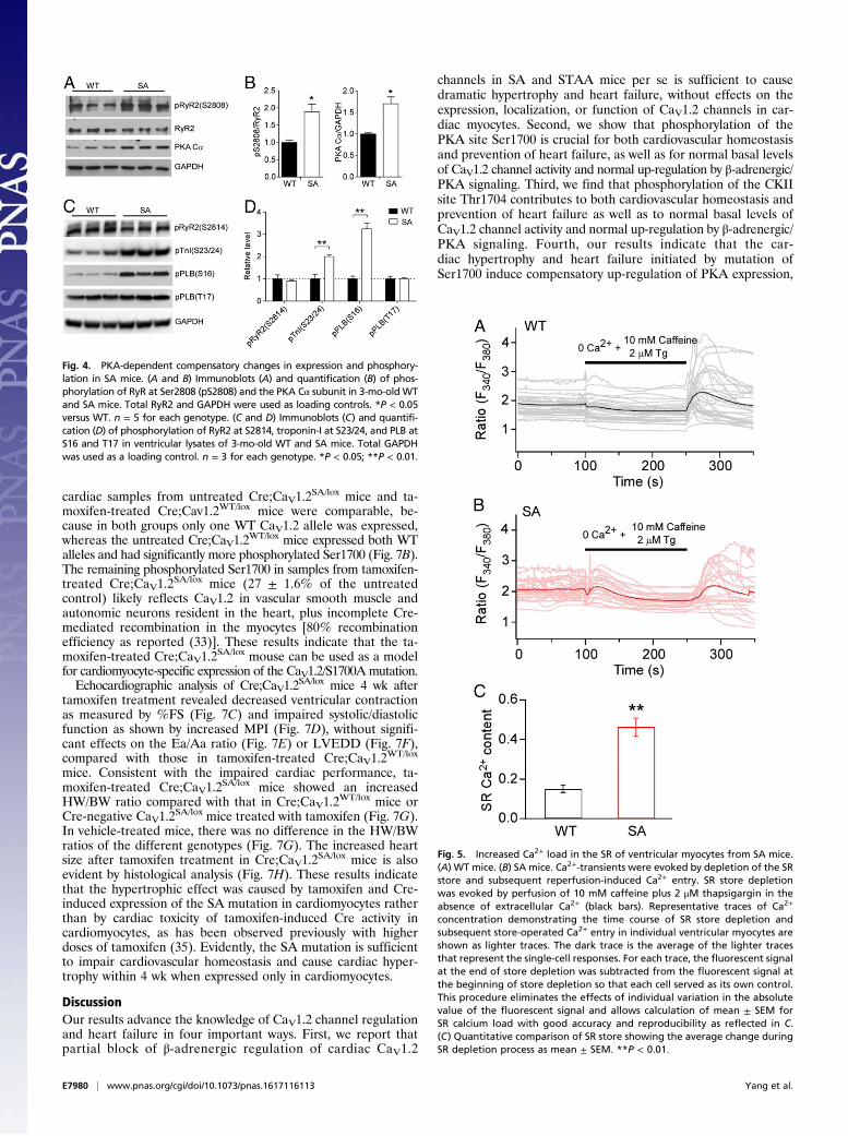
release from the SR through RyR2 channels, whose activity isincreased after PKA-mediated phosphorylation at Ser2808 (31).We found that both the level of expression of the PKA catalyticsubunit Cα and the level of phosphorylation of RyR2 at Ser2808were increased significantly in the hearts of SA mice comparedwith WT controls (Fig. 4 A and B), suggesting that the increasesin PKA activity and RyR2 activity might contribute to thepreservation of cardiac contractility in SA mice. In addition,phosphorylation of PKA regulatory sites in PLB (Ser16) andtroponin-I (Ser23/24) was also increased, consistent with theincreased level of PKA expression (Fig. 4 C and D). In contrast,phosphorylation by calcium/calmodulin-dependent protein ki-nase II at Ser2814 in RyR2 and Thr17 in PLB was not increased,suggesting specific hyperactivation of the PKA-signaling pathway(Fig. 4 C and D).
Increased SR Calcium, Hyperactivated Calcineurin, and Improvementin Cardiac Performance by Calcineurin Inhibition in SA Mice. In-creased phosphorylation of PLB at Ser16 would relieve theinhibition of SERCA2 and increase pumping of Ca2+ into theSR (4). Consistent with this effect, we found that SA mice haveincreased Ca2+ load in the SR, as determined from Ca2+ im-aging of the release of SR content by activation of RyR2 withcaffeine and inhibition of SERCA with thapsigargin (Fig. 5).Increased Ca2+ in the SR and increased Ca2+ release via RyR2activated by phosphorylation of Ser2808 leads to activation of the
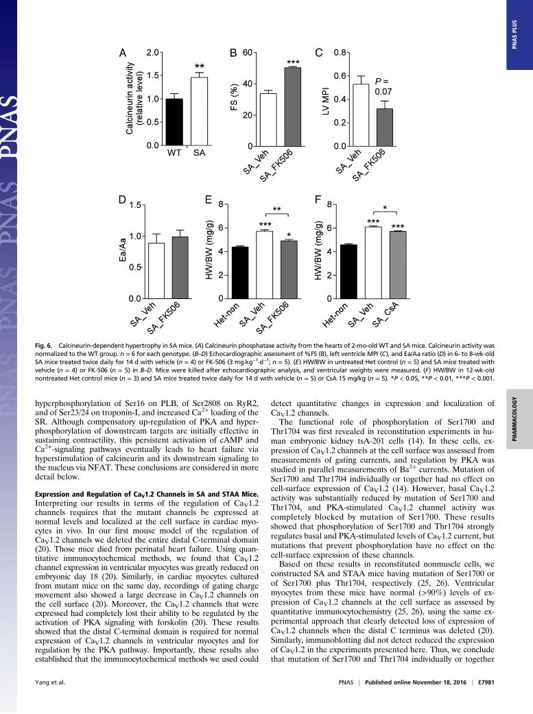
Ca2+/calmodulin-dependent phosphoprotein phosphatase calcineurinand downstream signaling to the nuclear factor of activated Tcells (NFAT) pathway, which in turn is sufficient to cause cardiachypertrophy if chronically activated (32). Consistent with theircardiac hypertrophy, SA mice have significantly increased calci-neurin phosphatase activity in their hearts compared with WTcontrol mice (Fig. 6A). Furthermore, SA mice treated for 2 wkwith the calcineurin inhibitor FK-506 exhibited significantly im-proved cardiac function compared with vehicle-treated controls(Fig. 6 B–D). Similarly, SA mice treated with either FK-506 orthe calcineurin inhibitor cyclosporine A (CsA) showed signifi-cant regression of cardiac hypertrophy as measured by the HW/BW ratio (Fig. 6 E and F). These data suggest that the persistentcompensatory increases in PKA, SERCA, and RyR2 activity inSA cardiomyocytes eventually lead to calcineurin activation andcardiac hypertrophy.
Cardiomyocyte-Specific Expression of the SA Mutation Leads toCardiac Hypertrophy and Impaired Contractility. CaV1.2 is alsoexpressed in vascular smooth muscle, autonomic neurons, andendocrine cells. To exclude effects of the SA mutation in theseother cell types, we generated mice with cardiomyocyte-specificexpression of the SA mutation. By crossing SA mice (CaV1.2
SA/SA)with transgenic mice expressing tamoxifen-inducible Cre recombi-nase under the control of the α-myosin heavy-chain promoter(αMHC-MerCreMer, herein referred to as “Cre”) and CaV1.2
lox/lox
mice (33, 34), we obtained Cre;CaV1.2SA/lox and control Cre;
CaV1.2WT/lox mice. After tamoxifen treatment, the floxed CaV1.2
allele was inactivated selectively in cardiomyocytes, leaving onlyone mutant SA allele expressed in Cre;CaV1.2
SA/lox mice and oneWT allele expressed in control Cre;CaV1.2
WT/lox mice. In othertissues, both WT and SA alleles were expressed heterozygouslyin Cre;CaV1.2
SA/lox mice. Based on our results (Figs. 1E and 2),SA/WT heterozygosity had no effect on cardiac function. Asshown in Fig. 7 A and B, tamoxifen treatment inactivated thefloxed CaV1.2 allele and substantially reduced the level ofphosphorylated Ser1700 in heart extracts from Cre;CaV1.2
SA/lox
mice. As expected, the levels of phosphorylated Ser1700 in
Fig. 3. Increased sensitivity to isoproterenol-induced hypertrophy and exercise in SA mice. (A) HW/BW of WT and SA mice after treatment as indicated withvehicle (Veh), isoproterenol (ISO, 10 mg·kg−1·d−1), isoproterenol plus metoprolol (ISO + Metop, 60 mg·kg−1·d−1), or isoproterenol plus carvedilol (ISO + Carv,10 mg·kg−1·d−1). Mice were injected i.p. once per day for 14 consecutive days (n = 4 or 5 for each group). (B and C) Running-wheel activity of WT (n = 6) andSA (n = 6) mice during a 4-d period. (D) HW/BW of WT (n = 6) and SA (n = 6) mice with or without 4 wk of running-wheel exercise. (E–H) Echocardiographicassessment of %FS (E), LV MPI (F), LVEDD (G), and HW/BW (H) of age-matched SA mice after treatment with vehicle (Veh; n = 5) or metoprolol (Metop,2.5 mg·kg−1·h−1; n = 6) for 4 wk with an s.c. Alzet osmotic minipump. *P < 0.05, **P < 0.01.
Yang et al. PNAS | Published online November 18, 2016 | E7979
PHARM
ACO
LOGY
PNASPL
US
cardiac samples from untreated Cre;CaV1.2SA/lox mice and ta-
moxifen-treated Cre;Cav1.2WT/lox mice were comparable, be-cause in both groups only one WT CaV1.2 allele was expressed,whereas the untreated Cre;CaV1.2
WT/lox mice expressed both WTalleles and had significantly more phosphorylated Ser1700 (Fig. 7B).The remaining phosphorylated Ser1700 in samples from tamoxifen-treated Cre;CaV1.2
SA/lox mice (27 ± 1.6% of the untreatedcontrol) likely reflects CaV1.2 in vascular smooth muscle andautonomic neurons resident in the heart, plus incomplete Cre-mediated recombination in the myocytes [80% recombinationefficiency as reported (33)]. These results indicate that the ta-moxifen-treated Cre;CaV1.2
SA/lox mouse can be used as a modelfor cardiomyocyte-specific expression of the CaV1.2/S1700A mutation.Echocardiographic analysis of Cre;CaV1.2
SA/lox mice 4 wk aftertamoxifen treatment revealed decreased ventricular contractionas measured by %FS (Fig. 7C) and impaired systolic/diastolicfunction as shown by increased MPI (Fig. 7D), without signifi-cant effects on the Ea/Aa ratio (Fig. 7E) or LVEDD (Fig. 7F),compared with those in tamoxifen-treated Cre;CaV1.2
WT/lox
mice. Consistent with the impaired cardiac performance, ta-moxifen-treated Cre;CaV1.2
SA/lox mice showed an increasedHW/BW ratio compared with that in Cre;CaV1.2
WT/lox mice orCre-negative CaV1.2
SA/lox mice treated with tamoxifen (Fig. 7G).In vehicle-treated mice, there was no difference in the HW/BWratios of the different genotypes (Fig. 7G). The increased heartsize after tamoxifen treatment in Cre;CaV1.2
SA/lox mice is alsoevident by histological analysis (Fig. 7H). These results indicatethat the hypertrophic effect was caused by tamoxifen and Cre-induced expression of the SA mutation in cardiomyocytes ratherthan by cardiac toxicity of tamoxifen-induced Cre activity incardiomyocytes, as has been observed previously with higherdoses of tamoxifen (35). Evidently, the SA mutation is sufficientto impair cardiovascular homeostasis and cause cardiac hyper-trophy within 4 wk when expressed only in cardiomyocytes.
DiscussionOur results advance the knowledge of CaV1.2 channel regulationand heart failure in four important ways. First, we report thatpartial block of β-adrenergic regulation of cardiac CaV1.2
channels in SA and STAA mice per se is sufficient to causedramatic hypertrophy and heart failure, without effects on theexpression, localization, or function of CaV1.2 channels in car-diac myocytes. Second, we show that phosphorylation of thePKA site Ser1700 is crucial for both cardiovascular homeostasisand prevention of heart failure, as well as for normal basal levelsof CaV1.2 channel activity and normal up-regulation by β-adrenergic/PKA signaling. Third, we find that phosphorylation of the CKIIsite Thr1704 contributes to both cardiovascular homeostasis andprevention of heart failure as well as to normal basal levels ofCaV1.2 channel activity and normal up-regulation by β-adrenergic/PKA signaling. Fourth, our results indicate that the car-diac hypertrophy and heart failure initiated by mutation ofSer1700 induce compensatory up-regulation of PKA expression,
Fig. 5. Increased Ca2+ load in the SR of ventricular myocytes from SA mice.(A) WT mice. (B) SA mice. Ca2+-transients were evoked by depletion of the SRstore and subsequent reperfusion-induced Ca2+ entry. SR store depletionwas evoked by perfusion of 10 mM caffeine plus 2 μM thapsigargin in theabsence of extracellular Ca2+ (black bars). Representative traces of Ca2+
concentration demonstrating the time course of SR store depletion andsubsequent store-operated Ca2+ entry in individual ventricular myocytes areshown as lighter traces. The dark trace is the average of the lighter tracesthat represent the single-cell responses. For each trace, the fluorescent signalat the end of store depletion was subtracted from the fluorescent signal atthe beginning of store depletion so that each cell served as its own control.This procedure eliminates the effects of individual variation in the absolutevalue of the fluorescent signal and allows calculation of mean ± SEM forSR calcium load with good accuracy and reproducibility as reflected in C.(C) Quantitative comparison of SR store showing the average change duringSR depletion process as mean ± SEM. **P < 0.01.
Fig. 4. PKA-dependent compensatory changes in expression and phosphory-lation in SA mice. (A and B) Immunoblots (A) and quantification (B) of phos-phorylation of RyR at Ser2808 (pS2808) and the PKA Cα subunit in 3-mo-old WTand SA mice. Total RyR2 and GAPDH were used as loading controls. *P < 0.05versus WT. n = 5 for each genotype. (C and D) Immunoblots (C) and quantifi-cation (D) of phosphorylation of RyR2 at S2814, troponin-I at S23/24, and PLB atS16 and T17 in ventricular lysates of 3-mo-old WT and SA mice. Total GAPDHwas used as a loading control. n = 3 for each genotype. *P < 0.05; **P < 0.01.
E7980 | www.pnas.org/cgi/doi/10.1073/pnas.1617116113 Yang et al.
hyperphosphorylation of Ser16 on PLB, of Ser2808 on RyR2,and of Ser23/24 on troponin-I, and increased Ca2+ loading of theSR. Although compensatory up-regulation of PKA and hyper-phosphorylation of downstream targets are initially effective insustaining contractility, this persistent activation of cAMP andCa2+-signaling pathways eventually leads to heart failure viahyperstimulation of calcineurin and its downstream signaling tothe nucleus via NFAT. These conclusions are considered in moredetail below.
Expression and Regulation of CaV1.2 Channels in SA and STAA Mice.Interpreting our results in terms of the regulation of CaV1.2channels requires that the mutant channels be expressed atnormal levels and localized at the cell surface in cardiac myo-cytes in vivo. In our first mouse model of the regulation ofCaV1.2 channels we deleted the entire distal C-terminal domain(20). Those mice died from perinatal heart failure. Using quan-titative immunocytochemical methods, we found that CaV1.2channel expression in ventricular myocytes was greatly reduced onembryonic day 18 (20). Similarly, in cardiac myocytes culturedfrom mutant mice on the same day, recordings of gating chargemovement also showed a large decrease in CaV1.2 channels onthe cell surface (20). Moreover, the CaV1.2 channels that wereexpressed had completely lost their ability to be regulated by theactivation of PKA signaling with forskolin (20). These resultsshowed that the distal C-terminal domain is required for normalexpression of CaV1.2 channels in ventricular myocytes and forregulation by the PKA pathway. Importantly, these results alsoestablished that the immunocytochemical methods we used could
detect quantitative changes in expression and localization ofCaV1.2 channels.The functional role of phosphorylation of Ser1700 and
Thr1704 was first revealed in reconstitution experiments in hu-man embryonic kidney tsA-201 cells (14). In these cells, ex-pression of CaV1.2 channels at the cell surface was assessed frommeasurements of gating currents, and regulation by PKA wasstudied in parallel measurements of Ba2+ currents. Mutation ofSer1700 and Thr1704 individually or together had no effect oncell-surface expression of CaV1.2 (14). However, basal CaV1.2activity was substantially reduced by mutation of Ser1700 andThr1704, and PKA-stimulated CaV1.2 channel activity wascompletely blocked by mutation of Ser1700. These resultsshowed that phosphorylation of Ser1700 and Thr1704 stronglyregulates basal and PKA-stimulated levels of CaV1.2 current, butmutations that prevent phosphorylation have no effect on thecell-surface expression of these channels.Based on these results in reconstituted nonmuscle cells, we
constructed SA and STAA mice having mutation of Ser1700 orof Ser1700 plus Thr1704, respectively (25, 26). Ventricularmyocytes from these mice have normal (>90%) levels of ex-pression of CaV1.2 channels at the cell surface as assessed byquantitative immunocytochemistry (25, 26), using the same ex-perimental approach that clearly detected loss of expression ofCaV1.2 channels when the distal C terminus was deleted (20).Similarly, immunoblotting did not detect reduced the expressionof CaV1.2 in the experiments presented here. Thus, we concludethat mutation of Ser1700 and Thr1704 individually or together
Fig. 6. Calcineurin-dependent hypertrophy in SAmice. (A) Calcineurin phosphatase activity from the hearts of 2-mo-old WT and SAmice. Calcineurin activity wasnormalized to the WT group. n = 6 for each genotype. (B–D) Echocardiographic assessment of %FS (B), left ventricle MPI (C), and Ea/Aa ratio (D) in 6- to 8-wk-oldSA mice treated twice daily for 14 d with vehicle (n = 4) or FK-506 (3 mg·kg−1·d−1; n = 5). (E) HW/BW in untreated Het control (n = 5) and SA mice treated withvehicle (n = 4) or FK-506 (n = 5) in B–D. Mice were killed after echocardiographic analysis, and ventricular weights were measured. (F) HW/BW in 12-wk-oldnontreated Het control mice (n = 3) and SA mice treated twice daily for 14 d with vehicle (n = 5) or CsA 15 mg/kg (n = 5). *P < 0.05, **P < 0.01, ***P < 0.001.
Yang et al. PNAS | Published online November 18, 2016 | E7981
PHARM
ACO
LOGY
PNASPL
US

does not alter cell-surface expression of CaV1.2 in ventricularmyocytes in vivo.Because cell-surface expression is unchanged, reduced CaV1.2
currents in ventricular myocytes must reflect reductions inchannel activity. In both SA mice and STAA mice, we found thatthe basal CaV1.2 current was reduced by ∼65% (25, 26). Theseresults showed that the basal activity of CaV1.2 channels inventricular myocytes depends on the phosphorylation of thesetwo residues, as we observed in reconstituted nonmuscle cells(14). Similarly, we found that the increment in β-adrenergic–stimulated CaV1.2 current was reduced by 67% [19.4 picoampere/picofarad (pA/pF) for WT mice vs. 6.5 pA/pF for SA mice (25)].These results support the conclusion that two-thirds of theβ-adrenergic–stimulated CaV1.2 current depends on the phos-phorylation of Ser1700, whereas one-third involves a separatemechanism and potentially additional phosphorylation sites (25).Measurements of β-adrenergic stimulation of CaV1.2 current
traditionally have relied on calculation of the ratio of stimulatedcurrent [S+B] over basal current (B), where S is the increment ofCaV1.2 current caused by β-adrenergic stimulation. However,inhibition of protein kinases in ventricular myocytes with abroad-spectrum inhibitor reduces basal CaV1.2 current by 50–60% (36). In this situation, use of the traditional [S+B]/B ratio asa measure of β-adrenergic regulation is potentially misleadingbecause the denominator B is not a fixed quantity. Mutations
that preferentially reduce basal current will reduce B and para-doxically will make the [S+B]/B ratio larger. Similarly mutationsthat affect basal and stimulated CaV1.2 current, as we haveshown for SA and STAA mice, will have the same effects on Sand B and therefore the [S+B]/B ratio will remain approximatelyunchanged even though large changes in the magnitude ofβ-adrenergic–stimulated CaV1.2 current may have occurred. Onthe other hand, mutations that preferentially reduce β-adrener-gic–stimulated CaV1.2 current will reduce S, and the [S+B]/Bratio also will be reduced; however, no mutations in CaV1.2channels that have this specific functional effect on stimulatedCaV1.2 currents have been found to date. Our results suggestthat the same phosphorylation sites that are involved in regula-tion of basal CaV1.2 currents, as assessed with broad-spectrumkinase inhibitors (36), are also involved in up-regulation byβ-adrenergic stimulation, as we have observed for Ser1700 andThr1704 (14, 25, 26). In fact, there may not be any regulatorysites that are completely unphosphorylated under basal condi-tions and that are phosphorylated only in response to β-adren-ergic stimulation in ventricular myocytes.β-Adrenergic regulation of the CaV1.2 current also has been
analyzed in a transgenic mouse model in which exogenous WT ormutant CaV1.2 channels were studied after the expression of thetransgene was induced with doxicyclin (9). Because CaV1.2 isexpressed from a transgene that lacks the regulatory sequences
Fig. 7. Mice with the cardiomyocyte-specific SA mutation show cardiac hypertrophy and impaired ventricular performance. (A and B) Immunoblot (A) andquantitation (B) of phosphorylation of CaV1.2 at Ser1700 (pS1700) in heart lysates from mice 10 d after starting treatment with vehicle (Veh) or tamoxifen(TAM, 30 mg/kg) i.p. once daily for three consecutive days. n = 3 for each genotype. (C–F) Echocardiographic assessment of %FS (C), left ventricle MPI (D), Ea/Aa(E), and LVEDD (F) of αMHC-MerCreMer (Cre);Cav1.2WT/lox (n = 5) and Cre;Cav1.2SA/lox (n = 6) mice 4 wk after tamoxifen treatment. (G) HW/BW of the indicatedgenotypes 5 wk after treatment with vehicle or tamoxifen (n = 5–10 for each group). (H) H&E staining of histological sections of hearts of Cre;Cav1.2SA/lox mice5 wk after vehicle or tamoxifen treatment. *P < 0.05, ***P < 0.001.
E7982 | www.pnas.org/cgi/doi/10.1073/pnas.1617116113 Yang et al.
present in the native gene and its transcribed mRNA, control ofchannel expression, localization, subunit composition, and as-sociation with regulatory proteins may be altered. Moreover, inthis model, the basal level of CaV1.2 current is determined by thelevel of induced transgene expression of CaV1.2 and thereforecannot be studied easily as an independent physiological pa-rameter. In this experimental setting, measurements of the tra-ditional [S+B]/B ratio did not reveal effects of Ser1700 orThr1704 mutation, in apparent disagreement with our work (9).However, as noted above, the [S+B]/B ratio in ventricularmyocytes did not change very much with the Ser1700 andThr1704 mutations in our hands either, because these mutationsreduce both the basal and the stimulated CaV1.2 current tocomparable extents (25, 26). If the physiologically relevant sitesfor β-adrenergic stimulation of CaV1.2 current are partiallyphosphorylated under basal physiological conditions, it wouldbe difficult to identify them by mutation when using a methodthat relies on measurements of the traditional [S+B]/B ratio be-cause both S and B would change together, obscuring significanteffects.
β-Adrenergic Regulation of Cardiac CaV1.2 Channels per se Is aTipping Point in Heart Failure. Increased Ca2+ influx via CaV1.2channels in cardiomyocytes has been proposed to be responsiblefor cardiac hypertrophy and pathological remodeling of the ven-tricles in both animal models and human patients (27, 28, 37–39).In keeping with these results, overexpression of CaV1.2 channels inthe hearts of transgenic mice leads to cardiac hypertrophy andheart failure (40). Moreover, CaV1.2 blockers effectively inhibitcardiac remodeling and prevent cardiomyopathy in animal modelswith pressure overload (41, 42). Paradoxically, in light of theseexperimental results, decreased expression of CaV1.2 protein in theheart also leads to heart failure (43). In addition to direct effects onCa2+ signaling, changes in CaV1.2 protein levels also would altersubcellular dyadic structures in cardiomyocytes that depend on thepresence of the CaV1.2 protein (44–46) and would modify the leveland composition of the supramolecular complex of CaV1.2 withother signaling proteins such as the β2-adrenergic receptor,AKAPs, adenylyl cyclases, PKA and other kinases, phosphodi-esterases, and phosphoprotein phosphatases, including cal-cineurin (47–49). Therefore, altered expression of CaV1.2 proteincould cause heart failure, or exacerbate it, by cell biologicalchanges in subcellular membrane compartments or macromolec-ular signaling complexes as well as by altered Ca2+ signaling.In contrast to this previous work, we specifically disrupted
β-adrenergic regulation of CaV1.2 channels by protein phos-phorylation without changes in channel expression, localization,or function (25, 26), and we probed the pathophysiological effectsof these specific regulatory changes through studies of cardiachypertrophy and heart failure. Remarkably, mutation of a singlePKA substrate residue in CaV1.2, Ser1700, which removes only itshydroxyl group, induces dramatic cardiac hypertrophy and 100%lethal heart failure in mice. These results demonstrate the ex-ceptional sensitivity of the heart to even partial disruption of thenormal up-regulation of CaV1.2 channels by phosphorylation ofSer1700 by the β-adrenergic/PKA signaling pathway. Surprisingly,the SA mutation leads to a greater decrease in Ca2+ current inisolated adult cardiomyocytes than did cardiomyocyte-specificdeletion of CaV1.2 in previous studies, but SA mice have lessimpairment of cardiac function and live much longer than car-diomyocyte-specific CaV1.2-knockout mice (compare refs. 24 and43 and Fig. 1C). This comparison suggests that mechanisms inaddition to decreased Ca2+ influx contribute to heart failure inmice having decreased CaV1.2 protein. Therefore, our resultsprovide direct evidence that loss of cardiac CaV1.2 channel reg-ulation per se is sufficient to induce cardiac hypertrophy and heartfailure without changes in channel expression or localization.
Phosphorylation of Ser1700 Is Crucial for β-Adrenergic Regulation andCardiac Homeostasis. Ser1700 (or its equivalent) is phosphorylatedin vivo in response to β-adrenergic stimulation in skeletal andcardiac muscle (23, 24) and is required for the normal β-adren-ergic/PKA–stimulated increase in CaV1.2 channel activity intransfected cells and in cardiac myocytes (14, 25, 26). The ex-periments presented here show that phosphorylation of this siteis required for cardiac homeostasis and the prevention of heartfailure in vivo. The mechanism of β-adrenergic/PKA stimulationof CaV1.2 channel activity in the fight-or-flight response has beencontroversial (7–9). However, the results of our previous ex-periments and those presented here leave no doubt about theimportance of phosphorylation of Ser1700 in cardiovascularhomeostasis and function. Mutation of this serine residue re-duces basal L-type calcium current and impairs β-adrenergic/PKA stimulation of CaV1.2 channels in transfected cells andcardiac myocytes (14, 25, 26). Moreover, mutation of Ser1700leads to dramatic cardiac hypertrophy and 100% lethal heartfailure in vivo. Together, these results demonstrate clearly thatphosphorylation of Ser1700 is crucial in cardiovascular regula-tion and homeostasis.
Phosphorylation of the CKII Site Thr1704 Enhances β-AdrenergicRegulation and Contributes to Cardiac Homeostasis. The sub-stantially shorter lifespan of STAA mice compared with SA mice(Fig. 1C) shows that phosphorylation of Thr1704 acts synergisti-cally with phosphorylation of Ser1700 in cardiac regulation in vivo.We found that ∼25% of isolated STAA cardiomyocytes exhibitedcontraction failure or arrhythmic contractions in response toβ-adrenergic stimulation (26); these deficits were not observed inSA cardiomyocytes (25). In STAA cardiomyocytes, but not in SAcardiomyocytes, the IC50 for isoproterenol stimulation of CaV1.2activity is approximately fivefold higher than that in WT car-diomyocytes, suggesting that phosphorylation of Thr1704 andSer1700 acts synergistically in the regulation of CaV1.2channels in cardiomyocytes (25). Evidently, loss of this syn-ergistic regulation in STAA mice has dire consequences forcontractility, arrhythmia, and premature death. Interestingly,synergistic regulation by PKA- and CKII-mediated phos-phorylation has been identified for other protein targets (50).
Mutation of Ser1700 Initiates a PKA-Dependent CompensatoryResponse. Cardiac myocytes from SA mice have a mild im-pairment in contractility, despite their major deficit in basaland β-adrenergic–stimulated Ca2+ currents (25), suggesting acompensatory response that increases the sensitivity of con-traction to Ca2+ entry via CaV1.2 channels. Our experimentspresented here show that PKA expression is increased andthree key downstream regulatory targets of PKA are hyper-phosphorylated. Increased PKA phosphorylation of PLB Ser16would increase SERCA pumping activity and increase the poolof Ca2+ in the SR (4). Ca2+ imaging studies indicate that thelevel of Ca2+ in the SR is indeed increased in SA mice. In thiscontext, increased PKA phosphorylation of RyR2 on Ser2808would increase Ca2+ release from the SR (3). This increasedCa2+ would have increased effectiveness in activating con-tractions because of the hyperphosphorylation of troponin-I byPKA. Together, this series of compensatory events would leadto a substantial increase in the sensitivity of contractions tothe diminished Ca2+ entering through CaV1.2 channels andthereby would sustain cellular contractility in the face of im-paired basal and β-adrenergic–stimulated Ca2+ currents. How-ever, persistent activation of this compensatory pathway causesincreased sensitivity to stress and hyperactivated signalingvia calcineurin, eventually leading to hypertrophy and heartfailure.
Yang et al. PNAS | Published online November 18, 2016 | E7983
PHARM
ACO
LOGY
PNASPL
US
SA Mice Show Increased Cardiovascular Pathology in Response toStress. The SA mutation greatly decreases CaV1.2 channel acti-vation by β-adrenergic stimulation but causes a much lesserreduction in the contractility of dissociated SA cardiomyocytes(25), suggesting compensation in the downstream signalingpathway. Consistent with that possibility, we show here that PKAis up-regulated and Ser2808 in RyR2 is hyperphosphorylated inSA mice. With the hyperactivation of this Ca2+ signaling path-way, which initially may help preserve contractility, two normallywell-tolerated cardiovascular stresses, continuous treatment withisoproterenol and voluntary wheel-running, both led to signifi-cant increases in cardiac hypertrophy in SA mice. These resultsreveal at a mechanistic level how seemingly effective compen-satory changes that preserve cardiac contractility in the shortterm can further endanger the heart and advance the process ofcardiac hypertrophy and heart failure when they are persistent.
Calcineurin, CaV1.2, and Heart Failure. The increased calcineurinactivity and the improvement of cardiac function by FK-506 andCsA treatment suggest that hyperactive calcineurin signaling causescardiac hypertrophy and heart failure in SA mice. Calcineurin/NFAT signaling has been well documented as a prohypertrophicpathway in pathological conditions (32, 51). Calcineurin bound tothe dCT of CaV1.2 channels has sequential effects in response toCa2+ entry: it contributes to Ca2+-dependent inactivation and op-poses up-regulation by PKA on the millisecond timescale (52), andit signals through NFAT to gene transcription in the nucleus on alonger timescale (53). In mice with heterozygous cardiac knockoutof CaV1.2 channels, calcineurin is hyperactivated, and its signalingto the nucleus via the NFAT pathway triggers hypertrophy andheart failure (43). The increased activation of calcineurin in thesemice is potentially caused by chronically increased RyR2 activa-tion and Ca2+ leak from the SR (43), as we propose here for SAmice.Our results add two crucial elements to this mechanism of heartfailure. First, we show that this pathway to hypertrophy andheart failure can be engaged by specific impairment of theβ-adrenergic regulation of CaV1.2 channels in cardiomyocytesper se, without effects on protein expression, localization,or function. Second, we demonstrate that up-regulation ofPKA expression is potentially a key link in CaV1.2 channeldysfunction, cardiac hypertrophy, and heart failure. Becausecalcineurin/NFAT signaling is engaged to cause cardiac hy-pertrophy induced by increased CaV1.2 protein expression (32,40), decreased CaV1.2 protein expression (43), and impairedregulation of CaV1.2 (this work), this mechanism gains in-creased emphasis as a common pathway leading to hypertro-phy and heart failure.
Materials and MethodsAnimals. The STAA and SA mouse lines were generated using standardmethods as described previously (25, 26) and were backcrossed to C57BL/6Jmice for more than eight generations. Conditional Cav1.2lox/lox (JAX 024714)and αMHC-MerCreMer (JAX 005657) mice were purchased from JacksonLaboratory and have been described previously (33, 34). Mice with heart-specific expression of the SA mutation were generated by crossing theαMHC-MerCreMer transgenic line to SA mice to obtain αMHC-MerCreMer;CavSA/WT mice, which then were crossed to Cav1.2lox/lox mice to obtain αMHC-MerCreMer;Cav1.2SA/lox mice. Littermates with αMHC-MerCreMer;Cav1.2WT/lox
and Cre-negative Cav1.2SA/lox genotypes were used as controls in bio-chemical and physiological studies. To induce αMHC-MerCreMer activation,mice were injected i.p. with tamoxifen (T5648; Sigma) once a day at30 mg·kg−1·d−1 for three consecutive days (35). All mice used in this studywere pathogen free and were housed in the University of Washington An-imal Facility at constant temperature (22 °C), on 12-h light/12-h dark cycles,with ad libitum access to food and water. Mice were housed in groups (threeto five mice per cage) except for running-wheel studies. The use of animalsin this study was approved by the Institutional Animal Care and Use Com-mittee at the University of Washington.
Immunoblot and Histological Analysis. Immunoblot analysis was performedusing mouse ventricles snap-frozen in liquid nitrogen and stored at −80 °C.Ventricles were homogenized in lysis buffer [50 mM Tris·HCl (pH 7.4), 150 mMNaCl, 1% Triton X-100, 0.25% sodium deoxycholate, 1 mM EDTA, supplementedwith protease inhibitors (Roche cOmplete ULTRA tablets) and phosphatase in-hibitors (Roche PhosSTOP)]. Homogenates were centrifuged at 12,000 × g, 4 °Cfor 10 min, and the supernatants were used for blotting. Twenty micrograms ofprotein were loaded on 4–20% Tris-glycine polyacrylamide gels and transferredto nitrocellulose membranes. Antibodies against total CaV1.2 (CNC1) and CaV1.2-pS1700 were generated as described previously (14, 15). Antibodies against RyR2(ARR-002; Alomone Labs), RyR2-pS2808 (ab59225; Abcam), PKA Cα subunit (sc-903; Santa Cruz), RyR2-pS2814 (A010-31; Badrilla), troponin-I-pS23/24 (4004; CellSignaling), phospholamban-pS16 (A010-12; Badrilla), phospholamban-pT17(A010-13; Badrilla), and GAPDH (AM4300; Invitrogen) were used. Immunoblotswere analyzed using the NIH ImageJ program.
For histological studies, adult mice were anesthetized with isoflurane andtranscardially perfused with PBS followed by ice-cold PBS-buffered 4%paraformaldehyde. Hearts were frozen in optimum cutting temperature(OCT) compound and were cryosectioned (10 μm). For gross histologicalexaminations, sections were stained with H&E. For CaV1.2-pS1700 immuno-histochemistry, sections were washed in PBS and incubated sequentially inblocking solution [1× PBS, 0.1% Tween 20 (PBST) and 10% normal goatserum] for >1 h and primary antibody (anti-pS1700, 1:200 in blocking solu-tion) at 4 °C overnight. Sections then were washed four or five times in PBSTand were incubated with goat anti-rabbit Alexa Fluor 488-labeled secondaryantibody (1:1,000 in blocking solution) for 2–3 h. Sections were washed withPBST and mounted with VECTASHIELD mounting medium and coverslips.Images were collected using a Leica SL confocal microscope in the KeckImaging Facility at the University of Washington.
Echocardiography, Drug Treatment, and Running-Wheel Exercise. For mice fromall genotypes or treatment groups, anesthesia was induced with 2% isofluraneand sustained with 0.5% isoflurane. Echocardiography was performed with anAcuson CV-70 cardiovascular ultrasound system (Siemens) using standard im-aging planes: M-mode, conventional and Tissue Doppler imaging as describedpreviously (54). To test the effect of persistent β-adrenergic stimulation, 3-mo-old SA mice and age-matched WT mice were injected i.p. once daily withisoproterenol (10 mg·kg−1·d−1) or vehicle (saline) for 14 consecutive days. Twoadditional groups of mice were injected i.p. with isoproterenol (10 mg·kg−1·d−1)in combination with either the β1-selective blocker metoprolol (60 mg·kg−1·d−1)or the nonselective β-blocker carvedilol (10 mg·kg−1·d−1). For long-termmetoprolol treatment, Alzet osmotic minipumps (2004; Durect Corp.) con-taining isoproterenol (2.5 mg·kg−1·h−1) or saline were surgically inserted s.c.in 3-mo-old mice under isoflurane anesthesia. At the fourth week followingsurgery, echocardiography was performed to assess cardiac function; thenmice were killed, and the HW/BW ratio was measured. To test the effect ofcalcineurin inhibition on cardiac function, SA mice at 4–6 wk of age wereinjected s.c. with FK-506 (3 mg·kg−1·d−1) (Abcam) or vehicle twice daily for 14consecutive days followed by echocardiographic assessment. For CsA (EMDMillipore) injection, 10-wk-old SA mice were injected s.c. with CsA (15 mg/kg)or vehicle twice daily for 14 d. For running-wheel exercise, 12-wk-old micewere housed singly with free access to a running wheel for 4 wk, and therunning activity was automatically recorded. Age-matched mice housedsingly without access to a running wheel were used as control.
Calcineurin Activity Assay. Calcineurin activity was measured by using thecalcineurin cellular assay kit (BML-AK816; Enzo Life Sciences) according to themanufacturer’s instructions. Briefly, ventricles from WT and SA mice werehomogenized in the lysis buffer with the protease inhibitors provided in thekit. Homogenates were centrifuged at 10,000 × g, 4 °C for 10 min. Excessphosphate and nucleotides were removed from the supernatant extract by gelfiltration. Five micrograms of protein were used for the assay. Calcineurinactivity was measured as the dephosphorylation of a synthetic phosphopep-tide substrate (RII peptide) in the presence or absence of EGTA. The amount ofPO4 released was determined photometrically by using the BIOMOL GREENreagent provided in the kit. The phosphate release was normalized to the WTgroup to obtain relative calcineurin activity.
Statistics. Statistical analysis was performed using Prism GraphPad software.A log-rank test was performed to compare the survival curves of WT, STAA,and SA mice. The other comparisons were analyzed by two-tailed Student’s ttest for two groups and by one-way or two-way ANOVA for three or moregroups. ANOVA was followed by Bonferroni posttests for multiple com-parisons. Values are presented as means ± SEM. Differences were consideredsignificant at P < 0.05.
E7984 | www.pnas.org/cgi/doi/10.1073/pnas.1617116113 Yang et al.
1. Bers DM (2002) Cardiac excitation-contraction coupling. Nature 415(6868):198–205.2. Ogawa Y, Kurebayashi N, Murayama T (1999) Ryanodine receptor isoforms in exci-
tation-contraction coupling. Adv Biophys 36:27–64.3. Kushnir A, Marks AR (2010) The ryanodine receptor in cardiac physiology and disease.
Adv Pharmacol 59:1–30.4. MacLennan DH, Kranias EG (2003) Phospholamban: A crucial regulator of cardiac
contractility. Nat Rev Mol Cell Biol 4(7):566–577.5. Reuter H (1983) Calcium channel modulation by neurotransmitters, enzymes and
drugs. Nature 301(5901):569–574.6. Osterrieder W, et al. (1982) Injection of subunits of cyclic AMP-dependent protein
kinase into cardiac myocytes modulates Ca2+ current. Nature 298(5874):576–578.7. Catterall WA (2015) Regulation of cardiac calcium channels in the fight-or-flight re-
sponse. Curr Mol Pharmacol 8(1):12–21.8. Weiss S, Oz S, Benmocha A, Dascal N (2013) Regulation of cardiac L-type Ca²⁺ channel
CaV1.2 via the β-adrenergic-cAMP-protein kinase A pathway: Old dogmas, advances,and new uncertainties. Circ Res 113(5):617–631.
9. Yang L, et al. (2013) β-adrenergic regulation of the L-type Ca2+ channel does notrequire phosphorylation of α1C Ser1700. Circ Res 113(7):871–880.
10. Striessnig J (1999) Pharmacology, structure and function of cardiac L-type Ca2+
channels. Cell Physiol Biochem 9(4-5):242–269.11. Catterall WA (2011) Voltage-gated calcium channels. Cold Spring Harb Perspect Biol
3(8):a003947.12. Wu J, et al. (2015) Structure of the voltage-gated calcium channel Cav1.1 complex.
Science 350(6267):aad2395.13. Hulme JT, Yarov-Yarovoy V, Lin TW-C, Scheuer T, Catterall WA (2006) Autoinhibitory
control of the CaV1.2 channel by its proteolytically processed distal C-terminal do-main. J Physiol 576(Pt 1):87–102.
14. Fuller MD, Emrick MA, Sadilek M, Scheuer T, Catterall WA (2010) Molecular mecha-nism of calcium channel regulation in the fight-or-flight response. Sci Signal 3(141):ra70.
15. De Jongh KS, et al. (1996) Specific phosphorylation of a site in the full-length form ofthe α1 subunit of the cardiac L-type calcium channel by adenosine 3′,5′-cyclicmonophosphate-dependent protein kinase. Biochemistry 35(32):10392–10402.
16. Hell JW, et al. (1996) N-methyl-D-aspartate receptor-induced proteolytic conversionof postsynaptic class C L-type calcium channels in hippocampal neurons. Proc NatlAcad Sci USA 93(8):3362–3367.
17. Hell JW, Appleyard SM, Yokoyama CT, Warner C, Catterall WA (1994) Differentialphosphorylation of two size forms of the N-type calcium channel α1 subunit whichhave different COOH termini. J Biol Chem 269(10):7390–7396.
18. Hulme JT, et al. (2005) Sites of proteolytic processing and noncovalent association ofthe distal C-terminal domain of CaV1.1 channels in skeletal muscle. Proc Natl Acad SciUSA 102(14):5274–5279.
19. Wei X, et al. (1994) Modification of Ca2+ channel activity by deletions at the carboxylterminus of the cardiac α1 subunit. J Biol Chem 269(3):1635–1640.
20. Fu Y, et al. (2011) Deletion of the distal C terminus of CaV1.2 channels leads to loss ofβ-adrenergic regulation and heart failure in vivo. J Biol Chem 286(14):12617–12626.
21. Hulme JT, Lin TW, Westenbroek RE, Scheuer T, Catterall WA (2003) β-adrenergicregulation requires direct anchoring of PKA to cardiac CaV1.2 channels via a leucinezipper interaction with A kinase-anchoring protein 15. Proc Natl Acad Sci USA100(22):13093–13098.
22. Hulme JT, Ahn M, Hauschka SD, Scheuer T, Catterall WA (2002) A novel leucine zippertargets AKAP15 and cyclic AMP-dependent protein kinase to the C terminus of theskeletal muscle Ca2+ channel and modulates its function. J Biol Chem 277(6):4079–4087.
23. Emrick MA, Sadilek M, Konoki K, Catterall WA (2010) β-adrenergic-regulated phos-phorylation of the skeletal muscle Ca(V)1.1 channel in the fight-or-flight response.Proc Natl Acad Sci USA 107(43):18712–18717.
24. Lundby A, et al. (2013) In vivo phosphoproteomics analysis reveals the cardiac targetsof β-adrenergic receptor signaling. Sci Signal 6(278):rs11.
25. Fu Y, Westenbroek RE, Scheuer T, Catterall WA (2014) Basal and β-adrenergic regu-lation of the cardiac calcium channel CaV1.2 requires phosphorylation of serine 1700.Proc Natl Acad Sci USA 111(46):16598–16603.
26. Fu Y, Westenbroek RE, Scheuer T, Catterall WA (2013) Phosphorylation sites requiredfor regulation of cardiac calcium channels in the fight-or-flight response. Proc NatlAcad Sci USA 110(48):19621–19626.
27. Bristow MR (2000) beta-adrenergic receptor blockade in chronic heart failure.Circulation 101(5):558–569.
28. Lohse MJ, Engelhardt S, Eschenhagen T (2003) What is the role of β-adrenergic sig-naling in heart failure? Circ Res 93(10):896–906.
29. Balakumar P, Singh AP, Singh M (2007) Rodent models of heart failure. J PharmacolToxicol Methods 56(1):1–10.
30. Allen DL, et al. (2001) Cardiac and skeletal muscle adaptations to voluntary wheelrunning in the mouse. J Appl Physiol 90(5):1900–1908.
31. Shan J, et al. (2010) Role of chronic ryanodine receptor phosphorylation in heartfailure and β-adrenergic receptor blockade in mice. J Clin Invest 120(12):4375–4387.
32. Molkentin JD, et al. (1998) A calcineurin-dependent transcriptional pathway forcardiac hypertrophy. Cell 93(2):215–228.
33. Sohal DS, et al. (2001) Temporally regulated and tissue-specific gene manipulations inthe adult and embryonic heart using a tamoxifen-inducible Cre protein. Circ Res 89(1):20–25.
34. Seisenberger C, et al. (2000) Functional embryonic cardiomyocytes after disruption ofthe L-type α1C (Cav1.2) calcium channel gene in the mouse. J Biol Chem 275(50):39193–39199.
35. Bersell K, et al. (2013) Moderate and high amounts of tamoxifen in αMHC-MerCreMermice induce a DNA damage response, leading to heart failure and death. Dis ModelMech 6(6):1459–1469.
36. duBell WH, Rogers TB (2004) Protein phosphatase 1 and an opposing protein kinaseregulate steady-state L-type Ca2+ current in mouse cardiac myocytes. J Physiol 556(Pt1):79–93.
37. Richard S, Leclercq F, Lemaire S, Piot C, Nargeot J (1998) Ca2+ currents in compensatedhypertrophy and heart failure. Cardiovasc Res 37(2):300–311.
38. Mukherjee R, Spinale FG (1998) L-type calcium channel abundance and function withcardiac hypertrophy and failure: A review. J Mol Cell Cardiol 30(10):1899–1916.
39. Keung EC (1989) Calcium current is increased in isolated adult myocytes from hy-pertrophied rat myocardium. Circ Res 64(4):753–763.
40. Muth JN, Bodi I, Lewis W, Varadi G, Schwartz A (2001) A Ca2+-dependent transgenicmodel of cardiac hypertrophy: A role for protein kinase Calpha. Circulation 103(1):140–147.
41. Liao Y, et al. (2005) Benidipine, a long-acting calcium channel blocker, inhibits cardiacremodeling in pressure-overloaded mice. Cardiovasc Res 65(4):879–888.
42. Semsarian C, et al. (2002) The L-type calcium channel inhibitor diltiazem preventscardiomyopathy in a mouse model. J Clin Invest 109(8):1013–1020.
43. Goonasekera SA, et al. (2012) Decreased cardiac L-type Ca²⁺ channel activity induceshypertrophy and heart failure in mice. J Clin Invest 122(1):280–290.
44. Shaw RM, Colecraft HM (2013) L-type calcium channel targeting and local signallingin cardiac myocytes. Cardiovasc Res 98(2):177–186.
45. Scriven DR, Asghari P, Moore ED (2013) Microarchitecture of the dyad. Cardiovasc Res98(2):169–176.
46. Sipido KR, Cheng H (2013) T-tubules and ryanodine receptor microdomains: On theroad to translation. Cardiovasc Res 98(2):159–161.
47. Flynn R, Altier C (2013) A macromolecular trafficking complex composed of β₂-adrenergicreceptors, A-kinase anchoring proteins and L-type calcium channels. J Recept SignalTransduct Res 33(3):172–176.
48. Harvey RD, Hell JW (2013) CaV1.2 signaling complexes in the heart. J Mol Cell Cardiol58:143–152.
49. Nikolaev VO, et al. (2010) β2-adrenergic receptor redistribution in heart failurechanges cAMP compartmentation. Science 327(5973):1653–1657.
50. Huang G, et al. (2007) Protein kinase A and casein kinases mediate sequentialphosphorylation events in the circadian negative feedback loop. Genes Dev 21(24):3283–3295.
51. Wilkins BJ, et al. (2004) Calcineurin/NFAT coupling participates in pathological, butnot physiological, cardiac hypertrophy. Circ Res 94(1):110–118.
52. Dittmer PJ, Dell’Acqua ML, Sather WA (2014) Ca2+/calcineurin-dependent inactivationof neuronal L-type Ca2+ channels requires priming by AKAP-anchored protein kinaseA. Cell Reports 7(5):1410–1416.
53. Murphy JG, et al. (2014) AKAP-anchored PKA maintains neuronal L-type calciumchannel activity and NFAT transcriptional signaling. Cell Reports 7(5):1577–1588.
54. Dai DF, et al. (2009) Overexpression of catalase targeted to mitochondria attenuatesmurine cardiac aging. Circulation 119(21):2789–2797.
Yang et al. PNAS | Published online November 18, 2016 | E7985
PHARM
ACO
LOGY
PNASPL
US