BDM attenuates β-adrenergic response of cardiac L-type Ca2 ...
11
© 2016 Journal of Pharmacy & Pharmacognosy Research, 4 (6), 206-216, 2016 ISSN 0719-4250 http://jppres.com/jppres Original Article | Artículo Original _____________________________________ 2,3-Butanedione monoxime attenuates the β-adrenergic response of the L-type Ca 2+ current in rat ventricular cardiomyocytes [La 2,3-butanodiona monoxima atenúa la respuesta β-adrenérgica de la corriente de Ca 2+ tipo L en cardiomiocitos ventriculares de rata] Julio Alvarez-Collazo 1,2,# , Ana Iris López-Medina 1,# , Loipa Galán-Martínez 1 , Julio L. Alvarez 1* 1 Laboratorio de Electrofisiología. Instituto de Cardiología y Cirugía Cardiovascular. Paseo y 17, Vedado, CP 10400, La Habana. Cuba. 2 Laboratory of Ion Channel Research and TRP Research Platform Leuven, Department of Cellular and Molecular Medicine, KU Leuven, Leuven, Belgium. # Both authors contributed equally. *E-mail: [email protected] Abstract Resumen Context: 2,3-Butanedione monoxime (BDM), an uncoupler of cardiac contraction, is commonly used in enzymatic dissociations to prevent hypercontraction of cardiomyocytes and in cardioplegic solutions to decrease oxygen demand during surgery. However, BDM affects multiple cellular systems including the L-type Ca 2+ current (ICaL). If its phosphatase activity is the mechanism underlying the decrease ICaL in cardiomyocytes is a still unresolved question. Aims: To study the effects of BDM on ICaL of rat ventricular cardiomyocytes focusing our attention on the response of ICaL to β- adrenergic stimulation. Methods: The whole-cell patch-clamp method was used to study ICaL in enzymatically dissociated rat ventricular cardiomyocytes. Results: Extracellular BDM (5 mM) decreased peak ICaL by ≈45%, slowed its fast inactivation but accelerated its slow inactivation. Cardiomyocytes incubated in BDM (≥ 30 min; 5 mM) perfused with normal extracellular solution, showed normal ICaL properties. However, extracellular BDM (in cardiomyocytes incubated in BDM or not) markedly reduced the response of ICaL to isoproterenol (1 μM). BDM also strongly attenuated the increase of ICaL in cardiomyocytes intracellularly perfused with cyclic AMP (50 μM). Conclusions: The decrease of basal ICaL by BDM is not related to its dephosphorylation action. Its effect on the Ca 2+ channel occurs most probably in a site in the extracellular side or within the sarcolemmal membrane. Due to its phosphatase action, BDM strongly attenuates the response of ICaL to β-adrenergic stimulation. These actions of BDM must be taken into account both for its use in the dissociation of cardiomyocytes and in cardioplegic solutions and myocardial preservation. Contexto: La 2,3-butanodiona monoxima (BDM), un desacoplador de la contracción cardíaca, es comúnmente utilizada en la disociación enzimática para prevenir la hipercontracción de los cardiomiocitos y en soluciones de cardioplejia para reducir la demanda de oxígeno durante la cirugía. No obstante, la BDM reduce la corriente de Ca 2+ tipo L (ICaL) pero su mecanismo de acción no ha sido dilucidado definitivamente. Objetivos: Estudiar los efectos de la BDM sobre ICaL de cardiomiocitos ventriculares de rata, centrando la atención en la respuesta de ICaL al isoproterenol (ISO). Métodos: Se utilizó la técnica de patch-clamp para registrar ICaL en cardiomiocitos ventriculares de rata disociados enzimáticamente. Resultados: La BDM extracelular (5 mM) redujo ICaL en ≈45% y modificó su inactivación rápida. Los cardiomiocitos incubados en BDM (≥ 30 min; 5 mM) y perfundidos con solución extracelular normal, mostraron ICaL normales. No obstante, la BDM extracelular (en cardiomiocitos incubados en BDM o no incubados), redujo marcadamente la respuesta de ICaL al ISO (1 μM). La BDM atenuó fuertemente el aumento de ICaL en cardiomiocitos perfundidos intracelularmente con AMP cíclico. Conclusiones: La reducción de ICaL basal por BDM no está relacionada a su actividad desfosforiladora. Su efecto sobre el canal de Ca 2+ ocurre probablemente en un sitio extracelular. Debido a su acción como fosfatasa, la BDM atenúa fuertemente la respuesta de ICaL al ISO. Estas acciones de la BDM deben ser consideradas tanto para su utilización en la disociación de cardiomiocitos como en las soluciones de cardioplejia y la preservación miocárdica. Keywords: 2,3-butanedione monoxime; calcium channels; cardiomyocytes; myocardial preservation; patch-clamp. Palabras Clave: 2,3-butanodiona monoxima; canales de calcio; cardiomiocitos; patch-clamp; preservación miocárdica. ARTICLE INFO Received | Recibido: August 12, 2016. Received in revised form | Recibido en forma corregida: September 23, 2016. Accepted | Aceptado: September 23, 2016. Available Online | Publicado en Línea: September 28, 2016. Declaration of interests | Declaración de Intereses: The authors declare no conflict of interest. Funding | Financiación: The study was supported by the Ministry of Public Health of Cuba (Research Project 1104012). Academic Editor | Editor Académico: Marisol Fernández.
Transcript of BDM attenuates β-adrenergic response of cardiac L-type Ca2 ...
© 2016 Journal of Pharmacy & Pharmacognosy Research, 4 (6), 206-216, 2016 ISSN 0719-4250
http://jppres.com/jppres
Original Article | Artículo Original
_____________________________________
2,3-Butanedione monoxime attenuates the β-adrenergic response of the L-type Ca2+ current in rat ventricular cardiomyocytes
[La 2,3-butanodiona monoxima atenúa la respuesta β-adrenérgica de la corriente de Ca2+ tipo L en cardiomiocitos ventriculares de rata]
Julio Alvarez-Collazo1,2,#, Ana Iris López-Medina1,#, Loipa Galán-Martínez1, Julio L. Alvarez1*
1Laboratorio de Electrofisiología. Instituto de Cardiología y Cirugía Cardiovascular. Paseo y 17, Vedado, CP 10400, La Habana. Cuba. 2Laboratory of Ion Channel Research and TRP Research Platform Leuven, Department of Cellular and Molecular Medicine, KU Leuven, Leuven, Belgium.
#Both authors contributed equally. *E-mail: [email protected]
Abstract Resumen
Context: 2,3-Butanedione monoxime (BDM), an uncoupler of cardiac contraction, is commonly used in enzymatic dissociations to prevent hypercontraction of cardiomyocytes and in cardioplegic solutions to decrease oxygen demand during surgery. However, BDM affects multiple cellular systems including the L-type Ca2+ current (ICaL). If its phosphatase activity is the mechanism underlying the decrease ICaL in cardiomyocytes is a still unresolved question.
Aims: To study the effects of BDM on ICaL of rat ventricular cardiomyocytes focusing our attention on the response of ICaL to β-adrenergic stimulation.
Methods: The whole-cell patch-clamp method was used to study ICaL in enzymatically dissociated rat ventricular cardiomyocytes.
Results: Extracellular BDM (5 mM) decreased peak ICaL by ≈45%, slowed its fast inactivation but accelerated its slow inactivation. Cardiomyocytes incubated in BDM (≥ 30 min; 5 mM) perfused with normal extracellular solution, showed normal ICaL properties. However, extracellular BDM (in cardiomyocytes incubated in BDM or not) markedly reduced the response of ICaL to isoproterenol (1 µM). BDM also strongly attenuated the increase of ICaL in cardiomyocytes intracellularly perfused with cyclic AMP (50 µM).
Conclusions: The decrease of basal ICaL by BDM is not related to its dephosphorylation action. Its effect on the Ca2+ channel occurs most probably in a site in the extracellular side or within the sarcolemmal membrane. Due to its phosphatase action, BDM strongly attenuates the response of ICaL to β-adrenergic stimulation. These actions of BDM must be taken into account both for its use in the dissociation of cardiomyocytes and in cardioplegic solutions and myocardial preservation.
Contexto: La 2,3-butanodiona monoxima (BDM), un desacoplador de la contracción cardíaca, es comúnmente utilizada en la disociación enzimática para prevenir la hipercontracción de los cardiomiocitos y en soluciones de cardioplejia para reducir la demanda de oxígeno durante la cirugía. No obstante, la BDM reduce la corriente de Ca2+ tipo L (ICaL) pero su mecanismo de acción no ha sido dilucidado definitivamente.
Objetivos: Estudiar los efectos de la BDM sobre ICaL de cardiomiocitos ventriculares de rata, centrando la atención en la respuesta de ICaL al isoproterenol (ISO).
Métodos: Se utilizó la técnica de patch-clamp para registrar ICaL en cardiomiocitos ventriculares de rata disociados enzimáticamente.
Resultados: La BDM extracelular (5 mM) redujo ICaL en ≈45% y modificó su inactivación rápida. Los cardiomiocitos incubados en BDM (≥ 30 min; 5 mM) y perfundidos con solución extracelular normal, mostraron ICaL normales. No obstante, la BDM extracelular (en cardiomiocitos incubados en BDM o no incubados), redujo marcadamente la respuesta de ICaL al ISO (1 µM). La BDM atenuó fuertemente el aumento de ICaL en cardiomiocitos perfundidos intracelularmente con AMP cíclico.
Conclusiones: La reducción de ICaL basal por BDM no está relacionada a su actividad desfosforiladora. Su efecto sobre el canal de Ca2+ ocurre probablemente en un sitio extracelular. Debido a su acción como fosfatasa, la BDM atenúa fuertemente la respuesta de ICaL al ISO. Estas acciones de la BDM deben ser consideradas tanto para su utilización en la disociación de cardiomiocitos como en las soluciones de cardioplejia y la preservación miocárdica.
Keywords: 2,3-butanedione monoxime; calcium channels; cardiomyocytes; myocardial preservation; patch-clamp.
Palabras Clave: 2,3-butanodiona monoxima; canales de calcio; cardiomiocitos; patch-clamp; preservación miocárdica.
ARTICLE INFO Received | Recibido: August 12, 2016. Received in revised form | Recibido en forma corregida: September 23, 2016. Accepted | Aceptado: September 23, 2016. Available Online | Publicado en Línea: September 28, 2016. Declaration of interests | Declaración de Intereses: The authors declare no conflict of interest. Funding | Financiación: The study was supported by the Ministry of Public Health of Cuba (Research Project 1104012). Academic Editor | Editor Académico: Marisol Fernández.
Alvarez-Collazo et al. BDM attenuates β-adrenergic response of cardiac L-type Ca2+ current
http://jppres.com/jppres J Pharm Pharmacogn Res (2016) 4(6): 207
INTRODUCTION
Isolated adult cardiomyocytes stand for the ex-perimental model of choice in studies of the bio-chemical, biophysical, electrical and contractile ac-tivities of normal and diseased myocardium and also in pharmacological studies (Bers, 2001). Dissocia-tion methods of adult cardiomyocytes are quite di-verse and almost every laboratory uses its own pro-tocol. However, one feature in common to all methods is to try to prevent the well-known “calci-um paradox phenomenon” (Daly et al. 1987) that re-sults in hypercontraction and death of cardiomyo-cytes after calcium re-admission following perfu-sion of hearts will nominally calcium-free solutions. Because of its ability to uncouple cardiac (and skel-etal) muscle contraction 2,3-butanedione monox-ime (BDM), was profiled to have “cardioprotective” properties and has been used to prevent hypercon-traction of Ca2+ -intolerant cardiomyocytes in many dissociation protocols (e. g. Kivistö et al., 1995; Chung et
al., 2015; see for review Abi-Gerges et al., 2013). Originally developed as a reactivator of acetylcholinesterase (Wilson and Ginsburg, 1955), BDM has been considered as a “chemical phosphatase”. This compound affects serine/threonine protein phosphorylation (Stapleton
et al., 1998) and has been shown to affect the activi-ties of proteins like myosin-II light chain kinase (Siegman et al., 1994); BDM is known to increase the equilibrium constant for ATP hydrolysis inhibiting the rate of phosphate release and stabilizing the M.ADP.PI intermediate (Herrmann et al., 1992) whose overall effect is the inhibition of the myosin ATPase rate and a decrease in force production. Yet, BDM is not considered to be a general myosin ATPase in-hibitor (Ostap, 2003). BDM has been also used in car-dioplegic solutions to suppress contraction in order to preserve the myocardium by decreasing oxygen demand thus preserving energy (ATP) during sur-gery (Stringham et al., 1994; Vahl et al., 1995; Habazettl et al.,
1998; Jayawant et al., 1999; Warnecke et al., 2002; Chambers,
2005; Reichert et al., 2013; Lee et al., 2016). While these actions of BDM may be useful to ob-
tain high yield of non-contracted myocytes after enzymatic digestion and even to preserve myocar-dium during cardioplegic arrest, BDM is known to affect other cellular mechanisms such as the activi-ty of connexins (Verrechia and Hervé, 1997), to block the
Na-Ca exchanger (Watanabe et al., 2006) and the ex-pression (Borlak and Zwadlo; 2004) and the activity of ionic channels (e.g. Schlichter et al., 1992; Lopatin and
Nichols, 1993). It has been shown that BDM promotes inhibition of mitochondrial respiration by acting directly on electron transport chain reducing cell viability (Hall and Hausenloy, 2016). BDM inhibits the L-type Ca2+ (ICaL) in cardiac ventricular myocytes (Cou-
lombe et al., 1990) and in guinea-pig taenia caeci (Lang
and Paul, 1991); the effects on ICaL could be more marked in ventricular myocytes from spontaneous-ly hypertensive rats (Xiao and McArdle, 1995). The de-crease of ICaL was accompanied by an acceleration of its inactivation (Coulombe et al., 1990; Chapman, 1993; Al-
len and Chapman, 1995). Although Schwinger et al. (1994) suggested that BDM does not affect the β-adrenergic response in human myocardium, Chap-man (1993) and Allen and Chapman (1995) showed that, due to its phosphatase activity, BDM inter-fered with the β-adrenergic response of ICaL in ven-tricular myocytes. Nonetheless, this has been ques-tioned by Eisfeld et al. (1997) and Allen et al. (1998)
who demonstrated that BDM does not interfere with the interaction sites between PKA and cardiac Ca2+ channel expressed in HEK 293 cells and Xenopus oocytes, respectively. It can be argued, however, that heterologous expression systems do not exactly reproduce native cellular systems leav-ing open the question of whether BDM affects or not the β-adrenergic response of ICaL.
Regarding the mechanism of the decrease of ICaL by BDM, the prevailing idea has been that due to its phosphatase like activity, this oxime affects the phosphorylated state of the L-type Ca2+ channel (Coulombe et al., 1990; Chapman, 1993; Allen and Chapman,
1995). Also, in murine DRG neurons, Huang and McArdle (1992) suggested that the decrease of an L-type Ca2+ current by BDM could be related to alter-ations in PKA regulation of ICaL. On the other hand, Lang and Paul (1991) in guinea-pig taenia caeci cells suggested that blockade of ICaL by BDM could be related to the interactions of this oxime on resting and/or inactivated states of the Ca2+ channel. Fer-reira et al. (1997) pointed out that the effects of BDM on ICaL and its increase in inactivation time con-stants could be mechanistically consistent with dephosphorylation but also with a dihydropyridine-like action; nevertheless, they ruled-out an open
Alvarez-Collazo et al. BDM attenuates β-adrenergic response of cardiac L-type Ca2+ current
http://jppres.com/jppres J Pharm Pharmacogn Res (2016) 4(6): 208
channel block as a mechanism of BDM on the L-type Ca2+ current. A clear-cut mechanism of ICaL de-crease by BDM has not been established.
The aim of the present study was to re-evaluate the effects of BDM on ICaL of rat ventricular cardio-myocytes focusing our attention on the changes in the response of ICaL to β-adrenergic stimulation. Our results show that when cardiomyocytes were incubated in BDM, the β-adrenergic response of ICaL is greatly attenuated.
MATERIAL AND METHODS
Chemicals
2,3-Butanedione monoxime (C4H7NO2; Pub-Chem CID: 6409633; CAS Number: 57-71-6; >98%) was purchased from Sigma Aldrich and was pre-pared in ethanol as stock solution. All other chemi-cals were also from Sigma Aldrich.
Animals
Experiments were performed using male adult Wistar (7 - 8 weeks) rats according to the proce-dures approved by the National Center for Labora-tory Animal Reproduction (CENPALAB; Santiago de Las Vegas, La Habana, Cuba). Prior to experi-ment, animals were adapted for seven days to la-boratory conditions (controlled temperature 25 ± 2°C, relative humidity 60 ± 10% and 12 h light/dark cycles). Tap water and standard diet for rodents supplied by CENPALAB were freely provided. All procedures were also conducted according to the European Commission guide-lines for the use and care of laboratory animals and approved by the Committee for Animal Care in Research of the Cen-ter. The minimum number of animals and duration of observation required to obtain consistent data were employed.
Enzymatic isolation of ventricular cardiomyocytes
Ventricular cardiomyocytes were isolated as pre-viously described in detail (Alvarez-Collazo et al., 2012) and were kept in a K+-Tyrode solution containing 1 mM Ca2+ at room temperature (21 ± 2 °C) and used for experiments for 6 h.
Patch-clamp recordings
Aliquots of cardiomyocytes were transferred to a Petri dish (with the same K+-Tyrode solution) on the stage of an inverted microscope. Relaxed, non-contracting, cardiomyocytes exhibiting clear striat-ed pattern were selected for patch-clamping. Whole-cell currents were recorded at room temper-ature (Alvarez-Collazo et al., 2012). Currents were fil-tered at 3 kHz and digitized at 50-μs intervals, stored on a computer and analyzed off-line with the ACQUIS1 software (version 2.0, CNRS License, France). To study L-type Ca2+ currents, K+ currents were blocked by substituting all potassium by ce-sium in extracellular and “intracellular” solutions. The extracellular solution contained (in millimo-lars): 117 NaCl, 20 CsCl, 10 HEPES, 2 CaCl2, 1.8 MgCl2, and 10 glucose, pH 7.4. The standard pipette (intracellular) solution contained (in millimolars): 130 CsCl, 0.4 Na2GTP, 5 Na2ATP, Na2-creatine phos-phate, 2.0 MgCl2, 11 EGTA, 4.7 CaCl2 (free Ca2+ ≈108 nM), and 10 HEPES, with pH adjusted to 7.2 with CsOH. In the experiments, cells were first left to lie in Petri dishes filled with K+-Tyrode solution with 1 mM Ca2+. Cells attached to the micropipette could be positioned on the extremity of each of six micro-capillaries (i.d. 250 μm) through which the different extracellular Cs+-containing solutions were per-fused by gravity (≈15 μL/min), allowing rapid changes (≈1 s) of the extracellular medium.
Pipette resistance was 1.0 - 1.2 MΩ. Membrane capacitance (Cm) and series resistance (Rs) were calculated on voltage-clamped cardiomyocytes as previously described (Alvarez et al., 2000). Average Cm and uncompensated Rs were 168 ± 8.6 pF and 3.3 ± 0.5 MΩ, respectively (n = 44). Rs could be electron-ically compensated up to 50% without ringing and was continually monitored during the experiment. Liquid junction potential was compensated before establishing the gigaseal. No leak or capacitance subtractions were performed in the recordings.
For routine monitoring of the L-type Ca2+ cur-rent (ICaL) a double pulse voltage-clamp protocol was employed: from a holding potential (HP) of -80 mV every 4s the cell membrane was depolarized by a prepulse to -40 mV for 50 ms to inactivate the fast Na+ current. From this membrane potential, a 200-ms test pulse to 0 mV evoked ICaL. Current-to-voltage relationships (I-V) and availability curves
Alvarez-Collazo et al. BDM attenuates β-adrenergic response of cardiac L-type Ca2+ current
http://jppres.com/jppres J Pharm Pharmacogn Res (2016) 4(6): 209
were obtained using the same prepulse protocol but interpolating 300-ms pulses from -40 to +50 mV between the pre- and test pulses. Pulses for I-V and availability curves were applied at 8 s intervals. The inactivation time course of ICaL was fitted to a dou-ble exponential using the fitting procedures of the ACQUIS1 software.
Statistical analysis
Results are expressed as means and standard er-rors of means. Statistical significance was evaluated by means of paired or unpaired Student’s t test ac-cording to the experimental situation. Differences were considered statistically significant for p < 0.05.
RESULTS
Effects of BDM on basal ICaL
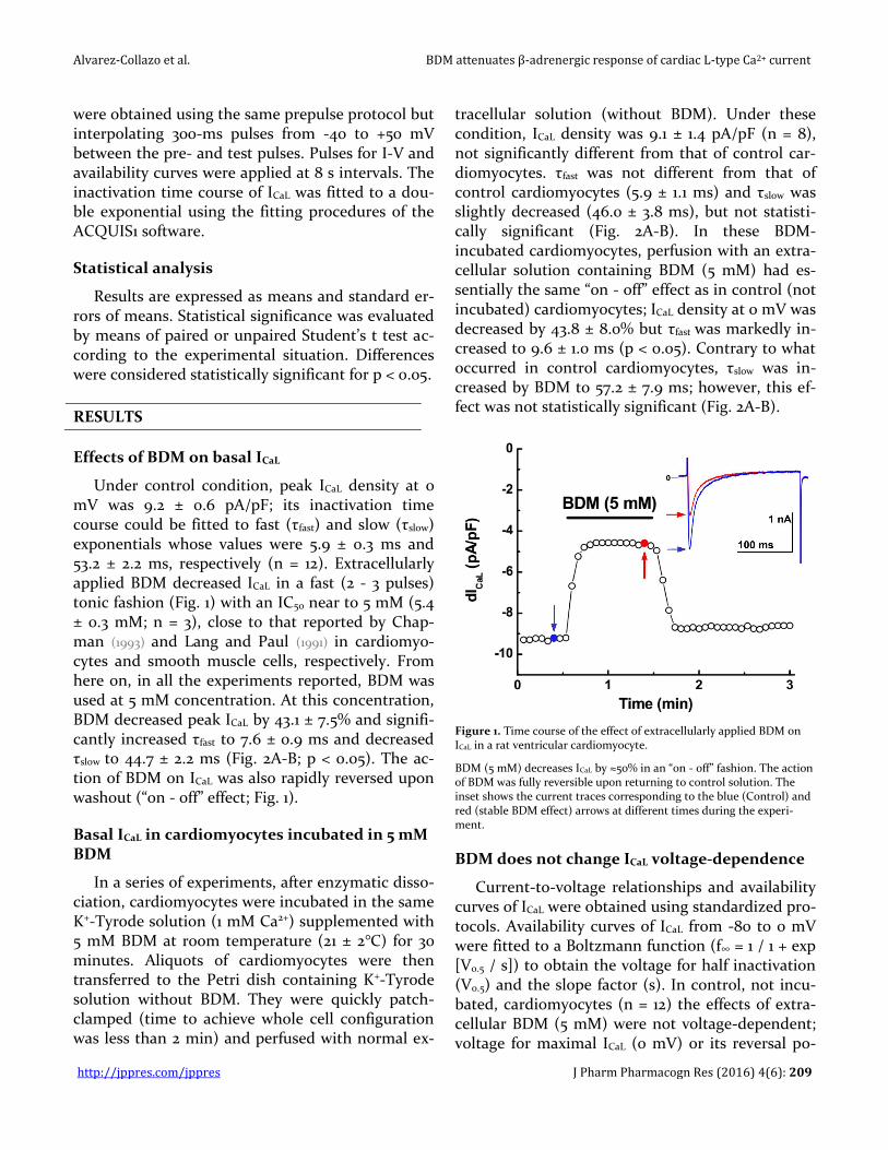
Under control condition, peak ICaL density at 0 mV was 9.2 ± 0.6 pA/pF; its inactivation time course could be fitted to fast (τfast) and slow (τslow) exponentials whose values were 5.9 ± 0.3 ms and 53.2 ± 2.2 ms, respectively (n = 12). Extracellularly applied BDM decreased ICaL in a fast (2 - 3 pulses) tonic fashion (Fig. 1) with an IC50 near to 5 mM (5.4 ± 0.3 mM; n = 3), close to that reported by Chap-man (1993) and Lang and Paul (1991) in cardiomyo-cytes and smooth muscle cells, respectively. From here on, in all the experiments reported, BDM was used at 5 mM concentration. At this concentration, BDM decreased peak ICaL by 43.1 ± 7.5% and signifi-cantly increased τfast to 7.6 ± 0.9 ms and decreased τslow to 44.7 ± 2.2 ms (Fig. 2A-B; p < 0.05). The ac-tion of BDM on ICaL was also rapidly reversed upon washout (“on - off” effect; Fig. 1).
Basal ICaL in cardiomyocytes incubated in 5 mM BDM
In a series of experiments, after enzymatic disso-ciation, cardiomyocytes were incubated in the same K+-Tyrode solution (1 mM Ca2+) supplemented with 5 mM BDM at room temperature (21 ± 2°C) for 30 minutes. Aliquots of cardiomyocytes were then transferred to the Petri dish containing K+-Tyrode solution without BDM. They were quickly patch-clamped (time to achieve whole cell configuration was less than 2 min) and perfused with normal ex-
tracellular solution (without BDM). Under these condition, ICaL density was 9.1 ± 1.4 pA/pF (n = 8), not significantly different from that of control car-diomyocytes. τfast was not different from that of control cardiomyocytes (5.9 ± 1.1 ms) and τslow was slightly decreased (46.0 ± 3.8 ms), but not statisti-cally significant (Fig. 2A-B). In these BDM-incubated cardiomyocytes, perfusion with an extra-cellular solution containing BDM (5 mM) had es-sentially the same “on - off” effect as in control (not incubated) cardiomyocytes; ICaL density at 0 mV was decreased by 43.8 ± 8.0% but τfast was markedly in-creased to 9.6 ± 1.0 ms (p < 0.05). Contrary to what occurred in control cardiomyocytes, τslow was in-creased by BDM to 57.2 ± 7.9 ms; however, this ef-fect was not statistically significant (Fig. 2A-B).
Figure 1. Time course of the effect of extracellularly applied BDM on ICaL in a rat ventricular cardiomyocyte.
BDM (5 mM) decreases ICaL by ≈50% in an “on - off” fashion. The action of BDM was fully reversible upon returning to control solution. The inset shows the current traces corresponding to the blue (Control) and red (stable BDM effect) arrows at different times during the experi-ment.
BDM does not change ICaL voltage-dependence
Current-to-voltage relationships and availability curves of ICaL were obtained using standardized pro-tocols. Availability curves of ICaL from -80 to 0 mV were fitted to a Boltzmann function (f∞ = 1 / 1 + exp [V0.5 / s]) to obtain the voltage for half inactivation (V0.5) and the slope factor (s). In control, not incu-bated, cardiomyocytes (n = 12) the effects of extra-cellular BDM (5 mM) were not voltage-dependent; voltage for maximal ICaL (0 mV) or its reversal po-
Alvarez-Collazo et al. BDM attenuates β-adrenergic response of cardiac L-type Ca2+ current
http://jppres.com/jppres J Pharm Pharmacogn Res (2016) 4(6): 210
tential (≈+50 mV) were not shifted (Fig. 3A). Con-sequently, availability curves were barely modified; V0.5 was -29.5 ± 0.3 mV in control and -31.7 ± 0.3 mV in the presence of BDM (Fig. 3B; n = 8). The slope factor, s, was not significantly changed (5.9 ± 0.4 mV vs 5.6 ± 0.3 mV). In BDM-incubated cardiomy-ocytes (n = 8) perfused with normal extracellular solution (as described above) voltage for maximal
ICaL and its reversal potential were similar to those of control cardiomyocytes (Fig. 3C). V0.5 and s were not significantly different from control cardiomyo-cytes (-30.9 ± 0.2 mV and 5.5 ± 0.2 mV, respectively; Fig. 3D). Perfusion of these cardiomyocytes with extracellular BDM (5 mM; Fig. 3D) produced no significant effects on V0.5 (-31.0 ± 0.2 mV) and s (5.3 ± 0.4 mV).
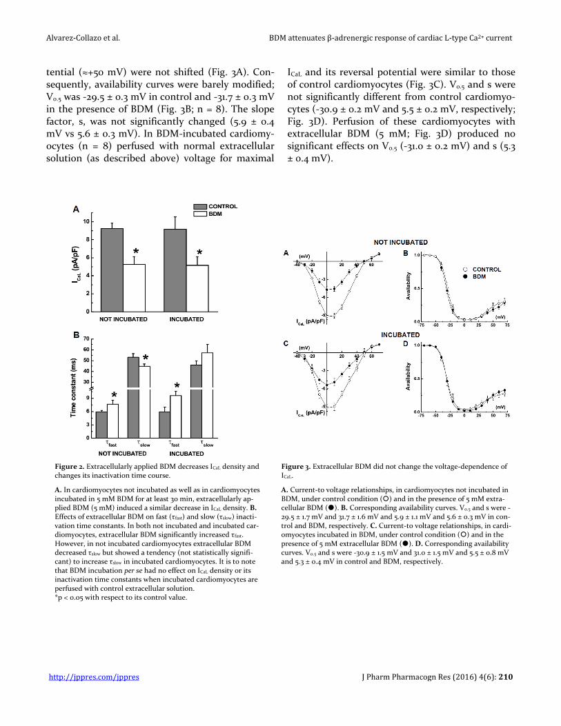
Figure 2. Extracellularly applied BDM decreases ICaL density and changes its inactivation time course.
A. In cardiomyocytes not incubated as well as in cardiomyocytes incubated in 5 mM BDM for at least 30 min, extracellularly ap-plied BDM (5 mM) induced a similar decrease in ICaL density. B. Effects of extracellular BDM on fast (τfast) and slow (τslow) inacti-vation time constants. In both not incubated and incubated car-diomyocytes, extracellular BDM significantly increased τfast. However, in not incubated cardiomyocytes extracellular BDM decreased τslow but showed a tendency (not statistically signifi-cant) to increase τslow in incubated cardiomyocytes. It is to note that BDM incubation per se had no effect on ICaL density or its inactivation time constants when incubated cardiomyocytes are perfused with control extracellular solution. *p < 0.05 with respect to its control value.
Figure 3. Extracellular BDM did not change the voltage-dependence of ICaL.
A. Current-to voltage relationships, in cardiomyocytes not incubated in BDM, under control condition () and in the presence of 5 mM extra-cellular BDM (). B. Corresponding availability curves. V0.5 and s were -29.5 ± 1.7 mV and 31.7 ± 1.6 mV and 5.9 ± 1.1 mV and 5.6 ± 0.3 mV in con-trol and BDM, respectively. C. Current-to voltage relationships, in cardi-omyocytes incubated in BDM, under control condition () and in the presence of 5 mM extracellular BDM (). D. Corresponding availability curves. V0.5 and s were -30.9 ± 1.5 mV and 31.0 ± 1.5 mV and 5.5 ± 0.8 mV and 5.3 ± 0.4 mV in control and BDM, respectively.
Alvarez-Collazo et al. BDM attenuates β-adrenergic response of cardiac L-type Ca2+ current
http://jppres.com/jppres J Pharm Pharmacogn Res (2016) 4(6): 211
BDM attenuates the response of ICaL to β-adrenergic stimulation
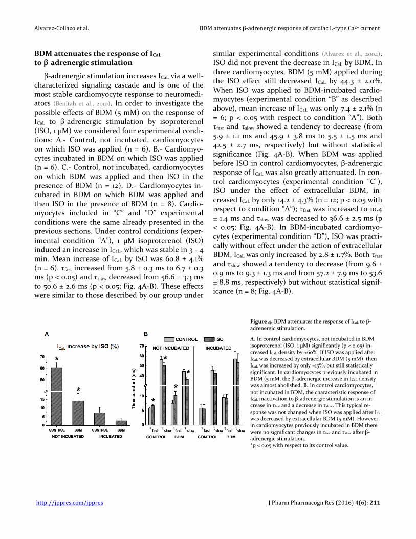
β-adrenergic stimulation increases ICaL via a well-characterized signaling cascade and is one of the most stable cardiomyocyte response to neuromedi-ators (Bénitah et al., 2010). In order to investigate the possible effects of BDM (5 mM) on the response of ICaL to β-adrenergic stimulation by isoproterenol (ISO, 1 µM) we considered four experimental condi-tions: A.- Control, not incubated, cardiomyocytes on which ISO was applied (n = 6). B.- Cardiomyo-cytes incubated in BDM on which ISO was applied (n = 6). C.- Control, not incubated, cardiomyocytes on which BDM was applied and then ISO in the presence of BDM (n = 12). D.- Cardiomyocytes in-cubated in BDM on which BDM was applied and then ISO in the presence of BDM (n = 8). Cardio-myocytes included in “C” and “D” experimental conditions were the same already presented in the previous sections. Under control conditions (exper-imental condition “A”), 1 µM isoproterenol (ISO) induced an increase in ICaL, which was stable in 3 - 4 min. Mean increase of ICaL by ISO was 60.8 ± 4.1% (n = 6). τfast increased from 5.8 ± 0.3 ms to 6.7 ± 0.3 ms (p < 0.05) and τslow decreased from 56.6 ± 3.3 ms to 50.6 ± 2.6 ms (p < 0.05; Fig. 4A-B). These effects were similar to those described by our group under
similar experimental conditions (Alvarez et al., 2004). ISO did not prevent the decrease in ICaL by BDM. In three cardiomyocytes, BDM (5 mM) applied during the ISO effect still decreased ICaL by 44.3 ± 2.0%. When ISO was applied to BDM-incubated cardio-myocytes (experimental condition “B” as described above), mean increase of ICaL was only 7.4 ± 2.1% (n = 6; p < 0.05 with respect to condition “A”). Both τfast and τslow showed a tendency to decrease (from 5.9 ± 1.1 ms and 45.9 ± 3.8 ms to 5.5 ± 1.5 ms and 42.5 ± 2.7 ms, respectively) but without statistical significance (Fig. 4A-B). When BDM was applied before ISO in control cardiomyocytes, β-adrenergic response of ICaL was also greatly attenuated. In con-trol cardiomyocytes (experimental condition “C”), ISO under the effect of extracellular BDM, in-creased ICaL by only 14.2 ± 4.3% (n = 12; p < 0.05 with respect to condition “A”); τfast was increased to 10.4 ± 1.4 ms and τslow was decreased to 36.6 ± 2.5 ms (p < 0.05; Fig. 4A-B). In BDM-incubated cardiomyo-cytes (experimental condition “D”), ISO was practi-cally without effect under the action of extracellular BDM, ICaL was only increased by 2.8 ± 1.7%. Both τfast and τslow showed a tendency to decrease (from 9.6 ± 0.9 ms to 9.3 ± 1.3 ms and from 57.2 ± 7.9 ms to 53.6 ± 8.8 ms, respectively) but without statistical signif-icance (n = 8; Fig. 4A-B).
Figure 4. BDM attenuates the response of ICaL to β-adrenergic stimulation.
A. In control cardiomyocytes, not incubated in BDM, isoproterenol (ISO, 1 µM) significantly (p < 0.05) in-creased ICaL density by ≈60%. If ISO was applied after ICaL was decreased by extracellular BDM (5 mM), then ICaL was increased by only ≈15%, but still statistically significant. In cardiomyocytes previously incubated in BDM (5 mM, the β-adrenergic increase in ICaL density was almost abolished. B. In control cardiomyocytes, not incubated in BDM, the characteristic response of ICaL inactivation to β-adrenergic stimulation is an in-crease in τfast and a decrease in τslow. This typical re-sponse was not changed when ISO was applied after ICaL was decreased by extracellular BDM (5 mM). However, in cardiomyocytes previously incubated in BDM there were no significant changes in τfast and τslow after β-adrenergic stimulation. *p < 0.05 with respect to its control value.

Alvarez-Collazo et al. BDM attenuates β-adrenergic response of cardiac L-type Ca2+ current
http://jppres.com/jppres J Pharm Pharmacogn Res (2016) 4(6): 212
From these results, it is clear that BDM attenu-ates β-adrenergic response of ICaL. We next studied whether BDM was also able to attenuate the re-sponse of ICaL to intracellular 3',5'-cyclic adenosine monophosphate (cAMP) a well-known activator of protein kinase A. Not incubated (n = 4) and BDM-incubated (n = 4) cardiomyocytes were patch-clamped with pipettes containing the normal “in-tracellular” solution but added with 50 µM cAMP. Immediately after patch rupture ICaL was continu-ously monitored. In not incubated cardiomyocytes, ICaL increased by 167.6 ± 22.0% from its initial value in about 2 min (Fig. 5A; see also, Alvarez-Collazo et al.,
2012). In BDM-incubated cardiomyocytes, however, ICaL was barely increased by 10.0 ± 6.0% from its ini-tial value (Fig. 5B). Moreover, in two control, not incubated, cardiomyocytes, application of extracel-lular BDM after the steady-state cAMP effect, still decreased ICaL by 42 and 48%. The effect was “on - off” but after washout, ICaL never recovered its max-imal attained value (Fig. 6).
Figure 5. BDM attenuates the response of ICaL to intracellularly applied cyclic adenosine monophosphate (3', 5'-cAMP; 50 µM).
A. Example of a control cardiomyocyte (not incubated in BDM) intra-cellularly perfused with cAMP in which there is a huge increase in ICaL density. B. In a cardiomyocyte previously incubated in 5 mM BDM, there was almost no effect of cAMP on ICaL.
Figure 6. Extracellularly applied BDM is able to decrease ICaL density in cardiomyocytes intracellularly perfused with cAMP.
In a cardiomyocyte not incubated in BDM, after ICaL was maximally increased by intracellular cAMP, extracellular perfusion with 5 mM BDM is still able to decrease ICaL by ≈50%. Upon washout with normal extracellular solution, ICaL never returned to its maximal value previous to BDM. The inset shows the current traces corresponding to the blue (Control), red (stable BDM effect) and green (washout of BDM) arrows at different times during the experiment.
DISCUSSION
The main outcome of the present investigation is that, in isolated rat ventricular cardiomyocytes, BDM attenuates the response of ICaL to β-adrenergic stimulation. Our results also suggest that BDM could inhibit the L-type Ca2+ channel by acting on a site in the external side of the sarcolemmal mem-brane.
Our results show that extracellular BDM de-crease ICaL in an “on - off” manner with an IC50 around 5 mM, similar to that commonly reported for cardiac myocytes (Coulombe et al., 1990; Chapman,
1993; but see Xiao and McArdle, 1995) and smooth muscle cells (Lang and Paul, 1991) but that is lower than the IC50 reported for the inhibition of an L-type Ca2+ current in neurons (Huang and McArdle, 1992) and of the human L-type Ca2+ channel expressed HEK 293 cells (Eisfeld et al., 1997) and Xenopus oocytes (Allen et
al., 1998). The decrease of ICaL by BDM in the present
Alvarez-Collazo et al. BDM attenuates β-adrenergic response of cardiac L-type Ca2+ current
http://jppres.com/jppres J Pharm Pharmacogn Res (2016) 4(6): 213
experiments was not voltage-dependent since nei-ther the I-V relationships nor the availability curves were modified by the oxime. This is in agreement with the results of Huang and McArdle (1992) in neurons, Lang and Paul (1991) in smooth muscle cells and Eisfled et al. (1997) in HEK-293 cells expressing the human L-type Ca2+ channel. Coulombe et al. (1990) and Ferreira et al. (1997) in rat and guinea-pig ventricular cardiac myocytes, respectively and Allen et al. (1998) in L-type Ca2+ channels expressed in Xenopus oocytes found a 4 - 6 mV leftward shift in ICaL availability but at much higher concentrations of BDM. In the present experiments, the inactiva-tion time course of ICaL was affected by BDM; τfast was consistently increased while τslow was de-creased. Similar results were reported by Allen and Chapman (1995) for the exponential and sustained phases of ICaL in guinea-pig ventricular cardiomyo-cytes using Ca2+ or Ba2+ as charge carriers. Although the effects of BDM on the fast inactivation of ICaL could be interpreted in terms of dephosphorylation or direct effects on channel gating (e.g. Allen and
Chapman, 1995; Ferreira et al., 1997) it should be consid-ered that τfast is related to the Ca2+-dependent inac-tivation (CDI; for review see Bénitah et al., 2010), which depends on the Ca2+ load of the sarcoplasmic re-ticulum (SR). It has been reported by Tripathi et al. (1999) that BDM is able to increase the open proba-bility of SR Ca2+ channels. Additionally, BDM de-creases peak ICaL. It is thus possible that under the action of BDM the SR Ca2+ load is decreased and CDI is diminished increasing τfast. The decrease we observed in τslow is most probably related to an ef-fect of BDM on channel gating (e.g.; Ferreira et al.,
1997). On the other hand, Eisfled et al. (1997) and Al-len et al. (1998) reported accelerations in the inacti-vation time course of ICaL under the action of BDM. It is, however, difficult to explain such a discrepan-cy since those results were obtained measuring cur-rents through L-type Ca2+ channels expressed in heterologous systems using Ba2+ as charge carrier.
One important finding of the present results is that incubating cardiomyocytes in a BDM-contai-ning solution did not affect ICaL density or its inacti-vation time course recorded when cardiomyocytes were perfused with control extracellular solution as described above. This is an expected result since the effect of extracellular BDM on ICaL was quickly es-
tablished an also rapidly washed out (“on - off”). Moreover, in those cardiomyocytes perfusion with an extracellular solution containing BDM produced essentially the same changes in ICaL properties as in not incubated cardiomyocytes. These results sug-gest that inhibition of basal ICaL by BDM is probably not related to its (intracellular) phosphatase activity (see Chapman, 1993; Allen and Chapman, 1995) but to a di-rect action on the L-type Ca2+ channel through a site located in the extracellular sarcolemmal inter-phase. Another possibility is that BDM, due to its lipophilicity, could penetrate the membrane and produce its ICaL blocking action either by directly interacting with the Ca2+ channel or by disturbing the lipid domains around the channel. Both possi-bilities (outside or within the membrane) are con-sistent with the fast decrease and washout (“on - off”) of BDM effect on ICaL. Our results are also con-sistent with the idea that there is a minor role of PKA in the maintenance of basal ICaL (see for review
Weiss et al., 2013). It should be noted here that the re-sults of Eisfled et al. (1997) and Allen et al. (1998), us-ing heterologous expression systems, supported the idea that BDM effects on basal ICaL were not medi-ated by dephosphorylation. However, as will be dis-cussed below, the intracellular phosphatase activity of BDM, reflected in a decreased β-adrenergic re-sponse, is long lasting.
The most important result of the present exper-iments is that BDM markedly attenuated the re-sponse of ICaL to β-adrenergic stimulation. In rat ventricular cardiomyocytes, ICaL usually respond to ISO (1 µM) with a ≥60% increase (see Alvarez et al.,
2004; present results). However, our results show that when extracellular BDM was first applied to cardi-omyocytes (basal ICaL is decreased) ISO was much less effective in increasing ICaL (≈15%). Furthermore, when cardiomyocytes incubated in BDM (at least 30 min) were patch-clamped and perfused with nor-mal extracellular solution (time to achieve whole cell configuration was less than 2 min), the re-sponse of ICaL to ISO was greatly diminished or even abolished (see Fig. 4). These results are agreement with those of Lang and Paul (1991) in smooth muscle cells but are in contrast to those of Chapman (1993) and Allen and Chapman (1995) in cardiomyocytes and Huang and McArdle (1992) in neurons who found that cAMP-dependent phosphorylation could
Alvarez-Collazo et al. BDM attenuates β-adrenergic response of cardiac L-type Ca2+ current
http://jppres.com/jppres J Pharm Pharmacogn Res (2016) 4(6): 214
revert BDM effects on the L-type Ca2+ current. In order to clarify this aspect we conducted experi-ments in cardiomyocytes intracellularly perfused with cAMP to fully activate PKA and the results show that when cardiomyocytes were previously incubated in BDM, the cAMP-mediated increase in ICaL (>160% in control cardiomyocytes) was almost suppressed. Moreover, in cardiomyocytes not incu-bated in BDM and intracellularly perfused with cAMP, extracellular BDM was still able to decrease the stimulated ICaL by an amount similar to that ob-served in control conditions.
CONCLUSIONS
Overall the present results indicate that the de-crease of basal ICaL by BDM is not related to the dephosphorylation action of this oxime and that this action of BDM on the L-type Ca2+ channel oc-curs most probably in a site in the extracellular side or within the sarcolemmal membrane. However, due to its phosphatase action, BDM strongly atten-uates the response of ICaL to β-adrenergic stimula-tion. The experiments with BDM-incubated cardi-omyocytes indicate that intracellular phosphatase-like action of BDM could be long lasting. These ac-tions of BDM must be taken into account both for its use in the dissociation and preservation of iso-lated myocytes, and for its utilization in cardiople-gic solutions and myocardial preservation. We should remark that the concentrations of BDM used in the present experiments were lower than those commonly reported by other authors.
CONFLICT OF INTEREST
The authors declare no conflict of interest.
ACKNOWLEDGEMENT
This work supported by the Ministry of Public Health of Cuba (Research Project 1104012).
REFERENCES
Abi-Gerges N, Pointon A, Pullen GF, Morton MJ, Oldman KL, Armstrong D, Valentin J-P, Pollard CE (2013) Preservation of cardiomyocytes from the adult heart. J Mol Cell Cardiol 64: 108-119.
Allen TJ, Chapman RA (1995) The effect of a chemical phosphatase on single calcium channels and the inactivation of whole-cell calcium current from isolated
guinea-pig ventricular myocytes Pflügers Arch Eur J Physiol 430: 68-80.
Allen TJ, Mikala G, Wu X, Dolphin AC (1998) Effects of 2,3-butanedione monoxime (BDM) on calcium channels expressed in Xenopus oocytes. J Physiol (L) 508: 1-14.
Alvarez-Collazo J, Díaz García CM, López Medina AI, Vassort G, Alvarez JL (2012) Zinc modulation of basal and β-adrenergically stimulated L-type Ca2+ current in rat ventricular cardiomyocytes: consequences in cardiac diseases. Pflügers Archiv Eur J Physiol 464: 459-470.
Alvarez JL, Aimond F, Lorente P, Vassort G (2000) Late post-myocardial infarction induces a tetrodotoxin-resistant Na+ current in rat cardiomyocytes. J Mol Cell Cardiol 32: 1169-1179.
Alvarez JL, Hamplova J, Hohaus A, Morano I, Haase H, Vassort G (2004) Calcium current in rat cardiomyocytes is modulated by the carboxy-terminal ahnak domain. J Biol Chem 279: 12456-12461.
Bénitah JP, Alvarez JL, Gómez AM (2010) L-type Ca2+ current in ventricular cardiomyocytes. J Mol Cell Cardiol 48: 26-36.
Bers DM (2001) Excitation-contraction Coupling and Cardiac Contractile Force. Second edition Kluwer Academic Press, The Netherlands: Dordrecht.
Borlak J, Zwadlo C (2004) The myosin ATPase inhibitor 2,3-butanedione monoxime dictates transcriptional activation of ion channels and Ca2+-handling proteins. Mol Pharmacol 66: 708-717.
Chambers DJ (2005) Mechanism of cardiac damage associated with cardiac surgery. Heart Metab 29: 5-9.
Chapmann RA (1993) The effect of oximes on the dihydropyridine-sensitive Ca2+ current of isolated guinea-pig ventricular myocytes. Pflügers Arch Eur J Physiol 422: 325-331.
Chung CS, Mechas C, Campbell KS (2015) Myocyte contractility can be maintained by storing cells with the myosin ATPase inhibitor 2,3-butanedione monoxime. Physiol Rep 3: e12445 doi: 1014814/phy212445.
Coulombe A, Lefebvre IA, Deroubaix E, Thuringer D, Coraboeuf E (1990) Effect of 2,3-butanedione monoxime on slow inward and transient outward currents in rat ventricular myocytes. J Mol Cell Cardiol 22: 921-932.
Daly MJ, Elys JS, Nayler WG (1987) Contracture and the calcium paradox in the rat heart. Circ Res 61: 560-569.
Eisfeld J, Mikala G, Varadi G, Schwartz A, Klockner U (1997) Inhibition of cloned human L-type cardiac calcium channels by 2,3-butanedione monoxime does not require PKA-dependent phosphorylation sites. Biochem Biophys Res Comm 230: 489-492.
Ferreira G, Artigas P, Pizarro G, Brum G (1997) Butanedione monoxime promotes voltage-dependent inactivation of L-type calcium channels in heart. Effects on gating currents. J Mol Cell Cardiol 29: 777-787.
Habazettl H, Voigtlander J, Leiderer R, Messmer K (1998) Efficacy of myocardial initial reperfusion with 2,3 butanedione monoxime after cardioplegic arrest is time-dependent. Cardiovasc Res 37: 684-690.
Hall AR, Hausenloy DJ (2016) Mitochondrial respiratory inhibition by 2,3-butanedione monoxime (BDM): Implications for culturing isolated mouse ventricular
Alvarez-Collazo et al. BDM attenuates β-adrenergic response of cardiac L-type Ca2+ current
http://jppres.com/jppres J Pharm Pharmacogn Res (2016) 4(6): 215
cardiomyocytes. Physiol Rep 4: e12606 doi: 1014814/phy212606.
Herrmann C, Wray J, Travers F, Barman, T (1992) Effect of 2,3-butanedione monoxime on myosin and myofibrillar ATPases: An example of an uncompetitive inhibitor. Biochemistry 31: 1222-12232.
Huang GJ, McArdle JJ (1992) Novel suppression of an L-type calcium channel in neurones of murine dorsal root ganglia by 2,3-butanedione monoxime. J Physiol (L) 447: 257-274.
Jayawant AM, Stephenson ES, Damiano RJ (1999) 2,3-Butanedione monoxime cardioplegia: Advantages over hyperkalemia in blood-perfused isolated hearts. Ann Thorac Surg 67: 618-623.
Kivistö T, Makiranta M, Oikarinen E-L, Karhu S, Weckström M, Sellin LC (1995) 2,3-Butanedione monoxime (BDM) increases initial yields and improves long-term survival of isolated cardiac myocytes. Jap J Physiol 45: 203-210.
Lang RJ, Paul RJ (1991) Effects of 2,3-butanedione monoxime on whole-cell Ca2+ channel currents in single cells of the guinea-pig taenia caeci. J Physiol (L) 433: 1-24.
Lee BK, Kim MJ, Jeung KW, Choi SS, Park SW, Yun SW, Lee SM, Lee DH, Min YI (2016) 2,3-Butanedione monoxime facilitates successful resuscitation in a dose-dependent fashion in a pig model of cardiac arrest. Am J Emerg Med 34: 1053-1058.
Lopatin AN, Nichols CG (1993) Block of delayed rectifier (DRK1) K+ channels by internal 2,3-butanedione monoxime in Xenopus oocytes. Receptors Channels 1: 279-286.
Ostap EM (2003) 2,3-Butanedione monoxime (BDM) as a myosin inhibitor. J Mus Res Cell Motil 23: 305-308.
Reichert KL, Pereira do Carmo, HR, Lima F, Torina AG, de Souza Vilarinho KA, Martins de Oliveira PP, Silveira Filho LM, Barbosa de Oliveira Severino ES, Petrucci O (2013) Development of cardioplegic solution without potassium: Experimental study in rat. Rev Bras Cir Cardiovasc 28: 524-30.
Schlichter LC, Pahapill PA, Chung I (1992) Dual action of 2,3-butanedione monoxime (BDM) on K+ current in human T lymphocytes. J Pharmacol Exp Ther 261: 438-46.
Schwinger RH, Bohm M, Koch A, Morano I, Ruegg JC, Erdmann E (1994) Inotropic effect of the cardioprotective agent 2,3-butanedione monoxime in failing and nonfailing human myocardium. J Pharmacol Exp Ther 269: 778-786.
Siegman MJ, Mooers SU, Warren TB, Warshaw DM, Ikebe M, Butler TM (1994) Comparison of the effects of 2,3-butanedione monoxime on force production, myosin light chain phosphorylation and chemical energy usage in intact and permeabilized smooth and skeletal muscles. J Mus Res Cell Motil 15: 457-472.
Stapleton MT, Fuchsbauer CM, Allshire AP (1998) BDM drives protein dephosphorylation and inhibits adenine nucleotide exchange in cardiomyocytes. Am J Physiol 275: H1260-H1266.
Stringham JC, Paulsen KL, Southhard JA, Mentzer M, Belzer FO (1994) Prolonging myocardial preservation with a modified University of Wisconsin solution containing 2,3-butanedione monoxime and Ca2+. J Thorac Cardiovasc Surg 107: 764-775.
Tripathi A, Xu L, Pasek DA, Meissner G (1999) Effects of 2,3-butanedione 2-monoxime on Ca2+ release channels (ryanodine receptors) of cardiac and skeletal muscle. J Membr Biol 169: 189-198.
Vahl CF, Bonz A, Hagl C, Timek T, Herold U, Fuchs H, Kochsiek N, Hagl S (1995) Cardioplegia on the contractile apparatus level: evaluation of a new concept of myodardial preservation in perfused pig hearts. Thorac Cardiovasc Surg 43: 185-193.
Verrechia F, Hervé JC (1997) Reversible blockade of gap junctional communication by 2,3-butanedione monoxime in rat cardiac myocytes. Am J Physiol 272: C875-C885.
Warnecke G, Schulze B, Hagi C, Haverich A, Klima U (2002) Improved right heart function after myocardial preservation with 2,3-butanedione 2-monoxime in a porcine model of allogenic heart transplantation. J Thorac Cardiovasc Surg 123: 81-88.
Watanabe Y, Koide Y, Kimura J (2006) Topics on the Na+/Ca2+ exchanger: pharmacological characterization of Na+/Ca2+ exchanger inhibitors. J Pharmacol Sci 102: 7-16.
Weiss S, Oz S, Benmocha A, Dascal N (2013) Regulation of cardiac L-type Ca2+ channel CaV 1.2 via the β-adrenergic-cAMP-protein kinase pathway: old dogmas, advances and new uncertainties. Circ Res 113: 617-631.
Wilson IR, Ginsburg S (1955) A powerful reactivator of alkylphosphate-inhibited acetylcholinesterase. Biochim Biophys Acta 18: 168-170.
Xiao YF, McArdle JJ (1995) Effects of 2,3-butanedione monoxime on blood pressure, myocardial Ca2+ currents, and action potentials of rats. Am J Hypertens 8: 1232-1240.
_________________________________________________________________________________________________________________
Alvarez-Collazo et al. BDM attenuates β-adrenergic response of cardiac L-type Ca2+ current
http://jppres.com/jppres J Pharm Pharmacogn Res (2016) 4(6): 216
Author contributions:
Contribution Álvarez-Collazo J López-Medina AI Galán-Martínez L Álvarez JL
Concepts or Ideas X
Design X X X
Definition of intellectual content X X
Literature search X X X X
Experimental studies X X X X
Data acquisition X X X
Data analysis X X X X
Statistical analysis X X X X
Manuscript preparation X X X X
Manuscript editing X X
Manuscript review X X X X
Citation Format: Álvarez-Collazo J, López-Medina AI, Galán-Martínez L, Álvarez JL (2016) 2,3-Butanedione monoxime attenuates the β-adrenergic response of the L-type Ca2+ current in rat ventricular cardiomyocytes. J Pharm Pharmacogn Res 4(6): 206-216.

![Ca2+ Entry (SOCE) Contributes to Muscle Contractility in ... · physiological role in young and aged skeletal muscle. We found that reagents that prevent [Ca2+] o entry reduce contractile](https://static.fdocument.org/doc/165x107/5fbbf98d4e86af3f2a7e3a76/ca2-entry-soce-contributes-to-muscle-contractility-in-physiological-role.jpg)