Long-Lived, High-Strength States of ICAM-1 Bonds to β2 Integrin, I: Lifetimes of Bonds to...
9
Long-Lived, High-Strength States of ICAM-1 Bonds to b 2 Integrin, I: Lifetimes of Bonds to Recombinant a L b 2 Under Force Evan Evans, †‡ * Koji Kinoshita, †‡ Scott Simon, § and Andrew Leung ‡ † Department of Biomedical Engineering, Boston University, Boston, Massachusetts; ‡ Departments of Physics and Pathology, University of British Columbia, Vancouver, Canada; and § Department of Biomedical Engineering, University of California, Davis, California ABSTRACT Using single-molecule force spectroscopy to probe ICAM-1 interactions with recombinant a L b 2 immobilized on microspheres and b 2 integrin on neutrophils, we quantified an impressive hierarchy of long-lived, high-strength states of the integrin bond, which start from basal levels with integrin activation in solutions of divalent cations and shift dramatically upward to hyperactivated states with cell signaling in leukocytes. Taking advantage of very rare events, we used repeated measure- ments of bond lifetimes under steady ramps of force to achieve a direct assay for the off-rates of ICAM-1 from b 2 integrin in each experiment. Of fundamental importance, the assay for off-rates does not depend on how the force is applied over time, and remains valid when the rates of dissociation change with different levels of force. In this first article, we present results from tests of a monovalent ICAM-1 probe against immobilized a L b 2 in environments of divalent cations (Ca 2þ , Mg 2þ , and Mn 2þ ) and demonstrate in detail the method for assay of off-rates. When extrapolated to zero force, the force-free values for the off-rates are found to be consistent with published solution-based assays of soluble ICAM-1 dissociation from immobilized LFA-1, i.e., ~10 2 /s in Mg 2þ or Mn 2þ and ~1/s in Ca 2þ . At the same time, as expected for adhesive function, we find that the b 2 integrin bonds activated in Mn 2þ or Mg 2þ possess significant and persistent mechanical strength (e.g., >20 pN for >1 s) even when subjected to slow force ramps (<10 pN/s). As discussed in our companion article, using the same assay, we find that although the rates of dissociation for diICAM-1fc bonds to LFA-1 on neutrophils in Mn 2þ are similar to those for mICAM-1 bonds to recombinant a L b 2 on microspheres, they appear to represent a dimeric attachment to a pair of tightly clustered integrin het- erodimers. The mechanical strengths and lifetimes of the dimeric interactions increase dramatically when the neutrophils are stimulated by the chemokine IL-8 or are bound with an allosterically activating (anti-CD18) monoclonal antibody, demonstrating the major impact of cell signaling on LFA-1. INTRODUCTION Circulating leukocytes selectively adhere to and migrate through the vascular endothelium proximal to sites of tissue injury or infection. This recruitment process is mediated by multiple sets of adhesion molecules (1) that are stimulated to appear or disappear, or become active or inactive, through specific signaling molecules that are either endogenous to the vascular cells (cytokines) or arise from other pathogenic sources (chemokines). Of these molecules, the cell-surface integrins are known to enable firm adhesion of leukocytes, cell spreading, and emigration into tissues (2). The family of integrins is itself made up of at least 24 heterodimers formed from 18 a-chains and eight b-chains (2,3). In the context of our study reported here, the particular subset of four b 2 integrins and their interactions with super-Ig family ligands (ICAMs) are prominent because of their role in recruiting circulating leukocytes into inflamed or injured tissues. Electron microscopy has guided much of the current thinking about integrin interactions by providing striking images of large, multidomain structures. These structures range from a bent or collapsed shape correlated with imper- ceptible adhesion to extended or erect shapes correlated with different levels of adhesiveness (4–7). Phenomenological insight into b 2 integrin adhesiveness has been gained by studies of rolling and tethering in flow chamber assays (8,9). By comparison, much less is known in detail about the mechanical performance of individual b 2 integrin bonds; in particular, it remains to be determined how the different states of activation regulate the lifetime of an integrin bond under stress. Thus, our objective in this article is to show the persistence of single ICAM-1 attachments to a L b 2 immobilized on microspheres over a wide range of tensile force values under basal activation by divalent cations (Mn 2þ , Mg 2þ , and Ca 2þ ). Then, in our companion article (10), we demonstrate the major impact of cell signaling in leukocytes on the persistence of ICAM-1 attachments to the native b 2 integrin under stress. As with all integrins, ligand specificity to a b 2 integrin is defined by the a-chain. The binding site for ICAM, which has been studied extensively for the b 2 integrin, exists on the outer A domain of the a-chain (also labeled as the inserted I domain). Because of its proximity to a nearby site for binding divalent metal cations, the location for binding ICAM on the aA domain is called the metal ion- dependent adhesion site (MIDAS) (3). In addition to the Submitted May 13, 2009, and accepted for publication September 24, 2009. *Correspondence: [email protected] Abbreviations used: AFM, atomic force microscopy; BFP, biomembrane force probe; ICAM-1, intercellular adhesion molecule-1; LFA-1, lympho- cyte function-associated antigen-1; mICAM-1, monomeric ICAM-1; PEG, polyethylene glycol; PMN, polymorphonuclear leukocyte or neutrophil; SD, standard deviation. Editor: Peter Hinterdorfer. Ó 2010 by the Biophysical Society 0006-3495/10/04/1458/9 $2.00 doi: 10.1016/j.bpj.2009.09.067 1458 Biophysical Journal Volume 98 April 2010 1458–1466
Transcript of Long-Lived, High-Strength States of ICAM-1 Bonds to β2 Integrin, I: Lifetimes of Bonds to...
1458 Biophysical Journal Volume 98 April 2010 1458–1466
Long-Lived, High-Strength States of ICAM-1 Bonds to b2 Integrin, I:Lifetimes of Bonds to Recombinant aL b2 Under Force
Evan Evans,†‡* Koji Kinoshita,†‡ Scott Simon,§ and Andrew Leung‡
†Department of Biomedical Engineering, Boston University, Boston, Massachusetts; ‡Departments of Physics and Pathology,University of British Columbia, Vancouver, Canada; and §Department of Biomedical Engineering, University of California, Davis, California
ABSTRACT Using single-molecule force spectroscopy to probe ICAM-1 interactions with recombinant aL b2 immobilized onmicrospheres and b2 integrin on neutrophils, we quantified an impressive hierarchy of long-lived, high-strength states of theintegrin bond, which start from basal levels with integrin activation in solutions of divalent cations and shift dramatically upwardto hyperactivated states with cell signaling in leukocytes. Taking advantage of very rare events, we used repeated measure-ments of bond lifetimes under steady ramps of force to achieve a direct assay for the off-rates of ICAM-1 from b2 integrin ineach experiment. Of fundamental importance, the assay for off-rates does not depend on how the force is applied over time,and remains valid when the rates of dissociation change with different levels of force. In this first article, we present resultsfrom tests of a monovalent ICAM-1 probe against immobilized aL b2 in environments of divalent cations (Ca2þ, Mg2þ, andMn2þ) and demonstrate in detail the method for assay of off-rates. When extrapolated to zero force, the force-free values forthe off-rates are found to be consistent with published solution-based assays of soluble ICAM-1 dissociation from immobilizedLFA-1, i.e., ~10�2/s in Mg2þ or Mn2þ and ~1/s in Ca2þ. At the same time, as expected for adhesive function, we find that the b2
integrin bonds activated in Mn2þ or Mg2þ possess significant and persistent mechanical strength (e.g., >20 pN for >1 s) evenwhen subjected to slow force ramps (<10 pN/s). As discussed in our companion article, using the same assay, we find thatalthough the rates of dissociation for diICAM-1fc bonds to LFA-1 on neutrophils in Mn2þ are similar to those for mICAM-1 bondsto recombinant aL b2 on microspheres, they appear to represent a dimeric attachment to a pair of tightly clustered integrin het-erodimers. The mechanical strengths and lifetimes of the dimeric interactions increase dramatically when the neutrophils arestimulated by the chemokine IL-8 or are bound with an allosterically activating (anti-CD18) monoclonal antibody, demonstratingthe major impact of cell signaling on LFA-1.
INTRODUCTION
Circulating leukocytes selectively adhere to and migrate
through the vascular endothelium proximal to sites of tissue
injury or infection. This recruitment process is mediated by
multiple sets of adhesion molecules (1) that are stimulated
to appear or disappear, or become active or inactive, through
specific signaling molecules that are either endogenous to the
vascular cells (cytokines) or arise from other pathogenic
sources (chemokines). Of these molecules, the cell-surface
integrins are known to enable firm adhesion of leukocytes,
cell spreading, and emigration into tissues (2). The family
of integrins is itself made up of at least 24 heterodimers
formed from 18 a-chains and eight b-chains (2,3). In the
context of our study reported here, the particular subset of
four b2 integrins and their interactions with super-Ig family
ligands (ICAMs) are prominent because of their role in
recruiting circulating leukocytes into inflamed or injured
tissues. Electron microscopy has guided much of the current
thinking about integrin interactions by providing striking
Submitted May 13, 2009, and accepted for publication September 24, 2009.
*Correspondence: [email protected]
Abbreviations used: AFM, atomic force microscopy; BFP, biomembrane
force probe; ICAM-1, intercellular adhesion molecule-1; LFA-1, lympho-
cyte function-associated antigen-1; mICAM-1, monomeric ICAM-1; PEG,
polyethylene glycol; PMN, polymorphonuclear leukocyte or neutrophil;
SD, standard deviation.
Editor: Peter Hinterdorfer.
� 2010 by the Biophysical Society
0006-3495/10/04/1458/9 $2.00
images of large, multidomain structures. These structures
range from a bent or collapsed shape correlated with imper-
ceptible adhesion to extended or erect shapes correlated with
different levels of adhesiveness (4–7). Phenomenological
insight into b2 integrin adhesiveness has been gained by
studies of rolling and tethering in flow chamber assays
(8,9). By comparison, much less is known in detail about
the mechanical performance of individual b2 integrin bonds;
in particular, it remains to be determined how the different
states of activation regulate the lifetime of an integrin bond
under stress. Thus, our objective in this article is to show
the persistence of single ICAM-1 attachments to aL b2
immobilized on microspheres over a wide range of tensile
force values under basal activation by divalent cations
(Mn2þ, Mg2þ, and Ca2þ). Then, in our companion article
(10), we demonstrate the major impact of cell signaling in
leukocytes on the persistence of ICAM-1 attachments to
the native b2 integrin under stress.
As with all integrins, ligand specificity to a b2 integrin is
defined by the a-chain. The binding site for ICAM, which
has been studied extensively for the b2 integrin, exists on
the outer A domain of the a-chain (also labeled as the
inserted I domain). Because of its proximity to a nearby
site for binding divalent metal cations, the location for
binding ICAM on the aA domain is called the metal ion-
dependent adhesion site (MIDAS) (3). In addition to the
doi: 10.1016/j.bpj.2009.09.067
Lifetimes of Integrin Bonds Under Force 1459
requisite binding of divalent cation to the MIDAS, b2 integ-
rin affinity for ICAM is also affected by binding divalent
metal cations to sites on the outer A domain of the b-chain
(an I-like domain that also possesses a MIDAS region that
is important in ligand interactions). The other ion-binding
sites include a ligand-induced metal ion-binding site and
an adjacent-to-metal ion-dependent adhesion site. The latter
is particularly notable because it mediates both the negative
regulatory affects of high Ca2þ and the induction of a high-
affinity state of the b2 integrin when Ca2þ is replaced by
Mn2þ (3). While obviously positioning the outer aA domain
for easier access to the binding site, the extended shapes of
b2 integrins are also accompanied by important shifts in
secondary and tertiary structure local to the MIDAS region
of the aA domain. Coupled to the metal-ion coordination
bond and other peptide interactions, these local changes in
molecular conformation appear to play an important role in
regulating ICAM affinity to the b2 integrin (3).
In this article, we describe the impact of force on the
lifetimes of mICAM-1 interactions with a leucine-zipper
construct of aL b2 (11) immobilized on microspheres in
millimolar environments of the divalent cations Mn2þ,
Mg2þ, and Ca2þ. Forming rare point attachments with
a high probability of single ligand interaction and applying
controlled ramps of tensile forces with precision, we used
an ultrasensitive BFP (12,13) covalently linked with a very
low density of ligand to test the lifetimes of mICAM-1 inter-
actions with the b2 integrin. Although diICAM-1 interac-
tions with cell-surface LFA-1 have been probed with other
force methods (e.g., AFM (14,15) and flow chamber exper-
iments (16); see comments in the Supporting Material), the
results have not provided a clear picture of the lifetimes for
the LFA-1 bond at different levels of force and different
states of activation. For this reason, we employed a novel
method for analysis of bond lifetimes under force that
provides a direct assay of off-rates throughout the course
of each experiment. By establishing a baseline reference
for pairwise interactions between monovalent ICAM-1 and
aL b2, we show that extrapolation of the off-rate measure-
ments to zero force yields force-free values consistent with
solution-based assays, i.e., ~10�2/s in Mn2þ or Mg2þ and
~1/s in Ca2þ. When probed in Mn2þ or Mg2þ, mICAM-1
bonds to the b2 integrin are found to respond with significant
mechanical strength under ramps of force in the range of
~1–10,000 pN/s. Yet, throughout each ramp, the expected
lifetimes of these bonds diminish rapidly, decreasing from
their force-free persistence of ~100 s to <1 ms at ~70 pN
of force.
MATERIALS AND METHODS
Soluble, five-domain mICAM-1, a recombinant human protein, was
purchased from R&D Systems (Minneapolis, MN) and used without further
purification. Another recombinant, the soluble construct of aL b2 (LFA-1),
was made available to us through an ongoing collaboration between
Dr. Scott Simon and Dr. D. E. Staunton (ICOS Corp., Bothell, WA).
This heterodimer was engineered by replacing the transmembrane and cyto-
plasmic domains of CD11a and CD18 with an acidic/basic leucine zipper
cassette (11).
Linkage to glass microspheres
To immobilize ICAM-1 on BFP tips and aL b2 heterodimer on targets, glass
spheres (2–3 mm; Duke Scientific, Palo Alto, CA) were cleaned and func-
tionalized with a very low concentration of each protein as previously
described (13). For BFP tips, a large amount of PEG-biotin was also added
to enable strong binding to a PEG-streptavidinated red cell transducer. In the
preparation procedure, microspheres were first bound with mercapto-silane
groups. Then, mono-and bifunctional thiol reactive PEGs (Nektar Therapeu-
tics, Huntsville, AL) were used to anchor PEG-biotin and the protein,
respectively. Before experiments were conducted, the integrin microspheres
were washed with 5 mM EDTA and 1.5 mM EGTA solution and then sus-
pended in 0.1 M NaCl buffer containing 2 mM of MnCl2, MgCl2, or CaCl2.
In control experiments, tests were performed in microscope chambers that
contained either the HEPES buffer with 2 mM of a divalent salt (MnCl2or MgCl2), or buffer with divalent cation plus the blocking anti CD18 mono-
clonal R15.7.
Red blood cells for BFP transducers
Fresh human blood samples were collected with a sterile syringe containing
10 U/mL heparin (Elkins-Sinn, Cherry Hill, NJ). Red cells were extracted
from the sample by low-speed centrifugation and then linked with PEG-
biotin, followed by streptavidin. To assemble the BFP transducer (12,13),
a 2-mm-diameter biotinylated and protein-functionalized microsphere was
attached to the avidinated red cell held by a micropipet (Fig. 1 A).
Single-molecule force spectroscopy
When a tense BFP red cell is pressurized into a spherical shape, pulling or
pushing on the microsphere tip with a small force results in an axial deflec-
tion that increases in proportion to the force (12). Within a reproducible
geometric parameter factor of order unity, the effective spring constant, kf,
of a BFP is set by tension in the red cell membrane, which is controlled
by the applied micropipet suction DP and scaled by pipet radius Rp.
In this way, the BFP spring constant can be accurately selected in a range
from 0.25 < kf < 1.5 pN/nm (chosen as either 0.25 pN/nm or 0.5 pN/nm
for the experiments reported here). The range of compressive to tensile
forces accessible with a BFP is �30 pN to þ300 pN, which are reported
with 51–2 pN precision. The precision in force is governed by the
53–4 nm SD accuracy in tracking of the BFP tip displacement, which is
performed with an online video-image analysis system at a sampling rate
of ~1400/s.
Mounted on a microscope stage and directed by computer, a second
micropipet holding a receptor target is moved repeatedly by a linear-piezo
translator to/from contact with the BFP tip following a programmed
sequence of velocities over time. Attempts to form an attachment between
the BFP and a receptor target are limited to a brief 0.1 s period of contact.
To sense and control contact with the BFP tip, the target approach is pro-
grammed to stop when a preset value of negative (compressive) BFP
deflection is detected (Fig. 1 B). The impingement force is typically chosen
to be ~�10 pN. Once detected, the target is then retracted at a fixed speed,
Vpull, which results in either an increasing positive (tensile) deflection of the
BFP if held by an attachment, or a return to baseline in the absence of an
attachment. As shown by slow retraction of a target held by attachment at
20 nm/s in Fig. 1 B (left plot), the linear increase in probe deflection often
commences a short time (Dto) after the zero crossing because of a small
molecular-scale length contributed by the receptor and ligand structures.
Although they are variable because of interface topography and impercep-
tible under fast pulling (Fig. 1 B, right), these time delays yield estimates
of Vpull Dto ~ 10–20 nm when integrin bonds form between the
Biophysical Journal 98(8) 1458–1466
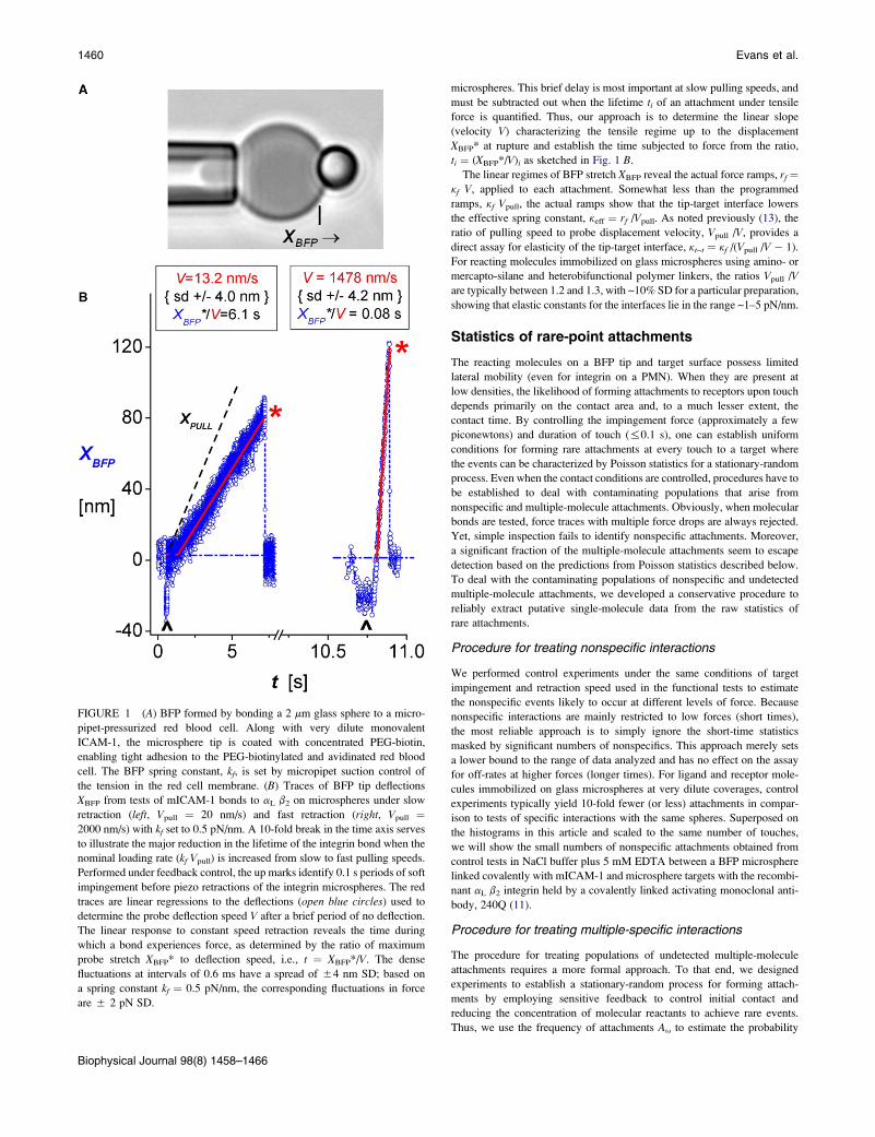
FIGURE 1 (A) BFP formed by bonding a 2 mm glass sphere to a micro-
pipet-pressurized red blood cell. Along with very dilute monovalent
ICAM-1, the microsphere tip is coated with concentrated PEG-biotin,
enabling tight adhesion to the PEG-biotinylated and avidinated red blood
cell. The BFP spring constant, kf, is set by micropipet suction control of
the tension in the red cell membrane. (B) Traces of BFP tip deflections
XBFP from tests of mICAM-1 bonds to aL b2 on microspheres under slow
retraction (left, Vpull ¼ 20 nm/s) and fast retraction (right, Vpull ¼2000 nm/s) with kf set to 0.5 pN/nm. A 10-fold break in the time axis serves
to illustrate the major reduction in the lifetime of the integrin bond when the
nominal loading rate (kf Vpull) is increased from slow to fast pulling speeds.
Performed under feedback control, the up marks identify 0.1 s periods of soft
impingement before piezo retractions of the integrin microspheres. The red
traces are linear regressions to the deflections (open blue circles) used to
determine the probe deflection speed V after a brief period of no deflection.
The linear response to constant speed retraction reveals the time during
which a bond experiences force, as determined by the ratio of maximum
probe stretch XBFP* to deflection speed, i.e., t ¼ XBFP*/V. The dense
fluctuations at intervals of 0.6 ms have a spread of 54 nm SD; based on
a spring constant kf ¼ 0.5 pN/nm, the corresponding fluctuations in force
are 5 2 pN SD.
Biophysical Journal 98(8) 1458–1466
1460 Evans et al.
microspheres. This brief delay is most important at slow pulling speeds, and
must be subtracted out when the lifetime ti of an attachment under tensile
force is quantified. Thus, our approach is to determine the linear slope
(velocity V) characterizing the tensile regime up to the displacement
XBFP* at rupture and establish the time subjected to force from the ratio,
ti ¼ (XBFP*/V)i as sketched in Fig. 1 B.
The linear regimes of BFP stretch XBFP reveal the actual force ramps, rf ¼kf V, applied to each attachment. Somewhat less than the programmed
ramps, kf Vpull, the actual ramps show that the tip-target interface lowers
the effective spring constant, keff ¼ rf /Vpull. As noted previously (13), the
ratio of pulling speed to probe displacement velocity, Vpull /V, provides a
direct assay for elasticity of the tip-target interface, kt~t ¼ kf /(Vpull /V � 1).
For reacting molecules immobilized on glass microspheres using amino- or
mercapto-silane and heterobifunctional polymer linkers, the ratios Vpull /V
are typically between 1.2 and 1.3, with ~10% SD for a particular preparation,
showing that elastic constants for the interfaces lie in the range ~1–5 pN/nm.
Statistics of rare-point attachments
The reacting molecules on a BFP tip and target surface possess limited
lateral mobility (even for integrin on a PMN). When they are present at
low densities, the likelihood of forming attachments to receptors upon touch
depends primarily on the contact area and, to a much lesser extent, the
contact time. By controlling the impingement force (approximately a few
piconewtons) and duration of touch (%0.1 s), one can establish uniform
conditions for forming rare attachments at every touch to a target where
the events can be characterized by Poisson statistics for a stationary-random
process. Even when the contact conditions are controlled, procedures have to
be established to deal with contaminating populations that arise from
nonspecific and multiple-molecule attachments. Obviously, when molecular
bonds are tested, force traces with multiple force drops are always rejected.
Yet, simple inspection fails to identify nonspecific attachments. Moreover,
a significant fraction of the multiple-molecule attachments seem to escape
detection based on the predictions from Poisson statistics described below.
To deal with the contaminating populations of nonspecific and undetected
multiple-molecule attachments, we developed a conservative procedure to
reliably extract putative single-molecule data from the raw statistics of
rare attachments.
Procedure for treating nonspecific interactions
We performed control experiments under the same conditions of target
impingement and retraction speed used in the functional tests to estimate
the nonspecific events likely to occur at different levels of force. Because
nonspecific interactions are mainly restricted to low forces (short times),
the most reliable approach is to simply ignore the short-time statistics
masked by significant numbers of nonspecifics. This approach merely sets
a lower bound to the range of data analyzed and has no effect on the assay
for off-rates at higher forces (longer times). For ligand and receptor mole-
cules immobilized on glass microspheres at very dilute coverages, control
experiments typically yield 10-fold fewer (or less) attachments in compar-
ison to tests of specific interactions with the same spheres. Superposed on
the histograms in this article and scaled to the same number of touches,
we will show the small numbers of nonspecific attachments obtained from
control tests in NaCl buffer plus 5 mM EDTA between a BFP microsphere
linked covalently with mICAM-1 and microsphere targets with the recombi-
nant aL b2 integrin held by a covalently linked activating monoclonal anti-
body, 240Q (11).
Procedure for treating multiple-specific interactions
The procedure for treating populations of undetected multiple-molecule
attachments requires a more formal approach. To that end, we designed
experiments to establish a stationary-random process for forming attach-
ments by employing sensitive feedback to control initial contact and
reducing the concentration of molecular reactants to achieve rare events.
Thus, we use the frequency of attachments Au to estimate the probability
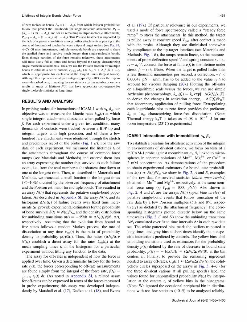
Lifetimes of Integrin Bonds Under Force 1461
of zero molecular bonds, P0 ¼ (1 � Au), from which Poisson probabilities
follow that predict the likelihoods for single-molecule attachment, P1 ¼(Au � 1) ln(1 � Au), and for all remaining multiple-molecule attachments,
PnR2 ¼ Au þ (1 � Au) ln(1 � Au). This Poisson treatment is supported by
the lack of apparent correlations among outlier attachments appearing in the
course of thousands of touches between a tip and target surface (see Fig. S1,
A–C). Of most importance, multiple-molecule bonds are expected to share
the applied force and survive much longer than single-molecule bonds.
Even though partition of the force remains unknown, these attachments
will most likely fail at times and forces beyond the range characterizing
single-molecule attachments. Thus, we use the Poisson fraction for multiple
bonds to estimate a set of outliers, PnR2 /Au (z Au /2 þ Au2 /6 þ .),
which is appropriate for exclusion at the longest times (largest forces).
Although this represents small percentages (typically ~10%) for the experi-
ments described here, truncating the raw statistics by this number of outliers
results in arrays of lifetimes N(ti) that have appropriate convergence for
single-molecule statistics at long times.
RESULTS AND ANALYSIS
In probing molecular interactions of ICAM-1 with aL b2, our
objective was to measure the kinetic rates koff(t) at which
single integrin attachments dissociate when pulled by force
f. For each experiment under a given test condition, many
thousands of contacts were tracked between a BFP tip and
integrin targets with high precision, and of these a few
hundred rare attachments were identified through the stretch
and precipitous recoil of the probe (Fig. 1 B). For the raw
data of each experiment, we measured the lifetimes ti of
the attachments throughout the course of common force
ramps (see Materials and Methods) and ordered them into
an array expressing the number that survived to each failure
event, i.e., from the total number at the shortest time down to
one at the longest time. Then, as described in Materials and
Methods, we truncated a small fraction of the longest times
(%~10%) dictated by the observed frequency of attachment
and the Poisson estimator for multiple bonds. This resulted in
an array N(ti) that represents the putative single-bond popu-
lation. As described in Appendix SI, the array N(ti), and its
histogram DNk(tk) of failure events over fixed time incre-
ments Dt, provide experimental estimators for the probability
of bond survival S(t) z N(ti)/No, and the density distribution
for unbinding transitions p(t) ¼ �dS/dt z DNk(tk)/(No Dt),respectively. Assuming that the evolution from bound to
free states follows a random Markov process, the rate of
dissociation at any time koff(t) is the ratio of probability
density to probability p(t)/S(t). Thus, the ratios (DNk/Dt)/N(tk) establish a direct assay for the rates koff(tk) at the
mean sampling times tk in the histogram for a particular
experiment without fitting any function to the data.
The assay for off-rates is independent of how the force is
applied over time. Given a deterministic history for the force
rate rf(t), the forces corresponding to off-rate measurements
are found simply from the integral of the force rate, f(tk) ¼!o/tk rf (t) dt. (As noted in Appendix SI, a related assay
for off-rates can be expressed in terms of the forces measured
in probe experiments; this assay was developed indepen-
dently by Marshall et al. (17), Dudko et al. (18), and Evans
et al. (19).) Of particular relevance in our experiments, we
used a mode of force spectroscopy called a ‘‘steady force
ramp’’ to stress the attachments. In this method, the target
is pulled away at constant speed Vpull after contact is made
with the probe. Although they are diminished somewhat
by compliance at the tip-target interface (see Materials and
Methods, Fig. 1 B), the ramps remain linear, so the measure-
ments of probe deflection speed V and spring constant kf, i.e.,
rf ¼ kf V, connect the force at failure fi to the lifetime under
stress, fi ¼ rf ti. (Note: When the target is pulled faster than
a few thousand nanometers per second, a correction, ~V �0.00048 pN $ s/nm, has to be added to the value rf ti to
account for viscous damping (20).) Plotting the off-rates
on a logarithmic scale versus the forces, we can use simple
Arrhenius phenomenology, koff(fk) ¼ ko exp[�DG(fk)/kBT],
to derive the changes in activation energy, �DG(fk)/kBT,
that accompany application of pulling force. Extrapolating
each logarithmic plot to zero force provides the prefactor,
ko ¼ 1/to, characterizing force-free dissociation. (Note:
Thermal energy kBT is taken as ~4.08 � 10�21 J for our
room-temperature (23�C) experiments.)
ICAM-1 interactions with immobilized aL b2
To establish a baseline for allosteric activation of the integrin
in environments of divalent cations, we focus on tests of a
mICAM-1 probe against recombinant aL b2 linked to micro-
spheres in separate solutions of Mn2þ, Mg2þ, or Ca2þ at
2 mM concentration. As demonstrations of the procedure
to obtain experimental estimators for bound-state probabili-
ties S(ti) z N(ti)/No, we show in Fig. 2, A and B, examples
of the raw data for survival statistics (black open circles)
obtained in Mn2þ and Mg2þ, respectively, at the same nom-
inal force ramp (kf Vpull ¼ 1000 pN/s). Also shown in
Fig. 2, A and B, are the arrays N(ti) (open blue circles) of
putative single-bond events that follow truncation of the
raw data by a few Poisson multiples (5% and 8%, respec-
tively) as dictated by the attachment frequency. The corre-
sponding histograms plotted directly below on the same
timescales (Fig. 2, C and D) show the unbinding transitions
DNk cumulated over fixed time intervals Dt in each raw data
set. The white-patterned bins mark the outliers truncated at
long times, and gray bins at short times identify the nonspe-
cific interactions predicted by controls. The yellow bins label
unbinding transitions used as estimators for the probability
density p(tk) defined by the rate of decrease in bound state
probability, p(tk) ¼ � [dS/dt]k z (DNk/Dt)/N(0), at the bin
centers tk. Finally, to provide the remaining ingredient
needed to assay off-rates, koff(tk) z (DNk/Dt)/N(tk), the solid-
yellow circles superposed on the arrays in Fig. 3, A–C (for
the three divalent cations at all pulling speeds) label the
values found for unnormalized probability N(tk) by interpo-
lation at the centers tk of yellow bins in the histograms.
(Note: We ignored the occasional peripheral bin in distribu-
tions with too few statistics (~0–5) to be analyzed reliably.
Biophysical Journal 98(8) 1458–1466

FIGURE 2 Lifetimes for mICAM-1 bonds to aL b2 on
microspheres subjected to a nominal force ramp of kf
Vpull ¼1000 pN/s in 2 mM solutions of each divalent
metal cation (measured ramps are marked on each panel).
(A and B) Raw data (black open circles) for bond survival
obtained in Mn2þ and Mg2þ are shown along with arrays
N(ti) (open blue circles) characterizing the unnormalized
probability for single-bond survival obtained after trunca-
tion of a small Poisson fraction of multiple bonds (0.05
and 0.08, respectively). (C and D) Histograms plotted
directly below on the same timescales show the unbinding
transitions DNk cumulated over fixed time intervals Dt in
each raw data set. White-patterned bins identify the outliers
truncated at long times, and gray bins at short times are
nonspecific interactions obtained from control experiments
rescaled to the same number of touches. Intervening yellow
bins show the transitions used as estimators for the rates of
decrease in bound state probabilities, p(tk)¼� [dS1/dt]k z(DNk/Dt)/N(0), at the bin centers tk.
1462 Evans et al.
This had no effect on the values of off-rates determined for
the other bins.)
Extending the off-rates examined to much lower values,
we show arrays of putative single-bond lifetimes in Fig. 3,
A–C, that were obtained with slower nominal force ramps
along with the interpolated values corresponding to bins in
histograms of the data (not shown). Again determining the
ratios (DNk/Dt)/N(tk) for all the data, we plot the off-rates
in Fig. 3, D–F, on a log scale versus the forces, fk ¼ rf tk,as defined by the ramp rates (5 SD) multiplied by the times
at bin centers. (Also plotted in Fig. 3, D and E (open circles),
are results from experiments performed at 2 � Vpull and
1/2 � kf, demonstrating that the off-rates depend only on
the instantaneous force, f ¼ (keff Vpull) t, rather than the pull-
ing speed or BFP spring constant.) As shown by linear
regressions (dotted lines) matched to each data set in
Fig. 3, D–F, the logarithms of off-rates follow a linear depen-
dence on the force level characterized by a force scale fb[¼ 1/(ln10� slope) ] and a zero-force intercept ko. Together,
the data and regressions in Fig. 3, D–F, exhibit the well-
known exponential behavior proposed by George Bell (21)
many years ago, i.e., koff z ko exp[f xb /kBT], in which the
distance xb represents the thermally averaged length gained
Biophysical Journal 98(8) 1458–1466
in the direction of force when a bond is dissociated. In the
context of the measurements, the length xb corresponds to
the force needed to lower the energy barrier governing off-
rate by one thermal energy unit, i.e., fb ¼ kBT/xb. Table 1
summarizes the parameters obtained from the linear regres-
sions to the data for the three divalent cations over a range
of force ramps between 10 and 1000 pN/s, which most likely
encompass conditions experienced by b2 integrin bonds
in vivo (22). As listed in Table 1, the stress-free rates ko char-
acterizing spontaneous mICAM-1 dissociation from aL b2
on microspheres exhibit the well-known hierarchy of allo-
steric activation expected for solutions of the different metal
cations (23), i.e., from 0.008/s in Mg2þ to 0.02/s in Mn2þ
and ~2/s in Ca2þ.
Ultrafast dissociation of ICAM-1 from immobilizedaL b2
As illustrated by sample data from tests of mICAM-1 against
aL b2 spheres in Mg2þ and Ca2þ (Fig. 4, A and B,), numer-
ous long-lived states in statistical arrays emerged at
extremely fast ramps (kf Vpull R 10000 pN/s) that could
not be attributed to multiple bonds. As demonstrated by
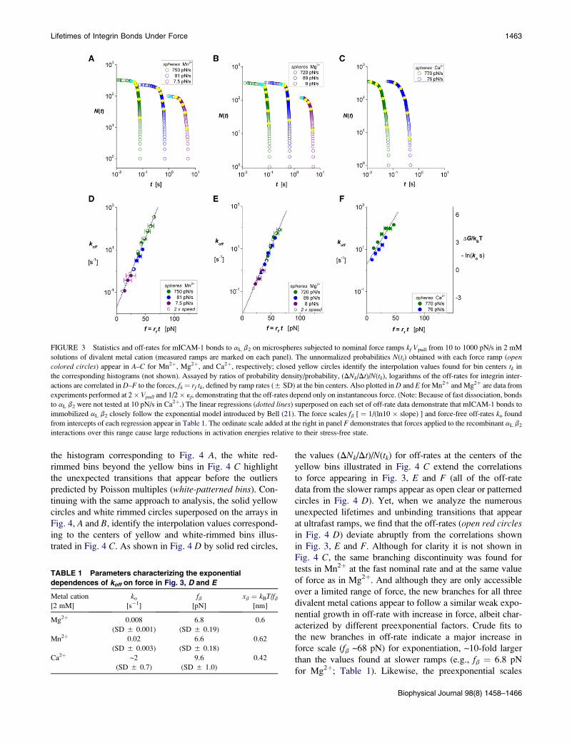
FIGURE 3 Statistics and off-rates for mICAM-1 bonds to aL b2 on microspheres subjected to nominal force ramps kf Vpull from 10 to 1000 pN/s in 2 mM
solutions of divalent metal cation (measured ramps are marked on each panel). The unnormalized probabilities N(ti) obtained with each force ramp (opencolored circles) appear in A–C for Mn2þ, Mg2þ, and Ca2þ, respectively; closed yellow circles identify the interpolation values found for bin centers tk in
the corresponding histograms (not shown). Assayed by ratios of probability density/probability, (DNk/Dt)/N(tk), logarithms of the off-rates for integrin inter-
actions are correlated in D–F to the forces, fk¼ rf tk, defined by ramp rates (5 SD) at the bin centers. Also plotted in D and E for Mn2þ and Mg2þ are data from
experiments performed at 2� Vpull and 1/2� kf, demonstrating that the off-rates depend only on instantaneous force. (Note: Because of fast dissociation, bonds
to aL b2 were not tested at 10 pN/s in Ca2þ.) The linear regressions (dotted lines) superposed on each set of off-rate data demonstrate that mICAM-1 bonds to
immobilized aL b2 closely follow the exponential model introduced by Bell (21). The force scales fb [ ¼ 1/(ln10 � slope) ] and force-free off-rates ko found
from intercepts of each regression appear in Table 1. The ordinate scale added at the right in panel F demonstrates that forces applied to the recombinant aL b2
interactions over this range cause large reductions in activation energies relative to their stress-free state.
Lifetimes of Integrin Bonds Under Force 1463
the histogram corresponding to Fig. 4 A, the white red-
rimmed bins beyond the yellow bins in Fig. 4 C highlight
the unexpected transitions that appear before the outliers
predicted by Poisson multiples (white-patterned bins). Con-
tinuing with the same approach to analysis, the solid yellow
circles and white rimmed circles superposed on the arrays in
Fig. 4, A and B, identify the interpolation values correspond-
ing to the centers of yellow and white-rimmed bins illus-
trated in Fig. 4 C. As shown in Fig. 4 D by solid red circles,
TABLE 1 Parameters characterizing the exponential
dependences of koff on force in Fig. 3, D and E
Metal cation
[2 mM]
ko
[s�1]
fb[pN]
xb ¼ kBT/fb[nm]
Mg2þ 0.008
(SD 5 0.001)
6.8
(SD 5 0.19)
0.6
Mn2þ 0.02
(SD 5 0.003)
6.6
(SD 5 0.18)
0.62
Ca2þ ~2
(SD 5 0.7)
9.6
(SD 5 1.0)
0.42
the values (DNk/Dt)/N(tk) for off-rates at the centers of the
yellow bins illustrated in Fig. 4 C extend the correlations
to force appearing in Fig. 3, E and F (all of the off-rate
data from the slower ramps appear as open clear or patterned
circles in Fig. 4 D). Yet, when we analyze the numerous
unexpected lifetimes and unbinding transitions that appear
at ultrafast ramps, we find that the off-rates (open red circlesin Fig. 4 D) deviate abruptly from the correlations shown
in Fig. 3, E and F. Although for clarity it is not shown in
Fig. 4 C, the same branching discontinuity was found for
tests in Mn2þ at the fast nominal rate and at the same value
of force as in Mg2þ. And although they are only accessible
over a limited range of force, the new branches for all three
divalent metal cations appear to follow a similar weak expo-
nential growth in off-rate with increase in force, albeit char-
acterized by different preexponential factors. Crude fits to
the new branches in off-rate indicate a major increase in
force scale (fb ~68 pN) for exponentiation, ~10-fold larger
than the values found at slower ramps (e.g., fb ¼ 6.8 pN
for Mg2þ; Table 1). Likewise, the preexponential scales
Biophysical Journal 98(8) 1458–1466
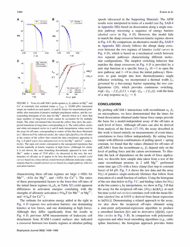
FIGURE 4 Tests of a mICAM-1 probe against aL b2 spheres in Mg2þ and
Ca2 at extremely fast nominal ramps kf Vpull R 10,000 pN/s (measured
ramps are marked on each panel). (A and B) Arrays for unnormalized prob-
ability after truncation of putative multiple-attachment outliers, and the cor-
responding histogram of raw data for Mg2þ directly below in C show that
large numbers of long-lived events cannot be accounted for by multiple
bonds. The white red-rimmed bins beyond the yellow bins show the unex-
pected transitions at long times corresponding to A. The solid yellow circles
and white red-rimmed circles in A and B mark interpolation values used in
the assay for off-rates, corresponding to centers of bins like those illustrated
in C. Shown in D by solid red circles, the values (DNk/Dt)/N(tk) for off-rates
at the centers of the yellow bins extend the data correlations appearing in
Fig. 3, E and F (open clear and patterned circles for Mg2þ and Ca2þ, respec-
tively). The open red circles correspond to the unexpected transitions that
deviate markedly in kinetic response at high forces. (Although for clarity
it is not shown, the same branching discontinuity appeared in tests with
Mn2þ under a ramp of 7510 pN/s.) As discussed in the text, the new
branches of off-rates in D appear to be best described by a model (solid
curves) based on a force-driven switch between different molecular config-
urations than by a model (dotted curves) based on a single pathway with two
activation barriers.
1464 Evans et al.
characterizing these off-rate regimes are large (~180/s for
Mn2þ, ~63/s for Mg2þ, and ~145/s for Ca2þ). The ratios
of these preexponential factors to the force-free off-rates of
the initial linear regimes (ko2/ko in Table S2) yield apparent
differences in activation energies correlating with the
strengths of allosteric activation (~9 kBT for Mn2þ or Mg2þ,
and ~4 kBT for Ca2þ).
The ordinate for activation energy added at the right in
Fig. 4 D exposes two activation barriers: one dominating
kinetics at low forces, and one dominating at high forces.
Although they differ significantly from the results in
Fig. 4 D, previous AFM measurements of leukocytic cell
detachment from ICAM-1-coated surfaces also indicated
a crossover between two kinetic regimes at ultrafast pulling
Biophysical Journal 98(8) 1458–1466
speeds (discussed in the Supporting Material). The AFM
results were interpreted in terms of a model (see Eq. SAII-4
in Appendix SII)) based on dissociation along a single reac-
tion pathway traversing a sequence of energy barriers
(dashed curve in Fig. 4 D). However, this model fails
to match the sharp crossover between kinetic regimes shown
in Fig. 4 D. By comparison, an alternative model (Eq. SAII-9
in Appendix SII) closely follows the abrupt sharp cross-
over between the two regimes of kinetics (solid curves in
Fig. 4 D), which is based on a mechanical switch between
two separate pathways characterizing different molec-
ular configurations. The simplest switching behavior that
matches the sharp crossovers in Fig. 4 D is provided by a
unit step function at a specific force f5 (0/1 to open the
new pathway and 1/0 to close the initial pathway). How-
ever, to gain insight into how thermodynamics might
influence switching, we incorporated a thermal width f12
governed by a free-energy barrier separating the two con-
figurations (24), which provides continuous switching,
exp[�(f5� f /f12)] /{1þ exp[�(f5� f /f12)]}, with the limit
of a step response as f12 / 0.
CONCLUSIONS
By probing mICAM-1 interactions with recombinant aL b2
on microspheres, we have demonstrated that the times for
bond dissociation obtained under linear force ramps provide
the basis for a model-independent assay of the off-rates at
each level of force. Although off-rates can also be derived
from analysis of the forces (17–19), the assay described in
this work is based entirely on measurements of event times;
correlations to force follow independently. By setting force
ramps with different combinations of pulling speed � spring
constant, we found that the values obtained for off-rates of
mICAM-1 from the recombinant aL b2 depend only on the
level of pulling force and the cation environment. To illus-
trate the lack of dependence on the mode of force applica-
tion, we describe here sample data taken from a test of the
same recombinant proteins in 2 mM Mg2þ performed
some time ago (25) with a force clamp (fast step to constant
force) of 64 pN. Fig. 5 A shows the raw data and the array
N(ti) of putative single-molecule lifetimes that follow from
truncation of a small fraction of outliers. Using the histogram
of the raw data below in Fig. 5 C, and finding values for N(tk)at the bin centers tk by interpolation, we show in Fig. 5 B that
the assay for the reciprocal off-rate, [N(tk) Dt/DNk], at each
bin time (solid red circles) correlates well with the mean life-
time (dashed line at 0.019 s) derived from linear regression
to ln[N(ti)]. Demonstrating a related approach to the assay,
we also show the reciprocal off-rates obtained using
a nine-point polynomial-regression algorithm to produce
a smoothed function N(t) and its derivative [dN/dt]i (openblue circles in Fig. 5 B). In comparison with polynomial-
regression and other local smoothing algorithms (e.g., cubic
spline functions), the histogram approach provides better
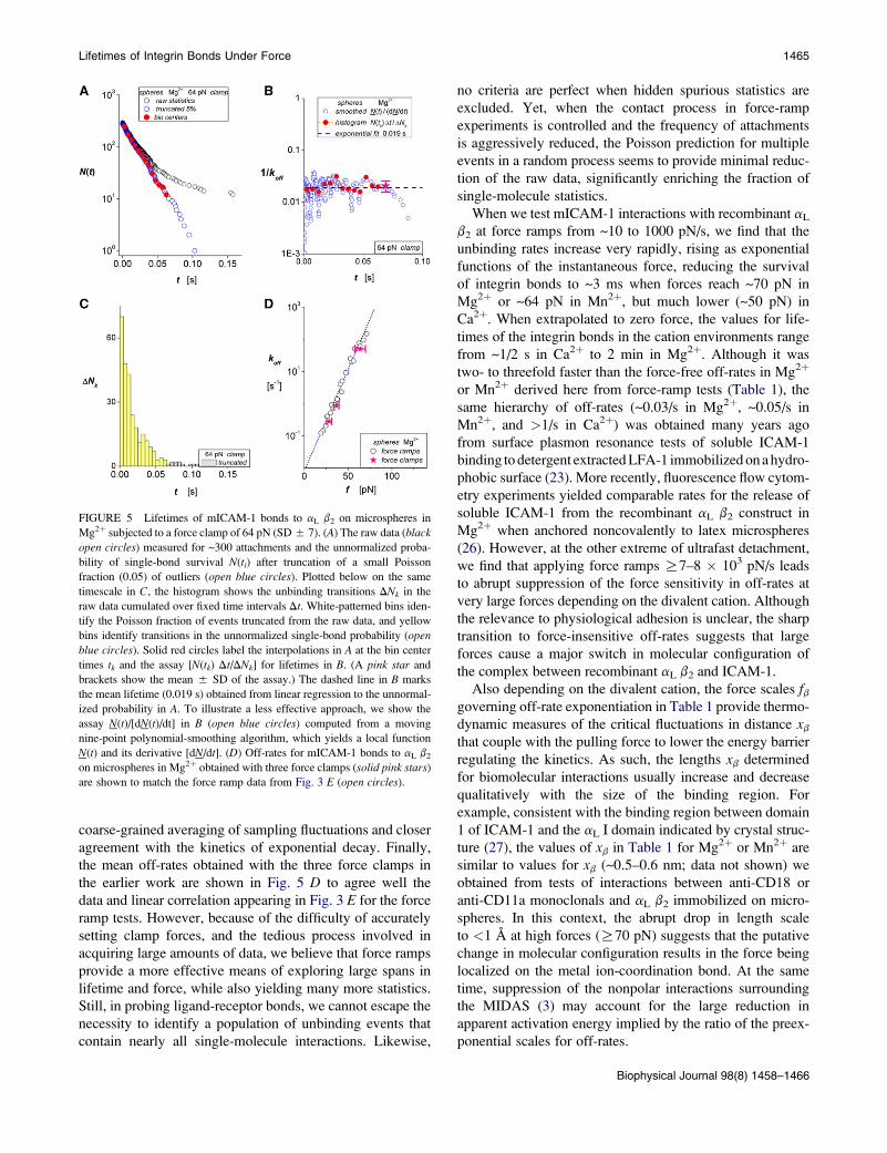
FIGURE 5 Lifetimes of mICAM-1 bonds to aL b2 on microspheres in
Mg2þ subjected to a force clamp of 64 pN (SD 5 7). (A) The raw data (black
open circles) measured for ~300 attachments and the unnormalized proba-
bility of single-bond survival N(ti) after truncation of a small Poisson
fraction (0.05) of outliers (open blue circles). Plotted below on the same
timescale in C, the histogram shows the unbinding transitions DNk in the
raw data cumulated over fixed time intervals Dt. White-patterned bins iden-
tify the Poisson fraction of events truncated from the raw data, and yellow
bins identify transitions in the unnormalized single-bond probability (open
blue circles). Solid red circles label the interpolations in A at the bin center
times tk and the assay [N(tk) Dt/DNk] for lifetimes in B. (A pink star and
brackets show the mean 5 SD of the assay.) The dashed line in B marks
the mean lifetime (0.019 s) obtained from linear regression to the unnormal-
ized probability in A. To illustrate a less effective approach, we show the
assay N(t)/[dN(t)/dt] in B (open blue circles) computed from a moving
nine-point polynomial-smoothing algorithm, which yields a local function
N(t) and its derivative [dN/dt]. (D) Off-rates for mICAM-1 bonds to aL b2
on microspheres in Mg2þ obtained with three force clamps (solid pink stars)
are shown to match the force ramp data from Fig. 3 E (open circles).
Lifetimes of Integrin Bonds Under Force 1465
coarse-grained averaging of sampling fluctuations and closer
agreement with the kinetics of exponential decay. Finally,
the mean off-rates obtained with the three force clamps in
the earlier work are shown in Fig. 5 D to agree well the
data and linear correlation appearing in Fig. 3 E for the force
ramp tests. However, because of the difficulty of accurately
setting clamp forces, and the tedious process involved in
acquiring large amounts of data, we believe that force ramps
provide a more effective means of exploring large spans in
lifetime and force, while also yielding many more statistics.
Still, in probing ligand-receptor bonds, we cannot escape the
necessity to identify a population of unbinding events that
contain nearly all single-molecule interactions. Likewise,
no criteria are perfect when hidden spurious statistics are
excluded. Yet, when the contact process in force-ramp
experiments is controlled and the frequency of attachments
is aggressively reduced, the Poisson prediction for multiple
events in a random process seems to provide minimal reduc-
tion of the raw data, significantly enriching the fraction of
single-molecule statistics.
When we test mICAM-1 interactions with recombinant aL
b2 at force ramps from ~10 to 1000 pN/s, we find that the
unbinding rates increase very rapidly, rising as exponential
functions of the instantaneous force, reducing the survival
of integrin bonds to ~3 ms when forces reach ~70 pN in
Mg2þ or ~64 pN in Mn2þ, but much lower (~50 pN) in
Ca2þ. When extrapolated to zero force, the values for life-
times of the integrin bonds in the cation environments range
from ~1/2 s in Ca2þ to 2 min in Mg2þ. Although it was
two- to threefold faster than the force-free off-rates in Mg2þ
or Mn2þ derived here from force-ramp tests (Table 1), the
same hierarchy of off-rates (~0.03/s in Mg2þ, ~0.05/s in
Mn2þ, and >1/s in Ca2þ) was obtained many years ago
from surface plasmon resonance tests of soluble ICAM-1
binding to detergent extracted LFA-1 immobilized on a hydro-
phobic surface (23). More recently, fluorescence flow cytom-
etry experiments yielded comparable rates for the release of
soluble ICAM-1 from the recombinant aL b2 construct in
Mg2þ when anchored noncovalently to latex microspheres
(26). However, at the other extreme of ultrafast detachment,
we find that applying force ramps R7–8 � 103 pN/s leads
to abrupt suppression of the force sensitivity in off-rates at
very large forces depending on the divalent cation. Although
the relevance to physiological adhesion is unclear, the sharp
transition to force-insensitive off-rates suggests that large
forces cause a major switch in molecular configuration of
the complex between recombinant aL b2 and ICAM-1.
Also depending on the divalent cation, the force scales fbgoverning off-rate exponentiation in Table 1 provide thermo-
dynamic measures of the critical fluctuations in distance xb
that couple with the pulling force to lower the energy barrier
regulating the kinetics. As such, the lengths xb determined
for biomolecular interactions usually increase and decrease
qualitatively with the size of the binding region. For
example, consistent with the binding region between domain
1 of ICAM-1 and the aL I domain indicated by crystal struc-
ture (27), the values of xb in Table 1 for Mg2þ or Mn2þ are
similar to values for xb (~0.5–0.6 nm; data not shown) we
obtained from tests of interactions between anti-CD18 or
anti-CD11a monoclonals and aL b2 immobilized on micro-
spheres. In this context, the abrupt drop in length scale
to <1 A at high forces (R70 pN) suggests that the putative
change in molecular configuration results in the force being
localized on the metal ion-coordination bond. At the same
time, suppression of the nonpolar interactions surrounding
the MIDAS (3) may account for the large reduction in
apparent activation energy implied by the ratio of the preex-
ponential scales for off-rates.
Biophysical Journal 98(8) 1458–1466
1466 Evans et al.
SUPPORTING MATERIAL
One figure and two tables are available at http://www.biophysj.org/biophysj/
supplemental/S0006-3495(10)00084-6.
This work was supported by National Institutes of Health grants HL65333
and HL31579 (to E.E.) and AI47294 (to S.S.).
REFERENCES
1. McEver, R. P. 2001. Adhesive interactions of leukocytes, platelets, andthe vessel wall during hemostasis and inflammation. Thromb. Haemost.86:746–756.
2. Hynes, R. O. 2002. Integrins: bi-directional, allosteric, signalingmachines. Cell. 110:673–687.
3. Luo, B. H., C. V. Carman, and T. A. Springer. 2007. Structural basis ofintegrin regulation and signaling. Annu. Rev. Immunol. 25:619–647.
4. Shimaoka, M., J. Takagi, and T. A. Springer. 2002. Conformationalregulation of integrin structure and function. Annu. Rev. Biophys.Biomol. Struct. 31:485–556.
5. Arnaout, M. A., B. Mahalingam, and J. P. Xiong. 2005. Integrin struc-ture, allostery, and bidirectional signaling. Annu. Rev. Cell Dev. Biol.21:381–410.
6. Luo, B. H., and T. A. Springer. 2006. Integrin structures and conforma-tional signaling. Curr. Opin. Cell Biol. 18:579–586.
7. Nishida, N., C. Xie, ., T. A. Springer. 2006. Activation of leukocyte b2
integrins by conversion from bent to extended conformations. Immu-nity. 25:583–594.
8. Salas, A., M. Shimaoka, ., T. A. Springer. 2004. Rolling adhesionthrough an extended conformation on integrin aL b2 and relation toa I and b I-like domain interaction. Immunity. 20:393–406.
9. Shamri, R., V. Grabovsky, ., R. Alon. 2005. Lymphocyte arrestrequires instantaneous induction of an extended LFA-1 conformationmediated by endothelium-bound chemokines. Nat. Immunol. 6:497–506.
10. Kinoshita, K., A. Leung, S. I. Simon, and E. Evans. 2009. Long-livedhigh-strength states of ICAM-1 bonds to aL b2 integrin: II. Lifetimesof LFA-1 bonds under force in leukocyte signaling. Biophys. J.98:1467–1475.
11. Lupher, Jr., M. L., E. A. Harris, ., D. E. Staunton. 2001. Cellularactivation of leukocyte function-associated antigen-1 and its affinityare regulated at the I domain allosteric site. J. Immunol. 167:1431–1439.
12. Evans, E., K. Ritchie, and R. Merkel. 1995. Sensitive force technique toprobe molecular adhesion and structural linkages at biological inter-faces. Biophys. J. 68:2580–2587.
Biophysical Journal 98(8) 1458–1466
13. Evans, E., V. Heinrich, ., K. Kinoshita. 2005. Nano-to-micro scaledynamics of P-selectin detachment from leukocyte interfaces: I.membrane separation from the cytoskeleton. Biophys. J. 88:2288–2298.
14. Zhang, X., E. P. Wojcikiewicz, and V. T. Moy. 2002. Force spectros-copy of the leukocyte function-associated antigen-1/intercellular adhe-sion molecule-1 interaction. Biophys. J. 83:2270–2279.
15. Wojcikiewicz, E. P., M. H. Abdulreda, ., V. T. Moy. 2006. Forcespectroscopy of LFA-1 and its ligands ICAM-1 and ICAM-2. Bioma-cromolecules. 7:3188–3195.
16. Vitte, J., A. Pierres, ., P. Bongrand. 2004. Direct quantification of themodulation of interaction between cell- or surface-bound LFA-1 andICAM-1. J. Leukoc. Biol. 76:594–602.
17. Marshall, B. T., K. K. Sarangapani, ., C. Zhu. 2005. Force historydependence of receptor-ligand dissociation. Biophys. J. 88:1458–1466.
18. Dudko, O. K., G. Hummer, and A. Szabo. 2008. Theory, analysis, andinterpretation of single-molecule force spectroscopy experiments. Proc.Natl. Acad. Sci. USA. 105:15755–15760.
19. Evans, E., K. Halvorsen, ., W. P. Wong. 2009. A new approach toanalysis of single molecule force experiments. In Handbook ofSingle-Molecule Biophysics. P. Hinterdorfer and A. M. van Oijen,editors. Springer Science, New York, In press.
20. Evans, E., A. Leung, ., S. Simon. 2001. Chemically-distinct transitionstates govern rapid detachment of single bonds to L-selectin underforce. Proc. Natl. Acad. Sci. USA. 98:3784–3789.
21. Bell, G. I. 1978. Models for the specific adhesion of cells to cells.Science. 200:618–627.
22. Alon, R., and M. L. Dustin. 2007. Force as a facilitator of integrinconformational changes during leukocyte arrest on blood vessels andantigen-presenting cells. Immunity. 26:17–27.
23. Labadia, M. E., D. D. Jeanfavre, ., M. M. Morelock. 1998. Molecularregulation of the interaction between leukocyte function-associatedantigen-1 and soluble ICAM-1 by divalent metal cations. J. Immunol.161:836–842.
24. Evans, E., A. Leung, ., C. Zhu. 2004. Mechanical switching andcoupling between two dissociation pathways in a P-selectin adhesionbond. Proc. Natl. Acad. Sci. USA. 101:11281–11286.
25. Evans, E., and K. Kinoshita. 2007. Using force to probe single-moleculereceptor-cytoskeletal anchoring beneath the surface of a living cell.Methods Cell Biol. 83:373–396.
26. Sarantos, M. R., S. Raychaudhure, ., S. I. Simon. 2005. Leukocytefunction-associated antigen 1-mediated adhesion stability is dynami-cally regulated through affinity and valency during bond formationwith intercellular adhesion molecule-1. J. Biol. Chem. 280:28290–28298.
27. Shimaoka, M., T. Xiao, ., T. A. Springer. 2003. Structures of the aL Idomain and its complex with ICAM-1 reveal a shape-shifting pathwayfor integrin regulation. Cell. 112:99–111.