Pdia4 regulates β‐cell pathogenesis in diabetes: molecular ...
Diabetologia (2006) 49: 1587–1598DOI 10.1007/s00125-006-0265-9
ARTICLE
E. D. Targonsky . F. Dai . V. Koshkin . G. T. Karaman .A. V. Gyulkhandanyan . Y. Zhang . C. B. Chan .M. B. Wheeler
α-Lipoic acid regulates AMP-activated protein kinase and inhibitsinsulin secretion from beta cells
Received: 15 October 2005 / Accepted: 27 February 2006 / Published online: 13 May 2006# Springer-Verlag 2006
Abstract Aims/hypothesis: The antioxidant compound α-lipoic acid (α-LA) possesses antidiabetic and anti-obesityproperties. In the hypothalamus, α-LA suppresses appetiteand prevents obesity by inhibiting AMP-activated proteinkinase (AMPK). Given the therapeutic potential of α-LAfor the treatment of type 2 diabetes and obesity, and theimportance of AMPK in beta cells, we examined the effectof α-LA on pancreatic beta cell function. Materials andmethods: Isolated rat islets and MIN6 beta cells weretreated acutely (15–90 min) or chronically (18–24 h) withα-LA or the known AMPK-activating compounds 5′-amino-imidazole-4-carboxamide ribonucleoside (AICAR)and metformin. Insulin secretion, the AMPK-signallingpathway, mitochondrial function and cell growth wereassessed. Results: Acute or chronic treatment of islets andMIN6 cells with α-LA led to dose-dependent rises inphosphorylation of the AMPK α-subunit and acetyl CoAcarboxylase. Chronic exposure to α-LA, AICAR ormetformin caused a reduction in insulin secretion. α-LAinhibited the p70 s6 kinase translational control pathway,and inhibited MIN6 growth in a manner similar torapamycin. Unlike AICAR and metformin, α-LA alsoacutely inhibited insulin secretion. Examination of theeffect of α-LA on mitochondrial function showed that
acute treatment with this compound elevated reactiveoxygen species (ROS) production and enhanced mito-chondrial depolarisation induced by Ca2+. Conclusions/interpretation: This study is the first to demonstrate thatα-LA directly affects beta cell function. The chronic effectsof α-LA include AMPK activation and reductions ininsulin secretion and content, and cell growth. Acutely,α-LA also inhibits insulin secretion, an effect probablyinvolving the ROS-induced impairment of mitochondrialfunction.
Keywords AMPK . Insulin secretion . α-Lipoic acid .Mitochondrial permeability transition . mTOR . Pancreaticbeta cell . Reactive oxygen species
Abbreviations α-LA: α-lipoic acid . ΔΨm:Mitochondrial membrane potential . ACC: acetyl CoAcarboxylase . AICAR: 5′-amino-imidazole-4-carboxamideribonucleoside . AMPK: AMP-activated protein kinase .DCF: dichlorofluorescein . GSIS: glucose-stimulatedinsulin secretion . KRBH: Krebs–Ringer buffer withHEPES . MPT: mitochondrial permeability transition .mTOR: mammalian target of rapamycin . p70 s6k: p70 s6kinase . ROS: reactive oxygen species
Introduction
α-Lipoic acid (α-LA) is a short-chain fatty acid that acts asa cofactor of enzymes involved in mitochondrial respira-tion [1]. Beneficial anti-oxidative properties for α-LA havebeen described in several tissues, including the brain [2],kidney [3] and heart [4]. In recent years, evidence hasaccumulated suggesting that α-LA has a wide range ofbenefits in the treatment of diabetes. Bitar et al. [5]demonstrated that α-LA could partly ameliorate insulinresistance in type 2 diabetic rats, while others had reportedadditional antidiabetic effects in earlier rodent [4, 6], andhuman studies [7]. In vitro approaches have also shownthat α-LA can enhance glucose disposal in skeletal muscle[8] and adipocytes [9], and suppress hepatic gluconeogen-
E. D. Targonsky . F. Dai . V. Koshkin . G. T. Karaman .A. V. Gyulkhandanyan . Y. Zhang . M. B. Wheeler (*)Department of Physiology, University of Toronto,1 King’s College Circle,Toronto, ON, Canada M5S 1A8e-mail: [email protected].: +416-978-1969Fax: +416-978-4940
M. B. WheelerDepartment of Medicine, University of Toronto,Toronto, ON, Canada
C. B. ChanDepartment of Biomedical Sciences,Atlantic Veterinary College,University of Prince Edward Island,PE, Canada
esis [10]. The exact molecular mechanisms responsible forthese effects are currently not known.
AMP-activated protein kinase (AMPK) functions as anenergy sensor in mammalian cells (reviewed in [11]).AMPK is a heterotrimeric enzyme that is activated bystress signals [12], such as an elevated intracellular AMP:ATP ratio. When AMP levels rise, the AMPK α-subunitbecomes phosphorylated and activated, leading to thesuppression of anabolic processes, including protein andlipid synthesis. This is accompanied by an increase in ATP-generating pathways, including the β-oxidation of fattyacids. AMPK mediates insulin-independent glucose uptakein skeletal muscle [13], suppresses glucose productionfrom the liver [14], and is activated by the hormones leptin[15] and adiponectin [16], and the antidiabetic drugmetformin [17]. As such, AMPK has attracted muchattention as a potential target for diabetes therapy.
Recently, the properties of α-LA were linked to AMPKin a report describing its ability to prevent obesity in rats[18]. α-LA treatment reduced body weight and feeding andincreased energy expenditure in these rodents. Theseeffects were shown to be, at least in part, due to the abilityof α-LA to suppress AMPK activity in the hypothalamus.Injection of rats with 5′-amino-imidazole-4-carboxamideribonucleoside (AICAR), an AMP analogue and potentactivator of AMPK, reversed the effects of α-LA andprevented protection from obesity.
In pancreatic islets of Langerhans, AMPK possesses aunique role, connecting cellular energy status to thecapacity of beta cells to synthesise and secrete insulin. Anumber of studies have demonstrated that AMPK, whenactivated, transmits the signal of low glucose availabilityand leads to an inhibition of insulin secretion andexpression [19–22]. While a role for AMPK in the betacell continues to unfold, evidence for a role for α-LA inislet function is lacking. Given that α-LA modulatesAMPK activity in the hypothalamus, and AMPK regulatesinsulin secretion, we investigated the possible role of α-LAin beta cell function using isolated rat islets and the MIN6beta cell line.
Materials and methods
Animals
Male C57/Bl6 mice, 2 to 3 months of age, were purchasedfrom Charles River (Wilmington, MA, USA). All animalswere handled according to the guidelines of the CanadianCouncil on Animal Care. All the protocols and procedureswere approved by the Animal Care and Use Committee atthe University of Toronto.
Reagents
α-LA and metformin (1,1-dimethylbiguanide hydrochlo-ride) were obtained from Sigma (Oakville, ON, Canada).AICAR was from Toronto Research Chemicals (North
York, ON, Canada). Rapamycin and all primary antibodiesexcept anti-p70 s6 kinase (p70 s6k; Upstate, Lake Placid,NY, USA) were from Cell Signaling Technology (Beverly,MA, USA). Materials for rat insulin RIA were obtainedfrom Linco Research (St Charles, MO, USA).
Cell culture
MIN6 insulinoma cells (passage 38–52), a gift from S.Seino (Chiba University, Chuo-ku, Japan), were cultured inDulbecco’s modified Eagle’s medium with 25 mmol/lglucose, 10% FBS, 100 U/ml penicillin G sodium, 100 μg/ml streptomycin sulphate, and β-mercaptoethanol (1.7μl/500 ml) at 37°C and 5% CO2. For overnight treatments,cells were cultured in growth medium with α-LA, metfor-min or AICAR at the specified concentrations. For acutetreatment for AMPK immunoblots, MIN6 cells werewashed twice in KRBH buffer (128.8 mmol/l NaCl,4.8 mmol/l KCl, 1.2 mmol/l KH2PO4, 1.2 mmol/lMgSO4, 2.5 mmol/l CaCl2, 5 mmol/l NaH2CO3, and10 mmol/l HEPES, pH 7.4 with 0.1% BSA), 2.8 or15 mmol/l glucose, and then incubated in the same mediumfor 30 min or 1 h. Cells were then washed twice beforetreatment with α-LA.
Islet isolations
Islets of Langerhans were isolated from male Wistar rats bycollagenase digestion and separated by density gradientcentrifugation. Islets were maintained in RPMI containing11.1 mmol/l glucose, 10 mmol/l HEPES, 10% FBS, 100 U/ml penicillin G sodium and 100 μg/ml streptomycinsulphate and cultured at 37°C and 5% CO2.
Immunoblotting
Samples were separated by SDS-PAGE on 8 to 12%polyacrylamide gels and transferred to PVDF-Plus mem-branes (BIORAD). Primary antibodies (see above) weredetected with donkey anti-rabbit or anti-mouse (AmershamBiosciences, Baie d’Urfe, QC, Canada) at 1:7,500 for 1.5 hat room temperature. Visualisation was by chemilumines-cence (ECL or ECL Plus; Amersham Biosciences) andexposure to Kodak film (Eastman Kodak, Rochester, NY,USA). Phospho-blots were normalised by strippingmembranes and re-probing with polyclonal antibodies toAMPK, Akt or p70 s6k to control for loading. Banddensities were quantified using Scion Image software(Scion Corporation, Frederick, MD, USA).
Insulin secretion
MIN6 cells were seeded in a 24-well plate at a density of∼2×10⁵ per well. Twenty-four hours later, cells weretreated with the indicated reagents for overnight incuba-
1588
tion. Cells were then washed in glucose-free KRBH threetimes and pre-incubated twice for 30 min with the samemedium. Following two more washes in KRBH, cells wereincubated for 1 h in KRBH containing either 0 or 15 mmol/l glucose as previously described [23]. Medium was savedand assayed for insulin content by RIA. For experimentsinvolving acute treatment of MIN6 cells with α-LA,AICAR or metformin, the respective compounds wereadded during the 1-h secretion period. All values werenormalised to total DNA content.
Freshly isolated rat islets were cultured overnight andtransferred to 6- or 12-well plates (20–25 islets per well)containing medium with the indicated treatments. Follow-ing overnight incubation, islets were washed in KRBHcontaining 2.8 mmol/l glucose, pre-incubated for 30 min inthe same medium and then transferred to KRBH(2.8 mmol/l glucose) for 90 min, followed by 90 min inKRBH with 16.7 mmol/l glucose. Medium was collectedfor insulin RIA. For acute treatments, α-LA was includedduring the 90-min secretion stages, and islets were used24 h after isolation. Islets were lysed in 70% acid-ethanolsolution for DNA quantification and subsequent normalisation.
Perfusions
Surgery and pancreatic perfusions were performed asdescribed [24] on male C57/Bl6 mice, with minormodifications. KRBH medium was the same as forexperiments in isolated islets and MIN6 cells except forthe addition of 3% dextran and a BSA concentration of0.25%. Samples were collected after a 20-min pre-perfu-sion period.
Cell growth
Cell growth was assessed using the XTT cell proliferationassay (Proliferation Kit II; Roche Applied Sciences, Laval,QC, Canada) according to the manufacturer’s instructions.Briefly, 2.5×10⁴ cells per well were seeded into a 96-welltissue culture plate. Twenty-four hours after seeding, cellswere treated with either 2.0 mmol/l α-LA or 5 nmol/lrapamycin and cultured for 24 to 72 h, with daily refreshingof medium. Absorbance readings at 490 nm were recordedfollowing the indicated treatment and 6 h of incubationwith the XTT reagent.
Microscopy
MIN6 cells were seeded onto coverslips, and treated for24 h with either 2.0 mmol/l α-LA or 6 μmol/l staurosporine(Sigma). Cells were then incubated for 20 min withpropidium iodide (Sigma) in medium containing (inmmol/l) 130 NaCl, 5 KCl, 5 NaHCO3, 2 CaCl2, 1 MgCl2,10 HEPES, pH 7.4. Coverslips were washed andtransferred to an open chamber with the same medium.Fluorescent imaging was carried out using an Olympus
BX51W1 microscope fitted with a 20×/0.95 waterimmersion objective and cooled CCD camera at 36±1°C. For excitation, a xenon-lamp-based DeltaRam high-speed monochromator (Photon Technology International[PTI], London, ON, Canada) was used. Excitation andemission wavelengths were 540 and 660 nm, respectively.ImageMaster 3 software (PTI) was used for monochro-mator and videocamera control, as well as for imaging anddata collection.
Mitochondrial membrane potential (ΔΨm), oxygenconsumption and reactive oxygen species (ROS)production
Measurements were performed as previously described[25], except that α-LA was added to the cells prior tomeasurements. Oxygen consumption measurements wereperformed using a Clark-type electrode coupled to anOxygraph unit (Hansatech, Pentney, UK). ΔΨm wasmonitored by observing safranin O fluorescence using aFluoroCount plate reader (Packard Instruments, Meriden,CT, USA) at excitation/emission wavelengths of530/590 nm. A decrease in fluorescence corresponded toan increase in ΔΨm. ROS production was assayed ashydrogen peroxide-induced formation of dichlorofluores-cein (DCF; Sigma) from the non-fluorescent reduced form,and monitored at excitation/emission wavelengths485/530 nm. DCF was obtained from the stable compoundDCF diacetate by alkaline hydrolysis.
Statistics
Data are presented as means±SE. Significance betweengroups was determined using an unpaired two-tailedStudent’s t-test or one-way ANOVA when appropriate.Significance was established at p<0.05.
Results
Activation of AMPK signalling
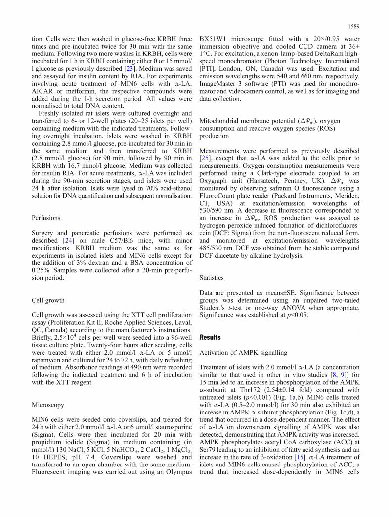
Treatment of islets with 2.0 mmol/l α-LA (a concentrationsimilar to that used in other in vitro studies [8, 9]) for15 min led to an increase in phosphorylation of the AMPKα-subunit at Thr172 (2.54±0.14 fold) compared withuntreated islets (p<0.001) (Fig. 1a,b). MIN6 cells treatedwith α-LA (0.5–2.0 mmol/l) for 30 min also exhibited anincrease in AMPK α-subunit phosphorylation (Fig. 1c,d), atrend that occurred in a dose-dependent manner. The effectof α-LA on downstream signalling of AMPK was alsodetected, demonstrating that AMPK activity was increased.AMPK phosphorylates acetyl CoA carboxylase (ACC) atSer79 leading to an inhibition of fatty acid synthesis and anincrease in the rate of β-oxidation [15]. α-LA treatment ofislets and MIN6 cells caused phosphorylation of ACC, atrend that increased dose-dependently in MIN6 cells
1589
(Fig. 1). Figure 1e shows that AMPK is inhibited byglucose, and that α-LA activated AMPK at both low(2.8 mmol/l) and high (15 mmol/l) glucose concentrations.
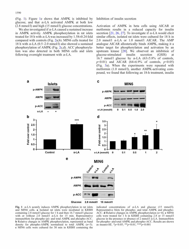
We also investigated if α-LA caused a sustained increasein AMPK activity. AMPK phosphorylation in rat isletstreated for 18 h with α-LAwas increased by 1.58±0.24 foldcompared with controls (Fig. 2a,b). MIN6 cells treated for18 h with α-LA (0.5–2.0 mmol/l) also showed a sustainedphosphorylation of AMPK (Fig. 2c,d). ACC phosphoryla-tion was also detected in both MIN6 cells and isletsfollowing overnight treatment with α-LA.
Inhibition of insulin secretion
Activation of AMPK in beta cells using AICAR ormetformin results in a reduced capacity for insulinsecretion [21, 26, 27]. To investigate if α-LA would elicitsimilar effects, isolated rat islets were cultured for 18 h in2.0 mmol/l α-LA or 1.0 mmol/l AICAR. The AMPanalogue AICAR allosterically binds AMPK, making it abetter target for phosphorylation and activation by anupstream kinase [28]. We observed an inhibition ofglucose-stimulated insulin secretion (GSIS) at16.7 mmol/l glucose by α-LA (63±5.8% of controls,p<0.01) and AICAR (64±6.9% of controls, p<0.05)(Fig. 3a). When the experiments were repeated withmetformin (1.0 mmol/l), another AMPK-activating com-pound, we found that following an 18-h treatment, insulin
Fig. 1 α-LA acutely induces AMPK phosphorylation in rat isletsand MIN6 cells. a Isolated rat islets were incubated in KRBHcontaining 2.8 mmol/l glucose for 1 h and then 16.7 mmol/l glucosewith or without 2.0 mmol/l α-LA for 15 min. Representativeimmunoblots for phospho (p)- and total AMPK and phospho-ACC.b Relative changes in AMPK phosphorylation, represented as banddensity for phospho-AMPK normalised to total AMPK (n=3).c MIN6 cells were cultured for 30 min in KRBH containing the
indicated concentrations of α-LA and glucose (15 mmol/l).Representative blots for phospho- and total AMPK and phospho-ACC. d Relative changes in AMPK phosphorylation (n=4). e MIN6cells were treated for 1 h in KRBH containing 2.8 or 15 mmol/lglucose in the presence or absence of 2 mmol/l α-LA. Immunoblotsfor phospho- and total AMPK and phospho-ACC. Results are shownas means±SE. *p<0.05; **p<0.01; ***p<0.001
1590
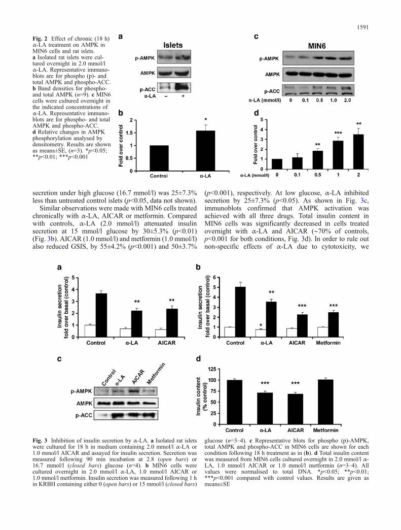
secretion under high glucose (16.7 mmol/l) was 25±7.3%less than untreated control islets (p<0.05, data not shown).
Similar observations were made with MIN6 cells treatedchronically with α-LA, AICAR or metformin. Comparedwith controls, α-LA (2.0 mmol/l) attenuated insulinsecretion at 15 mmol/l glucose by 30±5.3% (p<0.01)(Fig. 3b). AICAR (1.0 mmol/l) and metformin (1.0 mmol/l)also reduced GSIS, by 55±4.2% (p<0.001) and 50±3.7%
(p<0.001), respectively. At low glucose, α-LA inhibitedsecretion by 25±7.3% (p<0.05). As shown in Fig. 3c,immunoblots confirmed that AMPK activation wasachieved with all three drugs. Total insulin content inMIN6 cells was significantly decreased in cells treatedovernight with α-LA and AICAR (∼70% of controls,p<0.001 for both conditions, Fig. 3d). In order to rule outnon-specific effects of α-LA due to cytotoxicity, we
Fig. 2 Effect of chronic (18 h)α-LA treatment on AMPK inMIN6 cells and rat islets.a Isolated rat islets were cul-tured overnight in 2.0 mmol/lα-LA. Representative immuno-blots are for phospho (p)- andtotal AMPK and phospho-ACC.b Band densities for phospho-and total AMPK (n=9). c MIN6cells were cultured overnight inthe indicated concentrations ofα-LA. Representative immuno-blots are for phospho- and totalAMPK and phospho-ACC.d Relative changes in AMPKphosphorylation analysed bydensitometry. Results are shownas means±SE, (n=3). *p<0.05;**p<0.01; ***p<0.001
Fig. 3 Inhibition of insulin secretion by α-LA. a Isolated rat isletswere cultured for 18 h in medium containing 2.0 mmol/l α-LA or1.0 mmol/l AICAR and assayed for insulin secretion. Secretion wasmeasured following 90 min incubation at 2.8 (open bars) or16.7 mmol/l (closed bars) glucose (n=4). b MIN6 cells werecultured overnight in 2.0 mmol/l α-LA, 1.0 mmol/l AICAR or1.0 mmol/l metformin. Insulin secretion was measured following 1 hin KRBH containing either 0 (open bars) or 15 mmol/l (closed bars)
glucose (n=3–4). c Representative blots for phospho (p)-AMPK,total AMPK and phospho-ACC in MIN6 cells are shown for eachcondition following 18 h treatment as in (b). d Total insulin contentwas measured from MIN6 cells cultured overnight in 2.0 mmol/l α-LA, 1.0 mmol/l AICAR or 1.0 mmol/l metformin (n=3–4). Allvalues were normalised to total DNA. *p<0.05; **p<0.01;***p<0.001 compared with control values. Results are given asmeans±SE
1591
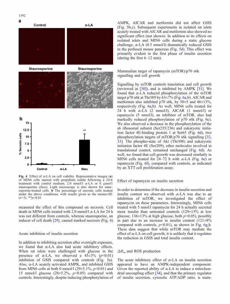
measured the effect of this compound on necrosis. Celldeath in MIN6 cells treated with 2.0 mmol/l α-LA for 24 hwas not different from controls, whereas staurosporine, aninducer of cell death [29], caused marked necrosis (Fig. 4).
Acute inhibition of insulin secretion
In addition to inhibiting secretion after overnight exposure,we found that α-LA also had acute inhibitory effects.When rat islets were challenged with glucose in thepresence of α-LA, we observed a 45±3% (p<0.01)inhibition of GSIS compared with controls (Fig. 5a).Also, α-LA acutely activated AMPK, and inhibited GSISfrom MIN6 cells at both 0 mmol/l (29±5.1%, p<0.01) and15 mmol/l glucose (28±5.2%, p<0.05) compared withcontrols. Interestingly, despite inducing phosphorylation of
AMPK, AICAR and metformin did not affect GSIS(Fig. 5b,c). Subsequent experiments in isolated rat isletsacutely treated with AICAR and metformin also showed nosignificant effect (not shown). In addition to its effects onisolated islets and MIN6 cells during a static glucosechallenge, α-LA (0.5 mmol/l) dramatically reduced GSISin the perfused mouse pancreas (Fig. 5d). This effect wasprimarily evident in the first phase of insulin secretion(during the first 6–12 min).
Mammalian target of rapamycin (mTOR)/p70 s6ksignalling and cell growth
Signalling by mTOR controls translation and cell growth(reviewed in [30]), and is inhibited by AMPK [31]. Wefound that α-LA reduced phosphorylation of the mTORtarget p70 s6k at Thr389 by 63±7% (Fig. 6a,b). AICAR andmetformin also inhibited p70 s6k, by 50±5 and 46±13%,respectively (Fig. 6a,b). As well, MIN6 cells treated for24 h with α-LA (2 mmol/l), AICAR (1 mmol/l) orrapamycin (5 nmol/l), an inhibitor of mTOR, also hadmarkedly reduced phosphorylation of p70 s6k (Fig. 6c).We also observed a decrease in the phosphorylation of thes6 ribosomal subunit (Ser235/236) and eukaryotic initia-tion factor 4E-binding protein 1 at Ser65 (Fig. 6d), twophosphorylation targets of mTOR/p70 s6k signalling [32,33]. The phospho-state of Akt (Thr308) and eukaryoticinitiation factor 4E (Ser209), other molecules involved intranslational control, remained unchanged (Fig. 6d). Aswell, we found that cell growth was decreased similarly inMIN6 cells treated for 24–72 h with α-LA (Fig. 6e) orrapamycin (Fig. 6f), compared with controls, as indicatedby an XTT cell proliferation assay.
Effect of rapamycin on insulin secretion
In order to determine if the decrease in insulin secretion andinsulin content we observed with α-LA was due to aninhibition of mTOR, we investigated the effect ofrapamycin on these parameters. Interestingly, MIN6 cellstreated with 5 nmol/l rapamycin for 24 h actually secretedmore insulin than untreated controls (129±13% at lowglucose, 136±13% at high glucose, both p<0.05), possiblyin part due to an increase in insulin content (121±8%compared with controls, p<0.01), as shown in Fig. 6g,h.These data suggest that while mTOR may mediate theeffect of α-LA on cell growth, it is unlikely that it regulatesthe reduction in GSIS and total insulin content.
ΔΨm and ROS production
The acute inhibitory effect of α-LA on insulin secretionappeared to have an AMPK-independent component.Given the reported ability of α-LA to induce a mitochon-drial uncoupling effect [34], and that the primary regulatorof insulin secretion, cytosolic ATP:ADP ratio, is main-
Fig. 4 Effect of α-LA on cell viability. Representative images (a)of MIN6 cells stained with propidium iodide following a 24-htreatment with control medium, 2.0 mmol/l α-LA or 6 μmol/lstaurosporine (Stau). Light microscopy is also shown for staur-osporine-treated cells. b The percentage of necrotic cells treatedunder the above conditions, with results given as the means±SE(n=3). **p<0.01
1592
tained by mitochondrial oxidative phosphorylation [35],we tested the effect of this compound on mitochondrialfunction in permeabilised MIN6 cells. Oxidative phos-phorylation was not affected by α-LA (200 μmol/l), butmitochondrial Ca2+ handling, which is another importantmitochondrial function, was clearly dependent on α-LA.Incremental addition of Ca2+ to respiring mitochondriacauses acceleration of oxygen consumption and membranedepolarisation due to Ca2+ uptake into the matrix [25], bothof which were more pronounced in the presence of α-LA(Fig. 7a,b,d). Both parameters could be reverted to initiallevels with cyclosporin A, a classic inhibitor of Ca2+-induced mitochondrial permeabilisation (so-called mito-chondrial permeability transition [MPT] [36]). MPT is anopening of non-specific pores in the mitochondrial innermembrane and is induced by Ca2+ in the presence of avariety of co-inducers, such as inorganic phosphate, thioloxidants and fatty acids (reviewed in [36]). Next, weconsidered how α-LA might influence MPT opening.Since MPT can be induced by ROS [36], and α-LA canbehave as both a pro-oxidant [34] and inducer of MPT [37],we tested the effect of α-LA on mitochondrial ROSproduction. Indeed, kinetics of respiration-dependent DCFoxidation (Fig. 7c,e), which reflects oxygen radicalproduction in the respiratory chain, demonstrated increased
mitochondrial ROS production in the presence of α-LA(200 μmol/l). Thus, our data suggest that α-LA can elevatethe mitochondrial level of ROS and probability of MPTopening.
Discussion
In this report, we have demonstrated several novelproperties of α-LA, including the activation of AMPK inrat islets and MIN6 cells. The role of α-LA as a regulator ofAMPK has only recently been demonstrated. Whereas Kimet al. [18] showed that this compound inhibited hypotha-lamic AMPK activity, we have shown the opposite to betrue in beta cells. Our observation of AMPK activation byα-LA in pancreatic beta cells suggests that AMPK isresponsible for some of the reported effects found with α-LA in other tissues. For example, α-LA facilitates glucoseuptake in both skeletal muscle and adipose tissue in vitro[8, 9]. Indeed, others have demonstrated that AMPKactivation in these tissues leads to improved insulinsensitivity and enhanced glucose uptake and utilisation[13, 15–17]. Furthermore, both AMPK activation [14, 17]and α-LA [10] are associated with suppression of hepaticglucose output. Very recently, two reports described that
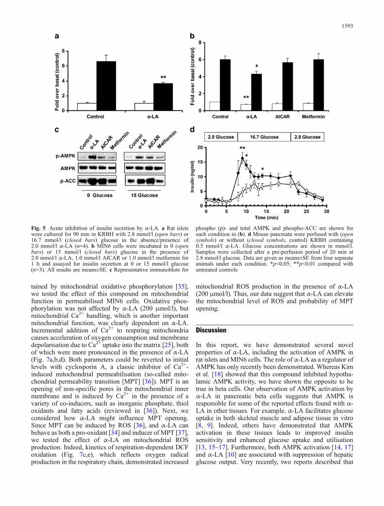
Fig. 5 Acute inhibition of insulin secretion by α-LA. a Rat isletswere cultured for 90 min in KRBH with 2.8 mmol/l (open bars) or16.7 mmol/l (closed bars) glucose in the absence/presence of2.0 mmol/l α-LA (n=4). b MIN6 cells were incubated in 0 (openbars) or 15 mmol/l (closed bars) glucose in the presence of2.0 mmol/l α-LA, 1.0 mmol/l AICAR or 1.0 mmol/l metformin for1 h and assayed for insulin secretion at 0 or 15 mmol/l glucose(n=3). All results are means±SE. c Representative immunoblots for
phospho (p)- and total AMPK and phospho-ACC are shown foreach condition in (b). d Mouse pancreata were perfused with (opensymbols) or without (closed symbols, control) KRBH containing0.5 mmol/l α-LA. Glucose concentrations are shown in mmol/l.Samples were collected after a pre-perfusion period of 20 min at2.8 mmol/l glucose. Data are given as means±SE from four separateanimals under each condition. *p<0.05; **p<0.01 compared withuntreated controls
1593
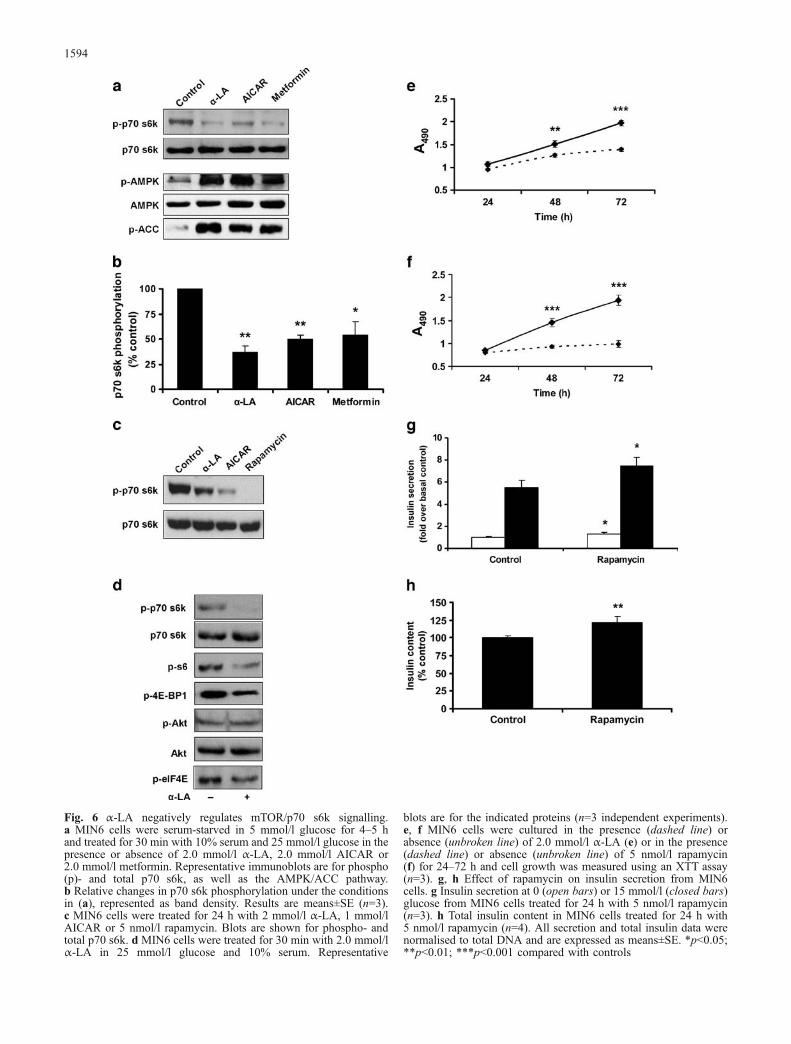
Fig. 6 α-LA negatively regulates mTOR/p70 s6k signalling.a MIN6 cells were serum-starved in 5 mmol/l glucose for 4–5 hand treated for 30 min with 10% serum and 25 mmol/l glucose in thepresence or absence of 2.0 mmol/l α-LA, 2.0 mmol/l AICAR or2.0 mmol/l metformin. Representative immunoblots are for phospho(p)- and total p70 s6k, as well as the AMPK/ACC pathway.b Relative changes in p70 s6k phosphorylation under the conditionsin (a), represented as band density. Results are means±SE (n=3).c MIN6 cells were treated for 24 h with 2 mmol/l α-LA, 1 mmol/lAICAR or 5 nmol/l rapamycin. Blots are shown for phospho- andtotal p70 s6k. d MIN6 cells were treated for 30 min with 2.0 mmol/lα-LA in 25 mmol/l glucose and 10% serum. Representative
blots are for the indicated proteins (n=3 independent experiments).e, f MIN6 cells were cultured in the presence (dashed line) orabsence (unbroken line) of 2.0 mmol/l α-LA (e) or in the presence(dashed line) or absence (unbroken line) of 5 nmol/l rapamycin(f) for 24–72 h and cell growth was measured using an XTT assay(n=3). g, h Effect of rapamycin on insulin secretion from MIN6cells. g Insulin secretion at 0 (open bars) or 15 mmol/l (closed bars)glucose from MIN6 cells treated for 24 h with 5 nmol/l rapamycin(n=3). h Total insulin content in MIN6 cells treated for 24 h with5 nmol/l rapamycin (n=4). All secretion and total insulin data werenormalised to total DNA and are expressed as means±SE. *p<0.05;**p<0.01; ***p<0.001 compared with controls
1594
α-LA acted as an insulin sensitiser in skeletal muscle [38],and prevented endothelial cell dysfunction in obese rats[39], effects shown to be attributable to AMPK activation.Thus, it is likely that AMPK is responsible for many of thediverse antidiabetic effects of this compound.
The beneficial properties of α-LA, in particular as anantioxidant [1, 3, 4] and stimulator of glucose uptake, makethis a potentially useful agent for diabetes therapy.However, we have shown here that α-LA inhibits insulinsecretion in vitro from MIN6 cells and isolated rat islets.These findings are supported by other reports describingsimilar inhibitory effects with metformin or AICAR [21,26, 27]. It is likely that the negative regulation of insulinsecretion and positive effect on insulin sensitivity by thesethree drugs are mediated by AMPK. Several studies haveshown that AMPK suppresses insulin secretion andsynthesis in beta cells [19, 21, 22], consistent with itsrole as an energy-conserving enzyme. There are a numberof possible mechanisms by which α-LA may inhibit insulinsecretion through AMPK. This enzyme has been shown tohave an important role in suppressing preproinsulinpromoter activity [21]. It has also been demonstrated that
activation of AMPK leads to modulation of secretoryvesicle dynamics [22]. More recently, Richards et al. [40]showed that mouse islets infected with a constitutivelyactive AMPK α-subunit had reduced glucose oxidationand insulin secretion, and were associated with poorerglycaemic control when transplanted into streptozotocin-diabetic mice compared with islets infected with null ordominant-negative AMPK viruses.
AMPK is an important regulator of the cellular responseto nutrient availability, including protein translation andcell growth, and when activated, AMPK inhibits mTOR/p70 s6k signalling [31]. However, whether mTOR isinvolved in AMPK-mediated inhibition of insulin secretionis currently not known. Consistent with a positive role forp70 s6k in insulin secretion, mice lacking this enzyme haveimpaired GSIS and reduced beta cell mass [41]. We haveshown in this report that α-LA inhibits mTOR/p70 s6ksignalling and cell growth in MIN6 cells, and reduces GSISand insulin content. While rapamycin and α-LA bothcaused inhibitory effects on cell growth and p70 s6kphosphorylation, the effect we observed with rapamycin onGSIS—a stimulatory one—is not consistent with a positive
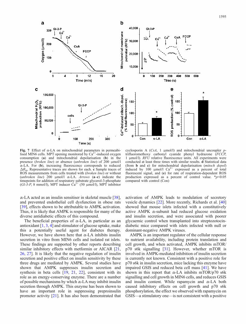
Fig. 7 Effect of α-LA on mitochondrial parameters in permeabi-lised MIN6 cells. MPT opening monitored by Ca2+-induced oxygenconsumption (a) and mitochondrial depolarisation (b) in thepresence (broken line) or absence (unbroken line) of 200 μmol/lα-LA. For (b), increasing fluorescence corresponds to reducedΔΨm. Representative traces are shown for each. c Sample traces ofROS measurements from cells treated with (broken line) or without(unbroken line) 200 μmol/l α-LA. Arrows (a–c) indicate thetimepoints for addition of respiratory substrate glycerol-3-phosphate(Gl-3-P, 8 mmol/l), MPT inducer Ca2+ (50 μmol/l), MPT inhibitor
cyclosporin A (CsA, 1 μmol/l) and mitochondrial uncoupler p-trifluoromethoxy carbonyl cyanide phenyl hydrazone (FCCP,1 μmol/l). RFU relative fluorescence units. All experiments wereconducted at least three times with similar results. d Statistical data(from b and c) for mitochondrial depolarisation (mitoch depol)induced by 100 μmol/l Ca2+ expressed as a percent of totalfluorescent signal, and (e) for rate of respiration-dependent ROSproduction expressed as a percent of control value. *p<0.05compared with control (Con)
1595
role for mTOR in insulin secretion. In agreement with ourdata, one report demonstrated that rapamycin improvedinsulin secretion in vivo in dogs following islet autografts[42]. While others have shown that rapamycin hasinhibitory effects in rat islets, these observations weremade only at high doses and treatment times of greater than3 days [43]. Another report demonstrated that rapamycintreatment (48 h) was detrimental to GSIS from HIT-T15cells, but not rat islets treated for 24 h [44]. Thesediscrepancies may be due to differential sensitivity torapamycin in assorted cell lines or animal species, orvariations in experimental protocols. The fact that p70 s6kknock-out mice have reduced GSIS [41] may be areflection of a total-body loss of this enzyme and thus islikely to include contributions from the effects of p70 s6kdeficiency in other tissues, including enhanced insulinsensitivity in the periphery [45]. More studies are requiredin order to clearly understand if rapamycin and/or mTORexhibit any significant effect on insulin secretion, and ifAMPK is involved in this process.
We demonstrated here that in addition to prolongedexposure to α-LA, acute treatment also inhibited insulinsecretion from isolated rat islets, MIN6 cells and theperfused mouse pancreas. Previous reports [19, 21]suggested that acute AMPK activation inhibits GSIS.Although we cannot completely rule out this possibility,our data suggest that acute activation of AMPK was notexclusively responsible for our observations, given thatAMPK and ACC phosphorylation were induced byAICAR, and to a lesser extent, metformin, yet thesedrugs did not have an acute inhibitory effect on insulinsecretion (Fig. 5b,c). It is possible that potential non-specific effects of AICAR, or differences in cell lines orexperimental protocols, could account for the differencesobserved here compared with those of the Hardie [19] andRutter [21] laboratories. Our data indicate that α-LA mayacutely inhibit insulin secretion via an alternative routeinvolving direct actions on beta cell mitochondria—aprocess consisting of increased ROS production andinduction of MPT opening. This would partially depolarisethe mitochondria, thereby reducing the proton-motive forceand thus ATP synthesis and, consequently, insulin secre-tion. Observations of increased ROS in isolated rat soleusmuscle treated with α-LA [34], and ROS-induced MPT inrat liver mitochondria by α-LA [37], support our data thatthis compound can possess pro-oxidant properties. Ourfindings are important, given the clinical relevance of α-LA as an antioxidant [5]. It is possible that at particularconcentrations, or in the absence of other pro-oxidants, α-LA itself acts as a pro-oxidant, whereas under conditions ofoxidative stress, such as diabetes [5], this compoundexhibits beneficial protective properties. As such, theclinical usefulness of α-LA may be contingent upon aparticular pathophysiological state. It is not known if α-LA-induced ROS generation or mitochondrial depolarisa-tion serves as a stimulus for AMPK activation by anupstream kinase such as LKB1 [12] or Ca2+/calmodulin-dependent protein kinase kinase [46], but this remains aninteresting possibility. Figure 8 depicts a potential scheme
by which α-LA exerts its multiple effects on beta cellfunction.
It can be argued that caution should be used whenconsidering AMPK activators such as α-LA and metforminfor the treatment of diabetes; there appears to be a trade-offof increased insulin sensitivity for reduced insulin secre-tion. However, simultaneous activation of AMPK incanonical insulin-sensitive tissues and the beta cell in aphysiological state may be beneficial, as improved insulinuse is accompanied by a smaller demand for insulinproduction, which may protect beta cells from exhaustion.Furthermore, while AMPK-activating compounds arethought to exert negative effects on GSIS under standardculture conditions, several reports suggest that AMPK canbe protective in beta cells under pathological conditions.Marchetti et al. [47] demonstrated that metformin restoresbeta cell function and prevents apoptosis in diabetic humanislets, while others reported beneficial properties forAICAR in clonal beta cells under lipotoxic conditions[48]. Whether α-LA behaves in a similar manner is aninteresting possibility, especially if this compound wouldact as an antioxidant under these conditions.
In conclusion, our data reveal several novel pharmaco-logical properties of α-LA, including the inhibition ofinsulin secretion from rat islets, the perfused mousepancreas and MIN6 cells. α-LA also acts as a signallingmolecule, activating AMPK and inhibiting p70 s6k andsubsequently cell growth. α-LA directly modulates themitochondrial state via an acute increase in ROS produc-tion and depolarisation of the mitochondrial membrane.Our findings provide some insight into a possibleexplanation for the insulin-sensitising properties of α-LA,which may occur in peripheral tissues in an AMPK-dependent manner. Further studies on α-LA and itsrelationship with AMPK are needed to gain a better
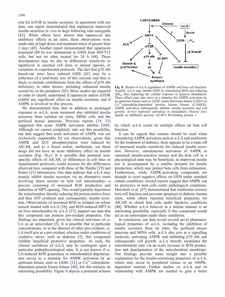
Fig. 8 Model of α-LA regulation of AMPK and beta cell function.Acutely, α-LA may inhibit GSIS by stimulating ROS and reducingΔΨm, thus impairing the cellular response to glucose stimulation.These effects may also serve as a stimulus for AMPK activation byan upstream kinase such as LKB1 serine/threonine kinase (LKB1) orCa2+/calmodulin-dependent protein kinase kinase (CAMKK).AMPK activation subsequently inhibits insulin secretion and cellgrowth. Arrows represent activation or stimulation; blunted linessignify an inhibitory process. 4E-BP1 4E-binding protein 1
1596

understanding of the antidiabetic properties of this mole-cule and the precise mechanism by which AMPK regula-tion occurs.
Acknowledgements This work was supported by a research grantfrom the Canadian Institutes of Health Research (CIHR) to M.B.Wheeler (MOP-12898). M. B. Wheeler is a CIHR investigator. E. D.Targonsky is supported by a research scholarship from the NaturalSciences and Engineering Research Council of Canada. F. Dai issupported by a CIHR New Emerging Teams Grant in DiabetesComplications (NET-54012). We thank G. Bikopoulos for hisassistance with islet isolations and A. Klip for her valuable adviceregarding experimental design.
References
1. Lovell MA, Xie C, Xiong S, Markesbery WR (2003) Protectionagainst amyloid beta peptide and iron/hydrogen peroxidetoxicity by alpha lipoic acid. J Alzheimers Dis 5:229–239
2. Piotrowski P, Wierzbicka K, Smialek M (2001) Neuronal deathin the rat hippocampus in experimental diabetes and cerebralischaemia treated with antioxidants. Folia Neuropathol 39:147–154
3. Mervaala E, Finckenberg P, Lapatto R et al (2003) Lipoic acidsupplementation prevents angiotensin II-induced renal injury.Kidney Int 64:501–508
4. Midaoui AE, Elimadi A, Wu L, Haddad PS, de Champlain J(2003) Lipoic acid prevents hypertension, hyperglycemia, andthe increase in heart mitochondrial superoxide production. AmJ Hypertens 16:173–179
5. Bitar MS, Wahid S, Pilcher CW, Al Saleh E, Al Mulla F (2004)Alpha-lipoic acid mitigates insulin resistance in Goto–Kakizakirats. Horm Metab Res 36:542–549
6. Khamaisi M, Potashnik R, Tirosh A et al (1997) Lipoic acidreduces glycemia and increases muscle GLUT4 content instreptozotocin-diabetic rats. Metabolism 46:763–768
7. Konrad T, Vicini P, Kusterer K et al (1999) alpha-Lipoic acidtreatment decreases serum lactate and pyruvate concentrationsand improves glucose effectiveness in lean and obese patientswith type 2 diabetes. Diabetes Care 22:280–287
8. Konrad D, Somwar R, Sweeney G et al (2001) The antihyper-glycemic drug alpha-lipoic acid stimulates glucose uptake viaboth GLUT4 translocation and GLUT4 activation: potentialrole of p38 mitogen-activated protein kinase in GLUT4activation. Diabetes 50:1464–1471
9. Yaworsky K, Somwar R, Ramlal T, Tritschler HJ, Klip A(2000) Engagement of the insulin-sensitive pathway in thestimulation of glucose transport by alpha-lipoic acid in 3T3-L1adipocytes. Diabetologia 43:294—303
10. Blumenthal SA (1984) Inhibition of gluconeogenesis in ratliver by lipoic acid. Evidence for more than one site of action.Biochem J 219:773—780
11. Hardie DG (2004) The AMP-activated protein kinasepathway—new players upstream and downstream. J CellSci 117:5479–5487
12. Shaw RJ, Kosmatka M, Bardeesy N et al (2004) The tumorsuppressor LKB1 kinase directly activates AMP-activatedkinase and regulates apoptosis in response to energy stress.Proc Natl Acad Sci U S A 101:3329–3335
13. Mu J, Brozinick JT Jr, Valladares O, Bucan M, Birnbaum MJ(2001) A role for AMP-activated protein kinase in contraction-and hypoxia-regulated glucose transport in skeletal muscle.Mol Cell 7:1085–1094
14. Iglesias MA, Ye JM, Frangioudakis G et al (2002) AICARadministration causes an apparent enhancement of muscle andliver insulin action in insulin-resistant high-fat-fed rats.Diabetes 51:2886–2894
15. Minokoshi Y, Kim YB, Peroni OD et al (2002) Leptinstimulates fatty-acid oxidation by activating AMP-activatedprotein kinase. Nature 415:339–343
16. Yamauchi T, Kamon J, Minokoshi Y et al (2002) Adiponectinstimulates glucose utilization and fatty-acid oxidation byactivating AMP-activated protein kinase. Nat Med 8:1288–1295
17. Zhou G, Myers R, Li Y et al (2001) Role of AMP-activatedprotein kinase in mechanism of metformin action. J Clin Invest108:1167–1174
18. Kim MS, Park JY, Namkoong C et al (2004) Anti-obesityeffects of alpha-lipoic acid mediated by suppression ofhypothalamic AMP-activated protein kinase. Nat Med10:727–733
19. Salt IP, Johnson G, Ashcroft SJ, Hardie DG (1998) AMP-activated protein kinase is activated by low glucose in cell linesderived from pancreatic beta cells, and may regulate insulinrelease. Biochem J 335:533–539
20. Da Silva Xavier G, Leclerc I, Salt IP et al (2000) Role of AMP-activated protein kinase in the regulation by glucose of isletbeta cell gene expression. Proc Natl Acad Sci U S A 97:4023–4028
21. Da Silva Xavier G, Leclerc I, Varadi A, Tsuboi T, Moule SK,Rutter GA (2003) Role for AMP-activated protein kinase inglucose-stimulated insulin secretion and preproinsulin geneexpression. Biochem J 371:761–774
22. Tsuboi T, Da Silva Xavier G, Leclerc I, Rutter GA (2003) 5′-AMP-activated protein kinase controls insulin-containing se-cretory vesicle dynamics. J Biol Chem 278:52042–52051
23. Wang X, Li H, De Leo D et al (2004) Gene and protein kinaseexpression profiling of reactive oxygen species-associatedlipotoxicity in the pancreatic beta-cell line MIN6. Diabetes53:129–140
24. Heron-Milhavet L, Haluzik M, Yakar S et al (2004) Muscle-specific overexpression of CD36 reverses the insulin resistanceand diabetes of MKR mice. Endocrinology 145:4667–4676
25. Koshkin V, Bikopoulos G, Chan CB, Wheeler MB (2004) Thecharacterization of mitochondrial permeability transition inclonal pancreatic beta-cells. Multiple modes and regulation.J Biol Chem 279:41368–41376
26. Leclerc I, Woltersdorf WW, Da Silva Xavier G et al (2004)Metformin, but not leptin, regulates AMP-activated proteinkinase in pancreatic islets: impact on glucose-stimulated insulinsecretion. Am J Physiol Endocrinol Metab 286:E1023–E1031
27. Kefas BA, Cai Y, Kerckhofs K et al (2004) Metformin-inducedstimulation of AMP-activated protein kinase in beta-cellsimpairs their glucose responsiveness and can lead to apoptosis.Biochem Pharmacol 68:409–416
28. Corton JM, Gillespie JG, Hawley SA, Hardie DG (1995) 5-aminoimidazole-4-carboxamide ribonucleoside. A specificmethod for activating AMP-activated protein kinase in intactcells? Eur J Biochem 229:558–565
29. Scarlett JL, Sheard PW, Hughes G, Ledgerwood EC, Ku HH,Murphy MP (2000) Changes in mitochondrial membranepotential during staurosporine-induced apoptosis in Jurkatcells. FEBS Lett 475:267–272
30. Rowinsky EK (2004) Targeting the molecular target ofrapamycin (mTOR). Curr Opin Oncol 16:564–575
31. Inoki K, Zhu T, Guan KL (2003) TSC2 mediates cellularenergy response to control cell growth and survival. Cell115:577–590
32. Brown EJ, Beal PA, Keith CT, Chen J, Shin TB, Schreiber SL(1995) Control of p70 s6 kinase by kinase activity of FRAP invivo. Nature 377:441–446
33. Hara K, Yonezawa K, Kozlowski MT et al (1997) Regulation ofeIF-4E BP1 phosphorylation by mTOR. J Biol Chem272:26457–26463
34. Dicter N, Madar Z, Tirosh O (2002) Alpha-lipoic acid inhibitsglycogen synthesis in rat soleus muscle via its oxidative activityand the uncoupling of mitochondria. J Nutr 132:3001–3006
1597
35. Maechler P, Wollheim CB (2001) Mitochondrial function innormal and diabetic beta-cells. Nature 414:807–812
36. Bernardi P, Scorrano L, Colonna R, Petronilli V, Di Lisa F(1999) Mitochondria and cell death. Mechanistic aspects andmethodological issues. Eur J Biochem 264:687–701
37. Saris NE, Karjalainen A, Teplova VV, Lindros KO (1998) Thestimulation of the mitochondrial permeability transition bydihydrolipoate and alpha-lipoate. Biochem Mol Biol Int44:127–134
38. Lee WJ, Song KH, Koh EH et al (2005) Alpha-lipoic acidincreases insulin sensitivity by activating AMPK in skeletalmuscle. Biochem Biophys Res Commun 332:885–891
39. Lee WJ, Lee IK, Kim HS et al (2005) Alpha-lipoic acidprevents endothelial dysfunction in obese rats via activation ofAMP-activated protein kinase. Arterioscler Thromb Vasc Biol25:2488–2494
40. Richards SK, Parton LE, Leclerc I, Rutter GA, Smith RM(2005) Over-expression of AMP-activated protein kinaseimpairs pancreatic {beta}-cell function in vivo. J Endocrinol187:225–235
41. Pende M, Kozma SC, Jaquet M et al (2000) Hypoinsulinaemia,glucose intolerance and diminished beta-cell size in S6K1-deficient mice. Nature 408:994–997
42. Kneteman NM, Lakey JR, Wagner T, Finegood D (1996) Themetabolic impact of rapamycin (sirolimus) in chronic canineislet graft recipients. Transplantation 61:1206–1210
43. Bell E, Cao X, Moibi JA et al (2003) Rapamycin has adeleterious effect on MIN-6 cells and rat and human islets.Diabetes 52:2731–2739
44. Paty BW, Harmon JS, Marsh CL, Robertson RP (2002)Inhibitory effects of immunosuppressive drugs on insulinsecretion from HIT-T15 cells and Wistar rat islets. Transplan-tation 73:353–357
45. Um SH, Frigerio F, Watanabe M et al (2004) Absence of S6K1protects against age- and diet-induced obesity while enhancinginsulin sensitivity. Nature 431:200–205
46. Hawley SA, Pan DA, Mustard KJ et al (2005) Calmodulin-dependent protein kinase kinase-beta is an alternative upstreamkinase for AMP-activated protein kinase. Cell Metab 2:9–19
47. Marchetti P, Del Guerra S, Marselli L et al (2004) Pancreaticislets from type 2 diabetic patients have functional defects andincreased apoptosis that are ameliorated by metformin. J ClinEndocrinol Metab 89:5535–5541
48. Yamashita T, Eto K, Okazaki Y et al (2004) Role of uncouplingprotein-2 up-regulation and triglyceride accumulation inimpaired glucose-stimulated insulin secretion in a beta-celllipotoxicity model overexpressing sterol regulatory element-binding protein-1c. Endocrinology 145:3566–3577
1598