JPET Fast Forward. Published on August 11, 2009 as...
55
JPET #152975 1 TITLE: GSI-953 (begacestat): A novel, selective thiophene sulfonamide inhibitor of APP -secretase for the treatment of Alzheimer’s disease Robert L. Martone, Hua Zhou, Kevin Atchison, Thomas Comery, Jane Z. Xu, Xinyi Huang, Xioahai Gong, Mei Jin, Anthony Kreft, Boyd Harrison, Scott C. Mayer, Suzan Aschmies, Cathleen Gonzales, Margaret M. Zaleska, David R. Riddell, Erik Wagner, Peimin Lu, Shaiu-Ching Sun, June Sonnenberg-Reines, Aram Oganesian, Karissa Adkins, Michael Leach, David W. Clarke, Donna Huryn, Magid Abou-Gharbia, Ronald Magolda, Jonathan Bard, Glen Frick, Sangeeta Raje, S. Bradley Forlow, Carrie Balliet, Michael E. Burczynski, Peter H. Reinhart, Hong I. Wan, Menelas N. Pangalos, J. Steven Jacobsen* Wyeth Research, Departments of Discovery Neuroscience (R.L.M., H.Z., K.A., T.C., J.Z.X., X.G., M.J., S.A., C.G., M.M.Z., D.R.R., E.W., P.L., R.S., J.S.-R., J.B., P.H.R., M.N.P., J.S.J.) and Chemical Screening Sciences (X.H., A.K., B.H., S.M. D.H., M.A.-G., R.M.), CN-8000, Princeton, NJ 08543; Exploratory Drug Safety (K.A., M.L.), One Burtt Road, Andover, MA 01810; Drug Safety and Metabolism (D.W.C.), 461 Ridge Road, Chazy, NY 12921; and Drug Safety and Metabolism (A.O.), Early Development and Clinical Pharmacology (G.F., S.R.), and Clinical Translational Medicine (S.B.F., C.B., M.E.B., H.I.W.), 500 Arcola Road, Collegeville, PA 19426 JPET Fast Forward. Published on August 11, 2009 as DOI:10.1124/jpet.109.152975 Copyright 2009 by the American Society for Pharmacology and Experimental Therapeutics. This article has not been copyedited and formatted. The final version may differ from this version. JPET Fast Forward. Published on August 11, 2009 as DOI: 10.1124/jpet.109.152975 at ASPET Journals on June 6, 2018 jpet.aspetjournals.org Downloaded from
-
Upload
phunghuong -
Category
Documents
-
view
214 -
download
1
Transcript of JPET Fast Forward. Published on August 11, 2009 as...
JPET #152975
1
TITLE: GSI-953 (begacestat): A novel, selective thiophene sulfonamide inhibitor of
APP γ-secretase for the treatment of Alzheimer’s disease
Robert L. Martone, Hua Zhou, Kevin Atchison, Thomas Comery, Jane Z. Xu, Xinyi
Huang, Xioahai Gong, Mei Jin, Anthony Kreft, Boyd Harrison, Scott C. Mayer, Suzan
Aschmies, Cathleen Gonzales, Margaret M. Zaleska, David R. Riddell, Erik Wagner,
Peimin Lu, Shaiu-Ching Sun, June Sonnenberg-Reines, Aram Oganesian, Karissa
Adkins, Michael Leach, David W. Clarke, Donna Huryn, Magid Abou-Gharbia, Ronald
Magolda, Jonathan Bard, Glen Frick, Sangeeta Raje, S. Bradley Forlow, Carrie Balliet,
Michael E. Burczynski, Peter H. Reinhart, Hong I. Wan, Menelas N. Pangalos, J. Steven
Jacobsen*
Wyeth Research, Departments of Discovery Neuroscience (R.L.M., H.Z., K.A., T.C.,
J.Z.X., X.G., M.J., S.A., C.G., M.M.Z., D.R.R., E.W., P.L., R.S., J.S.-R., J.B., P.H.R.,
M.N.P., J.S.J.) and Chemical Screening Sciences (X.H., A.K., B.H., S.M. D.H., M.A.-G.,
R.M.), CN-8000, Princeton, NJ 08543; Exploratory Drug Safety (K.A., M.L.), One Burtt
Road, Andover, MA 01810; Drug Safety and Metabolism (D.W.C.), 461 Ridge Road,
Chazy, NY 12921; and Drug Safety and Metabolism (A.O.), Early Development and
Clinical Pharmacology (G.F., S.R.), and Clinical Translational Medicine (S.B.F., C.B.,
M.E.B., H.I.W.), 500 Arcola Road, Collegeville, PA 19426
JPET Fast Forward. Published on August 11, 2009 as DOI:10.1124/jpet.109.152975
Copyright 2009 by the American Society for Pharmacology and Experimental Therapeutics.
This article has not been copyedited and formatted. The final version may differ from this version.JPET Fast Forward. Published on August 11, 2009 as DOI: 10.1124/jpet.109.152975
at ASPE
T Journals on June 6, 2018
jpet.aspetjournals.orgD
ownloaded from
JPET #152975
2
Running Title:
GSI-953: A novel inhibitor of γ-secretase
*Address correspondence to:
J. Steven Jacobsen
Discovery Neuroscience
Wyeth Research
CN-8000
Princeton, NJ 08543
732-274-4238 (Phone)
732-274-4755 (FAX)
email: [email protected]
Text Pages: 38
Tables: 7
Figures: 4
References: 40
Abstract (words): 243
Introduction (words): 634
Discussion (words): 1559
This article has not been copyedited and formatted. The final version may differ from this version.JPET Fast Forward. Published on August 11, 2009 as DOI: 10.1124/jpet.109.152975
at ASPE
T Journals on June 6, 2018
jpet.aspetjournals.orgD
ownloaded from
JPET #152975
3
Abbreviations: αAPPs, APP α-secretase-cleaved soluble fragment; Aβ, β-amyloid
peptide; AD, Alzheimer’s disease; APP, Amyloid Precursor Protein; βCTF, APP β-
secretase-cleaved carboxy-terminal fragment; PS, Presenilin; GS, γ-secretase; GSI, γ-
secretase inhibitor; Amgen GSI, 4-fluoro-N-[(1R,2R,4S)-1,3,3-
trimethylbicyclo[2.2.1]hept-2-yl]benzenesulfonamide;
BMS GSI, (2R)-2-{[5-chloro-2-(hydroxymethyl)phenyl][(4-
chlorophenyl)sulfonyl]amino}propylpropylcarbamate; DAPT, [N-(3,5-difluoro-
phenylacetyl)L-alanyl)-S-phenylglycine; GSI-953, 5-chloro-N-[(1S)-3,3,3-trifluoro-1-
(hydroxymethyl)-2- (trifluoromethyl)propyl] thiophene-2-sulfonamide; GSI-953 analog,
5-chloro-N-[(1S,2R)-4,4,4-trifluoro-1-(hydroxyl[3H2]methyl)-2-methylbutyl]thiophene-2-
sulfonamide; LY411575, N2-[(2S)-2-(3,5-difluorophenyl)-2-hydroxyethanoyl]-N1-[(7S)-
5-methyl-6-oxo-6,7-dihydro-5H-dibenzo[b,d]azepin-7-yl]-L-alaninamide; LY450139,
(2S)-2-hydroxy-3-methyl-N-((1S)-1-methyl-2-{[(1S)-3-methyl-2-oxo-2,3,4,5- tetrahydro-
1H-3-benzazepin-1-yl]amino}-2-oxoethyl)butanamide; CFC, contextual fear
conditioning; NICD, Notch intracellular domain; SEAP, secreted alkaline phosphatase
Recommended Section: Neuropharmacology
This article has not been copyedited and formatted. The final version may differ from this version.JPET Fast Forward. Published on August 11, 2009 as DOI: 10.1124/jpet.109.152975
at ASPE
T Journals on June 6, 2018
jpet.aspetjournals.orgD
ownloaded from
JPET #152975
4
ABSTRACT
The presenilin containing γ-secretase complex is responsible for the regulated
intramembraneous proteolysis of the Amyloid Precursor Protein (APP), the Notch
receptor, and a multitude of other substrates. Gamma secretase catalyzes the final step in
the generation of Aβ40 and Aβ42 peptides from APP. Aβ peptides aggregate to form
neurotoxic oligomers, senile plaques and congophilic angiopathy, some of the cardinal
pathologies associated with Alzheimer’s disease (AD). While inhibition of this protease
acting on APP may result in potentially therapeutic reductions of neurotoxic Aβ peptides,
non-selective inhibition of the enzyme may cause severe adverse events as a result of
impaired Notch receptor processing. Here, we report the preclinical pharmacological
profile of GSI-953 (begacestat), a novel thiophene sulfonamide γ-secretase inhibitor
(GSI) that selectively inhibits cleavage of APP over Notch. This GSI inhibits Aβ
production with low nanomolar potency in cellular and cell-free assays of γ-secretase
function, and displaces a tritiated analog of GSI-953 from enriched γ-secretase enzyme
complexes with similar potency. Cellular assays of Notch cleavage reveal that this
compound is approximately 16-fold selective for the inhibition of APP cleavage. In the
human APP overexpressing Tg2576 transgenic mouse, treatment with this orally active
compound results in a robust reduction in brain, plasma, and CSF Aβ levels, and a
reversal of contextual fear conditioning deficits that are correlated with Aβ load. In
healthy human volunteers, oral administration of a single dose of GSI-953 produces dose-
dependent changes in plasma Aβ levels, confirming pharmacodynamic activity of GSI-
953 in humans.
This article has not been copyedited and formatted. The final version may differ from this version.JPET Fast Forward. Published on August 11, 2009 as DOI: 10.1124/jpet.109.152975
at ASPE
T Journals on June 6, 2018
jpet.aspetjournals.orgD
ownloaded from
JPET #152975
5
Introduction
Alzheimer’s disease (AD) is a progressive neurodegenerative disorder
characterized by amyloid β-peptide (Aβ) deposition and amyloidosis of the brain
parenchyma and vasculature, as well as neuronal inclusions of hyperphosphorylated tau.
Aβ is derived from the amyloid precursor protein (APP) by the sequential proteolysis of
β-secretase (BACE) and γ-secretase (GS) (Tanzi and Bertram, 2005). Aβ peptides
aggregate to form soluble oligomers as well as insoluble fibrils and plaques. While
soluble oligomers are reported to be neurotoxic (Walsh et al., 2002; Klyubin et al., 2005),
deposits of amyloid fibrils elicit reactive gliosis, as well as neuroinflammatory responses,
and may compromise the integrity of the blood-brain barrier. Therefore, the reduction of
Aβ levels by secretase inhibition is a widely pursued therapeutic strategy for AD (Tomita
and Iwatsubo, 2004; Jacobsen et al., 2005).
GS is a membrane-associated complex containing presenilin (PS), a protease that
belongs to a family of enzymes that mediate regulated intramembraneous proteolysis, and
that includes rhomboid, signal peptide peptidase and site 2 protease. PS is unique in this
family of proteases in that it requires cofactors for enzymatic activity. Nicastrin, Aph-1
and pen-2 are additional essential components of GS and are required for reconstituted
GS activity (Edbauer et al., 2003; Kimberly et al., 2003), while CD147 (Zhou et al.,
2005) and TMP21 (Chen et al., 2006) are GS associated regulatory proteins. Along with
APP, more than 20 other GS substrates have been reported (Parks and Curtis, 2007)
including the Notch receptors (DeStrooper et al., 1999). While the consequences of
inhibiting GS processing of many of these substrates are currently unknown, the
inhibition of Notch processing is known to have specific adverse events (Geling et al.,
This article has not been copyedited and formatted. The final version may differ from this version.JPET Fast Forward. Published on August 11, 2009 as DOI: 10.1124/jpet.109.152975
at ASPE
T Journals on June 6, 2018
jpet.aspetjournals.orgD
ownloaded from
JPET #152975
6
2002; Van den Brandt et al., 2004). GS cleavage of the Notch receptor is required for the
release of the Notch intracellular domain (NICD) that is translocated to the nucleus where
it regulates HES-driven gene transcription. PS knock-out yields an embryonic lethal
phenotype that closely resembles Notch 1 knockout (Shen et al., 1997). Peptidomimetic
GS inhibitors (GSIs), such as DAPT (Dovey et al., 2001) and LY4115751, potently
inhibit Notch processing and consequently alter Notch signaling causing embryonic
defects in zebrafish, and altered thymocyte differentiation, gastroenteric disorders
(mucoid enteropathy and goblet cell dysplasia), and skin lesions (Van den Brandt et al.,
2004; Geling et al., 2002; Wong et al., 2004).
There are several strategies to alter the generation of Aβ by GS while limiting the
liabilities associated with inhibition of Notch processing. Cleavage site modulators of GS
such as flurbiprofen can either affect the ability of a transition state analogue to bind in
the absence of substrate (Beher et al., 2004), or bind APP and alter the substrate cleavage
site, resulting in reduced levels of the reportedly more toxic Aβ42 isoform, and increased
levels of the non-toxic Aβ38 isoform. Hence, modulators such as Flurbiprofen may cause
no net alterations in total levels of Aβ or other secretase products such as the NICD
(Kukar et al., 2008). Additionally, there are modulators of GS substrate selectivity, that
are based on the requirement of nucleotide binding to the GS enzyme complex for APP
processing, but not for Notch processing. Some protein kinase inhibitors that bind and
displace nucleotides from the GS complex can modulate APP binding to the GS enzyme,
and thereby decrease Aβ production while leaving Notch processing unaltered (Fraering
et al., 2005). Finally, several allosteric inhibitors of GS that have APP selective
properties, but do not alter Notch processing have been reported (Barten et al., 2005), and
This article has not been copyedited and formatted. The final version may differ from this version.JPET Fast Forward. Published on August 11, 2009 as DOI: 10.1124/jpet.109.152975
at ASPE
T Journals on June 6, 2018
jpet.aspetjournals.orgD
ownloaded from
JPET #152975
7
strategies to identify safe therapeutic windows for these compounds are being
investigated (Choi and Norstrom, 2007; Hyde et al., 2006).
Here we report the pharmacological properties of a novel thiophene sulfonamide
GSI, GSI-953 (Figure 1A), an APP-selective GSI that is well-tolerated in mouse and dog
toxicity studies and has been advanced to human clinical trials.
This article has not been copyedited and formatted. The final version may differ from this version.JPET Fast Forward. Published on August 11, 2009 as DOI: 10.1124/jpet.109.152975
at ASPE
T Journals on June 6, 2018
jpet.aspetjournals.orgD
ownloaded from
JPET #152975
8
Materials and Methods
APP stable cell line. The characteristics of a CHO-cell line stably expressing an
APP reporter construct containing the Swedish KM/NL mutation (APP-Rep-751NL) with
an amino-terminal deletion were reported previously (Jacobsen et al., 1994). Cells were
plated in 96-well plates and allowed to adhere overnight in Dulbecco's modified Eagle
medium (DMEM, Invitrogen, Carlsbad, CA) supplemented with 10% certified fetal
bovine serum. For compound testing, compounds were diluted from stock solutions in
dimethylsulfoxide (DMSO, Sigma Aldrich, St. Louis, MO) to yield a final concentration
of 0.1% DMSO in media. Cells were treated for 24 h at 37oC.
Cellular Aβ assay. Aβ levels in conditioned media were measured by sandwich
Aβ40 and Aβ42 end-specific ELISA using monoclonal antibody 6E10 (Signet/Covance
Labs, Dedham, MA) for capture, and rabbit C-terminal specific antibodies to Aβ40 or
Aβ42 (Biosource, Camarillo, CA) coupled with an alkaline phosphatase-conjugated anti-
Rabbit detection antibody and attophos (alkaline phosphatase substrate) for detection.
ELISA plates were read using a Cytofluor fluorescence plate reader to determine the
concentration of the fluorescent product of the alkaline phosphatase activity. Synthetic
Aβ40 and Aβ42 peptides (AnaSpec, St. Jose, CA) were used as standards. Reductions in
Aβ levels were measured relative to control cells treated with 0.1% DMSO and expressed
as a percent inhibition. Data from 5 doses in triplicate were fitted to a four-parameter
logistical model using LSW software to determine EC50 values. Cells were washed in
PBS and a CellTiter 96 Aqueous Non-Radioactive Cell Proliferation Assay (MTS:
Promega, Madison, WI) was used to assess cell viability.
Radiolabeled APP cellular assay. Compound activity as examined in
This article has not been copyedited and formatted. The final version may differ from this version.JPET Fast Forward. Published on August 11, 2009 as DOI: 10.1124/jpet.109.152975
at ASPE
T Journals on June 6, 2018
jpet.aspetjournals.orgD
ownloaded from
JPET #152975
9
radiolabeled APP cell assays. Cells were labeled with [35S]-methionine (NEN, Perkin
Elmer, Waltham, MA) in methionine-free Dulbecco’s Modified Eagle Medium (DMEM,
Invitrogen, Carlsbad, CA) at 37°C/5%CO2. After 3 h, the conditioned media was
removed and clarified by centrifugation at 1000 g. Immunoprecipitation buffer (IPB: 50
mM Tris HCl, pH 7.2, 150 mM NaCl, 5 mM EDTA, 0.5% IGEPAL CA-630, 0.5%
sodium deoxycholate) containing protease inhibitors (2 μg/ml pepstatin A, 50 μg/ml
leupeptin, 10 μg/ml aprotinin, 250 μg/ml PMSF) was added, and SDS was added to a
final concentration of 0.35%. Cells were lysed in IPB for 10 min on ice then centrifuged
at 14,000 g. The supernatant was collected, and SDS was added to a final concentration
of 0.35%. Conditioned media and lysates were boiled briefly, cooled on ice, then
immunoprecipitated overnight at 4°C with 6E10, an anti-Aβ monoclonal antibody linked
to protein A sepharose (PAS, Sigma Aldrich, St. Louis MO) using a rabbit anti-mouse
antibody (Jackson Immunoresearch, West Grove, PA). Samples were fractionated on
16.5% Tris-Tricine gels (Invitrogen, Carlsbad, CA), the gels dried, and 6E10
immunoprecipitated proteins were visualized by autoradiography with a Storm 860
phosphorimager (Molecular Dynamics GE Healthcare, Piscataway, NJ).
Cell-free γ-secretase cleavage assay. The EC50 value for Aβ40 lowering by GSI-
953 was determined in a cell-free assay by incubating approximately 30 μg of solubilized
GS prepared using published methods (Tian et al., 2002) with 2 μM recombinant peptide
comprising the C-terminal 100 amino acids of APP (APP-C100) in MES buffer, pH 6.5
in the presence of GSI-953 for 2 h at 37°C. The reaction mix was immunoprecipitated
overnight with an antibody to the C-terminus of APP (Sigma Aldrich, St. Louis, MO)
bound to PAS. The immune complexes were washed and denatured, and then
This article has not been copyedited and formatted. The final version may differ from this version.JPET Fast Forward. Published on August 11, 2009 as DOI: 10.1124/jpet.109.152975
at ASPE
T Journals on June 6, 2018
jpet.aspetjournals.orgD
ownloaded from
JPET #152975
10
supernatants were collected after filtration through 96-well Millipore filter plates. Aβ40
levels were determined using a 6E10 anti-Aβ40 sandwich ELISA.
Radiolabeled γ-secretase binding and displacement assay. A tritiated analog of
GSI-953 ([3H]-GSI-analog; Figure 1A) was prepared as an 11.00 mCi/ml ethanol stock
solution with specific activity of 26.6 Ci/mole and radiochemical purity of >99%. GS
enzyme was prepared as CHAPSO-solubilized microsomes from human neuroblastoma
SH-SY5Y cells. Filter plates were purchased from Millipore. Microscint-20 was
purchased from Perkin Elmer. Polyethyleneimine solution (PEI, 50% solution in water)
was purchased from Sigma Aldrich (St. Louis, MO). A Multiscreen 96-well FB filter
plate was prepared incubated with 200 μl of 0.6% (w/v) PEI solution overnight at 4°C.
The filter plate was vacuum filtered and washed twice with 200 μl of water and twice
with 200 μl of assay buffer (25 mM MES, pH 6.5, 1 mM EDTA, 0.01% β-
mercaptoethanol, 0.01% BSA). Reactions were set up in Costar 96-well clear
polypropylene plates (Corning, Corning, NY) with the following addition sequence: 160
μl γ-secretase enriched microsomes diluted 1:10 in assay buffer, 20 μl test compound
serially diluted in 10% DMSO and 90% assay buffer and 20 μl 250 nM [3H] GSI-analog
in assay buffer. Each plate contained 3 test compounds and cold-GSI-analog (as positive
control) in duplicate. The final concentrations in the reactions were 25 nM [3H] GSI-
analog, 0-2 μM or 0-10 μM test compound, 1% DMSO, and ~92 pM GS. Following a 1 h
incubation at room temperature with shaking, the 180 μl reactions were transferred to the
filter plate. The filter plate was then vacuum filtered, washed 4 times with 200 μl wash
buffer (5 mM Tris-HCl, pH 7.4) and vacuum dried for 5 min. The filter plate bottom was
removed and the plate was blotted dry with paper towel. Microscint-20 (30 μl) was added
This article has not been copyedited and formatted. The final version may differ from this version.JPET Fast Forward. Published on August 11, 2009 as DOI: 10.1124/jpet.109.152975
at ASPE
T Journals on June 6, 2018
jpet.aspetjournals.orgD
ownloaded from
JPET #152975
11
and 3H radioactivity was counted on a Wallac Model# 1450 Microbeta (Perkin Elmer,
Waltham, MA). Data were analyzed in GraphPad Prism 4.0 using the following equation:
Counts = a + (b-a)*IC50^n/(Conc^n+IC50^n), where n is the Hill number and IC50 is the
compound concentration that results in 50% competition.
Notch assay. In order to determine the effect of test compounds on Notch
signaling, we generated a constitutively active (ΔE) mouse Notch construct containing
the M1726V mutation as previously described (Kopan et al., 1996) and a reporter
construct employing secreted alkaline phosphatase (SEAP) driven by the HES1 promoter
(pSEAP-Basic vector, Clontech, Mountain View, CA). CHO K1 cells were transiently
transfected with both constructs using Polyfect (Qiagen, Valencia, CA) transfection
reagent in Opti-MEM media. Cells were plated in 96 well plates and treated with
compounds for 48 h. SEAP levels in the conditioned media were assessed using the Great
EscAPe SEAP Chemiluminescence detection kit (Clontech, Mountain View, CA)
according to manufacturer’s instruction. Briefly, 15 μl of conditioned media was mixed
with a dilution buffer and incubated at 65°C for 45 min. After cooling, assay buffer was
added, and the samples were incubated with substrate. Luminescence was measured in a
Wallac 1450 Victor luminescence counter.
Measurement of Aβ from in vivo samples. Twelve to twenty week - old Tg
2576 mice (n=7-10/condition) received oral doses of GSI-953 in Phosal PG 50:Tween
80:H20 (w:w:v-10:2:88) and were sacrificed at the times indicated. CSF was collected
from the cisterna magna and plasma was isolated from blood collected by cardiac
puncture. Brain tissue was collected and extracted in guanidine. Plasma Aβ40 and brain
and CSF Aβ40 and Aβ42 measurements were conducted by sandwich ELISA as published
This article has not been copyedited and formatted. The final version may differ from this version.JPET Fast Forward. Published on August 11, 2009 as DOI: 10.1124/jpet.109.152975
at ASPE
T Journals on June 6, 2018
jpet.aspetjournals.orgD
ownloaded from
JPET #152975
12
(Jacobsen et al., 2006). All samples were analyzed in duplicate and the average of results
from 2-3 independent experiments are reported.
In human studies, plasma was obtained following a single oral administration of
GSI-953 in healthy young subjects. Measurement of the Aβ40 concentrations in EDTA
plasma samples was performed using a validated ELISA as described by the
manufacturer (Wako Chemicals, Richmond, VA).
Contextual fear conditioning (CFC) model. We have previously reported that
Tg2576 transgenic animals exhibit an age- and Aβ-dependent deficit in CFC (Comery et
al., 2005). Following acute dosing by oral gavage with 0, 2.5, 5 or 10 mg/kg GSI-953 or
30 mg/kg of its inactive enantiomer WAY-210952 (Figure 1A), 20 week old Tg2576
mice (n=11/genotype/treatment) were trained and tested on two consecutive days as
described previously (Comery et al., 2005). Contextual freezing was analyzed using a
two-way ANOVA and post hoc pairwise comparison made using SAS Statistical
Software (SAS Institute, Inc., Cary, NC). All data is presented as mean ± SEM.
Pharmacokinetic parameters. PK parameters for GSI-953 were established in
transgenic Tg2576 or wild-type male mice following a single oral administration of
compound. Corresponding exposures to various dose levels, expressed as Cmax and AUC
0-α (area under the curve), are provided for brain and plasma of Tg2576 mice, and in
brain, cerebral spinal fluid (CSF) and plasma of wild-type mice (see Table 5).
Toxicity studies in vivo. GSI-953 was administered orally to Sprague-Dawley
rats at dosages of 0, 200, 600, or 2000 mg/kg/day for 10 (5 males/group and 5 females at
600 mg/kg/day) or 28 (10/sex/group) consecutive days. The control groups of male and
female rats received the vehicle (2.0% polysorbate 80 and 0.5% methylcellulose in
This article has not been copyedited and formatted. The final version may differ from this version.JPET Fast Forward. Published on August 11, 2009 as DOI: 10.1124/jpet.109.152975
at ASPE
T Journals on June 6, 2018
jpet.aspetjournals.orgD
ownloaded from
JPET #152975
13
water). Evaluations consisted of mortality, clinical observations, body weight,
hematology, clinical chemistry, organ weights, and macroscopic and microscopic
examinations. A full tissue list was examined in the toxicity studies, including the
lymphoid tissues (gut associated lymphoid tissue, spleen, mesenteric lymph node,
mandibular lymph node, and thymus), and the digestive tract [tongue, esophagus,
stomach (squamous and glandular), duodenum, jejunum, ileum, cecum and colon].
Thymocytes were examined using flow cytometry for proportions of single-positive (SP)
CD4+ or CD8+, and double-positive (DP) CD4+/CD8+ at the end of the 10-day study.
Evaluation of peripheral blood CD4, CD8 and CD45 was conducted at the end of the 28-
day study. The plasma pharmacokinetics of GSI-953 were determined in satellite groups
in both studies.
This article has not been copyedited and formatted. The final version may differ from this version.JPET Fast Forward. Published on August 11, 2009 as DOI: 10.1124/jpet.109.152975
at ASPE
T Journals on June 6, 2018
jpet.aspetjournals.orgD
ownloaded from
JPET #152975
14
Results
Optimization of a phenylsulfonamide lead obtained from high-throughput
screening from Wyeth and ArQule (Woburn, MA) compound libraries (Kreft et al., 2008)
led to the identification of GSI-953 (Mayer et al., 2008; Fig. 1A).
Reduction of Aβ production in a cell-based assay. GSI-953 is a potent inhibitor
of both Aβ40 and Aβ42 production in a cell line that stably expresses human recombinant
APP. Average EC50 values of 14.8 and 12.4 nM were determined for the lowering of
Aβ40 and Aβ42, respectively (Fig. 1B and Table 1). There was no cell toxicity (assessed by
MTS assay) observed at concentrations up to 30 μM (data not shown). Treatment with
WAY-210952 (Figure 1A), the inactive enantiomer of GSI-953, displayed very low
potency for Aβ reduction with EC50 values of ~10,000 and ~18,000 nM, respectively, for
the lowering of Aβ40 and Aβ42 (Fig. 1B and Table 1). Several benchmark GSIs including
DAPT (Dovey et al., 2001), LY4115751, LY4501392, L685458 (Shearman et al., 2000)
and DuPont E (Seiffert et al., 2000) were evaluated to compare potency and are
summarized in Table 1.
Alteration of APP processing is consistent with the inhibition of GS activity.
To demonstrate that lowering of Aβ levels in the cellular model is attributed to the
inhibition of GS activity, we performed radiolabeled continuous pulse cellular assays to
assess compound effects on APP processing (Fig. 1C and Table 2). Treatment with GSI-
953 demonstrated a dose-dependent reduction in total Aβ levels (EC50 value for Aβ
lowering was 7.3 nM). Treatment with WAY-210952 (inactive enantiomer) resulted in no
reduction of Aβ levels (data not shown). These compounds displayed little or no effect on
levels of the full length APP or the APP soluble fragment that is secreted following
This article has not been copyedited and formatted. The final version may differ from this version.JPET Fast Forward. Published on August 11, 2009 as DOI: 10.1124/jpet.109.152975
at ASPE
T Journals on June 6, 2018
jpet.aspetjournals.orgD
ownloaded from
JPET #152975
15
cleavage by α-secretase (αAPPs). It was further observed that treatment with GSI-953,
but not WAY-210952 (inactive enantiomer), caused a dose-dependent increase in levels
of APP β-secretase-cleaved carboxy-terminal fragment (βCTF), the substrate that is
otherwise cleaved by GS (EC50 value for βCTF increase was 6.6 nM). Similar
observations are reported with additional reference GSIs in Table 2. These observations
suggest that the Aβ reductions in the cellular model are due to alterations in APP
processing rather than changes in total protein synthesis, transport or secretion, and that
inhibition of GS activity results in both the accumulation of βCTF substrate and reduction
of Aβ production.
Inhibition of enriched GS activity in a cell-free system. To support binding and
inhibitor displacement studies, we confirmed that GSI-953 binds GS in vitro by
demonstrating the inhibition of GS proteolytic activity in microsome preparations. The
inhibitory effects of GSI-953 in this cell-free system were demonstrated by the dose-
dependent reduction of Aβ40 generated by the cleavage of APP-C100, a βCTF-like
recombinant protein (EC50 value for the reduction of Aβ40 was 16.5 nM; Fig. 1D). This
observation supports the conclusion that GSI-953 is an inhibitor of GS activity in both
cell-free and cellular models, and confirms that GSI-953 readily permeates cell
membranes.
In vitro binding and displacement studies. To determine whether GSI-953 binds
to sites on GS, we performed displacement studies to compare the binding affinity of
GSI-953 with previously described reference compounds (Fig. 1E and Table 3). The
direct interaction of GSI-953 on GS was confirmed by displacement assays using a
tritiated structural analog to GSI-953. GSI-953 displaced the [3H]-GSI-analog (Figure
This article has not been copyedited and formatted. The final version may differ from this version.JPET Fast Forward. Published on August 11, 2009 as DOI: 10.1124/jpet.109.152975
at ASPE
T Journals on June 6, 2018
jpet.aspetjournals.orgD
ownloaded from
JPET #152975
16
1A) with an IC50 value of 8 nM (Fig. 1E). In contrast, WAY-210952 (inactive
enantiomer) did not displace the [3H]-GSI-analog (IC50 value >10,000 nM). A total of 30
GSI-953 analogs and reference GSIs including DAPT, LY411575, LY450139, DuPont E,
Amgen GSI (Rishton et al., 2000) and BMS GSI3 were tested in the displacement assay.
Unlike GSI-953, the transition state inhibitor L685458 only partially displaced the [3H]-
GSI-analog (Fig. 1E) suggesting that GSI-953 interacts with GS at a site removed from
the catalytic domain (Tian et al. 2002). When IC50 values from displacement studies of
these GSIs were plotted against the EC50 values for Aβ42 reduction in the Aβ cellular
assay, a linear relationship was demonstrated (r2 = 0.99, Fig. 1F), suggesting that the
binding of GSI to GS is directly linked to the inhibition of Aβ42 activity in the cell-based
assay.
Notch selectivity studies in vitro. To demonstrate the selectivity of GSI-953 for
inhibiting GS activity and the cleavage of different substrates, we compared the
inhibition of APP processing (reduction of Aβ) to that of Notch processing (reduction of
NICD). Consistent with its function as a GSI, high concentrations of GSI-953 were
needed to inhibit Notch processing in a dose-dependent manner using a cellular assay that
detects a chemiluminescent signal generated by the cleavage of Notch substrate and the
translocation of NICD into the nucleus (Fig. 1G and Table 4). However, GSI-953 was
significantly more selective against Notch signaling in this assay than the reference GSI,
DAPT, with EC50 values of 208.5 and 14.9 nM, respectively (Table 4). As a measure of
selectivity, a ratio was calculated comparing the EC50 value for Notch inhibition to the
EC50 value for reduction of Aβ42. Using this analysis GSI-953 is 16.8-fold selective (e.g.,
208.5/12.4, Table 4) and preferentially inhibits APP processing by GS. In contrast, DAPT
This article has not been copyedited and formatted. The final version may differ from this version.JPET Fast Forward. Published on August 11, 2009 as DOI: 10.1124/jpet.109.152975
at ASPE
T Journals on June 6, 2018
jpet.aspetjournals.orgD
ownloaded from
JPET #152975
17
is 0.5-fold selective (e.g., 14.9/27.7) and inhibits Notch processing by GS. Both
LY411575 and LY450139 are potent GSIs for Aβ42 reduction, but lack Notch selectivity
with ratios of 0.9 and 0.8, respectively (Table 4). These selectivity data suggest that GSI-
953 may provide little or no alteration of Notch processing by the inhibition of GS
activity in comparison to non-selective reference GSIs evaluated.
GSI-953 reduces Aβ levels in the brains of transgenic APP mice. In vivo
efficacy of GSI-953 was first demonstrated in the transgenic APP (Tg2576) mouse model
with the lowering of Aβ40 and Aβ42 levels in brain and CSF, and Aβ40 levels in plasma
(Fig. 2A). GSI-953 was orally administered at a single high dose (100 mg/kg, n=20 per
time point) and Aβ levels measured in brain, CSF and plasma at 0, 2, 4 and 6 hr post-
dosing. Aβ40 was reduced ~88% in both CSF (p < 0.001, Fig. 2A, center panel) and
plasma (p < 0.001, Fig. 2A, bottom panel) by 2 h (p < 0.001), while brain Aβ40 levels
(Fig. 2A, top panel) reached a maximum reduction of ~60% at 6 h (p < 0.001).
Subsequently, a 24 h time course experiment was conducted following a single
oral dose (30 mg/kg) to determine the optimal time of compound exposure for Aβ
lowering (Fig. 2B). Following compound administration, Aβ was reduced from 0.25 to
10 h (p < 0.001) in brain with maximal Aβ reduction of approximately 67% (Aβ40) and
52% (Aβ42) between 4 and 6 h. Efficacy and compound exposure (data not shown) were
tightly correlated and a 4 h terminal time point was used for subsequent studies. The
finding that brain Aβ levels were significantly lowered within 15 min following the
administration of GSI-953 demonstrated that the compound was rapidly absorbed, able to
cross the blood-brain barrier (BBB), and inhibit de novo synthesis of Aβ.
A dose-response study to measure Aβ levels 4 h following a single oral
This article has not been copyedited and formatted. The final version may differ from this version.JPET Fast Forward. Published on August 11, 2009 as DOI: 10.1124/jpet.109.152975
at ASPE
T Journals on June 6, 2018
jpet.aspetjournals.orgD
ownloaded from
JPET #152975
18
administration of GSI-953 was performed (Fig. 2C). The minimal efficacious dose
(MED) was determined to be 1 mg/kg with statistically significant lowering of Aβ40 (p <
0.01); a significant lowering of both Aβ40 and Aβ42 was observed at 2.5 mg/kg (p < 0.05).
GSI-953 reverses contextual fear conditioning deficits in Tg2576 mice. To
examine the ability of GSI-953 to reverse CFC deficits, we used contextual fear
conditioning (CFC), a test of hippocampal-dependent learning and memory in young
(pre-plaque) Tg2576 mice as previously described (Comery et al., 2005; Jacobsen et al.,
2006). Contextual learning involves the association of an aversive stimulus (footshock)
with a novel testing environment (context). In this model, memory is expressed as a
context-dependent freezing behavior in the absence of the shock 24 h post learning.
Vehicle-dosed Tg2576 mice displayed significant deficits compared to vehicle-treated
WT control animals (p < 0.05, Fig. 3). However, treatment of Tg2576 mice with GSI-953
resulted in a dose-dependent reversal of these deficits when compound is orally
administered 3 h prior to training. Significant deficits were observed following treatment
with 2.5 mg/kg of GSI-953 (p < 0.05), and there was some reversal of this at 5 mg/kg and
full reversal at 10 mg/kg compared to vehicle-dosed Tg2576 mice (p < 0.05). WAY-
210952 (inactive enantiomer of GSI-953) was not active at 30 mg/kg and did not reverse
deficits in the CFC test (Fig. 3). Neither compound displayed effects on CFC
performance in WT animals. These results demonstrate that GSI-953 is efficacious for
improving CFC deficits in the APP transgenic mouse model, consistent with previous
observations in Tg2576 mice showing that administration of DAPT also completely
reverses such deficits (Comery et al., 2005).
This article has not been copyedited and formatted. The final version may differ from this version.JPET Fast Forward. Published on August 11, 2009 as DOI: 10.1124/jpet.109.152975
at ASPE
T Journals on June 6, 2018
jpet.aspetjournals.orgD
ownloaded from
JPET #152975
19
Pharmacokinetic parameters and efficacious exposure. Cmax and AUC
exposures of GSI-953 concentrations achieved in mice at various dose levels are shown
in Table 5. These PK parameters in brain, CSF and plasma compartments suggest that
GSI-953 readily penetrated the BBB and achieved a brain/plasma ratio of approximately
1 or more at all doses evaluated. GSI-953 exposure increased with a dose-proportional
manner in all three compartments. There was no accumulation of compound based on a
comparison of single or multiple daily dosing at 2.5 mg/kg. The MED, based on the
lowering of brain Aβ40 levels in Tg2576 mice (see above), was 1 mg/kg and resulted in a
plasma Cmax=101 ng/mL (AUC0-α=246 hr•ng/mL) and a brain Cmax=128 ng/g (AUC0-α
=258 hr•ng/gm) at 4 h.
Lack of Notch-related toxicity in rats and dogs following GSI-953
administration. GSI-953-related effects on thymic lymphocyte populations were
evaluated in a 10-day rat study to demonstrate that thymocyte maturation was not
inhibited (data not shown). A dosage-related trend of slightly lower percentages (%) of
single-positive (SP) CD4+ cells in males at all dosages (SP CD4+ cells = ~11 % in
controls compared with ~7% to ~9% in GSI-953-dosed animals) and females at
2000 mg/kg/day (SP CD4+ cells = ~10% in controls compared with ~8% in GSI-953-
dosed animals) was observed. These differences occurred concomitantly with a dosage-
related higher proportion of double positive (DP) CD4+/CD8+ precursor cells in males at
all dosages (DP CD4+/CD8+ cells = ~80% in controls compared to ~84 to ~86% in GSI-
953-dosed animals). These changes were statistically significant compared with the
controls. However, differences in absolute counts of these thymocyte subtypes were not
statistically significant between GSI-953-dosed and control groups. The percentage of
This article has not been copyedited and formatted. The final version may differ from this version.JPET Fast Forward. Published on August 11, 2009 as DOI: 10.1124/jpet.109.152975
at ASPE
T Journals on June 6, 2018
jpet.aspetjournals.orgD
ownloaded from
JPET #152975
20
thymic CD45+ (CD3) B cells were negligible for all groups on study. The GSI-953-
related alterations in relative lymphocyte populations occurred in the absence of changes
in absolute cell numbers/g tissue. There were no microscopic correlates for the altered
percentage of thymic lymphocyte populations. The toxicological significance of the
changes in subtype proportions is not clear, but they are not consistent with Notch
inhibition as reported (Hadland et al., 2001) where the inhibition of progression from
immature double-negative (DN) CD4-/CD8- precursor cells to intermediate DP
CD4+/CD8+ precursor cells was observed. In the 28-day study, no GSI-953-related effect
on peripheral blood lymphocyte subtype counts or on the CD4/CD8 ratio was observed.
GSI-953 was tolerated when administered to male and female rats at dosages of up to
2000 mg/kg/day for 10 or 28 days, and there were no dose-limiting effects.
Slight to mild GSI-953-related hypertrophy and hyperplasia of the mucous cells
of the small and large intestine was observed in the 28-day study throughout the small
and large intestines, and was more pronounced in the large intestine (data not shown).
The mucous cell changes were present at all GSI-953 dosage levels in the cecum and
colon, at 600 and 2000 mg/kg/day in the duodenum, and at 2000 mg/kg/day in the
jejunum and ileum. Slight to mild colonic glandular dilation was seen at 600 and 2000
mg/kg/day. There were no clinical signs associated with the intestinal changes.
After 10 days, mean body weight gain was lower (21% and 29%, respectively) in
groups of males at 600 and 2000 mg/kg/day, compared with controls; statistically
significant lower final body weights were observed for these same groups compared with
the control group (8% and 10%, respectively). Weight loss was not observed. After 28
days of dosing, mean body weights were lower (9%) in the group of males at 2000
This article has not been copyedited and formatted. The final version may differ from this version.JPET Fast Forward. Published on August 11, 2009 as DOI: 10.1124/jpet.109.152975
at ASPE
T Journals on June 6, 2018
jpet.aspetjournals.orgD
ownloaded from
JPET #152975
21
mg/kg/day and in groups of females (4% and 8%, respectively) at 600 and 2000
mg/kg/day, compared with control groups.
In the 10-day study, exposure to GSI-953 increased with increasing dosage in a
less than dose-proportional manner in male rats. On Day 10, the WAY-210953 mean
(± SE) Cmax values were 636 ± 208, 1981 ± 333 and 2100 ± 568 ng/mL in male rats given
200, 600 and 2000 mg/kg/day, respectively. The corresponding mean (± SE) AUC0-24
values were 4145 ± 453, 6991 ± 977 and 19233 ± 4398 ng•hr/mL, respectively. In female
rats given 600 mg/kg/day, the mean (± SE), Cmax and AUC0-24 values were 5098 ± 2662
ng/mL and 22102 ± 2608 ng•hr/mL, respectively. The Cmax was significantly higher
(approximately 3 times) in female rats at 600 mg/kg/day (the only dosage evaluated)
compared with male rats. All these values were much higher than the plasma Cmax of
101 ng/mL (AUC=2460-α hr•ng/mL) following the MED dosage of 1 mg/kg in Tg2576
mice.
In the 28-day study, exposure to GSI-953 increased with increasing dosage in an
approximately dose-proportional manner in male and female rats. On Day 28, the mean
(± SE) Cmax values were 252 ± 85, 2050 ± 641, and 4646 ± 1178 ng/mL in male rats, and
2497 ± 537, 11205 ± 6845, and 22855 (n=2) ng/mL in female rats. The corresponding
mean (± SE) AUC0-24 values were 1073 ± 164, 5407 ± 551, and 13074 ± 1268 ng•hr/mL
in males, and 4418 ± 501, 18272 ± 4076, and 31316 ± 10510 ng•hr/mL in females,
respectively. Exposure was higher in females compared to males. In conclusion, GSI-
953 was tolerated when administered to male and female rats at dosages of up to 2000
mg/kg/day for 10 or 28 days, and there were no dose-limiting effects. A maximum
This article has not been copyedited and formatted. The final version may differ from this version.JPET Fast Forward. Published on August 11, 2009 as DOI: 10.1124/jpet.109.152975
at ASPE
T Journals on June 6, 2018
jpet.aspetjournals.orgD
ownloaded from
JPET #152975
22
tolerated dose (MTD) was not achieved and was considered to be > 2000 mg/kg/day, the
highest dosage tested.
Neither 13-week nor 52-week repeat-dose toxicity studies in dog revealed
microscopic changes in the gastrointestinal (GI) tract (data not shown). Peak plasma
exposure of GSI-953 was 13 µM, a plasma exposure which is ~50-fold higher than the
plasma Cmax for an efficacious Aβ–lowering dosage of GSI-953 (1 mg/kg is 258 nM) and
~65-fold higher than the plasma Cmax for the in vitro EC50 for Notch inhibition (208.5
nM). However, in a short-term 14-day repeat dose toxicity study, potential Notch-related
effects were observed in the GI tract including fecal alterations (e.g., soft, liquid, mucoid
feces) clinically with slight to moderate crypt abscess and dilated mucosal glands
observed microscopically in the duodenum and colon, respectively, that are considered
adverse at dosages that are above those that are clinically relevant. Microscopic changes
in the GI tract were observed at dosages with peak plasma concentrations at ~23 µM, a
plasma exposure which is ~100-fold higher than the plasma Cmax for both an efficacious
Aβ–lowering dosage of GSI-953 and the in vitro EC50 for Notch inhibition. In a 7-day
study comparing GSI-953 and WAY-201952 (an inactive enantiomer of GSI-953 which
has >1000-fold less activity towards γ-secretase and Notch), similar clinical and
microscopic changes described for the 14-day study are observed for GSI-953, but not the
inactive enantiomer, indicating that the GI effects may be related to either γ-secretase or
Notch inhibition (data not shown).
GSI-953 demonstrates target engagement in human. After performing dose-
ranging and IND-enabling toxicity studies in mice, rats and dogs, and demonstrating the
lack of Notch-related adverse events in vivo, we initiated a first-in-human single
This article has not been copyedited and formatted. The final version may differ from this version.JPET Fast Forward. Published on August 11, 2009 as DOI: 10.1124/jpet.109.152975
at ASPE
T Journals on June 6, 2018
jpet.aspetjournals.orgD
ownloaded from
JPET #152975
23
ascending dose study with healthy subjects to investigate safety and tolerability of GSI-
953 (to be published elsewhere). Following single oral doses of GSI-953 (3-600 mg), to
healthy young subjects (ages 18-55 years), plasma samples were collected over a 24 h
period and assayed for Aβ40 levels (Fig. 4 and Table 7). The lowering of plasma Aβ40
levels, as measured by the mean percent change from baseline, was observed in a dose-
dependent manner. Transient reductions of plasma Aβ40 levels were observed at 1 h for
the 10-600 mg doses, while more sustained reductions were observed at 2 h for the 150-
600 mg doses, and at 4 h for the 600 mg dose. This initial reduction was followed by a
subsequent increase in plasma Aβ40 levels (data not shown).
This article has not been copyedited and formatted. The final version may differ from this version.JPET Fast Forward. Published on August 11, 2009 as DOI: 10.1124/jpet.109.152975
at ASPE
T Journals on June 6, 2018
jpet.aspetjournals.orgD
ownloaded from
JPET #152975
24
Discussion
GS is widely regarded as a viable target to achieve therapeutically relevant
reductions of Aβ in AD, and multiple classes of GSIs have been reported including
peptidomimetics and sulfonamides (for review, see Imbimbo, 2008). Here, we report the
preclinical pharmacological and safety profile, as well as clinical biomarker data for GSI-
953, a novel thiophene sulfonamide GSI that was observed to selectively inhibit the
cleavage of APP over Notch. We have shown that GSI-953 is a potent inhibitor of Aβ
production in vitro, and in the transgenic APP Tg2576 mouse model, is orally active and
displays a robust reduction in brain, plasma and CSF Aβ levels, and reverses contextual
fear conditioning deficits that are correlated with Aβ load. In healthy volunteers, oral
administration of a single dose of GSI-953 produced dose-dependent changes in plasma
Aβ levels, confirming target engagement of GSI-953 in humans.
GSI-953 was shown to inhibit Aβ production in a cellular assay with low
nanomolar potency (Aβ40 EC50 = 14.8 nM, Aβ42 EC50 = 12.4 nM) with a corresponding
increase in cellular βCTF levels, a property shared by other benchmark GSIs. The
inhibition of cleavage of recombinant GS substrate in a cell-free microsome system
coupled with the ability to displace a tritiated analog of GSI-953 that was established to
bind to GS, and the linear relationship between GS binding and the reduction of Aβ
levels, confirms that GSI-953 inhibits GS activity. The treatment of transgenic APP
Tg2576 mice with GSI-953 caused a rapid dose-dependent reduction of Aβ40 and Aβ42
levels in the brains of these mice that correlated with changes in both CSF and plasma Aβ
levels. GSI-953 time course studies revealed significant reductions in brain Aβ levels as
early as 15 min after administration of compound, indicating rapid compound absorption,
This article has not been copyedited and formatted. The final version may differ from this version.JPET Fast Forward. Published on August 11, 2009 as DOI: 10.1124/jpet.109.152975
at ASPE
T Journals on June 6, 2018
jpet.aspetjournals.orgD
ownloaded from
JPET #152975
25
robust inhibition of GS, and turnover of Aβ in the CNS. Brain Aβ reductions extended for
24 h, with maximum inhibition observed at 4-6 h. We performed additional GSI-953
dose-response studies, collected tissue samples at 4 h, and observed a significant
reduction of brain Aβ40 levels following a single oral dosage of 1 mg/kg. Similar
observations of dose-dependent reductions in brain or CSF Aβ levels have been
demonstrated for other GSIs, including LY411575 in Tg2576 mice (Lanz et al., 2004)
and rats (Best et al., 2005), and for LY450139 (Lanz et al., 2006) in guinea pig.
Since Aβ is believed to have a detrimental effect on cognition (Klyubin et al.,
2005; Jacobsen et al., 2006), we predicted that reduction in brain Aβ levels mediated by
GSI-953 inhibition of GS would have cognitive enhancing effects. We observed that
contextual fear conditioning (CFC) deficits in the Tg2576 transgenic model were
partially reversed 3 h following a single oral dosage of GSI-953 at 5 mg/kg, a dose that
lowered parenchymal Aβ levels by 25-35%, but were completely reversed 3 h following
the administration of GSI-953 at a dosage of 10 mg/kg. It is possible that the
pharmacokinetic and pharmacodynamic relationship for demonstrating the lowering of
brain Aβ levels may not optimally translate for improvement in CFC deficits. For
example, more robust reversal of these deficits at lower doses of GSI-953 may be
achieved by maintaining reduced Aβ levels over an extended period of time either prior
to or following training in the CFC model. The inhibition of Aβ fibril formation is not
likely to account for the reversal of CFC deficits as these animals are only 5 months old
and hence of pre-plaque deposition age. In contrast, WAY-210952, an inactive
enantiomer of GSI-953 that did not lower Aβ levels in the cellular assays, also did not
improve CFC deficits in vivo. These observations support that efficacy associated with
This article has not been copyedited and formatted. The final version may differ from this version.JPET Fast Forward. Published on August 11, 2009 as DOI: 10.1124/jpet.109.152975
at ASPE
T Journals on June 6, 2018
jpet.aspetjournals.orgD
ownloaded from
JPET #152975
26
GSI-953 can be attributed to the inhibition of GS activity.
A key property of GSI-953 is that cellular assays detecting APP and Notch
cleavage demonstrate this compound to be selective for the inhibition of APP cleavage.
At high concentrations GSI-953 inhibited processing of a Notch reporter construct (EC50
= 208.5 nM), consistent with a mechanism involving the inhibition of GS. However,
when EC50 values for the Notch reporter were expressed as a ratio to EC50 values for Aβ
reduction, GSI-953 was found to have greater than a 16-fold selectivity in vitro for the
preferential inhibition of APP processing by GS. This selectivity is in sharp contrast to
that of DAPT, which displayed a selectivity ratio <1, suggesting greater potency in vitro
for the inhibition of Notch processing by GS. Using similar analysis, other benchmark
GSIs tested also lacked Notch selectivity in vitro. Similarly, a ratio comparing the
maximum plasma drug concentration (Cmax) to the Notch EC50 values determined in vitro
was used to estimate Notch selectivity in vivo. For example (assuming that drug is
unbound), the plasma Cmax for an efficacious dosage of LY411575 at 1 mg/kg is 47.2 nM
for Aβ–lowering (taken from Table 1 in Lanz et al., 2006) and the in vitro EC50 for Notch
inhibition is 0.3 nM (Table 4). Therefore, at concentrations required to lower Aβ in the
brain, plasma drug exposures will be approximately 157 times the Notch EC50. This
suggests that LY411575 may likely modulate peripheral Notch processing at these
concentrations. In contrast, the plasma Cmax for an efficacious Aβ–lowering dosage of
GSI-953 at 1 mg/kg is 258 nM and the in vitro EC50 for Notch inhibition is 208.5 nM. It
is therefore unlikely that GSI-953 drug exposure in the plasma will reach sufficiently
high levels to completely inhibit Notch processing (Table 6). Furthermore, in vitro
protein-binding studies simulating plasma or brain environments (data not shown)
This article has not been copyedited and formatted. The final version may differ from this version.JPET Fast Forward. Published on August 11, 2009 as DOI: 10.1124/jpet.109.152975
at ASPE
T Journals on June 6, 2018
jpet.aspetjournals.orgD
ownloaded from
JPET #152975
27
suggest that only 17% of GSI-953 is free and not protein-bound (i.e., 83% is protein-
bound). If the plasma Cmax is corrected for the amount of free GSI-953, the potential for
inhibiting peripheral Notch processing becomes less likely as the corrected plasma drug
exposure is only 0.2 times the Notch EC50 value (Table 6). This is important since partial
inhibition of Notch is unlikely to impact the physiological influences of NICD (Barten et
al., 2005). This is further supported by the lack of Notch-related changes in thymic
lymphocyte subpopulations or peripheral blood lymphocyte populations in rats in studies
of up to 28 days in duration and achieving plasma concentrations up to 11.8 µM, or
Notch related gastrointestinal changes in dogs in studies of up to 13-weeks in duration at
plasma concentrations up to ~7.4 µM. These peak plasma concentrations are
approximately 56- and 36-fold the Notch EC50 value, respectively, and are total
concentrations of GSI-953 without correction for plasma protein binding. However, in a 7
day study in dogs, Notch-related adverse crypt abscess and dilated mucosal glands were
observed microscopically in the duodenum and colon, respectively, and occurred at
plasma concentrations of GSI-953 higher than 23 µM (100-fold the Notch EC50). These
Notch-related adverse microscopic events were not observed using the inactive
enantiomer, WAY-210952, as a comparator at similar exposures.
Interestingly, it was recently reported (Kukar et al., 2008) that GS modulators
such as flurbiprofen actually bind to APP substrate, rather than GS as originally thought,
and thereby reduce the generation of Aβ42 only while not altering Notch processing.
Furthermore, the recent report announcing the failure of a Phase 3 clinical trial evaluating
Flurizan™, Myriad Genetics, Inc.4, raises doubt as to the mechanism of action and
therapeutic utility for a γ-secretase modulator. Thus, a GSI that selectively inhibits APP
This article has not been copyedited and formatted. The final version may differ from this version.JPET Fast Forward. Published on August 11, 2009 as DOI: 10.1124/jpet.109.152975
at ASPE
T Journals on June 6, 2018
jpet.aspetjournals.orgD
ownloaded from
JPET #152975
28
processing and reduces brain levels of both Aβ42 and Aβ40 without inhibiting Notch may
potentially achieve the therapeutic objectives of a γ-secretase modulator, but with
superior safety. As far as we are aware, BMS-7081635 and GSI-953 are the only GSIs
currently in clinical evaluation that selectively inhibit APP processing. This selectivity
may offer an advantage over other compounds currently in clinical development for AD
that display little or no Notch selectivity in vitro.
During first-in-human single ascending dose studies in healthy young subjects, we
assessed the pharmacodynamic effects of GSI-953 and observed dose-dependent changes
of plasma Aβ40 levels following single oral doses of compound. The initial decrease in
plasma Aβ40 levels at early time points following all doses and subsequent increases in
plasma Aβ40 levels at later time points are similar to what has been described in
preclinical (Burton et al., 2008) and clinical (Siemers et al., 2005, 2007) studies with
other GSIs. This effect on a pharmacodynamic biomarker has provided preliminary
evidence for target engagement in humans, but additional studies of GSI-953
phamacokinetic (PK) and pharmacodynamic (PD) parameters in human CSF are required
to understand target engagement in the CNS. The plasma drug exposures obtained in the
first-in-human study reached and exceeded the plasma exposure in rodents where brain
Aβ reduction was observed. Taken together with the dose-dependent plasma Aβ lowering
observed at these doses, this initial PK/PD results provides rationale for advancing such
dose levels to further clinical testing. While the amyloid hypothesis is an important driver
of therapeutic strategies, clinical evaluation of this and other GSIs in longer studies in
AD patients will provide critical testing of this hypothesis.
In summary, the preclinical data for GSI-953 demonstrating potent Aβ lowering
This article has not been copyedited and formatted. The final version may differ from this version.JPET Fast Forward. Published on August 11, 2009 as DOI: 10.1124/jpet.109.152975
at ASPE
T Journals on June 6, 2018
jpet.aspetjournals.orgD
ownloaded from
JPET #152975
29
and in vitro selectivity against Notch processing, robust in vivo efficacy for the lowering
of brain, CSF and plasma Aβ levels, reversal of Aβ-dependent cognitive deficits in
Tg2576 mice, and the lowering of plasma Aβ levels in humans, provides evidence
supporting that GSI-953 (begacestat) treatment has the potential for disease modification
in the development of AD.
This article has not been copyedited and formatted. The final version may differ from this version.JPET Fast Forward. Published on August 11, 2009 as DOI: 10.1124/jpet.109.152975
at ASPE
T Journals on June 6, 2018
jpet.aspetjournals.orgD
ownloaded from
JPET #152975
30
References
Barten DM, Guss VL, Corsa JA, Loo A, Hansel SB, Zheng M, Munoz B, Srinivasan
K,Wang B, Robertson BJ, Polson CT, Wang J, Roberts SB, Hendrick JP, Anderson
JJ, Loy JK, Denton R, Verdoorn TA, Smith DW and Felsenstein KM (2005)
Dynamics of β-amyloid reductions in brain, cerebrospinal fluid, and plasma of β-
amyloid precursor protein transgenic mice treated with a γ-secretase inhibitor. J
Pharmacol Exp Ther 312:635-643.
Beher D, Clarke EE, Wrigley JD, Martin AC, Nadin A, Churcher I and Shearman MS
(2004) Selected non-steroidal anti-inflammatory drugs and their derivatives target
gamma-secretase at a novel site. Evidence for an allosteric mechanism. J Biol Chem
279:43419-43426.
Best JD, Jay MT, Otu F, Ma J, Nadin A, Ellis S, Lewis HD, Pattison C, Reilly M,
Harrison T, Shearman MS, Williamson TL and Atack JR (2005) Quantitative
measurement of changes in amyloid-beta(40) in the rat brain and cerebrospinal fluid
following treatment with the gamma-secretase inhibitor LY-411575 [N2-[(2S)-2-(3,5-
difluorophenyl)-2-hydroxyethanoyl]-N1-[(7S)-5-methyl-6-oxo-6,7-dihydro-5H-
dibenzo[b,d]azepin-7-yl]-L-alaninamide]. J Pharmacol Exp Ther 313:902-908.
Burton CR, Meredith JE, Barten DM, Goldstein ME, Krause CM, Kieras CJ, Sisk L, Iben
LG, Polson C, Thompson MW, Lin XA, Corsa J, Fiedler T, Pierdomenico M, Cao Y,
This article has not been copyedited and formatted. The final version may differ from this version.JPET Fast Forward. Published on August 11, 2009 as DOI: 10.1124/jpet.109.152975
at ASPE
T Journals on June 6, 2018
jpet.aspetjournals.orgD
ownloaded from
JPET #152975
31
Roach AH, Cantone JL, Ford MJ, Drexler DM, Olson RE, Yang MG, Bergstrom CP,
McElhone KE, Bronson JJ, Macor JE, Blat Y, Grafstrom RH, Stern AM, Seiffert DA,
Zaczek R, Albright CF and Toyn JH (2008) The amyloid-beta rise and gamma-
secretase inhibitor potency depend on the level of substrate expression. J Biol Chem
283:22992-23003.
Chen H, Hasegawa H, Schmitt-Ulms G, Kawarai T, Bohm C, Katayama T, Gu Y, Sanjo
N, Glista M, Rogaeva E, Wakutani Y, Pardossi-Piquard R, Ruan X, Tandon A,
Checler F, Marambaud P, Hansen K, Westaway D, St George-Hyslop P and Fraser P
(2006) TMP21 is a presenilin complex component that modulates gamma secretase
but not epsilon-secretase activity. Nature 440:1208-1212.
Choi SH and Norstrom E (2007) Moderate reduction of γ-secretase: Is there a therapeutic
sweet spot? J Neurosci 27:13579-13580.
Comery TA, Martone RL, Aschmies S, Atchison KP, Diamantidis G, Gong X, Zhou H,
Kreft A, Pangalos MN, Sonnenberg-Reines J, Jacobsen JS and Marquis KL (2005)
Acute γ-secretase inhibition improves contextual fear conditioning in the Tg2576
mouse model of Alzheimer's disease. J Neurosci 25:8898-8902.
De Strooper B, Annaert W, Cupers P, Saftig P, Craessaerts K, Mumm JS, Schroeter EH,
Schrijvers V, Wolfe MS, Ray WJ, Goate A and Kopan R (1999) A presenilin-1-
dependent gamma-secretase-like protease mediates release of Notch intracellular
This article has not been copyedited and formatted. The final version may differ from this version.JPET Fast Forward. Published on August 11, 2009 as DOI: 10.1124/jpet.109.152975
at ASPE
T Journals on June 6, 2018
jpet.aspetjournals.orgD
ownloaded from
JPET #152975
32
domain. Nature 398:466-467.
Dovey HF, John V, Anderson JP, Chen LZ, de Saint Andrieu P, Fang LY, Freedman SB,
Folmer B, Goldbach E, Holsztynska EJ, Hu KL, Johnson-Wood KL, Kennedy SL,
Kholodenko D, Knops JE, Latimer LH, Lee M, Liao Z, Lieberburg IM, Motter RN,
Mutter LC, Nietz J, Quinn KP, Sacchi KL, Seubert PA, Shopp GM, Thorsett ED,
Tung JS, Wu J, Yang S, Yin CT, Schenk DB, May PC, Altstiel LD, Bender MH,
Boggs LN, Britton TC, Clemens JC, Czilli DL, Dieckman-McGinty DK, Droste JJ,
Fuson KS, Gitter BD, Hyslop PA, Johnstone EM, Li WY, Little SP, Mabry TE,
Miller FD and Audia JE (2001) Functional gamma-secretase inhibitors reduce beta-
amyloid peptide levels in brain. J Neurochem 76:173-181.
Edbauer D, Winkler E, Regula JT, Pesold B, Steiner H and Haass C (2003)
Reconstitution of gamma-secretase activity Nat Cell Biol 5:486-488.
Fraering PC, Ye W, LaVoie MJ, Ostaszewski BL, Selkoe DJ and Wolfe MS (2005)
gamma-Secretase substrate selectivity can be modulated directly via interaction with
a nucleotide-binding site. J Biol Chem 280:41987-41996.
Geling A, Steiner H, Willem M, Bally-Cuif L and Haass C (2002) A gamma-secretase
inhibitor blocks Notch signaling in vivo and causes a severe neurogenic phenotype in
zebrafish. EMBO Rep 3:688-694.
This article has not been copyedited and formatted. The final version may differ from this version.JPET Fast Forward. Published on August 11, 2009 as DOI: 10.1124/jpet.109.152975
at ASPE
T Journals on June 6, 2018
jpet.aspetjournals.orgD
ownloaded from
JPET #152975
33
Hadland BK, Manley NR, Su D, Longmore GD, Moore CL, Wolfe MS, Schroeter EH
and Kopan R. (2001) γ-Secretase inhibitors repress thymocyte development. Proc
Natl Acad Sci USA 98:7487-7491.
Hyde LA, McHugh NA, Chen J, Zhang Q, Manfra D, Nomeir AA, Josien H, Bara T,
Clader JW, Zhang L, Parker EM and Higgins GA (2006) Studies to investigate the in
vivo therapeutic window of the gamma-secretase Inhibitor N2-[(2S)-2-(3,5-
difluorophenyl)-2-hydroxyethanoyl]-N1-[(7S)-5-methyl-6-oxo-6,7-dihydro-5H-
dibenzo[b,d]azepin-7-yl]-L-alaninamide (LY411,575) in the CRND8 mouse. J
Pharmacol Exp Ther 319:1133-1143.
Imbimbo BP (2008) Therapeutic potential of γ-secretase inhibitors and modulators. Curr
Top Med Chem 8:54-61.
Jacobsen JS, Reinhart PH and Pangalos MN (2005) Current concepts in therapeutic
strategies targeting cognitive decline and disease modification in Alzheimer’s
Disease. NeuroRx 2:612-626.
Jacobsen JS, Spruyt MA, Brown AM, Sahasrabudhe SR, Blume AJ, Vitek MP, Muenkel
HA and Sonnenberg-Reines J (1994) The release of Alzheimer's disease beta amyloid
peptide is reduced by phorbol treatment. J Biol Chem 269:8376-8382.
Jacobsen JS, Wu CC, Redwine JM, Comery TA, Arias R, Bowlby M, Martone R,
This article has not been copyedited and formatted. The final version may differ from this version.JPET Fast Forward. Published on August 11, 2009 as DOI: 10.1124/jpet.109.152975
at ASPE
T Journals on June 6, 2018
jpet.aspetjournals.orgD
ownloaded from
JPET #152975
34
Morrison JH, Pangalos MN, Reinhart PH and Bloom FE (2006) Early-onset
behavioral and synaptic deficits in a mouse model of Alzheimer's disease. Proc Natl
Acad Sci USA 103:5161-5166.
Kimberly WT, LaVoie MJ, Ostaszewski BL, Ye W, Wolfe MS and Selkoe DJ (2003)
Gamma-secretase is a membrane protein complex comprised of presenilin, nicastrin,
Aph-1, and Pen-2. Proc Natl Acad Sci USA 100:6382-6387.
Klyubin I, Walsh DM, Lemere CA, Cullen WK, Shankar GM, Betts V, Spooner ET,
Jiang L, Anwyl R, Selkoe DJ and Rowan MJ (2005) Amyloid beta protein
immunotherapy neutralizes Abeta oligomers that disrupt synaptic plasticity in vivo.
Nat Med 11:556-561.
Kopan R, Schroeter EH, Weintraub H and Nye JS (1996) Signal transduction by
activated mNotch: importance of proteolytic processing and its regulation by the
extracellular domain. Proc Natl Acad Sci USA 93:1683-1688.
Kreft A, Harrison B, Aschmies S, Atchison K, Casebier D, Cole D, Diamantidis G,
Ellingboe J, Hauze D, Hu Y, Huryn D, Jin M, Kubrak D, Lu P, Lundquist J, Mann C,
Martone R, Moore W, Oganesian A, Porte A, Riddell D, Sonnenberg-Reines J, Stock
J, Sun S-C, Wagner E, Woller K, Xu Z, Zhou H and Jacobsen JS (2008) Discovery
of a novel series of Notch-sparing γ-secretase inhibitors. Bioorg Med Chem Lett
18:4232-4236.
This article has not been copyedited and formatted. The final version may differ from this version.JPET Fast Forward. Published on August 11, 2009 as DOI: 10.1124/jpet.109.152975
at ASPE
T Journals on June 6, 2018
jpet.aspetjournals.orgD
ownloaded from
JPET #152975
35
Kukar TL, Ladd TB, Bann M, Fraering P, Narlawar R, Maharvi G, Healy B, Chapman R,
Welzel A, Price R, Moore B, Rangachari V, Cusack B, Eriksen J, Jansen-West K,
Verbeeck C, Yager D, Eckman C, Ye W, Sagi S, Cottrell B, Torpey J, Rosenberry T,
Fauq A, Wolfe M, Schmidt B, Walsh D, Koo E and Golde T (2008) Substrate-
targeting γ-secretase modulators. Nature 453:925-929.
Lanz TA, Hosley JD, Adams WJ and Merchant KM (2004) Studies of Abeta
pharmacodynamics in the brain, cerebrospinal fluid, and plasma in young (plaque-
free) Tg2576 mice using the gamma-secretase inhibitor N2-[(2S)-2-(3,5-
difluorophenyl)-2-hydroxyethanoyl]-N1-[(7S)-5-methyl-6-oxo-6,7-dihydro-5H-
dibenzo[b,d]azepin-7-yl]-L-alaninamide (LY411575). J Pharmacol Exp Ther 309:49-
55.
Lanz TA, Karmilowicz MJ, Wood KM, Pozdnyakov N, Du P, Piotrowski MA, Brown
TM, Nolan CE, Richter KE, Finley JE, Fei Q, Ebbinghaus CF, Chen YL, Spracklin
DK, Tate B, Geoghegan KF, Lau LF, Auperin DD and Schachter JB (2006)
Concentration-dependent modulation of amyloid-beta in vivo and in vitro using the
gamma-secretase inhibitor, LY450139. J Pharmacol Exp Ther 319:924-933.
Mayer SC, Kreft AF, Harrison B, Abou-Gharbia M, Antane M, Aschmies S, Atchison K,
Chlenov M, Cole D, Comery TA, Diamantidis G, Ellingboe J, Fan K, Galante R,
Gonzales C, Ho DM, Hoke ME, Hu Y, Huryn D, Jain U, Jin M, Kremer K, Kubrak
This article has not been copyedited and formatted. The final version may differ from this version.JPET Fast Forward. Published on August 11, 2009 as DOI: 10.1124/jpet.109.152975
at ASPE
T Journals on June 6, 2018
jpet.aspetjournals.orgD
ownloaded from
JPET #152975
36
D, Lin M, Lu P, Magolda R, Martone R, Moore W, Oganesian A, Pangalos MN,
Porte A, Reinhart P, Resnick L, Riddell D, Sonnenberg-Reines J, Stock J, Sun S-C,
Wagner E, Wang T, Woller K, Xu Z, Zaleska MM, Zeldis J, Zhang M, Zhou H and
Jacobsen JS (2008) Discovery of Begacestat, a Notch-1-sparing γ-secretase inhibitor
for the treatment of Alzheimer’s disease. J Med Chem 51:7348-7351.
Parks AL and Curtis Z (2007) Presenilin diversifies its portfolio. TRENDS in Genetics
23:140-150.
Rishton GM, Retz DM, Tempest PA, Novotny J, Kahn S, Treanor JJS, Lile JD, Citron M
(2000) Fenchylamine sulfonamide inhibitors of amyloid beta-peptide production by
the gamma-secretase proteolytic pathway. Potential small-molecule therapeutic
agents for the treatment of Alzheimer's disease. J Med Chem 43:2297-2299.
Shen J, Bronson RT, Chen DF, Xia W, Selkoe DJ and Tonegawa S (1997) Skeletal and
CNS defects in Presenilin-1-deficient mice. Cell 89:629-639.
Shearman MS, Beher D, Clarke EE, Lewis HD, Harrison T, Hunt P, Nadin A, Smith AL,
Stevenson G and Castro JL (2000) L-685,458, an aspartyl protease transition state
mimic, is a potent inhibitor of amyloid β-protein precursor γ-secretase activity.
Biochemistry 39:8698-8704.
Siemers E, Skinner M, Dean RA, Gonzales C, Satterwhite J, Farlow M, Ness D and May
This article has not been copyedited and formatted. The final version may differ from this version.JPET Fast Forward. Published on August 11, 2009 as DOI: 10.1124/jpet.109.152975
at ASPE
T Journals on June 6, 2018
jpet.aspetjournals.orgD
ownloaded from
JPET #152975
37
PC (2005) Safety, tolerability, and changes in amyloid beta concentrations after
administration of a gamma-secretase inhibitor in volunteers. Clin Neuropharmacol
28:126-132.
Siemers ER, Dean RA, Friedrich S, Ferguson-Sells L, Gonzales C, Farlow MR and May
PC (2007) Safety, tolerability and effects on plasma and cerebrospinal fluid
amyloid-β after inhibition of γ-secretase. Clin Neuropharmacol 30:317-325.
Seiffert D, Bradley JD, Rominger CM, Rominger DH, Yang F, Meredith JE Jr, Wang Q,
Roach AH, Thompson LA, Spitz SM, Higaki JN, Prakash SR, Combs AP, Copeland
RA, Arneric SP, Hartig PR, Robertson DW, Cordell B, Stern AM, Olson RE and
Zaczek R (2000) Presenilin-1 and -2 are molecular targets for gamma-secretase
inhibitors. J Biol Chem 275:34086-34091.
Tanzi RE and Bertram L (2005) Twenty years of the Alzheimer's disease amyloid
hypothesis: a genetic perspective. Cell 120:545-555.
Tian G, Sobotka-Briner CD, Zysk J, Liu X, Birr C, Sylvester MA, Edwards PD, Scott CD
and Greenberg BD (2002) Linear non-competitive inhibition of solubilized human
gamma-secretase by pepstatin A methylester, L685458, sulfonamides, and
benzodiazepines. J Biol Chem 277:31499-31505.
Tomita T and Iwatsubo T (2004) The inhibition of gamma-secretase as a therapeutic
This article has not been copyedited and formatted. The final version may differ from this version.JPET Fast Forward. Published on August 11, 2009 as DOI: 10.1124/jpet.109.152975
at ASPE
T Journals on June 6, 2018
jpet.aspetjournals.orgD
ownloaded from
JPET #152975
38
approach to Alzheimer's disease. Drug News Perspect 17:321-325.
Van den Brandt J, Voss K, Schott M, Hunig T, Wolfe MS and Reichardt HM (2004)
Inhibition of Notch signaling biases rat thymocyte development towards the NKcell
lineage. Eur J Immunol 34:1405-1413.
Walsh DM, Klyubin I, Fadeeva JV, Rowan MJ and Selkoe DJ (2002) Amyloid-beta
oligomers: their production, toxicity and therapeutic inhibition. Biochem Soc Trans
30:552-557.
Wong GT, Manfra D, Poulet FM, Zhang Q, Josien H, Bara T, Engstrom L, Pinzon-Ortiz
M, Fine JS, Lee HJ, Zhang L, Higgins GA and Parker EM (2004) Chronic treatment
with the gamma-secretase inhibitor LY411575 inhibits beta-amyloid peptide
production and alters lymphopoiesis and intestinal cell differentiation. J Biol Chem
279:12876-12882.
Zhou S, Zhou H, Walian PJ and Jap BK (2005) CD147 is a regulatory subunit of the
gamma-secretase complex in Alzheimer’s disease amyloid beta-peptide production.
Proc Natl Acad Sci USA 102:7499-7504.
This article has not been copyedited and formatted. The final version may differ from this version.JPET Fast Forward. Published on August 11, 2009 as DOI: 10.1124/jpet.109.152975
at ASPE
T Journals on June 6, 2018
jpet.aspetjournals.orgD
ownloaded from
JPET #152975
39
Footnotes:
Unnumbered footnotes to the title page
This research was supported by Wyeth Research.
Numbered footnotes to citation of abstracts where the work was previously
presented
1 May PC et al. Chronic treatment with a functional gamma-secretase inhibitor reduces
beta-amyloid burden and plaque pathology in PDAPP mice. International Conference
on Alzheimer’s Disease, 20-25 July, 2002, Stockholm, Sweden. (Abstract S7-503).
2 May PC et al. Multi-compartmental pharmaco-dynamic assessment of the functional
gamma-secretase inhibitor LY450139 dihydrate in PDAPP transgenic mice and non-
transgenic mice. International Conference on Alzheimer’s Disease, 17-22 July 2004,
Philadelphia PA. (Abstract O3-06-07).
3 Smith DW et al. PCT Int. Appl. (2002) WO 2000050391.
4 Press release by Market Wire via Comtrex New Network “Myriad Genetics Reports
Results of U.S. Phase 3 Trial of Flurizan™ in Alzheimer's Disease” was reported on the
web page for Myriad Genetics, Inc (June 20, 2008).
5 Albright C et al. BMS-708163, a potent and selective γ-secretase inhibitor, decreases CSF Aβ
This article has not been copyedited and formatted. The final version may differ from this version.JPET Fast Forward. Published on August 11, 2009 as DOI: 10.1124/jpet.109.152975
at ASPE
T Journals on June 6, 2018
jpet.aspetjournals.orgD
ownloaded from
JPET #152975
40
at safe and tolerable doses in animals and humans. International Conference on Alzheimer’s
Disease, 26-31 July 2008, Chicago, Illinois. (Abstract HT-01-05).
This article has not been copyedited and formatted. The final version may differ from this version.JPET Fast Forward. Published on August 11, 2009 as DOI: 10.1124/jpet.109.152975
at ASPE
T Journals on June 6, 2018
jpet.aspetjournals.orgD
ownloaded from
JPET #152975
41
Legends for Figures
Figure 1. (A) Structure of GSI-953, WAY-210952 (inactive enantiomer) and tritiated
GSI-analog. (B) Concentration-dependent inhibition of Aβ production by GSI-953 and
WAY-210952 in a cellular assay. Levels of secreted Aβ40 (dashed line) and Aβ42 (solid
line) are expressed as a fraction of Aβ levels in samples from vehicle-treated control cells
(± SEM). Levels of Aβ40 and Aβ42 are reduced to similar extent by treatment with GSI-
953. In contrast, WAY-210952, the inactive enantiomer of GSI-953, is >700-fold less
potent. The EC50 values for Aβ inhibition of these and benchmark GSIs are presented in
Table 1. (C) Effects of GSI-953 on APP processing. Cells overexpressing human APP
were pulse labeled with [35S]-methionine in the presence of vehicle or GSI-953 (13, 35 or
130 nM). Both cell lysates and conditioned media were immunoprecipitated with
monoclonal antibody 6E10 in order to examine effects of compound on APP and βCTF
(lysates), and secreted αAPPs and Aβ (conditioned media). A dose-dependent increase in
βCTF and decrease in Aβ was observed. Molecular weights of the full-length APP
reporter construct and associated fragments are presented in kDa on the right. EC50 values
for βCTF increases and Aβ reduction are presented in Table 2. (D) Concentration-
dependent inhibition of Aβ production by GSI-953 in a cell-free GS assay. A
recombinant peptide comprising the APP C-terminal 100 amino acids (APP-C100) was
incubated with GS enzyme isolated from human SH-SY5Y cells as described, and levels
of Aβ40 measured by ELISA. The results are presented as mean ECLU values (± SEM).
Treatment with GSI-953 caused a concentration-dependent reduction in AB40 generation
resulting in an EC50 value of 16.5 nM. (E) GSI-953 displaces a tritiated GSI-953 analog
from GS. GSI-953 (closed circle) and LY450139 (open square), but not WAY-210952
This article has not been copyedited and formatted. The final version may differ from this version.JPET Fast Forward. Published on August 11, 2009 as DOI: 10.1124/jpet.109.152975
at ASPE
T Journals on June 6, 2018
jpet.aspetjournals.orgD
ownloaded from
JPET #152975
42
(inactive enantiomer; open triangle), displace [H3]-GSI-953-analog from GS prepared
from isolated human SH-SY5Y cells in a concentration-dependent manner. L685458
(closed triangle) only partially displaces the ligand. Results are presented as average
percent residual CPM (± SEM). IC50 values for GSI-953 and reference GSI are presented
in Table 3. (F) Relationship between the binding of GSI to GS and the inhibition of GS
activity. Plot of in vitro displacement of GSI (Binding IC50 values, nM) vs reduction of
cellular Aβ42 by GSI (Aβ42 EC50 values, nM) suggests that the binding of GSI to GS is
directly linked to the inhibition of Aβ42 activity in the cellular assay. See Table 3 for data.
(G) Concentration-dependent inhibition of Notch gene transactivation. The Notch
intracellular domain (NICD) generated by GS proteolytic activity on a constitutively
active Notch construct was used to transactivate expression of the soluble alkaline
phosphatase gene (SEAP) driven by the HES1 promoter. SEAP levels in conditioned
media are expressed as the average fraction of vehicle-treated cells (± SEM). The
reference GSI LY450139, and to a lesser extent GSI-953, inhibit GS-dependent Notch
signaling, while WAY-210952 (inactive enantiomer) has no effect on GS-mediated
Notch signaling. EC50 values for inhibition of Notch function, as well as selectivity ratios
for Notch vs Aβ inhibition are presented in Table 4.
Figure 2. GSI-953 is efficacious for Aβ reduction in Tg2576 transgenic mice (A) Effects
of 100 mg/kg oral dosage of GSI-953 on Aβ levels in the brain, plasma and CSF of Tg-
2576 Mice. GSI-953- or vehicle-treated animals were sacrificed after dosing at times
indicated. Brain (Top panel), CSF (middle panel) and plasma (bottom panel) samples
were collected and Aβ40 (open bars) and Aβ42 (filled bars) levels were measured. Aβ
This article has not been copyedited and formatted. The final version may differ from this version.JPET Fast Forward. Published on August 11, 2009 as DOI: 10.1124/jpet.109.152975
at ASPE
T Journals on June 6, 2018
jpet.aspetjournals.orgD
ownloaded from
JPET #152975
43
levels are expressed as the percent of levels measured in tissues from vehicle treated
control animals. Aβ levels in brain were reduced to a maximum ~60%. Aβ levels in CSF
and plasma were rapidly reduced to near background levels. All Aβ reductions were
highly significant compared to the 0 h control (***, p < 0.001), except as noted (**, p <
0.01). (B) Time course to determine the effects of oral dosage (30 mg/kg) of GSI-953 on
Aβ levels in the brain of Tg2576 mice. GSI-953- or vehicle-treated animals were
sacrificed after dosing at times indicated. Brain levels of Aβ40 (open bars) and Aβ42
(filled bars) were significantly reduced at time points between 15 min and 24 h with
maximum effects between 4 and 6 h. All Aβ40 and Aβ42 reductions between 0.25 and 10 h
(***, p < 0.001), and at 24 h (*, p < 0.05) were significant. (C) Minimal efficacious dose
for the reduction of Aβ levels in the brain of Tg2576 mice by GSI-953. Animals received
dosages of GSI-953 as indicated and were sacrificed at the previously determined time
point of 4 h. Brain levels of Aβ40 (open bars) were significantly reduced at 1 mg/kg (**,
p < 0.01) and at higher doses (***, p < 0.001). Brain levels of Aβ42 (filled bars) were
significantly reduced at 2.5 mg/kg (*, p < 0.05) and at 5 mg/kg (***, p < 0.001).
Figure 3. GSI-953 reverses contextual memory deficits in Tg2576 transgenic mice.
Twenty week-old Tg2576 transgenic (filled bars) or wild-type control (open bars)
animals received oral dosages of GSI-953, WAY-210952 (inactive enantiomer) or
vehicle 3 h prior to training. Initial contextual memory deficits observed in vehicle-
treated Tg2576 mice compared to WT mice (*, p < 0.05) were reversed in a dose-
dependent manner following treatment with GSI-953. Memory deficits are significant in
Tg2576 mice treated with 2.5 mg/kg of GSI-953 (*, p < 0.05), but are significantly
This article has not been copyedited and formatted. The final version may differ from this version.JPET Fast Forward. Published on August 11, 2009 as DOI: 10.1124/jpet.109.152975
at ASPE
T Journals on June 6, 2018
jpet.aspetjournals.orgD
ownloaded from
JPET #152975
44
reversed by treatment with 10 mg/kg of GSI-953 compared to vehicle-treated Tg2575
animals (#, p < 0.05). Memory is not improved following treatment of Tg2576 mice with
30 mg/kg of WAY-210952 (inactive enantiomer) and deficits are significant compared to
WT-treated animals (*, p < 0.05).
Figure 4. GSI-953 alters plasma Aβ40 levels in human subjects after acute dosing.
Human subjects received 3-600 mg oral doses of GSI-953 in a single ascending dose
study. Plasma samples were collected over a 24 h period and assayed for Aβ40 levels
(expressed as percent change from baseline; data from the 0-4 h period is shown).
Transient reductions of Aβ40 in plasma at 1 h were observed for 10-600 mg doses.
Sustained reductions of Aβ40 in plasma at 2 h were observed for subjects treated with
150-600 mg doses; or reductions at 4 h treated with 600 mg. Plasma Aβ40 reductions were
followed by reversible increases in plasma Aβ40 levels at all doses. Doses (mg) are 0
(vehicle, no symbol), 3 (closed triangle), 10 (closed inverted triangle), 30 (closed
diamond), 60 (closed circle), 100 (open square), 150 (open triangle), 200 (open inverted
triangle), 300 (open diamond), 450 (open circle) and 600 (asterisk).
This article has not been copyedited and formatted. The final version may differ from this version.JPET Fast Forward. Published on August 11, 2009 as DOI: 10.1124/jpet.109.152975
at ASPE
T Journals on June 6, 2018
jpet.aspetjournals.orgD
ownloaded from
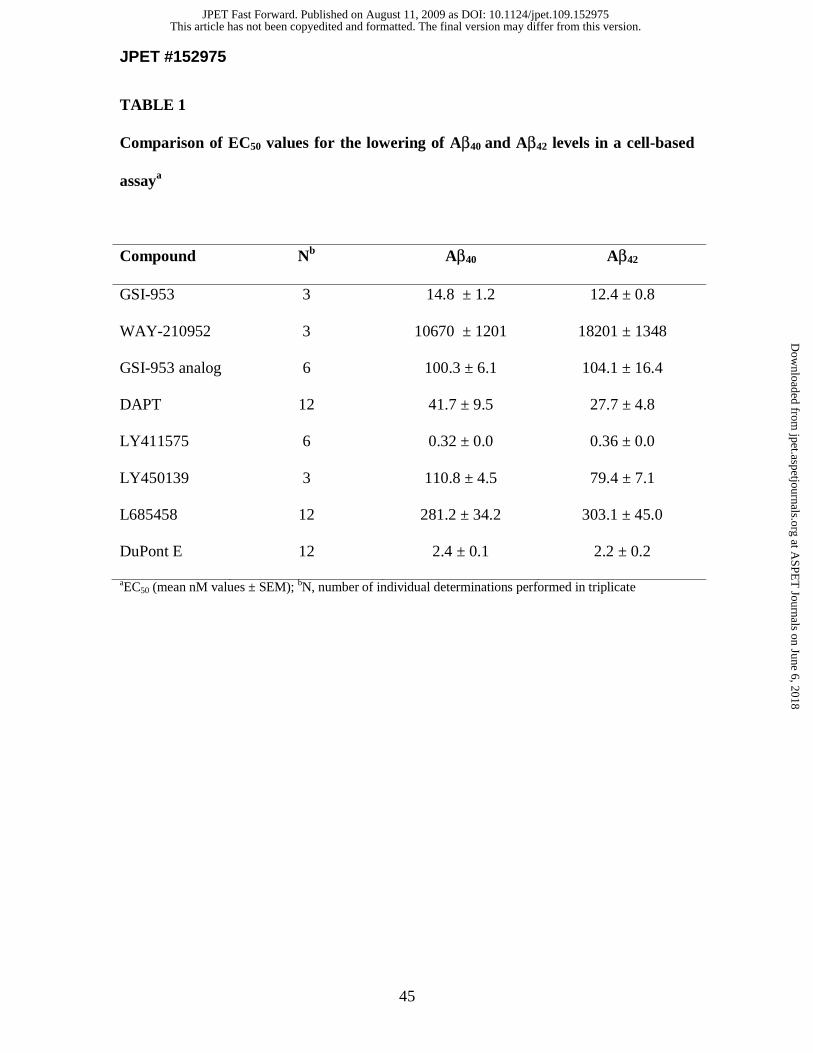
JPET #152975
45
TABLE 1
Comparison of EC50 values for the lowering of Aβ40 and Aβ42 levels in a cell-based
assaya
Compound Nb Aβ40 Aβ42
GSI-953 3 14.8 ± 1.2 12.4 ± 0.8
WAY-210952 3 10670 ± 1201 18201 ± 1348
GSI-953 analog 6 100.3 ± 6.1 104.1 ± 16.4
DAPT 12 41.7 ± 9.5 27.7 ± 4.8
LY411575 6 0.32 ± 0.0 0.36 ± 0.0
LY450139 3 110.8 ± 4.5 79.4 ± 7.1
L685458 12 281.2 ± 34.2 303.1 ± 45.0
DuPont E 12 2.4 ± 0.1 2.2 ± 0.2
aEC50 (mean nM values ± SEM); bN, number of individual determinations performed in triplicate
This article has not been copyedited and formatted. The final version may differ from this version.JPET Fast Forward. Published on August 11, 2009 as DOI: 10.1124/jpet.109.152975
at ASPE
T Journals on June 6, 2018
jpet.aspetjournals.orgD
ownloaded from
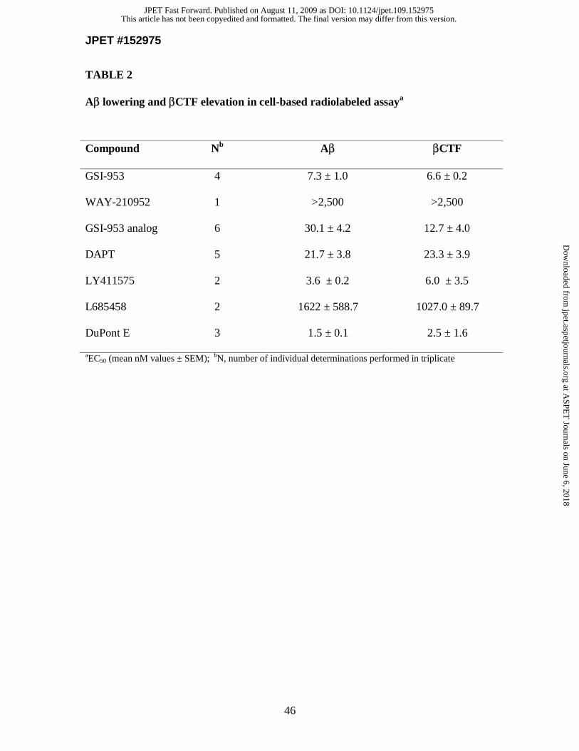
JPET #152975
46
TABLE 2
Aβ lowering and βCTF elevation in cell-based radiolabeled assaya
Compound Nb Aβ βCTF
GSI-953 4 7.3 ± 1.0 6.6 ± 0.2
WAY-210952 1 >2,500 >2,500
GSI-953 analog 6 30.1 ± 4.2 12.7 ± 4.0
DAPT 5 21.7 ± 3.8 23.3 ± 3.9
LY411575 2 3.6 ± 0.2 6.0 ± 3.5
L685458 2 1622 ± 588.7 1027.0 ± 89.7
DuPont E 3 1.5 ± 0.1 2.5 ± 1.6
aEC50 (mean nM values ± SEM); bN, number of individual determinations performed in triplicate
This article has not been copyedited and formatted. The final version may differ from this version.JPET Fast Forward. Published on August 11, 2009 as DOI: 10.1124/jpet.109.152975
at ASPE
T Journals on June 6, 2018
jpet.aspetjournals.orgD
ownloaded from
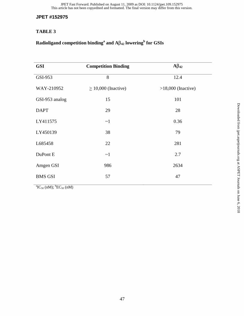
JPET #152975
47
TABLE 3
Radioligand competition bindinga and Aβ42 loweringb for GSIs
GSI Competition Binding Aβ42
GSI-953 8 12.4
WAY-210952 ≥ 10,000 (Inactive) >18,000 (Inactive)
GSI-953 analog 15 101
DAPT 29 28
LY411575 ~1 0.36
LY450139 38 79
L685458 22 281
DuPont E ~1 2.7
Amgen GSI 986 2634
BMS GSI 57 47
aIC50 (nM); bEC50 (nM)
This article has not been copyedited and formatted. The final version may differ from this version.JPET Fast Forward. Published on August 11, 2009 as DOI: 10.1124/jpet.109.152975
at ASPE
T Journals on June 6, 2018
jpet.aspetjournals.orgD
ownloaded from
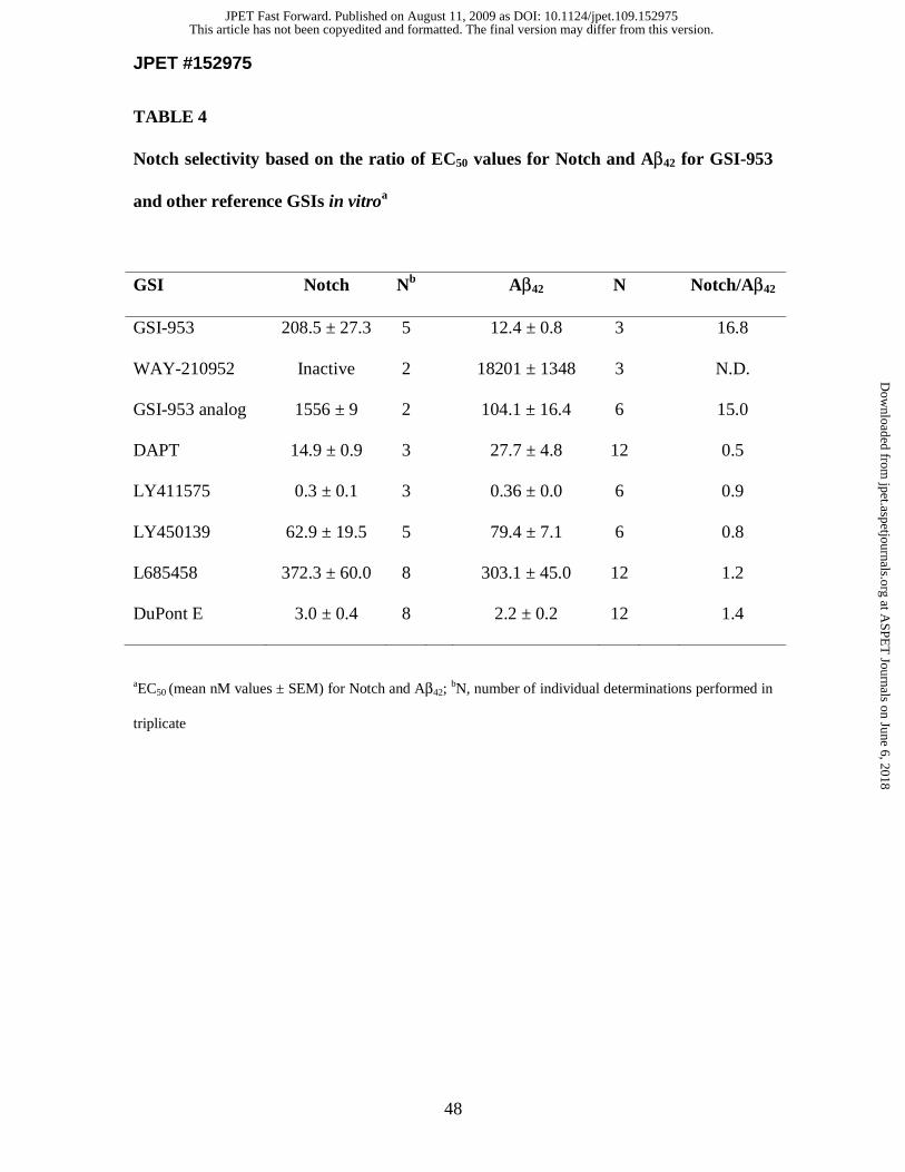
JPET #152975
48
TABLE 4
Notch selectivity based on the ratio of EC50 values for Notch and Aβ42 for GSI-953
and other reference GSIs in vitroa
GSI Notch Nb Aβ42 N Notch/Aβ42
GSI-953 208.5 ± 27.3 5 12.4 ± 0.8 3 16.8
WAY-210952 Inactive 2 18201 ± 1348 3 N.D.
GSI-953 analog 1556 ± 9 2 104.1 ± 16.4 6 15.0
DAPT 14.9 ± 0.9 3 27.7 ± 4.8 12 0.5
LY411575 0.3 ± 0.1 3 0.36 ± 0.0 6 0.9
LY450139 62.9 ± 19.5 5 79.4 ± 7.1 6 0.8
L685458 372.3 ± 60.0 8 303.1 ± 45.0 12 1.2
DuPont E 3.0 ± 0.4 8 2.2 ± 0.2 12 1.4
aEC50 (mean nM values ± SEM) for Notch and Aβ42; bN, number of individual determinations performed in
triplicate
This article has not been copyedited and formatted. The final version may differ from this version.JPET Fast Forward. Published on August 11, 2009 as DOI: 10.1124/jpet.109.152975
at ASPE
T Journals on June 6, 2018
jpet.aspetjournals.orgD
ownloaded from
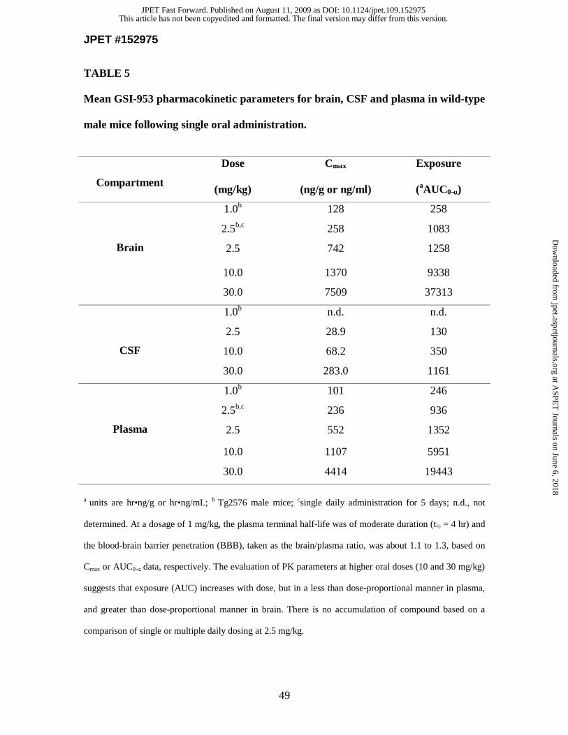
JPET #152975
49
TABLE 5
Mean GSI-953 pharmacokinetic parameters for brain, CSF and plasma in wild-type
male mice following single oral administration.
Compartment
Dose Cmax Exposure
(mg/kg) (ng/g or ng/ml) (aAUC0-α)
Brain
1.0b 128 258
2.5b,c 258 1083
2.5 742 1258
10.0 1370 9338
30.0 7509 37313
CSF
1.0b n.d. n.d.
2.5 28.9 130
10.0 68.2 350
30.0 283.0 1161
Plasma
1.0b 101 246
2.5b,c 236 936
2.5 552 1352
10.0 1107 5951
30.0 4414 19443
a units are hr•ng/g or hr•ng/mL; b Tg2576 male mice; csingle daily administration for 5 days; n.d., not
determined. At a dosage of 1 mg/kg, the plasma terminal half-life was of moderate duration (t½ = 4 hr) and
the blood-brain barrier penetration (BBB), taken as the brain/plasma ratio, was about 1.1 to 1.3, based on
Cmax or AUC0-α data, respectively. The evaluation of PK parameters at higher oral doses (10 and 30 mg/kg)
suggests that exposure (AUC) increases with dose, but in a less than dose-proportional manner in plasma,
and greater than dose-proportional manner in brain. There is no accumulation of compound based on a
comparison of single or multiple daily dosing at 2.5 mg/kg.
This article has not been copyedited and formatted. The final version may differ from this version.JPET Fast Forward. Published on August 11, 2009 as DOI: 10.1124/jpet.109.152975
at ASPE
T Journals on June 6, 2018
jpet.aspetjournals.orgD
ownloaded from
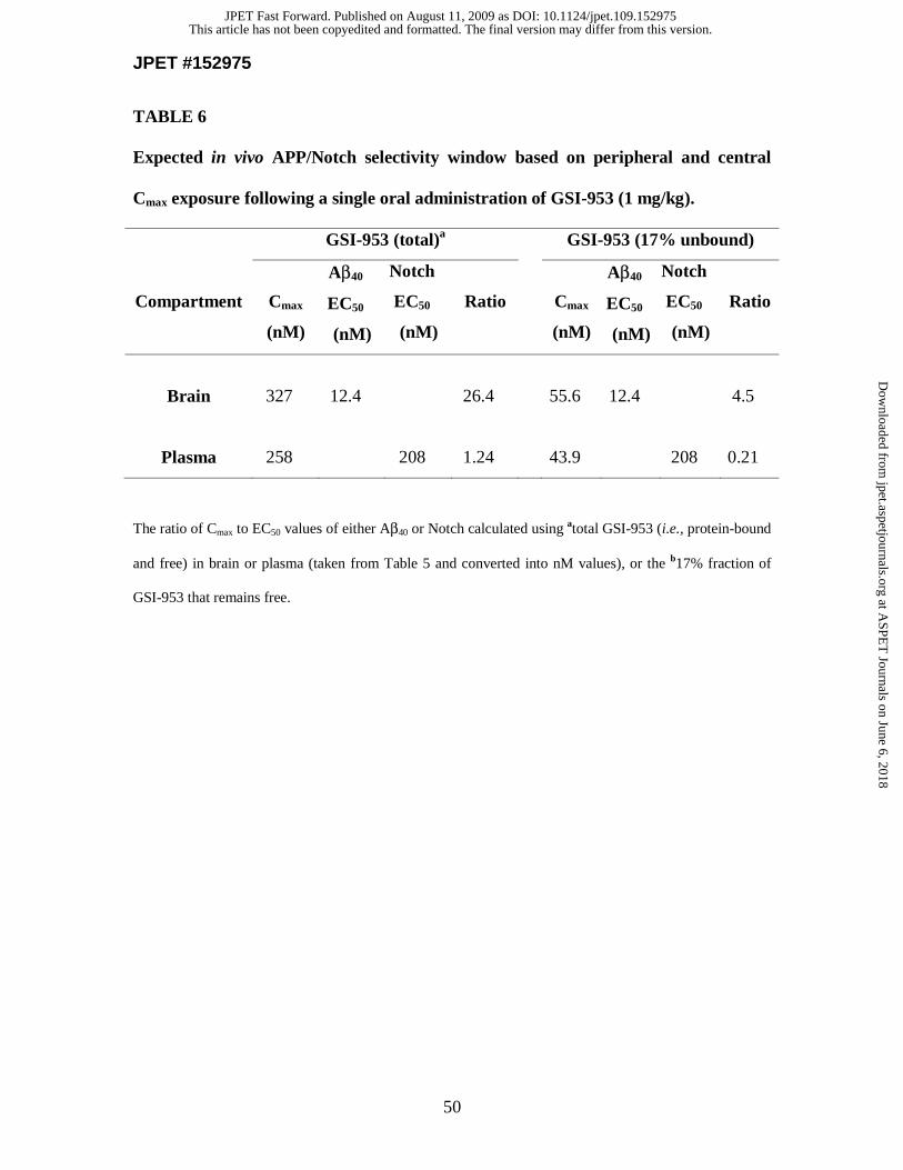
JPET #152975
50
TABLE 6
Expected in vivo APP/Notch selectivity window based on peripheral and central
Cmax exposure following a single oral administration of GSI-953 (1 mg/kg).
GSI-953 (total)a GSI-953 (17% unbound)
Compartment
Cmax
(nM)
Aβ40
EC50
(nM)
Notch
EC50
(nM)
Ratio
Cmax
(nM)
Aβ40
EC50
(nM)
Notch
EC50
(nM)
Ratio
Brain 327 12.4 26.4
55.6 12.4 4.5
Plasma 258 208 1.24
43.9 208 0.21
The ratio of Cmax to EC50 values of either Aβ40 or Notch calculated using atotal GSI-953 (i.e., protein-bound
and free) in brain or plasma (taken from Table 5 and converted into nM values), or the b17% fraction of
GSI-953 that remains free.
This article has not been copyedited and formatted. The final version may differ from this version.JPET Fast Forward. Published on August 11, 2009 as DOI: 10.1124/jpet.109.152975
at ASPE
T Journals on June 6, 2018
jpet.aspetjournals.orgD
ownloaded from
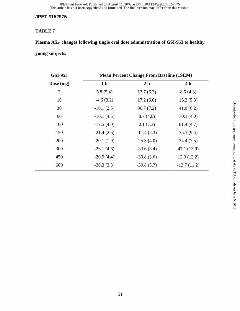
JPET #152975
51
TABLE 7
Plasma Aβ40 changes following single oral dose administration of GSI-953 to healthy
young subjects.
GSI-953
Dose (mg)
Mean Percent Change From Baseline (±SEM)
1 h 2 h 4 h
3 5.8 (5.4) 13.7 (6.3) 8.5 (4.3)
10 -4.6 (3.2) 17.2 (6.6) 15.3 (5.3)
30 -10.1 (2.5) 36.7 (7.2) 41.0 (6.2)
60 -16.1 (4.5) 8.7 (4.0) 70.1 (4.0)
100 -17.5 (4.0) 0.1 (7.3) 81.4 (4.7)
150 -21.4 (2.6) -11.4 (2.3) 75.3 (9.4)
200 -20.1 (1.9) -25.3 (4.0) 34.4 (7.5)
300 -26.1 (4.6) -33.6 (3.4) 47.1 (13.9)
450 -20.9 (4.4) -30.8 (3.6) 12.3 (12.2)
600 -30.3 (3.3) -39.8 (5.7) -13.7 (11.2)
This article has not been copyedited and formatted. The final version may differ from this version.JPET Fast Forward. Published on August 11, 2009 as DOI: 10.1124/jpet.109.152975
at ASPE
T Journals on June 6, 2018
jpet.aspetjournals.orgD
ownloaded from
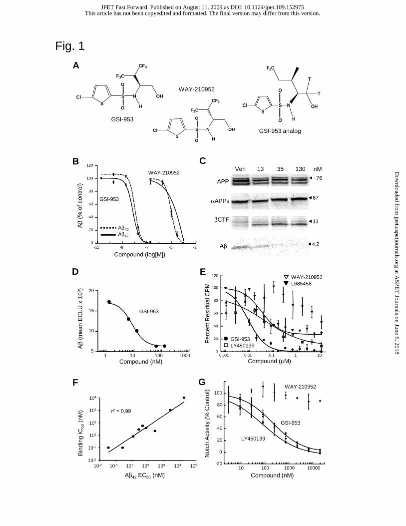
Fig. 1
-11 -9 -7 -5 -30
20
40
60
80
100
120
Aβ
(% o
f con
trol
)
Compound (log[M])
Aβ40
Aβ42
GSI-953
WAY-210952
-11 -9 -7 -5 -3
120
100
80
60
40
20
0
A
GSI-953
S
S NCl
H
OH
CF3
F3C
O
O
S
S NCl
H
OH
CF3
F3C
O
O
WAY-210952
B C
D
Not
ch A
ctiv
ity (
% C
ontr
ol)
GSI-953
LY450139
WAY-210952
10 100 1000 10000-20
0
20
40
60
80
100
Compound (nM)
Compound (nM)1 10 100 1000
Aβ
(mea
n E
CLU
x 1
03 )
5
10
15
20
GSI-953
F G
αAPPs
βCTF
APP
Aβ
Veh 13 35 130 nM
�~76
�67
�11
�4.2
Aβ42 EC50 (nM)
Bin
ding
IC50
(nM
)
10510410310210110-110-2
104
103
102
101
10-1
10-2
r2 = 0.99
E
0.001 0.01 0.1 1 100
20
40
60
80
100
120
Compound (μM)
Per
cent
Res
idua
l CP
M
WAY-210952L685458
GSI-953LY450139
GSI-953 analog
Cl S NS
OH
F3C
T
TO
O H
This article has not been copyedited and formatted. The final version may differ from this version.JPET Fast Forward. Published on August 11, 2009 as DOI: 10.1124/jpet.109.152975
at ASPE
T Journals on June 6, 2018
jpet.aspetjournals.orgD
ownloaded from
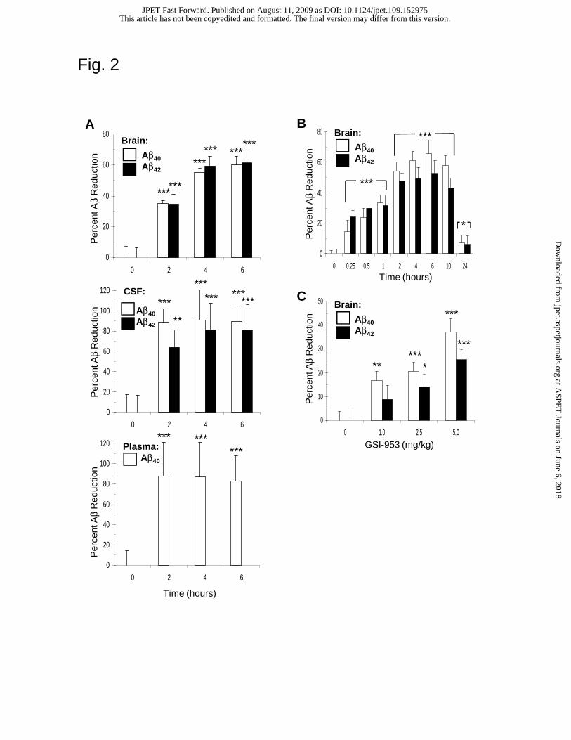
Fig. 2
0
20
40
60
80
0 2 4 6
Brain Ab40
Brain Ab42
0
20
40
60
80
100
120
0 2 4 6
CSF Ab40
CSF Ab42
0
20
40
60
80
100
120
0 2 4 6
Plasma Ab40
Per
cent
Aβ
Red
uctio
nP
erce
nt A
βR
educ
tion
Per
cent
Aβ
Red
uctio
n
CSF:
Aβ40Aβ42
Brain:
Aβ40Aβ42
Plasma: Aβ40
A
Time (hours)
**
*** ******
******
******
0
20
40
60
80
0 0.25 0.5 1 2 4 6 10 24
Ab 40
Ab42
******
***
****** ***
***
B
Per
cent
Aβ
Red
uctio
n
Time (hours)
CP
erce
nt A
βR
educ
tion
0
10
20
30
40
50
0 1.0 2.5 5.0
Ab40
Ab42
GSI-953 (mg/kg)
***
***
******
***
Brain:
Aβ40Aβ42
Brain:
Aβ40Aβ42
***
***
*
This article has not been copyedited and formatted. The final version may differ from this version.JPET Fast Forward. Published on August 11, 2009 as DOI: 10.1124/jpet.109.152975
at ASPE
T Journals on June 6, 2018
jpet.aspetjournals.orgD
ownloaded from
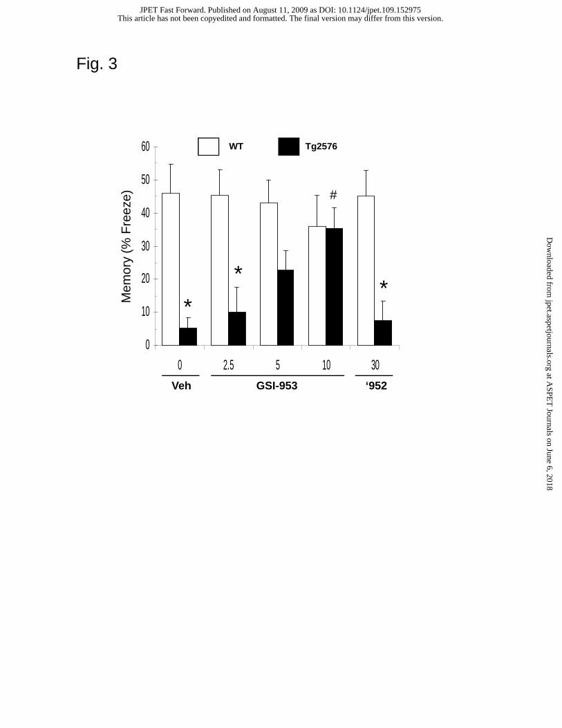
Fig. 3
Mem
ory
(% F
reez
e)
*
**
#
WT Tg2576
0
10
20
30
40
50
60
0 2.5 5 10 30Veh GSI-953 ‘952
*
** **
*#
This article has not been copyedited and formatted. The final version may differ from this version.JPET Fast Forward. Published on August 11, 2009 as DOI: 10.1124/jpet.109.152975
at ASPE
T Journals on June 6, 2018
jpet.aspetjournals.orgD
ownloaded from
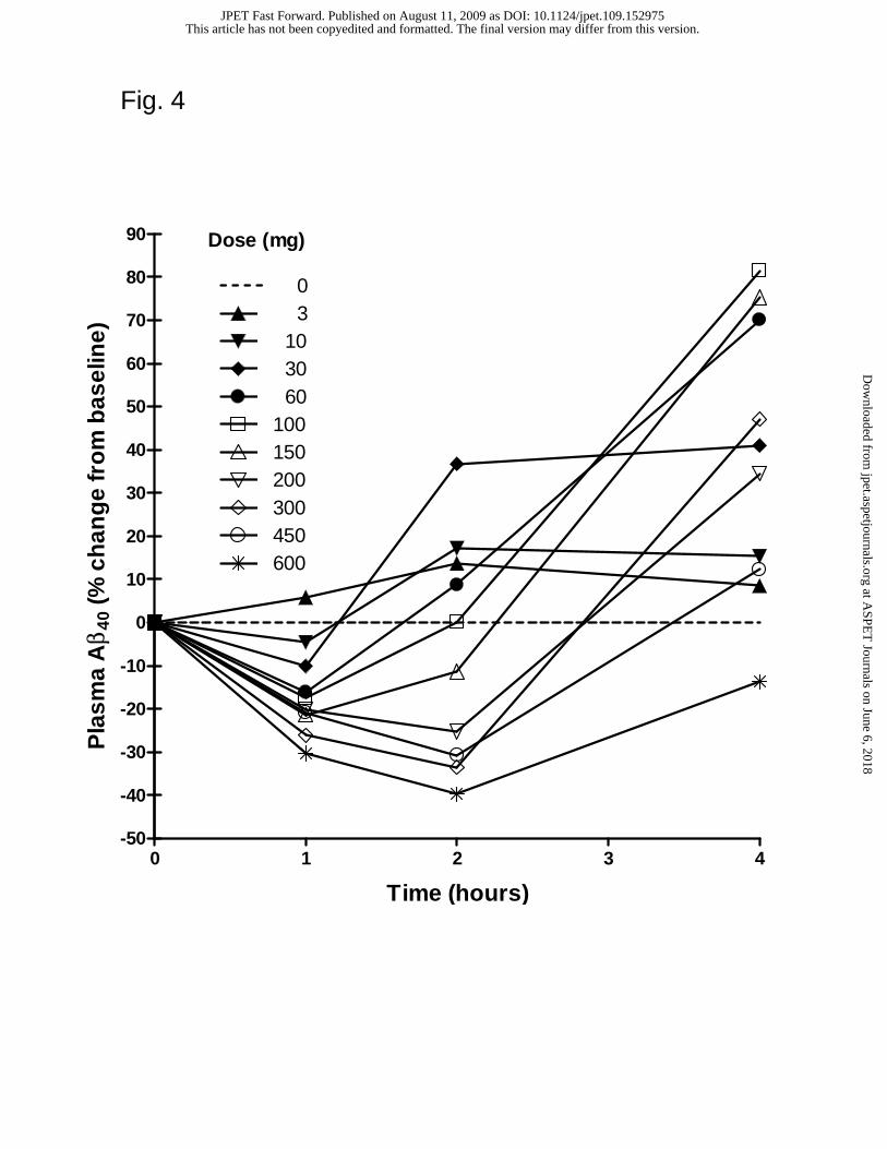
0 1 2 3 4-50
-40
-30
-20
-10
0
10
20
30
40
50
60
70
80
90
0 3 10 30 60 100 150 200 300 450 600
Dose (mg)
Time (hours)
Pla
sma
Aβ 4
0 (%
ch
ang
e fr
om
bas
elin
e)Fig. 4
This article has not been copyedited and formatted. The final version may differ from this version.JPET Fast Forward. Published on August 11, 2009 as DOI: 10.1124/jpet.109.152975
at ASPE
T Journals on June 6, 2018
jpet.aspetjournals.orgD
ownloaded from