Downloaded from jpet.aspetjournals.org at ASPET...
42
JPET #209346 1 The anti-angiogenic insulin receptor substrate-1 (IRS-1) antisense oligonucleotide, aganirsen, impairs AU-rich mRNA stability by reducing 14-3-3β-tristetraprolin protein complex reducing inflammation and psoriatic lesion size in patients Sylvie Colin * (PhD), Bernadette Darné* (MD, PhD), Amin Kadi (MD), Antoine Ferry (MD), Marlyne Favier (Ing), Corine Lesaffre (MSc), Jean-Pascal Conduzorgues (D.Pharm), Salman Al-Mahmood* (PhD), Najibe Doss* (MD) Affiliations Gene Signal SAS, 4, Rue Pierre Fontaine, 91000 EVRY, France (SC, A.F, S.A-M); Monitoring Force Group, 6, bis rue du Marechal Foch, 78600 Maisons-Laffitte, France (B.D, A.K); Institut Cochin, INSERM U1016, CNRS UMR8104, Morphology and histology platform, 27 rue du Faubourg-Saint-Jacques, 75014 Paris, and Université Paris Descartes, 75006 Paris, France (M.F, C.L); AMATSI, 17, Parc des Vautes, 34980 St Gely du Fesc, France (J-P.C); Université de Tunis El Manar, Faculté de Médecine de Tunis, Military Hospital of Tunis, Department of Dermatology, 1089 Montfleury, Tunis, Tunisia (N.D) JPET Fast Forward. Published on February 6, 2014 as DOI:10.1124/jpet.113.209346 Copyright 2014 by the American Society for Pharmacology and Experimental Therapeutics. This article has not been copyedited and formatted. The final version may differ from this version. JPET Fast Forward. Published on February 6, 2014 as DOI: 10.1124/jpet.113.209346 at ASPET Journals on September 12, 2018 jpet.aspetjournals.org Downloaded from
Transcript of Downloaded from jpet.aspetjournals.org at ASPET...
JPET #209346
1
The anti-angiogenic insulin receptor substrate-1 (IRS-1) antisense oligonucleotide, aganirsen,
impairs AU-rich mRNA stability by reducing 14-3-3β-tristetraprolin protein complex
reducing inflammation and psoriatic lesion size in patients
Sylvie Colin* (PhD), Bernadette Darné* (MD, PhD), Amin Kadi (MD), Antoine Ferry (MD),
Marlyne Favier (Ing), Corine Lesaffre (MSc), Jean-Pascal Conduzorgues (D.Pharm), Salman
Al-Mahmood* (PhD), Najibe Doss* (MD)
Affiliations
Gene Signal SAS, 4, Rue Pierre Fontaine, 91000 EVRY, France (SC, A.F, S.A-M);
Monitoring Force Group, 6, bis rue du Marechal Foch, 78600 Maisons-Laffitte, France (B.D,
A.K); Institut Cochin, INSERM U1016, CNRS UMR8104, Morphology and histology
platform, 27 rue du Faubourg-Saint-Jacques, 75014 Paris, and Université Paris Descartes,
75006 Paris, France (M.F, C.L); AMATSI, 17, Parc des Vautes, 34980 St Gely du Fesc,
France (J-P.C); Université de Tunis El Manar, Faculté de Médecine de Tunis, Military
Hospital of Tunis, Department of Dermatology, 1089 Montfleury, Tunis, Tunisia (N.D)
JPET Fast Forward. Published on February 6, 2014 as DOI:10.1124/jpet.113.209346
Copyright 2014 by the American Society for Pharmacology and Experimental Therapeutics.
This article has not been copyedited and formatted. The final version may differ from this version.JPET Fast Forward. Published on February 6, 2014 as DOI: 10.1124/jpet.113.209346
at ASPE
T Journals on Septem
ber 12, 2018jpet.aspetjournals.org
Dow
nloaded from
JPET #209346
2
Running title: Aganirsen improves clinical signs of psoriasis
Correspondence
Dr Salman Al-Mahmood, Gene Signal SAS, 4, Rue Pierre Fontaine, 91000 Evry, France.
Tel : +33 (0)675 244 999; Email : [email protected]
Recommended section: Drug discovery and translational medicine.
Cellular and molecular.
Table count: 2
Figure count: 8
Abstratct: 248 words
Introduction: 423 words
Discussion: 1298 words
Supplemental data: 1
Abbreviations: CK-16, cytokeratin 16; hEC, human endothelial cell; IRS-1, insulin receptor
substrate-1; ITT, intent-to-treat; PGA, physician’s global assessment; PASI, psoriasis area
severity index; SPN, soluble perinuclear-nuclear fraction; TTP, tristetraprolin protein; TSS,
total severity sign score.
This article has not been copyedited and formatted. The final version may differ from this version.JPET Fast Forward. Published on February 6, 2014 as DOI: 10.1124/jpet.113.209346
at ASPE
T Journals on Septem
ber 12, 2018jpet.aspetjournals.org
Dow
nloaded from

JPET #209346
3
AbstractIncreased inflammation and aberrant angiogenesis underlie psoriasis. Here, we report that the
inhibition of IRS-1 expression with aganirsen resulted in a dose-dependent reduction
(P<0.0001) in IRS-1 protein at the cytoplasm, while IRS-1 protein remained quantitatively
unchanged in the perinuclear environment. Aganirsen induced a dose-dependent increase in
serine-phosphorylated IRS-1 (pIRS-1Ser) in the soluble perinuclear-nuclear fraction (SPN),
inducing IRS-1-14-3-3β protein association (P<0.001), thereby impairing 14-3-3β-
tristetraprolin protein (TTP) complex and AU-rich mRNA’s stability (P<0.001). Accordingly,
aganirsen inhibited (P<0.001) in vitro the expression of interleukin-8 (IL-8), IL-12, IL-22 and
tumour necrosis factor alpha (TNFα), four inflammatory mediators containing mRNA with
AU-rich regions. To demonstrate the clinical relevance of this pathway, we tested the efficacy
of aganirsen by topical application in a pilot, “double-blind”, randomized, dose-ranging study
in 12 psoriatic human patients. After 6-weeks of treatment, Least Square means differences
with placebo were -38.9% (95%CI [-75.8%; -2.0%]) and -37.4% [-74.3%; -0.5%] at the doses
of 0.86 and 1.72 mg/g, respectively. Lesion size reduction was associated with reduced
expression of IRS-1 (P<0.01), TNFα (P<0.0001) and VEGF (P<0.01), reduced keratinocytes
proliferation (P<0.01), and the restoration (P<0.02) of normal levels of infiltrating CD4+ and
CD3+ lymphocytes in psoriatic skin lesions. These results suggest that aganirsen is a first in
class of a new generation of anti-angiogenic medicines combining anti-inflammatory
activities. Aganirsen-induced down-regulation of inflammatory mediators characterized by
AU-rich mRNA likely underlies its beneficial clinical outcome in psoriasis. These results
justify further large-scale clinical studies to establish the dose of aganirsen and its long-term
efficacy in psoriasis.
This article has not been copyedited and formatted. The final version may differ from this version.JPET Fast Forward. Published on February 6, 2014 as DOI: 10.1124/jpet.113.209346
at ASPE
T Journals on Septem
ber 12, 2018jpet.aspetjournals.org
Dow
nloaded from
JPET #209346
4
Introduction
Psoriasis is a common chronic disease occurring in approximately 2% of the population in
Western countries (Nestle et al., 2009; Sabat et al., 2007). Extensive work has established that
increased inflammation, aberrant angiogenesis and vascular remodelling as well as
keratinocyte hyper-proliferation are involved in the pathogenesis of psoriasis (Heidenreich et
al., 2009).
T-cell–mediated immune response to an as-yet unknown auto-antigen seems to play a key role
in the pathogenesis of psoriasis. This deregulated immune response is primarily driven by
CD4+ T cells with a T(h)1 and/or T(h)17 phenotype (Nestle et al., 2009; Coimbra et al., 2012;
Nograles et al., 2008), and by the production of TNFα (Caldarola et al., 2009; Orlinick and
Chao 1998). A significant increase in the development of the microvasculature (angiogenesis)
has been observed in psoriatic lesions compared with healthy skin (Heidenreich et al., 2009),
especially in the early phase of the development of the lesions (Henno et al., 2010). Vascular
endothelial growth factor (VEGF), which has a central role in angiogenesis, is up-regulated in
psoriasis (Detmar et al., 1994), and its levels of expression represent a good indicator of
active psoriatic arthritis (Heidenreich et al., 2009).
Conventional systemic therapies for psoriasis, such as methotrexate, cyclosporin A, retinoids
or psoralen and ultraviolet A (PUVA) therapies, can result in long-term toxicity and may not
be effective. The development of novel therapies targeting inflammatory factors including
TNFα, IL-12 and IL-22 now provide new and efficient treatment options (Weger 2010).
Nonetheless, because both angiogenesis and inflammation are involved in the development of
psoriasis, we speculated that aganirsen (GS-101) could be efficient in psoriatic patients.
Aganirsen is an antisense oligonucleotide that inhibits the expression of IRS-1 and has anti-
angiogenic activities including inhibition of both VEGF and IL-1β expressions (Al-Mahmood
et al., 2009; Cloutier et al., 2012; Andrieu-Soler et al., 2005). In this study, we show that by
This article has not been copyedited and formatted. The final version may differ from this version.JPET Fast Forward. Published on February 6, 2014 as DOI: 10.1124/jpet.113.209346
at ASPE
T Journals on Septem
ber 12, 2018jpet.aspetjournals.org
Dow
nloaded from
JPET #209346
5
inhibiting IRS-1 expression in the cytoplasmic compartment, aganirsen impaired 14-3-3β-
TTP complex formation leading to the inhibition of the expression of several cytokines with
AU-rich mRNA, including IL-8, IL-12, IL-22 and TNFα. We tested the efficacy of aganirsen
by a 6-week topical application in patients with chronic mild-to-moderate plaque psoriasis
and showed that aganirsen reduced plaque area. This was associated with a reduction in the
expression of VEGF and TNFα protein as well as a normalization of the levels of CD4+ and
CD3+ lymphocytes, and reduced keratinocyte proliferation in psoriatic skin lesion biopsies.
These data strongly support that the combined anti-angiogenic and anti-inflammatory
properties of aganirsen synergized to improve the clinical status of the patients.
This article has not been copyedited and formatted. The final version may differ from this version.JPET Fast Forward. Published on February 6, 2014 as DOI: 10.1124/jpet.113.209346
at ASPE
T Journals on Septem
ber 12, 2018jpet.aspetjournals.org
Dow
nloaded from
JPET #209346
6
Materials and Methods
Real-Time Reverse Transcription-Polymerase Chain Reaction Assay. The human
microvascular endothelial cell line (hEC) was provided by Dr E.W. Ades (Center for Disease
Control and Prevention, Atlanta, GA, USA) who established this line by transfecting human
dermal endothelial cells with SV40A gene product and large T antigen (Ades et al., 1992).
Cells were cultured in EGM2-MV medium (Lonza, Levallois, France). After exposure to
various concentrations of aganirsen (0-10 μM) or vehicle for 24 h, hEC (5 × 10⁵ cells/ml) total
mRNAs were isolated using the NucleoSpin RNA II kit. RNA yields and purity were assessed
by spectrophotometric analysis. The real-time RT-PCR was performed as described
previously (Al-Mahmood et al., 2009). In brief, 0.5 μg of total RNA was reverse-transcribed
with random hexamer primers and Moloney murine leukemia virus (200 U; Invitrogen), and
the synthesized cDNA was used immediately for real-time PCR amplification using the DNA-
binding dye SYBR Green I for the detection of PCR products and the following primers:
TNFα (sense, 5′-GCTGCAGCACATTATAATACAGAGA-3′; antisense, 5′-
GGTGTTTGTCGCGACTCC-3′); IL-8 (sense, 5′-AGTGGACCACACTGCGCCAAC-3′;
antisense, 5′-CCACAACCCTCTGCACCCAGT-3′); IL-12 (sense, 5’-
GAATGCAAAGCTTCTGATGGA-3’; antisense 5’-GTGGCACAGTCTCACTGTTGA-3’);
IL-22 (sense, 5’-CCCTCAATCTGATAGGTTCCAG-3’; antisense 5’-
GCAGGTCATCACCTTCAATATG-3’) and GAPDH (sense, 5′-
TGAAGGTCGGAGTCAACGGA-3′; antisense 5′-CATTGATGACAAGCTTCCCG-3′). The
real-time PCR reactions were carried out with the DNA Light Cycler 480 (Roche Diagnostic,
Meylan, France). The results were quantified using the equation: CopyTF/CopyGAPDH =
2C(t)GAPDH - C(t)TF. All PCR products were analyzed by electrophoresis on a 1.5% agarose
gel, visualized with ethidium bromide, and analyzed using the Genesnap 6.00.26 software
This article has not been copyedited and formatted. The final version may differ from this version.JPET Fast Forward. Published on February 6, 2014 as DOI: 10.1124/jpet.113.209346
at ASPE
T Journals on Septem
ber 12, 2018jpet.aspetjournals.org
Dow
nloaded from
JPET #209346
7
(Syngene, Frederick, MD). Densitometric analysis was performed using GeneTools Analysis
Software version 3.02.00 (Syngene).
Subcellular protein fractioning and protein quantification. Subcellular protein fractioning
was realized using the Thermo Scientific subcellular protein fractionation kit for cultured cells
(Product No. 78840; Pierce Biotechnology, Rockford, USA) according to the manufacturer's
instructions. The fractionation kit permitted the separation and preparation of five fractions:
membrane, cytoplasmic, soluble peri-nuclear and nuclear (SPN), chromatin-bound and
cytoskeletal protein fractions from hECs. IRS-1 protein was only detected in the cytoplasmic
and the SPN fractions. The protein content of the different fractions was measured by
Bradford’s method and adjusted. For purity control within the subfractions, equivalent
amounts of proteins were resolved by SDS-PAGE, followed by the transfer of proteins to a
polyvinylidene difluoride (PVDF) membrane. The membrane was incubated with 5% defatted
milk solution in PBS-0.5% Tween for 1 h, followed by two washes, and immunoblotting with
an anti-epidermal growth factor receptor (EGFR) mAb (clone D38B1, rabbit mAb, Cell
Signaling Technology Inc., Danvers, MA) as plasma membrane marker; an anti-heat shock
protein p90 (Hsp90) (cytoplasmic fraction marker) (clone C45G5, rabbit mAb, Cell Signaling
Technology Inc., Danvers, MA); an anti-histone deacetylase 2 (HDAC 2) mAb (rabbit mAb,
Cell Signaling Technology Inc., Danvers, MA) and an anti-transcription factor Sp1 mAb as
markers of the soluble nuclear and peri-nuclear fraction; an anti-vimentin mAb (clone D21H3
rabbit mAb, Cell Signaling Technology Inc., Danvers, MA) as a marker of the cytoskeletal
fraction; an anti-histone 3 mAb as a marker of the chromatin-bound insoluble nuclear
fraction. The results of the purity controls of the obtained fractions were shown in
supplemental Figure 1.
This article has not been copyedited and formatted. The final version may differ from this version.JPET Fast Forward. Published on February 6, 2014 as DOI: 10.1124/jpet.113.209346
at ASPE
T Journals on Septem
ber 12, 2018jpet.aspetjournals.org
Dow
nloaded from
JPET #209346
8
Serum-deprived hECs were incubated with different concentrations of aganirsen or scramble
oligonucleotide at 37°C under 5% CO2 for 6 h. After three washes with ice-cold PBS, cells
were suspended with the protein extraction buffer (20 mM Tris-HCl pH 7.5, 150 mM NaCl, 1
mM EDTA, 1 mM EGTA, 1% Triton, 25 mM sodium pyrophosphate, 1 mM β-
glycerophosphate, 1 mM Na3VO4, 1 μg/ml leupeptin, 1 μM phenylmethylsulfonyl fluoride).
The protein content was measured by Bradford. IRS-1 concentration in the extracts was
determined by the Path-Scan Total IRS-1 Sandwich ELISA kit (Cell Signaling Technology
Inc., Danvers, MA) according to the manufacturer's instructions. The data were collected from
four separate experiments performed in duplicate and expressed relative to control cells
(vehicle).
To confirm the results obtained with the Path-Scan Total IRS-1 Sandwich ELISA kit, equal
volumes of the adjusted cell extracts were also resolved by SDS-PAGE, followed by the
transfer of proteins to a PVDF membrane. The membrane was incubated with 5% defatted
milk solution in PBS-0.5% Tween for 1 h, followed by two washes, and immunoblotting with
either an anti-IRS-1-HRP conjugate (1:300 dilution; Santa Cruz Biotechnology, Inc., Santa
Cruz, CA), anti-GAPDH-HRP conjugate (Santa Cruz Biotechnology, Inc.), goat anti-TNFα
(1:200 dilution, reference sc52746; Santa Cruz Biotechnology, Inc.), and anti-goat-HRP
conjugate (1:20 000 dilution; reference sc2302; Santa Cruz Biotechnology, Inc.). Proteins
were then monitored by ECLplus (GE Healthcare, Chalfont St. Giles, UK).
For the quantification of IL-8, IL-12, IL-22 and TNFα in the culture medium, hECs in culture
medium without serum were incubated with different concentrations of aganirsen or scramble
oligonucleotide at 37°C under 5% CO2 for 24 h. Culture medium was recovered and used to
quantify TNFα, IL-8, IL-12 and IL-22 using human TNF sandwich ELISA kit (reference 900-
k25, Peprotech, France) and human IL8, IL-12 and IL-22 sandwich ELISA kits (reference
This article has not been copyedited and formatted. The final version may differ from this version.JPET Fast Forward. Published on February 6, 2014 as DOI: 10.1124/jpet.113.209346
at ASPE
T Journals on Septem
ber 12, 2018jpet.aspetjournals.org
Dow
nloaded from
JPET #209346
9
900-k18, 900-K96 and 900-K246, Peprotech, France), respectively, according to the
manufacturer's instructions.
Immunoprecipitation. Human ECs in culture medium without serum were incubated with
different concentrations of aganirsen or scramble oligonucleotide at 37°C under 5% CO2 for 6
h. The cells were washed twice with cold PBS, and lysed with 1% Triton X-100 buffer for 1 h
at 4°C. The protein 14-3-3 was immuno-precipitated by adding 4 µg of anti-14-3-3β mAb
(reference sc628; Santa Cruz Biotechnology, Inc.) and the immune complexes were harvested
with protein G-Sepharose beads (Santa Cruz), washed with lysis buffer, resolved in SDS–
PAGE, and proteins were transferred to PVDF membrane and revealed with the indicated
antibody.
Pilot clinical study. The clinical study was approved by the institutional ethic committee and
was conducted in accordance with Good Clinical Practices (GCPs) including International
Conference of Harmonization (ICH) Guidelines and all applicable local laws and regulatory
requirements. It was designed as a “double-blind”, randomized, control versus placebo, dose-
ranging, Latin-square design single-centre study from the department of dermatology of the
Military Hospital of Tunis (see Study protocol and results as supplementary file). It was set up
to evaluate the safety and the efficacy of two doses of aganirsen versus placebo in 12 patients
with chronic mild-to-moderate plaque psoriasis (Feldman and Krueger 2005). Each patient
had 3 psoriasis plaques (including 2 refractory lesions; refractory area are elbow and knee).
Each lesion was treated either by a low-dose (0.86 mg/g) or a high-dose (1.72 mg/g) of
aganirsen or the ointment alone (placebo; 60 % paraffin oil and 40 % white Vaseline, v/v). At
the inclusion and randomization visit (Visit 1), the investigator selected for treatment at least
2 discoids or plaques from 2 refractory sites (elbow and knee) representative of the disease
This article has not been copyedited and formatted. The final version may differ from this version.JPET Fast Forward. Published on February 6, 2014 as DOI: 10.1124/jpet.113.209346
at ASPE
T Journals on Septem
ber 12, 2018jpet.aspetjournals.org
Dow
nloaded from
JPET #209346
10
and a third plaque in a refractory or not refractory site. At Visit 1 (inclusion in the study) and
at Visit 2 (3-week post-inclusion), 75 tubes were delivered, i.e. 25 tubes of each type (A, B
and C). The investigator completed a form that was given to the patient together with the
ointment tubes explaining what type (A, B and C) of ointment tube must be used for each
plaque. The order of plaques corresponded to the order recorded in the case report form. “A”
and “B” types were assigned to refractory sites only. “A” ointment tubes were used for the
first plaque, “B” ointment tubes were used for the second plaque and “C” ointment tubes were
used for the third plaque. Always the same one type of ointment tube was used to treat a given
psoriasis plaque throughout the study for a given patient.
Each index psoriasis plaque was photographed at inclusion before starting the treatment and
every three weeks. A study nurse was trained to take these photos according to standards
specifically developed for this study.
Population of the pilot clinical study. Before starting the pilot clinical study, the
percutaneous absorption of aganirsen was studied quantitatively ex vivo on human dermatome
skin biopsies mounted in Franz™ diffusion cells. Results showed that single topical
application of aganirsen ointment resulted in a therapeutic concentration in both the epidermis
and the dermis (supplemental Fig. 2A-2B). No change in protocol of the pilot clinical study
occurred. The main characteristics of the included patients were summarized in Table 1.
Immunohistochemistry analysis. We collected 3 types of biopsies from 3 patients randomly
selected at the end of the trial: healthy skin; placebo-treated psoriatic skin; and aganirsen
(0.86 mg/g)-treated psoriatic skin. Tissue sections were stored at -80°C. For immuno-
labelling, they were thawed at room temperature, followed by incubation with 3% of a bovine
serum albumin solution in phosphate buffer solution (PBS) for one hour at room temperature.
This article has not been copyedited and formatted. The final version may differ from this version.JPET Fast Forward. Published on February 6, 2014 as DOI: 10.1124/jpet.113.209346
at ASPE
T Journals on Septem
ber 12, 2018jpet.aspetjournals.org
Dow
nloaded from
JPET #209346
11
Then, they were labelled with the indicated mAb diluted to 1/100 in PBS/bovine serum
albumin 3% for 1 to 2 hours; anti-TNFα mAb (mouse anti-human TNFα; clone 52B83;
reference sc-52746; Cliniscience; Montrouge, France), and anti-VEGF mAb (clone C-1; Santa
Cruz), overnight at 4oC for anti-human CD3 polyclonal antibody (Affymetrix eBioscience
reference 14-0038-82) and anti-human CD4 polyclonal antibody (Affymetrix eBioscience
reference 14-0048-82). Tissue sections were then washed with PBS at room temperature, and
incubated with the horse anti-mouse IgG (H+L)-Fluorescein conjugate (reference FI-2000;
Cliniscience) diluted to 1/100 in PBS for 1 hour. Following washes with PBS, tissue sections
were mounted in aqueous medium (Vectashield; Vector. Ref: H�1500). The labelled tissue
sections were examined with a Leica DMR Fluorescence microscope equipped with a
DC300F camera for image acquisition. The quantification of the labelling was performed
using ImageJ software from NIH Image and staining data were normalized and presented as
% labelling area per lesion area. For each labelling, the “n” corresponds to the number of
tissue sections analysed. Immunohistochemistry procedure and analysis were repeated 3 times
in independent series of experiments using the same tissue sections. This measure was made
blinded to treatment.
Study endpoints. The primary efficacy endpoint was the percentage evolution of area of
plaque size between baseline and week 6 as measured on photographs. This measure was
made blinded to treatment. This efficacy assessment relied on a millimetric surface scale
measurement of target lesions (pictures). All lesions within the scale square were measured.
The sum of all areas of all lesions within the square was used. Therefore, the primary efficacy
parameter was defined as:
area at Visit 3 – area at Visit 1 x 100 area at Visit 1
This article has not been copyedited and formatted. The final version may differ from this version.JPET Fast Forward. Published on February 6, 2014 as DOI: 10.1124/jpet.113.209346
at ASPE
T Journals on Septem
ber 12, 2018jpet.aspetjournals.org
Dow
nloaded from
JPET #209346
12
Major secondary efficacy endpoints were the percentage change in area after 3 weeks from
baseline and change after 3 weeks and after 6 weeks from baseline in Total Severity Sign
score (TSS) on physical examination and in Physician’s Global Assessment (PGA). Safety
assessments were made at all visits by investigating adverse events, serious adverse events,
and routine hematologic and laboratory values.
Statistical analysis. Primary and secondary efficacy analyses were based on the intent-to-
treat (ITT) population. The ITT population included all randomized patients. Safety analyses
were based on the dataset comprising all randomized patients who received at least one dose
of study treatment. All statistical tests were two-sided at the 5% significance level. The power
level of the study was not considered given the small sample size. Normality of areas of target
lesions, percentage changes in area of target lesions and lesion diameter variables were
checked visually and with the Kolmogorov-Smirnov test and Anderson-Darling test
(Anderson and Darling 1954). All parameters related to psoriatic lesion data were summarised
by treatment group (placebo, 0.86 mg/g and 1.72 mg/g) and target lesions. Continuous
parameters, such as primary efficacy, were compared using ANOVA. All data were analysed
using the SAS software version 9.2.
This article has not been copyedited and formatted. The final version may differ from this version.JPET Fast Forward. Published on February 6, 2014 as DOI: 10.1124/jpet.113.209346
at ASPE
T Journals on Septem
ber 12, 2018jpet.aspetjournals.org
Dow
nloaded from
JPET #209346
13
Results
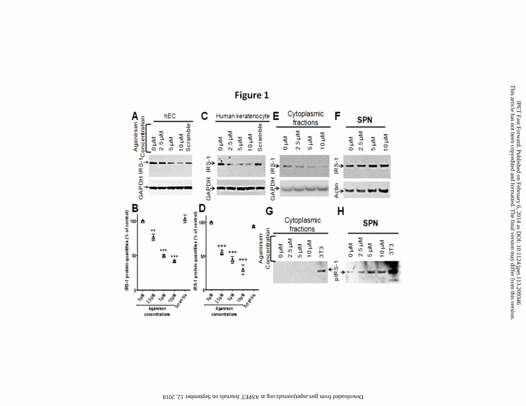
Aganirsen reduced IRS-1 protein expression in the cytoplasmic fraction, but not in the
soluble peri-nuclear/nuclear (SPN) fraction. Aganirsen dose-dependently inhibited IRS-1
expression in hEC (Fig. 1, A and B) and human keratinocytes (Fig. 1, C and D). Subcellular
fractionation of hEC permitted the separation of five fractions: membrane, cytoplasmic, SPN,
chromatin-bound and cytoskeletal protein. IRS-1 protein was only detected in the cytoplasmic
and the SPN fractions. Closer examination of IRS-1 protein expression revealed that
aganirsen inhibited IRS-1 protein expression in the cytoplasmic fraction only (Fig. 1E) and
not in the SPN fraction (Fig. 1F). Analysis of the serine phosphorylation state of IRS-1
protein revealed that pIRS-1Ser was undetectable at the cytoplasmic compartment (Fig. 1G); in
contrast, there was a dose-dependent increase in pIRS-1Ser in the SPN fraction (Fig. 1H).
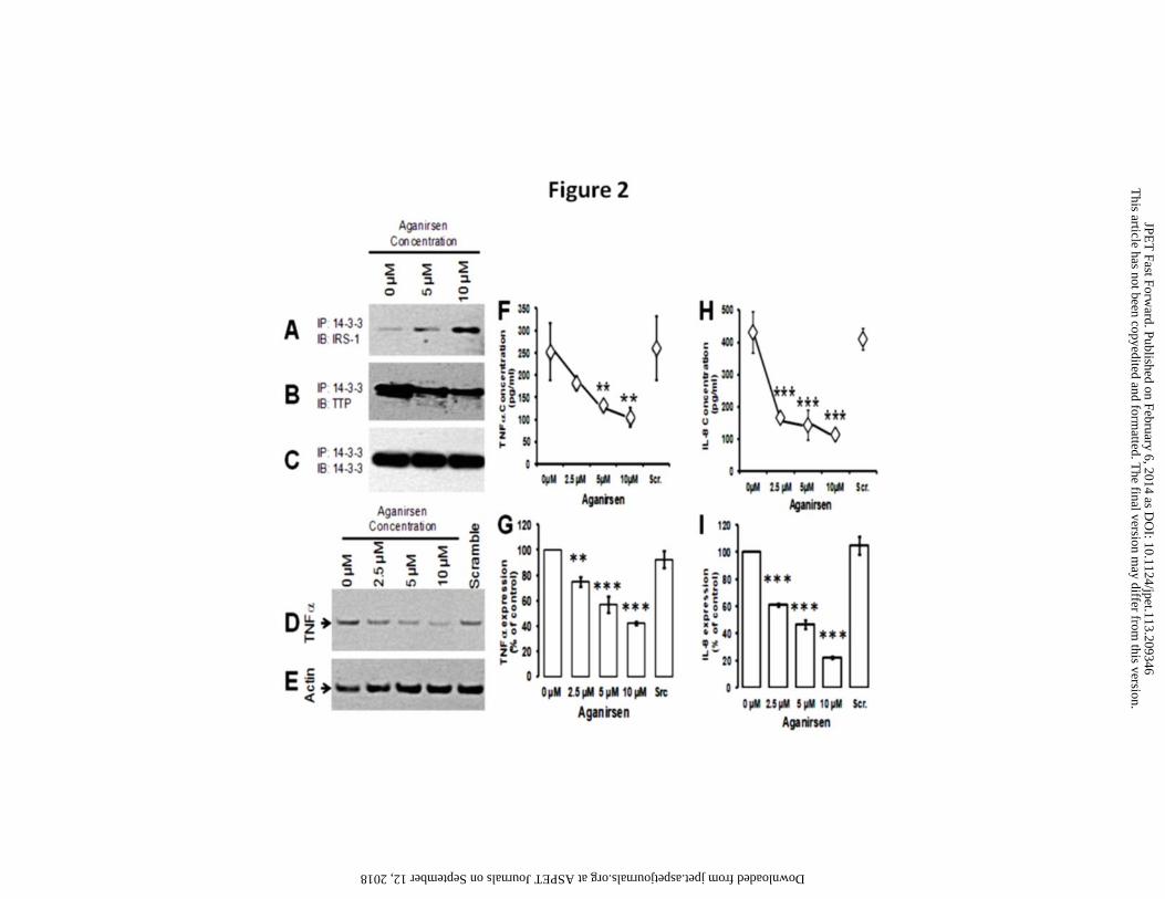
Aganirsen impairs 14-3-3β-TTP complex leading to the inhibition of TNFα expression. In
the SPN fraction, the increased serine phosphorylation of IRS-1 (Fig. 1H) paralleled the
increased association of IRS-1 with the protein 14-3-3β. Increasing amounts of IRS-1 co-
immunoprecipitated with the protein 14-3-3β (Fig. 2A); this was accompanied with
decreasing quantities of the zinc finger protein TTP co-immunoprecipitated with 14-3-3β
(Fig. 2B). These results suggest that the association 14-3-3β/IRS-1 impaired the formation of
the complex 14-3-3β/TTP. TTP is a hyper-phosphorylated protein that destabilizes mRNA by
binding to their AU-rich elements (Cao et al., 2003; Blackshear et al., 2003). The mRNA of
many cytokines contains AU-rich elements including those of TNFα, IL-8, IL-12 and IL-22
(Cao et al., 2003). The scramble oligonucleotide had no effect on the membrane-bound TNFα
in hEC (Fig. 2D) In contrastthere was a dose-dependent decrease in membrane-bound TNFα
in hEC incubated with aganirsen (Fig. 2D). This dose-dependent inhibition of TNFα
expression by aganirsen was confirmed by the measurement of the secreted TNFα protein in
This article has not been copyedited and formatted. The final version may differ from this version.JPET Fast Forward. Published on February 6, 2014 as DOI: 10.1124/jpet.113.209346
at ASPE
T Journals on Septem
ber 12, 2018jpet.aspetjournals.org
Dow
nloaded from
JPET #209346
14
the culture medium (Fig. 2F) and by the measurement of TNFα transcripts by qRT-PCR (Fig.
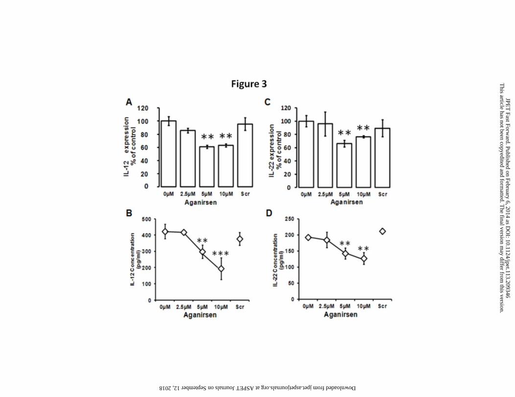
2G). In addition, aganirsen led to a dose-dependent inhibition of IL-8 expression both at the
translational (Fig. 2H) and the transcriptional levels (Fig. 2I), of IL-12 expression both at the
transcriptional (Fig. 3A) and the translational levels (Fig. 3B), and of IL-22 expression both at
the transcriptional (Fig. 3C) and the translational levels (Fig. 3D).
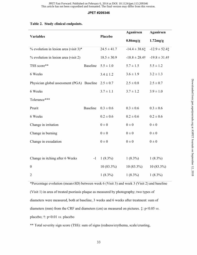
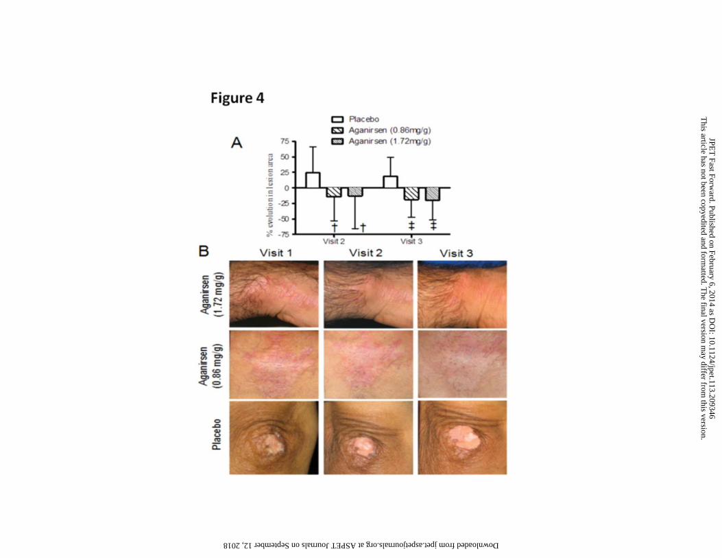
Efficacy of aganirsen in the treatment of psoriatic lesions. Treatment of 12 patients with
topical application of 0.86 mg/g or 1.72 mg/g of aganirsen for 6 weeks led to a significant
reduction (p<0.05) in the area of the treated lesions compared to placebo (Fig. 4A and 4B,
Table 2). By contrast, a slight increase in the lesion area was observed (median 22%) in the
placebo-treated group. Least square means (LSMeans) differences with placebo were -38.9%
(95%CI [-75.8%; -2.0%]) and -37.4% [-74.3%; -0.5%] for the 0.86 mg/g and 1.72 mg/g
groups, respectively. A significant decrease in aganirsen-treated lesion size was observed as
early as 3 weeks (Fig. 4A and 4B, Table 2) of treatment compared to placebo (p<0.01).
LSMeans differences with placebo were -37.3% [-62.7%;-11.9%] and -38.3% [-63.8%; -
12.9%] for the 0.86 mg/g and 1.72 mg/g groups, respectively.
We found no difference in the evolution of the TSS score (p=0.63 and p=0.82 at week 3 and
week 6, respectively, Table 2) between the 3 groups. At baseline, TSS had a median of 6, at
week 3 a median of 4 and at week 6 a median of 3 in the placebo group. In the 0.86 mg/g-
treated group, the median TSS score was of 5 at both baseline and week 3, and of 4 at week 6,
whereas in the 1.72 mg/g-treated group, the median TSS score was 6 at baseline, 4 at week 3
and 3 at week 6. Likewise, the PGA score remained stable over the 6-week period and was
similar in the three groups (Table 2).
Safety of Aganirsen. Safety analysis was carried out on the 12 patients. Aganirsen safety was
This article has not been copyedited and formatted. The final version may differ from this version.JPET Fast Forward. Published on February 6, 2014 as DOI: 10.1124/jpet.113.209346
at ASPE
T Journals on Septem
ber 12, 2018jpet.aspetjournals.org
Dow
nloaded from
JPET #209346
15
good without any reported adverse effects. Patients reported no irritation, burning or
exudation. In each treatment group, mild itching was reported for 1 lesion at baseline, no
itching was reported at 3 weeks and a moderate itching for 1 lesion at 6 weeks.
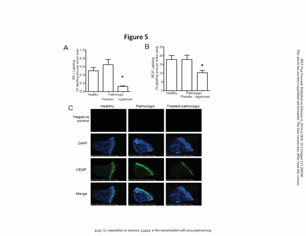
Aganirsen inhibits IRS-1 and VEGF expression in psoriatic lesions of patients. Treatment
with aganirsen (0.86 mg/g) reduced IRS-1 expression by 81±12% (p=0.0132; n=22) in human
psoriatic skin lesions relative to placebo-treated psoriatic skin lesions (Fig. 5A, and
supplemental Fig. 3). No difference in IRS-1 expression was detected between biopsies of
healthy skin and placebo-treated lesions (Fig. 5A). In psoriatic lesion biopsies, we found that
VEGF labelling was lowered by 42±8% (p=0.0074) in (0.86 mg/g)-treated psoriatic lesion
biopsies compared to placebo-treated pathologic skin lesions (Fig. 5B).
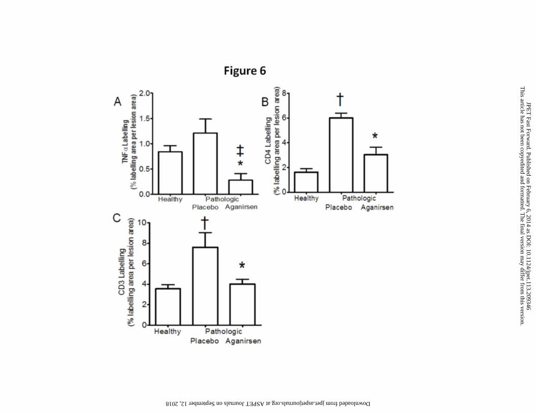
Effects of aganirsen on TNFα-expression, inflammatory cell recruitment and keratinocyte
proliferation in psoriatic lesions of patients. Treatment with aganirsen (0.86 mg/g) was also
associated with a lower TNFα labelling by 77±11% (p=0.0047; n=14) (Fig. 6A and
supplemental Fig. 4) in skin biopsies of psoriatic patients compared to placebo-treated
psoriatic skin samples. Measurement of CD4+ labelling indicated that psoriatic skin contained
65±21% more cells (p=0.013, n=12) than healthy skin. In contrast, CD4+ labelling in
aganirsen (0.86 mg/g)-treated pathologic tissue samples was lower by 43±6% (n=12;
p=0.035) compared with placebo-treated pathologic tissues (Fig. 6B and supplemental Fig. 5).
Similar results were obtained with CD3+ labelling. The psoriatic skin contained 115±40%
more (p=0.0089, n=14) CD3+ labelling than healthy skin, confirming our precedent results
with CD4+ cells and supporting further the role of inflammation in psoriasis. CD3+ labelling
in aganirsen-treated pathologic skin biopsies was reduced (47±6%; p=0.0355) compared to
placebo-treated pathologic tissues (Fig. 6C and supplemental Fig. 6); we found CD3+-
This article has not been copyedited and formatted. The final version may differ from this version.JPET Fast Forward. Published on February 6, 2014 as DOI: 10.1124/jpet.113.209346
at ASPE
T Journals on Septem
ber 12, 2018jpet.aspetjournals.org
Dow
nloaded from
JPET #209346
16
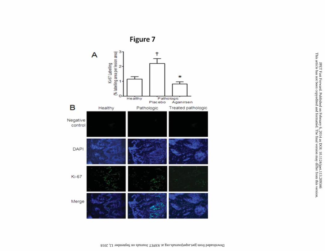
labelling to be similar between healthy skin tissues and aganirsen-treated skin tissues. Finally,
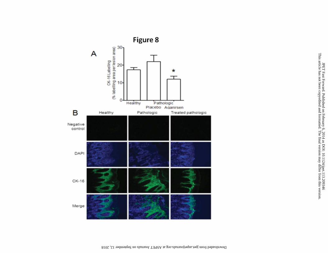
after 6 weeks of treatment with aganirsen, expression of Ki-67 and CK-16 were lower by
59±3% (p=0.0132, n=10) and by 46±8% (p=0.0265, n=5), respectively, compared to placebo-
treated lesions (Fig. 7A-7B and Fig. 8A-8B). Taken together, these findings demonstrate that
aganirsen has potent anti-inflammatory effects and inhibits both the infiltration of
lymphocytes and the proliferation of keratinocytes in the psoriatic skin.
This article has not been copyedited and formatted. The final version may differ from this version.JPET Fast Forward. Published on February 6, 2014 as DOI: 10.1124/jpet.113.209346
at ASPE
T Journals on Septem
ber 12, 2018jpet.aspetjournals.org
Dow
nloaded from
JPET #209346
17
Discussion
This study demonstrates that aganirsen is the first in its class of a new generation of anti-
angiogenic medicines that combines both anti-angiogenic and anti-inflammatory activities and
could be an effective treatment for patients with psoriasis. This statement is supported by
several direct evidences including: i) in vitro, aganirsen induced IRS-1/14-3-3β association
which impaired 14-3-3β/TTP complex, leading to the inhibition of the expression of cytokines
with AU-rich mRNA including IL-8, IL-12, IL-22 and TNFα; ii) in psoriatic patients,
aganirsen inhibited the expression of both VEGF and TNFα, reducing thereby the infiltration
of inflammatory CD4+ and CD3+ lymphocytes; and accordingly iii) aganirsen inhibited
keratinocyte proliferation and consequently reduced psoriatic lesion area in patients.
Psoriasis is a chronic inflammatory disease where many inflammatory cytokines, including
IL-12, Il-17, IL-22 and TNFα, play important pathological roles. Thus, the most desired
therapy is one that targets altogether these inflammatory factors. The mRNA of IL-12, IL-17,
IL-22 and TNFα shares one characteristic, containing AU-rich elements at their 3’-
untranslated regions (Bak and Mikkelsen 2010; Khabar 2010). TTP is a mRNA-binding
protein with high-binding specificity for the so-called class II AU-rich elements within the 3′-
untranslated mRNAs (Cao et al., 2003; Blackshear et al., 2003); specific binding of TTP to
the AU-rich elements results in the destabilization and the decay of the mRNAs (Carballo et
al.,1998; Lai et al.,1999). TTP occurs in a complex with 14-3-3 preventing TTP from binding
to and/or directing mRNA to the degradation machinery (Johnson et al., 2002; Zhao et al.,
2011). Our results show that aganirsen-induced association of IRS-1 with 14-3-3β protein
impairs TTP-14-3-3β complex, revealing a novel regulatory role for IRS-1. Indeed, IRS-1
associates with many of the seven isoforms of 14-3-3 proteins (Ogihara et al., 1997; Kosaki et
al., 1998; Xiang et al., 2002; Oriente et al., 2005); yet, the functional consequence for these
associations was unknown. Our results are therefore the first to demonstrate that by promoting
This article has not been copyedited and formatted. The final version may differ from this version.JPET Fast Forward. Published on February 6, 2014 as DOI: 10.1124/jpet.113.209346
at ASPE
T Journals on Septem
ber 12, 2018jpet.aspetjournals.org
Dow
nloaded from
JPET #209346
18
the association of IRS-1 to 14-3-3β protein and impairing TTP/14-3-3β complex, aganirsen
inhibits the expression of IL-8, IL12, IL22 and TNFα at both the transcriptional and the
translational levels. This is in line with the importance of the liberation of TTP from the
TTP/14-3-3 complex, and with the role of TTP in the regulation of expression of AU-rich
elements in the destabilization and the decay of mRNAs (Carballo et al.,1998; Lai et
al.,1999). These results, combined with those showing that aganirsen inhibited VEGF and IL-
1β expression (Al-Mahmood et al., 2009; Andrieu-Soler et al., 2005), identify aganirsen as a
dual-target therapeutic agent that is highly desirable for the treatment of psoriasis.
Topical applications of aganirsen lead to therapeutic concentrations in both
epidermis and dermis of human skin (supplemental Fig. 2A and 2B). It has been suggested
that the impaired barrier function of psoriatic lesions (Schittek 2011) and the amphipathic
nature of aganirsen (Cloutier et al., 2012) could facilitate uptake of antisense drugs in general
and aganirsen in particular. In agreement, topical applications of aganirsen decreased psoriatic
plaque area in both groups of patients (0.86 and 1.72 mg/g) compared to placebo.
Furthermore, aganirsen had a rapid onset of action as illustrated by the significant decrease in
lesion area after 3 weeks of treatment of -14% and -13% in the 0.86 and 1.72 mg/g treated
groups, respectively. By contrast, the plaque area increased by ~15% in the placebo-treated
group over this period. We did not find any significant differences in the evolution of TSS
and PGA scores either after 3 or 6 weeks of treatment. This is most likely related to the short
term of the trial; at least 12-16 weeks of treatment are required to confirm drug efficacy
against psoriatic lesions (Papp et al., 2012a,b). In addition, our study did not reveal a dose-
dependent effect of aganirsen; both 0.86 and 1.72 mg/g led to a similar decrease in psoriatic
lesion area. These results suggest that the two doses tested in this pilot study induced a
maximal effect, at least within the timeframe of the current protocol. Lower daily doses of
aganirsen combined with a longer period of treatment will therefore have to be tested in future
This article has not been copyedited and formatted. The final version may differ from this version.JPET Fast Forward. Published on February 6, 2014 as DOI: 10.1124/jpet.113.209346
at ASPE
T Journals on Septem
ber 12, 2018jpet.aspetjournals.org
Dow
nloaded from
JPET #209346
19
clinical studies. Importantly, aganirsen was well tolerated locally and no serious adverse
effects were observed in any group. Nonetheless, our study was neither large enough nor of
sufficiently long duration to ascertain uncommon adverse effects.
Psoriasis is characterized by chronic inflammation and enhanced lymphocytes
infiltration (Nestle et al., 2009; Coimbra et al., 2012). Consistently, psoriatic lesions contained
more CD4+ and CD3+ labelling compared to biopsies of healthy skin. Lymphocyte
recruitment requires blood vessels, and aberrant angiogenesis has been linked to psoriasis
(Sabat et al., 2007; Heidenreich et al., 2009; Detmar et al., 1998). In agreement with the anti-
angiogenic property of aganirsen, our results showed that VEGF expression was significantly
lower in psoriatic lesions treated with aganirsen when compared to placebo-treated psoriatic
lesions, although VEGF expression was not elevated in the latter compared to that in the
healthy skin. In contrast, CD31+ and CD34+ labelling was increased in psoriatic lesions
compared to the healthy skin, and this increase was normalized following the treatment with
aganirsen. On aggregate, these results suggest that the anti-inflammatory activity of aganirsen
rather than its anti-angiogenic activity underlies the observed beneficial clinical outcome.
VEGF is, nonetheless, chemo-attractant (Sawano et al., 2001; Ferrara et al., 2003) and
promotes lymphocyte rolling and adhesion in skin microvessels (Detmar et al., 1998).
Consequently, we would like to suggest that the inhibition of VEGF expression by aganirsen
contributes to limit the recruitment of immune cells within the psoriatic lesions. This would
explain the important decrease in TNFα protein in aganirsen-treated psoriatic lesions, which
is mostly produced by activated leukocytes (Orlinick and Chao 1998).
Psoriasis is characterized by epidermal thickening and keratinocyte hyper-proliferation,
which is directly linked to the inflammation within the psoriatic lesion (Nestle et al., 2009;
Sabat et al., 2007). The expression of VEGF and TNFα was reduced by aganirsen in psoriatic
lesions compared to placebo-treated lesions, and was associated with more than 50%
This article has not been copyedited and formatted. The final version may differ from this version.JPET Fast Forward. Published on February 6, 2014 as DOI: 10.1124/jpet.113.209346
at ASPE
T Journals on Septem
ber 12, 2018jpet.aspetjournals.org
Dow
nloaded from
JPET #209346
20
inhibition of keratinocyte proliferation as measured by Ki-67 and CK-16 labelling. The loss of
CK-16-associated fluorescence following a 6-week therapy is a crucial finding since CK-16 is
an important marker predicting therapeutic outcome (Krueger et al., 1995). The potent
inhibitory action of aganirsen on keratinocyte proliferation is most probably linked to its anti-
inflammatory action through inhibition of the expression of VEGF, TNFα, IL-12 and IL-22.
Finally, the levels of VEGF and TNFα were elevated in the biopsies of healthy skin,
confirm the global pro-inflammatory status of these patients (Nestle et al., 2009; Sabat et al.,
2007; Weger 2010). Many anti-inflammatory drugs targeting TNFα, IL-12, IL-17 and 1L-23
have been approved or are in advanced development stage for the treatment of psoriasis
(Coimbra et al., 2012; Papp et al., 2012a). Unlike aganirsen that is active topically, these
orally administrated new drugs have been, however, associated with numerous side effects.
Nonetheless, these new biologics have been used in patients with moderate-to-severe forms of
psoriasis, while aganirsen was used in mild-to-moderate forms of the disease (PASI score
ranged 4-7) and therefore cannot be compared.
In conclusion, our results demonstrate that aganirsen inhibits the expression of
cytokines, which mRNA contains AU-rich elements at their 3’-untranslated regions, including
TNFαILβ IL-8, IL-12 and IL-22, known targets of both inflammation and angiogenesis. On
the other hand, we confirme that aganirsen is a potent inhibitor of VEGF expression, the only
known growth factor that induces inflammation and promotes angiogenesis and
lymphangiogenesis (Al-Mahmood et al., 2009; Ji, 2012; Koch et al., 2011). Despite the
limitations of our trial (the small sample size and the short-term duration of the treatment),
these results suggest that aganirsen is a dual anti-angiogenic and anti-inflammatory agent that
could represent an innovative safe and effective alternative for the treatment of psoriasis, a
disease in need for new and safe therapeutic options.
This article has not been copyedited and formatted. The final version may differ from this version.JPET Fast Forward. Published on February 6, 2014 as DOI: 10.1124/jpet.113.209346
at ASPE
T Journals on Septem
ber 12, 2018jpet.aspetjournals.org
Dow
nloaded from
JPET #209346
21
Acknowledgments
The authors thank Drs Ekat Kritikou, Eric Thorin and Mr Eric Viaud for their help in the
writing and editing of the manuscript, and Mrs Céline Steverlynck (research engineer, Gene
Signal SAS) and Maud Bongaerts (technician, Gene Signal SAS) for their expert technical
assistance.
Authorship Contributions
Participated in research design: A.F., N.D., S.A-M. Conducted experiments: S.C., B.D., M.F.,
C.L. and A.K. Contributed new reagents or analytic tools: J-P.C., S.A-M.. Performed data
analysis: S.A-M. Wrote or contributed to the writing of the manuscript: S.A-M.
This article has not been copyedited and formatted. The final version may differ from this version.JPET Fast Forward. Published on February 6, 2014 as DOI: 10.1124/jpet.113.209346
at ASPE
T Journals on Septem
ber 12, 2018jpet.aspetjournals.org
Dow
nloaded from
JPET #209346
22
References
Ades, E.W., Candal, F.J., Swerlick, R.A., George, V.G., Summers, S., Bosse, D.C. Lawley,
T.J. (1992) HMEC-1: establishment of an immortalized human endothelial cell line. The
Journal of Investigative Dermatology 99: 683–690.
Al-Mahmood S, Colin S, Farhat N et al. (2009) Potent in vivo antiangiogenic effects of GS-
101 (5'-TATCCGGAGGGCTCGCCATGCTGCT-3'), an antisense oligonucleotide
preventing the expression of insulin receptor substrate-1. The Journal of pharmacology
and experimental therapeutics 329: 496-504.
Anderson TW, Darling DA. (1954) A Test of Goodness of Fit. Journal of the American
Statistical Association 49: 765-9.
Andrieu-Soler C, Berdugo M, Doat M et al. (2005) Downregulation of IRS-1 expression
causes inhibition of corneal angiogenesis. Investigative ophthalmology & visual science
46: 4072-8.
Bak RO, Mikkelsen JG. (2010) Regulation of cytokines by small RNAs during skin
inflammation. Journal of Biomedical Science 17:53.
Blackshear PJ, Lai WS, Kennington EA, Brewer G, Wilson GM, Guan X, Zhou P.(2003)
Characteristics of the interaction of a synthetic human tristetraprolin tandem zinc finger
peptide with AU-rich element containing RNA substrates. J. Biol. Chem 278:19947–
19955.
Cao H, Dzineku F, Blackshear PJ. (2003) Expression and purification of recombinant
tristetraprolin that can bind to tumor necrosis factor-α mRNA and serve as a substrate for
mitogen-activated protein kinases. Arch. Biochem. Biophys 412:106–120.
Caldarola G, De Simone C, Carbone A et al. (2009) TNFalpha and its receptors in psoriatic
skin, before and after treatment with etanercept. International journal of immunopathology
and pharmacology 22: 961-6.
This article has not been copyedited and formatted. The final version may differ from this version.JPET Fast Forward. Published on February 6, 2014 as DOI: 10.1124/jpet.113.209346
at ASPE
T Journals on Septem
ber 12, 2018jpet.aspetjournals.org
Dow
nloaded from
JPET #209346
23
Carballo E, Lai WS, Blackshear PJ. (1998) Feedback inhibition of macrophage tumor
necrosis factor-α production by tristetraprolin. Science 281:1001–1005.
Cloutier F, Lawrence M, Goody R et al. (2012) Antiangiogenic activity of aganirsen in
nonhuman primate and rodent models of retinal neovascular disease after topical
administration. Investigative ophthalmology & visual science 53: 1195-203.
Coimbra S, Figueiredo A, Castro E et al. (2012) The roles of cells and cytokines in the
pathogenesis of psoriasis. International journal of dermatology 51: 389-95; quiz 95-8.
Detmar M, Brown LF, Claffey KP et al. (1994) Overexpression of vascular permeability
factor/vascular endothelial growth factor and its receptors in psoriasis. The Journal of
experimental medicine 180: 1141-6.
Detmar M, Brown LF, Schon MP et al. (1998) Increased microvascular density and enhanced
leukocyte rolling and adhesion in the skin of VEGF transgenic mice. The Journal of
investigative dermatology 111: 1-6.
Feldman SR, Krueger GG. (2005) Psoriasis assessment tools in clinical trials. Annals of the
rheumatic diseases 64 Suppl 2: ii65-8; discussion ii9-73.
Ferrara N, Gerber HP, LeCouter J. (2003) The biology of VEGF and its receptors. Nature
medicine 9: 669-76.
Heidenreich R, Rocken M, Ghoreschi K. (2009) Angiogenesis drives psoriasis pathogenesis.
International journal of experimental pathology 90: 232-48.
Henno A, Blacher S, Lambert CA et al. (2010) Histological and transcriptional study of
angiogenesis and lymphangiogenesis in uninvolved skin, acute pinpoint lesions and
established psoriasis plaques: an approach of vascular development chronology in
psoriasis. Journal of dermatological science 57: 162-9.
Ji RC. (2012) Macrophages are important mediators of either tumor- or inflammation-induced
lymphangiogenesis. Cellular and molecular life sciences : CMLS 69: 897-914.
This article has not been copyedited and formatted. The final version may differ from this version.JPET Fast Forward. Published on February 6, 2014 as DOI: 10.1124/jpet.113.209346
at ASPE
T Journals on Septem
ber 12, 2018jpet.aspetjournals.org
Dow
nloaded from
JPET #209346
24
Johnson BA, Stehn JR, Yaffe MB, Blackwell TK. (2002) Cytoplasmic localization of
tristetraprolin involves 14-3-3-dependent and -independent mechanisms. J. Biol. Chem
277:18029–18036.
Khabar KSA. (2010) Post-transcriptional control during chronic inflammation and cancer: a
focus on AU-rich elements. Cell. Mol. Life Sci. 67:2937–2955.Koch S, Tugues S, Li X et
al. (2011) Signal transduction by vascular endothelial growth factor receptors. The
Biochemical journal 437: 169-83.
Kosaki A, Yamada K, Suga J, Otaka A, Kuzuya H. (1998) 14-3-3beta protein associates with
insulin receptor substrate 1 and decreases insulin-stimulated phosphatidylinositol 3’-kinase
activity in 3T3L1 adipocytes. J Biol Chem 273:940–944.
Krueger JG, Wolfe JT, Nabeya RT et al. (1995) Successful ultraviolet B treatment of psoriasis
is accompanied by a reversal of keratinocyte pathology and by selective depletion of
intraepidermal T cells. The Journal of experimental medicine 182: 2057-68.
Lai WS, Carballo E, Strum JR, Kennington EA, Phillips RS, Blackshear PJ. (1999) Evidence
that tristetraprolin binds to AU-rich elements and promotes the deadenylation and
destabilization of tumor necrosis factor α mRNA. Mol. Cell. Biol 19:4311–4323.
Nestle FO, Kaplan DH, Barker J. (2009) Psoriasis. The New England journal of medicine 361:
496-509.
Nograles KE, Zaba LC, Guttman-Yassky E et al. (2008) Th17 cytokines interleukin (IL)-17
and IL-22 modulate distinct inflammatory and keratinocyte-response pathways. The British
journal of dermatology 159: 1092-102.
Ogihara T, Isobe T, Ichimura T, Taoka M, Funaki M, et al. (1997) 14-3-3 protein binds to
insulin receptor substrate-1, one of the binding sites of which is in the phosphotyrosine
binding domain. J Biol Chem 272: 25267–25274.
This article has not been copyedited and formatted. The final version may differ from this version.JPET Fast Forward. Published on February 6, 2014 as DOI: 10.1124/jpet.113.209346
at ASPE
T Journals on Septem
ber 12, 2018jpet.aspetjournals.org
Dow
nloaded from

JPET #209346
25
Oriente F, Andreozzi F, Romano C, Perruolo G, Perfetti A, et al. (2005) Protein kinase C-
alpha regulates insulin action and degradation by interacting with insulin receptor
substrate-1 and 14-3-3 epsilon. J Biol Chem 280: 40642–40649.
Orlinick JR, Chao MV. (1998) TNF-related ligands and their receptors. Cellular signalling
10: 543-51.
Papp KA, Leonardi C, Menter A et al. (2012a) Brodalumab, an anti-interleukin-17-receptor
antibody for psoriasis. The New England journal of medicine 366: 1181-9.
Papp K, Cather JC, Rosoph L et al. (2012b) Efficacy of apremilast in the treatment of
moderate to severe psoriasis: a randomised controlled trial. Lancet.
Sabat R, Sterry W, Philipp S et al.(2007) Three decades of psoriasis research: where has it led
us? Clinics in dermatology 25: 504-9.
Sawano A, Iwai S, Sakurai Y et al. (2001) Flt-1, vascular endothelial growth factor receptor
1, is a novel cell surface marker for the lineage of monocyte-macrophages in humans.
Blood 97: 785-91.
Schittek B. (2011) The antimicrobial skin barrier in patients with atopic dermatitis. Current
problems in dermatology 41: 54-67.
Weger W. (2010) Current status and new developments in the treatment of psoriasis and
psoriatic arthritis with biological agents. British journal of pharmacology 160: 810-20.
Xiang X, Yuan M, Song Y, Ruderman N, Wen R, et al. (2002) 14-3-3 facilitates insulin-
stimulated intracellular trafficking of insulin receptor substrate 1. Mol Endocrinol 16: 552–
562.
Zhao J, Meyerkord CL, Du Y, Khuri FR, Fu H. (2011) 14-3-3 proteins as potential therapeutic
targets. Semin Cell Dev Biol. 22: 705–712.
This article has not been copyedited and formatted. The final version may differ from this version.JPET Fast Forward. Published on February 6, 2014 as DOI: 10.1124/jpet.113.209346
at ASPE
T Journals on Septem
ber 12, 2018jpet.aspetjournals.org
Dow
nloaded from

JPET #209346
26
Footnotes
*: these authors equally contributed to the work.
Funding
This study was supported by Gene Signal SAS, 4, Rue Pierre Fontaine, 91000 Evry, France.
Conflict of interests
Bernadette Darné, Amin Kadi, Maryline Favier, Corinne Lesaffre, and Neijib Doss declare no
conflict of interest. Jean-Pascal Conduzorgues is a consultant in product formulation for Gene
Signal. Sylvie Colin and Salman Al-Mahmood are cofounders of Gene Signal, have stock
ownership and are employees of Gene Signal. Antoine Ferry detains stock ownership of Gene
Signal.
This article has not been copyedited and formatted. The final version may differ from this version.JPET Fast Forward. Published on February 6, 2014 as DOI: 10.1124/jpet.113.209346
at ASPE
T Journals on Septem
ber 12, 2018jpet.aspetjournals.org
Dow
nloaded from
JPET #209346
27
Figure legends
Figure 1: Aganirsen reduced IRS-1 protein expression in the cytoplasmic compartment,
but not in the SPN. A) Equivalent amounts of proteins extracts from hEC incubated with
increasing concentrations of aganirsen were used to quantify IRS-1 protein by Western blot
(upper panel). Membranes were stripped and re-probed with anti-GAPDH mAb to control for
equal loading (lower panel). B) The same hEC proteins extracts as in A were used to quantify
IRS-1 protein by ELISA sandwich assay. C) Equivalent amounts of proteins extracts from
human keratinocytes incubated with increasing concentrations of aganirsen were used to
quantify IRS-1 protein by Western blot (upper panel). Membranes were stripped and re-
probed with anti-GAPDH mAb to control for equal loading (lower panel). D) The same
human keratinocyte protein extracts as in C were used to quantify IRS-1 protein by ELISA
sandwich assay. E) Equivalent amounts of the cytoplasmic fractions from hEC incubated with
increasing concentrations of aganirsen were resolved by SDS-PAGE, transferred to PVDF
membranes and probed with anti-IRS-1 mAb (Upper panel). The membranes were stripped
and re-probed with anti-GAPDH mAb to control for equal loading (lower panel). F)
Equivalent amounts of SPN fractions from hEC incubated with increasing concentrations of
aganirsen were resolved by SDS-PAGE, transferred to PVDF membrane and probed with
anti-serine-phosphorylated IRS-1 (pIRS-1) mAb (Upper panel). The same membranes were
stripped and re-probed with anti-actin mAb to control for equal loading (lower panel). G)
Equivalent amounts of the cytoplasmic fraction from hEC incubated with increasing
concentrations of aganirsen were resolved by SDS-PAGE, transferred to PVDF membrane
and probed with anti-serine-phosphorylated IRS-1 (pIRS-1) mAb. H) Equivalent amounts of
SPN fractions from hEC incubated with increasing concentrations of aganirsen were resolved
by SDS-PAGE, transferred to PVDF membrane and probed with anti-serine-phosphorylated
IRS-1 (pIRS-1) mAb. Where indicated: *, p < 0.05; **, p<0.001; ***, p<0.0001 versus
This article has not been copyedited and formatted. The final version may differ from this version.JPET Fast Forward. Published on February 6, 2014 as DOI: 10.1124/jpet.113.209346
at ASPE
T Journals on Septem
ber 12, 2018jpet.aspetjournals.org
Dow
nloaded from
JPET #209346
28
vehicle-treated group (0.9% NaCl). Scr corresponded to the scramble oligonucleotide (10
µM).
Figure 2: Aganirsen impairs 14-3-3β/TTP complex leading to the inhibition of both IL-8
and TNFα expression. A) Human EC incubated with increasing concentrations of aganirsen
were lysed and proteins were immunoprecipitated with anti-14-3-3β mAb. Equivalent
amounts of the immunoprecipitates were resolved by SDS-PAGE, transferred to PVDF
membranes and probed with anti-IRS-1 mAb. B) The same equivalent amounts of
immunoprecipitates as in A were resolved by SDS-PAGE, transferred to PVDF membranes
and probed with anti-TTP mAb. C) The membranes used in A were stripped and re-probed
with anti-14-3-3β mAb to control for equal loading in A and B. D) Human EC were incubated
with increasing concentrations of aganirsen were lysed and equivalent amounts of proteins
were resolved by SDS-PAGE, transferred to PVDF membranes and probed with anti-TNFα
mAb to reveal the membrane bound TNFα. E) The PVDF membranes used in D were
stripped and re-probed with anti-actin mAb to control for equal loading in D. F) Human EC
were incubated with increasing concentrations of aganirsen and TNFα mRNA was measured
by qRT-PCR and expressed as mean ± S.E.M. (n = 4). G) Human EC in serum-deprived
culture medium were incubated overnight with increasing concentrations of aganirsen and the
culture supernatants were collected to quantify secreted TNFα proteins by ELISA sandwich
assay. Results are expressed as pg of TNFα per ml of culture supernatant and presented as
mean ± S.E.M. (n = 4). H) Human EC were incubated with increasing concentrations of
aganirsen and IL-8 mRNA was measured by qRT-PCR and expressed as mean ± S.E.M. (n =
4). I) Human EC in serum-deprived culture medium were incubated over-night with
increasing concentrations of aganirsen and the culture supernatants were collected to quantify
secreted IL-8 protein by ELISA sandwich assay. Results were expressed as pg of IL-8 per ml
This article has not been copyedited and formatted. The final version may differ from this version.JPET Fast Forward. Published on February 6, 2014 as DOI: 10.1124/jpet.113.209346
at ASPE
T Journals on Septem
ber 12, 2018jpet.aspetjournals.org
Dow
nloaded from
JPET #209346
29
of culture supernatant and presented as mean ± S.E.M. (n = 4). Where indicated; *, p < 0.05;
**, p<0.001; ***, p<0.0001 versus vehicle-treated group (0.9% NaCl). Scr correspond to the
scramble oligonucleotide (10 µM).
Figure 3: Aganirsen inhibits both IL-12 and IL-22 expression. A) Human EC were
incubated with increasing concentrations of aganirsen and IL-12 mRNA was measured by
qRT-PCR and expressed as mean ± S.E.M. (n = 4). B) Human EC in serum-deprived culture
medium were incubated overnight with increasing concentrations of aganirsen and the culture
supernatant was collected to quantify secreted IL-12 protein by ELISA sandwich assay.
Results were expressed as pg of IL-12 per ml of culture supernatant and presented as mean ±
S.E.M. (n = 4). C) Human EC were incubated with increasing concentrations of aganirsen and
IL-22 mRNA was measured by qRT-PCR and expressed as mean ± S.E.M. (n = 4). D) Human
EC in serum-deprived culture medium were incubated overnight with increasing
concentrations of aganirsen and the culture supernatant was collected to quantify secreted IL-
22 protein by ELISA sandwich assay. Results were expressed as pg of IL-22 per ml of culture
supernatant and presented as mean ± S.E.M. (n = 4). Where indicated: *, p < 0.05; **,
p<0.001; ***, p<0.0001 versus vehicle-treated group (0.9% NaCl). Scr correspond to the
scramble oligonucleotide (10 µM).
Figure 4: Topical application of aganirsen reduced psoriatic lesion area compared to
placebo. (A) Treatment of 12 patients with topical application of 0.86 mg/g or 1.72 mg/g of
aganirsen led to a significant reduction at 3 (†: p<0.01) and 6 weeks (‡: p<0.05) in the treated
lesions compared to placebo. Data are means±SD. (B) Representative images of the evolution
of psoriatic lesions of patients treated with aganirsen at 1.72 mg/g (upper panel), patients
treated with aganirsen at 0.86 mg/g (middle panel) and patients treated with placebo (lower
This article has not been copyedited and formatted. The final version may differ from this version.JPET Fast Forward. Published on February 6, 2014 as DOI: 10.1124/jpet.113.209346
at ASPE
T Journals on Septem
ber 12, 2018jpet.aspetjournals.org
Dow
nloaded from
JPET #209346
30
panel). Visit 1, visit 2 and visit 3 correspond to baseline, after 3 week and after 6 week of
treatment, respectively.
Figure 5: Topical application of aganirsen for 6 weeks decreased both IRS-1 and VEGF
expression in psoriatic lesion biopsies. (A) Inhibition of IRS-1 expression (measured by
immunofluorescence using an IRS-1 antibody) after daily topical treatment for 6 weeks with
aganirsen (0.86 mg/g of ointment) in psoriatic skin lesion biopsies from patients. Data are
means±SEM. *, p=0.0008 vs. placebo-treated psoriatic lesions. (B) Inhibition of VEGF
expression after daily topical treatment for 6 weeks with aganirsen (0.86 mg/g of ointment).
Data are means±SEM. *, p=0.0074 vs. placebo-treated psoriatic lesions. (C) Representative
images of vascular endothelial growth factor (VEGF) labeling in human skin biopsies using
anti-VEGF monoclonal antibody in healthy untreated (left panel), psoriatic lesions placebo-
treated (middle panel) and aganirsen-treated (right panel). Negative controls (secondary
antibody only) are presented in the top row of images, cell nuclei are stained with DAPI in
blue (second row of images), immunostaining of VEGF in green (third row of images) and the
merge images incorporate both nuclei and VEGF immunostaining-derived fluorescence
(fourth row of images).
Figure 6: Topical application of aganirsen inhibits TNFα, CD4+ and CD3+ lymphocyte
infiltration in psoriatic lesions. Twice daily topical treatment for 6 weeks with aganirsen at
0.86 mg/g of ointment inhibited TNFα expression (A) in psoriatic skin lesions and reduced
the levels of infiltrated CD4+ (B) and CD3+ (C) lymphocytes compared to placebo-treated
psoriatic lesions. Data are means±SEM. A: *, p=0.047 vs. pathologic placebo-treated and ‡,
p=0.007 vs. healthy skin; B: *, p=0.002 vs. pathologic placebo-treated, †: p<0.0001 vs.
healthy skin; C: *, p=0.0355 vs. pathologic placebo-treated, †: p=0.0089 vs. healthy skin.
This article has not been copyedited and formatted. The final version may differ from this version.JPET Fast Forward. Published on February 6, 2014 as DOI: 10.1124/jpet.113.209346
at ASPE
T Journals on Septem
ber 12, 2018jpet.aspetjournals.org
Dow
nloaded from
JPET #209346
31
Figure 7. Topical application of aganirsen inhibits keratinocyte proliferation in
psoriatic lesions. (A) Twice daily topical treatment for 6 weeks with aganirsen at 0.86 mg/g
of ointment inhibited keratinocyte proliferation as evidenced by the reduced expression of the
keratinocyte proliferation marker Ki-67 in psoriatic lesions. Data are means±SEM. *,
p=0.0022 vs. pathologic placebo-treated and †, p=0.021 vs. healthy skin. (B) Representative
images of Ki-67 labelling in human skin samples using anti-Ki-67 monoclonal antibody in
healthy non-treated (left panel) and psoriatic lesions either placebo-treated (middle panel) or
aganirsen-treated (right panel). Negative controls (secondary antibody only) are presented in
the top row of images. Cell nuclei are stained with DAPI in blue (second row of images),
immunostaining of Ki-67 in green (third row of images) and the merge images incorporate
both nuclei and Ki-67 immunostaining-derived fluorescence (fourth row of images).
Figure 8. Topical application of aganirsen inhibits cytokeratin 16 expression in psoriatic
lesions. (A) Twice daily topical treatment for 6 weeks with aganirsen at 0.86 mg/g of
ointment inhibited keratinocyte proliferation as evidenced by the reduced expression of CK-
16 in psoriatic lesions. Data are means±SEM. *, p=0.0265 vs. pathologic placebo-treated. (B)
Representative images of CK-16 labelling in human skin samples using anti-CK-16
monoclonal antibody in healthy non-treated (left panel), and psoriatic lesions either placebo-
treated (middle panel) or aganirsen-treated (right panel). Negative controls (secondary
antibody only) are presented in the top row of images. Cell nuclei are stained with DAPI in
blue (second row of images), immunostaining of KC-16 in green (third row of images) and
the merge images incorporate both nuclei and KC-16 immunostaining-derived fluorescence
(fourth row of images).
This article has not been copyedited and formatted. The final version may differ from this version.JPET Fast Forward. Published on February 6, 2014 as DOI: 10.1124/jpet.113.209346
at ASPE
T Journals on Septem
ber 12, 2018jpet.aspetjournals.org
Dow
nloaded from
JPET #209346
32
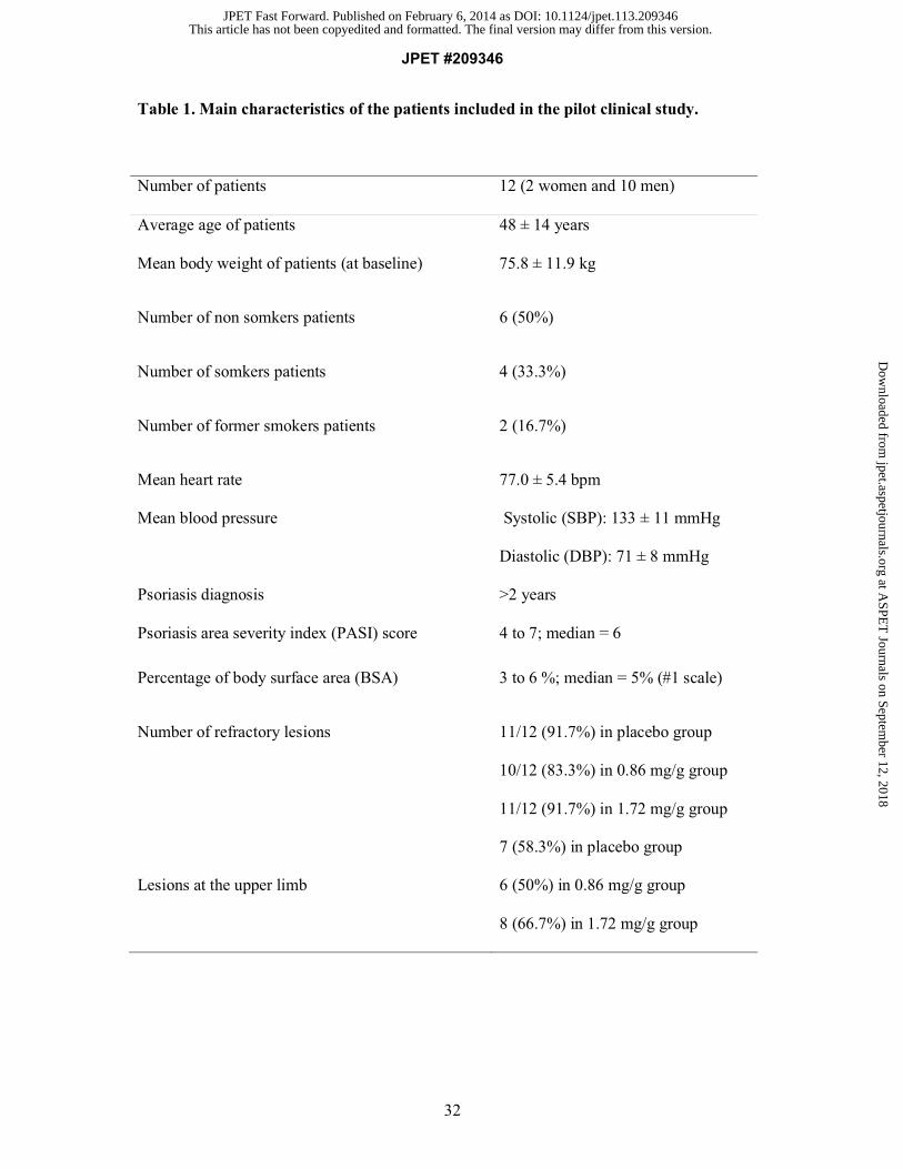
Table 1. Main characteristics of the patients included in the pilot clinical study.
Number of patients 12 (2 women and 10 men)
Average age of patients 48 ± 14 years
Mean body weight of patients (at baseline) 75.8 ± 11.9 kg
Number of non somkers patients 6 (50%)
Number of somkers patients 4 (33.3%)
Number of former smokers patients 2 (16.7%)
Mean heart rate 77.0 ± 5.4 bpm
Mean blood pressure Systolic (SBP): 133 ± 11 mmHg
Diastolic (DBP): 71 ± 8 mmHg
Psoriasis diagnosis >2 years
Psoriasis area severity index (PASI) score 4 to 7; median = 6
Percentage of body surface area (BSA) 3 to 6 %; median = 5% (#1 scale)
Number of refractory lesions 11/12 (91.7%) in placebo group
10/12 (83.3%) in 0.86 mg/g group
11/12 (91.7%) in 1.72 mg/g group
Lesions at the upper limb
7 (58.3%) in placebo group
6 (50%) in 0.86 mg/g group
8 (66.7%) in 1.72 mg/g group
This article has not been copyedited and formatted. The final version may differ from this version.JPET Fast Forward. Published on February 6, 2014 as DOI: 10.1124/jpet.113.209346
at ASPE
T Journals on Septem
ber 12, 2018jpet.aspetjournals.org
Dow
nloaded from
JPET #209346
33
Table 2. Study clinical endpoints.
Variables Placebo Aganirsen
0.86mg/g
Aganirsen
1.72mg/g
% evolution in lesion area (visit 3)* 24.5 ± 41.7 -14.4 ± 38.6‡ -12.9 ± 52.4‡
% evolution in lesion area (visit 2) 18.5 ± 30.9 -18.8 ± 28.4† -19.8 ± 31.4†
TSS score** Baseline
6 Weeks
5.5 ± 1.0
3.4 ± 1.2
5.7 ± 1.5
3.6 ± 1.9
5.5 ± 1.2
3.2 ± 1.3
Physician global assessment (PGA) Baseline
6 Weeks
2.5 ± 0.7
3.7 ± 1.1
2.5 ± 0.8
3.7 ± 1.2
2.5 ± 0.7
3.9 ± 1.0
Tolerance***
Prurit Baseline
6 Weeks
Change in irritation
Change in burning
Change in exsudation
0.3 ± 0.6
0.2 ± 0.6
0 ± 0
0 ± 0
0 ± 0
0.3 ± 0.6
0.2 ± 0.6
0 ± 0
0 ± 0
0 ± 0
0.3 ± 0.6
0.2 ± 0.6
0 ± 0
0 ± 0
0 ± 0
Change in itching after 6 Weeks -1 1 (8.3%) 1 (8.3%) 1 (8.3%)
0 10 (83.3%) 10 (83.3%) 10 (83.3%)
2 1 (8.3%) 1 (8.3%) 1 (8.3%)
*Percentage evolution (mean±SD) between week 6 (Visit 3) and week 3 (Visit 2) and baseline
(Visit 1) in area of treated psoriasis plaque as measured by photography; two types of
diameters were measured, both at baseline, 3 weeks and 6 weeks after treatment: sum of
diameters (mm) from the CRF and diameters (cm) as measured on pictures. ‡: p<0.05 vs.
placebo; †: p<0.01 vs. placebo
** Total severity sign score (TSS): sum of signs (redness/erythema, scale/crusting,
This article has not been copyedited and formatted. The final version may differ from this version.JPET Fast Forward. Published on February 6, 2014 as DOI: 10.1124/jpet.113.209346
at ASPE
T Journals on Septem
ber 12, 2018jpet.aspetjournals.org
Dow
nloaded from

JPET #209346
34
thickening/elevation) and symptoms (pruritus) using 4-point scales (e.g. 0=None/clear, Mild
=1, Moderate =2, Extensive=3). Score varies from 0 to 12 for each lesion
***Local tolerance of treatment: patient’s assessment on a 4-point scale of: irritation, itching,
pruritus, sensation of burning and exudation.
For Physician Global Assessment (PGA), three items were noted: erythema, induration and
scale, according to Feldman and Krueger 2005.
This article has not been copyedited and formatted. The final version may differ from this version.JPET Fast Forward. Published on February 6, 2014 as DOI: 10.1124/jpet.113.209346
at ASPE
T Journals on Septem
ber 12, 2018jpet.aspetjournals.org
Dow
nloaded from
This article has not been copyedited and form
atted. The final version m
ay differ from this version.
JPET
Fast Forward. Published on February 6, 2014 as D
OI: 10.1124/jpet.113.209346
at ASPET Journals on September 12, 2018 jpet.aspetjournals.org Downloaded from
This article has not been copyedited and form
atted. The final version m
ay differ from this version.
JPET
Fast Forward. Published on February 6, 2014 as D
OI: 10.1124/jpet.113.209346
at ASPET Journals on September 12, 2018 jpet.aspetjournals.org Downloaded from
This article has not been copyedited and form
atted. The final version m
ay differ from this version.
JPET
Fast Forward. Published on February 6, 2014 as D
OI: 10.1124/jpet.113.209346
at ASPET Journals on September 12, 2018 jpet.aspetjournals.org Downloaded from
This article has not been copyedited and form
atted. The final version m
ay differ from this version.
JPET
Fast Forward. Published on February 6, 2014 as D
OI: 10.1124/jpet.113.209346
at ASPET Journals on September 12, 2018 jpet.aspetjournals.org Downloaded from
This article has not been copyedited and form
atted. The final version m
ay differ from this version.
JPET
Fast Forward. Published on February 6, 2014 as D
OI: 10.1124/jpet.113.209346
at ASPET Journals on September 12, 2018 jpet.aspetjournals.org Downloaded from
This article has not been copyedited and form
atted. The final version m
ay differ from this version.
JPET
Fast Forward. Published on February 6, 2014 as D
OI: 10.1124/jpet.113.209346
at ASPET Journals on September 12, 2018 jpet.aspetjournals.org Downloaded from
This article has not been copyedited and form
atted. The final version m
ay differ from this version.
JPET
Fast Forward. Published on February 6, 2014 as D
OI: 10.1124/jpet.113.209346
at ASPET Journals on September 12, 2018 jpet.aspetjournals.org Downloaded from
This article has not been copyedited and form
atted. The final version m
ay differ from this version.
JPET
Fast Forward. Published on February 6, 2014 as D
OI: 10.1124/jpet.113.209346
at ASPET Journals on September 12, 2018 jpet.aspetjournals.org Downloaded from
![JPET #201616jpet.aspetjournals.org/content/jpet/early/2013/01/08/jpet.112.201616.full.pdf[D-Ala2, NMe-Phe4, Gly-ol5]-This article has not been copyedited and formatted. The final version](https://static.fdocument.org/doc/165x107/5e3960ce75216306724b28d2/jpet-d-ala2-nme-phe4-gly-ol5-this-article-has-not-been-copyedited-and-formatted.jpg)