JPET #155630 1 Title Page Structure-Activity Relationship Studies ...
31
JPET #155630 1 Title Page Structure-Activity Relationship Studies of Fostriecin, Cytostatin and Key Analogues, with PP1, PP2A, PP5, and ( β12- β13)-chimeras (PP1/PP2A and PP5/PP2A) Provide Further Insight Into the Inhibitory Actions of Fostriecin Family Inhibitors Mark R. Swingle * , Lauren Amable * , Brian G. Lawhorn, Suzanne B. Buck, Christopher P. Burke, Pukar Ratti, Kimberly L. Fischer, Dale L. Boger and Richard E. Honkanen Department of Biochemistry and Molecular Biology, University of South Alabama College of Medicine, Mobile, Alabama 36688 (M.R.S., L.A., P.R., K.L.F., R.E.H.) Department of Chemistry and The Skaggs Institute for Chemical Biology, The Scripps Research Institute, 10550 North Torrey Pines Road, La Jolla, California 92037 (B.G.L., S.B.B., C.P.B., D.L.B.) JPET Fast Forward. Published on July 10, 2009 as DOI:10.1124/jpet.109.155630 Copyright 2009 by the American Society for Pharmacology and Experimental Therapeutics. This article has not been copyedited and formatted. The final version may differ from this version. JPET Fast Forward. Published on July 10, 2009 as DOI: 10.1124/jpet.109.155630 at ASPET Journals on January 30, 2018 jpet.aspetjournals.org Downloaded from
-
Upload
hoangduong -
Category
Documents
-
view
221 -
download
0
Transcript of JPET #155630 1 Title Page Structure-Activity Relationship Studies ...
JPET #155630
1
Title Page
Structure-Activity Relationship Studies of Fostriecin, Cytostatin and Key Analogues, with
PP1, PP2A, PP5, and (β12-β13)-chimeras (PP1/PP2A and PP5/PP2A) Provide Further
Insight Into the Inhibitory Actions of Fostriecin Family Inhibitors
Mark R. Swingle*, Lauren Amable*, Brian G. Lawhorn, Suzanne B. Buck, Christopher P. Burke,
Pukar Ratti, Kimberly L. Fischer, Dale L. Boger and Richard E. Honkanen
Department of Biochemistry and Molecular Biology, University of South Alabama College of Medicine,
Mobile, Alabama 36688 (M.R.S., L.A., P.R., K.L.F., R.E.H.) Department of Chemistry and The Skaggs
Institute for Chemical Biology, The Scripps Research Institute, 10550 North Torrey Pines Road, La Jolla,
California 92037 (B.G.L., S.B.B., C.P.B., D.L.B.)
JPET Fast Forward. Published on July 10, 2009 as DOI:10.1124/jpet.109.155630
Copyright 2009 by the American Society for Pharmacology and Experimental Therapeutics.
This article has not been copyedited and formatted. The final version may differ from this version.JPET Fast Forward. Published on July 10, 2009 as DOI: 10.1124/jpet.109.155630
at ASPE
T Journals on January 30, 2018
jpet.aspetjournals.orgD
ownloaded from
JPET #155630
2
Running Title Page
Running title: Structure activity relationship studies of fostriecin
Address Correspondence to: Richard E. Honkanen, Department of Biochemistry and Molecular Biology,
MSB 2362, 307 University BLVD North, Mobile, AL 36688; Phone: 251 460-6859; Fax 251 460-6850;
E-mail [email protected]
Text pages (double spaced including title pages and references); 22
Number of tables: 1
Number of Figures: 5
Number of words (excluding references)
Abstract: 235
Introduction: 743
Discussion: 1440
References: 39
ABBREVITIONS: PP1c; ser/thr protein phosphatase 1 catalytic subunit, PP2Ac; ser/thr protein
phosphatase 2A catalytic subunit, PP5c; ser/thr protein phosphatase 5 catalytic subunit, PPase;
serine/threonine phosphatase
This article has not been copyedited and formatted. The final version may differ from this version.JPET Fast Forward. Published on July 10, 2009 as DOI: 10.1124/jpet.109.155630
at ASPE
T Journals on January 30, 2018
jpet.aspetjournals.orgD
ownloaded from
JPET #155630
3
Abstract
Fostriecin and cytostatin are structurally related natural inhibitors of serine/threonine phosphatases, with
promising antitumor activity. The total synthesis of these antitumor agents has enabled the production of
structural analogues, which are useful to explore the biological significance of features contained in the
parent compounds. Here, the inhibitory activity of fostriecin, cytostatin, and 10 key structural analogues
were tested in side-by-side phosphatase assays to further characterize their inhibitory activity against
PP1c, PP2Ac, PP5c, and chimeras of PP1 and PP5, in which key residues predicted for inhibitor contact
with PP2A were introduced into PP1 and PP5 using site-directed mutagenesis. The data confirms the
importance of the C9-phosphate and C11-alcohol for general inhibition and further demonstrates the
importance of a predicted C3 interaction with a unique cysteine (C269) in the β12-β13 loop of PP2A. The
data also indicates that additional features beyond the unsaturated lactone contribute to inhibitory potency
and selectivity. Notably, a derivative of fostriecin lacking the entire lactone subunit demonstrated marked
potency and selectivity for PP2A, while having substantially reduced and similar activity against PP1 and
PP1/PP2A- PP5/PP2A-chimeras that have greatly increased sensitivity to both fostriecin and cytostatin.
This suggests that other features [e.g. the (Z,Z,E)-triene] also contribute to inhibitory selectivity. When
considered together with previous data, these studies suggest that despite the high structural conservation
of the catalytic site in PP1, PP2A and PP5, the development of highly selective catalytic inhibitors should
be feasible.
This article has not been copyedited and formatted. The final version may differ from this version.JPET Fast Forward. Published on July 10, 2009 as DOI: 10.1124/jpet.109.155630
at ASPE
T Journals on January 30, 2018
jpet.aspetjournals.orgD
ownloaded from
JPET #155630
4
Introduction
Fostriecin and cytostatin are structurally related phosphate monoesters produced by
Streptromyces (S. pulveraceus and S. MJ654-Nf4, respectively) that display cytotoxicity and antitumor
activity [for review see; (Lewy et al., 2002)]. Cytostatin has cytotoxic activity towards melanoma and
leukemia cell lines and has been shown to inhibit lung tumor metastasis (Masuda et al., 1995; Kawada et
al., 1999). The antitumor activity of fostriecin (also called CI-920, NSC 339638, or PD 110,161) has
been evaluated extensively [for review see; (de Jong et al., 1997; Lewy et al., 2002; Honkanen, 2005)]. It
demonstrates marked cytotoxicity against many cancer cell lines and potent antitumor activity in animals
[for review see (de Jong et al., 1997; Lewy et al., 2002; Honkanen, 2005)]. To evaluate its potential for
use as an anti-tumor agent in humans, fostriecin entered NCI-sponsored clinical trials (Le et al., 2004).
Although limited, the data obtained from the Phase 1 trials suggests that plasma levels of fostriecin shown
to have antitumor activity in animals can be achieved in humans (Leopold et al., 1984; Susick et al., 1990;
Le et al., 2004). Unfortunately, the trials were discontinued before the MTD (maximum tolerated dose)
was established, when concerns related to the storage stability of the naturally produced material surfaced
(Le et al., 2004).
The biological actions of fostriecin were initially ascribed to its ability to inhibit topoisomerase II;
however, its cell cycle effects and potency are inconsistent with this target of action [for review see;
(Lewy et al., 2002; Honkanen, 2005)]. Subsequently, fostriecin (Walsh et al., 1997; Buck et al., 2003),
cytostatin (Bialy and Waldmann, 2004; Lawhorn et al., 2006) and structurally related natural products
[phospholine, leustroducsin and phoslactomycins (Usui et al., 1999; Kawada et al., 2003); Figure 1] have
all been shown to inhibit a subset of PPP-family serine/threonine protein phosphatases. Fostriecin acts
as a potent inhibitor of PP2A/PP4 (IC50 0.2-4 nM) and a weak inhibitor of PP1 and PP5 (PP2A/PP4 vs
PP1/PP5 selectivity >104) (Walsh et al., 1997; Buck et al., 2003). Cytostatin is also a potent and selective
inhibitor of PP2A (PP2A IC50 =20-400 nM; PP2A vs PP1/PP5 > 103) (Bialy and Waldmann, 2004;
Lawhorn et al., 2006). Phospholine, leustroducsin H, and phoslactomycins are weaker inhibitors of PP2A
(Usui et al., 1999; Kawada et al., 2003) and have not been examined using other phosphatases.
This article has not been copyedited and formatted. The final version may differ from this version.JPET Fast Forward. Published on July 10, 2009 as DOI: 10.1124/jpet.109.155630
at ASPE
T Journals on January 30, 2018
jpet.aspetjournals.orgD
ownloaded from
JPET #155630
5
Synthetic efforts have provided methods for producing fostriecin and related PP2A-inhibitors.
Following the first total synthesis of fostriecin (Boger et al., 2001), at least nine total or formal syntheses
have been reported (Chavez and Jacobsen, 2001; Esumi et al., 2002; Reddy and Falck, 2002; Miyashita et
al., 2003; Maki et al., 2005; Trost et al., 2005). The total synthesis of cytostatin (Bialy and Waldmann,
2004; Lawhorn et al., 2006; Jung et al., 2008) and cytostatin analogs (Lawhorn et al., 2006; Jung et al.,
2008) have also been reported. These studies have sparked renewed interest in the potential for
development of more stable derivatives that may have utility as novel anti-tumor drugs.
In efforts to identify the features of fostriecin-family compounds required for its potent and
selective inhibition of PP2A, we have reported the synthesis of structural derivatives of fostriecin (Buck
et al., 2003; Lawhorn et al., 2006) and more recently, cytostatin (Lawhorn et al., 2006), which share
distinctive phosphate monoester, (Z,Z,E)-triene, and α,β-unsaturated δ-lactone structural units (Figure 1).
Key analogues that have been synthesized include, dephosphofostriecin, dephosphocytostatin, the
C10,C11-diastereomers of cytostatin, a cyclic phosphodiester of fostriecin, a partial structure of fostriecin
lacking the entire lactone moiety, and derivatives of fostriecin in which the lactone ring is saturated or
contained other modifications that alter the electrophilic nature of C3 (Buck et al., 2003; Lawhorn et al.,
2006). The diastereomers of cytostatin demonstrated the importance of the C11-hydroxyl for PP2A
inhibition (Lawhorn et al., 2006). Compounds in which the lactone is modified suggest the lactone
contributes to potent inhibitory activity against PP2A (Buck et al., 2003; Lawhorn et al., 2006).
Compounds lacking the phosphate were less active against PP2A and less cytotoxic against cultured
cancer cell lines (Buck et al., 2003; Lawhorn et al., 2006).
The crystal structures of PP1 (Goldberg et al., 1995), PP2A (Xing et al., 2006; Cho and Xu, 2007)
and PP5 (Swingle et al., 2004) have been solved. Superpositions of PP1c, PP2Ac and PP5c (Swingle et
al., 2004) gives pairwise alpha carbon r.m.s. deviations of 1.3-1.5 Å within the ~285-residue aligned
region. This indicates that the similarity between the catalytic domains of PP1, PP2A and PP5 is striking.
Notably, the active site of all three PPases is located at the base of a shallow depression on the surface
that is formed by the inter-strand loops of a common central β-sandwich. When inhibition studies with
This article has not been copyedited and formatted. The final version may differ from this version.JPET Fast Forward. Published on July 10, 2009 as DOI: 10.1124/jpet.109.155630
at ASPE
T Journals on January 30, 2018
jpet.aspetjournals.orgD
ownloaded from
JPET #155630
6
fostriecin (Buck et al., 2003) and cytostatin (Lawhorn et al., 2006) are considered along with structural
studies of PP2A (Buck et al., 2003; Xing et al., 2006), we predicted that the unsaturated lactone reacts
with a cysteine near the active site of PP2A (Buck et al., 2003) and a hydrogen bond occurs between the
C11-hydroxyl and arg214, which is a conserved binding feature of the non-selective PP1 pharmacophore
(Colby and Chamberlin, 2006). Still, comparison of PP1, PP2A, and PP5, reveal sequence and
conformational differences near the active site, suggesting the feasibility of developing type-specific
inhibitors. Notably, differences occur in the β12-β13 loop, a region immediately adjacent to the active
site and known to participate in okadaic acid and microcystin mediated inhibition of PP1 (Goldberg et al.,
1995) and PP2A (Xing et al., 2006). Here, we performed site-directed mutagenesis of PP1c and PP5c,
altering domains predicted to be important for inhibitor binding in PP2Ac. Then head-to-head dose-
response studies were conducted with PP1c, PP2Ac, PP5c and chimeras (PP1/PP2A and PP5/PP2A),
testing key compounds for inhibitory activity.
Methods
Synthesis and Characterization of Inhibitors. The synthesis and structural characterization of
fostriecin, cytostatin and structural analogues (compounds 1, 2, 6-15) has been described (Boger et al.,
2001; Buck et al., 2003; Lawhorn et al., 2006).
Preparation of Phosphohistone Substrate and Determination of Phosphatase Activity.
Phosphohistone with a specific activity of >4.5 X10⁶ dpm/ nmole of incorporated phosphate, was
prepared by the phosphorylation of bovine brain histone with cAMP-dependent protein kinase (PKA)
from rabbit muscle in the presence of [32P]ATP. Histone (type-2AS) was phosphorylated with PKA in a
reaction containing 10 mg/ml histone, 2 mg/ml PKA, 6 mCi/ml [γ32P]ATP (200 mM ATP), 0.4 mM
cAMP, 40 mM PIPES pH 6.8 (at 37o C), 7 mM MgCl2, 0.1 mM EDTA, and 5 mM DTT as described
previously (Honkanen et al., 1990; Walsh et al., 1997; Swingle et al., 2007).
Protein phosphatase activity against phosphohistone was measured by the quantification of [32P]
liberated from phosphohistone, using established protocols (Honkanen et al., 1990; Swingle et al., 2007).
This article has not been copyedited and formatted. The final version may differ from this version.JPET Fast Forward. Published on July 10, 2009 as DOI: 10.1124/jpet.109.155630
at ASPE
T Journals on January 30, 2018
jpet.aspetjournals.orgD
ownloaded from
JPET #155630
7
Briefly, dephosphorylation reactions were conducted for 10 minutes at 30 o C. For all reactions the
dephosphorylation of substrate was kept to less than 5% of the total phosphorylated substrate, and the
reactions were linear with respect to enzyme concentration and time. For inhibition studies, compounds
were added to the enzymes 10 minutes prior to the initiation of the reaction by the addition of substrate.
In the literature, the reported strength of PP2A inhibition for fostriecin and cytostatin varies considerably
(e.g. IC50 values range from 0.2 nM to 40 nM for fostriecin). This likely reflects differences in the
amount of enzyme used in the assay, the choice of substrate, and/or handling/stability issues that are not
widely appreciated [e.g. the inhibitory activity of fostriecin can be greatly reduced by even a brief
exposure to weak acid (pH <5.5) or base pH >7.5 (Swingle et al., 2007)]. Therefore, when comparing the
inhibitory actions of an analogue series, it is important to consider stability issues and conduct side-by-
side measurements using similar amounts of protein with the same substrate. All of the phosphatases
employed were highly purified, demonstrating a single band upon SDS-PAGE and Coomassie staining.
For studies employing high affinity inhibitors, it is also important to ensure the free inhibitor
concentration in the assay is not reduced significantly through binding and sequestration of inhibitors by
the PPase in the assay (i.e. “titration”). Here, a Microcystin-titration assay [described in detail
previously; (Swingle et al., 2007)] was used to accurately determine the amount of PPase used in the
assays.
Mutagenesis, Cloning Expression and Purification of PP1 and PP5. Regions within the β12-
β13 loop of human PP1c-alpha and PP5c were mutated at the residues indicated, replacing the amino
acids endogenous to PP1 or PP5 with the corresponding residues contained in PP2Ac (267 YRCG 270,
and combinations there of) using the Stratagene Quick Change Site-Directed Mutagenesis kit. All
resulting products were sequenced to verify the fidelity of the mutations and the integrity of the
expression constructs. For expression, PP1c, PP5c and the mutants produced (PP1-YRCG, PP5-YRCG)
were cloned into a modified pMal-c2E expression vector (Swingle et al., 2004). Protein expression was
induced with the addition of IPTG during logarithmic growth (OD600= 0.5). Cells were harvested by
This article has not been copyedited and formatted. The final version may differ from this version.JPET Fast Forward. Published on July 10, 2009 as DOI: 10.1124/jpet.109.155630
at ASPE
T Journals on January 30, 2018
jpet.aspetjournals.orgD
ownloaded from
JPET #155630
8
centrifuging at 6,000 g for 20 min at 4ºC. The bacteria were resuspended in Buffer A (20 mM Tris pH
7.4, 10 µM EDTA, 0.001% Brij-35, 1 mM MnCl2, 0.007% β-mercaptoethanol, 20% glycerol) and lysed
using a French Press, followed by centrifugation at 45,000 g for 1 hour at 4º C. The proteins were purified
using a nickel-iminodiacetate column as described previously (Swingle et al., 2004). The purified fusion
proteins were then digested with TEV protease, and free PP5c was further purified via anion-exchange
chromatography using Q-Sepharose resin for PP5 as described previously (Swingle et al., 2004). Further
purification of PP1 was achieved using a 5 ml HiTrap Heparin column (Amersham Biosciences)
equilibrated with Buffer A. PP1c was eluted using a 1-100% linear gradient of Buffer B (20 mM Tris pH
7.4, 10 µM EDTA, 0.001% Brij-25, 1 mM MnCl2, 0.007% β-mercaptoethanol, 20% glycerol, 1 M NaCl).
Changes made to the β12-β13 loop did not significantly affect column retention. Active fractions were
identified by activity against p-nitrophenylphosphate (pNPP, Sigma). Fractions containing the highest
pNPP activity were pooled and stored at -80ºC. The final preparations were > 90% pure as judged by
SDS-PAGE. Native PP2Ac, was purified as described previously (Walsh et al., 1997).
Computer Modeling of PP1, PP2A, PP5 and Inhibitors. For inhibitor docking studies,
Autodock 3.05 (Morris et al., 1998) was modified to use a particle swarm optimization algorithm
(Kennedy and Eberhart, 1995) as the search algorithm according to methods of Chen et al. (Chen et al.,
2007). Coordinate files for fostriecin were generated with ghemical (Hassinen and Peräkylä, 2001). ADT
(http://autodock.scripps.edu/ resources/adt/index_html) was used as a graphical user interface to the
programs in the autodock suite (i.e. autotors, addsol, atmtobnd, protonate, autogrid3, autodock3 and
makelaunch) and for analysis of docking results.
Atomic coordinates for the protein phosphatase 2A catalytic subunit (Xing et al., 2006) were
obtained from the RCSB protein data bank (PDB: 2IE4). All water molecules and okadaic acid were
removed and polar hydrogen atoms were added. Kollman united atom template charges were assigned
with ADT. The solvation parameters (Stouten et al., 1993) were calculated with addsol. The prepared
PP2Ac structure was saved in the pdbqs format required by autogrid3 for calculating energy grid maps. A
This article has not been copyedited and formatted. The final version may differ from this version.JPET Fast Forward. Published on July 10, 2009 as DOI: 10.1124/jpet.109.155630
at ASPE
T Journals on January 30, 2018
jpet.aspetjournals.orgD
ownloaded from
JPET #155630
9
rather large grid (80x62x68 points with 0.375A spacing) was defined for the calculation of maps, which
encompassed the dinuclear catalytic center of PP2Ac as well as the “hydrophobic” and “acidic” grooves
on the protein's surface.
All atom models of fostriecin built with ghemical and optimized with the Tripos 5.2 force field.
Gasteiger charges (Gasteiger J., 1980) were assigned to all atoms. At physiological pH, the ligand
phosphoryl groups are likely to be predominately dianionic. Thus, for purposes of docking, phosphoryl
groups were built completely unprotonated. All non-polar hydrogens were merged within the united atom
approach with autotors, which was also used to define which bonds were treated as freely rotatable during
the docking run. Ligands were saved in the PDBQ format required by autodock3. In the united atom
approach, bonds to terminal methyl groups are not freely rotatable. However, hydroxyl groups are treated
as rotatable as the hydrogen is polar. Due to their partial double-bond character, bonds in the triene tail
were set as non-rotatable. The modified version of AutoDock described above was used to evaluate and
optimize ligand binding energies over the conformational search space using a particle swarm
optimization/local search hybrid algorithm. Population size was set to 50 and the number of energy
evaluations was set to 5 million.
Results
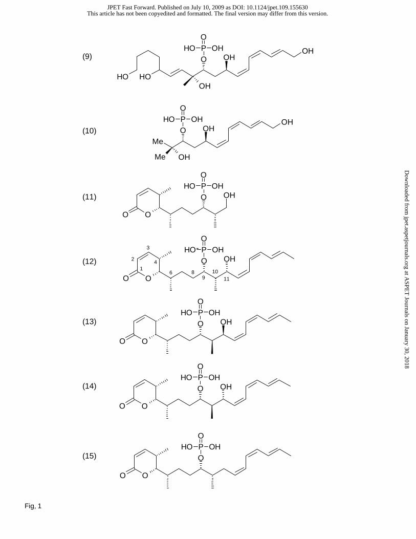
Inhibition of PP1, PP2A and PP5 Phosphatase Activity. Fostriecin, cytostatin, and a series of
10 structural analogs were synthesized using previously published methods (Buck et al., 2003; Lawhorn
et al., 2006). In side-by-side measurements using equal amounts of enzymes and [32P]phosphohistone
as substrate, both fostriecin (1; IC50 =1.4 +/-0.3 nM) and cytostatin (2; IC50 =29.0+/- 7.0 nM) potently
inhibit the activity of PP2A (Figure 2). In contrast, dephosphofostriecin (7) and dephosphocytostatin (8)
have minimal inhibitory activity against PP2A (IC50 >100 μM). Derivatives, in which the entire lactone
(10) or the entire (Z,Z,E)-triene (11) are deleted, strongly inhibit PP2A (IC50 = 0.1 +/- 0.02 and 4.2 +/-0.3
μM, respectively), while having little effect (IC50 >100 μM) on PP1 or PP5 (Figure 2C and D).
Inhibition of PP1/PP2A- and PP5/PP2A-chimeras by Fostriecin. Structural studies (Goldberg
This article has not been copyedited and formatted. The final version may differ from this version.JPET Fast Forward. Published on July 10, 2009 as DOI: 10.1124/jpet.109.155630
at ASPE
T Journals on January 30, 2018
jpet.aspetjournals.orgD
ownloaded from
JPET #155630
10
et al., 1995; Swingle et al., 2004) and mutational analysis (Zhang et al., 1996) indicate that both PP1 and
PP5 share a common catalytic mechanism with PP2A. Indeed PP1, PP2A, PP2B, and PP5 all contain a
common catalytic pocket consisting of 10 conserved amino acids (Figure 3). Computer models predicted
that fostriecin binds to a unique region of PP2A contained in the β12-β13 loop, and mutational analysis of
PP1 revealed considerable insight into the binding of natural inhibitors (Zhang et al., 1994; Zhang et al.,
1996). Therefore, we performed site-directed mutagenesis, replacing endogenous amino acids in the β12-
β13 loop of PP1 and PP5 with the corresponding amino acids (YRCG) in PP2A. Then, in side-by-side
dose-response studies, we tested the sensitivity of the PP1(YRCG)- and PP5(YRCG)-chimeras to
fostriecin, cytostatin and key analogs. In PP1, conversion to YRCG resulted in a ~600 fold increase in
sensitivity to fostriecin (Figure 4A; IC50 = 0.09 +/- 0.02 vs ~72 +/- 11 μM). With PP5, a ~200 fold
increase in sensitivity was produced by as similar (YRCG) mutation (Figure 4B; IC50 = 0.3 +/- 0.08 vs
60.5 +/ 0.6 μM). Mutation of a single residue in PP5 (PP5 M to C) increased sensitivity to fostriecin by
~50 fold (IC50 =1.2 +/- 0.5 μM). PP1(YRCG) and PP5(YRCG) were also sensitive to cytostatin (IC50 =
17 +/- 3.2 and 34 +/- 6.1 μM, respectively), which has little effect on native PP1 or PP5 (IC50 >100 μM).
Compounds, in which the lactone ring is deleted or disrupted (9) and (10), demonstrated no detectable
increase in inhibitory activity against PP1(YRCG) or PP5(YRCG). In contrast, compounds retaining the
phosphate monoester and unsaturated lactone (i.e. 1, 2, 11-15) demonstrated increased potency against the
chimeras (Table 1).
Discussion.
To date, >10 natural compounds have been identified that share a distinctive phosphate
monoester and α,β-unsaturated δ-lactone structural units with fostriecin (Figure 1). Fostriecin and
cytostatin, which both act as potent inhibitors of PP2A (Walsh et al., 1997; Buck et al., 2003; Lawhorn et
al., 2006) also share a (Z,Z,E)-triene. The total synthesis of fostriecin (1) (Boger et al., 2001; Chavez and
Jacobsen, 2001), cytostatin (2) (Lawhorn et al., 2006) and more recently several key analogs (compounds
6-15) (Buck et al., 2003; Lawhorn et al., 2006), have enabled further structure activity relationship studies
This article has not been copyedited and formatted. The final version may differ from this version.JPET Fast Forward. Published on July 10, 2009 as DOI: 10.1124/jpet.109.155630
at ASPE
T Journals on January 30, 2018
jpet.aspetjournals.orgD
ownloaded from
JPET #155630
11
to explore the inhibitory activity of the fostriecin family of inhibitors. As known from previous studies
(Buck et al., 2003; Lawhorn et al., 2006) and shown in Figure 2A, fostriecin (1; IC50 =1.4 nM) is an ~20
fold more potent inhibitor of PP2A than cytostatin (2; IC50 =29.0 nM). At much higher concentrations,
fostriecin is essentially an equipotent inhibitor of PP5 and PP1 (IC50 ~60 μM), and cytostatin has little
effect on PP1 or PP5 (IC50 >100 μM). Thus, the selectivity of (1) and (2) for PP2A is substantial
(PP2A/PP1-PP5 >104). The known (Buck et al., 2003; Lawhorn et al., 2006) importance of the C9
phosphate for both (1) and (2) is illustrated in Figure 2A, where dephosphofostriecin (7) and
dephosphocytostatin (8) have minimal inhibitory activity against PP2A (IC50 >100 μM). When the
phosphate is converted into a phosphodiester (6), inhibitory activity against PP2A is reduced ~ 103 fold
(IC50 3.2 +/- 1.1 μM). Both dephosphocompounds also have reduced inhibitory activity against PP1 and
PP5 (not shown) suggesting the phosphate interacts with conserved catalytic residues. However, (1) and
(2) also differ at C4 and C17. In addition, derivatives in which the entire lactone (10) or the entire
(Z,Z,E)-triene (11) are deleted still strongly inhibit PP2A, while having little effect on PP1 or PP5 (Figure
2C and D). We interpret this to indicate that both the lactone and the (Z,Z,E)-triene contribute to
selectivity.
Computer models of interactions between (1) or (2) with PPP-family phosphatases suggest that
the phosphate and the common C11-hydroxyl interact with regions conserved in PP1, PP2A and PP5 (i.e.
the catalytic metals and conserved active site residues that coordinate the metals and/or position the
incoming substrate for nucleophilic attack) which is consistent with the inhibition data presented above.
The models also predict that the unsaturated lactone contributes to the strong inhibitory actions of PP2A,
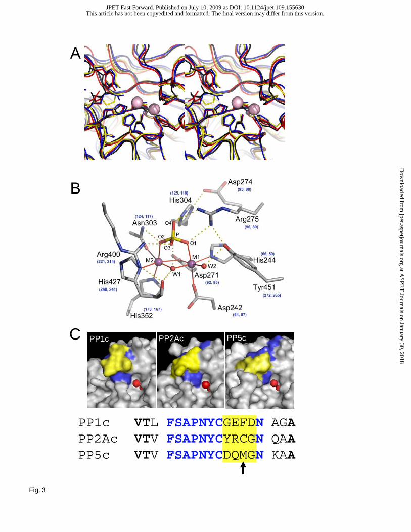
due to an electrophilic interaction with a cysteine (cys269) contained in the β12-β13 loop of PP2A that is
not present in PP1 and PP5 (Figure 3C). This prediction is supported by the decreased PP2A inhibitory
activity of (9), in which the electrophilic nature of C3 is absent, and (10), in which the entire lactone
moiety is deleted (Figure 2C and D). To further test this hypothesis, we performed site-directed
mutagenesis, replacing endogenous amino acids in the β12-β13 loop of PP1 and PP5 with the
This article has not been copyedited and formatted. The final version may differ from this version.JPET Fast Forward. Published on July 10, 2009 as DOI: 10.1124/jpet.109.155630
at ASPE
T Journals on January 30, 2018
jpet.aspetjournals.orgD
ownloaded from
JPET #155630
12
corresponding amino acids in PP2A. For both PP1 and PP5, the region needed for strong inhibition was
mapped to 4 amino acids immediately adjacent to the active site tyrosine (Tyr265). In PP1, conversion
from GEFD to YRCG (the sequence contained in PP2A) resulted in a ~600 fold increase in sensitivity to
fostriecin (Figure 4A). With PP5, a ~200 fold increase in sensitivity was produced by as similar (DQMG
to YRCG) mutation (Figure 4B). PP1- and PP5-YRCG mutants were also sensitive to cytostatin (IC50 =
17 +/- 3.2 and 34 +/- 6.1 μM, respectively), which has little effect on native PP1 or PP5 (IC50 >100 μM).
Compounds, in which the lactone ring is deleted or disrupted (9) and (10), demonstrated no detectable
increase in inhibitory activity against PP1(YRCG) or PP5(YRCG). In addition, (11), which lacks the
(Z,Z,E)-triene, demonstrated similar strength in the inhibition of PP2A, PP1(YRCG) and PP5(YRCG)
while having minimal activity against native PP1 or PP5. Together these studies support the critical role
of an interaction between C3 and cys269 in the β12-β13 loop of PP2A. The critical role of cys269 in
fostriecin sensitivity is also supported by our previous SAR studies using additional derivatives of
fostriecin (Buck et al., 2003) and studies in yeast, in which a 10-fold decrease in sensitivity to fostriecin
induced by random mutagenesis was associated with a homologous C269S mutation (Evans and Simon,
2001).
In addition to the lactone, (1), (2) and (11) share in common a C11-alcohol, and C10 of (2) and
(11) contains a methyl group not contained in (1), (9) or (10). Thus, four C10/C11 cytostatin
diastereomers [(12), (13), (14) and (15)] were tested on PP1, PP2A and PP1 (YRCG). As we reported
previously (Buck et al., 2003; Lawhorn et al., 2006) compared to the natural compound (2), each of these
cytostatin diastereomers were less potent inhibitors of PP2A. The C11 epimer (12), in which only the
stereochemistry of the alcohols is inverted, the C10-epimer (13), in which only the stereochemistry of the
methyl group is inverted, and the (10R,11R)-diastereomer (14), in which both the C11-methyl group and
the C11-alcohol are inverted, all proved to be stronger inhibitors of PP1 (YRCG) than PP1 (Table 1). All
three cytostatin epimers also demonstrated similar strength against PP1(YRCG) and PP2A. In addition,
(15), which lacks the alcohol at C11, and (11), which lacks the entire triene, also demonstrated similar
This article has not been copyedited and formatted. The final version may differ from this version.JPET Fast Forward. Published on July 10, 2009 as DOI: 10.1124/jpet.109.155630
at ASPE
T Journals on January 30, 2018
jpet.aspetjournals.orgD
ownloaded from
JPET #155630
13
strength of inhibition against PP2A and PP1(YRCG), while having little effect on native PP1 or PP5.
Together, these observations provide compelling data supporting the concept that much of the selectivity
for PP2A observed with fostriecin-family inhibitors is indeed derived from the interaction between C3 of
the inhibitors and the β12-β13 loop in PP2A. Nonetheless, because (9) and (10) are still highly selective
for PP2A (Figure 2), additional selectivity is likely derived from the (Z,Z,E)-triene.
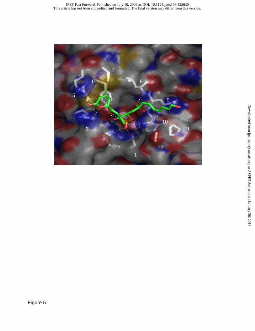
To gain additional insight into the interactions that aid the selective inhibition of PP2A, bound
conformations of (1) were generated via molecular docking with a modified version of autodock 3.05 (see
Materials and Methods). Cluster analysis of top-scoring poses from each of 100 independent docking
runs separated poses into non-overlapping groups based upon an r.m.s.d. threshold of 2.5Å. Each cluster
represents a group of similar binding poses, the degree of similarity being dependent upon the r.m.s.d.
cutoff. These clusters can be thought of as representing a binding mode plus snapshots of the associated
relative internal motions of the receptor-ligand complex (Ruvinsky and Kozintsev, 2005).
The docking of fostriecin to PP2Ac results in 33 non-overlapping clusters (8 with more than 2
members). The 3 clusters with the lowest estimated binding energy (ranked according to the best member)
show fostriecin bound (for a representative structure; see Figure 5) with the phosphate moiety coordinated
to the active site metals and forming hydrogen bonds with highly conserved active site residues (asn117,
his118, arg214, and tyr265), the (Z,Z,E)-triene placed in the acidic groove, and the unsaturated lactone ring
nestled in a pocket formed by the highly conserved arg89 and 4 residues in the β12-13 loop: tyr265, cys266,
arg268, and cys269. Importantly, this conformation places the gamma-sulfur of cys269 3.6 Å away from the
electrophilic C3, suggesting that binding to PP2A pre-positions the lactone ring for nucleophilic attack.
Two of the residues in the lactone binding pocket, arg268 and cys269, are nonconservatively substituted in
PP1 and PP5. Arg268 is substituted by glu and gln in PP1 and PP5, respectively, while cys269 is substituted
by phe and met. In addition to the absence of the thiolate nucleophile in cys269, the presence of the bulky
hydrophobic side chain of phe or met in the other two PPases alters the shape of the pocket such that the
lactone binding would be less favorable. The structure of PP5 also shows partial occupancy of this pocket
This article has not been copyedited and formatted. The final version may differ from this version.JPET Fast Forward. Published on July 10, 2009 as DOI: 10.1124/jpet.109.155630
at ASPE
T Journals on January 30, 2018
jpet.aspetjournals.orgD
ownloaded from
JPET #155630
14
by a methionine residue from the c-terminal J-helix that may interfere with binding. The C8-hydroxyl
forms a hydrogen bond with the tyr265 hydroxyl group and would be well placed to form a hydrogen bond
with the guanidinium group of arg89 if receptor flexibility allows for an induced fit of receptor residues
around this fostriecin conformation. The C11-hydroxyl forms a hydrogen bond with the backbone amide
of leu243 and the backbone carbonyl of the highly conserved his241, which has been implicated in helping
to orient the hydroxide nucleophile during phosphomonoester hydrolysis (Swingle et al., 2004). The
triene tail makes hydrophobic contacts with the side chains of pro213 and leu243, as well as the aliphatic
portion of gln242. These residues are not conserved with PP5 or, with the exception of gln242, PP1. These
structural differences with regard to interactions with the triene tail may account for part of the
differential affinity of fostriecin toward these enzymes. Unfortunately, unlike the β12-β13 loop, which is
contained on an exposed surface loop and can easily be modified without affecting that general structure
of PP1 and PP5, mutations of the residues in the acidic grove are likely to alter large regions of the
protein. Therefore, future SAR studies await the development of methods to synthesize addition
derivatives to further probe the importance of the triene.
This article has not been copyedited and formatted. The final version may differ from this version.JPET Fast Forward. Published on July 10, 2009 as DOI: 10.1124/jpet.109.155630
at ASPE
T Journals on January 30, 2018
jpet.aspetjournals.orgD
ownloaded from
JPET #155630
15
References
Bialy L and Waldmann H (2004) Total synthesis and biological evaluation of the protein
phosphatase 2A inhibitor cytostatin and analogues. Chemistry 10:2759-2780.
Boger DL, Ichikawa S and Zhong W (2001) Total synthesis of fostriecin (CI-920). J Am Chem
Soc 123:4161-4167.
Buck SB, Hardouin C, Ichikawa S, Soenen DR, Gauss CM, Hwang I, Swingle MR, Bonness
KM, Honkanen RE and Boger DL (2003) Fundamental role of the fostriecin unsaturated
lactone and implications for selective protein phosphatase inhibition. J Am Chem Soc
125:15694-15695.
Chavez DE and Jacobsen EN (2001) Total Synthesis of Fostriecin (CI-920) Angew Chem Int Ed
Engl 40:3667-3670.
Chen HM, Liu BF, Huang HL, Hwang SF and Ho SY (2007) SODOCK: swarm optimization for
highly flexible protein-ligand docking. J Comput Chem 28:612-623.
Cho US and Xu W (2007) Crystal structure of a protein phosphatase 2A heterotrimeric
holoenzyme. Nature 445:53-57.
Colby DA and Chamberlin AR (2006) Pharmacophore identification: the case of the ser/thr
protein phosphatase inhibitors. Mini Rev Med Chem 6:657-665.
de Jong RS, de Vries EG and Mulder NH (1997) Fostriecin: a review of the preclinical data.
Anticancer Drugs 8:413-418.
Esumi T, Okamoto N and Hatakeyama S (2002) Versatile enantiocontrolled synthesis of (+)-
fostriecin. Chem Commun (Camb):3042-3043.
Evans DR and Simon JA (2001) The predicted beta12-beta13 loop is important for inhibition of
PP2Acalpha by the antitumor drug fostriecin. FEBS Lett 498:110-115.
Gasteiger J. MM (1980) Iterative partial equalization of orbital electronegativity – a rapid access
This article has not been copyedited and formatted. The final version may differ from this version.JPET Fast Forward. Published on July 10, 2009 as DOI: 10.1124/jpet.109.155630
at ASPE
T Journals on January 30, 2018
jpet.aspetjournals.orgD
ownloaded from
JPET #155630
16
to atomic charges. . Tetrahedron 36:3219-3228.
Goldberg J, Huang HB, Kwon YG, Greengard P, Nairn AC and Kuriyan J (1995) Three-
dimensional structure of the catalytic subunit of protein serine/threonine phosphatase-1.
Nature 376:745-753.
Hassinen T and Peräkylä M (2001) New energy terms for reduced protein models implemented
in an off-lattice force field. J. Comput. Chem. 22:1229-1242.
Honkanen RE (2005) Serine/threonine protein phosphatase inhibitors with anti tumor activity.
Handbook of Exp. Pharm. 167:295-317.
Honkanen RE, Zwiller J, Moore RE, Daily SL, Khatra BS, Dukelow M and Boynton AL (1990)
Characterization of microcystin-LR, a potent inhibitor of type 1 and type 2A protein
phosphatases. J Biol Chem 265:19401-19404.
Jung WH, Guyenne S, Riesco-Fagundo C, Mancuso J, Nakamura S and Curran DP (2008)
Confirmation of the stereostructure of (+)-cytostatin by fluorous mixture synthesis of four
candidate stereoisomers. Angew Chem Int Ed Engl 47:1130-1133.
Kawada M, Amemiya M, Ishizuka M and Takeuchi T (1999) Differential induction of apoptosis
in B16 melanoma and EL-4 lymphoma cells by cytostatin and bactobolin. Jpn J Cancer
Res 90:219-225.
Kawada M, Kawatsu M, Masuda T, Ohba S, Amemiya M, Kohama T, Ishizuka M and Takeuchi
T (2003) Specific inhibitors of protein phosphatase 2A inhibit tumor metastasis through
augmentation of natural killer cells. Int Immunopharmacol 3:179-188.
Kennedy J and Eberhart RC (1995) Particle swarm optimization. . Proceedings of IEEE
International Conference on Neural Networks, Piscataway, NJ. :1942-1948.
Lawhorn BG, Boga SB, Wolkenberg SE, Colby DA, Gauss CM, Swingle MR, Amable L,
This article has not been copyedited and formatted. The final version may differ from this version.JPET Fast Forward. Published on July 10, 2009 as DOI: 10.1124/jpet.109.155630
at ASPE
T Journals on January 30, 2018
jpet.aspetjournals.orgD
ownloaded from
JPET #155630
17
Honkanen RE and Boger DL (2006) Total synthesis and evaluation of cytostatin, its C10-
C11 diastereomers, and additional key analogues: impact on PP2A inhibition. J Am Chem
Soc 128:16720-16732.
Le LH, Erlichman C, Pillon L, Thiessen JJ, Day A, Wainman N, Eisenhauer EA and Moore MJ
(2004) Phase I and pharmacokinetic study of fostriecin given as an intravenous bolus
daily for five consecutive days. Invest New Drugs 22:159-167.
Leopold WR, Shillis JL, Mertus AE, Nelson JM, Roberts BJ and Jackson RC (1984) Anticancer
activity of the structurally novel antibiotic Cl-920 and its analogues. Cancer Res
44:1928-1932.
Lewy DS, Gauss CM, Soenen DR and Boger DL (2002) Fostriecin: chemistry and biology. Curr
Med Chem 9:2005-2032.
Maki K, Motoki R, Fujii K, Kanai M, Kobayashi T, Tamura S and Shibasaki M (2005) Catalyst-
controlled asymmetric synthesis of fostriecin and 8-epi-fostriecin. J Am Chem Soc
127:17111-17117.
Masuda T, Watanabe S, Amemiya M, Ishizuka M and Takeuchi T (1995) Inhibitory effect of
cytostatin on spontaneous lung metastases of B16-BL6 melanoma cells. J Antibiot
(Tokyo) 48:528-529.
Miyashita K, Ikejiri M, Kawasaki H, Maemura S and Imanishi T (2003) Total synthesis of an
antitumor antibiotic, Fostriecin (CI-920). J Am Chem Soc 125:8238-8243.
Morris GM, Goodsell DS, Halliday RS, Huey R, Hart WE, Belew RK and Olson AJ (1998)
Automated docking using a Lamarckian genetic algorithm and an empirical binding free
energy function. J. Comput. Chem. 19:1639-1662.
Reddy YK and Falck JR (2002) Asymmetric total synthesis of (+)-fostriecin. Org Lett 4:969-
This article has not been copyedited and formatted. The final version may differ from this version.JPET Fast Forward. Published on July 10, 2009 as DOI: 10.1124/jpet.109.155630
at ASPE
T Journals on January 30, 2018
jpet.aspetjournals.orgD
ownloaded from
JPET #155630
18
971.
Ruvinsky AM and Kozintsev AV (2005) New and fast statistical-thermodynamic method for
computation of protein-ligand binding entropy substantially improves docking accuracy.
J Comput Chem 26:1089-1095.
Stouten PFW, Frömmel C, Nakamura H and Sander C (1993) An Effective Solvation Term
Based on Atomic Occupancies for Use in Protein Simulations Mol. Simulat. 10:97-120.
Susick RL, Jr., Hawkins KL and Pegg DG (1990) Preclinical toxicological evaluation of
fostriecin, a novel anticancer antibiotic, in rats. Fundam Appl Toxicol 15:258-269.
Swingle M, Ni L and Honkanen RE (2007) Small-molecule inhibitors of ser/thr protein
phosphatases: specificity, use and common forms of abuse. Methods Mol Biol 365:23-38.
Swingle MR, Honkanen RE and Ciszak EM (2004) Structural basis for the catalytic activity of
human serine/threonine protein phosphatase-5. J Biol Chem 279:33992-33999.
Trost BM, Frederiksen MU, Papillon JP, Harrington PE, Shin S and Shireman BT (2005)
Dinuclear asymmetric Zn aldol additions: formal asymmetric synthesis of fostriecin. J
Am Chem Soc 127:3666-3667.
Usui T, Marriott G, Inagaki M, Swarup G and Osada H (1999) Protein phosphatase 2A
inhibitors, phoslactomycins. Effects on the cytoskeleton in NIH/3T3 cells. J Biochem
125:960-965.
Walsh AH, Cheng A and Honkanen RE (1997) Fostriecin, an antitumor antibiotic with inhibitory
activity against serine/threonine protein phosphatases types 1 (PP1) and 2A (PP2A), is
highly selective for PP2A. FEBS Lett 416:230-234.
Xing Y, Xu Y, Chen Y, Jeffrey PD, Chao Y, Lin Z, Li Z, Strack S, Stock JB and Shi Y (2006)
Structure of protein phosphatase 2A core enzyme bound to tumor-inducing toxins. Cell
This article has not been copyedited and formatted. The final version may differ from this version.JPET Fast Forward. Published on July 10, 2009 as DOI: 10.1124/jpet.109.155630
at ASPE
T Journals on January 30, 2018
jpet.aspetjournals.orgD
ownloaded from
JPET #155630
19
127:341-353.
Zhang J, Zhang Z, Brew K and Lee EY (1996) Mutational analysis of the catalytic subunit of
muscle protein phosphatase-1. Biochemistry 35:6276-6282.
Zhang Z, Zhao S, Long F, Zhang L, Bai G, Shima H, Nagao M and Lee EY (1994) A mutant of
protein phosphatase-1 that exhibits altered toxin sensitivity. J Biol Chem 269:16997-
17000.
This article has not been copyedited and formatted. The final version may differ from this version.JPET Fast Forward. Published on July 10, 2009 as DOI: 10.1124/jpet.109.155630
at ASPE
T Journals on January 30, 2018
jpet.aspetjournals.orgD
ownloaded from

JPET #155630
20
Footnotes. *Indicates equal contribution by these two authors. This work was supported in part by grants
from the National Institutes of Health [CA42056; CA60750; MD002314]. This investigation was
conducted in a facility constructed with support from Research Facilities Improvement Program Grant
(C06 RR11174) from the National Center for Research Resources.
This article has not been copyedited and formatted. The final version may differ from this version.JPET Fast Forward. Published on July 10, 2009 as DOI: 10.1124/jpet.109.155630
at ASPE
T Journals on January 30, 2018
jpet.aspetjournals.orgD
ownloaded from
JPET #155630
21
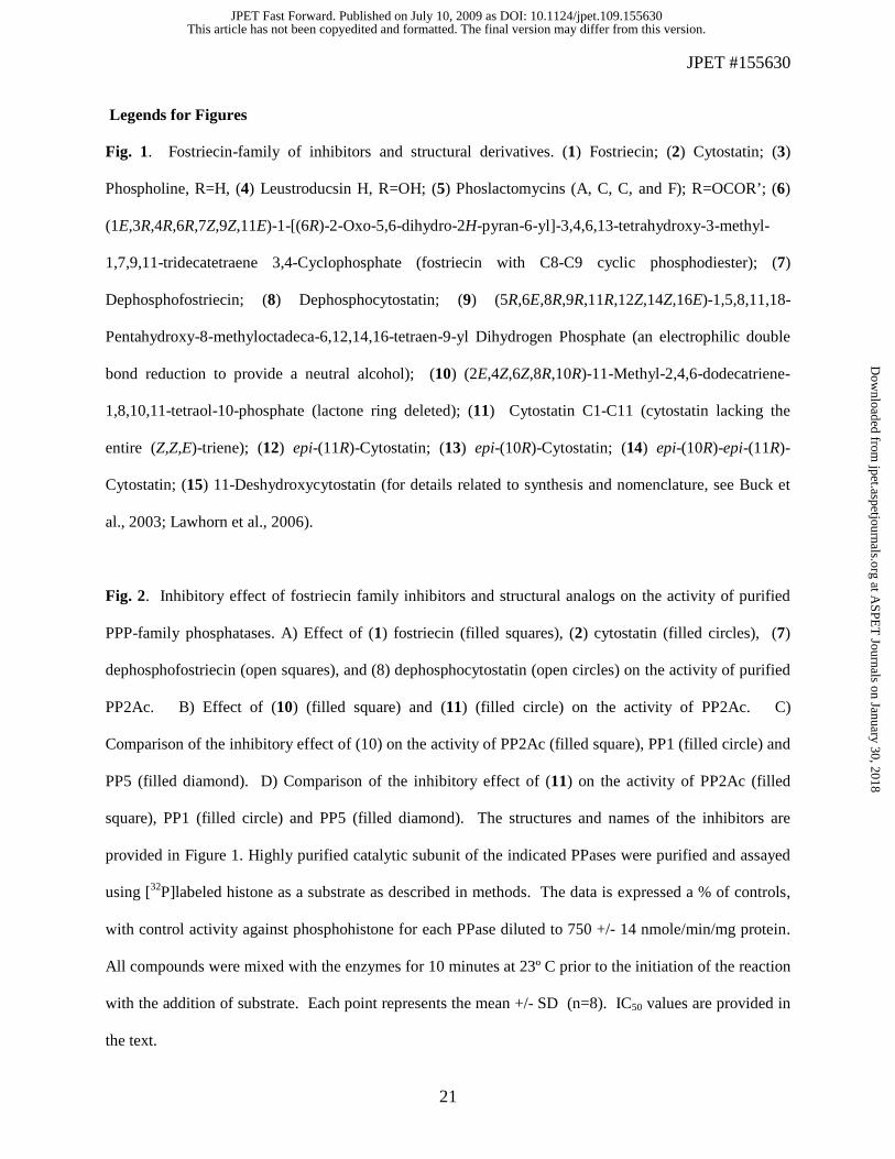
Legends for Figures
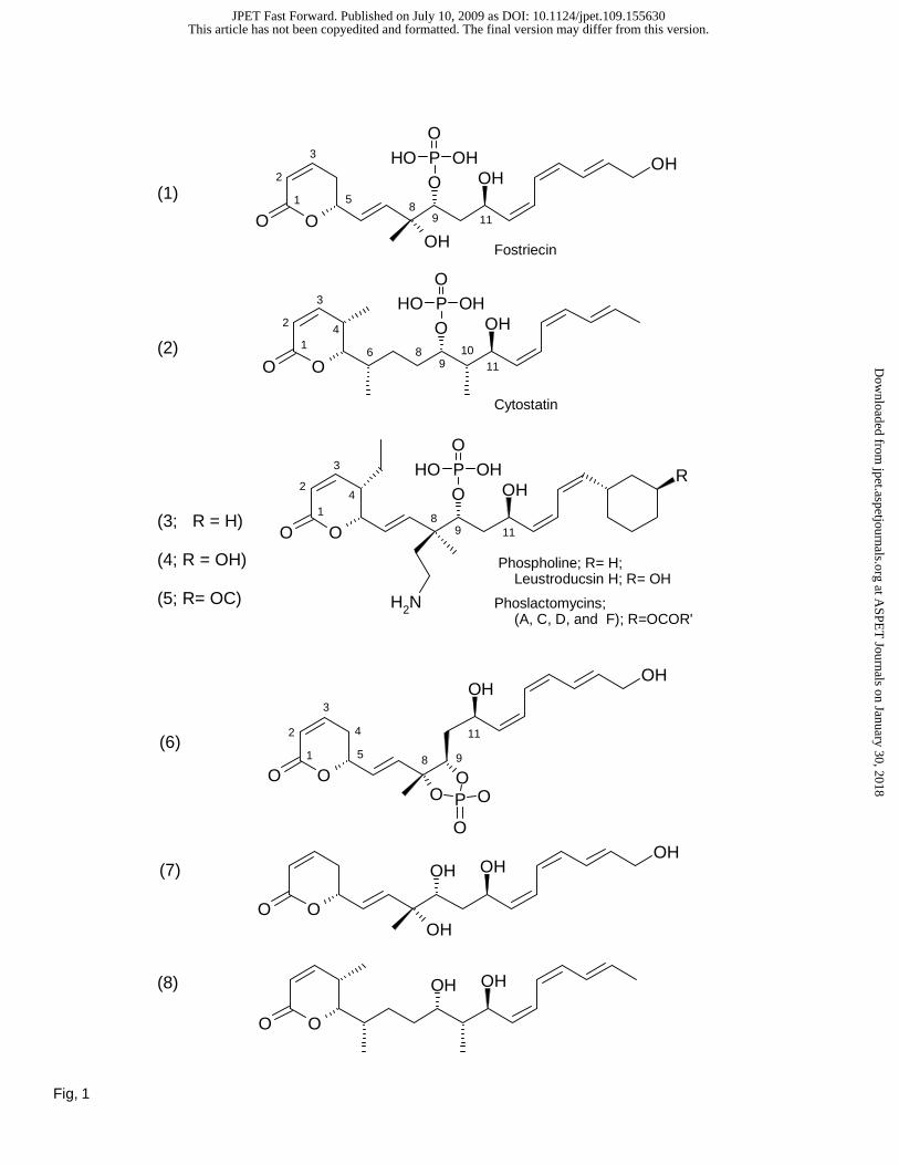
Fig. 1. Fostriecin-family of inhibitors and structural derivatives. (1) Fostriecin; (2) Cytostatin; (3)
Phospholine, R=H, (4) Leustroducsin H, R=OH; (5) Phoslactomycins (A, C, C, and F); R=OCOR’; (6)
(1E,3R,4R,6R,7Z,9Z,11E)-1-[(6R)-2-Oxo-5,6-dihydro-2H-pyran-6-yl]-3,4,6,13-tetrahydroxy-3-methyl-
1,7,9,11-tridecatetraene 3,4-Cyclophosphate (fostriecin with C8-C9 cyclic phosphodiester); (7)
Dephosphofostriecin; (8) Dephosphocytostatin; (9) (5R,6E,8R,9R,11R,12Z,14Z,16E)-1,5,8,11,18-
Pentahydroxy-8-methyloctadeca-6,12,14,16-tetraen-9-yl Dihydrogen Phosphate (an electrophilic double
bond reduction to provide a neutral alcohol); (10) (2E,4Z,6Z,8R,10R)-11-Methyl-2,4,6-dodecatriene-
1,8,10,11-tetraol-10-phosphate (lactone ring deleted); (11) Cytostatin C1-C11 (cytostatin lacking the
entire (Z,Z,E)-triene); (12) epi-(11R)-Cytostatin; (13) epi-(10R)-Cytostatin; (14) epi-(10R)-epi-(11R)-
Cytostatin; (15) 11-Deshydroxycytostatin (for details related to synthesis and nomenclature, see Buck et
al., 2003; Lawhorn et al., 2006).
Fig. 2. Inhibitory effect of fostriecin family inhibitors and structural analogs on the activity of purified
PPP-family phosphatases. A) Effect of (1) fostriecin (filled squares), (2) cytostatin (filled circles), (7)
dephosphofostriecin (open squares), and (8) dephosphocytostatin (open circles) on the activity of purified
PP2Ac. B) Effect of (10) (filled square) and (11) (filled circle) on the activity of PP2Ac. C)
Comparison of the inhibitory effect of (10) on the activity of PP2Ac (filled square), PP1 (filled circle) and
PP5 (filled diamond). D) Comparison of the inhibitory effect of (11) on the activity of PP2Ac (filled
square), PP1 (filled circle) and PP5 (filled diamond). The structures and names of the inhibitors are
provided in Figure 1. Highly purified catalytic subunit of the indicated PPases were purified and assayed
using [32P]labeled histone as a substrate as described in methods. The data is expressed a % of controls,
with control activity against phosphohistone for each PPase diluted to 750 +/- 14 nmole/min/mg protein.
All compounds were mixed with the enzymes for 10 minutes at 23º C prior to the initiation of the reaction
with the addition of substrate. Each point represents the mean +/- SD (n=8). IC50 values are provided in
the text.
This article has not been copyedited and formatted. The final version may differ from this version.JPET Fast Forward. Published on July 10, 2009 as DOI: 10.1124/jpet.109.155630
at ASPE
T Journals on January 30, 2018
jpet.aspetjournals.orgD
ownloaded from
JPET #155630
22
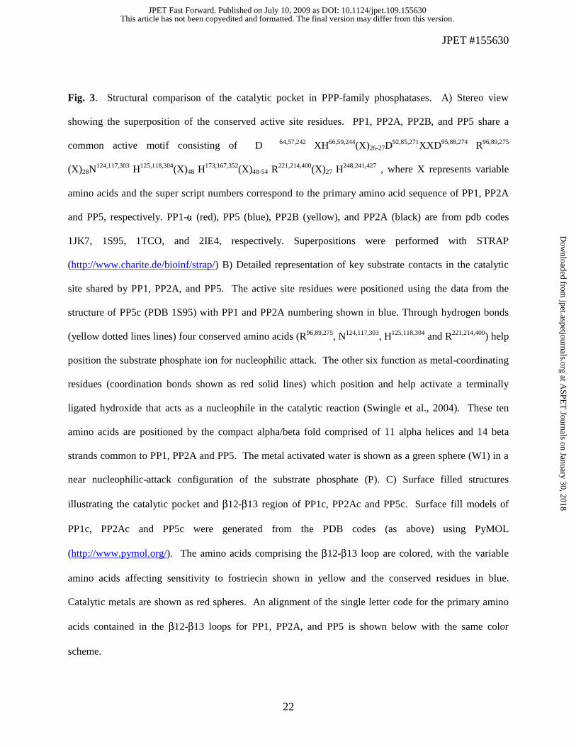
Fig. 3. Structural comparison of the catalytic pocket in PPP-family phosphatases. A) Stereo view
showing the superposition of the conserved active site residues. PP1, PP2A, PP2B, and PP5 share a
common active motif consisting of D 64,57,242 XH66,59,244(X)26-27D92,85,271XXD95,88,274 R96,89,275
(X)28N124,117,303 H125,118,304(X)48 H
173,167,352(X)48-54 R221,214,400(X)27 H
248,241,427 , where X represents variable
amino acids and the super script numbers correspond to the primary amino acid sequence of PP1, PP2A
and PP5, respectively. PP1-α (red), PP5 (blue), PP2B (yellow), and PP2A (black) are from pdb codes
1JK7, 1S95, 1TCO, and 2IE4, respectively. Superpositions were performed with STRAP
(http://www.charite.de/bioinf/strap/) B) Detailed representation of key substrate contacts in the catalytic
site shared by PP1, PP2A, and PP5. The active site residues were positioned using the data from the
structure of PP5c (PDB 1S95) with PP1 and PP2A numbering shown in blue. Through hydrogen bonds
(yellow dotted lines lines) four conserved amino acids (R96,89,275, N124,117,303, H125,118,304 and R221,214,400) help
position the substrate phosphate ion for nucleophilic attack. The other six function as metal-coordinating
residues (coordination bonds shown as red solid lines) which position and help activate a terminally
ligated hydroxide that acts as a nucleophile in the catalytic reaction (Swingle et al., 2004). These ten
amino acids are positioned by the compact alpha/beta fold comprised of 11 alpha helices and 14 beta
strands common to PP1, PP2A and PP5. The metal activated water is shown as a green sphere (W1) in a
near nucleophilic-attack configuration of the substrate phosphate (P). C) Surface filled structures
illustrating the catalytic pocket and β12-β13 region of PP1c, PP2Ac and PP5c. Surface fill models of
PP1c, PP2Ac and PP5c were generated from the PDB codes (as above) using PyMOL
(http://www.pymol.org/). The amino acids comprising the β12-β13 loop are colored, with the variable
amino acids affecting sensitivity to fostriecin shown in yellow and the conserved residues in blue.
Catalytic metals are shown as red spheres. An alignment of the single letter code for the primary amino
acids contained in the β12-β13 loops for PP1, PP2A, and PP5 is shown below with the same color
scheme.
This article has not been copyedited and formatted. The final version may differ from this version.JPET Fast Forward. Published on July 10, 2009 as DOI: 10.1124/jpet.109.155630
at ASPE
T Journals on January 30, 2018
jpet.aspetjournals.orgD
ownloaded from
JPET #155630
23
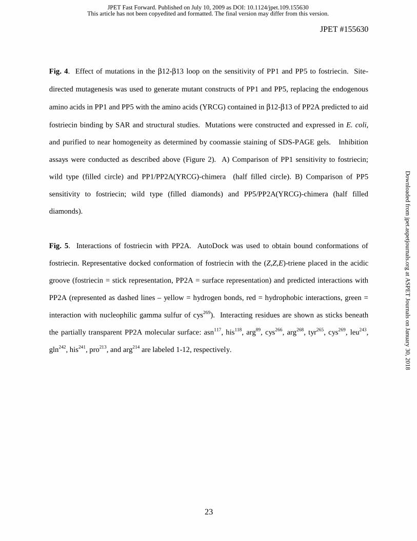
Fig. 4. Effect of mutations in the β12-β13 loop on the sensitivity of PP1 and PP5 to fostriecin. Site-
directed mutagenesis was used to generate mutant constructs of PP1 and PP5, replacing the endogenous
amino acids in PP1 and PP5 with the amino acids (YRCG) contained in β12-β13 of PP2A predicted to aid
fostriecin binding by SAR and structural studies. Mutations were constructed and expressed in E. coli,
and purified to near homogeneity as determined by coomassie staining of SDS-PAGE gels. Inhibition
assays were conducted as described above (Figure 2). A) Comparison of PP1 sensitivity to fostriecin;
wild type (filled circle) and PP1/PP2A(YRCG)-chimera (half filled circle). B) Comparison of PP5
sensitivity to fostriecin; wild type (filled diamonds) and PP5/PP2A(YRCG)-chimera (half filled
diamonds).
Fig. 5. Interactions of fostriecin with PP2A. AutoDock was used to obtain bound conformations of
fostriecin. Representative docked conformation of fostriecin with the (Z,Z,E)-triene placed in the acidic
groove (fostriecin = stick representation, PP2A = surface representation) and predicted interactions with
PP2A (represented as dashed lines – yellow = hydrogen bonds, red = hydrophobic interactions, green =
interaction with nucleophilic gamma sulfur of cys269). Interacting residues are shown as sticks beneath
the partially transparent PP2A molecular surface: asn117, his118, arg89, cys266, arg268, tyr265, cys269, leu243,
gln242, his241, pro213, and arg214 are labeled 1-12, respectively.
This article has not been copyedited and formatted. The final version may differ from this version.JPET Fast Forward. Published on July 10, 2009 as DOI: 10.1124/jpet.109.155630
at ASPE
T Journals on January 30, 2018
jpet.aspetjournals.orgD
ownloaded from
JPET #155630
24
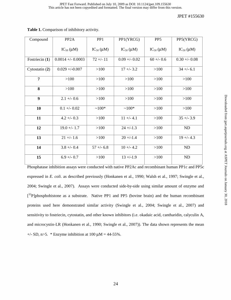
Table 1. Comparison of inhibitory activity.
Compound PP2A
IC50 (μM)
PP1
IC50 (μM)
PP1(YRCG)
IC50 (μM)
PP5
IC50 (μM)
PP5(YRCG)
IC50 (μM)
Fostriecin (1) 0.0014 +/- 0.0003 72 +/- 11 0.09 +/- 0.02 60 +/- 0.6 0.30 +/- 0.08
Cytostatin (2) 0.029 +/-0.007 >100 17 +/- 3.2 >100 34 +/- 6.1
7 >100 >100 >100 >100 >100
8 >100 >100 >100 >100 >100
9 2.1 +/- 0.6 >100 >100 >100 >100
10 0.1 +/- 0.02 ~100* ~100* >100 >100
11 4.2 +/- 0.3 >100 11 +/- 4.1 >100 35 +/- 3.9
12 19.0 +/- 1.7 >100 24 +/-1.3 >100 ND
13 21 +/- 1.6 >100 20 +/-1.4 >100 19 +/- 4.3
14 3.8 +/- 0.4 57 +/- 6.8 10 +/- 4.2 >100 ND
15 6.9 +/- 0.7 >100 13 +/-1.9 >100 ND
Phosphatase inhibition assays were conducted with native PP2Ac and recombinant human PP1c and PP5c
expressed in E. coli. as described previously (Honkanen et al., 1990; Walsh et al., 1997; Swingle et al.,
2004; Swingle et al., 2007). Assays were conducted side-by-side using similar amount of enzyme and
[32P]phosphohistone as a substrate. Native PP1 and PP5 (bovine brain) and the human recombinant
proteins used here demonstrated similar activity (Swingle et al., 2004; Swingle et al., 2007) and
sensitivity to fostriecin, cytostatin, and other known inhibitors (i.e. okadaic acid, cantharidin, calyculin A,
and microcystin-LR (Honkanen et al., 1990; Swingle et al., 2007)). The data shown represents the mean
+/- SD, n>5. * Enzyme inhibition at 100 μM = 44-55%.
This article has not been copyedited and formatted. The final version may differ from this version.JPET Fast Forward. Published on July 10, 2009 as DOI: 10.1124/jpet.109.155630
at ASPE
T Journals on January 30, 2018
jpet.aspetjournals.orgD
ownloaded from
OHOP OHO
OH
OO8
3
2
14
119106
OO
OHOP OHO
OH R
NH2
OO
OHOH
OH
OP OHO
OH
OOO
OP
OHOH
O
O
OO
OHOH
OH
OH
(6)
(7)
3
2
1
4
8
11
95
OHOH
OO
(3; R = H)
(4; R = OH)
(5; R= OC)
(2)
(1)
3
2
14
8119
3
2
18
119
5
Phospholine; R= H; Leustroducsin H; R= OH
Cytostatin
Fostriecin
Phoslactomycins; (A, C, D, and F); R=OCOR'
(8)
Fig, 1
This article has not been copyedited and formatted. The final version may differ from this version.JPET Fast Forward. Published on July 10, 2009 as DOI: 10.1124/jpet.109.155630
at ASPE
T Journals on January 30, 2018
jpet.aspetjournals.orgD
ownloaded from
OHOH
OHOH
OH
OP OHO
OH
OH
Me
OH
OH
OP OHO
OH
Me
OP OHO
OH
OO
OH
OHOP OHO
OH
OO
OHOP OHO
OH
OO
OP OHO
OH
OO
OHOP OHO
OH
OO8
3
2
14
119106
-
(9)
(10)
(11)
(12)
(13)
(14)
(15)
Fig, 1
This article has not been copyedited and formatted. The final version may differ from this version.JPET Fast Forward. Published on July 10, 2009 as DOI: 10.1124/jpet.109.155630
at ASPE
T Journals on January 30, 2018
jpet.aspetjournals.orgD
ownloaded from
Figure 2
A
B
This article has not been copyedited and formatted. The final version may differ from this version.JPET Fast Forward. Published on July 10, 2009 as DOI: 10.1124/jpet.109.155630
at ASPE
T Journals on January 30, 2018
jpet.aspetjournals.orgD
ownloaded from
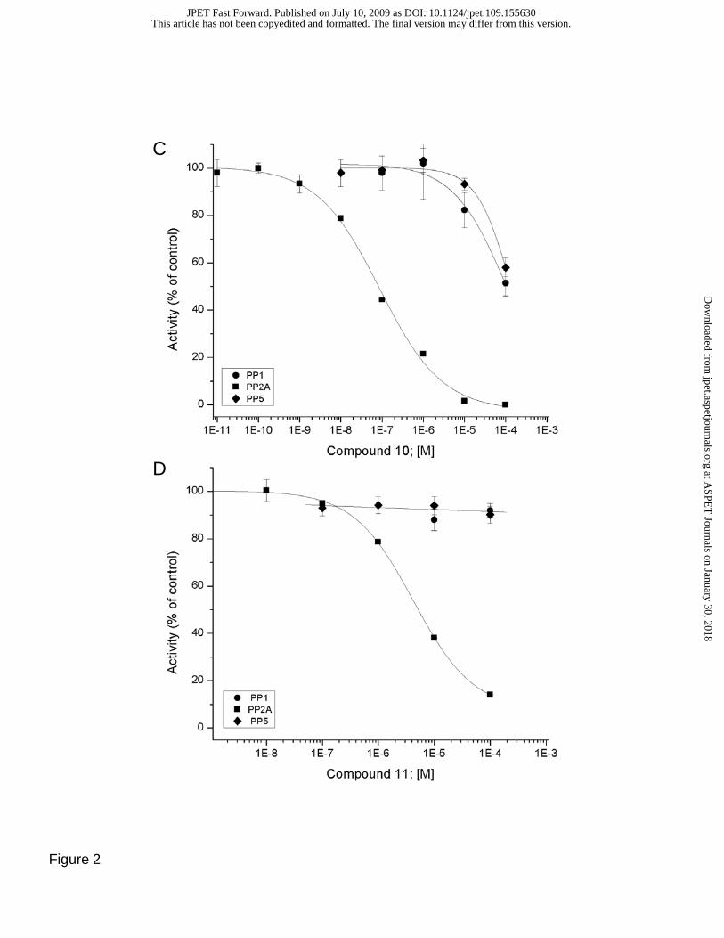
C
D
Figure 2
This article has not been copyedited and formatted. The final version may differ from this version.JPET Fast Forward. Published on July 10, 2009 as DOI: 10.1124/jpet.109.155630
at ASPE
T Journals on January 30, 2018
jpet.aspetjournals.orgD
ownloaded from
A
B
PP1c PP5cPP2AcC
PP1c VTL FSAPNYCGEFDN AGAPP2Ac VTV FSAPNYCYRCGN QAAPP5c VTV FSAPNYCDQMGN KAA
Fig. 3
This article has not been copyedited and formatted. The final version may differ from this version.JPET Fast Forward. Published on July 10, 2009 as DOI: 10.1124/jpet.109.155630
at ASPE
T Journals on January 30, 2018
jpet.aspetjournals.orgD
ownloaded from
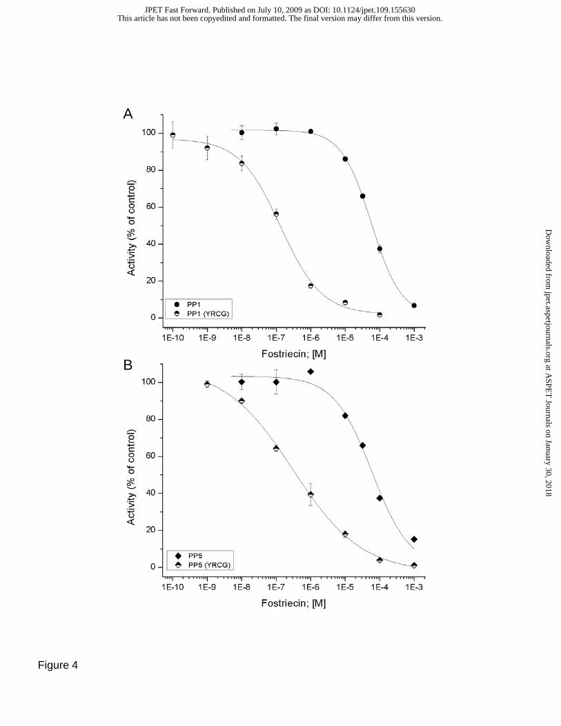
Figure 4
A
B
This article has not been copyedited and formatted. The final version may differ from this version.JPET Fast Forward. Published on July 10, 2009 as DOI: 10.1124/jpet.109.155630
at ASPE
T Journals on January 30, 2018
jpet.aspetjournals.orgD
ownloaded from
1
2
3
54
6
7
8
9
1110
12
Figure 5
This article has not been copyedited and formatted. The final version may differ from this version.JPET Fast Forward. Published on July 10, 2009 as DOI: 10.1124/jpet.109.155630
at ASPE
T Journals on January 30, 2018
jpet.aspetjournals.orgD
ownloaded from