Journal of Ovarian Research BioMed Central · itron-emitting radiolabeled molecules to display...
10
BioMed Central Page 1 of 10 (page number not for citation purposes) Journal of Ovarian Research Open Access Review Positron emission tomography in ovarian cancer: 18F-deoxy-glucose and 16α-18F-fluoro-17β-estradiol PET Yoshio Yoshida* 1 , Tetsuji Kurokawa 1 , Tetuya Tsujikawa 2 , Hidehiko Okazawa 2 and Fumikazu Kotsuji 1 Address: 1 Department of Obstetrics and Gynecology, Faculty of Medical Sciences, University of Fukui, 23-3 Matsuoka-Shimoaizuki, Eiheiji-cho, Fukui, Japan and 2 Biomedical Imaging Research Center, Faculty of Medical Sciences, University of Fukui, 23-3 Matsuoka-Shimoaizuki, Eiheiji-cho, Fukui, Japan Email: Yoshio Yoshida* - [email protected]; Tetsuji Kurokawa - [email protected]; Tetuya Tsujikawa - [email protected]; Hidehiko Okazawa - [email protected]; Fumikazu Kotsuji - [email protected] * Corresponding author Abstract The most frequently used molecular imaging technique is currently 18F-deoxy-glucose (FDG) positron emission tomography (PET). FDG-PET holds promise in the evaluation of recurrent or residual ovarian cancer when CA125 levels are rising and conventional imaging, such as ultrasound, CT, or MRI, is inconclusive or negative. Recently, integrated PET/CT, in which a full-ring-detector clinical PET scanner and a multidetector helical CT scanner are combined, has enabled the acquisition of both metabolic and anatomic imaging data using one device in a single diagnostic session. This can also provide precise anatomic localization of suspicious areas of increased FDG uptake and rule out false-positive PET findings. FDG-PET/CT is an accurate modality for assessing primary and recurrent ovarian cancer and may affect management. FDG-PET/CT may provide benefits for detection of recurrent of ovarian cancer and improve surgical planning. And FDG-PET has been shown to predict response to neoadjuvant chemotherapy and survival in advanced ovarian cancer. This review focuses on the role of FDG-PET and FDG-PET/CT in the management of patients with ovarian cancer. Recently, we have evaluated 16α-18F-fluoro-17β-estradiol (FES)-PET, which detects estrogen receptors. In a preliminary study we reported that FES-PET provides information useful for assessing ER status in advanced ovarian cancer. This new information may expand treatment choice for such patients. Background Ovarian cancer is the second most common gynecologic malignancy. It has a relatively poor prognosis, accounting for approximately half of all deaths related to gynecologic cancer [1]. Conventional imaging with ultrasonography (US), computed tomography (CT) and magnetic reso- nance imaging (MRI) has been used, but ability to diag- nose the primary tumor and accurately stage the ovarian cancer are variable. Such conventional imaging tools are also commonly used to guide the management of ovarian cancer patients. However, concerns remain that these imaging techniques may provide false negative results because of their inability to identify disease when normal anatomic landmarks have been lost because of surgery or radiation, or give false positive results related to their ina- bility to distinguish between viable tumor masses and Published: 16 June 2009 Journal of Ovarian Research 2009, 2:7 doi:10.1186/1757-2215-2-7 Received: 13 January 2009 Accepted: 16 June 2009 This article is available from: http://www.ovarianresearch.com/content/2/1/7 © 2009 Yoshida et al; licensee BioMed Central Ltd. This is an Open Access article distributed under the terms of the Creative Commons Attribution License (http://creativecommons.org/licenses/by/2.0 ), which permits unrestricted use, distribution, and reproduction in any medium, provided the original work is properly cited.
Transcript of Journal of Ovarian Research BioMed Central · itron-emitting radiolabeled molecules to display...
BioMed CentralJournal of Ovarian Research
ss
Open AcceReviewPositron emission tomography in ovarian cancer: 18F-deoxy-glucose and 16α-18F-fluoro-17β-estradiol PETYoshio Yoshida*1, Tetsuji Kurokawa1, Tetuya Tsujikawa2, Hidehiko Okazawa2 and Fumikazu Kotsuji1Address: 1Department of Obstetrics and Gynecology, Faculty of Medical Sciences, University of Fukui, 23-3 Matsuoka-Shimoaizuki, Eiheiji-cho, Fukui, Japan and 2Biomedical Imaging Research Center, Faculty of Medical Sciences, University of Fukui, 23-3 Matsuoka-Shimoaizuki, Eiheiji-cho, Fukui, Japan
Email: Yoshio Yoshida* - [email protected]; Tetsuji Kurokawa - [email protected]; Tetuya Tsujikawa - [email protected]; Hidehiko Okazawa - [email protected]; Fumikazu Kotsuji - [email protected]
* Corresponding author
AbstractThe most frequently used molecular imaging technique is currently 18F-deoxy-glucose (FDG)positron emission tomography (PET). FDG-PET holds promise in the evaluation of recurrent orresidual ovarian cancer when CA125 levels are rising and conventional imaging, such as ultrasound,CT, or MRI, is inconclusive or negative. Recently, integrated PET/CT, in which a full-ring-detectorclinical PET scanner and a multidetector helical CT scanner are combined, has enabled theacquisition of both metabolic and anatomic imaging data using one device in a single diagnosticsession. This can also provide precise anatomic localization of suspicious areas of increased FDGuptake and rule out false-positive PET findings. FDG-PET/CT is an accurate modality for assessingprimary and recurrent ovarian cancer and may affect management. FDG-PET/CT may providebenefits for detection of recurrent of ovarian cancer and improve surgical planning. And FDG-PEThas been shown to predict response to neoadjuvant chemotherapy and survival in advanced ovariancancer. This review focuses on the role of FDG-PET and FDG-PET/CT in the management ofpatients with ovarian cancer. Recently, we have evaluated 16α-18F-fluoro-17β-estradiol (FES)-PET,which detects estrogen receptors. In a preliminary study we reported that FES-PET providesinformation useful for assessing ER status in advanced ovarian cancer. This new information mayexpand treatment choice for such patients.
BackgroundOvarian cancer is the second most common gynecologicmalignancy. It has a relatively poor prognosis, accountingfor approximately half of all deaths related to gynecologiccancer [1]. Conventional imaging with ultrasonography(US), computed tomography (CT) and magnetic reso-nance imaging (MRI) has been used, but ability to diag-nose the primary tumor and accurately stage the ovarian
cancer are variable. Such conventional imaging tools arealso commonly used to guide the management of ovariancancer patients. However, concerns remain that theseimaging techniques may provide false negative resultsbecause of their inability to identify disease when normalanatomic landmarks have been lost because of surgery orradiation, or give false positive results related to their ina-bility to distinguish between viable tumor masses and
Published: 16 June 2009
Journal of Ovarian Research 2009, 2:7 doi:10.1186/1757-2215-2-7
Received: 13 January 2009Accepted: 16 June 2009
This article is available from: http://www.ovarianresearch.com/content/2/1/7
© 2009 Yoshida et al; licensee BioMed Central Ltd. This is an Open Access article distributed under the terms of the Creative Commons Attribution License (http://creativecommons.org/licenses/by/2.0), which permits unrestricted use, distribution, and reproduction in any medium, provided the original work is properly cited.
Page 1 of 10(page number not for citation purposes)
Journal of Ovarian Research 2009, 2:7 http://www.ovarianresearch.com/content/2/1/7
masses of necrotic or scar tissue [1-3]. New diagnosticimaging tools for primary and recurrent ovarian cancerhave therefore been anticipated.
Functional imaging methods such as positron emissiontomography (PET) can establish the metabolic or func-tional parameters of tissue. Instead of using anatomicaldeviations to identify areas of abnormality, PET uses pos-itron-emitting radiolabeled molecules to display molecu-lar interactions of biological processes in vivo. The mostcommonly used radioisotope tracer is 18F-deoxy-glucose(FDG), a glucose analog which is preferentially taken upby and retained within malignant cells. Depending on thearea or organ under study, baseline glucose metabolismmay be low, further highlighting the difference betweennormal background tissue and tumor [4]. However, FDG-PET has some limitations. It does not provide anatomicinformation, and precise localization of any suspiciouslesions may accordingly be difficult. FDG-PET is alsoimpaired by the presence of increased glucose uptake inphysiologic, non-physiologic, or inflammatory states [4-9]. Recently, integrated PET/CT, in which a full-ring-detec-tor clinical PET scanner and a multidetector helical CTscanner are combined, has enabled the acquisition ofboth metabolic and anatomic imaging data using onedevice in a single diagnostic session, and this providesprecise anatomic localization of suspicious areas ofincreased FDG uptake and eliminates false-positive PETfindings [9-15]. Bar-Shalom et al. demonstrated thatFDG-PET/CT provided additional information comparedwith the separate interpretation of PET and CT in 178 of53 sites (30%) imaged in 99 of 40 patients (49%). FDG-PET/CT improved characterization of equivocal lesions asdefinitely benign in 10% of sites and as definitely malig-nant in 5%. It precisely defined the anatomic location ofmalignant FDG uptake in 6% of sites, and it led to retro-spective lesion detection on PET or CT in 8%. The resultsof FDG-PET/CT had an impact on the management of 28patients (14%) whose management changed as a result ofFDG-PET/CT, obviating the need for further evaluation in5 (2%), guiding further diagnostic procedures in 7 (3%),and assisting in planning therapy for 16 patients (8%)[11]. Thus, compared with structural imaging techniques,FDG-PET and, moreover, FDG-PET/CT have the potentialto deliver greater accuracy in diagnosis, staging, and man-agement decisions in ovarian cancer.
In this review article, the role of FDG-PET and FDG-PET/CT in the diagnosis, staging, and management of ovariancancer will be discussed. For conciseness we will focus onresearch published within the past decade and drawextensively on the texts and summaries of the articles ref-erenced. Less recent citations are also included whendeemed useful to provide background information.
16α-[18F]fluoro-17β-estradiol (FES) is a radiopharmaceu-tical that binds to the estrogen receptor (ER), therebydemonstrating the existence of this receptor [16]. FES canhelp diagnose ER-positive breast cancer and determine theefficacy of hormonal therapy in these patients [17]. In thisarticle, we also discuss our preliminary studies indicatingthe usefulness of FES-PET imaging in the diagnosis ofgynecologic cancer and in determining the efficacy of hor-monal therapy [18,19], as a future PET method for evalu-ating ovarian cancer.
Imaging protocol for ovarian tumorsFDG is excreted through the urinary tract and also physio-logically accumulates in the bowel. Intense activity in theurinary or gastrointestinal tract can interfere with the opti-mal evaluation of the abdomen and pelvis. The most sim-ple solution to this is to request that the patient emptiestheir bladder just prior to imaging and to initiate imagingfrom the pelvis, before the bladder is full [8].
Other useful techniques to avoid false positives are blad-der catheterization and furosemide administration.Koyama et al. reported that continuous bladder irrigationis useful for eliminating FDG activity in the bladder dur-ing FDG-PET (FDG activity in the urinary tract was elimi-nated in 80% of patients). The technique had satisfactorydiagnostic utility with 100% sensitivity, 86% specificityand 98% accuracy for differentiating malignant from non-malignant lesions. However, there is no foolproofmethod for avoiding bowel uptake [20].
It is now hoped that FDG-PET/CT will increase both sen-sitivity and specificity of PET by identifying physiologictracer uptake and delineating cancerous lesions with lowor absent FDG uptake [21]
Physiological FDG uptake in the ovariesIncreased physiologic ovarian FDG uptake in menstruat-ing patients has been reported as an incidental finding.Lerman et al. evaluated patterns of FDG uptake during 4phases of the menstrual cycle in 246 pre- and postmeno-pausal women without gynecologic tumors. Increasedovarian uptake was detected in 21 premenopausalpatients, of whom 15 were at mid cycle and 3 reported oli-gomenorrhea. An ovarian standardized uptake value(SUV) of 7.9 differentiated benign from malignant uptakewith a sensitivity of 57% and specificity of 95% [22].Nishizawa et al. demonstrated focal ovarian FDG uptakein most premenopausal women examined 8 to 18 daysbefore their next menstruation. This period correspondsroughly to the late follicular to early luteal phase. Theyalso mentioned that physiological ovarian FDG uptaketypically appeared as a round or oval area and was notedas an intense focal abnormality singular with a SUVgreater than 3.0. It would therefore seem difficult to dis-
Page 2 of 10(page number not for citation purposes)
Journal of Ovarian Research 2009, 2:7 http://www.ovarianresearch.com/content/2/1/7
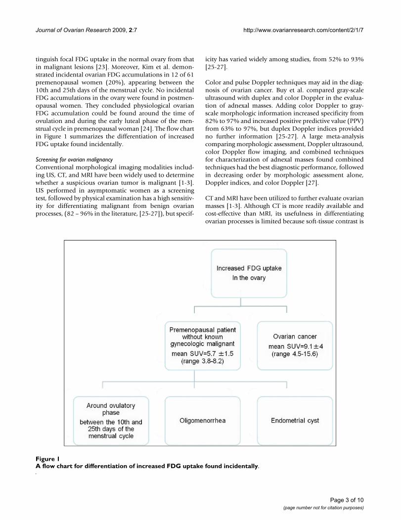
tinguish focal FDG uptake in the normal ovary from thatin malignant lesions [23]. Moreover, Kim et al. demon-strated incidental ovarian FDG accumulations in 12 of 61premenopausal women (20%), appearing between the10th and 25th days of the menstrual cycle. No incidentalFDG accumulations in the ovary were found in postmen-opausal women. They concluded physiological ovarianFDG accumulation could be found around the time ofovulation and during the early luteal phase of the men-strual cycle in premenopausal woman [24]. The flow chartin Figure 1 summarizes the differentiation of increasedFDG uptake found incidentally.
Screening for ovarian malignancyConventional morphological imaging modalities includ-ing US, CT, and MRI have been widely used to determinewhether a suspicious ovarian tumor is malignant [1-3].US performed in asymptomatic women as a screeningtest, followed by physical examination has a high sensitiv-ity for differentiating malignant from benign ovarianprocesses, (82 – 96% in the literature, [25-27]), but specif-
icity has varied widely among studies, from 52% to 93%[25-27].
Color and pulse Doppler techniques may aid in the diag-nosis of ovarian cancer. Buy et al. compared gray-scaleultrasound with duplex and color Doppler in the evalua-tion of adnexal masses. Adding color Doppler to gray-scale morphologic information increased specificity from82% to 97% and increased positive predictive value (PPV)from 63% to 97%, but duplex Doppler indices providedno further information [25-27]. A large meta-analysiscomparing morphologic assessment, Doppler ultrasound,color Doppler flow imaging, and combined techniquesfor characterization of adnexal masses found combinedtechniques had the best diagnostic performance, followedin decreasing order by morphologic assessment alone,Doppler indices, and color Doppler [27].
CT and MRI have been utilized to further evaluate ovarianmasses [1-3]. Although CT is more readily available andcost-effective than MRI, its usefulness in differentiatingovarian processes is limited because soft-tissue contrast is
A flow chart for differentiation of increased FDG uptake found incidentallyFigure 1A flow chart for differentiation of increased FDG uptake found incidentally.
Page 3 of 10(page number not for citation purposes)
Journal of Ovarian Research 2009, 2:7 http://www.ovarianresearch.com/content/2/1/7
relatively poor when compared with MRI, and MRI there-fore has higher diagnostic accuracy [1-3]. Reports in theliterature differ with regard to the sensitivity and specifi-city of MRI in the differentiation of benign and malignantadnexal lesions, ranging from 85% to 95% for sensitivityand from 87% to 96% for specificity [28-31].
The sensitivity of FDG-PET in the detection of ovariancancer was 78% in our study [32]; this was lower than theresults reported in the literature, which have been in therange of 83% to 86% [33-37]. We suspect that the reasonfor the comparatively low sensitivity in our study was thatour study population included a large number of falsenegative cases, such as patients with early mucinous ade-nocarcinoma and borderline mucinous adenocarcinoma.Rieber et al. reported that early carcinomas, mucinousadenocarcinomas, and particularly borderline tumors,present a problem because these tumors presumably lackthe typical pattern of FDG uptake as a result of the smallamount of transformed tissue [33], so they are likely togive false-negative results. Moreover, false-positive find-ings with FDG-PET occurred for endometriomas and der-moid cysts in our study. When the ovary is involved in aninflammatory process, inflammatory exudate may beaccompanied by FDG uptake in these regions [34]. Inaddition, schwannomas, serous cystadenomas, thecomas,mucinous cystadenomas, and corpus luteum cysts showincidentally increased glucose metabolism has beenreported in the literature [33-37].
In screening for ovarian cancer, US is the most importantmodality. MRI or FDG-PET, in addition to US, can providefurther information about ovarian tumors and improvespecificity. However, our study showed that the additionof FDG-PET to MRI does not yield additional informationfor the diagnosis of ovarian masses after US [32].
Recently, Castellucci et al. assessed the accuracy of FDG-PET/CT in distinguishing malignant from benign pelviclesions, compared with transvaginal ultrasonography(TVUS). Adding FDG-PET/CT increased specificity from61% to 100%, negative predictive value (NPV) from 78%to 81%, PPV from 80% to 100%, and accuracy from 80%to 92%. They concluded that FDG-PET/CT provides addi-tional information to TVUS in the differential diagnosis ofbenign from malignant pelvic lesions [37]. In conclusion,US is the most important modality in screening for ovar-ian malignancy. Although some investigators considerFDG-PET useful in the differential diagnosis of malig-nancy, most studies have shown that it is of little value[32-36]. However, FDG-PET/CT may provide useful addi-tional information when performed after TVUS in the dif-ferential diagnosis of malignancy [37].
PET in stagingA major problem in ovarian cancer is that a high propor-tion (75%) of patients have advanced stage disease at thetime of diagnosis, which results in a 5-year survival rate ofonly 41% [1]. Primary debulking surgery is not the onlytreatment option for ovarian cancer, and patients withbulky, nonresectable disease will not benefit from pri-mary surgery [1]. In addition, there is little survival benefitif the debulking is not optimal. The results of studiesregarding therapy for patients with advanced cancer of thepancreas and esophagus provide clear evidence that neo-adjuvant chemotherapy before surgery enables downstag-ing and thus improves operability as well as prognosis.The results of these studies strongly suggest the need toconsider neoadjuvant chemotherapy in patients withadvanced ovarian cancer [38]. Thereafter, accurate stagingof patients with ovarian cancer before treatment is neededto determine appropriate treatment for those who willpotentially benefit from it.
Our study is the first to show that the addition of FDG-PET to CT improves the staging accuracy of ovarian cancer[39]. The reason for this improvement was that FDG-PETfacilitated detection of metastases outside the pelvis. Forintrapelvic lesions, the sensitivity, specificity, PPV, NPV,and accuracy of CT alone increased from 72, 81, 48, 92,and 79%, respectively, to 76, 82, 50, 94, and 81%, respec-tively, when FDG-PET was added. Similarly, for lesionsoutside the pelvis, the sensitivity, specificity, PPV, NPV,and accuracy of CT alone increased from 24, 95, 44, 88,and 85%, respectively, to 63, 98, 88, 93, and 93%, respec-tively, with the addition of FDG-PET [39]. Although ourstudy did not provide an evaluation on a per patient basisor a statistical analysis, to the best of our knowledge, it isthe first to show that the addition of FDG-PET to CTimproves the staging accuracy of ovarian cancer [39].Recently, Kitajima et al. also reported that FDG-PET/con-trast-enhanced CT was a more accurate imaging modalityfor staging ovarian cancer and was more useful for select-ing appropriate treatment than enhanced CT alone [40].
In conclusion, FDG-PET is a useful and promising toolbut not an established procedure in the staging of ovariancancer patients. As studies in this field have been small-scale and have had variable results, a multicenter studywith more data and showing clinical utility for routine useis needed before the procedure can be applied routinelyfor patients with confirmed or suspected ovarian cancer[33,39-43].
Diagnosis of recurrent ovarian cancerRecurrent ovarian cancer is almost never curable, but earlydetection of recurrence theoretically increases the chancethat salvage treatment will result in prolonged remissionand sustained quality of life. Conventional imaging
Page 4 of 10(page number not for citation purposes)
Journal of Ovarian Research 2009, 2:7 http://www.ovarianresearch.com/content/2/1/7
modalities often give nonspecific results and are subopti-mal for the reliable detection of peritoneal recurrence. Theidentification of more accurate imaging modalitiesshould improve management decisions for patients withrecurrent ovarian cancer.
In 2002 we reported that FDG-PET was useful for follow-ing up an ovarian cancer patient in whom the only featuresuspicious of recurrence was a rising CA125 level withinthe normal range [44]. Havrilesky et al. performed a meta-analysis to assess the diagnostic performance of FDG-PETin comparison with that of CT and MRI in patients withovarian cancer. They concluded that FDG-PET did notappear to be useful in the routine surveillance of patientswith a history of ovarian cancer, and that it was unlikelyto improve the sensitivity of conventional modalities todetect microscopic intraperitoneal disease. There is fairevidence to support the use of PET for the detection ofrecurrent ovarian cancer when the CA-125 is elevated andconventional imaging is negative or equivocal, althoughwhether this results in improved patient outcome isunclear [45].
The use of FDG-PET/CT for detecting recurrent ovariancancer was first described by Makhija et al. in 2002 [46].In 2008, Gu et al. performed a systemic meta-analysis toassess the accuracy of CA-125, PET alone, FDG-PET/CT,CT alone, and MRI in diagnosing recurrent ovarian carci-noma. They demonstrated that CA-125 had the highestpooled specificity, 0.93 (95%CI: 0.89 – 0.95), and FDG-PET/CT had the highest pooled sensitivity, 0.91 (95% CI:0.88 – 0.94). They concluded FDG-PET/CT might be auseful supplement to current surveillance techniques, par-ticularly for patients with an increasing CA-125 level andnegative CT or MRI. However, regarding diagnostic accu-racy, interpreted CT may have limited additional valueover FDG-PET in detecting recurrent ovarian cancer [47].Recently, Kitajima et al. reported that PET/contrast-enhanced CT was able to detect more malignant lesionsthan FDG-PET/CT or enhanced CT alone in recurrentovarian cancer. Therefore, PET/contrast-enhanced CTcould lead to changes in the subsequent clinical manage-ment of 39% of these patients. Improved diagnostic accu-racy with PET/contrast-enhanced CT impactedmanagement in 16 patients (12%) diagnosed byenhanced CT alone and in three patients (2%) diagnosedby PET/non-contrast-enhanced CT [48]. They concludedthat PET/contrast-enhanced CT is an imaging modalitywith favorable accuracy for staging and for assessing ovar-ian cancer recurrence when compared with PET/non-con-trast-enhanced or enhanced CT.
In conclusion, FDG-PET may provide benefits for thosewith elevated CA-125 (>35 U/ml), CT- or MRI-definedlocalized recurrence amenable to local destructive proce-
dures, and clinically suspected recurrent or persistent can-cer when biopsy cannot be performed. Using FDG-PET/CT or PET/contrast-enhanced CT is reported to havehigher sensitivity and specificity than FDG-PET alone fordetecting recurrent disease. We have summarized sensitiv-ity and specificity for each imaging modality for the diag-nosis of primary and recurrent/metastatic ovarian cancerin Tables 1 and 2.
Usefulness of FDG-PET for assessing malignant activitySUV is the most common PET parameter measured in theclinical setting. Its calculation is simple, and most con-temporary FDG-PET/CT scanners display the imaging inthese units, provided the injected dose and the patientweight have been entered when setting up the PET acqui-sition [49]. The role of SUV in PET examination has beendiscussed at length; however, doubts remain due to fac-tors that can influence SUV calculation and reproducibil-ity. A study by Nahmias et al. [49] investigated thereproducibility of SUV in malignant tumors and foundthat a number of factors other than the natural history ofthe tumor could cause variability in the measured SUV.These factors included fluctuations in plasma glucose andpatient weight, errors in repositioning regions of interest(ROI) or image registration, and variations in the uptakeperiod. They concluded that repeated measurements ofmean SUV performed a few days apart were reproducible.A decrease of 0.5 SUV is statistically significant and maybe considered when establishing thresholds to predictsuccess of chemotherapy in patients with cancer.
We have previously assessed whether FDG-PET is usefulfor assessing malignant activity and gathering prognosticinformation in ovarian cancer [50]. We evaluated whetherFDG uptake, quantified as SUV by PET in ovarian epithe-lial tumors, correlates with clinical stage [51,52], tumorgrade [53], cell proliferation [54-56], or glucose metabo-lism [57], all of which are reported to be biomarkers forresponse to chemotherapy, prognosis, and overall survivalin ovarian cancer patients. Epithelial ovarian tumor spec-imens were graded histopathologically, and immunohis-tochemistry for MIB-1 (a proliferation index marker) andGLUT-1 (glucose transporter marker) was performed. Thecorrelations between FDG uptake and clinical stage,GLUT-1 expression, MIB-1 labeling index (LI), and histo-logical grade were determined. No positive correlation
Table 1: The following information shows the diagnosis of primary ovarian cancer
Sensitivity Specificity
Ultrasound (color and pulsed Doppler) 82%–96% 52%–93%MRI 85% – 95% 87% – 96%FDG-PET 58% – 86% 54% – 86%PET-CT 88% – 100% 85% – 88%
Page 5 of 10(page number not for citation purposes)
Journal of Ovarian Research 2009, 2:7 http://www.ovarianresearch.com/content/2/1/7
was observed between FDG uptake and clinical stage (P =0.14). On the other hand, the intensity of GLUT-1 expres-sion (r = 0.76, P = 0.001), MIB-1 LI (r = 0.457, P = 0.014),and histological grade (r = 0.692, P = 0.005) showed sta-tistically significant positive correlations with FDGuptake. Stepwise logistic regression analysis revealed thatthe expression of GLUT-1 transporters was the strongestpredictor of positive FDG uptake (r = 0.760, P = 0.0004)[50].
A study of GLUT-1 expression in ovarian carcinoma byCanturia et al. showed that GLUT-1 status is an independ-ent prognostic factor of response to chemotherapy inadvanced ovarian carcinoma, and that patients over-expressing this marker have a significantly shorter disease-free survival rate [58]. Furthermore, Avril et al. showed
that FDG-PET could predict response to neoadjuvantchemotherapy and survival in advanced ovarian cancer.Using a threshold for decrease in SUV from baseline of20% after the first course, the median overall survival wasfound to be 38.3 months in responders (23.1 months innon-responders). At a threshold of 55% decrease in SUVafter the third cycle, median overall survival was 38.9months in responders (19.7 months in non-responders).Although the number of cases was small, this prospectivestudy showed that sequential FDG-PET predicted patientoutcome as early as after the first cycle of neoadjuvantchemotherapy and was more accurate than CA-125.[59]
In conclusion, glucose consumption, as determined byanalysis of SUV in FDG-PET, may be a non-invasivebiomarker that can predict response to chemotherapy andsurvival in ovarian cancer.
Cost-effectiveness evaluation of FDG-PET in the management of patients with ovarian cancerPatients with advanced ovarian cancer who have com-pleted a planned course of chemotherapy have frequentlyundergone a systematic surgical exploration and may beasymptomatic. About 36% to 73% of patients may havepersistent disease detected at second-look procedures.Patients with residual disease should undergo continuous
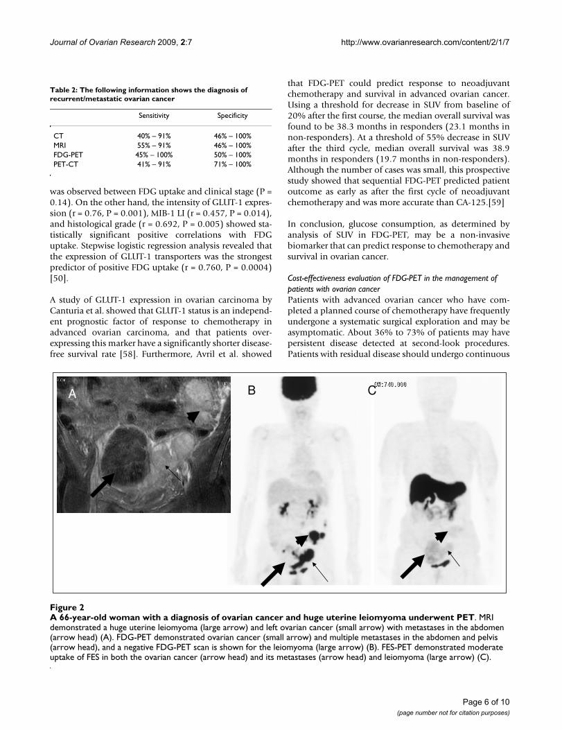
A 66-year-old woman with a diagnosis of ovarian cancer and huge uterine leiomyoma underwent PETFigure 2A 66-year-old woman with a diagnosis of ovarian cancer and huge uterine leiomyoma underwent PET. MRI demonstrated a huge uterine leiomyoma (large arrow) and left ovarian cancer (small arrow) with metastases in the abdomen (arrow head) (A). FDG-PET demonstrated ovarian cancer (small arrow) and multiple metastases in the abdomen and pelvis (arrow head), and a negative FDG-PET scan is shown for the leiomyoma (large arrow) (B). FES-PET demonstrated moderate uptake of FES in both the ovarian cancer (arrow head) and its metastases (arrow head) and leiomyoma (large arrow) (C).
A B C
Table 2: The following information shows the diagnosis of recurrent/metastatic ovarian cancer
Sensitivity Specificity
CT 40% – 91% 46% – 100%MRI 55% – 91% 46% – 100%FDG-PET 45% – 100% 50% – 100%PET-CT 41% – 91% 71% – 100%
Page 6 of 10(page number not for citation purposes)
Journal of Ovarian Research 2009, 2:7 http://www.ovarianresearch.com/content/2/1/7
adjunctive therapy, while those without disease may dis-continue adjunctive therapy. The cost-effectiveness andvalue of FDG-PET as a substitute for a second-look proce-dure have therefore been explored [14,60,61]. A detailedcost analysis of management of ovarian cancer with com-parison of FDG-PET and second-look procedure was per-formed by Smith et al. [60]. They demonstrated that FDG-PET led to a decrease in the proportion of patients whounderwent unnecessary laparotomy from 70% to 5%;35% of patients underwent the less-invasive procedure oflaparoscopy instead of laparatomy. Moreover, Kim et al.[61] reported the prognostic value of FDG-PET comparedwith that of a second-look procedure in patients withadvanced ovarian cancer treated with chemotherapy. Theyconcluded that PPV was 93% and NPV was 70% for FGD-PET, with no significant differences in progression-free
interval between FDG-PET groups and second-look proce-dures. Hence FDG-PET appears to be useful and cost effec-tive in the diagnosis of recurrent ovarian cancer.
A new PET tracer: potential applications in determining ER statusThe sensitivity of ovarian cancer to hormonal therapy hasa real, although modest, role in the treatment of advancedovarian cancers resistant to chemotherapy. Many agentshave been evaluated, including antiestrogens, estrogens,progesterones, androgens, aromatase inhibitors, andgonadotropin releasing hormone agonists (GnRH). Asanticancer agents, hormonal therapies produce anapproximate 10% response rate in previously treatedpatients. A correlation may exist between the presence ofhormone receptors and a response to therapy [1]. Thus,knowledge of hormone receptor status, for example estro-
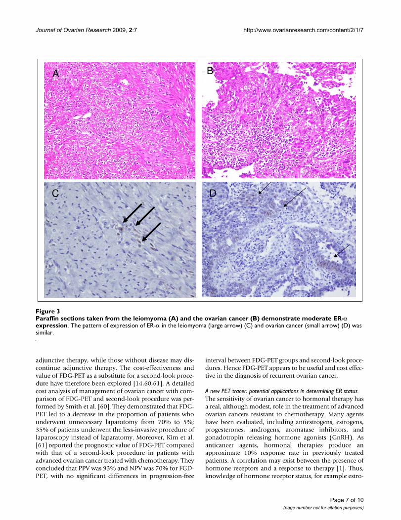
Paraffin sections taken from the leiomyoma (A) and the ovarian cancer (B) demonstrate moderate ER-α expressionFigure 3Paraffin sections taken from the leiomyoma (A) and the ovarian cancer (B) demonstrate moderate ER-α expression. The pattern of expression of ER-α in the leiomyoma (large arrow) (C) and ovarian cancer (small arrow) (D) was similar.
A B
C D
Page 7 of 10(page number not for citation purposes)
Journal of Ovarian Research 2009, 2:7 http://www.ovarianresearch.com/content/2/1/7
gen receptor (ER) status, is critically important for thetreatment of ovarian cancer. Tissue sampling is essentialbut difficult because it is associated with significant mor-bidity and sampling error. The most commonly usedmolecular imaging technique in body imaging is currentlyFDG-PET. This has become the method of choice for stag-ing and restaging in ovarian cancer, and it also hasbecome extremely valuable in monitoring the response toanticancer agents. New PET agents, such as 16α-18F-fluoro-17β-estradiol (FES) have potential in the evalua-tion of response to hormonal therapy for ovarian cancerafter FDG-PET. Although we do not have any experienceof the use of FES-PET in patients on long-term hormonetherapy to treat osteoporosis, we have already evaluatedFES-PET for patients without any previous treatment inthe differential diagnosis of benign and malignant uterinetumors [19].
Here, we first showed that FES uptake was observed at pri-mary and metastatic sites in three cases of advanced ovar-ian cancer. In these patients, we compared FES uptake andimmunohistochemistry results for surgical specimensfrom patients with both primary and metastatic sites.These data indicated that FES uptake in PET was associ-ated with ER status, particularly ER-α status, in ovariancancer. A representative case was that of a 66-year-oldwoman with huge uterine leiomyoma and ovarian serousadenocarcinoma who underwent FES-PET before andafter cytoreduction surgery. Before surgery, FES-PETshowed moderately increased uptake in both the leiomy-oma and ovarian cancer regions; the maximum SUV was2.5 and 2.1 (figure 2), respectively. After resection, boththe leiomyoma and ovarian cancer were found to befocally positive for estrogen receptor-α (ER-α) (figure 3).Hence FES uptake in PET was associated with ER-α statusin ovarian cancer in this case. Although this study wasonly a preliminary case report, to the best of our knowl-edge it is the first to suggest that FES-PET could provideuseful information about hormone status in advancedovarian cancer. This information may be useful inexpanding treatment choices for such patients.
ConclusionFDG-PET holds promise in the evaluation of cancerspread or recurrent or residual disease when other radio-graphic data are uncertain. FDG-PET/CT might be a usefulsupplemental investigation to detect primary and recur-rent ovarian cancer earlier than FDG-PET and other con-ventional imaging tools. In addition, FES-PET may havethe potential to provide useful information about hor-mone status in advanced ovarian cancer.
Competing interestsThe authors declare that they have no competing interests.
Authors' contributionsYY drafted the manuscript. TK, TT, OH, and FK conceptu-alized, edited, and revised the manuscript. All authorshave read and approved the final manuscript.
AcknowledgementsWe wish to express our sincere thanks to Dr. Yasuhisa Fujibayashi, Direc-tor, the Biomedical Imaging Research Center, and all staff of our depart-ment of gynecologic oncology.
References1. Ozols RF, Rubin SC, Thomas GM, Robboy SJ: Epithelial ovarian
cancer. In Principles and Practice of Gynecologic Oncology 4th edition.Edited by: William JH, Carlos AP, Robert CY. Philadelphia, LippincottWilliams & Wilkins; 2004:841-918.
2. Bragg DG, Hricak H: Imaging in gynecologic malignancies. Can-cer 1993, 71:1648-51.
3. Togashi K: Ovarian cancer: the clinical role of US, CT, andMRI. Eur Radiol. 2003, 13 Suppl 4:L87-L104.
4. Cook GJ, Maisey MN, Fogelman I: Normal variants, artefacts andinterpretative pitfalls in PET imaging with 18-fluoro-2-deox-yglucose and carbon-11 methionine. Eur J Nucl Med 1999,26:1363-78.
5. Kostakoglu L, Agress H Jr, Goldsmith SJ: Clinical role of FDG PETin evaluation of cancer patients. Radiographics 2003, 23:315-40.
6. Nakamoto Y, Saga T, Fujii S: Positron emission tomographyapplication for gynecologic tumors. Int J Gynecol Cancer 2005,15:701-9.
7. Pandit-Taskar N: Oncologic imaging in gynecologic malignan-cies. J Nucl Med 2005, 46:1842-50.
8. Kumar R, Chauhan A, Jana S, Dadparvar S: Positron emission tom-ography in gynecological malignancies. Expert Rev AnticancerTher 2006, 6:1033-44.
9. Mironov S, Akin O, Pandit-Taskar N, Hann LE: Ovarian cancer.Radiol Clin North Am 2007, 45:149-66.
10. Beyer T, Townsend DW, Brun T, Kinahan PE, Charron M, Roddy R,Jerin J, Young J, Byars L, Nutt R: A combined PET/CT scanner forclinical oncology. J Nucl Med 2000, 41:1369-79.
11. Bar-Shalom R, Yefremov N, Guralnik L, Gaitini D, Frenkel A, KutenA, Altman H, Keidar Z, Israel O: Clinical performance of PET/CTin evaluation of cancer: additional value for diagnostic imag-ing and patient management. J Nucl Med. 2003,44(8):1200-1209.
12. Yamamoto Y, Oguri H, Yamada R, Maeda N, Kohsaki S, Fukaya T:Preoperative evaluation of pelvic masses with combined18F-fluorodeoxyglucose positron emission tomography andcomputed tomography. Int J Gynaecol Obstet 2008, 102:124-7.
13. Iagaru AH, Mittra ES, McDougall IR, Quon A, Gambhir SS: 18F-FDGPET/CT evaluation of patients with ovarian carcinoma. NuclMed Commun 2008, 29:1046-51.
14. Basu S, Rubello D: PET imaging in the management of tumorsof testis and ovary: current thinking and future directions.Minerva Endocrinol 2008, 33:229-56.
15. Lucignani G: FDG-PET in gynaecological cancers: recentobservations. Eur J Nucl Med Mol Imaging 2008, 35:2133-9.
16. Tsuchida T, Okazawa H, Mori T, Kobayashi M, Yoshida Y, FujibayashiY, Itoh H: In vivo imaging of estrogen receptor concentrationin the endometrium and myometrium using FES PET – influ-ence of menstrual cycle and endogenous estrogen level. NuclMed Biol 2007, 34:205-10.
17. Mortimer JE, Dehdashti F, Siegel BA, Trinkaus A, KatzenellenbogenJA, Welch MJ: Metabolic flare: indicator of hormone respon-siveness in advanced breast cancer. J Clin Oncol 2001,19:2797-2803.
18. Yoshida Y, Kurokawa T, Sawamura Y, Shinagawa A, Okazawa H,Fujibayashi Y, Kotsuji F: The positron emission tomographywith F18 17beta-estradiol has the potential to benefit diag-nosis and treatment of endometrial cancer. Gynecol Oncol2007, 104:764-6.
19. Tsujikawa T, Yoshida Y, Mori T, Kurokawa T, Fujibayashi Y, Kotsuji F,Okazawa H: Uterine tumors: pathophysiologic imaging with16alpha-[18F]fluoro-17beta-estradiol and 18F fluorodeoxy-glucose PET – initial experience. Radiology 2008, 248:599-605.
Page 8 of 10(page number not for citation purposes)
Journal of Ovarian Research 2009, 2:7 http://www.ovarianresearch.com/content/2/1/7
20. Koyama K, Okamura T, Kawabe J, Ozawa N, Torii K, Umesaki N,Miyama M, Ochi H, Yamada R: Evaluation of 18F-FDG PET withbladder irrigation in patients with uterine and ovariantumors. J Nucl Med 2003, 44:353-8.
21. Schöder H, Gönen M: Screening for cancer with PET and PET/CT: potential and limitations. J Nucl Med 2007, 48:4-18.
22. Lerman H, Metser U, Grisaru D, Fishman A, Lievshitz G, Even-SapirE: Normal and abnormal 18F-FDG endometrial and ovarianuptake in pre- and postmenopausal patients: assessment byPET/CT. J Nucl Med 2004, 45:266-71.
23. Nishizawa S, Inubushi M, Okada H: Physiological 18F-FDGuptake in the ovaries and uterus of healthy female volun-teers. Eur J Nucl Med Mol Imaging 2005, 32:549-56.
24. Kim SK, Kang KW, Roh JW, Sim JS, Lee ES, Park SY: Incidentalovarian 18F-FDG accumulation on PET: correlation with themenstrual cycle. Eur J Nucl Med Mol Imaging 2005, 32:757-63.
25. Jain KA, Friedman DL, Pettinger TW, Alagappan R, Jeffrey RB Jr, Som-mer FG: Adnexal masses: comparison of specificity ofendovaginal US and pelvic MR imaging. Radiology 1993,186:697-704.
26. Buy JN, Ghossain MA, Hugol D, Hassen K, Sciot C, Truc JB, PoitoutP, Vadrot D: Characterization of adnexal masses: combina-tion of color Doppler and conventional sonography com-pared with spectral Doppler analysis alone and conventionalsonography alone. AJR Am J Roentgenol 1996, 166:385-93.
27. Kinkel K, Hricak H, Lu Y, Tsuda K, Filly RA: US characterizationof ovarian masses: a meta-analysis. Radiology 2000, 217:803-11.
28. Outwater EK, Dunton CJ: Imaging of the ovary and adnexa: clin-ical issues and applications of MR imaging. Radiology 1995,194:1-18.
29. Yamashita Y, Torashima M, Hatanaka Y, Harada M, Higashida Y, Taka-hashi M, Mizutani H, Tashiro H, Iwamasa J, Miyazaki K: Adnexalmasses: accuracy of characterization with transvaginal USand precontrast and postcontrast MR imaging. Radiology 1995,194:557-65.
30. Stevens SK, Hricak H, Stern JL: Ovarian lesions: detection andcharacterization with gadolinium-enhanced MRI at 1.5 T.Radiology 1991, 181:481-8.
31. Stevens SK, Hricak H, Campos Z: Teratomas versus cystic hem-orrhagic adnexal lesions: differentiation with proton-selec-tive fat-saturation MR imaging. Radiology 1993, 186:481-8.
32. Kawahara K, Yoshida Y, Kurokawa T, Suzuki Y, Nagahara K, TsuchidaT, Okazawa H, Fujibayashi Y, Yonekura Y, Kotsuji F: Evaluation ofpositron emission tomography with tracer 18-fluorodeoxy-glucose in addition to magnetic resonance imaging in thediagnosis of ovarian cancer in selected women after ultra-sonography. J Comput Assist Tomogr 2004, 28:505-16.
33. Rieber A, Nussle K, Stohr I, Grab D, Fenchel S, Kreienberg R, ReskeSN, Brambs HJ: Preoperative diagnosis of ovarian tumors withMR imaging: comparison with transvaginal sonography, pos-itron emission tomography, and histologic findings. AJR Am JRoentgenol 2001, 177:123-9.
34. Fenchel S, Grab D, Nuessle K, Kotzerke J, Rieber A, Kreienberg R,Brambs HJ, Reske SN: Asymptomatic adnexal masses: correla-tion of FDG PET and histopathologic findings. Radiology 2002,223:780-8.
35. Berger KL, Nicholson SA, Dehdashti F, Siegel BA: FDG PET evalu-ation of mucinous neoplasms: correlation of FDG uptakewith histopathologic features. AJR Am J Roentgenol 2000,174:1005-8.
36. Modesitt S, Tortolero-Luna G, Robinson J, Gershenson D, Wolf J:Ovarian and extraovarian endometriosis-associated cancer.Obstet Gynecol 2002, 100:788.
37. Castellucci P, Perrone AM, Picchio M, Ghi T, Farsad M, Nanni C,Messa C, Meriggiola MC, Pelusi G, Al-Nahhas A, Rubello D, Fazio F,Fanti S: Diagnostic accuracy of 18F-FDG PET/CT in charac-terizing ovarian lesions and staging ovarian cancer: correla-tion with transvaginal ultrasonography, computedtomography, and histology. Nucl Med Commun 2007, 28:589-95.
38. Kuhn W, Rutke S, Späthe K, Schmalfeldt B, Florack G, von Hun-delshausen B, Pachyn D, Ulm K, Graeff H: Neoadjuvant chemo-therapy followed by tumor debulking prolongs survival forpatients with poor prognosis in International Federation ofGynecology and Obstetrics Stage IIIC ovarian carcinoma.Cancer 2001, 92:2585-2591.
39. Yoshida Y, Kurokawa T, Kawahara K, Tsuchida T, Okazawa H, Fujiba-yashi Y, Yonekura Y, Kotsuji F: Incremental benefits of FDG pos-itron emission tomography over CT alone for thepreoperative staging of ovarian cancer. AJR Am J Roentgenol2004, 182:227-33.
40. Kitajima K, Murakami K, Yamasaki E, Kaji Y, Fukasawa I, Inaba N, Sug-imura K: Diagnostic accuracy of integrated FDG-PET/con-trast-enhanced CT in staging ovarian cancer: comparisonwith enhanced CT. Eur J Nucl Med Mol Imaging 2008, 35:1912-20.
41. Schröder W, Zimny M, Rudlowski C, Büll U, Rath W: The role of18F-fluoro-deoxyglucose positron emission tomography(18F-FDG PET) in diagnosis of ovarian cancer. Int J GynecolCancer 1999, 9:117-122.
42. Risum S, Høgdall C, Loft A, Berthelsen AK, Høgdall E, Nedergaard L,Lundvall L, Engelholm SA: The diagnostic value of PET/CT forprimary ovarian cancer – a prospective study. Gynecol Oncol2007, 105:145-9.
43. Risum S, Høgdall C, Loft A, Berthelsen AK, Høgdall E, Nedergaard L,Lundvall L, Engelholm SA: Prediction of suboptimal primarycytoreduction in primary ovarian cancer with combined pos-itron emission tomography/computed tomography-a pro-spective study. Gynecol Oncol 2008, 108:265-70.
44. Kurokawa T, Yoshida Y, Kawahara K, Tsuchida T, Fujibayashi Y,Yonekura Y, Kotsuji F: Whole-body PET with FDG is useful forfollowing up an ovarian cancer patient with only rising CA-125 levels within the normal range. Ann Nucl Med 2002,16:491-3.
45. Havrilesky LJ, Kulasingam SL, Matchar DB, Myers ER: FDG-PET formanagement of cervical and ovarian cancer. Gynecol Oncol2005, 97:183-91.
46. Makhija S, Howden N, Edwards R, Kelley J, Townsend DW, MeltzerCC: Positron emission tomography/computed tomographyimaging for the detection of recurrent ovarian and fallopiantube carcinoma: a retrospective review. Gynecol Oncol 2002,85:53-8.
47. Gu P, Pan LL, Wu SQ, Sun L, Huang G: CA 125, PET alone, PET-CT, CT and MRI in diagnosing recurrent ovarian carcinomaA systematic review and meta-analysis. Eur J Radiol 2008:29.
48. Kitajima K, Murakami K, Yamasaki E, Domeki Y, Kaji Y, Fukasawa I,Inaba N, Suganuma N, Sugimura K: Performance of integratedFDG-PET/contrast-enhanced CT in the diagnosis of recur-rent ovarian cancer: comparison with integrated FDG-PET/non-contrast-enhanced CT and enhanced CT. Eur J Nucl MedMol Imaging 2008, 35:1439-48.
49. Nahmias C, Wahl LM: Reproducibility of standardized uptakevalue measurements determined by 18F-FDG PET in malig-nant tumors. J Nucl Med 2008, 49:1804-8.
50. Kurokawa T, Yoshida Y, Kawahara K, Tsuchida T, Okazawa H, Fujiba-yashi Y, Yonekura Y, Kotsuji F: Expression of GLUT-1 glucosetransfer, cellular proliferation activity and grade of tumorcorrelate with [F-18]-fluorodeoxyglucose uptake by posi-tron emission tomography in epithelial tumors of the ovary.Int J Cancer 2004, 10;109:926-32.
51. Malkasian GD Jr, Melton LJ Jr, O'Brien PC, Greene MH: Prognosticsignificance of histologic classification and grading of epithe-lial malignancies of the ovary. Am J Obstet Gynecol 1984,149:274-84.
52. Omura GA, Brady MF, Homesley HD, Yordan E, Major FJ, BuchsbaumHJ, Park RC: Long-term follow-up and prognostic factor anal-ysis in advanced ovarian carcinoma: the Gynecologic Oncol-ogy Group experience. J Clin Oncol 1991, 9:1138-50.
53. Shimizu Y, Kamoi S, Amada S, Akiyama F, Silverberg SG: Toward thedevelopment of a universal grading system for ovarian epi-thelial carcinoma: testing of a proposed system in a series of461 patients with uniform treatment and follow-up. Cancer1998, 82:893-901.
54. Huettner PC, Weinberg DS, Lage JM: Assessment of proliferativeactivity in ovarian neoplasms by flow and static cytometry.Correlation with prognostic features. Am J Pathol 1992,141:699-706.
55. Garzetti GG, Ciavattini A, Goteri G, De Nictolis M, Stramazzotti D,Lucarini G, Biagini G: Ki67 antigen immunostaining (MIB 1monoclonal antibody) in serous ovarian tumors: index ofproliferative activity with prognostic significance. GynecolOncol 1995, 56:169-74.
Page 9 of 10(page number not for citation purposes)
http://www.ncbi.nlm.nih.gov/entrez/query.fcgi?cmd=Retrieve&db=PubMed&dopt=Abstract&list_uids=8430177
http://www.ncbi.nlm.nih.gov/entrez/query.fcgi?cmd=Retrieve&db=PubMed&dopt=Abstract&list_uids=8430177
http://www.ncbi.nlm.nih.gov/entrez/query.fcgi?cmd=Retrieve&db=PubMed&dopt=Abstract&list_uids=8553953
http://www.ncbi.nlm.nih.gov/entrez/query.fcgi?cmd=Retrieve&db=PubMed&dopt=Abstract&list_uids=8553953
http://www.ncbi.nlm.nih.gov/entrez/query.fcgi?cmd=Retrieve&db=PubMed&dopt=Abstract&list_uids=8553953
http://www.ncbi.nlm.nih.gov/entrez/query.fcgi?cmd=Retrieve&db=PubMed&dopt=Abstract&list_uids=7997533
http://www.ncbi.nlm.nih.gov/entrez/query.fcgi?cmd=Retrieve&db=PubMed&dopt=Abstract&list_uids=7997533
http://www.ncbi.nlm.nih.gov/entrez/query.fcgi?cmd=Retrieve&db=PubMed&dopt=Abstract&list_uids=7824738
http://www.ncbi.nlm.nih.gov/entrez/query.fcgi?cmd=Retrieve&db=PubMed&dopt=Abstract&list_uids=7824738
http://www.ncbi.nlm.nih.gov/entrez/query.fcgi?cmd=Retrieve&db=PubMed&dopt=Abstract&list_uids=7824738
http://www.ncbi.nlm.nih.gov/entrez/query.fcgi?cmd=Retrieve&db=PubMed&dopt=Abstract&list_uids=1924792
http://www.ncbi.nlm.nih.gov/entrez/query.fcgi?cmd=Retrieve&db=PubMed&dopt=Abstract&list_uids=1924792
http://www.ncbi.nlm.nih.gov/entrez/query.fcgi?cmd=Retrieve&db=PubMed&dopt=Abstract&list_uids=8421755
http://www.ncbi.nlm.nih.gov/entrez/query.fcgi?cmd=Retrieve&db=PubMed&dopt=Abstract&list_uids=8421755
http://www.ncbi.nlm.nih.gov/entrez/query.fcgi?cmd=Retrieve&db=PubMed&dopt=Abstract&list_uids=8421755
http://www.ncbi.nlm.nih.gov/entrez/query.fcgi?cmd=Retrieve&db=PubMed&dopt=Abstract&list_uids=6328995
http://www.ncbi.nlm.nih.gov/entrez/query.fcgi?cmd=Retrieve&db=PubMed&dopt=Abstract&list_uids=6328995
http://www.ncbi.nlm.nih.gov/entrez/query.fcgi?cmd=Retrieve&db=PubMed&dopt=Abstract&list_uids=6328995
http://www.ncbi.nlm.nih.gov/entrez/query.fcgi?cmd=Retrieve&db=PubMed&dopt=Abstract&list_uids=1904477
http://www.ncbi.nlm.nih.gov/entrez/query.fcgi?cmd=Retrieve&db=PubMed&dopt=Abstract&list_uids=1904477
http://www.ncbi.nlm.nih.gov/entrez/query.fcgi?cmd=Retrieve&db=PubMed&dopt=Abstract&list_uids=1904477
http://www.ncbi.nlm.nih.gov/entrez/query.fcgi?cmd=Retrieve&db=PubMed&dopt=Abstract&list_uids=9486579
http://www.ncbi.nlm.nih.gov/entrez/query.fcgi?cmd=Retrieve&db=PubMed&dopt=Abstract&list_uids=9486579
http://www.ncbi.nlm.nih.gov/entrez/query.fcgi?cmd=Retrieve&db=PubMed&dopt=Abstract&list_uids=9486579
http://www.ncbi.nlm.nih.gov/entrez/query.fcgi?cmd=Retrieve&db=PubMed&dopt=Abstract&list_uids=1381562
http://www.ncbi.nlm.nih.gov/entrez/query.fcgi?cmd=Retrieve&db=PubMed&dopt=Abstract&list_uids=1381562
http://www.ncbi.nlm.nih.gov/entrez/query.fcgi?cmd=Retrieve&db=PubMed&dopt=Abstract&list_uids=1381562
http://www.ncbi.nlm.nih.gov/entrez/query.fcgi?cmd=Retrieve&db=PubMed&dopt=Abstract&list_uids=7896180
Journal of Ovarian Research 2009, 2:7 http://www.ovarianresearch.com/content/2/1/7
Publish with BioMed Central and every scientist can read your work free of charge
"BioMed Central will be the most significant development for disseminating the results of biomedical research in our lifetime."
Sir Paul Nurse, Cancer Research UK
Your research papers will be:
available free of charge to the entire biomedical community
peer reviewed and published immediately upon acceptance
cited in PubMed and archived on PubMed Central
yours — you keep the copyright
Submit your manuscript here:http://www.biomedcentral.com/info/publishing_adv.asp
BioMedcentral
56. Viale G, Maisonneuve P, Bonoldi E, Di Bacco A, Bevilacqua P, Paniz-zoni GA, Radaelli U, Gasparini G: The combined evaluation ofp53 accumulation and of Ki-67 (MIB1) labelling index pro-vides independent information on overall survival of ovariancarcinoma patients. Ann Oncol 1997, 8:469-76.
57. Kalir T, Wang BY, Goldfischer M, Haber RS, Reder I, Demopoulos R,Cohen CJ, Burstein DE: Immunohistochemical staining ofGLUT1 in benign, borderline, and malignant ovarian epithe-lia. Cancer 2002, 94:1078-82.
58. Cantuaria G, Magalhaes A, Penalver M, Angioli R, Braunschweiger P,Gomez-Marin O, Kanhoush R, Gomez-Fernandez C, Nadji M:Expression of GLUT-1 glucose transporter in borderline andmalignant epithelial tumors of the ovary. Gynecol Oncol 2000,79:33-7.
59. Avril N, Sassen S, Schmalfeldt B, Naehrig J, Rutke S, Weber WA,Werner M, Graeff H, Schwaiger M, Kuhn W: Prediction ofresponse to neoadjuvant chemotherapy by sequential F-18-fluorodeoxyglucose positron emission tomography inpatients with advanced-stage ovarian cancer. J Clin Oncol 2005,23:7445-53.
60. Smith GT, Hubner KF, McDonald T, Thie JA: Cost Analysis of FDGPET for Managing Patients with Ovarian Cancer. Clin PositronImaging 1999, 2:63-70.
61. Kim S, Chung JK, Kang SB, Kim MH, Jeong JM, Lee DS, Lee MC:[18F]FDG PET as a substitute for second-look laparotomy inpatients with advanced ovarian carcinoma. Eur J Nucl Med MolImaging 2004, 31:196-201.
Page 10 of 10(page number not for citation purposes)
http://www.ncbi.nlm.nih.gov/entrez/query.fcgi?cmd=Retrieve&db=PubMed&dopt=Abstract&list_uids=9233527
http://www.ncbi.nlm.nih.gov/entrez/query.fcgi?cmd=Retrieve&db=PubMed&dopt=Abstract&list_uids=9233527


![Index []1631 Index a a emitters 422 A-DOXO-HYD 777, 778 A121 human ovarian tumor xenograft 1348 a2-macroglobulin 65 AAG (α1-acid glycoprotein) 1341AAV (adeno-associated virus) 1426,](https://static.fdocument.org/doc/165x107/60bed310ab987851c764f6d0/index-1631-index-a-a-emitters-422-a-doxo-hyd-777-778-a121-human-ovarian-tumor.jpg)