Journal of Experimental & Clinical Cancer Research BioMed ... · sol level less than 2.5 μg/dL (69...
9
BioMed Central Page 1 of 9 (page number not for citation purposes) Journal of Experimental & Clinical Cancer Research Open Access Research MASEP gamma knife radiosurgery for secretory pituitary adenomas: experience in 347 consecutive cases Heng Wan* 1 , Ohye Chihiro 2 and Shubin Yuan 3 Address: 1 Department of Neurology and Functional neurosurgery, West China Fourth hospital, Sichuan University, Chengdu, 610041, PR China, 2 Functional and Gamma Knife Surgery Center, Hidaka Hospital, Takasaki, Gunma, Japan and 3 Department of Stereotactic and Functional Neurosurgery, Chengdu Air-force 452 Hospital, Chengdu, 610010, PR China Email: Heng Wan* - [email protected]; Ohye Chihiro - [email protected]; Shubin Yuan - [email protected] * Corresponding author Abstract Background: Secretory pituitary adenomas are very common brain tumors. Historically, the treatment armamentarium for secretory pituitary adenomas included neurosurgery, medical management, and fractionated radiotherapy. In recent years, MASEP gamma knife radiosurgery (MASEP GKRS) has emerged as an important treatment modality in the management of secretory pituitary adenomas. The goal of this research is to define accurately the efficacy, safety, complications, and role of MASEP GKRS for treatment of secretory pituitary adenomas. Methods: Between 1997 and 2007 a total of 347 patients with secretory pituitary adenomas treated with MASEP GKRS and with at least 60 months of follow-up data were identified. In 47 of these patients some form of prior treatment such as transsphenoidal resection, or craniotomy and resection had been conducted. The others were deemed ineligible for microsurgery because of body health or private choice, and MASEP GKRS served as the primary treatment modality. Endocrinological, ophthalmological, and neuroradiological responses were evaluated. Results: MASEP GKRS was tolerated well in these patients under the follow-up period ranged from 60 to 90 months; acute radioreaction was rare and 17 patients had transient headaches with no clinical significance. Late radioreaction was noted in 1 patient and consisted of consistent headache. Of the 68 patients with adrenocorticotropic hormone-secreting(ACTH) adenomas, 89.7% showed tumor volume decrease or remain unchanged and 27.9% experienced normalization of hormone level. Of the 176 patients with prolactinomas, 23.3% had normalization of hormone level and 90.3% showed tumor volume decrease or remain unchanged. Of the 103 patients with growth hormone-secreting(GH) adenomas, 95.1% experienced tumor volume decrease or remain unchanged and 36.9% showed normalization of hormone level. Conclusion: MASEP GKRS is safe and effective in treating secretory pituitary adenomas. None of the patients in our study experienced injury to the optic apparatus or had other neuropathies related with gamma knife. MASEP GKRS may serve as a primary treatment method in some or as a salvage treatment in the others. However, treatment must be tailored to meet the patient's symptoms, tumor location, tumor morphometry, and overall health. Longer follow-up is required for a more complete assessment of late radioreaction and treatment efficacy. Published: 11 March 2009 Journal of Experimental & Clinical Cancer Research 2009, 28:36 doi:10.1186/1756-9966-28-36 Received: 10 December 2008 Accepted: 11 March 2009 This article is available from: http://www.jeccr.com/content/28/1/36 © 2009 Wan et al; licensee BioMed Central Ltd. This is an Open Access article distributed under the terms of the Creative Commons Attribution License (http://creativecommons.org/licenses/by/2.0 ), which permits unrestricted use, distribution, and reproduction in any medium, provided the original work is properly cited.
Transcript of Journal of Experimental & Clinical Cancer Research BioMed ... · sol level less than 2.5 μg/dL (69...
BioMed Central
Journal of Experimental & Clinical Cancer Research
ss
Open AcceResearchMASEP gamma knife radiosurgery for secretory pituitary adenomas: experience in 347 consecutive casesHeng Wan*1, Ohye Chihiro2 and Shubin Yuan3Address: 1Department of Neurology and Functional neurosurgery, West China Fourth hospital, Sichuan University, Chengdu, 610041, PR China, 2Functional and Gamma Knife Surgery Center, Hidaka Hospital, Takasaki, Gunma, Japan and 3Department of Stereotactic and Functional Neurosurgery, Chengdu Air-force 452 Hospital, Chengdu, 610010, PR China
Email: Heng Wan* - [email protected]; Ohye Chihiro - [email protected]; Shubin Yuan - [email protected]
* Corresponding author
AbstractBackground: Secretory pituitary adenomas are very common brain tumors. Historically, thetreatment armamentarium for secretory pituitary adenomas included neurosurgery, medicalmanagement, and fractionated radiotherapy. In recent years, MASEP gamma knife radiosurgery(MASEP GKRS) has emerged as an important treatment modality in the management of secretorypituitary adenomas. The goal of this research is to define accurately the efficacy, safety,complications, and role of MASEP GKRS for treatment of secretory pituitary adenomas.
Methods: Between 1997 and 2007 a total of 347 patients with secretory pituitary adenomastreated with MASEP GKRS and with at least 60 months of follow-up data were identified. In 47 ofthese patients some form of prior treatment such as transsphenoidal resection, or craniotomy andresection had been conducted. The others were deemed ineligible for microsurgery because ofbody health or private choice, and MASEP GKRS served as the primary treatment modality.Endocrinological, ophthalmological, and neuroradiological responses were evaluated.
Results: MASEP GKRS was tolerated well in these patients under the follow-up period rangedfrom 60 to 90 months; acute radioreaction was rare and 17 patients had transient headaches withno clinical significance. Late radioreaction was noted in 1 patient and consisted of consistentheadache. Of the 68 patients with adrenocorticotropic hormone-secreting(ACTH) adenomas,89.7% showed tumor volume decrease or remain unchanged and 27.9% experienced normalizationof hormone level. Of the 176 patients with prolactinomas, 23.3% had normalization of hormonelevel and 90.3% showed tumor volume decrease or remain unchanged. Of the 103 patients withgrowth hormone-secreting(GH) adenomas, 95.1% experienced tumor volume decrease or remainunchanged and 36.9% showed normalization of hormone level.
Conclusion: MASEP GKRS is safe and effective in treating secretory pituitary adenomas. None ofthe patients in our study experienced injury to the optic apparatus or had other neuropathiesrelated with gamma knife. MASEP GKRS may serve as a primary treatment method in some or asa salvage treatment in the others. However, treatment must be tailored to meet the patient'ssymptoms, tumor location, tumor morphometry, and overall health. Longer follow-up is requiredfor a more complete assessment of late radioreaction and treatment efficacy.
Published: 11 March 2009
Journal of Experimental & Clinical Cancer Research 2009, 28:36 doi:10.1186/1756-9966-28-36
Received: 10 December 2008Accepted: 11 March 2009
This article is available from: http://www.jeccr.com/content/28/1/36
© 2009 Wan et al; licensee BioMed Central Ltd. This is an Open Access article distributed under the terms of the Creative Commons Attribution License (http://creativecommons.org/licenses/by/2.0), which permits unrestricted use, distribution, and reproduction in any medium, provided the original work is properly cited.
Page 1 of 9(page number not for citation purposes)

Journal of Experimental & Clinical Cancer Research 2009, 28:36 http://www.jeccr.com/content/28/1/36
BackgroundPituitary adenomas are common lesions and represent20% of all primary brain tumors[1,2]. The epidemiologi-cal studies have demonstrated that nearly 20% of the gen-eral population harbor pituitary adenomas[3,4]. Pituitaryadenomas are broadly classified into two groups[5]. In thefirst category are those that secrete excess amounts of nor-mal pituitary hormones and present with a variety of clin-ical syndromes depending on the types of hormonessecreted. Meanwhile, some macroadenoma may presentwith pressure symptoms, often increase in size ifuntreated, and in some rare cases they may cause symp-toms related to mass effect in which the optic nerves andchiasm are compressed[6,7]. The second category of pitu-itary adenomas is nonfunctioning adenomas that do notsecrete any known biologically active pituitary hormones.Patients can also suffer hypopituitarism secondary tocompression of the normal functioning pituitarygland[8].
In the treatment of pituitary adenomas the goal is toremove the tumor mass or arrest further growth and whenpresent normalize hormonal hypersecretion. Transsphe-noidal surgery is established as one of the most reliabletreatment modalities. This modern microsurgical tech-nique can reduce tumor mass to protect surroundingstructures from potential compression, and achieve theendocrinological cure of the symptoms caused by hor-mone secreting tumors. Long term tumor control ratesafter transsphenoidal excision alone vary from 50 to80%[9]. However, in some cases, many patients arealready in poor physical condition caused by extendedproduction of the excess pituitary hormones, and generalanesthesia itself sometimes brings a certain risk for them.Also, they often show invasion to surrounding structuresincluding cavernous sinus. And for these types of pituitaryadenomas, incomplete tumor resection or recurrence as aresult of tumor invasion into surrounding structures isquite common[10].
In recent years, gamma knife radiosurgery(GKRS) hasemerged as an important treatment modality in the man-agement of secretory pituitary adenomas with its high effi-cacy. Radiosurgical treatment may deliver a high dose tothe adenomas with high accuracy and may not influencethe nearby neural structures to induce neurologicaldefect[11]. Recently, more and more reports have detailedtreatment results for secretory pituitary adenomas withGKRS, and there have been a number of reports of GKRSas a primary treatment for secretory pituitary adeno-mas[12]. However, most results of these reports werebased on the gamma knife with radioactive source stati-cally, and the time of the follow-up varied from monthsto years. This article reviews our 10 years of clinical expe-rience in performing rotary gamma knife in patients with
secretory pituitary adenomas. The focus of this research isto define accurately the efficacy, safety, complications,and role of rotary gamma knife for treatment of secretorypituitary adenomas.
MethodsCharacteristic of the patientsBetween 1997 and 2007, 1681 patients with a diagnosisof secretory pituitary adenoma were treated with MASEProtary gamma knife(MASEP instruments, Inc., Shenzhen,P.R. China) in our medical center. The patients with secre-tory pituitary adenoma treated in our studies are thoseloss convenience, intolerant of or resistant to medicaltherapies. Some of them were evaluated ineligible for neu-rosurgery because of body health and the others rejectedto surgery on private choice or economic condition. 347patients under medical therapies irregularly less than 3months after MASEP GKRS and getting follow-up with atleast 60 months were taken in our study, and those withfollow-up less than 60 months or taken medical therapiesregularly after MASEP GKRS were excluded. Our studypopulation comprised 162 men (46.7%) and 185 women(53.3%). Their age ranged from 17 to 86 years (mean41.8). The patients presented with a 1- to 19-year history(mean 2.7). In 47 of these patients some form of priortreatment such as transsphenoidal resection, or craniot-omy and resection had been conducted. The others weredeemed ineligible for microsurgery because of bodyhealth or private choice, and MASEP GKRS served as theprimary treatment modality. Endocrinological, ophthal-mological, and neuroradiological exams were taken for allof them. The diagnosis was made on the basis of magneticresonance imaging (MRI) findings, endocrinologicalexam findings, pathological findings (available for post-operative patients), and their clinic history. Of thesepatients treated, 68(19.6%) had a diagnosis of adrenocor-ticotropic hormone-secreting adenomas, 176(50.7%) hada diagnosis of prolactinomas, and 103(29.7%) hadgrowth hormone-secreting adenomas. The mean follow-up period was 67.3 months (range 60~90 months) (Table1).
Gamma knife processingPatients were fixed in the Leksell stereotactic head frameunder administration of local anesthesia. Before treat-ment, a high-resolution MRI with gadolinium-enhance-ment to obtain precise information on the shape, volume,and the three-dimensional coordinates of the tumors andthe surrounding anatomic structures is performed. Radio-surgery was performed using the MASEP rotary gammaknife. MASEP rotary gamma ray stereotactic extracranialsystem is equipped with 25 Co-60 sources. Each source isformed by certain amount of Φ1 × 1 cobalt granuleswelded into 2 layer stainless steel casing through argonfluorine welding technique to make it seal-tight. The total
Page 2 of 9(page number not for citation purposes)

Journal of Experimental & Clinical Cancer Research 2009, 28:36 http://www.jeccr.com/content/28/1/36
combined initial loading activity is 240.5 TBq ± 10%(6500 Ci ± 10%). Source specific activity is 300 Ci/g.Source active zone is Φ3.1 × 30. At initial loading thewater-absorption dose rate at focusing point is greaterthan 3 Gy/min. 25 cobalt sources are placed in the colli-mator passages. The commercially available software,MASEP Gamma-Plan (MASEP instruments, Inc., Shen-zhen, P.R. China) was used for complex dose planning.The radiosurgical planning was done jointly by neurosur-geons and radiation oncologists. Dose planning requiresdelineation of the targets and the adjacent structures,especially the optic chiasm. Though the MASEP gammaknife has five collimator sizes, 4, 8, 14, 18 and 22 mm, the4 mm and 8 mm collimator were used commonly. Theday before MASEP GKRS, patients were claimed to take1.5 mg hexadecadrol. The day after MASEP GKRS, patientswere desired to take intervenous drop infusion of 250 mlmannitol plus 10 mg hexadecadrol (twice a day) for 3
days to avoid radioreaction. Then they were dischargedand could return to their daily lives without any neurolog-ical deterioration.
Treatment planningTumor volume was 0.8~21.5 cm3(mean 5.2 cm3). For thepurpose of both growth control and hormonal remission,secretory pituitary adenomas were usually irradiated morethan 12 Gy (range 12~35 Gy) at the tumor margin. Thewhole tumor was covered within 50~70% isodose lines.The dosimetric goal in every case was complete tumor cov-erage. The prescribed marginal dose had to be decreasedoccasionally to keep the dose less than 10 Gy to the opticnerve, chiasma, and tract to avoid radiation-induced vis-ual disturbances, less than 12 Gy to the brainstem and lessthan 25 Gy to the internal carotid artery (Table 2).
Clinical observationAfter the treatment of MASEP GKRS, follow-up was sched-uled at intervals of 6 months, 1 year and annually thereaf-ter. The visit routinely assessed the change in symptomsafter treatment, endocrinological examination and neuro-radiological study.
In our series to evaluate ACTH-producing pituitary adeno-mas, we utilized the 24 h urine cortisol collection notexcess of 200 μg/dL (550 nmol/dL) and the plasma corti-sol level less than 2.5 μg/dL (69 nmol/dL) as the criteriafor endocrinological evaluation. For patients treated withprolactinomas, we used normal serum prolactin level forgender as cure criteria and the normal PRL range for non-pregnant women is <500 mU/L (20 μg/L) and for men<300 mU/L (12 μg/L). Meanwhile, we used the guidelinesfor a remission or cure as the GH level less than 1 ng/ml(2.5 mU/L) after glucose ingestion and a normal serumtype-1 insulin like growth factor(IGF-1) when matchedfor age and gender to define the results of radiosurgery forpatients with acromegaly.
After irradiation of pituitary tissue, regular surveillance isneeded to detect development of hypopituitarism, partic-ularly GH deficiency. Basal pituitary profiles, including
Table 1: Characteristics of patients with pituitary adenomas treated with MASEP GKRS
Characteristic Value(%)
The statistics of the populationSex
male 162(46.7)female 185(53.3)
Mean age(yrs) 41.8 (range17~86)Mean history(yrs) 2.7(range1 to 19)
No. of previous treatmentstranssphenoidal resection 27*craniotomy and resection 23
Mean follow-up after GKRS(mos) 67.3 (range 60~90)Type of adenomas
ACTH adenomas 68(19.6)microadenoma(size, cm3) 21 (0.8~1.1)macroadenoma(size, cm3) 47 (1.2~6.4)
Prolactinomas 176(50.7)microadenoma(size, cm3) 0macroadenoma(size, cm3) 176(1.2~17.9)
GH adenomas 103(29.7)microadenoma(size, cm3) 0macroadenoma(size, cm3) 103(2.3~21.5)
*three patient had performed repeated craniotomy and resection
Table 2: MASEP GKRS plan for patients with pituitary adenomas(mean)
Type Cases Margin dose(Gy) Treatment isodose(%) Tumor coverage(%)
ACTH 68microadenoma 21 15~28(18.9) 50 100macroadenoma 47 18~35(24.9) 50~70(54.7) 70~100(95.3)PRL 176microadenoma 0 0 0 0macroadenoma 176 15~35 (22.4) 50~70(55.3) 64~100(93.3)GH 103microadenoma 0 0 0 0macroadenoma 103 12~30 (21.4) 50~70(57.6) 55~100(88.6)
Page 3 of 9(page number not for citation purposes)

Journal of Experimental & Clinical Cancer Research 2009, 28:36 http://www.jeccr.com/content/28/1/36
measurement of TSH, ACTH, gonadotropins, growth hor-mone, IGF-1 and assessment for the clinical features ofGH deficiency or consequent gonadal failure, were per-formed regularly on follow-up.
The statistical analysisStatistical analysis was performed with the aid of commer-cially available software (StatView 4.5.1; Abacus Con-cepts, Inc., Berkeley, CA).
ResultsMASEP GKRS was tolerated well in these patients. Acuteradioreaction was rare and 17 patients had transient head-aches with no clinical significance. Consistent headachewas noted in 1 patient 4 years after radiosurgery and per-sisted for the entire 1 year during follow-up. There was nosignificant compression and the reason of headache wasstill unknown. Of the 68 patients with ACTH adenomas,61(89.7%) showed tumor volume decrease or remainunchanged and 19(27.9%) experienced normalization ofhormone level (Figure 1 and Figure 2). Of the other 5
patients with enlarged ACTH adenomas, 4 had repeatedMASEP GKRS. One had craniotomy and resection of themass after experiencing consistent vomiting. Another twocases with no clinical symptom with a neuroradiologicaldiagnosis of radiation necrosis received no more treat-ment. Of the 176 patients with prolactinomas, 41(23.3%)had normalization of hormone level and 159(90.3%)showed tumor volume decrease or remain unchanged(Figure 3 and Figure 4). Of the 12 patients with enlargedprolactinomas, 9 had repeated GKRS. Two had transsphe-noidal resection of the mass after experiencing consistentheadache. One case died 4 years after primary MASEPGKRS rejecting any medical intervention. Another 5 caseswith the diagnosis of radiation necrosis had no clinicalsymptoms and lived as usual. Of the 103 patients with GHadenomas, 98(95.1%) experienced tumor volumedecrease or remain unchanged and 38(36.9%) showednormalization of hormone level (Figure 5 and Figure 6).Of the other 3 patients with enlarged GH adenomas, 2had repeated MASEP GKRS. One had craniotomy andresection of the mass after experiencing consistent vomit-ing. Another 2 cases with no clinical treatment had a neu-roradiological diagnosis of radiation necrosis and wereunder observation.
Typical MRI scan changes in ACTH adenomaFigure 1Typical MRI scan changes in ACTH adenoma. Coronal T1-weighted postcontrast MRI scan at left and right, obtained in Patient 1, a 30-year-old man who presented with ACTH adenomas and consistent headache 2 years before undergo-ing GKRS. An enhancing mass lesion is seen in the sella turcia with extension to bilateral internal carotid artery. Patient 1's serum ACTH level was 381.6 pg/ml, and his blood pressure was over 180/120 mmHg. The patient was treated with MASEP GKRS, and MRI was performed for treatment plan-ning. 26 Gy defined to the 50% isodose line is used to cover the full extent of the pituitary tumor in all three planes.
Typical MRI scan changes in ACTH adenomaFigure 2Typical MRI scan changes in ACTH adenoma. No enhancing mass lesion is seen in the sella turcia under the T1-weighted postcontrast MRI scan performed 2 years after GKRS. Patient 1's clinical symptom did improve. His serum ACTH level came down to 40.4 pg/ml, and his blood pres-sure was controlled within 140/80 mmHg.
Page 4 of 9(page number not for citation purposes)
Journal of Experimental & Clinical Cancer Research 2009, 28:36 http://www.jeccr.com/content/28/1/36
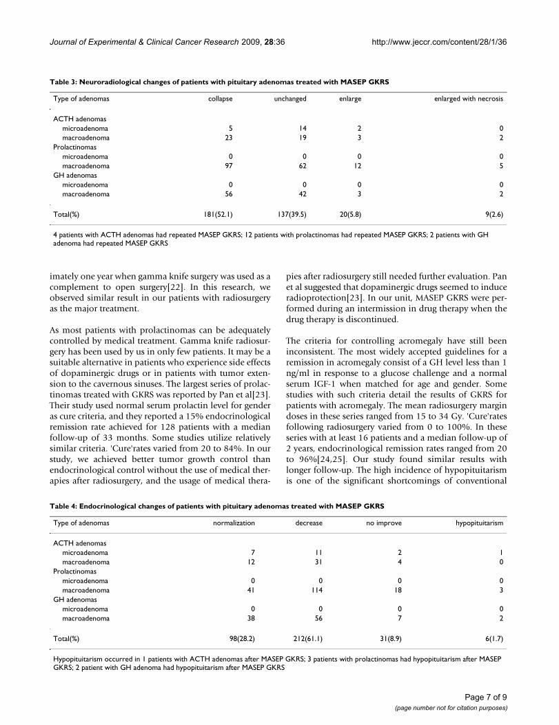
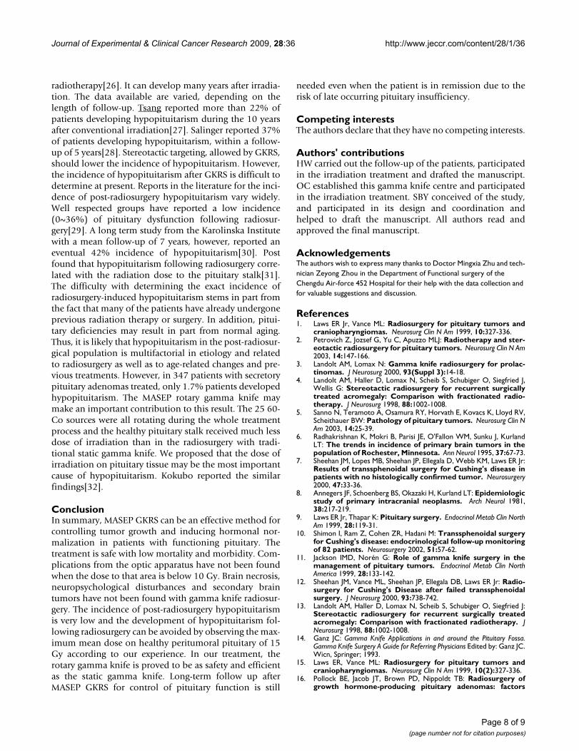
Regular endocrinological and neuroradiological re-exam-inations were available in all these patients. The data col-lected as of the end of 2007 are summed up in table 3 andtable 4.
Overall 91.6% of tumor control was achieved in 318 withonly mild and transient neurological complications insome cases. 28.2% of normalization of hormone level rateand 61.1% of decrease of hormone level rate were alsoachieved. Hypopituitarism occurred in 6(1.7%) patientswho received replacement therapy now.
DiscussionThere are multiple treatment modalities for pituitary ade-nomas. The individual treatment must be tailored to a
patient's symptoms, overall health, and tumor morphom-etry. GKRS has been found to be an effective, noninvasivemethod for treating patients with functioning pituitaryadenoma as a complement to the surgery. Tumors thatcompress the optic pathway should be removed withmicrosurgery, and residual tumor, especially in the cav-ernous sinus, is a good indication for radiosurgery. At ourinstitute, MASEP GKRS may be considered as an alterna-tive treatment to microsurgery if patients are reluctant toundergo surgical resection, or are unable to undergomicrosurgery under general anesthesia because of old ageor poor medical conditions. The purpose of GKRS, in thecase of secretory pituitary adenomas, is to control tumorgrowth and normalize endocrinological hypersecretion.Secretory adenomas seem to require a higher radiationdose than nonfunctioning pituitary adenomas[13]. Ganzsuggested that the effective dose for secretory adenomasshould be higher than 25 Gy according to the details[14].Laws and Vance estimated that a higher percentage of con-trol of hyper-functioning syndromes could be accom-plished with the higher margin dose[15]. The lowesteffective radiation dose in our study was 12 Gy deliveredto the tumor margin; the mean marginal dose was 22.2Gy. According to our experience, the suitable margin dose
Typical MRI scan changes in prolactinomas adenomaFigure 3Typical MRI scan changes in prolactinomas adenoma. Coronal T1-weighted postcontrast MRI scan at left and right, obtained in Patient 2, a 27-year-old woman who presented with prolactinomas adenomas and amenorrhea-galactorrhea 4 years before undergoing MASEP GKRS. An asymmetrically enhancing mass lesion is seen in the sella turcia with exten-sion to bilateral internal carotid artery. Patient 2's serum prolactin level was 183.7 ng/ml. The patient was treated with MASEP GKRS twice because of the huge volume of the mass. The second MASEP GKRS was performed 1 year after the first one. The tumor was treated separately with the lower and upper part in order to protect the optic chiasma. MRI was performed for treatment planning. 25 Gy defined to the 50% isodose line is used to cover the lower part of the pitui-tary tumor in the first treatment, and 18 Gy defined to the 50% isodose line is used to cover the upper part of the pitui-tary tumor in the second time.
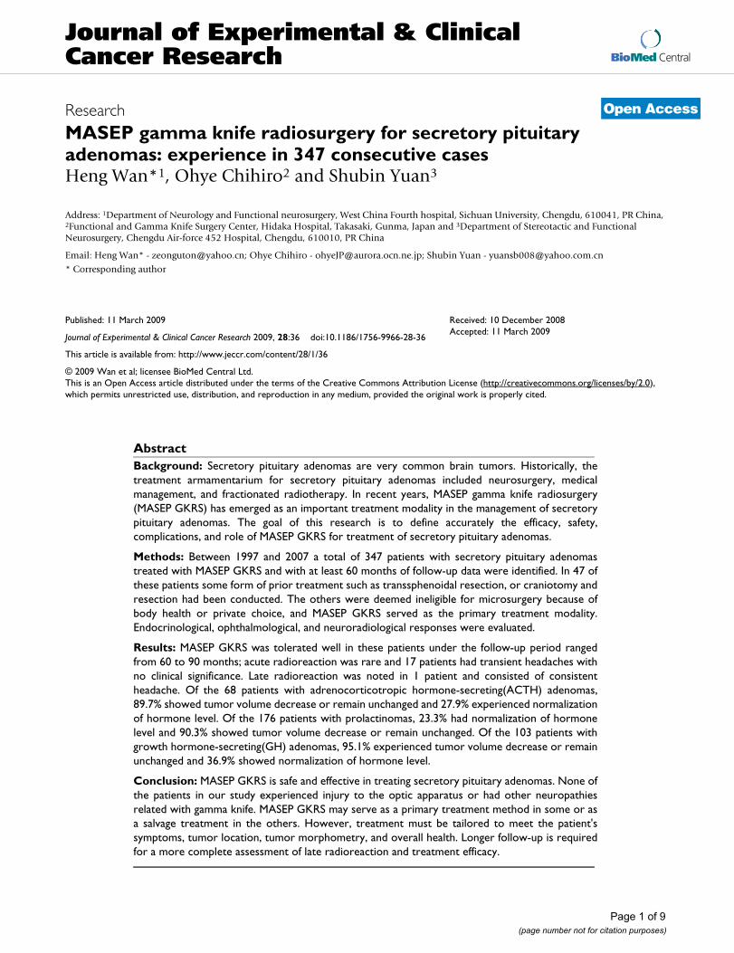
Typical MRI scan changes in prolactinomas adenomaFigure 4Typical MRI scan changes in prolactinomas adenoma. An enhancing mass lesion is seen in the sella turcia under the T1-weighted postcontrast MRI scan performed 1 year after MASEP GKRS, but the volume of the mass had collapsed for more than 50%. Patient 2's clinical symptom did improve. Her serum prolactin level came down to 14.5 ng/ml, and she got gestation and delivered a healthy baby recently.
Page 5 of 9(page number not for citation purposes)
Journal of Experimental & Clinical Cancer Research 2009, 28:36 http://www.jeccr.com/content/28/1/36
should depend on the endocrinological type of the secre-tory pituitary adenoma. However, the recent report of Pol-lock for functioning adenomas revealed the radiationdose was not related to endocrinological outcome[16].
In nearly all published series, stereotactic radiosurgeryafforded excellent control of tumor growth. Hayashireported that the tumor control rate for pituitary adenomaafter GKRS was between 93% and 94%, and that thetumor shrinkage rate ranged from 46% to 56.7%[17].Many studies reported a greater than 95% control oftumor size with follow-up varying from months toyears[18,19]. Some series have even demonstratedimprovement in visual function following radiosurgeryupon shrinkage of the tumor. Most pituitary adenomastend to be slow growing lesions. As such, it may be mis-leading to evaluate series of patients with relatively short
follow-up. In our previous study, the effects of MASEPGKRS may get stable within three years after the treat-ment, and this study shows concordant results within thefollow-up more than 5 years.
At the time when GKRS started, the results of microsurgerywere disappointing regarding ACTH-producing pituitaryadenomas and the role of GKRS as primary therapy wasevaluated. We have not seen any recurrences after MASEPGKRS in patients who obtained remission in contrast topituitary microsurgery with progressive increase of recur-rences of Cushing's syndrome with time. Cushing's dis-ease is a serious catabolic illness that requires rapidnormalization of cortisol hypersecretion. Thus pituitarymicrosurgery is the primary treatment for Cushing's dis-ease; gamma knife surgery can be applied when open sur-gery is contraindicated or refused or as a secondarytreatment when open surgery has failed or the tumorextends into the cavernous sinuses. Many series utilizedthe 24 h urine cortisol collection as part of the criteria forendocrinological evaluation, and the endocrinologi-cal'cure'rates ranged from 17 to 83%[20,21]. In a recentreview, Laws and Vance reported remission in about 60%of their patients with Cushing's disease followed for morethan 6 months with a mean time to remission of approx-
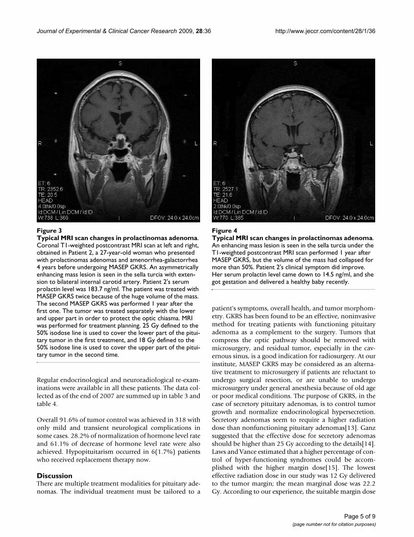
Typical MRI scan changes in GH adenomaFigure 5Typical MRI scan changes in GH adenoma. Coronal T1-weighted postcontrast MRI scan at upper left and right, obtained in Patient 3, a 33-year-old man who presented with GH adenomas and acromegaly 7 years before undergoing MASEP GKRS. (Figure 5) An enhancing mass lesion is seen in the sella turcia with extension into the left cavernous sinus. Patient 3's serum growth hormone level was 497.3 ng/ml ini-tially. He was treated with transsphenoidal surgery, and the tumor relapsed shortly with the serum growth hormone level reduced to 130.2 ng/ml. The patient was treated with MASEP GKRS, and MRI was performed for treatment plan-ning. 20 Gy defined to the 50% isodose line is used to cover the full extent of the pituitary tumor in the first radiosurgery, and 28 Gy defined to the 50% isodose line is used to cover the pituitary tumor in the second time one year later.
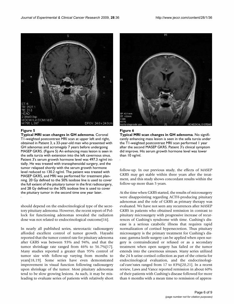
Typical MRI scan changes in GH adenomaFigure 6Typical MRI scan changes in GH adenoma. No signifi-cantly enhancing mass lesion is seen in the sella turcia under the T1-weighted postcontrast MRI scan performed 1 year after the second MASEP GKRS. Patient 3's clinical symptom did improve. His serum growth hormone level was lower than 10 ng/ml.
Page 6 of 9(page number not for citation purposes)
Journal of Experimental & Clinical Cancer Research 2009, 28:36 http://www.jeccr.com/content/28/1/36
imately one year when gamma knife surgery was used as acomplement to open surgery[22]. In this research, weobserved similar result in our patients with radiosurgeryas the major treatment.
As most patients with prolactinomas can be adequatelycontrolled by medical treatment. Gamma knife radiosur-gery has been used by us in only few patients. It may be asuitable alternative in patients who experience side effectsof dopaminergic drugs or in patients with tumor exten-sion to the cavernous sinuses. The largest series of prolac-tinomas treated with GKRS was reported by Pan et al[23].Their study used normal serum prolactin level for genderas cure criteria, and they reported a 15% endocrinologicalremission rate achieved for 128 patients with a medianfollow-up of 33 months. Some studies utilize relativelysimilar criteria. 'Cure'rates varied from 20 to 84%. In ourstudy, we achieved better tumor growth control thanendocrinological control without the use of medical ther-apies after radiosurgery, and the usage of medical thera-
pies after radiosurgery still needed further evaluation. Panet al suggested that dopaminergic drugs seemed to induceradioprotection[23]. In our unit, MASEP GKRS were per-formed during an intermission in drug therapy when thedrug therapy is discontinued.
The criteria for controlling acromegaly have still beeninconsistent. The most widely accepted guidelines for aremission in acromegaly consist of a GH level less than 1ng/ml in response to a glucose challenge and a normalserum IGF-1 when matched for age and gender. Somestudies with such criteria detail the results of GKRS forpatients with acromegaly. The mean radiosurgery margindoses in these series ranged from 15 to 34 Gy. 'Cure'ratesfollowing radiosurgery varied from 0 to 100%. In theseseries with at least 16 patients and a median follow-up of2 years, endocrinological remission rates ranged from 20to 96%[24,25]. Our study found similar results withlonger follow-up. The high incidence of hypopituitarismis one of the significant shortcomings of conventional
Table 3: Neuroradiological changes of patients with pituitary adenomas treated with MASEP GKRS
Type of adenomas collapse unchanged enlarge enlarged with necrosis
ACTH adenomasmicroadenoma 5 14 2 0macroadenoma 23 19 3 2
Prolactinomasmicroadenoma 0 0 0 0macroadenoma 97 62 12 5
GH adenomasmicroadenoma 0 0 0 0macroadenoma 56 42 3 2
Total(%) 181(52.1) 137(39.5) 20(5.8) 9(2.6)
4 patients with ACTH adenomas had repeated MASEP GKRS; 12 patients with prolactinomas had repeated MASEP GKRS; 2 patients with GH adenoma had repeated MASEP GKRS
Table 4: Endocrinological changes of patients with pituitary adenomas treated with MASEP GKRS
Type of adenomas normalization decrease no improve hypopituitarism
ACTH adenomasmicroadenoma 7 11 2 1macroadenoma 12 31 4 0
Prolactinomasmicroadenoma 0 0 0 0macroadenoma 41 114 18 3
GH adenomasmicroadenoma 0 0 0 0macroadenoma 38 56 7 2
Total(%) 98(28.2) 212(61.1) 31(8.9) 6(1.7)
Hypopituitarism occurred in 1 patients with ACTH adenomas after MASEP GKRS; 3 patients with prolactinomas had hypopituitarism after MASEP GKRS; 2 patient with GH adenoma had hypopituitarism after MASEP GKRS
Page 7 of 9(page number not for citation purposes)
Journal of Experimental & Clinical Cancer Research 2009, 28:36 http://www.jeccr.com/content/28/1/36
radiotherapy[26]. It can develop many years after irradia-tion. The data available are varied, depending on thelength of follow-up. Tsang reported more than 22% ofpatients developing hypopituitarism during the 10 yearsafter conventional irradiation[27]. Salinger reported 37%of patients developing hypopituitarism, within a follow-up of 5 years[28]. Stereotactic targeting, allowed by GKRS,should lower the incidence of hypopituitarism. However,the incidence of hypopituitarism after GKRS is difficult todetermine at present. Reports in the literature for the inci-dence of post-radiosurgery hypopituitarism vary widely.Well respected groups have reported a low incidence(0~36%) of pituitary dysfunction following radiosur-gery[29]. A long term study from the Karolinska Institutewith a mean follow-up of 7 years, however, reported aneventual 42% incidence of hypopituitarism[30]. Postfound that hypopituitarism following radiosurgery corre-lated with the radiation dose to the pituitary stalk[31].The difficulty with determining the exact incidence ofradiosurgery-induced hypopituitarism stems in part fromthe fact that many of the patients have already undergoneprevious radiation therapy or surgery. In addition, pitui-tary deficiencies may result in part from normal aging.Thus, it is likely that hypopituitarism in the post-radiosur-gical population is multifactorial in etiology and relatedto radiosurgery as well as to age-related changes and pre-vious treatments. However, in 347 patients with secretorypituitary adenomas treated, only 1.7% patients developedhypopituitarism. The MASEP rotary gamma knife maymake an important contribution to this result. The 25 60-Co sources were all rotating during the whole treatmentprocess and the healthy pituitary stalk received much lessdose of irradiation than in the radiosurgery with tradi-tional static gamma knife. We proposed that the dose ofirradiation on pituitary tissue may be the most importantcause of hypopituitarism. Kokubo reported the similarfindings[32].
ConclusionIn summary, MASEP GKRS can be an effective method forcontrolling tumor growth and inducing hormonal nor-malization in patients with functioning pituitary. Thetreatment is safe with low mortality and morbidity. Com-plications from the optic apparatus have not been foundwhen the dose to that area is below 10 Gy. Brain necrosis,neuropsychological disturbances and secondary braintumors have not been found with gamma knife radiosur-gery. The incidence of post-radiosurgery hypopituitarismis very low and the development of hypopituitarism fol-lowing radiosurgery can be avoided by observing the max-imum mean dose on healthy peritumoral pituitary of 15Gy according to our experience. In our treatment, therotary gamma knife is proved to be as safety and efficientas the static gamma knife. Long-term follow up afterMASEP GKRS for control of pituitary function is still
needed even when the patient is in remission due to therisk of late occurring pituitary insufficiency.
Competing interestsThe authors declare that they have no competing interests.
Authors' contributionsHW carried out the follow-up of the patients, participatedin the irradiation treatment and drafted the manuscript.OC established this gamma knife centre and participatedin the irradiation treatment. SBY conceived of the study,and participated in its design and coordination andhelped to draft the manuscript. All authors read andapproved the final manuscript.
AcknowledgementsThe authors wish to express many thanks to Doctor Mingxia Zhu and tech-nician Zeyong Zhou in the Department of Functional surgery of the Chengdu Air-force 452 Hospital for their help with the data collection and for valuable suggestions and discussion.
References1. Laws ER Jr, Vance ML: Radiosurgery for pituitary tumors and
craniopharyngiomas. Neurosurg Clin N Am 1999, 10:327-336.2. Petrovich Z, Jozsef G, Yu C, Apuzzo MLJ: Radiotherapy and ster-
eotactic radiosurgery for pituitary tumors. Neurosurg Clin N Am2003, 14:147-166.
3. Landolt AM, Lomax N: Gamma knife radiosurgery for prolac-tinomas. J Neurosurg 2000, 93(Suppl 3):14-18.
4. Landolt AM, Haller D, Lomax N, Scheib S, Schubiger O, Siegfried J,Wellis G: Stereotactic radiosurgery for recurrent surgicallytreated acromegaly: Comparison with fractionated radio-therapy. J Neurosurg 1998, 88:1002-1008.
5. Sanno N, Teramoto A, Osamura RY, Horvath E, Kovacs K, Lloyd RV,Scheithauer BW: Pathology of pituitary tumors. Neurosurg Clin NAm 2003, 14:25-39.
6. Radhakrishnan K, Mokri B, Parisi JE, O'Fallon WM, Sunku J, KurlandLT: The trends in incidence of primary brain tumors in thepopulation of Rochester, Minnesota. Ann Neurol 1995, 37:67-73.
7. Sheehan JM, Lopes MB, Sheehan JP, Ellegala D, Webb KM, Laws ER Jr:Results of transsphenoidal surgery for Cushing's disease inpatients with no histologically confirmed tumor. Neurosurgery2000, 47:33-36.
8. Annegers JF, Schoenberg BS, Okazaki H, Kurland LT: Epidemiologicstudy of primary intracranial neoplasms. Arch Neurol 1981,38:217-219.
9. Laws ER Jr, Thapar K: Pituitary surgery. Endocrinol Metab Clin NorthAm 1999, 28:119-31.
10. Shimon I, Ram Z, Cohen ZR, Hadani M: Transsphenoidal surgeryfor Cushing's disease: endocrinological follow-up monitoringof 82 patients. Neurosurgery 2002, 51:57-62.
11. Jackson IMD, Norén G: Role of gamma knife surgery in themanagement of pituitary tumors. Endocrinol Metab Clin NorthAmerica 1999, 28:133-142.
12. Sheehan JM, Vance ML, Sheehan JP, Ellegala DB, Laws ER Jr: Radio-surgery for Cushing's Disease after failed transsphenoidalsurgery. J Neurosurg 2000, 93:738-742.
13. Landolt AM, Haller D, Lomax N, Scheib S, Schubiger O, Siegfried J:Stereotactic radiosurgery for recurrent surgically treatedacromegaly: Comparison with fractionated radiotherapy. JNeurosurg 1998, 88:1002-1008.
14. Ganz JC: Gamma Knife Applications in and around the Pituitary Fossa.Gamma Knife Surgery A Guide for Referring Physicians Edited by: Ganz JC.Wicn, Springer; 1993.
15. Laws ER, Vance ML: Radiosurgery for pituitary tumors andcraniopharyngiomas. Neurosurg Clin N Am 1999, 10(2):327-336.
16. Pollock BE, Jacob JT, Brown PD, Nippoldt TB: Radiosurgery ofgrowth hormone-producing pituitary adenomas: factors
Page 8 of 9(page number not for citation purposes)
http://www.ncbi.nlm.nih.gov/entrez/query.fcgi?cmd=Retrieve&db=PubMed&dopt=Abstract&list_uids=9609294
http://www.ncbi.nlm.nih.gov/entrez/query.fcgi?cmd=Retrieve&db=PubMed&dopt=Abstract&list_uids=9609294
http://www.ncbi.nlm.nih.gov/entrez/query.fcgi?cmd=Retrieve&db=PubMed&dopt=Abstract&list_uids=9609294
http://www.ncbi.nlm.nih.gov/entrez/query.fcgi?cmd=Retrieve&db=PubMed&dopt=Abstract&list_uids=7818260
http://www.ncbi.nlm.nih.gov/entrez/query.fcgi?cmd=Retrieve&db=PubMed&dopt=Abstract&list_uids=7818260
http://www.ncbi.nlm.nih.gov/entrez/query.fcgi?cmd=Retrieve&db=PubMed&dopt=Abstract&list_uids=7213145
http://www.ncbi.nlm.nih.gov/entrez/query.fcgi?cmd=Retrieve&db=PubMed&dopt=Abstract&list_uids=7213145
http://www.ncbi.nlm.nih.gov/entrez/query.fcgi?cmd=Retrieve&db=PubMed&dopt=Abstract&list_uids=9609294
http://www.ncbi.nlm.nih.gov/entrez/query.fcgi?cmd=Retrieve&db=PubMed&dopt=Abstract&list_uids=9609294

Journal of Experimental & Clinical Cancer Research 2009, 28:36 http://www.jeccr.com/content/28/1/36
Publish with BioMed Central and every scientist can read your work free of charge
"BioMed Central will be the most significant development for disseminating the results of biomedical research in our lifetime."
Sir Paul Nurse, Cancer Research UK
Your research papers will be:
available free of charge to the entire biomedical community
peer reviewed and published immediately upon acceptance
cited in PubMed and archived on PubMed Central
yours — you keep the copyright
Submit your manuscript here:http://www.biomedcentral.com/info/publishing_adv.asp
BioMedcentral
associated with biochemical remission. J Neurosurg 2007,106:833-838.
17. Hayashi M, Izawa M, Hiyama H, Nakamura S, Atsuchi S, Sato H,Nakaya K, Sasaki K, Ochiai T, Kubo O, Hori T, Takakura K: Gammaknife radiosurgery for pituitary adenomas. Stereotact FunctNeurosurg 1999, 72:111-118.
18. Thorén M, Höybye C, Grenbäck E, Degerblad M, Rähn T, Hulting AL:The role of gamma knife radiosurgery in the management ofpituitary adenomas. J Neurooncol 2001, 54:197-203.
19. Petrovich Z, Yu C, Gianotta SL, Zee CS, Apuzzo ML: Gamma kniferadiosurgery for pituitary adenoma:early results. Neurosur-gery 2003, 53:51-59.
20. Höybye C, Grenbäck E, Rähn T, Degerblad M, Thorén M, Hulting AL:ACTH-producing pituitary tumours 12–22 years follow upafter treatment with stereotactic radiosurgery. Neurosurgery2001, 49:284-291.
21. Thorén M, Rähn T, Hallengren B, Kaad PH, Nilsson KO, Ravn H,Ritzén M, Petersen KE, Aarskog D: Treatment of Cushing's dis-ease in Childhood and Adolescence by Stereotactic pituitaryirradiation. Acta Paediatr Scand 1986, 75:388-395.
22. Laws E, Vance ML: Radiosurgery for pituitary tumors and crani-opharyngiomas. Neurosurgery Clinics of North America 1999,10:327-336.
23. Pan L, Zhang N, Wang E, Wang B, Xu W: Pituitary adenomas: Theeffect of gamma knife radiosurgery on tumor growth andendocrinopathies. Stereotact Funct Neurosurg 1998, 70:119-126.
24. Choi JY, Chang JH, Chang JW, Ha Y, Park YG, Chung SS: Radiologi-cal and hormonal responses of functioning pituitary adeno-mas after gamma knife radiosurgery. Yonsei Med J 2003,44:602-607.
25. Kim MS, Lee SI, Sim JH: Gamma Knife radiosurgery for function-ing pituitary microadenoma. Stereotact Funct Neurosurg 1999,72:119-124.
26. Becker G, Kocher M, Kortmann RD, Paulsen F, Jeremic B, Muller RP,Bamberg M: Radiation therapy in the multimodal treatmentapproach of pituitary adenoma. Strahlenther Onkol 2002,178:173-186.
27. Tsang RW, Brierley JD, Panzarella T, Gospodarowicz MK, Sutcliffe SB,Simpson WJ: Role of radiation therapy in clinical hormonally-active pituitary adenomas. Radiother Oncol 1996, 41:45-53.
28. Salinger DJ, Brady LW, Miyamoto CT: Radiation therapy in thetreatment of pituitary adenomas. Am J Clin Oncol 1992,15:467-473.
29. McCord MW, Buatti JM, Fennell EM, Mendenhall WM, Marcus RB Jr,Rhoton AL, Grant MB, Friedman WA: Radiotherapy for pituitaryadenoma: long-term outcome and sequelae. Int J Radiat OncolBiol Phys 1997, 39:437-444.
30. Nishioka H, Hirano A, Haraoka J, Nakajima N: Histologicalchanges in the pituitary gland and adenomas following radi-otherapy. Neuropathology 2002, 22:19-25.
31. Post KD, Habas JE: Comparison of long term results betweenprolactin secreting adenomas and ACTH secreting adeno-mas. Can J Neurol Sci 1990, 17:74-77.
32. Kokubo M, Sasai K, Shibamoto Y, Aoki T, Oya N, Mitsumori M, Taka-hashi JA, Hashimoto N, Hiraoka M: Long-term results of radia-tion therapy for pituitary adenoma. J Neuro oncol 2000,47:79-84.
Page 9 of 9(page number not for citation purposes)
http://www.ncbi.nlm.nih.gov/entrez/query.fcgi?cmd=Retrieve&db=PubMed&dopt=Abstract&list_uids=3014807
http://www.ncbi.nlm.nih.gov/entrez/query.fcgi?cmd=Retrieve&db=PubMed&dopt=Abstract&list_uids=3014807
http://www.ncbi.nlm.nih.gov/entrez/query.fcgi?cmd=Retrieve&db=PubMed&dopt=Abstract&list_uids=3014807
http://www.ncbi.nlm.nih.gov/entrez/query.fcgi?cmd=Retrieve&db=PubMed&dopt=Abstract&list_uids=9782243
http://www.ncbi.nlm.nih.gov/entrez/query.fcgi?cmd=Retrieve&db=PubMed&dopt=Abstract&list_uids=9782243
http://www.ncbi.nlm.nih.gov/entrez/query.fcgi?cmd=Retrieve&db=PubMed&dopt=Abstract&list_uids=9782243
http://www.ncbi.nlm.nih.gov/entrez/query.fcgi?cmd=Retrieve&db=PubMed&dopt=Abstract&list_uids=8961367
http://www.ncbi.nlm.nih.gov/entrez/query.fcgi?cmd=Retrieve&db=PubMed&dopt=Abstract&list_uids=8961367
http://www.ncbi.nlm.nih.gov/entrez/query.fcgi?cmd=Retrieve&db=PubMed&dopt=Abstract&list_uids=1449108
http://www.ncbi.nlm.nih.gov/entrez/query.fcgi?cmd=Retrieve&db=PubMed&dopt=Abstract&list_uids=1449108
http://www.ncbi.nlm.nih.gov/entrez/query.fcgi?cmd=Retrieve&db=PubMed&dopt=Abstract&list_uids=9308948
http://www.ncbi.nlm.nih.gov/entrez/query.fcgi?cmd=Retrieve&db=PubMed&dopt=Abstract&list_uids=9308948
http://www.ncbi.nlm.nih.gov/entrez/query.fcgi?cmd=Retrieve&db=PubMed&dopt=Abstract&list_uids=2155694
http://www.ncbi.nlm.nih.gov/entrez/query.fcgi?cmd=Retrieve&db=PubMed&dopt=Abstract&list_uids=2155694