Isolation and properties of the porin of the outer ...disruption of cells are carried out according...
14
Methods in Enzymology Volume 97 Biomembranes Part Κ Membrane Biogenesis: Assembly and Targeting (Prokaryotes, Mitochondria, and Chloroplasts) EDITED BY Sidney Fleischer Becca Fleischer DEPARTMENT OF MOLECULAR BIOLOGY VANDERB1LT UNIVERSITY NASHVILLE, TENNESSEE Editorial Advisory Board David Baltimore Walter Neupert Günter Blobel George Palade Nam-Hai Chua David Sabatini 1983 ACADEMIC PRESS A Subsidiary of Harcouri Brace Jovanovich, Publishers New York London JParis San Diego San Francisco Säo Paulo Sydney Tokyo Toronto
Transcript of Isolation and properties of the porin of the outer ...disruption of cells are carried out according...
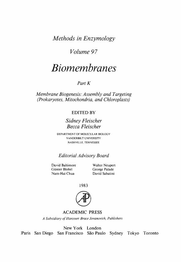
Methods in Enzymology
Volume 97
Biomembranes Part Κ
Membrane Biogenesis: Assembly and Targeting (Prokaryotes, Mitochondria, and Chloroplasts)
E D I T E D BY
Sidney Fleischer Becca Fleischer
DEPARTMENT OF MOLECULAR BIOLOGY VANDERB1LT UNIVERSITY
NASHVILLE, TENNESSEE
Editorial Advisory Board
David Baltimore Walter Neupert Günter Blobel George Palade Nam-Hai Chua David Sabatini
1983
A C A D E M I C PRESS
A Subsidiary of Harcouri Brace Jovanovich, Publishers
New York London JParis San Diego San Francisco Säo Paulo Sydney Tokyo Toronto
Table of Contents
CONTRIBUTORS T O V O L U M E 97 ix
P R E F A C E xiii
V O L U M E S IN SERIES xv
Sect ion I . P r o k a r y o t i c Membranes
A . G e n e r a l Methods
1. Genetic Analysis of Protein Export in Escherichia JONATHAN BECKWITH AND coli THOMAS J . SILHAVY 3
2. Isolation and Characterization of Mutants of Esche- THOMAS J . SILHAVY AND richia coli K l 2 Affected in Protein Localization JONATHAN BECKWITH 11
3. Purification and Characterization of Leader Pepti- P. B. W O L F E , C. ZWIZINSKI, dase from Escherichia coli AND W I L L I A M W I C K N E R 40
4. Molecular Genetics of Escherichia coli Leader Pep- T A K A Y A S U D A T E , tidase PAMELA SILVER, AND
W I L L I A M W I C K N E R 46
5. Pulse-Labeling Studies of Membrane Assembly and WILLIAM WICKNER, Protein Secretion in Intact Cells: M13 Coat Pro- T A K A Y A S U D A T E , tein RICHARD ZIMMERMANN,
AND K O R E A K I ITO 57
6. Synthesis of Proteins by Membrane-Associated PNANG C. T A I , Polysomes and Free Polysomes M I C H A E L P. C A U L F I E L D ,
AND BERNARD D. DAVIS 62
7. Preparation of Free and Membrane-Bound Poly- LINDA L. R A N D A L L AND somes from Escherichia coli SIMON J . S. H A R D Y 70
8. Analysis of Cotranslational Proteolytic Processing of LARS-GÖRAN JOSEFSSON Nascent Chains Using Two-Dimensional Gel AND LINDA L. R A N D A L L 77 Electrophoresis
B . Outer Membrane
9. Proteins Forming Large Channels from Bacterial HIROSHI NIKAIDO 85 and Mitochondrial Outer Membranes: Porins and Phage Lambda Receptor Protein
10. Phage λ Receptor (LamB Protein) in Escherichia coli M A X I M E SCHWARTZ 100
11. Synthesis and Assembly of the Outer Membrane IAN CROWLESMITH AND Proteins OmpA and OmpF of Escherichia coli KONRAD GAMON 112
12. Isolation of Mutants of the Major Outer Membrane J A C K COLEMAN, Lipoprotein of Escherichia coli for the Study of Its SUMIKO INOUYE, Assembly AND ΜASAYORI INOUYE 124
ν
VI T A B L E O F C O N T E N T S
C . Inner Membrane
13. Analysis of Μ13 Procoat Assembly into Membranes C O L I N WATTS, in Vitro J O E L M . GOODMAN,
PAMELA SILVER, AND WILLIAM WICKNER 130
14. Insertion of Proteins into Bacterial Membranes P E T E R M O D E L AND MARJORIE RUSSEL 138
15. Influence of Membrane Potential on the Insertion ROBERT C . LANDICK, and Transport of Proteins in Bacterial Membranes C H A R L E S J . DANIELS, AND
D A L E L. O X E N D E R 146
16. Penicillinase Secretion in Vivo and in Vitro JENNIFER Β. K. NIELSEN 153
17. Lactose Permease of Escherichia coli J . K. WRIGHT, R . M. T E A T H E R , AND P . O V E R A T H 158
18. Cloning of the Structural Genes of the Escherichia D A V I D A. JANS AND coli Adenosinetriphosphatase Complex F R A N K GIBSON 176
19. Biogenesis of an Oligomeric Membrane Protein W I L L I A M S. A. BRUSILOW, Complex: The Proton Translocating ATPase of ROBERT P. GUNSALUS, AND Escherichia coli ROBERT D . SIMONI 188
20. Analysis of Escherichia coli ATP Synthase Subunits JOHN E . W A L K E R AND by DNA and Protein Sequencing NICHOLAS J . G A Y 195
21. Biogenesis of Purple Membrane in Halobacteria D O R O T H E A - C H . NEUGEBAUER, H O R S T - P E T E R ZINGSHEIM, AND D I E T E R O E S T E R H E L T 218
22. Isolation of the Bacterioopsin Gene by Colony Hy- H E I K E VOGELSANG, bridization W O L F G A N G O E R T E L , AND
D I E T E R O E S T E R H E L T 226
Sect ion II . Mitochondria
23. Assessing Import of Proteins into Mitochondria: An SUSAN M. GASSER AND Overview R I C K H A Y 245
24. Molecular Cloning of Middle-Abundant mRNAs A D E L H E I D VIEBROCK, from Neurospora crassa A N G E L A PERZ, AND
W A L T E R SEBALD 254
25. Biogenesis of Cytochrome c in Neurospora crassa B E R N D HENNIG AND W A L T E R NEUPERT 261
26. Biosynthesis and Assembly of Nuclear-Coded RICHARD ZIMMERMANN Mitochondrial Membrane Proteins in Neurospora AND W A L T E R NEUPERT 275 crassa
T A B L E O F C O N T E N T S VÜ
27. Isolation and Properties of the Porin of the Outer Mitochondrial Membrane from Neurospora crassa
28. Synthesis and Assembly of Subunit 6 of the Mitochondrial ATPase in Yeast
29. Preparation and Use of Antibodies against Insoluble Membrane Proteins
30. Processing of Mitochondrial Polypeptide Precursors
in Yeast
31. Pulse Labeling of Yeast Cells and Spheroplasts
32. Import of Polypeptides into Isolated Yeast Mitochondria
33. A Yeast Mitochondrial Chelator-Sensitive Protease That Processes Cytoplasmically Synthesized Protein Precursors: Isolation from Yeast and Assay
34. Selection and Characterization of Nuclear Genes Coding Mitochondrial Proteins: Genetic Complementation of Yeast pet Mutants
35. Transformation of Nuclear Respiratory Deficient Mutants of Yeast
36. Analysis of Yeast Mitochondrial Genes
37. Genetics and Biogenesis of Cytochrome b
38. Synthesis and Intracellular Transport of Mitochondrial Matrix Proteins in Rat Liver: Studies in Vivo and in Vitro
39. Biosynthesis of Cytochrome c and Its Posttransla-tional Transfer into Mitochondria
40. Isolation of Mammalian Mitochondrial DNA and
RNA and Cloning of the Mitochondrial Genome
41. Analysis of Human Mitochondrial RNA
42. Isolation of a Hexokinase Binding Protein from the Outer Mitochondrial Membrane
H E L M U T F R E I T A G , R O L A N D BENZ, AND W A L T E R N E U P E R T 286
SANGKOT MARZUKI AND ANTHONY W. LINNANE 294
MORDECHAI SUISSA AND G R A E M E A. R E I D 305
P E T E R C . BÖHNI AND G Ü N T H E R D A U M 311
G R A E M E A . R E I D 324
SUSAN M . GASSER 329
P H Y L L I S C . M C A D A AND M I C H A E L G . DOUGLAS 337
K A R E N O ' M A L L E Y AND M I C H A E L G . DOUGLAS 344
C A R O L L . DIECKMANN AND A L E X A N D E R T Z A G O L O F F 355
C A R O L L . DIECKMANN AND A L E X A N D E R T Z A G O L O F F 361
PHILIP S. PERLMAN AND H E N R Y R . M A H L E R 374
GORDON C . SHORE, RICHARD A. RACHUBINSKI, C A R O L E ARGAN, RIMA R O Z E N , M A R C E L POUCHELET, C A R O L J . L U S T Y , AND Y V E S RAYMOND 396
TAKASHI MORIMOTO, SHIRO MATSUURA, AND MONIQUE ARPIN 408
DOUGLAS P. TAPPER, RIC H A R D A. V A N E T T E N , AND D A V I D A . C L A Y T O N 426
GIUSEPPE A T T A R D I AND JULIO MONTOYA 435
JOHN Ε . WILSON, JANICE L . MESSER, AND PHILIP L . F E L G N E R 469
νιπ T A B L E O F C O N T E N T S
43. Protein Synthesis by Isolated Plant Mitochondria C . J. L E A V E R , Ε. H A C K , AND Β. G . F O R D E
Sect ion III . Ch lorop las t s
44. Synthesis and Assembly of Thylakoid Membrane RICHARD S. WILLIAMS AND Proteins in Isolated Pea Chloroplasts
45. In Vitro Reconstitution of Synthesis, Uptake, and Assembly of Cytoplasmically Synthesized Chloro-plast Proteins
46. Structure and Synthesis of Chloroplast ATPase
47. Cloning in Physical Mapping of Maize Plastid Genes
JOHN BENNETT
JOHN E . M U L L E T AND NAM-HAI C H U A
NATHAN NELSON
L A W R E N C E BOGORAD, E A R L J . GUBBINS, ENNO K R E B B E R S , IGNACIO M . LARRINUA, BERNARD J . M U L L I G A N , K A R E N Μ . T . MUSKAVITCH, E L I Z A B E T H A . ORR, S T E V E N R . R O D E R M E L , R U D I SCHANTZ, ANDRE A . STEINMETZ, G U I D O D E VOS, AND Y u K U N Κ. Y E
48. Role of Thylakoid Polypeptide Phosphorylation and M . W E T T E R N , J. C. OWENS, Turnover in the Assembly and Function of Photo-system II
AND I . OHAD
Sect ion I V . S u m m a r y of Membrane Prote ins
49. Membrane Proteins: A Summary of Known Struc- D A V I D R . NELSON AND tural Information N E A L C. ROBINSON
A d d e n d u m
Addendum to Article [39] TAKASHI MORIMOTO, SHIRO MATSUURA, AND MONIQUE ARPIN
476
487
502
510
524
554
571
621
AUTHOR INDEX 625
SUBJECT INDEX 661
286 M I T O C H O N D R I A [27]
[27] Isolation and Properties of the Porin of the Outer Mitochondrial Membrane from
Neurospora crassa By H E L M U T F R E I T A G , R O L A N D B E N Z , and W A L T E R N E U P E R T
The outer mitochondrial membrane is freely permeable to low molecular weight components such as metal ions, substrates, and nucleotides. 1 2
Therefore pores in the outer membrane have been postulated. The size of these pores is not clearly defined. Molecules with apparent molecular weights up to 2000-8000 were found to permeate through these channels. Detergent extracts of mitochondrial outer membranes or purified pore proteins were incorporated into liposomes or bilayers. Formation of nonspecific pores with a diameter of about 20 A was observed. 3 - 8 The pore-
1 R. L. O'Brien and G. Brierley, J. Biol. Chem. 240, 4527 (1965). 2 E. Pfaff, M. Klingenberg, Ε. Ritt, and W. Vogell, Eur. J. Biochem. 5, 222 (1968). 3 M. Colombini, Nature (London) 279, 643 (1979). 4 M. Colombini, / . Membr. Biol. 53, 79 (1980). 5 L. S. Zalman, H. Nikaido, and Y. Kagawa, J. Biol. Chem. 255, 1771 (1980). 6 H. Freitag, W. Neupert, and R. Benz, Eur. J. Biochem. 123, 629 (1982). 7 N. Roos, R. Benz, and D. Brdiczka, Biochim. Biophys. Acta 686, 204 (1982) 8 H. Freitag, G. Genchi, R. Benz, F. Palmieri, and W. Neupert, FEBS Lett. 145, 72 (1982).
Copyright © 1983 by Academic Press. Inc. METHODS IN ENZYMOLOGY. VOL. 97 All rights of reproduction in anv form reserved.
ISBN 0-12-181997-3
[27] I S O L A T I O N O F M I T O C H O N D R I A L PORIN 287
forming protein, called mitochondrial porin, is a major component of outer mitochondrial membranes. It has an apparent molecular weight of about 31,000. In many respects the mitochondrial porin resembles the porins of outer membranes from gram-negative bacteria.
Here we describe two different isolation procedures for this protein and discuss some of its properties and its reconstitution into artificial bilayers.
Isolation of Mitochondrial Porin Growth of Neurospora crassa hyphae (wild-type 74 A) for 17 hr in 8-liter
cultures (inoculum 1 Χ 106 conidia/ml) in the presence of 2% sucrose and disruption of cells are carried out according to standard procedures.9 Mi tochondria are isolated in isolation medium (0.44 Μ sucrose, 2 mM EDTA, 30 mM Tris-HCl, p H 7.6, adjusted at 4°) with the addition of 0.5 m M phenylmethylsulfonyl fluoride (PMSF; final concentration), freshly dissolved in ethanol. The following centrifugation steps are carried out at 4°, employing a Sorvall RC 5Β centrifuge and a GS-3 rotor. The cell debris are removed by centrifugation for 20 min at 2000 g. The resulting supernatant is centrifuged for 45 min at 17,000 The upper layers of the resulting sediment (containing the mitochondria) are washed twice by resuspending them in a glass-Teflon potter and centrifuging for 45 min at 17,000 g. The final mitochondrial pellet is the starting material for the porin isolation. Under typical conditions, from an 8-liter culture of Neurospora crassa 180 g of hyphae (wet weight) are obtained and the yield of mitochondria is 230 mg protein.
Porin Isolation from Outer Membranes6
Principle
Mitochondrial outer membranes are isolated by swelling-shrinking of the mitochondria and subsequent separation by a sucrose step centrifugation. Differential detergent extraction of the outer membrane followed by DEAE-cellulose chromatography gives pure porin, which forms active pores after reconstitution into artificial bilayers.
Reagents Swelling medium: 2.5 mM potassium H 2 P 0 4 , adjusted to p H 7.3 with
K O H PMSF stock solution: 100 m M P M S F in ethanol, freshly prepared Shrinking medium: 2.2 Μ sucrose, 8 m M M g C l 2 , 8 mM ATP, adjusted to
p H 7.3 with K O H
9 W. Neupert and G. D. Ludwig, Eur. J. Biochem. 19, 523 (1971).
288 M I T O C H O N D R I A [27]
Isolation medium: 0.44 Μ sucrose, 2 m M EDTA, 30 mM Tris-HCl, pH 7.5
Sucrose step medium: 0.95 Μ sucrose, 10 m M Tris-HCl, pH 7.5 Tris buffer: 10 m M Tris-HCl, pH 7.5 Phosphate buffer: 100 m M potassium H 2 P 0 4 , adjusted to pH 7.3 with
K O H Octyl glucoside solution: 60 m M octyl-/?-D-glucoside, 5 m M Tris-HCl,
p H 7.5 (17.5 mg of octyl glucoside per milliliter) Genapol solution: 1% Genapol X-100 (Farbwerke Hoechst, Frankfurt,
FRG), 5 m M Tris-HCl, p H 7.5. Genapol X-100 contains polyethoxy chains like Tri ton X-100, but has isotridecyl groups instead of aromatic rings and therefore shows no absorption at 280 nm
DEAE-cellulose column: A column (15 ml , 1 cm diameter) is filled with DEAE-cellulose (DE-52, Whatman, Maidstone, England) and equilibrated with the Genapol solution. The flow rate was 10 ml/hr. In all solutions, the p H was adjusted at 4°.
Procedure
Isolation of Outer Membranes. Swelling solution, 100 ml containing 0.5 m M P M S F is used for 100 mg of mitochondrial protein. The mitochondrial pellet is suspended in the swelling medium with a glass-Teflon potter. The suspension is stirred for 15 min at 4°, and then 70 m l of the shrinking medium are added while stirring. During shrinking the suspension becomes more turbid. It is further stirred for 10 min. Then 60-ml portions of the suspension are transferred into a glass-teflon potter, and the pestle, driven by a motor at 500-700 rpm, is moved up and down five times. The membranes are then pelleted by centrifugation for 45 min at 98,000 g in a Beckman ultracentrifuge using a 45 T i rotor. The pellets are suspended in isolation medium with a glass-Teflon potter, then 35 ml of the sucrose step medium are fi//ed into a Beckman SW 25.2 centrifuge tube and overiayered with 20 ml of the mitochondrial suspension, containing the membranes from about 100 mg of mitochondria. Centrifugation is performed for 60 min at 70,000 g. The small band of red outer membranes on top of the 0.95 Μ sucrose solution is collected with a bended Pasteur pipette. The suspension is diluted with an equal volume of Tris buffer and centrifuged for 30 min at 166,000 g in a Beckman 50 T i rotor. The outer membrane pellets are suspended in phosphate buffer with the addition of 8% dimethyl sulfoxide (v/v) at a protein concentration of 3.5 mg/ml and stored in —20°. The yield of outer membranes is about 0.5% of protein with respect to whole mitochondria, which is about 25% of the theoretical amount. Samples should be stored not longer than 1 week, because prolonged storage alters the behavior of the membranes during the subsequent extraction procedures.
[27] I S O L A T I O N O F M I T O C H O N D R I A L PORIN 289
Isolation of Porin. The thawed suspension of outer membranes is first extracted with octyl glucoside, which removes most of the outer membrane proteins, except the porin. 6 For this extraction it is important to adjust the octyl glucoside concentration to 30 mM, and the octyl glucoside-to-protein ratio to 10 (w/w), to obtain reproducible results. One milliliter of outer membrane suspension is first diluted with phosphate buffer to 2 ml, then 2 ml o f 60 mM octyl glucoside solution are added. The mixture is kept at 4° for 45 min. Then it is transferred into Eppendorf tubes, which are placed in water-filled plastic adaptors and centrifuged for 30 min at 150,000 g in a Beckman 50 T i rotor. The membrane pellets are suspended in the Genapol solution by adding 1 ml per milligram of the original outer membrane protein. They are shaken for 30 min at 4°. After centrifugation in an Eppendorf centrifuge for 5 min, the supernatant is subjected to chromatography on a DEAE-cellulose column, equilibrated with the Ganapol solution. Elution is followed by monitoring the absorbance at 280 nm. The protein that elutes with the void volume is pure porin and can be stored for several months at —20°.
Porin Isolation from Whole Mitochondria8
Principle
Whole mitochondria are lysed with Genapol X-100 and chromato-graphed on hydroxyapatite, Celite, and DEAE-cellulose. This procedure gives pure and functionally active porin in high yield and can be easily scaled up.
Reagents Buffer A: 2.5% Genapol X-100 (Farbwerke Hoechst, Frankfurt, Federal
Republic of Germany), 50 m A / K C l , 10 mM potassium H 2 P 0 4 , 1 mM EDTA, 10 mM Tris adjusted to pH 7.0 with HCl
Genapol solution: 1% Genapol X-100, 5 mM Tris-HCl, pH 7.5 Dry hydroxyapatite (BioGel HTP, Bio-Rad, Richmond, California) Dry Celite (Celite 535, Roth, Karlsruhe', Federal Republic of Germany).
Procedure
For this isolation procedure, frozen mitochondria can be used. Two Pasteur pipettes plugged with cotton wool, one filled with 0.6 g of hydroxyapatite and the other filled with 0.6 g of a dry mixture of equal weights of hydroxyapatite and Celite, are prepared. To 4 - 6 mg of protein of a mitochondrial pellet, 0.7 ml of buffer A are added, mixed, and kept for 30 min at 4°, followed by centrifugation for 15 min at 27,000 The supernatant is applied to the hydroxyapatite pipette. When the solution has entered the
290 M I T O C H O N D R I A [27]
column, further buffer is applied until the eluate has reached the original volume (0.7 ml). This eluate is transferred to the hydroxyapatite-Celite pipette. Again 0.7 ml of the eluate is collected, which already contains the largely enriched porin. 8 The eluate is dialyzed overnight against 20 volumes of the Genapol solution and subjected to chromatography on DEAE-cellulose as described before.
There are two ways to scale up the isolation procedure. For up to 200 ßg of porin it is recommended to use 10 Pasteur pipettes in parallel and to combine the eluates for one DEAE-cellulose column of the size described above. In order to prepare porin in larger amounts, larger columns should be used. Mitochondria (1.2 g of protein) are dissolved in 130 ml of buffer A and chromatographed on columns containing 600 g of hydroxyapatite or hydroxyapatite-Celite mixture (diameter 3 cm) and over a DEAE-cellulose column of 40 X 3 cm. The yield is 3 - 4 mg of porin.
Comment
The porin contains an acid-labile bond. 8 Precipitation of porin from detergent-containing solutions with acids should be carried out in the cold. Add one-tenth volume of 3 Μ trichloroacetic acid and one-third volume of methanol (to prevent precipitation of the detergent). After 1 hr at 0 - 4 ° the porin can be separated by centrifugation.
Properties of the Mitochondrial Porin
General Properties
The mitochondrial porin (Mr 31,000) is the major protein of the outer membrane. It represents about 20% of total outer membrane protein and 0.4% of total mitochondrial protein. Upon isoelectric focusing, and purified porin gives two bands with pis of 7.7 (major band) and 7.8,8 The reason for this heterogeneity is not known, but porins from other sources also show this behavior. 1 0 1 1
From protease digestion experiments6 and from x-ray diffraction data, 1 2
i t was concluded that the porin is deeply embedded into the membrane and does not extend into the aqueous phase.
It has been demonstrated that the mitochondrial hexokinase binding protein, which binds brain hexokinase1 2 3 or hexokinase from fast growing tumor cells,1 3 is identical with the mitochondrial p o r i n . 1 1 1 4
1 0 H. Tokunaga, M. Tokunaga, and T. Nakae, Eur. J. Biochem. 95, 433 (1979). 1 1 M. Linden, P. Gellerfors, and B. D. Nelson, FEBS Lett. 141, 189 (1982). 1 2 C. A. Mannella, Biochim. Biophys. Acta 645, 33 (1981). I 2 a J. E. Wilson, J. L. Messer, and P. L. Feigner, this volume [42].
[27] I S O L A T I O N O F M I T O C H O N D R I A L PORIN 291
λ S cm"
10' 5-
0 10 20 30 40
/mm
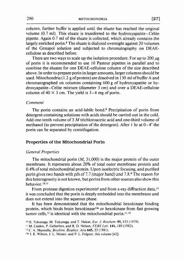
F I G . 1. Specific membrane conductance A as a function of time after addition of 100 ng of mitochondrial porin from Neurospora crassa per milliliter to a black lipid bilayer membrane from asolectin-/?-decane (arrow). The crosses represent a control experiment where only \0μ% of Genapol X-100 per milliliter were added to another membrane; 1 Μ KCl, pH 6: T= 25°.
Biosynthesis of the Porin15
Mitochondrial porin is synthesized on cytoplasmic free polysomes without an additional presequence. In contrast to the biogenesis of mitochondrial inner membrane or matrix proteins, 1 6 assembly of the porin is not dependent on energization of the mitochondria. By digitonin treatment it could be shown that the porin precursor in an in vitro transfer assay is incorporated into the outer membrane. Binding to the outer membrane precedes the incorporation into this membrane. 1 5
Reconstitution of the Porin in Lipid Bilayer Membranes
The addition of small quantities of mitochondrial porin to the aqueous phase bathing a black lipid bilayer membrane prepared according to established procedures1 7 results in a strong increase of the membrane conductance. A typical experiment is given in Fig. 1. Mitochondrial porin was added in a final concentration of 100 ng/ml to a black membrane from
1 3 E. Bustamente, H. P. Morris, and P. L. Pedersen, J. Biol. Chem. 254, 8699 (1981). 1 4 C. Fiek, R. Benz, Ν. Roos, and D. Brdiczka, Biochem. Biophys. Ada 688, 429 (1982). 1 5 H. Freitag, Μ. Janes, and W. Neupert, Eur. J. Biochem. 126, 197 (1982). 1 6 M. Schleyer, Β. Schmidt, and W. Neupert, Eur. J. Biochem. 125, 109 (1982). 1 7 R. Benz, Κ. Janko, W. Boos, and P. Läuger, Biochim. Biophys. Acta 511, 305 (1978).
292 M I T O C H O N D R I A [27]
asolect in-«-decane. After an initial lag of about 5 min, presumably caused by the diffusion of the protein through unstirred layers, the conductance increased by about four orders of magnitude within about 30 min. Only a slight further increase occurred after that time. The conductance increase was approximately linearly dependent on the protein concentration in the aqueous phase i f the same times after addition of the protein were considered (usually 20-30 min).
A considerable asymmetry of the action of the voltage on the single conductive unit was observed, i f the protein was added to only one side of the membrane. 6 7 This indicates an asymmetric insertion of the protein into the membrane.
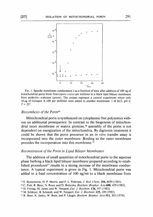
Figure 2 shows fluctuations of current observed with mitochondrial porin inserted into a asolectin membrane in the presence of 1 Μ K C l . Most of the fluctuations were directed upward, and terminating steps were only rarely observed at a membrane voltage of 5-10 m V membrane potential. 6
This indicated that the lifetime of the pores was long at low voltage and usually exceeded 5 min.
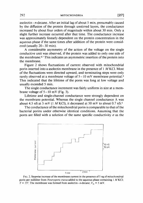
The single conductance increment was fairly uniform in size at a membrane voltage of 5 - 10 mV (Fig. 3).
Lifetime and single-channel conductance were strongly dependent on the membrane potential. Whereas the single channel conductance Λ was about 4.5 nS at 5 m V (1 Μ KCl) , i t decreased at 50 m V to about 0.7 nS. 6
The conductance of the mitochondrial porin is comparable to that o f the bacterial porins under otherwise identical conditions. Assuming that the pores are filled with a solution of the same specific conductivity σ as the
F I G . 2. Stepwise increase of the membrane current in the presence of 5 ng of mitochondrial porin per milliliter from Neurospora crassa added to the aqueous phase containing 1 Μ KCl; Τ = 25°. The membrane was formed from asolectin-w-decane; Vm = 5 mV.
[27] I S O L A T I O N O F M I T O C H O N D R I A L PORIN 293
F I G . 3. Histogram of the conductance fluctuations observed with membranes from asolec-tin-A7-decane in the presence of mitochondrial porin from Neurospora crassa. The aqueous phase contained 1 Μ KCl; pH 6; T= 25°. Applied voltage was 5 mV; the mean value of all observed conductance fluctuations was 4.5 nS for 253 single events.
external solution and assuming a pore length 7.5 n m (corresponding to the thickness of the outer membranes of bacteria and mitochondria), the average pore diameter d(= 2 r) and the cross section can be calculated according to the equation Λ = onr2ll. The table shows the diameter and the cross section for mitochondrial porins from Neurospora crassa and rat liver, as well as for the bacterial porins from different gram-negative bacteria. 6 7 ' 1 7- 2 1
1 8 R. Benz and R. E. W. Hancock, Biochim. Biophys. Acta 646, 298 (1981). 1 9 R. E. W. Hancock, G. M . Decad, and H. Nikaido, Biochim. Biophys. Acta 554, 323 (1979). 2 0 H. Nikaido and T. Nakae, Adv. Microbiol. Physiol. 20, 163 (1979). 2 1 R. Benz, J. Ishii, and T. Nakae, J. Membr. Biol. 56, 19 (1980).
CoMPAJUSOTi OF THE PORES FORMED BY MITOCHONDRIAL AND BACTERIAL PORINS IN LIPID B I L A Y E R MEMBRANE"
Λ d Area Reference Porin (nS) (nm) (nm2) footnote
Mitochondrial Neurospora crassa 4.5 2.0 3.1 6 Rat liver 4.3 1.9 2.9 7
Bacterial Escherichia coli 1.9 1.3 1.3 17 Salmonella typhimurium 2.4 1.4 1.6 21 Pseudomonas aeruginosa 5.6 2.2 3.8 18
The pore diameter d was calculated from the pore conductance in 1 Μ KCl according to Λ = anr2jl (using σ = 110 mS cm - 1 and length (/) = 7.5 nm).
294 M I T O C H O N D R I A [28]
The diameter of the pore from Pseudomonas aeruginosa protein F is about the same as that of the mitochondrial porin pores.18 This is consistent with the observations on the pores in reconstituted vesicles, which in both cases were permeable to hydrophilic solutes of molecular weights up to 6000. 5 · 1 9
Copyright © 1983 by Academic Press. Inc. METHODS IN ENZYMOLOGY. VOL. 97 All rights of reproduction in any form reserved.
ISBN 0-12-181997-3