Invasion of ovarian cancer cells is induced byPITX2 ...
15
RESEARCH Open Access Invasion of ovarian cancer cells is induced byPITX2-mediated activation of TGF-β and Activin-A Moitri Basu 1 , Rahul Bhattacharya 1 , Upasana Ray 1 , Satinath Mukhopadhyay 2 , Uttara Chatterjee 3 and Sib Sankar Roy 1* Abstract Background: Most ovarian cancers are highly invasive in nature and the high burden of metastatic disease make them a leading cause of mortality among all gynaecological malignancies. The homeodomain transcription factor, PITX2 is associated with cancer in different tissues. Our previous studies demonstrated increased PITX2 expression in human ovarian tumours. Growing evidence linking activation of TGF-β pathway by homeodomain proteins prompted us to look for the possible involvement of this signalling pathway in PITX2-mediated progression of ovarian cancer. Methods: The status of TGF-β signalling in human ovarian tissues was assessed by immunohistochemistry. The expression level of TGFB/INHBA and other invasion-associated genes was measured by quantitative-PCR (Q-PCR) and Western Blot after transfection/treatments with clones/reagents in normal/cancer cells. The physiological effect of PITX2 on invasion/motility was checked by matrigel invasion and wound healing assay. The PITX2- and activin-induced epithelial-mesenchymal transition (EMT) was evaluated by Q-PCR of respective markers and confocal/phase-contrast imaging of cells. Results: Human ovarian tumours showed enhanced TGF- β signalling. Our study uncovers the PITX2-induced expression of TGFB1/2/3 as well as INHBA genes (p < 0.01) followed by SMAD2/3-dependent TGF-β signalling pathway. PITX2-induced TGF-β pathway regulated the expression of invasion-associated genes, SNAI1, CDH1 and MMP9 (p < 0.01) that accounted for enhanced motility/invasion of ovarian cancers. Snail and MMP9 acted as important mediators of PITX2-induced invasiveness of ovarian cancer cells. PITX2 over-expression resulted in loss of epithelial markers (p < 0.01) and gain of mesenchymal markers (p < 0.01) that contributed significantly to ovarian oncogenesis. PITX2-induced INHBA expression (p < 0.01) contributed to EMT in both normal and ovarian cancer cells. Conclusions: Overall, our findings suggest a significant contributory role of PITX2 in promoting invasive behaviour of ovarian cancer cells through up-regulation of TGFB/INHBA. We have also identified the previously unknown involvement of activin-A in promoting EMT. Our work provides novel mechanistic insights into the invasive behavior of ovarian cancer cells. The extension of this study have the potential for therapeutic applications in future. Keywords: PITX2, TGF-β signalling, Activin-A, Invasion, EMT, IOSE, Ovarian cancer cells * Correspondence: [email protected] 1 Cell Biology and Physiology Division, CSIR-Indian Institute of Chemical Biology, Council of Scientific and Industrial Research, 4 Raja S. C. Mullick Road, Kolkata 700032, India Full list of author information is available at the end of the article © 2015 Basu et al. Open Access This article is distributed under the terms of the Creative Commons Attribution 4.0 International License (http://creativecommons.org/licenses/by/4.0/), which permits unrestricted use, distribution, and reproduction in any medium, provided you give appropriate credit to the original author(s) and the source, provide a link to the Creative Commons license, and indicate if changes were made. The Creative Commons Public Domain Dedication waiver (http://creativecommons.org/publicdomain/zero/1.0/) applies to the data made available in this article, unless otherwise stated. Basu et al. Molecular Cancer (2015) 14:162 DOI 10.1186/s12943-015-0433-y
Transcript of Invasion of ovarian cancer cells is induced byPITX2 ...
RESEARCH Open Access
Invasion of ovarian cancer cells is inducedbyPITX2-mediated activation of TGF-β andActivin-AMoitri Basu1, Rahul Bhattacharya1, Upasana Ray1, Satinath Mukhopadhyay2, Uttara Chatterjee3 and Sib Sankar Roy1*
Abstract
Background: Most ovarian cancers are highly invasive in nature and the high burden of metastatic diseasemake them a leading cause of mortality among all gynaecological malignancies. The homeodomaintranscription factor, PITX2 is associated with cancer in different tissues. Our previous studies demonstratedincreased PITX2 expression in human ovarian tumours. Growing evidence linking activation of TGF-β pathwayby homeodomain proteins prompted us to look for the possible involvement of this signalling pathway inPITX2-mediated progression of ovarian cancer.
Methods: The status of TGF-β signalling in human ovarian tissues was assessed by immunohistochemistry. Theexpression level of TGFB/INHBA and other invasion-associated genes was measured by quantitative-PCR (Q-PCR)and Western Blot after transfection/treatments with clones/reagents in normal/cancer cells. The physiologicaleffect of PITX2 on invasion/motility was checked by matrigel invasion and wound healing assay. The PITX2- andactivin-induced epithelial-mesenchymal transition (EMT) was evaluated by Q-PCR of respective markers andconfocal/phase-contrast imaging of cells.
Results: Human ovarian tumours showed enhanced TGF-β signalling. Our study uncovers the PITX2-inducedexpression of TGFB1/2/3 as well as INHBA genes (p < 0.01) followed by SMAD2/3-dependent TGF-β signallingpathway. PITX2-induced TGF-β pathway regulated the expression of invasion-associated genes, SNAI1, CDH1and MMP9 (p < 0.01) that accounted for enhanced motility/invasion of ovarian cancers. Snail and MMP9 actedas important mediators of PITX2-induced invasiveness of ovarian cancer cells. PITX2 over-expression resulted inloss of epithelial markers (p < 0.01) and gain of mesenchymal markers (p < 0.01) that contributed significantly toovarian oncogenesis. PITX2-induced INHBA expression (p < 0.01) contributed to EMT in both normal and ovariancancer cells.
Conclusions: Overall, our findings suggest a significant contributory role of PITX2 in promoting invasivebehaviour of ovarian cancer cells through up-regulation of TGFB/INHBA. We have also identified the previouslyunknown involvement of activin-A in promoting EMT. Our work provides novel mechanistic insights into theinvasive behavior of ovarian cancer cells. The extension of this study have the potential for therapeuticapplications in future.
Keywords: PITX2, TGF-β signalling, Activin-A, Invasion, EMT, IOSE, Ovarian cancer cells
* Correspondence: [email protected] Biology and Physiology Division, CSIR-Indian Institute of ChemicalBiology, Council of Scientific and Industrial Research, 4 Raja S. C. MullickRoad, Kolkata 700032, IndiaFull list of author information is available at the end of the article
© 2015 Basu et al. Open Access This article is distributed under the terms of the Creative Commons Attribution 4.0International License (http://creativecommons.org/licenses/by/4.0/), which permits unrestricted use, distribution, andreproduction in any medium, provided you give appropriate credit to the original author(s) and the source, provide a link tothe Creative Commons license, and indicate if changes were made. The Creative Commons Public Domain Dedication waiver(http://creativecommons.org/publicdomain/zero/1.0/) applies to the data made available in this article, unless otherwise stated.
Basu et al. Molecular Cancer (2015) 14:162 DOI 10.1186/s12943-015-0433-y
BackgroundOvarian cancer is a highly metastatic disease and is theleading cause of death among all gynaecological malignan-cies [1]. Unfortunately, the disease is often detected at anadvanced stage (stages III-IV) and progresses very rapidly,as it acquires an aggressive phenotype. A number of growthfactors, including transforming growth factor-β (TGF-β)regulate the proliferation of ovarian surface epithelial (OSE)cells and increases metastasis [2]. TGF-β binds to its spe-cific receptors, type-I (TβRI) and type-II (TβRII). Upon lig-and binding, TβRII trans-phosphorylates and activatesTβRI, which in turn phosphorylates Smad2 and Smad3[3, 4]. The latter two then bind to Smad4 and the com-plex translocates to the nucleus to regulate the expres-sion of target genes. Other member of the TGF-βsuperfamily, like activin transduces signals mainlythrough the SMAD2/3-dependent pathway [5]. INHBAforms a disulfide-linked homodimer, known as activin-A which is a polypeptide hormone of primarily gonadalorigin [6, 7]. The mojor gonadal sites of its productionis Sertoli cells of males and ovarian granulosa cells offemale origin [6, 7]. High levels of activin-βA subunit isdetected in majority of the patients with granulosa celltumors [8], but almost absent in ovarian epithelial tumorsexcept mucinous carcinoma [9]. In addition, increased ex-pression of activin-A is observed in esophageal [10] andcolorectal carcinomas [11]. High expression of activin-Awas found in stage IV colorectal cancer [12] and correlatedwith poor overall survival rate [11, 12]. However, there areno reports on the regulation of activin-A and its role inepithelial ovarian cancer progression.Highly invasive and metastatic behavior underpin the ag-
gressive nature of ovarian cancers. Epithelial-mesenchymaltransition (EMT) is a major mechanism for the conversionof early-stage tumors to invasive malignancies due to theloss of epithelial adherence and tight junctions [13, 14].Transcription factor like Snail acts as a key regulator in theinduction of cellular invasion, in part, by suppressing theexpression of the epithelial specific adhesion molecule, E-cadherin and by increasing the expression of matrix metal-loproteinases MMPs; [15]. TGF-β-signalling, on the otherhand, enhances the invasive properties of ovarian cancerspartially through up-regulation of MMPs [16].The homeobox genes are widely implicated in various
human cancers, acting as oncogenes or tumour suppres-sors [17–21]. Pituitary homeobox 2 (PITX2), a memberof the bicoid/paired-like homeobox gene family, is amultifunctional transcription factor [22–24]. Three dif-ferent isoforms of PITX2 (PITX2A/B/C), differ only intheir amino terminus and regulate the transcription oftarget genes differentially [25]. Recently, several reportshighlighted the association of PITX2 with progression ofbreast and colorectal cancers [26, 27]. We observed theup-regulated expression of PITX2 in ovarian tumours
[28] and simultaneously we found induced TGF-β sig-naling pathway in the same tissue sections. Consider-ing the importance of TGF-β signalling pathway inpromoting oncogenesis of several tissues, we aimed toinvestigate possible involvement of PITX2 in promot-ing invasiveness of ovarian cancer cells through theregulation of TGF-β signalling pathways. We also ex-plored the role of activin-A in the progression of epi-thelial ovarian cancers.
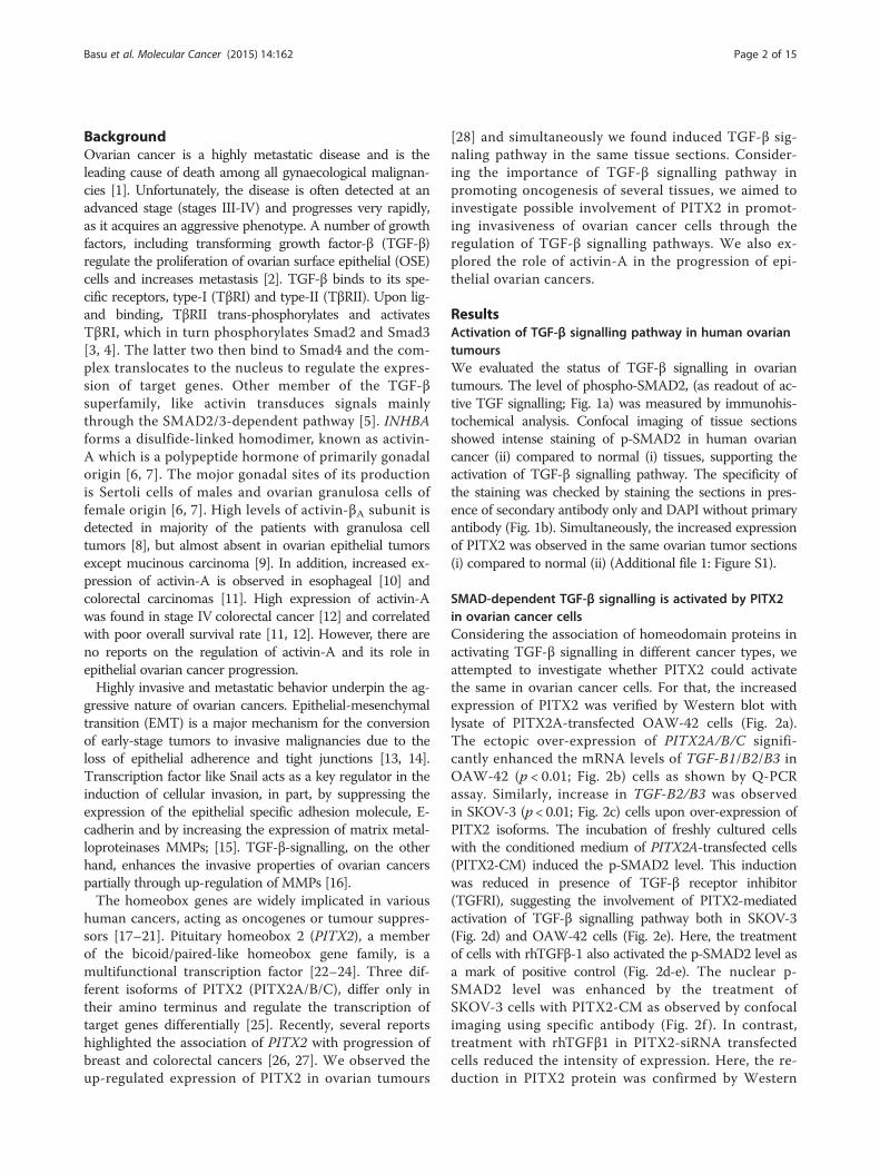
ResultsActivation of TGF-β signalling pathway in human ovariantumoursWe evaluated the status of TGF-β signalling in ovariantumours. The level of phospho-SMAD2, (as readout of ac-tive TGF signalling; Fig. 1a) was measured by immunohis-tochemical analysis. Confocal imaging of tissue sectionsshowed intense staining of p-SMAD2 in human ovariancancer (ii) compared to normal (i) tissues, supporting theactivation of TGF-β signalling pathway. The specificity ofthe staining was checked by staining the sections in pres-ence of secondary antibody only and DAPI without primaryantibody (Fig. 1b). Simultaneously, the increased expressionof PITX2 was observed in the same ovarian tumor sections(i) compared to normal (ii) (Additional file 1: Figure S1).
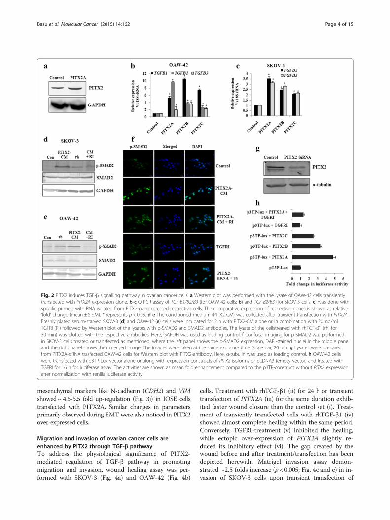
SMAD-dependent TGF-β signalling is activated by PITX2in ovarian cancer cellsConsidering the association of homeodomain proteins inactivating TGF-β signalling in different cancer types, weattempted to investigate whether PITX2 could activatethe same in ovarian cancer cells. For that, the increasedexpression of PITX2 was verified by Western blot withlysate of PITX2A-transfected OAW-42 cells (Fig. 2a).The ectopic over-expression of PITX2A/B/C signifi-cantly enhanced the mRNA levels of TGF-B1/B2/B3 inOAW-42 (p < 0.01; Fig. 2b) cells as shown by Q-PCRassay. Similarly, increase in TGF-B2/B3 was observedin SKOV-3 (p < 0.01; Fig. 2c) cells upon over-expression ofPITX2 isoforms. The incubation of freshly cultured cellswith the conditioned medium of PITX2A-transfected cells(PITX2-CM) induced the p-SMAD2 level. This inductionwas reduced in presence of TGF-β receptor inhibitor(TGFRI), suggesting the involvement of PITX2-mediatedactivation of TGF-β signalling pathway both in SKOV-3(Fig. 2d) and OAW-42 cells (Fig. 2e). Here, the treatmentof cells with rhTGFβ-1 also activated the p-SMAD2 level asa mark of positive control (Fig. 2d-e). The nuclear p-SMAD2 level was enhanced by the treatment ofSKOV-3 cells with PITX2-CM as observed by confocalimaging using specific antibody (Fig. 2f ). In contrast,treatment with rhTGFβ1 in PITX2-siRNA transfectedcells reduced the intensity of expression. Here, the re-duction in PITX2 protein was confirmed by Western
Basu et al. Molecular Cancer (2015) 14:162 Page 2 of 15
blot with PITX2-siRNA trasfected cell lysate (Fig. 2g).Further, the ectopic over-expression of PITX2A/B/C aug-mented the activity of TGFβ/SMAD-responsive reporterconstruct (p3TP-lux) by 3-5 folds (Fig. 2h) in OAW-42cells, however, TGFRI-treatment suppressed this trans-activation (Fig. 2h). Taken together, the results suggest acti-vation of TGF-β signalling pathway by PITX2.
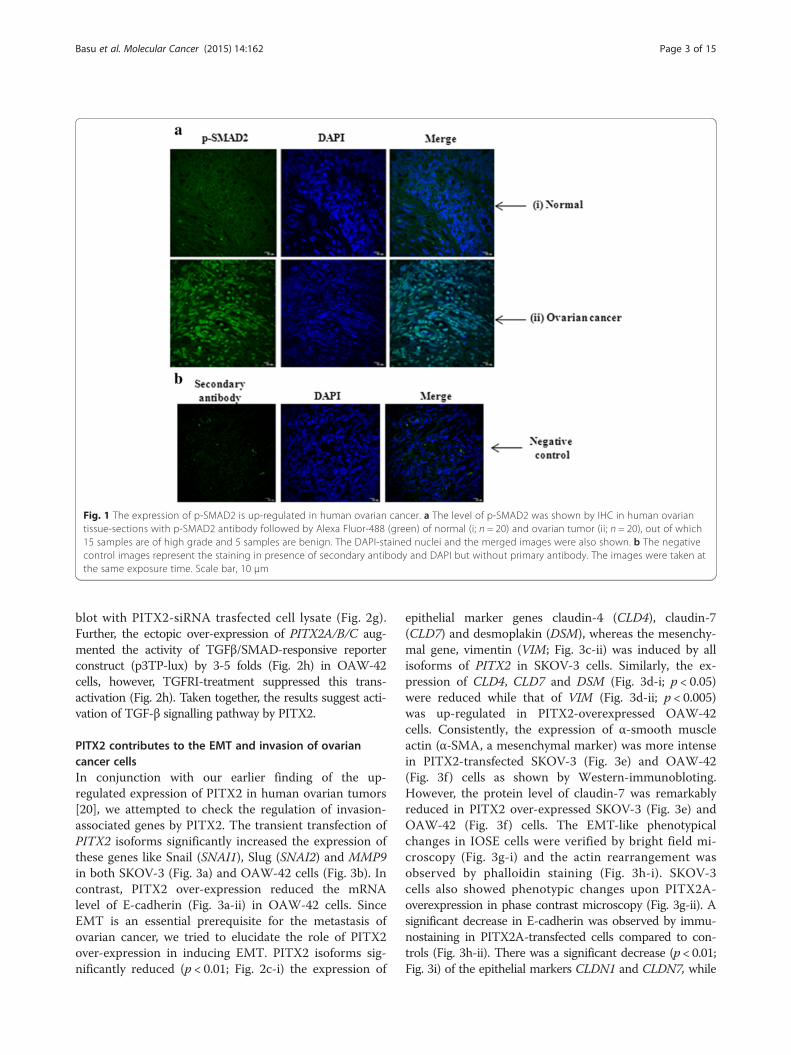
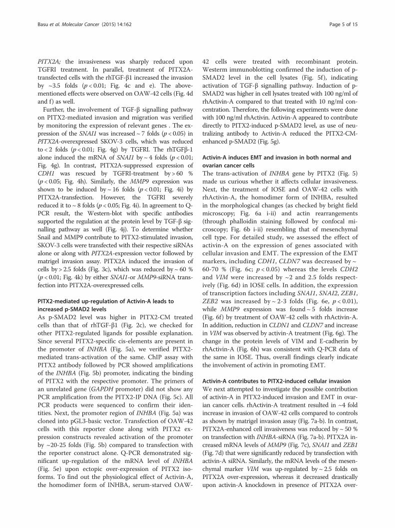
PITX2 contributes to the EMT and invasion of ovariancancer cellsIn conjunction with our earlier finding of the up-regulated expression of PITX2 in human ovarian tumors[20], we attempted to check the regulation of invasion-associated genes by PITX2. The transient transfection ofPITX2 isoforms significantly increased the expression ofthese genes like Snail (SNAI1), Slug (SNAI2) and MMP9in both SKOV-3 (Fig. 3a) and OAW-42 cells (Fig. 3b). Incontrast, PITX2 over-expression reduced the mRNAlevel of E-cadherin (Fig. 3a-ii) in OAW-42 cells. SinceEMT is an essential prerequisite for the metastasis ofovarian cancer, we tried to elucidate the role of PITX2over-expression in inducing EMT. PITX2 isoforms sig-nificantly reduced (p < 0.01; Fig. 2c-i) the expression of
epithelial marker genes claudin-4 (CLD4), claudin-7(CLD7) and desmoplakin (DSM), whereas the mesenchy-mal gene, vimentin (VIM; Fig. 3c-ii) was induced by allisoforms of PITX2 in SKOV-3 cells. Similarly, the ex-pression of CLD4, CLD7 and DSM (Fig. 3d-i; p < 0.05)were reduced while that of VIM (Fig. 3d-ii; p < 0.005)was up-regulated in PITX2-overexpressed OAW-42cells. Consistently, the expression of α-smooth muscleactin (α-SMA, a mesenchymal marker) was more intensein PITX2-transfected SKOV-3 (Fig. 3e) and OAW-42(Fig. 3f ) cells as shown by Western-immunobloting.However, the protein level of claudin-7 was remarkablyreduced in PITX2 over-expressed SKOV-3 (Fig. 3e) andOAW-42 (Fig. 3f ) cells. The EMT-like phenotypicalchanges in IOSE cells were verified by bright field mi-croscopy (Fig. 3g-i) and the actin rearrangement wasobserved by phalloidin staining (Fig. 3h-i). SKOV-3cells also showed phenotypic changes upon PITX2A-overexpression in phase contrast microscopy (Fig. 3g-ii). Asignificant decrease in E-cadherin was observed by immu-nostaining in PITX2A-transfected cells compared to con-trols (Fig. 3h-ii). There was a significant decrease (p < 0.01;Fig. 3i) of the epithelial markers CLDN1 and CLDN7, while
Fig. 1 The expression of p-SMAD2 is up-regulated in human ovarian cancer. a The level of p-SMAD2 was shown by IHC in human ovariantissue-sections with p-SMAD2 antibody followed by Alexa Fluor-488 (green) of normal (i; n = 20) and ovarian tumor (ii; n = 20), out of which15 samples are of high grade and 5 samples are benign. The DAPI-stained nuclei and the merged images were also shown. b The negativecontrol images represent the staining in presence of secondary antibody and DAPI but without primary antibody. The images were taken atthe same exposure time. Scale bar, 10 μm
Basu et al. Molecular Cancer (2015) 14:162 Page 3 of 15
mesenchymal markers like N-cadherin (CDH2) and VIMshowed ~ 4.5-5.5 fold up-regulation (Fig. 3j) in IOSE cellstransfected with PITX2A. Similar changes in parametersprimarily observed during EMT were also noticed in PITX2over-expressed cells.
Migration and invasion of ovarian cancer cells areenhanced by PITX2 through TGF-β pathwayTo address the physiological significance of PITX2-mediated regulation of TGF-β pathway in promotingmigration and invasion, wound healing assay was per-formed with SKOV-3 (Fig. 4a) and OAW-42 (Fig. 4b)
cells. Treatment with rhTGF-β1 (ii) for 24 h or transienttransfection of PITX2A (iii) for the same duration exhib-ited faster wound closure than the control set (i). Treat-ment of transiently transfected cells with rhTGF-β1 (iv)showed almost complete healing within the same period.Conversely, TGFRI-treatment (v) inhibited the healing,while ectopic over-expression of PITX2A slightly re-duced its inhibitory effect (vi). The gap created by thewound before and after treatment/transfection has beendepicted herewith. Matrigel invasion assay demon-strated ~2.5 folds increase (p < 0.005; Fig. 4c and e) in in-vasion of SKOV-3 cells upon transient transfection of
Fig. 2 PITX2 induces TGF-β signalling pathway in ovarian cancer cells. a Western blot was performed with the lysate of OAW-42 cells transientlytransfected with PITX2A expression clone. b-c Q-PCR assay of TGF-B1/B2/B3 (for OAW-42 cells; b) and TGF-B2/B3 (for SKOV-3 cells; c) was done withspecific primers with RNA isolated from PITX2-overexpressed respective cells. The comparative expression of respective genes is shown as relative‘fold’ change (mean ± S.E.M). * represents p < 0.05. d-e The conditioned-medium (PITX2-CM) was collected after transient transfection with PITX2A.Freshly plated serum-starved SKOV-3 (d) and OAW-42 (e) cells were incubated for 2 h with PITX2-CM alone or in combination with 20 ng/mlTGFRI (RI) followed by Western blot of the lysates with p-SMAD2 and SMAD2 antibodies. The lysate of the cellstreated with rhTGF-β1 (rh; for30 min) was blotted with the respective antibodies. Here, GAPDH was used as loading control. f Confocal imaging for p-SMAD2 was performedin SKOV-3 cells treated or transfected as mentioned, where the left panel shows the p-SMAD2 expression, DAPI-stained nuclei in the middle paneland the right panel shows their merged image. The images were taken at the same exposure time. Scale bar, 20 μm. g Lysates were preparedfrom PITX2A-siRNA trasfected OAW-42 cells for Western blot with PITX2-antibody. Here, α-tubulin was used as loading control. h OAW-42 cellswere transfected with p3TP-Lux vector alone or along with expression constructs of PITX2 isoforms or pcDNA3 (empty vector) and treated withTGFRI for 16 h for luciferase assay. The activities are shown as mean fold enhancement compared to the p3TP-construct without PITX2 expressionafter normalization with renilla luciferase activity
Basu et al. Molecular Cancer (2015) 14:162 Page 4 of 15
PITX2A; the invasiveness was sharply reduced uponTGFRI treatment. In parallel, treatment of PITX2A-transfected cells with the rhTGF-β1 increased the invasionby ~3.5 folds (p < 0.01; Fig. 4c and e). The above-mentioned effects were observed on OAW-42 cells (Fig. 4dand f) as well.Further, the involvement of TGF-β signalling pathway
on PITX2-mediated invasion and migration was verifiedby monitoring the expression of relevant genes . The ex-pression of the SNAI1 was increased ~ 7 folds (p < 0.05) inPITX2A-overexpressed SKOV-3 cells, which was reducedto < 2 folds (p < 0.01; Fig. 4g) by TGFRI. The rhTGFβ-1alone induced the mRNA of SNAI1 by ~ 4 folds (p < 0.01;Fig. 4g). In contrast, PITX2A-suppressed expression ofCDH1 was rescued by TGFRI-treatment by > 60 %(p < 0.05; Fig. 4h). Similarly, the MMP9 expression wasshown to be induced by ~ 16 folds (p < 0.01; Fig. 4i) byPITX2A-transfection. However, the TGFRI severelyreduced it to ~ 8 folds (p < 0.05; Fig. 4i). In agreement to Q-PCR result, the Western-blot with specific antibodiessupported the regulation at the protein level by TGF-β sig-nalling pathway as well (Fig. 4j). To determine whetherSnail and MMP9 contribute to PITX2-stimulated invasion,SKOV-3 cells were transfected with their respective siRNAsalone or along with PITX2A-expression vector followed bymatrigel invasion assay. PITX2A induced the invasion ofcells by > 2.5 folds (Fig. 3c), which was reduced by ~ 60 %(p < 0.01; Fig. 4k) by either SNAI1-or MMP9-siRNA trans-fection into PITX2A-overexpressed cells.
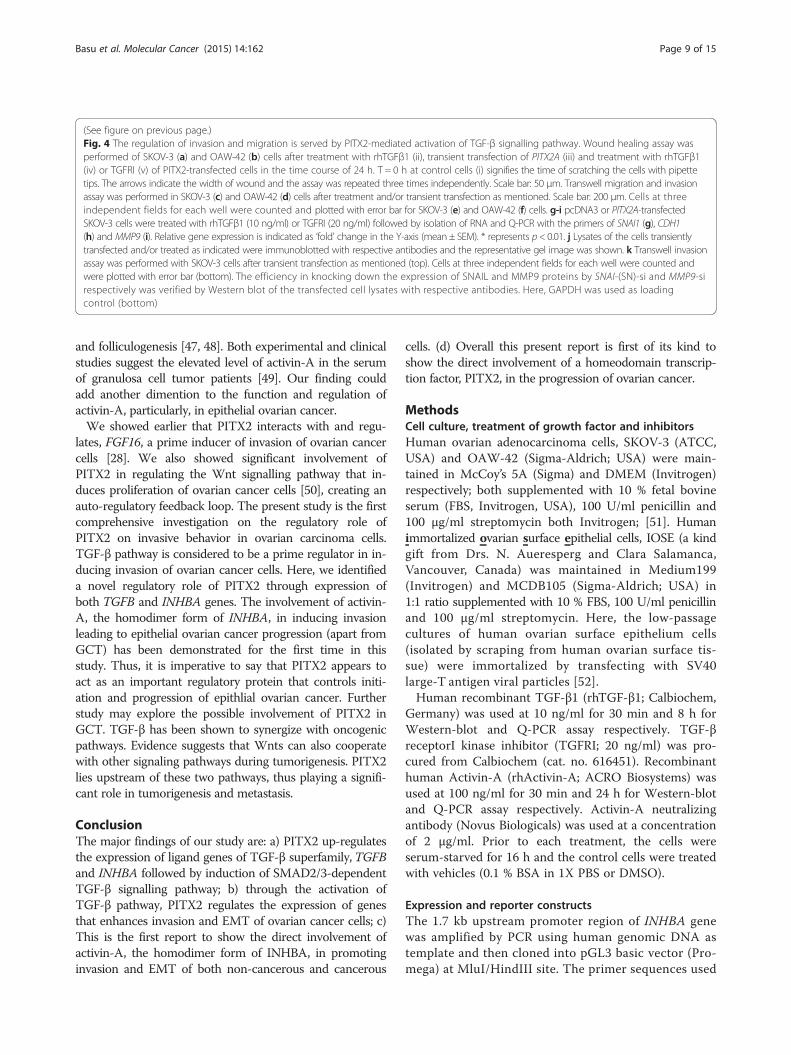
PITX2-mediated up-regulation of Activin-A leads toincreased p-SMAD2 levelsAs p-SMAD2 level was higher in PITX2-CM treatedcells than that of rhTGF-β1 (Fig. 2c), we checked forother PITX2-regulated ligands for possible explanation.Since several PITX2-specific cis-elements are present inthe promoter of INHBA (Fig. 5a), we verified PITX2-mediated trans-activation of the same. ChIP assay withPITX2 antibody followed by PCR showed amplificationsof the INHBA (Fig. 5b) promoter, indicating the bindingof PITX2 with the respective promoter. The primers ofan unrelated gene (GAPDH promoter) did not show anyPCR amplification from the PITX2-IP DNA (Fig. 5c). AllPCR products were sequenced to confirm their iden-tities. Next, the promoter region of INHBA (Fig. 5a) wascloned into pGL3-basic vector. Transfection of OAW-42cells with this reporter clone along with PITX2 ex-pression constructs revealed activation of the promoterby ~20-25 folds (Fig. 5b) compared to transfection withthe reporter construct alone. Q-PCR demonstrated sig-nificant up-regulation of the mRNA level of INHBA(Fig. 5e) upon ectopic over-expression of PITX2 iso-forms. To find out the physiological effect of Activin-A,the homodimer form of INHBA, serum-starved OAW-
42 cells were treated with recombinant protein.Westerm immunoblotting confirmed the induction of p-SMAD2 level in the cell lysates (Fig. 5f ), indicatingactivation of TGF-β signalling pathway. Induction of p-SMAD2 was higher in cell lysates treated with 100 ng/ml ofrhActivin-A compared to that treated with 10 ng/ml con-centration. Therefore, the following experiments were donewith 100 ng/ml rhActivin. Activin-A appeared to contributedirectly to PITX2-induced p-SMAD2 level, as use of neu-tralizing antibody to Activin-A reduced the PITX2-CM-enhanced p-SMAD2 (Fig. 5g).
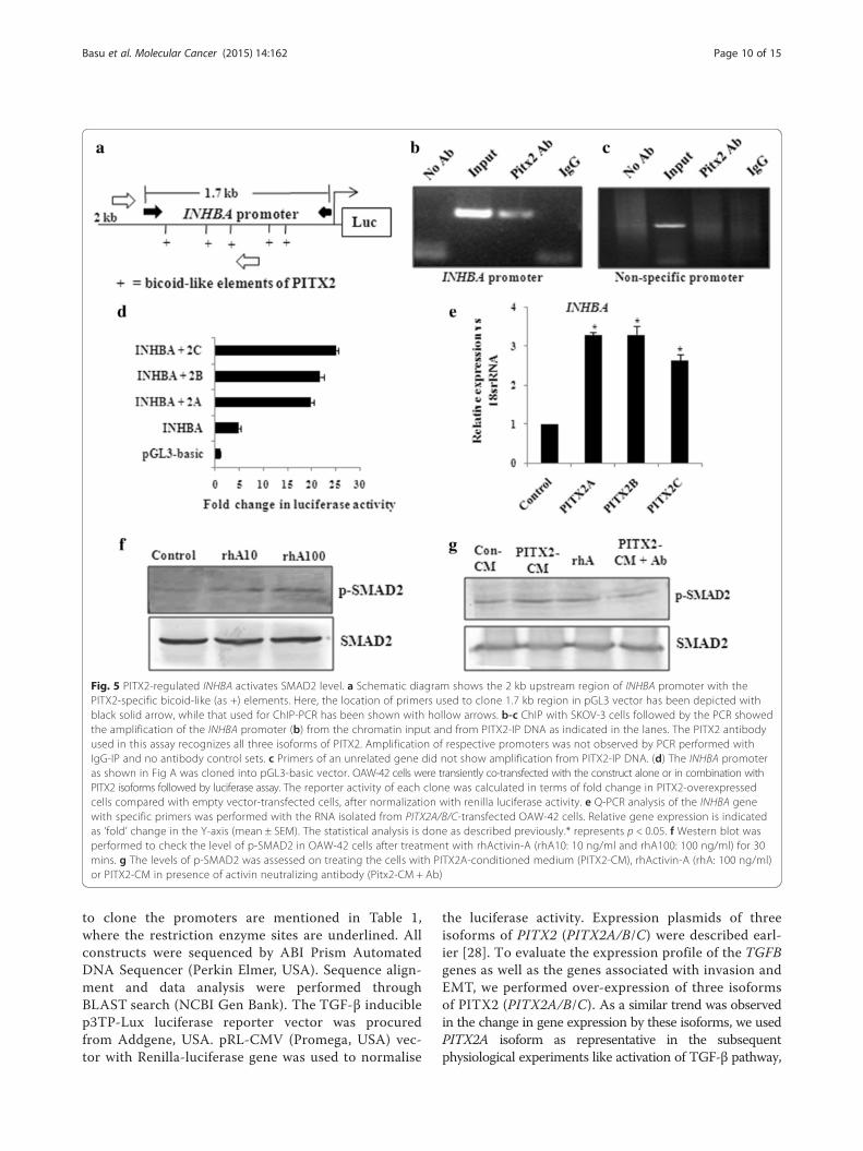
Activin-A induces EMT and invasion in both normal andovarian cancer cellsThe trans-activation of INHBA gene by PITX2 (Fig. 5)made us curious whether it affects cellular invasiveness.Next, the treatment of IOSE and OAW-42 cells withrhActivin-A, the homodimer form of INHBA, resultedin the morphological changes (as checked by bright fieldmicroscopy; Fig. 6a i-ii) and actin rearrangements(through phalloidin staining followed by confocal mi-croscopy; Fig. 6b i-ii) resembling that of mesenchymalcell type. For detailed study, we assessed the effect ofactivin-A on the expression of genes associated withcellular invasion and EMT. The expression of the EMTmarkers, including CDH1, CLDN7 was decreased by ~60-70 % (Fig. 6c; p < 0.05) whereas the levels CDH2and VIM were increased by ~2 and 2.5 folds respect-ively (Fig. 6d) in IOSE cells. In addition, the expressionof transcription factors including SNAI1, SNAI2, ZEB1,ZEB2 was increased by ~ 2-3 folds (Fig. 6e, p < 0.01),while MMP9 expression was found ~ 5 folds increase(Fig. 6f) by treatment of OAW-42 cells with rhActivin-A.In addition, reduction in CLDN1 and CLDN7 and increasein VIM was observed by activin-A treatment (Fig. 6g). Thechange in the protein levels of VIM and E-cadherin byrhActivin-A (Fig. 6h) was consistent with Q-PCR data ofthe same in IOSE. Thus, overall findings clearly indicatethe involvement of activin in promoting EMT.
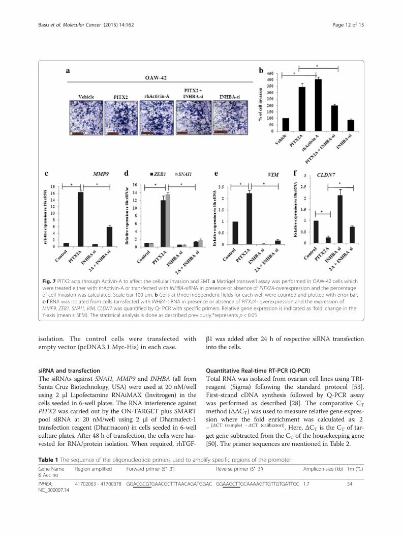
Activin-A contributes to PITX2-induced cellular invasionWe next attempted to investigate the possible contributionof activin-A in PITX2-induced invasion and EMT in ovar-ian cancer cells. rhActivin-A treatment resulted in ~4 foldincrease in invasion of OAW-42 cells compared to controlsas shown by matrigel invasion assay (Fig. 7a-b). In contrast,PITX2A-enhanced cell invasiveness was reduced by ~ 50 %on transfection with INHBA-siRNA (Fig. 7a-b). PITX2A in-creased mRNA levels of MMP9 (Fig. 7c), SNAI1 and ZEB1(Fig. 7d) that were significantly reduced by transfection withactivin-A siRNA. Similarly, the mRNA levels of the mesen-chymal marker VIM was up-regulated by ~ 2.5 folds onPITX2A over-expression, whereas it decreased drasticallyupon activin-A knockdown in presence of PITX2A over-
Basu et al. Molecular Cancer (2015) 14:162 Page 5 of 15
Fig. 3 (See legend on next page.)
Basu et al. Molecular Cancer (2015) 14:162 Page 6 of 15
expression (Fig. 7e). Knockdown of INHBA rescued thePITX2A-mediated suppression of the epithelial markerCLDN7 (Fig. 7f).
DiscussionHigh mortality rates of ovarian cancer patients are notonly due to the rapid resistance acquired by the tumorcells to conventional therapies but also due to theirovertly aggressive and invasive behaviour [29]. Metastaticcells undergo various genetic and epigenetic changes thatincrease motility and invasive behavior [29]. Evidence sup-ports a strong positive role of both TGF-β ligands and theirreceptors in promoting carcinogenesis through invasivetransformation. In tumor cells, increased secretion of theTGF-β ligands enhance metastasis and promotes tumori-genesis [30, 31]. The three mammalian isoforms, TGF-β1/β2/β3, are encoded by different genes which functionthroughtrans-membrane receptors with intrinsic cytoplas-mic serine-threonine kinase domains [32]. Among those,TGF-β1 is most frequently up-regulated in tumor cells [33]and is the focus of most studies, including ours.TGF-β/SMAD signalling pathway has been earlier dem-
onstrated to be operational in ovarian cancer cells [34] andour present report demonstrated its activation in humanovarian tumors (Fig. 1). We showed up-regulation of ahomeodomain transcription factor, PITX2, in similar tissuesections [28], indicating its possible involvement in ovariancancer. Recently the association of PITX2 with cancers ofthyroid, prostate, colon and breast [26, 27, 35] has beenhighlighted. In addition, inactivation of PITX2 leads toapoptosis of pituitary gonadotrophs [36] suggesting an ac-tive association between PITX2 and cancer, although nosuch report is available in the context of ovarian cancer.Considering the importance of the regulation of TGF-βpathway by homeodomain proteins including HOXB9 inpromoting tumorigenesis [37], we investigated the possibleassociation of PITX2 and TGF-β signalling in ovarian can-cer. Our experiments demonstrated that PITX2 isoformsdifferentially enhanced the expression of TGFB1/2/3 genes.In addition, PITX2-activated TGF-β signalling pathway(Fig. 1) was evidenced by p-SMAD2 induction as well as itsnuclear localization by PITX2-CM in ovarian cancer cells.We further investigated the role of PITX2-activated
TGF-β pathway in EMT and invasion, the key steps in
ovarian cancer progression [38]. We showed a significantregulatory role of PITX2 in inducing EMT through expres-sion of EMT markers (Fig. 2) and phenotypic changes inPITX2-overexpressed cells. Earlier studies demonstratedthe regulatory function of HoxB7 and HoxA10, the homeo-domain transcription factors, in controlling EMT [21, 39].Here we show an important role of PITX2 in promotingEMT. In addition, Snail and Slug are important effectorsfor invasiveness. They act through transcriptional repres-sion of E-cadherin, thereby [40] facilitating metastasis [41]through basement membrane degradation, associated withlower overall survival in ovarian cancer [42]. Moreover, ec-topic expression of Snail or Slug results in enhanced inva-siveness and tumorigenicity in the SKOV-3 cells [43]. Weshowed up-regulation of Snail/Slug and downregulation ofE-cadherin upon over-expression of PITX2 isoforms (Fig. 2).The activity of MMPs has been implicated in tumor growthand metastasis through ECM remodelling [44] and can beconsidered as an independent prognostic marker [45]. Wefound up-regulation of MMP9 by PITX2 in ovarian cancercells, suggesting PITX2 as a key regulator of relevant genesthat control invasion/metastasis of ovarian cancer cells.Our data suggests that either recombinant TGF-β1 or
PITX2 over-expression can induce motility/invasion inovarian cancer cells (Fig. 4). When PITX2-transfectedcells were treated with rhTGF-β1, a synergistic activityon cellular invasion was observed. Quite surprisingly,PITX2-transfected cell lysates showed higher level ofp-SMAD2 compared to rhTGF-β1-treated ones (Fig. 1c-d).We postulated involvement of some other factors up-regulated by PITX2-overexpression that could increasep-SMAD2. Indeed, we found activin-A as one such factorthat is enhanced by PITX2. In addition, PITX2 binds to thebicoid-like elements present in the promoter of INHBAgene and trans-activates it, enhancing the formation ofactivin-A (Fig. 3). In ovary, activin is predominantlyexpressed in the granulosa cell layer of follicles and playsimportant roles in several physiological processes, includingfolliculogenesis, steroid hormone production, and oocytematuration by acting either as a paracrine or autocrine fac-tor [46]. Through the activation of activin signaling medi-ated by ActRIB-Smad2 system, activin-A facilitates theexpression of FSH receptor and aromatase activity which isessential for ovarian granulosa cell function, differentiation
(See figure on previous page.)Fig. 3 Over-expression of PITX2 affects markers for EMT and invasion in ovarian cancer cells. a-d The expression level of SNAI1, SNAI2, MMP9,CLD4, CLD7, DSM and VIM genes were quantified by Q-PCR assay after ectopic over-expression of PITXA/B/C into SKOV-3 (a and c) and OAW-42(b and d) cells. The expression of CDH1 was assessed in SKOV-3 cells (Fig A-ii) upon transient transfection of PITX2 isoforms. The comparativeexpression of respective genes is shown as relative ‘fold’ change (mean ± S.E.M). e-f Western blot analysis of the proteins with respectiveantibodies was performed with the lysates of PITX2-tranfected SKOV-3 (e) and OAW-42 (f) cells. Here, GAPDH was used as internal loadingcontrol. g Bright field microscopy images exhibit the epithelial phenotype of the control and mesenchymal phenotype of PITX2A-transfected IOSE (G-i)and SKOV3 (G-ii) cells cells. h Phalloidin staining of IOSE cells (i) and imunofluorescence imaging of E-cadherin was performed for the SKOV-3 cells (ii).i-j Q-PCR assay was performed to check the expression of CLDN1, CLDN7, CDH2 and VIM in PITX2-overexpressed IOSE cells. *represents p< 0.05
Basu et al. Molecular Cancer (2015) 14:162 Page 7 of 15
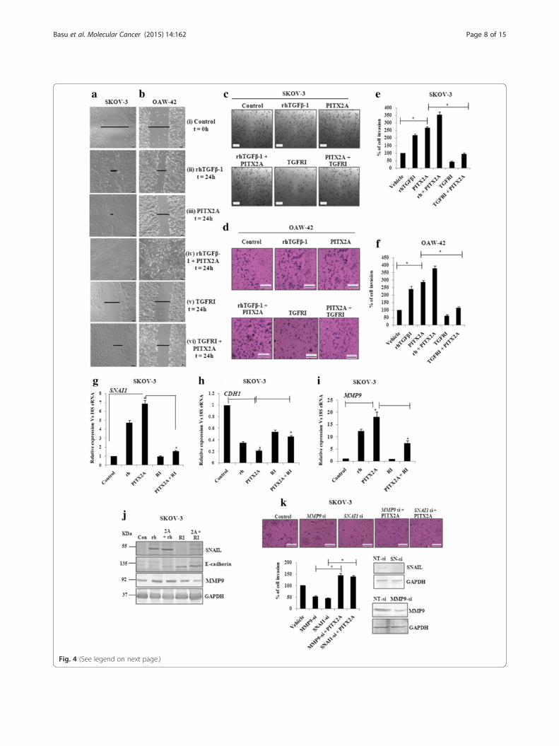
Fig. 4 (See legend on next page.)
Basu et al. Molecular Cancer (2015) 14:162 Page 8 of 15
and folliculogenesis [47, 48]. Both experimental and clinicalstudies suggest the elevated level of activin-A in the serumof granulosa cell tumor patients [49]. Our finding couldadd another dimention to the function and regulation ofactivin-A, particularly, in epithelial ovarian cancer.We showed earlier that PITX2 interacts with and regu-
lates, FGF16, a prime inducer of invasion of ovarian cancercells [28]. We also showed significant involvement ofPITX2 in regulating the Wnt signalling pathway that in-duces proliferation of ovarian cancer cells [50], creating anauto-regulatory feedback loop. The present study is the firstcomprehensive investigation on the regulatory role ofPITX2 on invasive behavior in ovarian carcinoma cells.TGF-β pathway is considered to be a prime regulator in in-ducing invasion of ovarian cancer cells. Here, we identifieda novel regulatory role of PITX2 through expression ofboth TGFB and INHBA genes. The involvement of activin-A, the homodimer form of INHBA, in inducing invasionleading to epithelial ovarian cancer progression (apart fromGCT) has been demonstrated for the first time in thisstudy. Thus, it is imperative to say that PITX2 appears toact as an important regulatory protein that controls initi-ation and progression of epithlial ovarian cancer. Furtherstudy may explore the possible involvement of PITX2 inGCT. TGF-β has been shown to synergize with oncogenicpathways. Evidence suggests that Wnts can also cooperatewith other signaling pathways during tumorigenesis. PITX2lies upstream of these two pathways, thus playing a signifi-cant role in tumorigenesis and metastasis.
ConclusionThe major findings of our study are: a) PITX2 up-regulatesthe expression of ligand genes of TGF-β superfamily,TGFBand INHBA followed by induction of SMAD2/3-dependentTGF-β signalling pathway; b) through the activation ofTGF-β pathway, PITX2 regulates the expression of genesthat enhances invasion and EMT of ovarian cancer cells; c)This is the first report to show the direct involvement ofactivin-A, the homodimer form of INHBA, in promotinginvasion and EMT of both non-cancerous and cancerous
cells. (d) Overall this present report is first of its kind toshow the direct involvement of a homeodomain transcrip-tion factor, PITX2, in the progression of ovarian cancer.
MethodsCell culture, treatment of growth factor and inhibitorsHuman ovarian adenocarcinoma cells, SKOV-3 (ATCC,USA) and OAW-42 (Sigma-Aldrich; USA) were main-tained in McCoy’s 5A (Sigma) and DMEM (Invitrogen)respectively; both supplemented with 10 % fetal bovineserum (FBS, Invitrogen, USA), 100 U/ml penicillin and100 μg/ml streptomycin both Invitrogen; [51]. Humanimmortalized ovarian surface epithelial cells, IOSE (a kindgift from Drs. N. Aueresperg and Clara Salamanca,Vancouver, Canada) was maintained in Medium199(Invitrogen) and MCDB105 (Sigma-Aldrich; USA) in1:1 ratio supplemented with 10 % FBS, 100 U/ml penicillinand 100 μg/ml streptomycin. Here, the low-passagecultures of human ovarian surface epithelium cells(isolated by scraping from human ovarian surface tis-sue) were immortalized by transfecting with SV40large-T antigen viral particles [52].Human recombinant TGF-β1 (rhTGF-β1; Calbiochem,
Germany) was used at 10 ng/ml for 30 min and 8 h forWestern-blot and Q-PCR assay respectively. TGF-βreceptorI kinase inhibitor (TGFRI; 20 ng/ml) was pro-cured from Calbiochem (cat. no. 616451). Recombinanthuman Activin-A (rhActivin-A; ACRO Biosystems) wasused at 100 ng/ml for 30 min and 24 h for Western-blotand Q-PCR assay respectively. Activin-A neutralizingantibody (Novus Biologicals) was used at a concentrationof 2 μg/ml. Prior to each treatment, the cells wereserum-starved for 16 h and the control cells were treatedwith vehicles (0.1 % BSA in 1X PBS or DMSO).
Expression and reporter constructsThe 1.7 kb upstream promoter region of INHBA genewas amplified by PCR using human genomic DNA astemplate and then cloned into pGL3 basic vector (Pro-mega) at MluI/HindIII site. The primer sequences used
(See figure on previous page.)Fig. 4 The regulation of invasion and migration is served by PITX2-mediated activation of TGF-β signalling pathway. Wound healing assay wasperformed of SKOV-3 (a) and OAW-42 (b) cells after treatment with rhTGFβ1 (ii), transient transfection of PITX2A (iii) and treatment with rhTGFβ1(iv) or TGFRI (v) of PITX2-transfected cells in the time course of 24 h. T = 0 h at control cells (i) signifies the time of scratching the cells with pipettetips. The arrows indicate the width of wound and the assay was repeated three times independently. Scale bar: 50 μm. Transwell migration and invasionassay was performed in SKOV-3 (c) and OAW-42 (d) cells after treatment and/or transient transfection as mentioned. Scale bar: 200 μm. Cells at threeindependent fields for each well were counted and plotted with error bar for SKOV-3 (e) and OAW-42 (f) cells. g-i pcDNA3 or PITX2A-transfectedSKOV-3 cells were treated with rhTGFβ1 (10 ng/ml) or TGFRI (20 ng/ml) followed by isolation of RNA and Q-PCR with the primers of SNAI1 (g), CDH1(h) and MMP9 (i). Relative gene expression is indicated as ‘fold’ change in the Y-axis (mean ± SEM). * represents p< 0.01. j Lysates of the cells transientlytransfected and/or treated as indicated were immunoblotted with respective antibodies and the representative gel image was shown. k Transwell invasionassay was performed with SKOV-3 cells after transient transfection as mentioned (top). Cells at three independent fields for each well were counted andwere plotted with error bar (bottom). The efficiency in knocking down the expression of SNAIL and MMP9 proteins by SNAI-(SN)-si and MMP9-sirespectively was verified by Western blot of the transfected cell lysates with respective antibodies. Here, GAPDH was used as loadingcontrol (bottom)
Basu et al. Molecular Cancer (2015) 14:162 Page 9 of 15
to clone the promoters are mentioned in Table 1,where the restriction enzyme sites are underlined. Allconstructs were sequenced by ABI Prism AutomatedDNA Sequencer (Perkin Elmer, USA). Sequence align-ment and data analysis were performed throughBLAST search (NCBI Gen Bank). The TGF-β induciblep3TP-Lux luciferase reporter vector was procuredfrom Addgene, USA. pRL-CMV (Promega, USA) vec-tor with Renilla-luciferase gene was used to normalise
the luciferase activity. Expression plasmids of threeisoforms of PITX2 (PITX2A/B/C) were described earl-ier [28]. To evaluate the expression profile of the TGFBgenes as well as the genes associated with invasion andEMT, we performed over-expression of three isoformsof PITX2 (PITX2A/B/C). As a similar trend was observedin the change in gene expression by these isoforms, we usedPITX2A isoform as representative in the subsequentphysiological experiments like activation of TGF-β pathway,
Fig. 5 PITX2-regulated INHBA activates SMAD2 level. a Schematic diagram shows the 2 kb upstream region of INHBA promoter with thePITX2-specific bicoid-like (as +) elements. Here, the location of primers used to clone 1.7 kb region in pGL3 vector has been depicted withblack solid arrow, while that used for ChIP-PCR has been shown with hollow arrows. b-c ChIP with SKOV-3 cells followed by the PCR showedthe amplification of the INHBA promoter (b) from the chromatin input and from PITX2-IP DNA as indicated in the lanes. The PITX2 antibodyused in this assay recognizes all three isoforms of PITX2. Amplification of respective promoters was not observed by PCR performed withIgG-IP and no antibody control sets. c Primers of an unrelated gene did not show amplification from PITX2-IP DNA. (d) The INHBA promoteras shown in Fig A was cloned into pGL3-basic vector. OAW-42 cells were transiently co-transfected with the construct alone or in combination withPITX2 isoforms followed by luciferase assay. The reporter activity of each clone was calculated in terms of fold change in PITX2-overexpressedcells compared with empty vector-transfected cells, after normalization with renilla luciferase activity. e Q-PCR analysis of the INHBA genewith specific primers was performed with the RNA isolated from PITX2A/B/C-transfected OAW-42 cells. Relative gene expression is indicatedas ‘fold’ change in the Y-axis (mean ± SEM). The statistical analysis is done as described previously.* represents p < 0.05. f Western blot wasperformed to check the level of p-SMAD2 in OAW-42 cells after treatment with rhActivin-A (rhA10: 10 ng/ml and rhA100: 100 ng/ml) for 30mins. g The levels of p-SMAD2 was assessed on treating the cells with PITX2A-conditioned medium (PITX2-CM), rhActivin-A (rhA: 100 ng/ml)or PITX2-CM in presence of activin neutralizing antibody (Pitx2-CM + Ab)
Basu et al. Molecular Cancer (2015) 14:162 Page 10 of 15
matrigel invasion, wound healing and cell proliferationassay etc.
Transient transfection and luciferase assayFor reporter assay, 5 × 10⁴ cells were seeded on 12-wellculture plates. After 24 h, p3TP-Lux vector (0.4 μg) wastransiently transfected alone or along with PITX2 ex-pression vectors (0.4 μg) with Lipofectamine 2000 (Invi-trogen). After 4 h of transfection, the medium wasreplaced with fresh incomplete one supplemented witheither TGFRI or DMSO for next 16 h. Each transfectionwas normalized with pRL-CMV vector (0.04 μg). In thefollowing day, cells were harvested and firefly/renillaluciferase activity was determined [50]. The transfectionof pGL3-reporter vectors and the subsequent assaywas performed following the same protocol. Each
transfection was performed in triplicate and the experi-ments were repeated thrice.To over-express PITX2 isoforms, 1 μg of expression
constructs were transfected per 105 cells/well in 6-wellplate using Lipofectamine 2000 (Invitrogen). After 24 hand 48 h of transfection, the cells were harvested to iso-late RNA and protein respectively. For the treatment ofrhTGF-β1/TGFRI, the medium was replaced after 4 h ofPITX2 transfection with fresh one supplemented withthese factors. The cells were harvested after 16 h forRNA/protein isolation. To collect conditioned medium(CM), PITX2A was transiently transfected as men-tioned earlier. After 6 h, the medium was replaced withfresh serum-free one, which was collected after 24 h oftransfection and added directly or in combination withrhTGF-β1/TGFR1 to the freshly plated cells. Thetreated cells were harvested after 2 h for protein
Fig. 6 Activin-A promotes EMT in ovarian non-cancerous and cancer cells. a-b IOSE and OAW-42 cells were treated with rhActivin-A andtheir morphological changes were observed through bright field (A i-ii) as well as through confocal microscopy after phalloidin staining(B i-ii). c-d RNA was isolated from the IOSE cells to check the expression of CDH1, CLDN7 (c) and CDH2, VIM (d) by Q-PCR. e-g RNA isolationwas done from the OAW-42 cells to quantify ZEB1, ZEB2,SNAI1, SNAI2 (e), MMP9 (f), CLDN1,CLDN7 and VIM (g) by Q-PCR with specific primers.Relative gene expression is indicated as ‘fold’ change in the Y-axis (mean ± SEM). The statistical analysis is done as described previously.*represents p < 0.05. h Western Blot was done with lysates of IOSE cells with respective antibodies as mentioned. GAPDH was used as theloading control
Basu et al. Molecular Cancer (2015) 14:162 Page 11 of 15
isolation. The control cells were transfected withempty vector (pcDNA3.1 Myc-His) in each case.
siRNA and transfectionThe siRNAs against SNAI1, MMP9 and INHBA (all fromSanta Cruz Biotechnology, USA) were used at 20 nM/wellusing 2 μl Lipofectamine RNAiMAX (Invitrogen) in thecells seeded in 6-well plates. The RNA interference againstPITX2 was carried out by the ON-TARGET plus SMARTpool siRNA at 20 nM/well using 2 μl of Dharmafect-1transfection reagent (Dharmacon) in cells seeded in 6-wellculture plates. After 48 h of transfection, the cells were har-vested for RNA/protein isolation. When required, rhTGF-
β1 was added after 24 h of respective siRNA transfectioninto the cells.
Quantitative Real-time RT-PCR (Q-PCR)Total RNA was isolated from ovarian cell lines using TRI-reagent (Sigma) following the standard protocol [53].First-strand cDNA synthesis followed by Q-PCR assaywas performed as described [28]. The comparative CT
method (ΔΔCT) was used to measure relative gene expres-sion where the fold enrichment was calculated as: 2− [ΔCT (sample) −ΔCT (calibrator)]. Here, ΔCT is the CT of tar-get gene subtracted from the CT of the housekeeping gene[50]. The primer sequences are mentioned in Table 2.
Table 1 The sequence of the oligonucleotide primers used to amplify specific regions of the promoter
Gene Name& Acc no
Region amplified Forward primer (5′- 3′) Reverse primer (5′- 3′) Amplicon size (kb) Tm (°C)
INHBA;NC_000007.14
41702063 - 41700378 GGACGCGTGAACGCTTTAACAGATGGAC GGAAGCTTGCAAAAGTTGTTGTGATTGC 1.7 54
Fig. 7 PITX2 acts through Activin-A to affect the cellular invasion and EMT. a Matrigel transwell assay was performed in OAW-42 cells whichwere treated either with rhActivin-A or transfected with INHBA-siRNA in presence or absence of PITX2A-overexpression and the percentageof cell invasion was calculated. Scale bar 100 μm. b Cells at three independent fields for each well were counted and plotted with error bar.c-f RNA was isolated from cells tarnsfected with INHBA-siRNA in presence or absence of PITX2A- overexpression and the expression ofMMP9, ZEB1, SNAI1, VIM, CLDN7 was quantified by Q- PCR with specific primers. Relative gene expression is indicated as ‘fold’ change in theY-axis (mean ± SEM). The statistical analysis is done as described previously.*represents p < 0.05
Basu et al. Molecular Cancer (2015) 14:162 Page 12 of 15
Chromatin-immunoprecipitation (ChIP)ChIP with SKOV-3 cells was performed following themethods described earlier [50]. For ChIP-PCR, theimmunoprecipitated (IP) and input DNA were used atequal quantity following the conditions: 95 °C for 30 s,annealing at specific temp for 30 s and extension at72 °C for 30 s, for 30 cycles. The information of theprimers is shown in Table 3.
Western blot analysisCell lysis and protein extraction was performed as de-scribed previously [51] and subjected to immunoblottingwith antibodies specific for the proteins including, PITX2(Chemicon, 1:1000), α-SMA (Sigma; 1:1000 dilution),claudin-7, MMP9 (both Santa Cruz; 1:1000 dilution),GAPDH (1:3000), E-cadherin, SNAIL, vimentin, p-SMAD2and SMAD2 (all 1:2000 dilution; all from Cell signallingtechnology, USA).
Confocal microscopyImmunofluorescence staining with anti-p SMAD2 (1:100)E-cadherin (1:100) and PITX2 (1:100) antibodies followedby Alexa-fluor 488-conjugated secondary antibody was
performed as described previously [28]. For phalloidinstaining 105 cells/well were plated in 6-well plate. After24 h of PITX2A transfection or rhActivin-A treatment,actin filament bundle formation was observed by phalloidinstaining as mentioned previously [54] followed by imagingwith confocal microscopy.
Wound healing assaySerum-starved cells at 70 % confluency were transfectedwith PITX2A-construct. After 4 h, medium was replacedby fresh and incomplete one supplemented with rhTGF-β1/TGFRI. In additional experiment, treatment was alsogiven in other set of serum-starved cells to check the ef-fect of only TGF-β1/TGFRI on migration. Scratchingwas carried out with a 200 μl pipette tip prior to thetreatment and mentioned as t = 0 h at the figure. Cellswere washed several times with PBS to remove the de-tached ones and supplied with new growth medium.Photographs of the scratches were taken at 0 and 24 husing an inverted microscope (Leica) equipped with aScion digital camera and in-built software (Leica appli-cation suite v3.0).
Table 2 The sequence, respective amplicon size and Tm of the oligonucleotide primers used in Q-PCR
Gene Name Forward primer (5′- 3′) Reverse primer (5′- 3′) Amplicon size (bp) Tm (°C)
18S rRNA GATTCCGTGGGTGGTGGTGC AAGAAGTTGGGGGACGCCGA 134 60
POLR2A TGGACCCACCGGCATGTTCT GCCCCTGGGGTCATTCCACT 141 60
CLDN4 GGCTGCTTTGCTGCAACT CAGAGCGGGCAGCAGAATAC 110 60
CLDN7 GTGGCAGATGAGCTCCTATGC CATCCACAGCCCCTTGTACA 80 60
DSM GCAGCAAAGGGCGGAGAT TGTTAATGTGCTGCTCCACTGA 80 60
VIM ACACCCTGCAATCTTTCAGACA GATTCCACTTTGCGTTCAAGGT 80 60
FN1 CCTTCATGGCAGCGGTTT AGCGTCCTAAAGACTCCATGATCT 94 60
PITX2 CGCGAAGAAATCGCTGTGT CGACGATTCTTGAACCAAACC 78 58
TGFB1 GTGACAGCAGGGATAACACACTG CATGAATGGTGGCCAGGTC 80 60
TGFB2 GCTGAGCGCTTTTCTGATCCT CGAGTGTGCTGCAGGTAGACA 80 60
TFGB3 CACCACAACCCTCATCTAATCCT CCTGGCCCGGGTTGTC 100 60
SNAI1 TCGGAAGCCTAACTACAGCGA AGATGAGCATTGGCAGCGAG 140 60
SNAI2 ATGAGGAATCTGGCTGCTGT CAGGAGAAAATGCCTTTGGA 119 60
CDH1 GTCACTGACACCAACGATAATCT TTTCAGTGTGGTGATTACGACGTA 100 60
MMP9 ACCTCGAACTTTGACAGCGAC GAGGAATGATCTAAGCCCAGC 113 60
INHBA GTGAGTGCCCGAGCCATATAG CATGCGGTAGTGGTTGATGACT 80 60
CDH2 CCATCAAGCCTGTGGGAATC GCAGATCGGACCGGATACTG 76 60
ZEB1 CAATGATCAGCCTCAATCTGCA CCATTGGTGGTTGATCCCA 117 60
ZEB2 AAGCCCCATCAACCCATACAAG AAATTCCTGAGGAAGGCCCA 124 60
Table 3 The sequence, respective amplicon size and Tm of the oligonucleotide primers used in ChIP-PCR
Gene Name & Acc no Region amplified Forward primer (5′- 3′) Reverse primer (5′- 3′) Amplicon size (kb) Tm (°C)
INHBA; NC_000007.14 41702097- 41701831 GGCTTATGTGTGGGAAAGAA ACCAGTGCATTCATAGACAG 265 55.5
Basu et al. Molecular Cancer (2015) 14:162 Page 13 of 15
In vitro invasion assayTranswell membranes coated with Matrigel (BDBiosciences, USA) were used to assay in vitro invasion asmentioned previously [53]. In brief, 2.5 × 10⁵ cells wereseeded in the upper chamber in serum-free medium andFBS or rhTGF-β1/TGFRI was added in the lower chamber.To check the effect of PITX2, MMP9 and SNAIL on TGF-β-mediated invasion, cells were transiently transfected withrespective construct or siRNAs on previous day, allowed torecover overnight and then serum-starved for additional16 h. The cells were then trypsinized, counted and equalnumber of transfected cells were added in the upper cham-ber and allowed to invade in presence of TGF-β orTGFRI.To understand the role of activin-A in PITX2-mediated cell invasion, the cells were transiently transfectedwith PITX2 expression construct or INHBA-siRNAs orboth on previous day, allowed to recover overnight andthen serum-starved for additional 16 h. The cells were thentrypsinized, counted and equal number of transfected cellswere added in the upper chamber and allowed to invade inpresence or absence of rhActivin-A. After incubating for22 h at 37 °C in 5 % CO2, the invaded cells were fixed,stained and counted under microscope. Three independ-ent experiments were performed followed by statisticalanalysis.
Immunohistochemistry (IHC) with immunofluorescence(IF)-based detectionTissue sample blocks used for IHC were archival materialsprovided by the Department of Pathology, Institute of PostGraduate Medical Education and Research and SSKM Hos-pital, Kolkata, India. Isolated tissues were fixed, processed,and sectioned as mentioned earlier [55]. The sections werethen blocked in 5 % BSA in 1XTBS-T for 30 min and incu-bated for 2 h with the anti-pSMAD2 (1:100) and antibodydiluted in 1XTBS containing 0.1 % BSA. The slides werethen washed and incubated for 1 h with the secondary anti-body (Alexa Fluor-488; 1:500) followed by staining withDAPI. The IF-staining was performed in all collected sam-ples and the representative images have been shown.
Statistical analysisAll data were expressed as mean ± SEM and the ± SEM arerepresented by error bars. The statistical significance wascalculated by two-tailed Student’s t-test. p < 0.05 was con-sidered as significant. The experiments were done at least 3times in duplicate unless otherwise stated.
Additional file
Additional file 1: Figure S1. The expression of PITX2 is up-regulatedin human ovarian cancer. (A) The level of PITX2 was observed by IHCin human ovarian tissue-sections with specific antibody followed byAlexa Fluor-488 (green) of low malignant potential (i; n = 20) and high
malignant potential metastatic adenocarcinoma patients (ii; n = 20).The DAPI-stained nuclei and the merged images were also shown.Scale bar, 10 μm. (TIFF 28495 kb)
AbbreviationDAPI: 4′, 6-Diamidino-2-phenylindole; TGF: Transforming growth factor;MMP: Matrix metalloprotease; SMAD: Mothers against decapentaplegic,Drosophila; EMT: Epithelial-mesenchymal transition; GAPDH: Glyceraldehyde-3phosphate dehydrogenase; ECM: Extracellular matrix; SMA: Smooth muscle actin;CLDN: Claudin; CDH1: E-cadherin; CDH2: N-cadherin; SNAI1: SNAIL; SNAI2: SLUG;INHBA: activin-A; IOSE: Immortalized ovarian surface epithelium.
Competing interestsThe authors declare that they have no competing interests.
Authors’ contributionsMB designed and performed the experiments and drafted the manuscript.RB planned and performed immunodetection and imaging, Q-PCR and matrigelassay. UR did the confocal imaging, Q-PCR and immunodetection. SNM partlydrafted and corrected the manuscript. UC collected the human tissue samplesand processed. SSR conceived the study, designed the experiments and draftedthe manuscript. All authors read and approved the final manuscript.
AcknowledgementsWe thankfully acknowledge Council of Scientific and Industrial Research (CSIR,project No. BSC 0101 and BSC 0206), Govt. of India, for funding. MB and RB arethe recipient of fellowship from CSIR, Govt. of India [award no. 10-2(5)/2007(II)-E.UIIand 31/002(0941)/2013-EMR-1 respectively]. UR is a recipient of fellowship fromUGC, Govt. of India [award no.19-06/2011(i)EU-IV]. The IOSE cells were a kind giftfrom Dr. Clara Salamanca, as mentioned in the methods section. The technicalassistance of Prabir Kumar Dey (CSIR-IICB) is gratefully acknowledged. Othermembers of SSR laboratory are thankfully acknowledged for their co-operation.
Author details1Cell Biology and Physiology Division, CSIR-Indian Institute of ChemicalBiology, Council of Scientific and Industrial Research, 4 Raja S. C. MullickRoad, Kolkata 700032, India. 2Department of Endocrinology and Metabolism,IPGMER and SSKM Hospital, 244 AJC Bose Road, Kolkata, India. 3Departmentof Pathology, IPGMER and SSKM Hospital, 244 AJC Bose Road, Kolkata, India.
Received: 18 March 2015 Accepted: 12 August 2015
References1. Yap TA, Carden CP, Kaye SB. Beyond chemotherapy: targeted therapies in
ovarian cancer. Nat Rev Cancer. 2009;9:167–81.2. Scully RE. Pathology of ovarian cancer precursors. J Cell Biochem Suppl.
1995;23:208–18.3. Gotzmann J, Mikula M, EgerSchulte-Hermann AR, Foisner R, Beug H, Mikulits
W. Molecular aspects of epithelial cell plasticity: implications for localtumour invasion and metastasis. Mutat Res. 2004;566:9–20.
4. Akhurst RJ, Derynck R. TGF-beta signaling in cancer-a double-edged sword.Trends Cell Biol. 2001;11:S44–51.
5. Shi Y, Massagué J. Mechanisms of TGF-beta signaling from cell membraneto the nucleus. Cell. 2003;113:685–700.
6. Burger HG. Inhibin. Rep Med Rev. 1992;1:1–20.7. De Kretser DM, Robertson DM. The isolation and physiology of inhibin and
related proteins. Biol Reprod. 1989;40:33–47.8. Choi YL, Kim HS, Ahn G. Immunoexpression of inhibin alpha subunit,
inhibin/activin betaA subunit and CD99 in ovarian tumors. Arch Pathol LabMed. 2000;124:563–9.
9. Petraglia F, Luisi S, Pautier P, Sabourin JC, Rey R, Lhomme C, et al. Inhibin Bis the major form of inhibin/activin family secreted by granulosa celltumors. J Clin Endocrinol Metab. 1998;83:1029–32.
10. Yoshinaga K, Mimori K, Inoue H, Kamohara Y, Yamashita K, Tanaka F, et al.Clinical significance of the expression of activin A in esophageal carcinoma.Int J Oncol. 2003;22:75–80.
11. Wildi S, Kleeff J, Maruyama H, Maurer CA, Büchler MW, Korc M.Overexpression of activin A in stage IV colorectal cancer. Gut.2001;49:409–17.
Basu et al. Molecular Cancer (2015) 14:162 Page 14 of 15
12. Okano M, Yamamoto H, Ohkuma H, Kano Y, Kim H, Nishikawa S, et al.Significance of INHBA expression in human colorectal cancer. Oncol Rep.2013;30:2903–8.
13. Petersen OW, Nielsen HL, Gudjonsson T, Villadsen RF, Rank E, NiebuhrMJ, et al. Epithelial to mesenchymal transition in human breast cancercan provide a nonmalignant stroma. Am J Pathol. 2003;162:391–402.
14. Thiery JP. Epithelial-mesenchymal transitions in tumour progression.Nat Rev Cancer. 2002;2:442–54.
15. Peinado H, Olmeda D, Cano A. Snail, Zeb and bHLH factors in tumourprogression: an alliance against the epithelial phenotype? Nat Rev Cancer.2007;7:415–28.
16. Sun L, Diamond ME, Ottaviano AJ, Joseph MJ, Ananthanarayan V, MunshiHG. Transforming growth factor-beta 1 promotes matrix metalloproteinase-9-mediated oral cancer invasion through snail expression. Mol Cancer Res.2008;6:10–20.
17. Abate-Shen C, Shen MM, Gelmann E. Integrating differentiation and cancer:the Nkx3.1 homeobox gene in prostate organogenesis and carcinogenesis.Differentiation. 2008;76:717–27.
18. Raman V, Martensen SA, Reisman D, Evron E, Odenwald WF, Jaffee E, et al.Compromised HOXA5 function can limit p53 expression in human breasttumours. Nature. 2000;405:974–8.
19. Crijns AP, de Graeff P, Geerts D, Ten Hoor KA, Hollema H, Van der Sluis Tet al. MEIS and PBX homeobox proteins in ovarian cancer. Eur J Cancer.2007;43:2495–505.
20. Tan Y, Cheung M, Pei J, Menges CW, Godwin AK, Testa JR. Upregulation ofDLX5 promotes ovarian cancer cell proliferation by enhancing IRS-2-AKTsignalling. Cancer Res. 2010;70:9197–206.
21. Wu X, Chen H, Parker B, Rubin E, Zhu T, Lee JS. HOXB7, a homeodomain protein,is overexpressed in breast cancer and confers epithelial-mesenchymal transition.Cancer Res. 2006;66:9527–34.
22. Hayashi M, Maeda S, Aburatani H, Kitamura K, Miyoshi H, Miyazono K, et al.PITX2 prevents osteoblastic trans differentiation of myoblasts by bonemorphogenetic proteins. J Biol Chem. 2008;283:557–65.
23. Kioussi C, Briata P, Baek SH, Rose DW, Hamblet NS, Herman T et al.Identification of a Wnt/Dvl/[beta]-catenin PITX2 pathway mediating cell-type-specific proliferation during development. Cell. 2002;111:673–85.
24. Lin CR, Kioussi C, O’Connell S, Briata P, Szeto D, Liu F, et al. Pitx2 regulateslung asymmetry, cardiac positioning and pituitary and toothmorphogenesis. Nature. 1999;401:279–82.
25. Cox CJ, Espinoza HM, McWilliams B, Chappell K, Morton L, Hjalt TA, et al.Differential regulation of gene expression by PITX2 isoforms. J Biol Chem.2002;277:25001–10.
26. Nimmrich I, Sieuwerts AM, Meijer-van Gelder ME, Schwope I, Bolt-de Vries J,Harbeck N, et al. DNA hypermethylation of PITX2 is a marker of poor prognosis inuntreated lymph node-negative hormone receptor-positive breast cancerpatients. Breast Cancer Res Treat. 2008;111:429–37.
27. Hirose H, Ishii H, Mimori K, Tanaka F, Takemasa I, Mizushima T, et al. Thesignificance of PITX2 overexpression in human colorectal cancer. Ann SurgOncol. 2011;18:3005–12.
28. Basu M, Mukhopadyay S, Chatterjee U, Roy SS. FGF16 promotes invasivebehavior of SKOV-3 ovarian cancer cells through the activation of MAPKsignaling pathway. J Biol Chem. 2014;289:1415–28.
29. DeVita VT, Hellman S, Rosenberg SA. Cancer: principles and practice ofoncology. 6th ed. New York: Lippincott-Raven; 2001.
30. Akhurst RJ, Balmain A. Genetic events and the role of TGF beta in epithelialtumour progression. J Pathol. 1999;187:82–90.
31. Chen RH, Ebner R, Derynck R. Inactivation of the type II receptor reveals tworeceptor pathways for the diverse TGF-beta activities. Science.1993;260:1335–8.
32. Massagué J. TGF-β signal transduction. Annu Rev Biochem. 1998;67:753–91.33. Derynck R, Goeddel DV, Ullrich A, Gutterman JU, Williams RD, Bringman TS,
et al. Synthesis of messenger RNAs for transforming growth factors α and βand the epidermal growth factor receptor by human tumors. Cancer Res.1987;47:707–12.
34. Dunfield LD, Dwyer EJ, Nachtigal MW. TGF beta-induced Smad signalingremains intact in primary human ovarian cancer cells. Endocrinology.2002;143:1174–81.
35. Huang Y, Guigon CJ, Fan J, Cheng SY, Zhu GZ. Pituitary homeobox 2 (PITX2)promotes thyroid carcinogenesis by activation of cyclin D2. Cell Cycle.2010;9:1333–41.
36. Acunzo J, Roche C, Defilles C, Thirion S, Quentien MH, Figarella-Branger D,et al. Inactivation of PITX2 transcription factor induced apoptosis ofgonadotroph tumoral cells. Endocrinology. 2011;152:3884–92.
37. Hayashida T, Takahashi F, Chiba N, Brachtel E, Takahashi M, Godin-HeymannN, et al. HOXB9, a gene overexpressed in breast cancer, promotestumorigenicity and lung metastasis. Proc Natl Acad Sci U S A.2010;107:1100–5.
38. Lee JM, Dedhar S, Kalluri R, Thompson EW. The epithelial-mesenchymaltransition: new insights in signaling, development, and disease. J Cell Biol.2006;172:973–81.
39. Yoshida H, Broaddus R, Cheng W, Xie S, Naora H. Deregulation of the HOXA10homeobox gene in endometrial carcinoma: role in epithelial-mesenchymaltransition. Cancer Res. 2006;66:889–97.
40. Batlle E, Sancho E, Francí C, Domínguez D, Monfar M, Baulida J, et al.The transcription factor snail is a repressor of E-cadherin gene expression inepithelial tumour cells. Nat Cell Biol. 2000;2:84–9.
41. Vleminckx K, Vakaet Jr L, Mareel M, Fiers W, van Roy F. Genetic manipulationof E-cadherin expression by epithelial tumor cells reveals an invasionsuppressor role. Cell. 1991;66:107–19.
42. Blechschmidt K, Sassen S, Schmalfeldt B, Schuster T, Ho¨fler H, Becker KF.The E-cadherin repressor Snail is associated with lower overall survival ofovarian cancer patients. Br J Cancer. 2008;98:489–95.
43. Kurrey NK, Amit K, Bapat SA. Snail and Slug are major determinants ofovarian cancer invasiveness at the transcription level. Gynecol Oncol.2005;97:155–65.
44. Roy R, Yang J, Moses MA. Matrix metalloproteinases as novel biomarkersand potential therapeutic targets in human cancer. J Clin Oncol.2009;27:5287–97.
45. Lengyel E, Schmalfeldt B, Konik E, Späthe K, Härting K, Fenn A, et al.Expression of latent matrix metalloproteinase 9 (MMP-9) predicts survival inadvanced ovarian cancer. Gynecol Oncol. 2001;82:291–8.
46. Peng C, Mukai ST. Activins and their receptors in female reproduction.Biochem Cell Biol. 2000;78:261–79.
47. Mukasa C, Nomura M, Tanaka T, Tanaka K, Nishi Y, Okabe T, et al. Activinsignaling through type IB activin receptor stimulates aromatase activity inthe ovarian granulosa cell-like human granulosa (KGN) cells. Endocrinology.2003;144:1603–11.
48. Nomura M, Sakamoto R, Morinaga H, Wang L, Mukasa C, Takayanagi R.Activin stimulates CYP19A gene expression in human ovarian granulosacell-like KGN cells via the Smad2 signaling pathway. Biochem Biophys ResCommun. 2013;436:443–8.
49. Ciriş M, Erhan Y, Zekioglu O, Bayramoglu H. Inhibin alpha and betaexpression in ovarian stromal tumors and their histological equivalences.Acta Obstet Gynecol Scand. 2004;83:491–6.
50. Basu M, Roy SS. Wnt/β-catenin pathway is regulated by PITX2homeodomain protein and thus contributes to the proliferation of humanovarian adenocarcinoma cell, SKOV-3. J Biol Chem. 2013;288:4355–67.
51. Ghosh S, Basu M, Roy SS. ETS-1 regulates vascular endothelial growth factor-induced matrix metalloproteinase-9 and matrix metalloproteinase-13expression in human ovarian carcinoma cell SKOV3. J Biol Chem 2012,287:15001-15.
52. Kruk PA, Maines-Bandiera SL, Auersperg N. A simplified method to culturehuman ovarian surface epithelium. Lab Invest 1990, 63:132–36.
53. Ghosh P, Saha SK, Nandi SS, Bhattacharya S, Roy SS. Involvement of Pitx2, ahomeodomain transcription factor, in hypothyroidism associatedreproductive disorders. Cell Physiol Biochem 2007, 20:887-98.
54. Faulstich H, Trischmann H, Mayer D. Preparation of tetramethylrhodaminyl-phalloidin and uptake of the toxin into short-term cultured hepatocytes byendocytosis. Exp Cell Res. 1983;144:73–82.
55. Nandi SS, Ghosh P, Roy SS. Expression of PITX2 homeodomain transcriptionfactor during rat gonadal development in a sexually dimorphic manner.Cell Physiol Biochem. 2011;27:159–70.
Basu et al. Molecular Cancer (2015) 14:162 Page 15 of 15


![MicroRNA-505 functions as a tumor suppressor in ... · nant tumors, including osteosarcoma, hepatic cancer, prostate cancer and breast cancer [20, 22, 26, 32, 33]. Recent studies](https://static.fdocument.org/doc/165x107/5f024f927e708231d403a367/microrna-505-functions-as-a-tumor-suppressor-in-nant-tumors-including-osteosarcoma.jpg)