Insulin-like growth factor binding protein-4 and -5 modulate ligand-dependent estrogen receptor-α...
8
Insulin-like growth factor binding protein-4 and -5 modulate ligand-dependent estrogen receptor-α activation in breast cancer cells in an IGF-independent manner ☆ Alexander Hermani a , Ashish Shukla a , Senad Medunjanin a, 1 , Haim Werner b , Doris Mayer a, ⁎ a Hormones and Signal Transduction Group, German Cancer Research Center, DKFZ-ZMBH Alliance, Heidelberg, Germany b Department of Human Molecular Genetics and Biochemistry, Sackler School of Medicine, Tel Aviv University, Tel Aviv 69978, Israel abstract article info Article history: Received 4 February 2013 Accepted 18 February 2013 Available online 14 March 2013 Keywords: IGF-binding proteins Akt/PKB pathway Estrogen receptor Breast cancer Insulin-like growth factor binding proteins (IGFBPs) are modulators of numerous cellular processes including cell proliferation. Although IGFBPs classically act by sequestration of extracellular insulin-like growth factors (IGFs), thereby contributing to the fine-tuning of growth factor signals, IGF-independent actions of IGFBPs have also been described. In the breast, growth factor signaling in association with estradiol (E2)-stimulated estrogen receptor function is organized in a complex cross-talk. The importance of phosphatidylinositol 3-kinase/protein kinase B (Akt/PKB) pathway components for the E2-induced activation of estrogen receptor-alpha (ERα) is well accepted. Here we show that in the absence of IGFs, IGFBP-4 or IGFBP-5, either overexpressed in MCF-7 breast cancer cells or added exogenously, decreased the capability of E2 to induce ERα transcriptional activity. In addition, overexpression or addition of recombinant IGFBP-4 or IGFBP-5 resulted in reduction of E2-induced phosphorylation of Akt/PKB, GSK-3α/β and ERα in MCF-7 cells. The ac- tivation of the Akt/PKB-pathway describes a non-genomic effect of E2, which did not involve activation/phos- phorylation of the IGF-I receptor (IGF-IR). Furthermore, knockdown of the IGF-IR did not affect the inhibition of E2-induced ERα phosphorylation by IGFBP-4 and 5. Moreover, IGFBP-4 and IGFBP-5 strongly decreased E2-triggered growth of MCF-7 cells. Our data suggest that IGFBPs interfere with the E2-induced activation of the Akt/PKB-pathway and prevent full hormone-dependent activation of ERα and breast cancer cell growth in an IGF- and IGF-IR-independent manner. © 2013 Elsevier Inc. All rights reserved. 1. Introduction The IGF (insulin-like growth factor) axis plays a crucial role in the regulation of cellular growth and differentiation, developmental pro- cesses and malignant cell transformation [1,2]. IGF-I and IGF-II are po- tent mitogenic and survival factors for both normal and cancer cells, and their effects on cell proliferation are mediated by the type 1 IGF ty- rosine kinase receptor (IGF-IR). Classically, IGF binding proteins (IGFBPs) modulate the bioavailability of the IGFs through IGF/IGFBP complex formation and contribute to the control of IGF-I and IGF- II-induced proliferation [3–6]. Six high-affinity IGFBPs have been identi- fied, which are structurally, functionally, and evolutionarily related and which usually inhibit IGF-I and IGF-II action [6–9]. IGFBP-1 to -4 have similar affinities for IGF-I and IGF-II, whilst IGFBP-5 and IGFBP-6 bind IGF-II with a much higher affinity. Recent evidence suggests intrinsic ligand-independent activities of IGFBPs at the cellular level, which lead to specific cellular actions or modulate the effects of other factors [10]. So far it is unclear whether these activities are mediated via puta- tive cell surface IGFBP receptors or by intracellular delivery [8–12]. In the mammary gland the components of the IGF system, including the IGFs, IGF-IR and IGFBPs, are important players in the development Cellular Signalling 25 (2013) 1395–1402 Abbreviations: BFA, brefeldin A; BP-4, IGFBP-4; BP-5, IGFBP-5; BSA, bovine serum albu- min; DCC, dextran-coated charcoal; DMEM, Dulbecco's modified Eagle's Medium; E2, 17β-estradiol; EGFP, enhanced green fluorescent protein; ERα, estrogen receptor-alpha; ERE, estrogen response element; FBS, fetal bovine serum; GSK-3, glycogen synthase kinase-3; IGF, insulin-like growth factor; IGFBP, IGF-binding protein; IGF-IR, type 1 IGF re- ceptor; PBS, phosphate-buffered saline; PI3K, phosphatidylinositol 3-kinase; PKB, protein kinase B; qPCR, quantitative real time polymerase chain reaction. ☆ Grant: This work was supported by the Cooperation Program in Cancer Research of the Deutsches Krebsforschungszentrum (DKFZ) and Israeli's Ministry of Science and Technology (MOST) (to D.M. and H.W.). The funding sources were not involved in study design, in the collection, analysis and interpretation of data, in writing the man- uscript and in the decision to submit the article for publication. ⁎ Corresponding author at: Hormones and Signal Transduction Group, German Cancer Research Center, Im Neuenheimer Feld 581, 69120 Heidelberg, Germany. Tel.: +49 6221 423238; fax: +49 6221 421559. E-mail addresses: [email protected] (A. Hermani), [email protected] (A. Shukla), [email protected] (S. Medunjanin), [email protected] (H. Werner), [email protected] (D. Mayer). 1 Present address: Department of Cardiology, Angiology, and Pneumology, Magdeburg University, Germany. 0898-6568/$ – see front matter © 2013 Elsevier Inc. All rights reserved. http://dx.doi.org/10.1016/j.cellsig.2013.02.018 Contents lists available at SciVerse ScienceDirect Cellular Signalling journal homepage: www.elsevier.com/locate/cellsig
Transcript of Insulin-like growth factor binding protein-4 and -5 modulate ligand-dependent estrogen receptor-α...
Cellular Signalling 25 (2013) 1395–1402
Contents lists available at SciVerse ScienceDirect
Cellular Signalling
j ourna l homepage: www.e lsev ie r .com/ locate /ce l l s ig
Insulin-like growth factor binding protein-4 and -5 modulateligand-dependent estrogen receptor-α activation in breast cancer cellsin an IGF-independent manner☆
Alexander Hermani a, Ashish Shukla a, Senad Medunjanin a,1, Haim Werner b, Doris Mayer a,⁎a Hormones and Signal Transduction Group, German Cancer Research Center, DKFZ-ZMBH Alliance, Heidelberg, Germanyb Department of Human Molecular Genetics and Biochemistry, Sackler School of Medicine, Tel Aviv University, Tel Aviv 69978, Israel
Abbreviations: BFA, brefeldin A; BP-4, IGFBP-4; BP-5, IGmin; DCC, dextran-coated charcoal; DMEM, Dulbecco's17β-estradiol; EGFP, enhanced green fluorescent protein;ERE, estrogen response element; FBS, fetal bovine serukinase-3; IGF, insulin-like growth factor; IGFBP, IGF-bindinceptor; PBS, phosphate-buffered saline; PI3K, phosphatidykinase B; qPCR, quantitative real time polymerase chain re☆ Grant: This work was supported by the Cooperationthe Deutsches Krebsforschungszentrum (DKFZ) and IsrTechnology (MOST) (to D.M. and H.W.). The fundingstudy design, in the collection, analysis and interpretatiouscript and in the decision to submit the article for pub⁎ Corresponding author at: Hormones and Signal T
Cancer Research Center, Im Neuenheimer Feld 581,Tel.: +49 6221 423238; fax: +49 6221 421559.
E-mail addresses: [email protected]@gmail.com (A. Shukla), senad.medunjanin(S. Medunjanin), [email protected] (H. Werner), d
1 Present address: Department of Cardiology, AngiologyUniversity, Germany.
0898-6568/$ – see front matter © 2013 Elsevier Inc. Allhttp://dx.doi.org/10.1016/j.cellsig.2013.02.018
a b s t r a c t
a r t i c l e i n f oArticle history:Received 4 February 2013Accepted 18 February 2013Available online 14 March 2013
Keywords:IGF-binding proteinsAkt/PKB pathwayEstrogen receptorBreast cancer
Insulin-like growth factor binding proteins (IGFBPs) are modulators of numerous cellular processes includingcell proliferation. Although IGFBPs classically act by sequestration of extracellular insulin-like growth factors(IGFs), thereby contributing to the fine-tuning of growth factor signals, IGF-independent actions of IGFBPshave also been described. In the breast, growth factor signaling in association with estradiol (E2)-stimulatedestrogen receptor function is organized in a complex cross-talk. The importance of phosphatidylinositol3-kinase/protein kinase B (Akt/PKB) pathway components for the E2-induced activation of estrogenreceptor-alpha (ERα) is well accepted. Here we show that in the absence of IGFs, IGFBP-4 or IGFBP-5, eitheroverexpressed in MCF-7 breast cancer cells or added exogenously, decreased the capability of E2 to induceERα transcriptional activity. In addition, overexpression or addition of recombinant IGFBP-4 or IGFBP-5resulted in reduction of E2-induced phosphorylation of Akt/PKB, GSK-3α/β and ERα in MCF-7 cells. The ac-tivation of the Akt/PKB-pathway describes a non-genomic effect of E2, which did not involve activation/phos-phorylation of the IGF-I receptor (IGF-IR). Furthermore, knockdown of the IGF-IR did not affect the inhibitionof E2-induced ERα phosphorylation by IGFBP-4 and 5. Moreover, IGFBP-4 and IGFBP-5 strongly decreasedE2-triggered growth of MCF-7 cells. Our data suggest that IGFBPs interfere with the E2-induced activationof the Akt/PKB-pathway and prevent full hormone-dependent activation of ERα and breast cancer cellgrowth in an IGF- and IGF-IR-independent manner.
© 2013 Elsevier Inc. All rights reserved.
FBP-5; BSA, bovine serum albu-modified Eagle's Medium; E2,ERα, estrogen receptor-alpha;m; GSK-3, glycogen synthaseg protein; IGF-IR, type 1 IGF re-linositol 3-kinase; PKB, proteinaction.Program in Cancer Research ofaeli's Ministry of Science andsources were not involved inn of data, in writing the man-lication.ransduction Group, German69120 Heidelberg, Germany.
m (A. Hermani),@[email protected] (D. Mayer)., and Pneumology, Magdeburg
rights reserved.
1. Introduction
The IGF (insulin-like growth factor) axis plays a crucial role in theregulation of cellular growth and differentiation, developmental pro-cesses and malignant cell transformation [1,2]. IGF-I and IGF-II are po-tent mitogenic and survival factors for both normal and cancer cells,and their effects on cell proliferation are mediated by the type 1 IGF ty-rosine kinase receptor (IGF-IR). Classically, IGF binding proteins(IGFBPs) modulate the bioavailability of the IGFs through IGF/IGFBPcomplex formation and contribute to the control of IGF-I and IGF-II-induced proliferation [3–6]. Six high-affinity IGFBPs have been identi-fied, which are structurally, functionally, and evolutionarily related andwhich usually inhibit IGF-I and IGF-II action [6–9]. IGFBP-1 to -4 havesimilar affinities for IGF-I and IGF-II, whilst IGFBP-5 and IGFBP-6 bindIGF-II with a much higher affinity. Recent evidence suggests intrinsicligand-independent activities of IGFBPs at the cellular level, whichlead to specific cellular actions or modulate the effects of other factors[10]. So far it is unclear whether these activities are mediated via puta-tive cell surface IGFBP receptors or by intracellular delivery [8–12].
In the mammary gland the components of the IGF system, includingthe IGFs, IGF-IR and IGFBPs, are important players in the development
1396 A. Hermani et al. / Cellular Signalling 25 (2013) 1395–1402
and progression of breast cancer. Besides IGF-IR and IGFs, all six IGFBPsare expressed in mammary tumors [3,7]. In particular, IGFBP-4 andIGFBP-5 are found expressed in primary breast cancer and breast cancercell lines [13–16] and studies have shown that both IGFBPs are producedpredominantly by ERα-positive tumors [17,18]. It is well establishedthat the expression of different components of the IGF axis, includingIGF-IR, IGFs, and IGFBPs, is under estrogenic control [7,18–21], andmod-ulation of human breast cancer cell proliferation by estrogens orantiestrogens has been associated with specific alterations in the accu-mulation of IGFBPs in the conditioned media [22]. In vitro and in vivostudies suggest that IGFBP-4 and IGFBP-5 modulate tumor growth byinfluencing autocrine and paracrine IGF actions or by as yet unknownIGF-independent mechanisms [12,23]. Furthermore, a complex cross-talk between the IGF and ERα signaling pathways in breast cancercells has been suggested [24,25] and IGFs and estrogens act in concertto regulate cell growth in different tissues. Although the regulation of es-trogen signaling by the IGF axis is not well defined, evidence has beenpresented showing that IGF-I regulates both the expression and the ac-tivity of ERα. In this context, the phosphatidylinositol 3-kinase (PI3K)/Akt pathway has been postulated to be responsible, at least in part, formediating the growth factor effects on ERα expression and activity [25].
Not only IGFs but also estrogenic hormones like E2 activate the PI3K/Akt pathway [26]. For example, hyperphosphorylation of Akt/PKB hasbeen observed upon short-term treatment of cells with E2 [27,28].Furthermore, glycogen synthase kinase-3 (GSK-3), a downstream targetof Akt/PKB that acts as a key player in ERα stabilization and function, isphosphorylated in response to E2 in MCF-7 cells under IGF-free andserum-free conditions [29,30]. As IGFBPs prevent the IGF-induced activa-tion of the Akt/PKB pathway, it was of interest to study whether IGFBPsalso interfere with the E2-induced activation of this pathway and thuscontribute to the cross-talk between the IGF axis and ERα-signaling.Here we show that recombinant or overexpressed IGFBP-4 and IGFBP-5 inhibit the E2-induced ERα activation andproliferation in breast cancercells in the absence of IGF and after downregulation of the IGF-IR. Thesedata are consistent with an IGF-independent action of IGFBP-4 and -5 inthe regulation of ERα activity.
2. Materials and methods
2.1. Cell lines and reagents
Experiments were performed in MCF-7 breast cancer cells (GermanCollection ofMicroorganisms and Cell Cultures, Braunschweig, Germany)andMELN cells, a cell line derived fromMCF-7 cells by stable transfectionof a reporter plasmid carrying luciferase under the control of an estrogenresponse element (ERE-luc) [31].MELN cellswere stably transfectedwithexpression vectors encoding either wild-type or kinase dead (K197A)Akt/PKB. Akt/PKB constructs were kindly provided by Dr. Brian Hem-mings [29,32]. Cells were routinely maintained in phenol red-freeDMEMsupplementedwith 10% fetal bovine serum(FBS), 100 μg/ml pen-icillin and 100 units/ml streptomycin at 37 °C in a humid atmospherecontaining 5% CO2. Before being utilized for experiments, cells werekept for 4 days in DMEM containing 10% dextran-coated charcoal(DCC)-treated-FBS to exclude a potential effect of E2 present in the nativeFBS [29].
The following antibodies were used: mouse anti-phospho-Akt(Ser473), rabbit anti-Akt-1/2, rabbit anti-phospho-GSK-3α/β (Ser21/9),mouse anti-GSK-3α/β, mouse anti-phospho-Ser118-ERα (Cell SignalingTechnology, Danvers, MA, USA), mouse anti-ERα (Novocastra, Newcas-tle upon Tyne, UK), rabbit anti-ERα, rabbit anti IGF-IRβ (Santa Cruz Bio-technology; Santa Cruz, CA, USA), mouse anti P-Tyr (clone 4G10), mouseanti-β-tubulin and mouse anti-IGF-II (Upstate Biotechnology Inc., LakePlacid, NY, USA). Endogenous IGFBP-4 and IGFBP-5 were detected byspecific mouse monoclonal antibodies (R&D Systems, Wiesbaden,Germany). Tagged IGFBPs were detected in western blots using amouse monoclonal anti-Flag-tag antibody (Sigma, München, Germany).
Peroxidase-labeled secondary antibodies were from Dianova (Hamburg,Germany). Recombinant human (rh) IGF-I, IGFBP-4 and IGFBP-5 pro-teins were purchased from R&D Systems.
Proteinwasdetermined by theDCassay kit fromBio-Rad (München,Germany). Protein A-agarose beads, complete mini EDTA-free proteaseinhibitors and PhosSTOP phosphatase inhibitors were from RocheApplied Biosciences (Mannheim, Germany).
2.2. Quantitative RT-PCR
Expression of IGFBP-1 to -6mRNAs inMCF-7 cellswas determined byquantitative real-time PCR (qPCR). Cells remained untreated or weretreated for 6 hwith 10 nME2. Total RNAwas extracted using the QiagenRNeasy kit. Reverse transcription of mRNA was performed using theQiagen quantiTect reverse transcription kit. SYBR Green I-based qPCRwas carried out on a MJ Research DNA Engine Opticon Continuous Fluo-rescence Detection System (Opticon Monitor II, Bio-Rad, CA, USA). Therelative expression of each gene was determined using the comparativeCT method. The expression of IGFBPs and progesterone receptor (PgR,studied for control) was normalized to glyceraldehyde 3-phosphate de-hydrogenase (GAPDH). The following primer pairs were used:
IGFBP-1 fwd: 5′’-TGTCAGAGGTCCCCGTTG-3′IGFBP-1 rev: 5′-CGACCTGGACAGTCAGCAG-3′IGFBP-2 fwd: 5′-GGTGGCAAGCATCACCTT-3′IGFBP-2 rev: 5′-TCCTGTTGGCAGGGAGTC-3′IGFBP-3 fwd: 5′-AACGCTAGTGCCGTCAGC-3′IGFBP-3 rev: 5′-CGGTCTTCCTCCGACTCAG-3′IGFBP-4 fwd: 5′-GAAGCACTTCGCCAAAATTC-3′IGFBP-4 rev: 5′-ATCCAGAGCTGGGTGACACT-3′IGFBP-5 fwd: 5′-GAGCTGAAGGCTGAAGCAGT-3′IGFBP-5 rev: 5′-GAATCCTTTGCGGTCACAAT-3′IGFBP-6 fwd: 5′-TGACCATCGAGGCTTCTACC-3′IGFBP-6 rev: 5′-CATCCGATCCACACACCA-3′PgR-fwd: 5′-GGCATGGTCCTTGGAGGT-3′PgR-rev: 5′-CAATGGCTGTGGGAGAGC-3′GAPDH-fwd: 5′-AGCCACATCGCTCAGAGA-3′GAPDH-rev: 5′-GCCCAATAGGACCAAATCC-3′
2.3. Plasmid construction and transient transfections
IGFBP-4 and IGFBP-5 expression constructswere generated by cloningthe respective human coding sequences (IGFBP-4, nucleotides 313 to1086, GenBank acc. no. NM_001552, and IGFBP-5, nucleotides 774 to1589, GenBank acc. no. NM_000599) into a pcDNA3.1-derived Flag-tagvector. Expression of these constructs yielded the full-length IGFBP pro-teins, each harboring a 2xFlag-tag peptide at the C-terminus. 3.5 × 10⁵
MCF-7 cells were plated perwell using 6-well plates in DMEM containing10% DCC-FBS. After 24 h transient transfection of IGFBP-4 and IGFBP-5was carried out with 200 ng/ml of construct or empty vector using theEffectene transfection reagent (Qiagen, Hilden, Germany) according tothe standard protocols provided by the manufacturer.
A pS2 reporter plasmid harboring the pS2 gene promoter sequence infront of a luciferase reporter gene (pGL3–pS2-prom)was kindly providedby Dr. George Reid (EMBL, Heidelberg, Germany). Transient transfectionsof MCF-7 cells were carried out using 500 ng of pGL3–pS2 or empty vec-tor andEffectene. 10 ngpRL-TKRenilla luciferase plasmid (Promega)wascotransfected for normalization of pS2–luciferase data.
2.4. IGF-IR knockdown
The ON-TARGETplus non-targeting siRNA #2 (CT2) and IGF-IRsiRNA (ON-TARGETplus SMARTpool) were purchased from Dharmacon(Boulder, CO, USA). 3 × 10⁵ cells/well were seeded in 6-well plates and
1397A. Hermani et al. / Cellular Signalling 25 (2013) 1395–1402
after 18 h cells were transfectedwith 25 nMsiRNAusing DharmaFECT1(Dharmacon) and the protocol recommended by the manufacturer.After 48 h medium was replaced by serum-free medium and IGFBPswere added at 200 ng/ml for 4 h. Then cells were treated with 10 nME2 or 50 ng/ml IGF-I for 20 min.
2.5. Immunoprecipitation and immunoblot analysis
Cells were washed with ice-cold PBS and lysed in lysis buffer (1.5%Triton X-100, 50 mM Hepes pH 7.6, 150 mM NaCl, 1.5 mM MgCl2,5 mM EDTA, 10% glycerol, protease and phosphatase inhibitors).Fifty microliters of protein A-agarose beads (50% suspension) werepre-equilibrated with lysis buffer. Lysates containing 750 μg proteinin 0.75 ml buffer were pre-cleared by a 2-h incubation with proteinA-agarose beads and incubated with fresh beads and 2 μg of IGF-IRpolyclonal antibodies (Santa Cruz Biotech.) overnight at 4 °C in a ro-tating shaker. The beads were washed thereafter with lysis bufferand LiCl Buffer (1 M LiCl, 100 mM Tris–HCl pH 8.0, 0.1% NaN3) and fi-nally resuspended in loading buffer, and boiled at 90 °C for 10 min toelute the proteins.
Immunoblot analysis of protein samples was performed as de-scribed [29,30]. Serum-free cell culture supernatantswere concentratedwith Amicon Centrifugal Filter Units with 5 kDa or 10 kDa cutoff mem-branes before SDS–PAGE and detection of IGFBPs.
2.6. Luciferase reporter assay
ERα transcriptional activitywasmeasured inMELN cells using a lucif-erase reporter gene assay. Cells were plated in DMEM/10% DCC-FBS at adensity of 3.5 × 10⁵ cells/well in 6-well plates. For transfection of theIGFBP expression constructs cells were washed with PBS the next day,and fresh medium was supplied before adding transfection mixes.Twenty-four hours after transfection medium was changed and cellswere incubated for an additional 24 h in DMEM/1% DCC-FBS. rhIGFBPs(200 ng/ml) were added for 4 h prior to incubation with E2. Cells werestimulated with E2 (10 nM final concentration) for 24 h. Cells werethen lysed in Luciferase Cell Culture Lysis Reagent (Promega, Mannheim,Germany) on ice for 30 min and luciferase activitywas determined usingthe Luciferase Assay Reagent (Promega) and a Biolumat LB9505luminometer (Berthold, Bad Wildbad, Germany), measuring eachsample in duplicates. Luciferase activity in MCF-7 cells cotransfectedwith pGL3–pS2-prom and pRL-TK was measured using the Dual Lucifer-ase Assay from Promega. At least four independent experiments wereperformed under the same conditions.
2.7. Cell growth assay
To assess the effect of IGFBPs on the E2-stimulated growth of MCF-7cells, cells were kept for 3 days on DMEM/10% DCC-FBS and then platedon 96-well culture plates (104 cells perwell) in DMEM/2%DCC-FBS. Cellswere treated with E2 (10 nM) or vehicle and with rhIGFBP-4, rhIGFBP-5(200 ng/ml) for 24, 36, and 48 h. Thereafter, cells werewashedwith PBS,fixed with 4% paraformaldehyde in PBS and stained with 0.1% crystalviolet. Staining intensity was recorded using a plate reader (MultiscanMX, Thermo, Dreieich, Germany).
2.8. Statistical analysis
Immunoblots were quantitatively evaluated using the Image J soft-ware (NIH, USA). Signal intensities of phospho-proteins were normal-ized to the corresponding protein signals. Data are presented asmean ± S.E.M. of at least three independent experiments. Significanceof differences between treatmentswas analyzed byANOVAand t-test. AP-value below 0.05 was considered statistically significant.
3. Results
3.1. Estradiol treatment increases IGFBP-4 and IGFBP-5 mRNA levels inMCF-7 cells
Treatment of MCF-7 cells with 10 nM E2 for 6 h resulted in in-crease of mRNA expression levels of IGFBP-4 (2.5-fold) and IGFBP-5(2.2-fold). The mRNA levels of other IGFBPs were not significantly al-tered by E2-treatment (IGFBP-1, 0.6-fold; IGFBP-2, 0.9-fold; IGFBP-3,1.0-fold; IGFBP-6, 1.0-fold). PgR mRNA level studied for comparisonwas induced 10.5-fold under the same conditions.
3.2. Secretion of IGFBP-4 and IGFBP-5 in MCF-7 cells
We detected only little endogenous IGFBP-4 and IGFBP-5 inlysates from serum-starved mock-transfected MCF-7 cells (Fig. 1A).We therefore overexpressed IGFBPs and studied whether they are se-creted. After transfection with Flag-tagged IGFBP-4 or IGFBP-5 con-structs or empty vector, cells were changed to serum-free mediumafter 24 h, and treated or not with 10 μg/ml brefeldin A (BFA), an in-hibitor of secretion which disrupts the structure and function of theGolgi apparatus, for an additional 5 h. Immunoblot analysis detectedabundant amounts of IGFBP-4 and IGFBP-5 in lysates from transfectedcells, both treated with BFA and untreated. IGFBP-4 and IGFBP-5 werealso detected in concentrated culture supernatants from cells nottreated with BFA. Only small amounts of IGFBP-4 and IGFBP-5 weredetected in supernatants from BFA-treated cells (Fig. 1A). Thesefindings suggest that the overexpressed IGFBPs are actively secretedand putatively may exert extracellular functions. The proper functionof Flag-tagged IGFBP-4 and IGFBP-5 was demonstrated by inhibitionof IGF-I-induced phosphorylation of Akt/PKB (Fig. 1B).
3.3. IGFBP-4 and IGFBP-5 suppress activation of the Akt/PKB pathway byE2 in MCF-7 cells
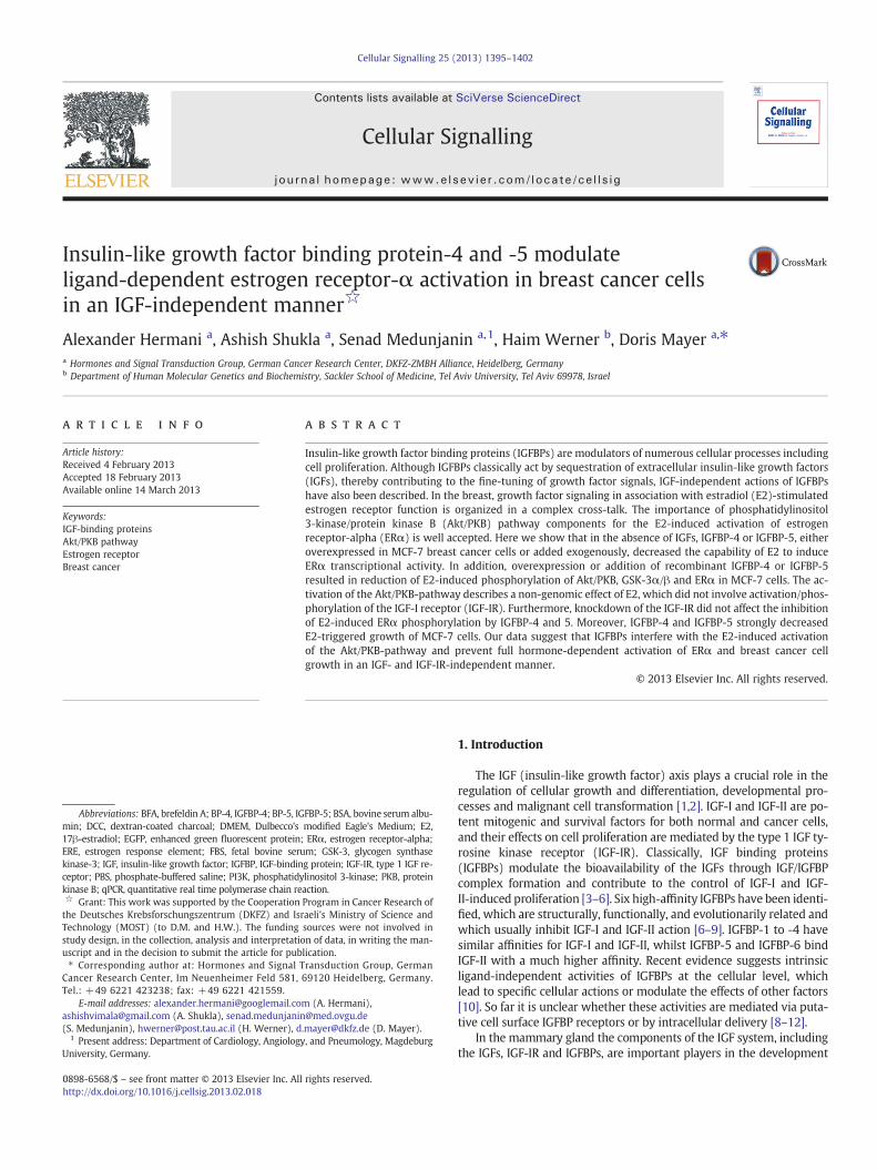
IGFBPs efficiently bind extracellular IGFs and prevent activation ofsignaling pathways such as the Akt/PKB pathway transduced by theIGF-IR [6]. Since Akt/PKB is not only activated by stimulation of thecells with IGF but is also phosphorylated in response to E2[27,28,33], we studied whether IGFBP-4 and IGFBP-5 expression af-fects the E2-induced phosphorylation of Akt/PKB and GSK-3α/β adownstream effector of Akt/PKB. In a first experiment Flag-taggedIGFBPs were overexpressed in MCF-7 cells. Then cells were kept over-night in 1% DCC-FBS and treated with 100 nM E2 for 20 min (Fig. 2)or with 10 nM E2 for 24 h (Suppl. Fig. 1). Immunoblot analysis of ly-sates from cells transfected with the empty vector and expressionconstructs showed that the expression of the IGFBPs did not affect ex-pression levels of Akt/PKB, GSK-3α/β and ERα and inhibited slightlythe basal phosphorylation of Akt/PKB at Ser-473 and GSK-3α/β atSer-21/9. Importantly, lysates from E2-treated cells revealed in-creased phosphorylation of Akt/PKB and of the downstream target ki-nase GSK-3α/β both at 20 min (Fig. 2A, B, C) and 24 h treatment(Suppl. Fig. 1). In cells transfected with IGFBP-4 or IGFBP-5 expres-sion constructs, E2-induced phosphorylation of Akt/PKB andGSK-3α/β was significantly reduced, indicating that both IGFBPsprevented E2-dependent activation of the Akt/PKB pathway.
3.4. E2-induced phosphorylation of ERα at Ser-118 is decreased byIGFBP-4 and IGFBP-5
Treatment of MCF-7 cells with E2 results in rapid phosphorylation ofERα at Ser-118 showing two bands which are caused by the well-known band-shift of ERα [29]. We previously reported that Akt/PKBand GSK-3 play a significant role in this phosphorylation [29,30]. SinceIGFBP-4 and IGFBP-5 decreased the E2-induced phosphorylation of Akt/PKB and GSK-3α/β, we addressed the question whether the two IGFBPs

Fig. 1. Overexpressed IGFBP-4 and IGFBP-5 are secreted by MCF-7 breast cancer cells and inhibit IGF-I induced phosphorylation of Akt/PKB. A. Cells were transiently transfectedwith IGFBP-4 (BP-4) or IGFBP-5 (BP-5) expression constructs containing a Flag-tag or with the empty vector (EVC) and after 24 h were incubated for additional 5 h inserum-free medium with or without 10 μg/ml brefeldin A (BFA). Overexpressed IGFBPs were detected using the respective IGFBP antibodies and an anti-Flag-tag antibody incell lysates and concentrated culture supernatants. Addition of BFA clearly decreased the amounts of IGFBPs in the culture supernatant. B. MCF-7 cells were transfected withIGFBP-4 or IGFBP-5 expression constructs or empty vector (EVC), kept in serum-free medium for 24 h and treated or not with 50 ng/ml IGF-I for 45 min. Immunoblots showthe phosphorylated form (pSer-473) of Akt/PKB, the total Akt/PKB protein and the Flag-tagged IGFBPs.
1398 A. Hermani et al. / Cellular Signalling 25 (2013) 1395–1402
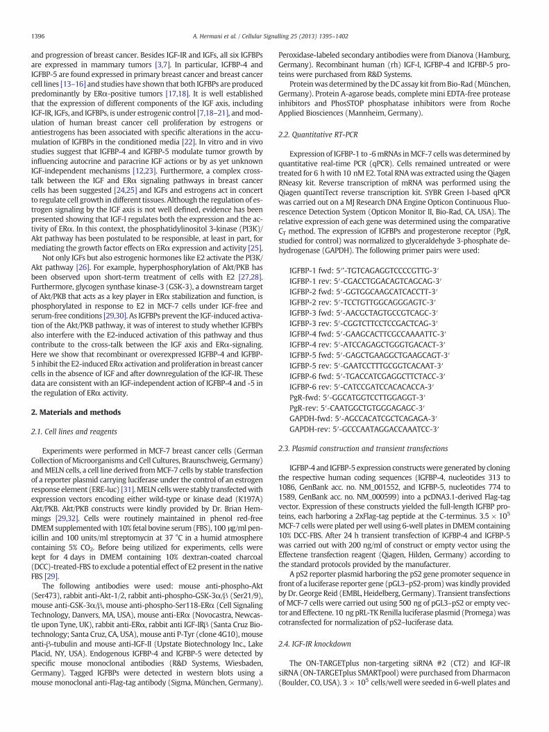
also influence phosphorylation of ERα at Ser-118. Short-term treatmentof cells with 100 nM E2 for 20 min resulted in phosphorylation of ERαin cells transfected with the empty vector. In IGFBP-4- and IGFBP-5-transfected cells we observed a significant reduction (35% and 49%, re-spectively) of the E2-induced phosphorylation of ERα at Ser-118 com-pared to empty vector-transfected cells, but no significant decrease ofbasal ERα phosphorylation (Fig. 2A, D). Similar results were obtainedwith recombinant human IGFBP-4 and IGFBP-5 added to the culture me-dium (Fig. 3). This suggests that IGFBP-4 and IGFBP-5 interfere with theE2-triggered phosphorylation of the ERα in the AF-1 domain.
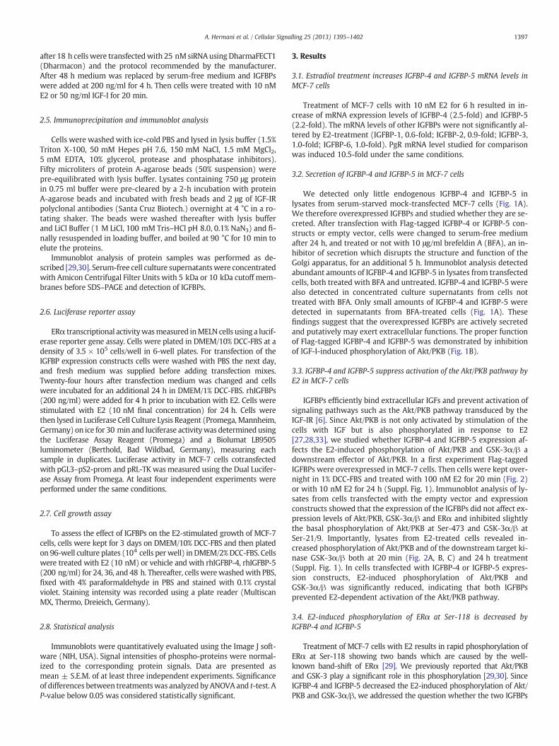
3.5. IGFBPs and kinase dead Akt/PKB prevent full ligand-induced ERαtranscriptional activity
Phosphorylation of Ser-118 is required for full transcriptional activityof ERα [34]. As shown in Figs. 2 and 3, overexpressed or recombinantIGFBP-4 or IGFBP-5 causes reduction of phosphorylation of ERα atSer-118.We therefore studied if IGFBPs also influence ERα transcription-al activity. For this purpose, MELN cells were transfectedwith IGFBP-4 orIGFBP-5 expression constructs and, after 24 h, cells were treated with10 nM E2 for additional 24 h and ERE-dependent luciferase activitywas determined. While E2 potently induced luciferase activity in cellstransfected with the empty vector, luciferase activity was significantlydiminished by about 25% in cells transfected with either IGFBP-4 orIGFBP-5 (Fig. 4A). Similarly, incubation of MELN cells with 200 ng/mlrhIGFBP-4 and rhIGFBP-5 for 4 h prior to E2 treatment caused a signifi-cant (~25%) decrease of the E2-induced luciferase activity (Fig. 4B). Toinvestigate the effect of IGFBP-4 and IGFBP-5 on E2-induced expressionof an ERα target gene we used another reporter gene construct harbor-ing the pS2 gene promoter, a well-known estrogen-inducible gene,fused to a firefly luciferase reporter gene. MCF-7 cells transiently
transfected with this reporter construct showed a 2.2-fold induction ofluciferase activity upon treatment with E2 for 24 h (Fig. 4C).Overexpression of IGFBP-4 or IGFBP-5 led to a significant decrease ofthe basal as well as E2-induced pS2-luciferase activity compared tocells transfectedwith empty vector and pS2 construct. This result furthersuggests an inhibitory effect of the IGFBPs on ERα target gene expression.
The data presented above suggest that the Akt/PKB-pathway is ofimportance for mediation of the inhibitory effects of IGFBP-4 andIGFBP-5 on E2-induced ERα activation. We therefore analyzed if block-ade of Akt/PKB affects E2-dependent luciferase activity. Stable expres-sion of a kinase-dead Akt/PKB mutant in MELN cells caused reductionof E2-induced luciferase activity by about 30%, whereas luciferase activ-ity was not altered in cells overexpressing wild type Akt/PKB (Fig. 4D).This suggests that activation of the Akt/PKB pathway indeed is requiredfor full ERα transcriptional activity in an ERE-dependent luciferase re-porter assay system.
3.6. IGF-IR phosphorylation is not involved in E2-induced activation ofthe Akt/PKB pathway
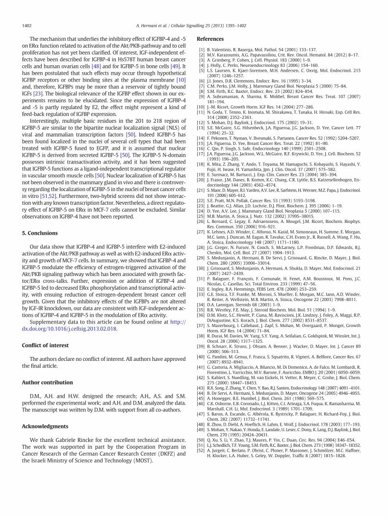
Since active Akt/PKB was of relevance for E2-induced ERα activationand phosphorylation/activation of Akt/PKB may be mediated by activat-ed IGF-IR, we studied whether E2-treatment of the cells results in tyro-sine phosphorylation of IGF-IRβ. IGF-IRβ immunoprecipitated fromlysates of untreated serum-starved cells showed only weak basal tyro-sine phosphorylation. E2-treatment did not result in increase ofIGF-IRβ phosphorylation during 60 min of treatment (Fig. 5A). In orderto study whether IGF-IR is required for IGFBP-mediated reduction ofE2-induced phosphorylation of ERα, IGF-IR was downregulated byRNAi. Fig. 5B shows that knockdown of IGF-IR by RNAi did not result inreduction of E2-induced ERα phosphorylation (Fig. 5B). Pretreatment
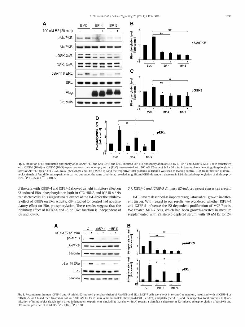
Fig. 2. Inhibition of E2-stimulated phosphorylation of Akt/PKB and GSK-3α/β and of E2-induced Ser-118 phosphorylation of ERα by IGFBP-4 and IGFBP-5. MCF-7 cells transfectedwith IGFBP-4 (BP-4) or IGFBP-5 (BP-5) expression constructs or empty vector (EVC) were treated with 100 nM E2 or vehicle for 20 min. A, Immunoblots detecting phosphorylatedforms of Akt/PKB (pSer-473), GSK-3α/β (pSer-21/9), and ERα (pSer-118) and the respective total proteins. β-Tubulin was used as loading control. B–D, Quantification of immu-noblot signals of four different experiments carried out under the same conditions, revealed a significant IGFBP-dependent decrease in E2-induced phosphorylation of all three pro-teins. ⁎P b 0.05 and ⁎⁎P b 0.005.
1399A. Hermani et al. / Cellular Signalling 25 (2013) 1395–1402
of the cellswith IGFBP-4 and IGFBP-5 showed a slight inhibitory effect onE2-induced ERα phosphorylation both in CT2 siRNA and IGF-IR siRNAtransfected cells. This suggests no relevance of the IGF-IR for the inhibito-ry effect of IGFBPs on ERα activity. IGF-I studied for control had no stim-ulatory effect on ERα phosphorylation. These results suggest that theinhibitory effect of IGFBP-4 and -5 on ERα function is independent ofIGF and IGF-IR.
Fig. 3. Recombinant human IGFBP-4 and -5 inhibit E2-induced phosphorylation of Akt/PKBrhIGFBP-5 for 4 h and then treated or not with 100 nM E2 for 20 min. A, Immunoblots shotification of immunoblot signals from three independent experiments (including that showERα in the presence of rhIGFBPs. ⁎P b 0.05, ⁎⁎P b 0.005.
3.7. IGFBP-4 and IGFBP-5 diminish E2-induced breast cancer cell growth
IGFBPsweredescribed as important regulators of cell growth indiffer-ent tissues. With regard to our results, we wondered whether IGFBP-4and IGFBP-5 influence the E2-dependent proliferation of MCF-7 cells.We treated MCF-7 cells, which had been growth-arrested in mediumsupplemented with 2% steroid-depleted serum, with 10 nM E2 for 24,
and ERα. MCF-7 cells were kept in serum-free medium, incubated with rhIGFBP-4 orw pAkt/PKB (Ser-473) and pERα (Ser-118) and the respective total proteins. B, Quan-n in A) reveals a significant decrease in E2-induced phosphorylation of Akt/PKB and
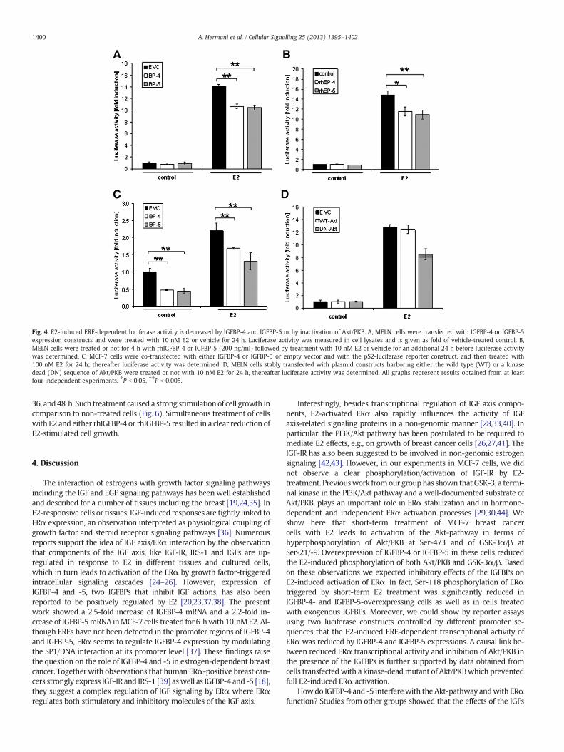
Fig. 4. E2-induced ERE-dependent luciferase activity is decreased by IGFBP-4 and IGFBP-5 or by inactivation of Akt/PKB. A, MELN cells were transfected with IGFBP-4 or IGFBP-5expression constructs and were treated with 10 nM E2 or vehicle for 24 h. Luciferase activity was measured in cell lysates and is given as fold of vehicle-treated control. B,MELN cells were treated or not for 4 h with rhIGFBP-4 or IGFBP-5 (200 ng/ml) followed by treatment with 10 nM E2 or vehicle for an additional 24 h before luciferase activitywas determined. C, MCF-7 cells were co-transfected with either IGFBP-4 or IGFBP-5 or empty vector and with the pS2-luciferase reporter construct, and then treated with100 nM E2 for 24 h; thereafter luciferase activity was determined. D, MELN cells stably transfected with plasmid constructs harboring either the wild type (WT) or a kinasedead (DN) sequence of Akt/PKB were treated or not with 10 nM E2 for 24 h, thereafter luciferase activity was determined. All graphs represent results obtained from at leastfour independent experiments. ⁎P b 0.05, ⁎⁎P b 0.005.
1400 A. Hermani et al. / Cellular Signalling 25 (2013) 1395–1402
36, and48 h. Such treatment caused a strong stimulation of cell growth incomparison to non-treated cells (Fig. 6). Simultaneous treatment of cellswith E2 and either rhIGFBP-4 or rhIGFBP-5 resulted in a clear reduction ofE2-stimulated cell growth.
4. Discussion
The interaction of estrogens with growth factor signaling pathwaysincluding the IGF and EGF signaling pathways has been well establishedand described for a number of tissues including the breast [19,24,35]. InE2-responsive cells or tissues, IGF-induced responses are tightly linked toERα expression, an observation interpreted as physiological coupling ofgrowth factor and steroid receptor signaling pathways [36]. Numerousreports support the idea of IGF axis/ERα interaction by the observationthat components of the IGF axis, like IGF-IR, IRS-1 and IGFs are up-regulated in response to E2 in different tissues and cultured cells,which in turn leads to activation of the ERα by growth factor-triggeredintracellular signaling cascades [24–26]. However, expression ofIGFBP-4 and -5, two IGFBPs that inhibit IGF actions, has also beenreported to be positively regulated by E2 [20,23,37,38]. The presentwork showed a 2.5-fold increase of IGFBP-4 mRNA and a 2.2-fold in-crease of IGFBP-5mRNA inMCF-7 cells treated for 6 hwith 10 nME2. Al-though EREs have not been detected in the promoter regions of IGFBP-4and IGFBP-5, ERα seems to regulate IGFBP-4 expression by modulatingthe SP1/DNA interaction at its promoter level [37]. These findings raisethe question on the role of IGFBP-4 and -5 in estrogen-dependent breastcancer. Togetherwith observations that human ERα-positive breast can-cers strongly express IGF-IR and IRS-1 [39] aswell as IGFBP-4 and -5 [18],they suggest a complex regulation of IGF signaling by ERα where ERαregulates both stimulatory and inhibitory molecules of the IGF axis.
Interestingly, besides transcriptional regulation of IGF axis compo-nents, E2-activated ERα also rapidly influences the activity of IGFaxis-related signaling proteins in a non-genomic manner [28,33,40]. Inparticular, the PI3K/Akt pathway has been postulated to be required tomediate E2 effects, e.g., on growth of breast cancer cells [26,27,41]. TheIGF-IR has also been suggested to be involved in non-genomic estrogensignaling [42,43]. However, in our experiments in MCF-7 cells, we didnot observe a clear phosphorylation/activation of IGF-IR by E2-treatment. Previouswork fromour grouphas shown that GSK-3, a termi-nal kinase in the PI3K/Akt pathway and a well-documented substrate ofAkt/PKB, plays an important role in ERα stabilization and in hormone-dependent and independent ERα activation processes [29,30,44]. Weshow here that short-term treatment of MCF-7 breast cancercells with E2 leads to activation of the Akt-pathway in terms ofhyperphosphorylation of Akt/PKB at Ser-473 and of GSK-3α/β atSer-21/-9. Overexpression of IGFBP-4 or IGFBP-5 in these cells reducedthe E2-induced phosphorylation of both Akt/PKB and GSK-3α/β. Basedon these observations we expected inhibitory effects of the IGFBPs onE2-induced activation of ERα. In fact, Ser-118 phosphorylation of ERαtriggered by short-term E2 treatment was significantly reduced inIGFBP-4- and IGFBP-5-overexpressing cells as well as in cells treatedwith exogenous IGFBPs. Moreover, we could show by reporter assaysusing two luciferase constructs controlled by different promoter se-quences that the E2-induced ERE-dependent transcriptional activity ofERαwas reduced by IGFBP-4 and IGFBP-5 expressions. A causal link be-tween reduced ERα transcriptional activity and inhibition of Akt/PKB inthe presence of the IGFBPs is further supported by data obtained fromcells transfectedwith a kinase-deadmutant of Akt/PKBwhich preventedfull E2-induced ERα activation.
Howdo IGFBP-4 and -5 interferewith the Akt-pathway andwith ERαfunction? Studies from other groups showed that the effects of the IGFs
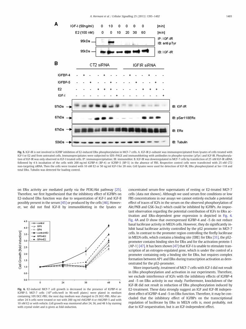
Fig. 5. IGF-IR is not involved in IGFBP inhibition of E2-induced ERα phosphorylation in MCF-7 cells. A. IGF-IR β-subunit was immunoprecipitated from lysates of cells treated withIGF-I or E2 and from untreated cells. Immunoprecipitates were subjected to SDS–PAGE and immunoblotting with antibodies to phospho-tyrosine (pTyr) and IGF-IR. Phosphoryla-tion of IGF-IR was only observed in IGF-I treated cells. IP: immunoprecipitation; IB: immunoblot. B. IGF-IR was downregulated in MCF-7 cells by transfection of 25 nM IGF-IR siRNAfollowed by 4 h incubation of the cells with 200 ng/ml IGFBP-4 (BP-4) or IGFBP-5 (BP-5) in the absence of FBS. Respective control cells were transfected with 25 nM CT2non-targeting siRNA. Then the cells were treated with 10 nM E2 or 50 ng/ml IGF-I for 20 min. Cell lysates were used for detection of IGF-IR, ERα phosphorylated at Ser-118 andtotal ERα. Tubulin was detected for loading control.
1401A. Hermani et al. / Cellular Signalling 25 (2013) 1395–1402
on ERα activity are mediated partly via the PI3K/Akt pathway [25].Therefore, we first hypothesized that the inhibitory effect of IGFBPs onE2-induced ERα function was due to sequestration of IGF-I and IGF-IIpossibly present in the serum [45] or produced by the cells [46]. Howev-er, we did not find IGF-II by immunoblotting in the lysates or
Fig. 6. E2-induced MCF-7 cell growth is decreased in the presence of IGFBP-4 orIGFBP-5. MCF-7 cells (104 cells/well in 96-well plates) were plated in mediumcontaining 10% DCC-FBS; the next day medium was changed to 2% DCC-FBS. After an-other 24 h cells were treated or not with 200 ng/ml rhIGFBP-4 or rhIGFBP-5 and with10 nM E2 or with vehicle. Cell growth was monitored after 24, 36, and 48 h by stainingwith crystal violet and is given as fold-induction.
concentrated serum-free supernatants of resting or E2-treated MCF-7cells (data not shown). Although we used serum free conditions or lowFBS concentrations in our assays we cannot entirely exclude a potentialeffect of traces of IGFs in the serum on the observed phosphorylation ofAkt/PKB and GSK-3α/β which could be inhibited by IGFBPs. An impor-tant observation regarding the potential contribution of IGFs to ERα ac-tivation and ERα-dependent gene expression is depicted in Fig. 4.Fig. 4A and D show that overexpressed IGFBP-4 and -5 do not reducebasal luciferase activity in MELN cells. However, they do significantly in-hibit basal luciferase activity controlled by the pS2 promoter in MCF-7cells. In contrast to the promoter region controlling the firefly luciferasein MELN cells, which contains a binding site (ERE) for ERα [31], the pS2-promoter contains binding sites for ERα and for the activation protein-1(AP-1) [47]. It has been shown [47] that IGF-I is unable to stimulate tran-scription of an estrogen-regulated gene, which is under the control of apromoter containing only a binding site for ERα, but requires complexformation between AP1 and ERα during transcription activation as dem-onstrated for the pS2-promoter.
More importantly, treatment of MCF-7 cells with IGF-I did not resultin ERα phosphorylation and activation in our experiments. Therefore,we exclude interference of IGFs with the inhibitory effects of IGFBP-4and -5 on ERα activity in our study. Furthermore, knockdown of theIGF-IR did not result in reduction of ERα phosphorylation induced byE2-treatment. These data strongly suggest an IGF and IGF-IR indepen-dent effect of IGFBP-4 and -5 on ERα function. Therefore, it may be con-cluded that the inhibitory effect of IGFBPs on the transcriptionalregulation of luciferase by ERα in MELN cells is, most probably, notdue to IGF-sequestration, but is an IGF-independent effect.
1402 A. Hermani et al. / Cellular Signalling 25 (2013) 1395–1402
Themechanism that underlies the inhibitory effect of IGFBP-4 and -5on ERα function related to activation of the Akt/PKB-pathway and to cellproliferation has not yet been clarified. Of interest, IGF-independent ef-fects have been described for IGFBP-4 in Hs578T human breast cancercells and human ovarian cells [48] and for IGFBP-5 in bone cells [49]. Ithas been postulated that such effects may occur through hypotheticalIGFBP receptors or other binding sites at the plasma membrane [10]and, therefore, IGFBPs may be more than a reservoir of tightly boundIGFs [23]. The biological relevance of the IGFBP effect shown in our ex-periments remains to be elucidated. Since the expression of IGFBP-4and -5 is partly regulated by E2, the effect might represent a kind offeed-back regulation of IGFBP expression.
Interestingly, multiple basic residues in the 201 to 218 region ofIGFBP-5 are similar to the bipartite nuclear localization signal (NLS) ofviral and mammalian transcription factors [50]. Indeed IGFBP-5 hasbeen found localized in the nuclei of several cell types that had beentreated with IGFBP-5 fused to EGFP, and it is assumed that nuclearIGFBP-5 is derived from secreted IGFBP-5 [50]. The IGFBP-5 N-domainpossesses intrinsic transactivation activity, and it has been suggestedthat IGFBP-5 functions as a ligand-independent transcriptional regulatorin vascular smooth muscle cells [50]. Nuclear localization of IGFBP-5 hasnot been observed in themammary gland in vivo and there is controver-sy regarding the localization of IGFBP-5 in the nuclei of breast cancer cellsin vitro [51,52]. Furthermore, two-hybrid screens did not show interac-tionwith any known transcription factor. Nevertheless, a direct regulato-ry effect of IGFBP-5 on ERα in MCF-7 cells cannot be excluded. Similarobservations on IGFBP-4 have not been reported.
5. Conclusions
Our data show that IGFBP-4 and IGFBP-5 interfere with E2-inducedactivation of the Akt/PKB pathway aswell as with E2-induced ERα activ-ity and growth ofMCF-7 cells. In summary, we showed that IGFBP-4 andIGFBP-5 modulate the efficiency of estrogen-triggered activation of theAkt/PKB signaling pathway which has been associated with growth fac-tor/ERα cross-talks. Further, expression or addition of IGFBP-4 andIGFBP-5 led to decreased ERα phosphorylation and transcriptional activ-ity, with ensuing reduction of estrogen-dependent breast cancer cellgrowth. Given that the inhibitory effects of the IGFBPs are not alteredby IGF-IR knockdown, our data are consistent with IGF-independent ac-tions of IGFBP-4 and IGFBP-5 in the modulation of ERα activity.
Supplementary data to this article can be found online at http://dx.doi.org/10.1016/j.cellsig.2013.02.018.
Conflict of interest
The authors declare no conflict of interest. All authors have approvedthe final article.
Author contribution
D.M., A.H. and H.W. designed the research; A.H., A.S. and S.M.performed the experimental work; and A.H. and D.M. analyzed the data.The manuscript was written by D.M. with support from all co-authors.
Acknowledgments
We thank Gabriele Rincke for the excellent technical assistance.The work was supported in part by the Cooperation Program inCancer Research of the German Cancer Research Center (DKFZ) andthe Israeli Ministry of Science and Technology (MOST).
References
[1] B. Valentinis, R. Baserga, Mol. Pathol. 54 (2001) 133–137.[2] M.V. Karazoumis, A.G. Papavassiliou, Crit. Rev. Oncol. Hematol. 84 (2012) 8–17.[3] A. Grimberg, P. Cohen, J. Cell. Physiol. 183 (2000) 1–9.[4] J. Holly, C. Perks, Neuroendocrinology 83 (2006) 154–160.[5] L.S. Laursen, K. Kjaer-Sorensen, M.H. Andersen, C. Oxvig, Mol. Endocrinol. 215
(2007) 1246–1257.[6] J.I. Jones, D.R. Clemmons, Endocr. Rev. 16 (1995) 3–34.[7] C.M. Perks, J.M. Holly, J. Mammary Gland Biol. Neoplasia 5 (2000) 75–84.[8] S.M. Firth, R.C. Baxter, Endocr. Rev. 23 (2002) 824–854.[9] A. Subramanian, A. Sharma, K. Mokbel, Breast Cancer Res. Treat. 107 (2007)
181–194.[10] J.-M. Ricort, Growth Horm. IGF Res. 14 (2004) 277–286.[11] N. Goda, T. Tenno, K. Inomata, M. Shirakawa, T. Tanaka, H. Hiroaki, Exp. Cell Res.
314 (2008) 2352–2361.[12] S. Mohan, D.J. Baylink, J. Endocrinol. 175 (2002) 19–31.[13] S.E. McGuire, S.G. Hilsenbeck, J.A. Figueroa, J.G. Jackson, D. Yee, Cancer Lett. 77
(1994) 25–32.[14] F. Pekonen, T. Nyman, V. Ilvesmaki, S. Partanen, Cancer Res. 52 (1992) 5204–5207.[15] J.A. Figueroa, D. Yee, Breast Cancer Res. Treat. 22 (1992) 81–90.[16] C. Qin, P. Singh, S. Safe, Endocrinology 140 (1999) 2501–2508.[17] J.A. Figueroa, J.G. Jackson, W.L. McGuire, R.F. Krywicki, D. Yee, J. Cell. Biochem. 52
(1993) 196–205.[18] K. Mita, Z. Zhang, Y. Ando, T. Toyama, M. Hamaguchi, S. Kobayashi, S. Hayashi, Y.
Fujii, H. Iwase, H. Yamashita, Jpn. J. Clin. Oncol. 37 (2007) 575–582.[19] E. Surmacz, M. Bartucci, J. Exp. Clin. Cancer Res. 23 (2004) 385–394.[20] J. Frasor, J.M. Danes, B. Komm, K.C. Chang, C.R. Lyttle, B.S. Katzenellenbogen, En-
docrinology 144 (2003) 4562–4574.[21] S.Maor, D.Mayer, R.I. Yarden, A.V. Lee, R. Sarfstein, H.Werner,M.Z. Papa, J. Endocrinol.
191 (2006) 605–612.[22] S.E. Pratt, M.N. Pollak, Cancer Res. 53 (1993) 5193–5198.[23] J. Beattie, G.J. Allan, J.D. Lochrie, D.J. Flint, Biochem. J. 395 (2006) 1–19.[24] D. Yee, A.V. Lee, J. Mammary Gland Biol. Neoplasia 5 (2000) 107–115.[25] M.B. Martin, A. Stoica, J. Nutr. 132 (2002) 3799S–3801S.[26] L. Bernard, C. Legay, E. Adriaenssens, A. Mougel, J.M. Ricort, Biochem. Biophys.
Res. Commun. 350 (2006) 916–921.[27] K. Lehnes, A.D. Winder, C. Alfonso, N. Kasid, M. Simoneaux, H. Summe, E. Morgan,
M.C. Iann, J. Duncan, M. Eagan, R. Tavaluc, C.H. Evans Jr., R. Russell, A. Wang, F. Hu,A. Stoica, Endocrinology 148 (2007) 1171–1180.
[28] J.G. Greger, N. Fursov, N. Cooch, S. McLarney, L.P. Freedman, D.P. Edwards, B.J.Cheskis, Mol. Cell. Biol. 27 (2007) 1904–1913.
[29] S. Medunjanin, A. Hermani, B. De Servi, J. Grisouard, G. Rincke, D. Mayer, J. Biol.Chem. 280 (2005) 33006–33014.
[30] J. Grisouard, S. Medunjanin, A. Hermani, A. Shukla, D. Mayer, Mol. Endocrinol. 21(2007) 2427–2439.
[31] P. Balaguer, F. François, F. Comunale, H. Fenet, A.M. Boussioux, M. Pons, J.C.Nicolas, C. Casellas, Sci. Total Environ. 233 (1999) 47–56.
[32] E. Ingley, B.A. Hemmings, FEBS Lett. 478 (2000) 253–259.[33] G.E. Stoica, T.F. Franke, M. Moroni, S. Mueller, E. Morgan, M.C. Iann, A.D. Winder,
R. Reiter, A. Wellstein, M.B. Martin, A. Stoica, Oncogene 22 (2003) 7998–8011.[34] D.A. Lannigan, Steroids 68 (2003) 1–9.[35] B.R. Westley, F.E. May, J. Steroid Biochem. Mol. Biol. 51 (1994) 1–9.[36] D.M. Klotz, S.C. Hewitt, P. Ciana, M. Raviscioni, J.K. Lindzey, J. Foley, A. Maggi, R.P.
DiAugustine, K.S. Korach, J. Biol. Chem. 277 (2002) 8531–8537.[37] S. Mazerbourg, I. Callebaut, J. Zapf, S. Mohan, M. Overgaard, P. Monget, Growth
Horm. IGF Res. 14 (2004) 71–84.[38] R. Durai, M. Davies, W. Yang, S.Y. Yang, A. Seifalian, G. Goldspink, M.Winslet, Int. J.
Oncol. 28 (2006) 1317–1325.[39] B. Schnarr, K. Strunz, J. Ohsam, A. Benner, J. Wacker, D. Mayer, Int. J. Cancer 89
(2000) 506–513.[40] G. Pandini, M. Genua, F. Frasca, S. Squatrito, R. Vigneri, A. Belfiore, Cancer Res. 67
(2007) 8932–8941.[41] G. Castoria, A. Migliaccio, A. Bilancio, M. Di Domenico, A. de Falco, M. Lombardi, R.
Fiorentino, L. Varricchio, M.V. Barone, F. Auricchio, EMBO J. 20 (2001) 6050–6059.[42] S. Kahlert, S. Nuedling, M. van Eickels, H. Vetter, R. Meyer, C. Grohe, J. Biol. Chem.
275 (2000) 18447–18453.[43] R.X. Song, Z. Zhang, Y. Chen, Y. Bao, R.J. Santen, Endocrinology 148 (2007) 4091–4101.[44] B. De Servi, A. Hermani, S. Medunjanin, D. Mayer, Oncogene 24 (2005) 4946–4955.[45] A. Honegger, R.E. Humbel, J. Biol. Chem. 261 (1986) 569–575.[46] C.K. Osborne, E.B. Coronado, L.J. Kitten, C.I. Arteaga, S.A. Fuqua, K. Ramasharma, M.
Marshall, C.H. Li, Mol. Endocrinol. 3 (1989) 1701–1709.[47] S. Baron, A. Escande, G. Albérola, K. Bystricky, P. Balaguer, H. Richard-Foy, J. Biol.
Chem. 282 (2007) 11732–11741.[48] R. Zhou, D. Diehl, A. Hoeflich, H. Lahm, E. Wolf, J. Endocrinol. 178 (2003) 177–193.[49] S.Mohan, Y. Nakao, Y. Honda, E. Landale, U. Leser, C. Dony, K. Lang,D.J. Baylink, J. Biol.
Chem. 270 (1995) 20424–20431.[50] Q. Xu, S. Li, Y. Zhao, T.J. Maures, P. Yin, C. Duan, Circ. Res. 94 (2004) E46–E54.[51] L.J. Schedlich, T.F. Young, S.M. Firth, R.C. Baxter, J. Biol. Chem. 273 (1998) 18347–18352.[52] A. Jurgeit, C. Berlato, P. Obrist, C. Ploner, P. Massoner, J. Schmölzer, M.C. Haffner,
H. Klocker, L.A. Huber, S. Geley, W. Doppler, Traffic 8 (2007) 1815–1828.