Increased constitutive aSMA and Smad2/3 expression in ... · RESEARCH Open Access Increased...
13
RESEARCH Open Access Increased constitutive αSMA and Smad2/3 expression in idiopathic pulmonary fibrosis myofibroblasts is K Ca 3.1-dependent Katy M Roach 1* , Heike Wulff 2 , Carol Feghali-Bostwick 3 , Yassine Amrani 1 and Peter Bradding 1 Abstract Background: Idiopathic pulmonary fibrosis is a common and invariably fatal disease with limited therapeutic options. Ca 2+ -activated K Ca 3.1 potassium channels play a key role in promoting TGFβ1 and bFGF-dependent profibrotic responses in human lung myofibroblasts (HLMFs). We hypothesised that K Ca 3.1 channel-dependent cell processes regulate HLMF αSMA expression via Smad2/3 signalling pathways. Methods: In this study we have compared the phenotype of HLMFs derived from non-fibrotic healthy control lungs (NFC) with cells derived from IPF lungs. HLMFs grown in vitro were examined for αSMA expression by immunofluorescence (IF), RT-PCR and flow cytommetry. Basal Smad2/3 signalling was examined by RT-PCR, western blot and immunofluorescence. Two specific and distinct K Ca 3.1 blockers (TRAM-34 200 nM and ICA-17043 [Senicapoc] 100 nM) were used to determine their effects on HLMF differentiation and the Smad2/3 signalling pathways. Results: IPF-derived HLMFs demonstrated increased constitutive expression of both α-smooth muscle actin (αSMA) and actin stress fibres, indicative of greater myofibroblast differentiation. This was associated with increased constitutive Smad2/3 mRNA and protein expression, and increased Smad2/3 nuclear localisation. The increased Smad2/3 nuclear localisation was inhibited by removing extracellular Ca 2+ or blocking K Ca 3.1 ion channels with selective K Ca 3.1 blockers (TRAM-34, ICA-17043). This was accompanied by de-differentiation of IPF-derived HLMFs towards a quiescent fibroblast phenotype as demonstrated by reduced αSMA expression and reduced actin stress fibre formation. Conclusions: Taken together, these data suggest that Ca 2+ - and K Ca 3.1-dependent processes facilitate “constitutive” Smad2/3 signalling in IPF-derived fibroblasts, and thus promote fibroblast to myofibroblast differentiation. Importantly, inhibiting K Ca 3.1 channels reverses this process. Targeting K Ca 3.1 may therefore provide a novel and effective approach for the treatment of IPF and there is the potential for the rapid translation of K Ca 3.1-directed therapy to the clinic. Keywords: Idiopathic pulmonary fibrosis (IPF), Fibrosis, Lung, Myofibroblast, K Ca 3.1, Ion channel, Differentiation, Smad 2, Smad 3 Introduction Idiopathic pulmonary fibrosis (IPF) has an unknown etiology [1] and is marked by progressive lung fibrosis leading to respiratory failure. The pathogenic mechanisms involved in its initiation and progression are poorly understood [2] and there are limited therapeutic options with poor efficacy [3,4]. Prognosis is bleak with a median survival of only 3 years, worse than many cancers [5]. IPF patients present with a mean age of between 60 to 65 years at diagnosis [4]. In the USA the overall incidence of IPF is 16 per 100,000 person-years [2] and the incidence is increasing by 11% annually in the UK [6]. The most favoured hypothesis regarding its development is that on-going multiple, microscopic, isolated episodes of alveoli epithelial injury lead to an abnormal wound healing response involving fibrotic repair mechanisms [7]. Fibroblasts are mesenchymal cells that serve a critical role in both normal and fibrotic repair processes, which when activated, become differentiated, highly secretory * Correspondence: [email protected] 1 Department of Infection, Immunity and Inflammation, Institute for Lung Health, University of Leicester, Leicester LE3 9QP, UK Full list of author information is available at the end of the article © 2014 Roach et al.; licensee BioMed Central Ltd. This is an Open Access article distributed under the terms of the Creative Commons Attribution License (http://creativecommons.org/licenses/by/4.0), which permits unrestricted use, distribution, and reproduction in any medium, provided the original work is properly credited. The Creative Commons Public Domain Dedication waiver (http://creativecommons.org/publicdomain/zero/1.0/) applies to the data made available in this article, unless otherwise stated. Roach et al. Respiratory Research 2014, 15:155 http://respiratory-research.com/content/15/1/155
Transcript of Increased constitutive aSMA and Smad2/3 expression in ... · RESEARCH Open Access Increased...
Roach et al. Respiratory Research 2014, 15:155http://respiratory-research.com/content/15/1/155
RESEARCH Open Access
Increased constitutive αSMA and Smad2/3expression in idiopathic pulmonary fibrosismyofibroblasts is KCa3.1-dependentKaty M Roach1*, Heike Wulff2, Carol Feghali-Bostwick3, Yassine Amrani1 and Peter Bradding1
Abstract
Background: Idiopathic pulmonary fibrosis is a common and invariably fatal disease with limited therapeutic options.Ca2+-activated KCa3.1 potassium channels play a key role in promoting TGFβ1 and bFGF-dependent profibrotic responsesin human lung myofibroblasts (HLMFs). We hypothesised that KCa3.1 channel-dependent cell processes regulate HLMFαSMA expression via Smad2/3 signalling pathways.
Methods: In this study we have compared the phenotype of HLMFs derived from non-fibrotic healthy control lungs (NFC)with cells derived from IPF lungs. HLMFs grown in vitro were examined for αSMA expression by immunofluorescence (IF),RT-PCR and flow cytommetry. Basal Smad2/3 signalling was examined by RT-PCR, western blot and immunofluorescence.Two specific and distinct KCa3.1 blockers (TRAM-34 200 nM and ICA-17043 [Senicapoc] 100 nM) were used to determinetheir effects on HLMF differentiation and the Smad2/3 signalling pathways.
Results: IPF-derived HLMFs demonstrated increased constitutive expression of both α-smooth muscle actin (αSMA)and actin stress fibres, indicative of greater myofibroblast differentiation. This was associated with increased constitutiveSmad2/3 mRNA and protein expression, and increased Smad2/3 nuclear localisation. The increased Smad2/3 nuclearlocalisation was inhibited by removing extracellular Ca2+ or blocking KCa3.1 ion channels with selective KCa3.1 blockers(TRAM-34, ICA-17043). This was accompanied by de-differentiation of IPF-derived HLMFs towards a quiescent fibroblastphenotype as demonstrated by reduced αSMA expression and reduced actin stress fibre formation.
Conclusions: Taken together, these data suggest that Ca2+- and KCa3.1-dependent processes facilitate “constitutive”Smad2/3 signalling in IPF-derived fibroblasts, and thus promote fibroblast to myofibroblast differentiation. Importantly,inhibiting KCa3.1 channels reverses this process. Targeting KCa3.1 may therefore provide a novel and effective approachfor the treatment of IPF and there is the potential for the rapid translation of KCa3.1-directed therapy to the clinic.
Keywords: Idiopathic pulmonary fibrosis (IPF), Fibrosis, Lung, Myofibroblast, KCa3.1, Ion channel, Differentiation,Smad 2, Smad 3
IntroductionIdiopathic pulmonary fibrosis (IPF) has an unknownetiology [1] and is marked by progressive lung fibrosisleading to respiratory failure. The pathogenic mechanismsinvolved in its initiation and progression are poorlyunderstood [2] and there are limited therapeutic optionswith poor efficacy [3,4]. Prognosis is bleak with a median
* Correspondence: [email protected] of Infection, Immunity and Inflammation, Institute for LungHealth, University of Leicester, Leicester LE3 9QP, UKFull list of author information is available at the end of the article
© 2014 Roach et al.; licensee BioMed Central LCommons Attribution License (http://creativecreproduction in any medium, provided the orDedication waiver (http://creativecommons.orunless otherwise stated.
survival of only 3 years, worse than many cancers [5]. IPFpatients present with a mean age of between 60 to 65 yearsat diagnosis [4]. In the USA the overall incidence of IPFis 16 per 100,000 person-years [2] and the incidenceis increasing by 11% annually in the UK [6]. Themost favoured hypothesis regarding its development isthat on-going multiple, microscopic, isolated episodesof alveoli epithelial injury lead to an abnormal woundhealing response involving fibrotic repair mechanisms [7].Fibroblasts are mesenchymal cells that serve a critical
role in both normal and fibrotic repair processes, whichwhen activated, become differentiated, highly secretory
td. This is an Open Access article distributed under the terms of the Creativeommons.org/licenses/by/4.0), which permits unrestricted use, distribution, andiginal work is properly credited. The Creative Commons Public Domaing/publicdomain/zero/1.0/) applies to the data made available in this article,
Roach et al. Respiratory Research 2014, 15:155 Page 2 of 13http://respiratory-research.com/content/15/1/155
and contractile smooth muscle-like cells termed myofibro-blasts [8]. Expression of alpha smooth muscle actin(αSMA) and αSMA-containing stress fibres is the hall-mark of these cells [9-12]. IPF evolves from dysfunc-tional interactions between the injured epithelium andfibroblasts which lead to pathologic lesions calledfibroblast foci, which are comprised of activated myo-fibroblasts [13]. In their activated state, myofibroblastsare the primary cell responsible for the synthesis, secre-tion and remodelling of the extracellular matrix in IPF[14]. The human lung myofibroblast (HLMF) is there-fore an attractive target for the treatment of IPF.αSMA is a key protein expressed by HLMFs as com-
pared to quiescent fibroblasts [15], and contributes to theformation of characteristic HLMF contractile stress fibres[8,16,17]. αSMA expression and stress fibre formation inmyofibroblasts is regulated in part by the TGFβ1/Smadsignalling pathway [18,19]. Smads are intracellular pro-teins which transduce TGFβ1-dependent signals. Follow-ing binding of TGFβ1 to the TGFβRII, Smad2/3 arephosphorylated and form hetero-oligomeric complexeswith Smad 4, leading to nuclear translocation and the regu-lation of gene transcription [20]. They therefore regulatemany biological effects in HLMFs that are under the controlof TGFβ1, including collagen secretion, proliferation, differ-entiation and contraction [18-21].Ion channels are attractive therapeutic targets for many
chronic diseases including fibrosis. Activated intermediateconductance Ca2+-activated K+ channels promote severalpro-fibrotic processes in HLMFs such as basic fibroblastgrowth factor (bFGF)-dependent proliferation, and TGFβ1-dependent wound healing, collagen secretion and contrac-tion [22]. KCa3.1 activity was also shown to contribute tothe upregulation of αSMA in response to TGFβ1 throughthe enhancement of Smad phosphorylation [23], andcontributed to diabetic [24] and surgically-induced kidneyfibrosis in rodents [25]. However, much of the researchpublished to-date has focussed on the activity of myofibro-blasts following TGFβ1 stimulation, and there are fewstudies investigating basal signalling differences betweennon fibrotic control (NFC) and IPF-derived myofibroblasts.Previously, IPF-derived HLMFs demonstrated significantlyhigher constitutive αSMA [26] and functional KCa3.1channel expression [22] compared to NFC-derived cells.However, whether these basal increases in αSMA inIPF-derived HLMFs are due to altered constitutive Smadpathway signalling or KCa3.1 activity is not known.We therefore hypothesized that Smad2/3 signalling is
increased at baseline in IPF-derived HLMFs comparedto NFC cells, and if true, that this would be susceptibleto KCa3.1 channel inhibition. We therefore investigatedconstitutive Smad2/3 nuclear localisation and αSMAexpression in NFC- and IPF-derived HLMFs, and therole of KCa3.1 ion channels in these processes.
Materials and methodsHuman lung myofibroblast isolation and cultureNon-fibrotic control (NFC) HLMFs were derived fromhealthy areas of lung from patients undergoing lungresection for carcinoma at Glenfield Hospital, Leicester,UK. No morphological evidence of disease was found inthe tissue samples used for HLMF isolation. IPF HLMFswere derived from patients undergoing lung biopsy fordiagnostic purposes at the University of PittsburghMedical Center, USA, and were shown to have UIP onhistological examination. Myofibroblasts were grown fromexplanted lung tissue from both sources under identicalconditions, using DMEM supplemented with 10% FBS,antibiotic/antimycotic agents and non-essential aminoacids [27,28]. The cells were cultured at 37°C in 5% CO2/95% air. Cells were studied at passages 4–5 for functionalstudies. HLMFs were characterised as previously described[22]. All NFC patients gave informed written consentand the study was approved by the National ResearchEthics Service (references 07/MRE08/42 and 10/H0402/12). Written informed consent was also obtained from allIPF subjects, in accordance with the responsible Universityof Pittsburgh Institutional Review Board.
Human myofibroblast characterisationHuman myofibroblasts were harvested from 80-90%confluent monolayers with 0.1% trypsin/0.1% EDTA.Cells were seeded into 8-well chamber slides, grown toconfluence, and to confirm a myofibroblast population thecells were immunostained using the following antibodies:FITC-conjugated mouse monoclonal anti-α-smooth muscleactin (αSMA) (F3777, 10 μg/ml, Sigma-Aldrich, Poole,Dorset, UK); mouse monoclonal anti-fibroblast surfaceprotein(FSP)(F4771, 4 μg/ml, Sigma-Aldrich); mousemonoclonal anti-fibroblast antigen THY-1 (CP28, 3 μg/ml,Calbiochem, San Diego, CA; rabbit polyclonal collagentype 1 (550346, 20 μg/ml, Millipore, Watford, UK), isotypecontrol rabbit IgG (20 μg/ml). To detect the presence ofcontaminating cells the following antibodies wereused; monoclonal mouse CD68 antibody (6.4 μg/ml,Dako) to detect the presence of macrophages, mast cellsand monocytes; mouse monoclonal CD3 (4.5 mg/mlDako) to detect T-cells, and mouse monoclonal CD34R-PE antibody (0.5 μg/ml, Catlag) was also used todetect progenitor cells. Secondary antibodies labelled withFITC or R-PE (F0313, Dako) were applied and the cellscounterstained with 4′,6-diamidino-2-phenylindole (DAPI,Sigma-Aldrich). All isotype controls were negative. Cellswere mounted with fluorescent mounting medium andcover-slipped. The results confirmed that the cells isolatedwere of myofibroblast-rich population of cells (99%), withno contaminating cells identified. Full details, results andaccompanying images have been previously published inRoach et al. [22].
Roach et al. Respiratory Research 2014, 15:155 Page 3 of 13http://respiratory-research.com/content/15/1/155
ImmunofluorescenceHLMFs were grown on 8-well chamber slides andserum-starved for 24 hours prior to the experiment.The cells were then treated for 24 hours with either0.1% DMSO, TRAM-34 (20 and 200 nM) or ICA-17043(10 and 100 nM). Cells were then immunostained asdescribed previously [22] using FITC-conjugated mousemonoclonal anti-α-SMA (F3777, 10 μg/ml, Sigma-Aldrich,Poole, Dorset, UK) and isotype control FITC-conjugatedmouse IgG2a (X0933, 10 μg/ml, Dako, Ely, UK). KCa3.1expression was examined by immunofluorescence usingrabbit polyclonal anti-KCa3.1 (AV35098, 5 μg/ml, Sigma)and appropriate isotype control. Secondary antibodieslabelled with FITC (F0313, Dako) were applied and thecells counterstained with 4′,6-diamidino-2-phenylindole(DAPI, Sigma-Aldrich). Cells were mounted with fluo-rescent mounting medium and cover-slipped. Originalimages were captured on an epifluorescent microscope(Olympus BX50, Olympus UK Ltd, Southend–on-sea);grey scale intensity was examined using Cell F imagingsoftware (Olympus UK Ltd). Matched exposures wereused for isotype controls.Actin stress fibres were calculated using a specialised
macro on image J designed by Dr Kees Straatman,University of Leicester. The macro is capable of providinga quantitative, unbiased score of the number of stressfibres per individual cell by determining the fluctuationsof grey scale intensity created by the αSMA stainingwithin the stress fibres.
Flow cytometryCells were grown on T25 flasks and serum-starved for24 hours prior to the experiment. The myofibroblastswere incubated for 24 hours in the presence of 0.1%DMSO control, TRAM-34 (200 nM) or ICA-17043(100 nM). Cells were detached using 0.1% trypsin/0.1% EDTA, washed then fixed and permeabilised in 4%paraformaldehyde plus 0.1% saponin (Sigma) respectivelyfor 20 minutes on ice. Myofibroblasts were labelledwith FITC-conjugated mouse monoclonal anti-α-smoothmuscle actin (sigma) or isotype control FITC-conjugatedmouse IgG2a. Secondary antibodies labelled with FITC(F0313, Dako) were applied. Analysis was performed usingsingle colour flow cytometry on a FACScan (BD, UK).
Smad nuclear localisationHLMFs were grown on 8-well chamber slides andserum-starved for 24 hours prior to the experiment. Thecells were then stimulated with TGFβ1 (10 ng/ml) inthe presence of either 0.1% DMSO control, TRAM-34(200 nM), ICA-17043 (100 nM) or Ca2+ free media. After1 hour cells were immunostained using rabbit monoclonalanti-Smad2/3 (0.174 μg/ml, Cell Signalling). Secondaryantibody labelled with FITC (F0313, Dako) was applied
and the cells counterstained with DAPI (Sigma-Aldrich).Cells were mounted with fluorescent mounting mediumand cover-slipped. Images were analysed as above. Theintensity of nuclear Smad2/3 staining was quantifiedby measuring the grey scale intensity of DAPI positivenuclei to whole cell staining.
Western blot for Smad proteinsCells were grown in T75 flasks, serum starved for24 hours, and then incubated with either 0.1% DMSOcontrol, TRAM-34 (200 nM), ICA-17043 (100 nM) orCa2+ free media for 1 hour. Cells were detached with0.1% Trypsin/EDTA and washed. Protein was isolatedusing the RIPA buffer lysis system (Santa Cruz, Germany)and total protein concentration was determined using theDC Bio-Rad protein Assay (Bio-Rad, UK). 30 μg of proteinwas resolved using 10% Mini-Protean TGX precast gels(Bio-Rad) and then transferred to an immunobilon-Ppolyvinylidene difluoride membrane, using Trans-blotTurbo transfer packs (Bio-Rad). Membranes were blockedwith 5% milk and incubated with rabbit monoclonal anti-phospho-Smad2/3 (0.231 μg/ml, Cell Signalling, USA),rabbit polyclonal anti-Smad2/3 (0.0087 μg/ml, CellSignalling), mouse monoclonal anti-TATA binding protein(TBP) (1 μg/ml, Abcam), or mouse monoclonal anti-β-actin antibody (0.2 μg/ml, SantaCruz). Protein bandswere identified by horseradish peroxidase-conjugatedsecondary antibody and enhanced chemiluminescencereagent (Amersham, UK). Immunolabelled proteins werevisualized using ImageQuant LAS 4000 (GE HealthcareLife Sciences, UK).
Nuclear fractionNFC and IPF-derived HLMFs were grown in T75 flasks,serum starved for 24 hours, then detached with 0.1%Trypsin/EDTA and washed. Nuclear and cytoplasmicextracts were isolated using the Nuclear Extract Kit(Abcam, ab113474). Protein concentrations of bothnuclear and cytoplasmic extract was then was isolatedusing the RIPA buffer lysis system determined usingthe DC Bio-Rad protein Assay. Western blot was thenperformed as described above.
qRT-PCRMyofibroblast RNA was isolated using the RNeasy Plus Kit(Qiagen, West Sussex, UK) according to the manufacturer’sinstructions. Primers were designed for Smad2 , forwardCGTCCATCTTGCCATTCACG and reverse CTCAAGCTCATCTAATCGTCCTG, product size 182 bp from NCB1Reference sequence NM_005901.5; and Smad3, forwardGCGTGCGGCTCTACTACATC and reverse GCACATTCGGGTCAACTGGTA product size 233 bp from referencesequence NM_005902.3 β-actin primers were analysedusing gene-specific Quantitect Primer Assay primers
Roach et al. Respiratory Research 2014, 15:155 Page 4 of 13http://respiratory-research.com/content/15/1/155
(Qiagen, Germany), HS_ACTB_1_SG. All expression datawere normalized to β-actin and corrected using the refer-ence dye ROX. Gene expression was quantified by real-timePCR using the Brilliant SYBR Green QRT-PCR 1-StepMaster Mix (Strategene, The Netherlands). PCR productswere run on a 1.5% agarose gel to confirm the productamplified was the correct size, and each of the productswere sequenced to confirm the specificity of the primers.
Statistical analysisExperiments from an individual donor were performedeither in duplicate or triplicate and a mean value wasderived for each condition. Cells from 8 IPF and 8 NFCdonors were used, and the number of donors used for eachexperimental condition is stated in the text and/or figurelegends. Data distribution across donors was tested for nor-mality using the Kolmogorov-Smirnov test. For parametricdata the 1-way ANOVA or repeated measures ANOVA foracross-group comparisons was used followed by the appro-priate multiple comparison post hoc test; otherwise anunpaired or paired t-test was used. For non-parametric datathe Friedman test was used for across group comparisonsfollowed by the appropriate multiple comparison post hoctest, or the Mann Whitney U test was used where therewere two unpaired groups. GraphPad Prism for windows(version 6, GraphPad Software, San Diego California USA)was used for these analyses. A value of P < 0.05 was takento assume statistical significance and data are representedas mean (± SEM) or median (IQR).
ResultsIPF myofibroblasts have increased basal αSMA expressionand stress fibre formationWe and others have shown previously that fibroblastsgrown from lung parenchyma express relatively high levelsof αSMA and are contractile [22,29], in keeping with amyofibroblast phenotype. Here both NFC and IPF-derivedHLMFs expressed αSMA protein, but this was signifi-cantly increased in IPF derived cells when assessed byimmunofluorescent staining (P = 0.0111, Figure 1A and B),western blot analysis (P = 0.0026, Figure 1C and D) andflow cytometry, P = 0.0159 (Figures 1E and F).Both NFC and IPF-derived cells displayed cytosolic
αSMA staining, but IPF-derived HLMFs displayedobvious αSMA-positive stress fibres. Immunofluorescentpictures were examined using an Image J macro whichmeasured the numbers of stress fibres per cell, this isshown in Figure 1G. We found significantly more αSMApositive stress fibres in IPF-derived cells in comparison toNFC donors, P = 0.0437 (Figure 1H).
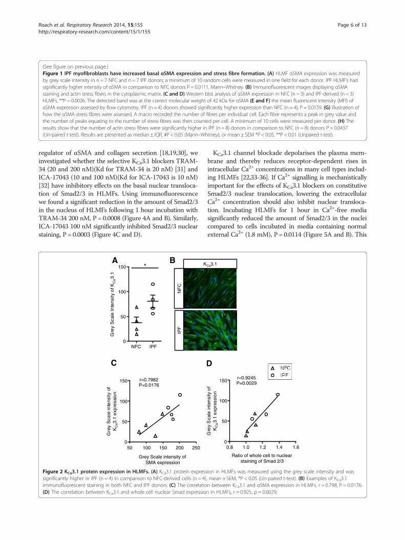
IPF myofibroblasts have increased basal KCa3.1 expressionFunctional KCa3.1 channels are increased in IPF-derivedHLMFs [22]. We therefore examined the expression of
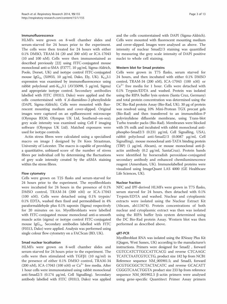
KCa3.1 by immunofluorescence and its relationship toαSMA expression. We found that IPF-derived HLMFshad a higher intensity of immunostaining than NFCcells, (Figure 2A and B), in keeping with previous patchclamp electrophysiology data [22]. Furthermore, KCa3.1expression correlated significantly with both basal αSMAexpression, r = 0.79, P = 0.0176, and basal Smad2/3 local-isation, r = 0.9245, P = 0.0029 in both NFC and IPF donors(Figure 2C and D).
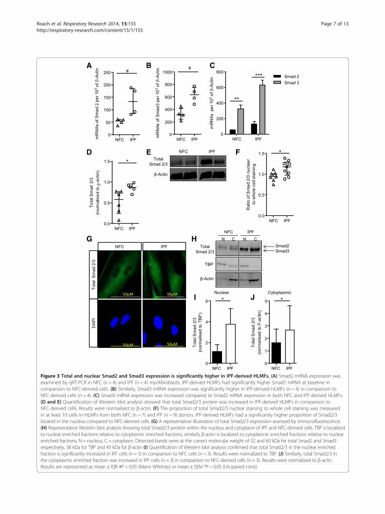
Smad2/3 expression is greater in IPF-derived HLMFsTo elucidate the molecular mechanisms underlying theobserved phenoytypic differences between NFC- andIPF-derived HLMFs we investigated the basal Smad2/3content as Smad2/3 signalling is key for myofibroblastdifferentiation and αSMA gene transcription RT-PCRresults confirmed that both Smad2 and Smad3 mRNAwere significantly upregulated in IPF-derived HLMFscompared to NFC-derived cells, P = 0.0286 and P = 0.0286respectively, Mann Whitney (Figure 3A and B). Smad3mRNA was more highly expressed than Smad2 mRNA(Figure 3C).Total Smad2/3 protein expression was significantly
increased in IPF-derived HLMF donors compared toNFC-derived cells, P = 0.0294, unpaired t test (Figure 3Dand E), although phosphorylated Smad2/3 was undetectablein both IPF and NFC-derived cells. Immunofluorescentstaining revealed that constitutive Smad2/3 nuclear stainingwas significantly greater in IPF-derived HLMFs incomparison to NFC-derived cells, P = 0.0202, unpairedt test (Figure 3F and G). To confirm the observed increasednuclear staining of IPF-derived cells, Western blot wasperformed examining total Smad2/3 protein expressionin the nuclear fraction and cytoplasmic extract of IPF andNFC-derived cells. Cytoplasmic and nuclear fractions wereprobed for the nuclear protein, TATA box binding protein(TBP) and β-actin to verify separation. As expected TBPwas observed primarily in the nuclear protein enrichedfractions, and served as a nuclear loading control foranalysis. β-actin was observed primarily in the cytoplasmicprotein enriched fractions and was used as the cytoplasmicloading control for subsequent analysis. The resultsdemonstrate that IPF-derived HLMFs have significantlygreater amounts of total Smad2/3 within the nucleus andcytoplasm in comparison to NFC HLMFs, P = 0.0488 andP = 0.0454 respectively (Figure 3H,I and J). This suggeststhat there may be increased constitutive Smad2/3 signallingin IPF-derived HLMFs.
Constitutive Smad 2/3 expression is inhibited by KCa3.1blockers and is Ca2+ dependentKCa3.1 channel activity is important for profibrotic HLMFprocesses such as proliferation, contraction and collagensecretion [22]. Because the Smad2/3 pathway is a major
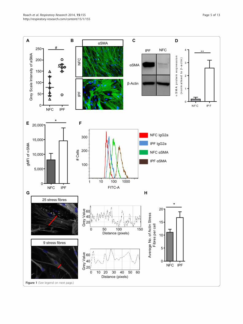
Figure 1 (See legend on next page.)
Roach et al. Respiratory Research 2014, 15:155 Page 5 of 13http://respiratory-research.com/content/15/1/155
(See figure on previous page.)Figure 1 IPF myofibroblasts have increased basal αSMA expression and stress fibre formation. (A) HLMF αSMA expression was measuredby grey scale intensity in n = 7 NFC and n = 7 IPF donors; a minimum of 10 random cells were measured in one field for each donor. IPF HLMF’s hadsignificantly higher intensity of αSMA in comparison to NFC donors P = 0.0111, Mann–Whitney. (B) Immunofluorescent images displaying αSMAstaining and actin stress fibers in the cytoplasmic matrix. (C and D) Western blot analysis of αSMA expression in NFC (n = 3) and IPF-derived (n = 3)HLMFs, **P = 0.0026. The detected band was at the correct molecular weight of 42 kDa for αSMA (E and F) the mean fluorescent intensity (MFI) ofαSMA expression assessed by flow cytometry. IPF (n = 4) donors showed significantly higher expression than NFC (n = 4), P = 0.0159. (G) Illustration ofhow the αSMA stress fibres were assessed. A macro recorded the number of fibres per individual cell. Each fibre represents a peak in grey value andthe number of peaks equating to the number of stress fibres was then counted per cell. A minimum of 10 cells were measured per donor. (H) Theresults show that the number of actin stress fibres were significantly higher in IPF (n = 8) donors in comparison to NFC (n = 8) donors P = 0.0437(Un-paired t-test). Results are presented as median ± IQR, #P < 0.05 (Mann–Whitney), or mean ± SEM *P < 0.05, **P < 0.01 (Unpaired t-test).
Roach et al. Respiratory Research 2014, 15:155 Page 6 of 13http://respiratory-research.com/content/15/1/155
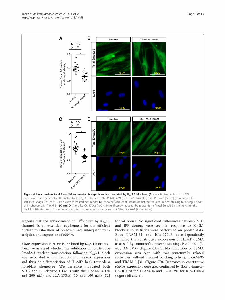
regulator of αSMA and collagen secretion [18,19,30], weinvestigated whether the selective KCa3.1 blockers TRAM-34 (20 and 200 nM)(Kd for TRAM-34 is 20 nM) [31] andICA-17043 (10 and 100 nM)(Kd for ICA-17043 is 10 nM)[32] have inhibitory effects on the basal nuclear transloca-tion of Smad2/3 in HLMFs. Using immunofluorescencewe found a significant reduction in the amount of Smad2/3in the nucleus of HLMFs following 1 hour incubation withTRAM-34 200 nM, P = 0.0008 (Figure 4A and B). Similarly,ICA-17043 100 nM significantly inhibited Smad2/3 nuclearstaining, P = 0.0003 (Figure 4C and D).
NF
C
IPF
BA K
C
NFC IPF0
50
100
150
Gre
yS
cale
Inte
nsity
ofK
Ca3
.1
*
50 100 150 200 2500
50
100
150
Grey Scale intensity ofSMA expression
Gre
yS
cale
inte
nsity
ofK
Ca3
.1e
xpre
ssio
n
r=0.7982P=0.0176
Figure 2 KCa3.1 protein expression in HLMFs. (A) KCa3.1 protein expresssignificantly higher in IPF (n = 4) in comparison to NFC-derived cells (n = 4)immunofluorescent staining in both NFC and IPF donors. (C) The correlatio(D) The correlation between KCa3.1 and whole cell nuclear Smad expressio
KCa3.1 channel blockade depolarises the plasma mem-brane and thereby reduces receptor-dependent rises inintracellular Ca2+ concentrations in many cell types includ-ing HLMFs [22,33-36]. If Ca2+ signalling is mechanisticallyimportant for the effects of KCa3.1 blockers on constitutiveSmad2/3 nuclear translocation, lowering the extracellularCa2+ concentration should also inhibit nuclear transloca-tion. Incubating HLMFs for 1 hour in Ca2+-free mediasignificantly reduced the amount of Smad2/3 in the nucleicompared to cells incubated in media containing normalexternal Ca2+ (1.8 mM), P = 0.0114 (Figure 5A and B). This
Ca3.1
D
0.8 1.0 1.2 1.4 1.60
50
100
150
Ratio of whole cell to nuclearstaining of Smad 2/3
Gre
yS
cale
i nte
ns i
tyo
fK
Ca3.
1e
xpre
ssio
n
r=0.9245P=0.0029
ion in HLMFs was measured using the grey scale intensity and was, mean ± SEM, *P < 0.05 (Un-paired t-test). (B) Examples of KCa3.1n between KCa3.1 and αSMA expression in HLMFs, r = 0.798, P = 0.0176.n in HLMFs, r = 0.925, p = 0.0029.
A
D
G H
I J
E F
B C
Figure 3 Total and nuclear Smad2 and Smad3 expression is significantly higher in IPF-derived HLMFs. (A) Smad2 mRNA expression wasexamined by qRT-PCR in NFC (n = 4) and IPF (n = 4) myofibroblasts. IPF-derived HLMFs had significantly higher Smad2 mRNA at baseline incomparison to NFC-derived cells. (B) Similarly, Smad3 mRNA expression was significantly higher in IPF-derived HLMFs (n = 4) in comparison toNFC-derived cells (n = 4). (C) Smad3 mRNA expression was increased compared to Smad2 mRNA expression in both NFC and IPF-derived HLMFs.(D and E) Quantification of Western blot analysis showed that total Smad2/3 protein was increased in IPF-derived HLMFs in comparison toNFC-derived cells. Results were normalised to β-actin. (F) The proportion of total Smad2/3 nuclear staining to whole cell staining was measuredin at least 10 cells in HLMFs from both NFC (n = 7) and IPF (n = 9) donors. IPF-derived HLMFs had a significantly higher proportion of Smad2/3located in the nucleus compared to NFC-derived cells. (G) A representative illustration of total Smad2/3 expression assessed by immunofluorescence.(H) Representative Western blot analysis showing total Smad2/3 protein within the nucleus and cytoplasm of IPF and NFC-derived cells. TBP is localizedto nuclear enriched fractions relative to cytoplasmic enriched fractions, similarly β-actin is localized to cytoplasmic enriched fractions relative to nuclearenriched fractions, N = nucleus, C = cytoplasm. Detected bands were at the correct molecular weight of 52 and 60 kDa for total Smad2 and Smad3respectively, 38 kDa for TBP and 43 kDa for β-actin (I) Quantification of Western blot analysis confirmed that total Smad2/3 in the nuclear enrichedfraction is significantly increased in IPF cells (n = 3) in comparison to NFC cells (n = 3). Results were normalized to TBP. (J) Similarly, total Smad2/3 inthe cytoplasmic enriched fraction was increased in IPF cells (n = 3) in comparison to NFC-derived cells (n = 3). Results were normalized to β-actin.Results are represented as mean ± IQR #P < 0.05 (Mann Whitney) or mean ± SEM *P = 0.05 (Un-paired t-test).
Roach et al. Respiratory Research 2014, 15:155 Page 7 of 13http://respiratory-research.com/content/15/1/155
Baseline TRAM-34 200nM
Tota
l Sm
ad2/
3D
AP
I
50µM
50µM
50µM
50µM
Baseline ICA-17043 100nM
Tota
l Sm
ad2/
3D
AP
I
50µM
50µM
50µM
50µM
A B
C D
0.0
0.5
1.0
1.5
Ra
tioof
Sm
ad2/
3n
ucle
ar
tow
hole
cell
stai
nin
g
*
0.0
0.5
1.0
1.5
Ra
tioof
Sm
ad2
/3n
ucle
arto
wh
ole
cell
sta
inin
g
*
Figure 4 Basal nuclear total Smad2/3 expression is significantly attenuated by KCa3.1 blockers. (A) Constitutive nuclear Smad2/3expression was significantly attenuated by the KCa3.1 blocker TRAM-34 (200 nM) (NFC n = 5 [triangles] and IPF n = 6 [circles] (data pooled forstatistical analysis, at least 10 cells were measured per donor). (B) Immunofluorescent images depict the reduced nuclear staining following 1 hourof incubation with TRAM-34. (C and D) Similarly, ICA-17043 (100 nM) significantly reduced the proportion of total Smad2/3 staining within thenuclei of HLMFs after a 1 hour incubation. Results are represented as mean ± SEM, *P < 0.05 (Paired t-test).
Roach et al. Respiratory Research 2014, 15:155 Page 8 of 13http://respiratory-research.com/content/15/1/155
suggests that the enhancement of Ca2+-influx by KCa3.1channels is an essential requirement for the efficientnuclear translocation of Smad2/3 and subsequent tran-scription and expression of αSMA.
αSMA expression in HLMF is inhibited by KCa3.1 blockersNext we assessed whether the inhibition of constitutiveSmad2/3 nuclear translocation following KCa3.1 blockwas associated with a reduction in αSMA expressionand thus de-differentiation of HLMFs back towards afibroblast phenotype. We therefore incubated bothNFC- and IPF-derived HLMFs with the TRAM-34 (20and 200 nM) and ICA-17043 (10 and 100 nM) [32]
for 24 hours. No significant differences between NFCand IPF donors were seen in response to KCa3.1blockers so statistics were performed on pooled data.Both TRAM-34 and ICA-17043 dose-dependentlyinhibited the constitutive expression of HLMF αSMAassessed by immunofluorescent staining, P < 0.0001 (2-way ANOVA) (Figure 6A-C). No inhibition of αSMAexpression was seen with two structurally relatedmolecules without channel blocking activity, TRAM-85and TRAM-7 [31] (Figure 6D). Decreases in constitutiveαSMA expression were also confirmed by flow cytometry(P = 0.0078 for TRAM-34 and P = 0.0391 for ICA-17043)(Figure 6E and F).
Ca2+ Free MediaControl Media
Tot
al S
mad
2/3
DA
PI
50µM
50µM 50µM
50µM
A
B
0.0
0.5
1.0
1.5
Rat
ioo
fSm
ad
2/3
n uc l
ear
s ta i
nin
gt o
wh
ole
cell
*
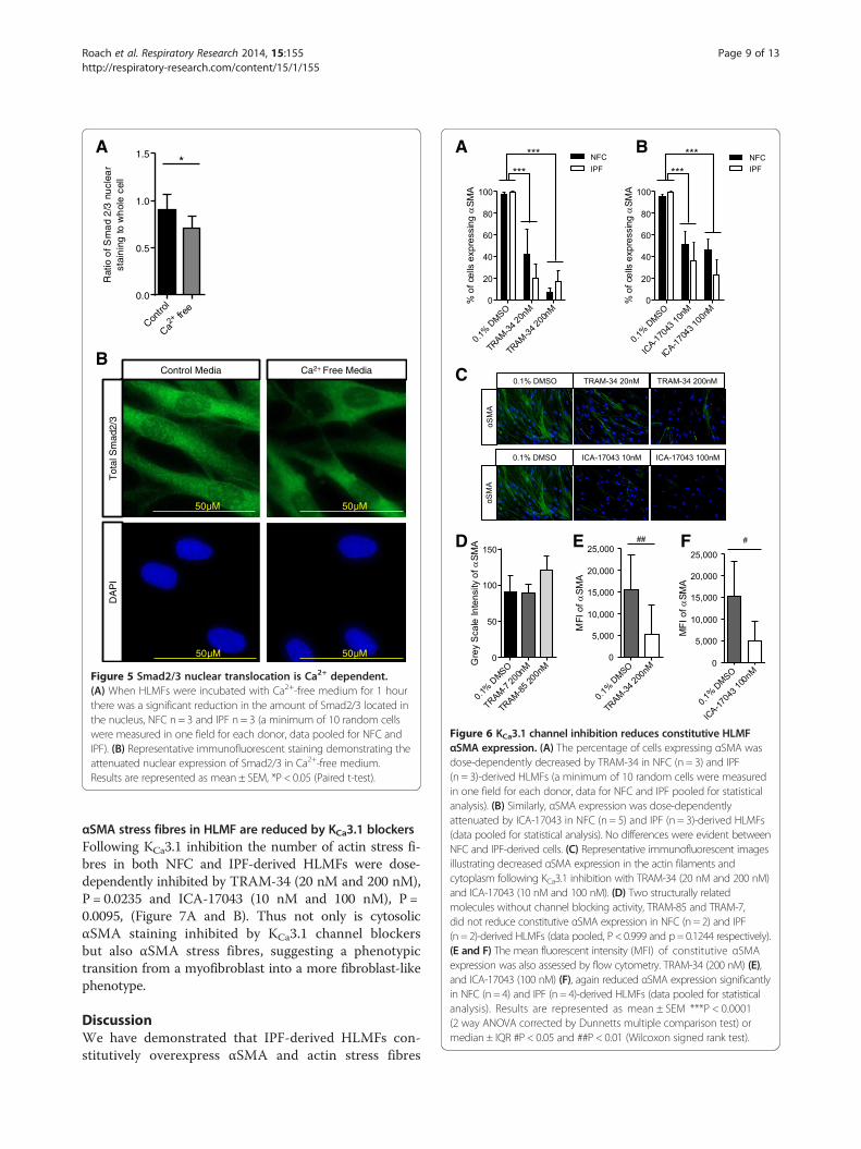
Figure 5 Smad2/3 nuclear translocation is Ca2+ dependent.(A) When HLMFs were incubated with Ca2+-free medium for 1 hourthere was a significant reduction in the amount of Smad2/3 located inthe nucleus, NFC n = 3 and IPF n = 3 (a minimum of 10 random cellswere measured in one field for each donor, data pooled for NFC andIPF). (B) Representative immunofluorescent staining demonstrating theattenuated nuclear expression of Smad2/3 in Ca2+-free medium.Results are represented as mean ± SEM, *P < 0.05 (Paired t-test).
A
C
D E F
B
Figure 6 KCa3.1 channel inhibition reduces constitutive HLMFαSMA expression. (A) The percentage of cells expressing αSMA wasdose-dependently decreased by TRAM-34 in NFC (n = 3) and IPF(n = 3)-derived HLMFs (a minimum of 10 random cells were measuredin one field for each donor, data for NFC and IPF pooled for statisticalanalysis). (B) Similarly, αSMA expression was dose-dependentlyattenuated by ICA-17043 in NFC (n = 5) and IPF (n = 3)-derived HLMFs(data pooled for statistical analysis). No differences were evident betweenNFC and IPF-derived cells. (C) Representative immunofluorescent imagesillustrating decreased αSMA expression in the actin filaments andcytoplasm following KCa3.1 inhibition with TRAM-34 (20 nM and 200 nM)and ICA-17043 (10 nM and 100 nM). (D) Two structurally relatedmolecules without channel blocking activity, TRAM-85 and TRAM-7,did not reduce constitutive αSMA expression in NFC (n = 2) and IPF(n = 2)-derived HLMFs (data pooled, P < 0.999 and p= 0.1244 respectively).(E and F) The mean fluorescent intensity (MFI) of constitutive αSMAexpression was also assessed by flow cytometry. TRAM-34 (200 nM) (E),and ICA-17043 (100 nM) (F), again reduced αSMA expression significantlyin NFC (n = 4) and IPF (n = 4)-derived HLMFs (data pooled for statisticalanalysis). Results are represented as mean ± SEM ***P < 0.0001(2 way ANOVA corrected by Dunnetts multiple comparison test) ormedian ± IQR #P < 0.05 and ##P < 0.01 (Wilcoxon signed rank test).
Roach et al. Respiratory Research 2014, 15:155 Page 9 of 13http://respiratory-research.com/content/15/1/155
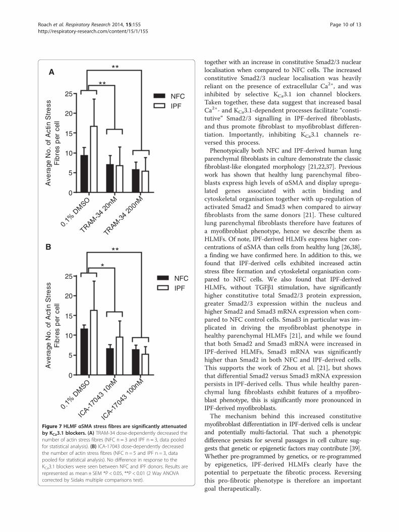
αSMA stress fibres in HLMF are reduced by KCa3.1 blockersFollowing KCa3.1 inhibition the number of actin stress fi-bres in both NFC and IPF-derived HLMFs were dose-dependently inhibited by TRAM-34 (20 nM and 200 nM),P = 0.0235 and ICA-17043 (10 nM and 100 nM), P =0.0095, (Figure 7A and B). Thus not only is cytosolicαSMA staining inhibited by KCa3.1 channel blockersbut also αSMA stress fibres, suggesting a phenotypictransition from a myofibroblast into a more fibroblast-likephenotype.
DiscussionWe have demonstrated that IPF-derived HLMFs con-stitutively overexpress αSMA and actin stress fibres
A
B
0
5
10
15
20
25
Ave
rage
No
.of
Act
inS
t re
ssF
ibre
sp
erce
ll
**
**
0
5
10
15
20
25
Ave
rage
No
.of
Act
inS
tre
ssF
ibre
sp
erce
ll
NFCIPF
*
**
NFCIPF
Figure 7 HLMF αSMA stress fibres are significantly attenuatedby KCa3.1 blockers. (A) TRAM-34 dose-dependently decreased thenumber of actin stress fibres (NFC n = 3 and IPF n = 3, data pooledfor statistical analysis). (B) ICA-17043 dose-dependently decreasedthe number of actin stress fibres (NFC n = 5 and IPF n = 3, datapooled for statistical analysis). No difference in response to theKCa3.1 blockers were seen between NFC and IPF donors. Results arerepresented as mean ± SEM *P < 0.05, **P < 0.01 (2 Way ANOVAcorrected by Sidaks multiple comparisons test).
Roach et al. Respiratory Research 2014, 15:155 Page 10 of 13http://respiratory-research.com/content/15/1/155
together with an increase in constitutive Smad2/3 nuclearlocalisation when compared to NFC cells. The increasedconstitutive Smad2/3 nuclear localisation was heavilyreliant on the presence of extracellular Ca2+, and wasinhibited by selective KCa3.1 ion channel blockers.Taken together, these data suggest that increased basalCa2+- and KCa3.1-dependent processes facilitate “consti-tutive” Smad2/3 signalling in IPF-derived fibroblasts,and thus promote fibroblast to myofibroblast differen-tiation. Importantly, inhibiting KCa3.1 channels re-versed this process.Phenotypically both NFC and IPF-derived human lung
parenchymal fibroblasts in culture demonstrate the classicfibroblast-like elongated morphology [21,22,37]. Previouswork has shown that healthy lung parenchymal fibro-blasts express high levels of αSMA and display upregu-lated genes associated with actin binding andcytoskeletal organisation together with up-regulation ofactivated Smad2 and Smad3 when compared to airwayfibroblasts from the same donors [21]. These culturedlung parenchymal fibroblasts therefore have features ofa myofibroblast phenotype, hence we describe them asHLMFs. Of note, IPF-derived HLMFs express higher con-centrations of αSMA than cells from healthy lung [26,38],a finding we have confirmed here. In addition to this, wefound that IPF-derived cells exhibited increased actinstress fibre formation and cytoskeletal organisation com-pared to NFC cells. We also found that IPF-derivedHLMFs, without TGFβ1 stimulation, have significantlyhigher constitutive total Smad2/3 protein expression,greater Smad2/3 expression within the nucleus andhigher Smad2 and Smad3 mRNA expression when com-pared to NFC control cells. Smad3 in particular was im-plicated in driving the myofibroblast phenotype inhealthy parenchymal HLMFs [21], and while we foundthat both Smad2 and Smad3 mRNA were increased inIPF-derived HLMFs, Smad3 mRNA was significantlyhigher than Smad2 in both NFC and IPF-derived cells.This supports the work of Zhou et al. [21], but showsthat differential Smad2 versus Smad3 mRNA expressionpersists in IPF-derived cells. Thus while healthy paren-chymal lung fibroblasts exhibit features of a myofibro-blast phenotype, this is significantly more pronounced inIPF-derived myofibroblasts.The mechanism behind this increased constitutive
myofibroblast differentiation in IPF-derived cells is unclearand potentially multi-factorial. That such a phenotypicdifference persists for several passages in cell culture sug-gests that genetic or epigenetic factors may contribute [39].Whether pre-programmed by genetics, or re-programmedby epigenetics, IPF-derived HLMFs clearly have thepotential to perpetuate the fibrotic process. Reversingthis pro-fibrotic phenotype is therefore an importantgoal therapeutically.
Roach et al. Respiratory Research 2014, 15:155 Page 11 of 13http://respiratory-research.com/content/15/1/155
Irrespective of the point from where pathologicalHLMF differentiation occurs in IPF, we have shown thatKCa3.1 channels play a key role in this process. By patchclamp electrophysiology we have shown previously thatfunctional KCa3.1 ion channels are increased in IPF-derivedHLMFs [22], and we found increased KCa3.1 ion channelprotein in this study. Importantly, we have shown here thatblocking KCa3.1 channels has the unique ability to de-differentiate HLMFs towards a fibroblast phenotype as indi-cated by a marked reduction in αSMA protein and stressfibre formation. This appears to operate via Ca2+-dependentprocesses as the reduction in constitutive nuclear Smad2/3expression induced by KCa3.1 blockers was mimicked by re-moving extracellular Ca2+, and KCa3.1 channel activity isknown to influence intracellular Ca2+ concentrations inmany cell types [34,35,40-42]. For example, we demon-strated previously that KCa3.1 activity is required for a risein intracellular Ca2+ that occurs following exposure ofHLMFs to TGFβ1 [22].Previous studies have focused mainly on TGFβ1-
dependent Smad2/3 processes and have been contradictorywith regards to the role of Ca2+. A study on oesteoblastsshowed that a TGFβ1-dependent Ca2+ signal was notrequired for Smad2 phosphorylation [43] where as inkidney fibroblasts, TGFβ1-dependent Smad functionwas controlled directly by Ca2+-calmodulin [44]. Wefound that constitutive Smad2/3 nuclear localisation ishighly dependent on Ca2+ and KCa3.1 channel activity.This suggests that the contribution of Ca2+ signals andKCa3.1 activity to Smad2/3 signalling may be cell-specific.This well documented heterogeneity in fibroblast biologyhighlights the importance of studying cells from therelevant species and tissue of interest. Thus these datafrom primary IPF-derived HLMFs demonstrate thepotential of KCa3.1 as a target for IPF.The increased nuclear localisation of Smad2/3 in IPF-
derived HLMFs in association with increased αSMA actinand KCa3.1 expression, and the parallel reductions inSmad2/3 nuclear localisation and αSMA expression fol-lowing KCa3.1 blockade, suggest that Smad2/3 signalling islikely to be increased constitutively in IPF-derived cells.However, we were unable to observe phosphoSmad2/3 inthe cytoplasm or nucleus. Under these basal conditions,phosphoSmad2/3 might be below the limit of detection,and dephosphorylation within the nucleus may be rela-tively dominant without a strong exogenous stimulus. ForSmad2/3 to enter the nucleus, prior phosphorylation is arequirement [20,45], so it likely that there is increasedconstitutive Smad2/3 activation in IPF-derived HLMFs.Further experiments will be required to prove this defini-tively. Similarly, it is likely that KCa3.1 is operating tocontrol αSMA expression via regulation of Smad2/3 sig-nalling, but further experiments involving Smad2/3 down-regulation would be required to confirm this.
We used two distinct and selective KCa3.1 blockers at theIC50 (20 nM for TRAM-34 and 10 nM for ICA-17043) andat 10× the IC50 where >95% of channels will be blocked.These concentrations are physiologically relevant, and fur-thermore, two structurally similar drugs without channelblocking activity, TRAM-7 and TRAM-85, were without ef-fect here and in previous experiments [22]. Importantly,the concentration of ICA-17043 used here can be achievedin vivo in humans with oral dosing [46], indicating thatthe targeting of KCa3.1 in IPF is feasible.
ConclusionKCa3.1 channel inhibition attenuates many TGFβ1- andbFGF-dependent profibrotic activities in HLMFs [22].However, as shown here, blocking KCa3.1 channelspromotes the de-differentiation of IPF-derived HLMFstowards a quiescent fibroblast phenotype. This suggeststhat KCa3.1-dependent cell processes may be a commondenominator in IPF pathophysiology. KCa3.1 knockoutanimals are relatively healthy, and the KCa3.1 blockerICA-17043 (Senicapoc), when delivered orally, was welltolerated for 12 months in a phase III clinical trial of sicklecell disease [46]. Targeting KCa3.1 may therefore provide anovel and effective approach for the treatment of IPFand there is the potential for the rapid translation ofKCa3.1-directed therapy to the clinic.
AbbreviationsIPF: Idiopathic pulmonary fibrosis; NFC: Non fibrotic control; αSMA: α-smoothmuscle actin; HLMF: Human lung myofibroblast; TGFβ1: Transforming growthfactor Beta 1; TGFβRII: Transforming growth factor beta receptor II;DMEM: Dulbecco’s modified eagle medium; FBS: Fetal bovine serum;IQR: Interquartile range; SEM: Standard error of the mean.
Competing interestsThe authors declare that they have no competing interests.
Authors’ contributionsKMR performed all of the laboratory studies on human lung myofibroblastsunder the supervision of PB, analyzed the data and drafted the manuscript.CF-B recruited and characterised patients with IPF for inclusion in the study,advised on myofibroblast cell culture, reviewed the data, and revised themanuscript critically for intellectual content. HW provided TRAM-34 for thein vitro studies, reviewed the data, and revised the manuscript critically forintellectual content. PB and YA conceived and designed the research,supervised KMR, analyzed the data, and drafted the manuscript. All authorsapproved the final manuscript.
AcknowledgementsThis project was supported by The Dunhill Medical Trust, project grantR270/1112. The work was also supported in part by the National Institute forHealth Research Leicester Respiratory Biomedical Research Unit. The viewsexpressed are those of the author(s) and not necessarily those of the NHS,the NIHR or the Department of Health. HW was supported by RO1GM076063 from the National Institute of Health.
Author details1Department of Infection, Immunity and Inflammation, Institute for LungHealth, University of Leicester, Leicester LE3 9QP, UK. 2Department ofPharmacology, University of California, Davis, California 95616, USA.3Department of Medicine, Division of Rheumatology and Immunology,University of South Carolina, Columbia, USA.
Roach et al. Respiratory Research 2014, 15:155 Page 12 of 13http://respiratory-research.com/content/15/1/155
Received: 18 September 2014 Accepted: 21 November 2014
References1. Katzenstein AL, Myers JL: Idiopathic pulmonary fibrosis: clinical relevance
of pathologic classification. Am J Respir Crit Care Med 1998,157(4 Pt 1):1301–1315.
2. Raghu G, Weycker D, Edelsberg J, Bradford WZ, Oster G: Incidence andprevalence of idiopathic pulmonary fibrosis. Am J Respir Crit Care Med2006, 174(7):810–816.
3. Flaherty KR, King TE Jr, Raghu G, Lynch JP 3rd, Colby TV, Travis WD,Gross BH, Kazerooni EA, Toews GB, Long Q, Murray S, Lama VN, Gay SE,Martinez FJ: Idiopathic interstitial pneumonia: what is the effect of amultidisciplinary approach to diagnosis? Am J Respir Crit Care Med 2004,170(8):904–910.
4. Raghu G, Collard HR, Egan JJ, Martinez FJ, Behr J, Brown KK, Colby TV,Cordier JF, Flaherty KR, Lasky JA, Lynch DA, Ryu JH, Swigris JJ, Wells AU,Ancochea J, Bouros D, Carvalho C, Costabel U, Ebina M, Hansell DM, Johkoh T,Kim DS, King TE Jr, Kondoh Y, Myers J, Muller NL, Nicholson AG, Richeldi L,Selman M, Dudden RF, et al: An Official ATS/ERS/JRS/ALAT Statement:Idiopathic Pulmonary Fibrosis: Evidence-based Guidelines for Diagnosisand Management. Am J Respir Crit Care Med 2011, 183(6):788–824.
5. Schwartz DA, Helmers RA, Galvin JR, Van Fossen DS, Frees KL, Dayton CS,Burmeister LF, Hunninghake GW: Determinants of survival in idiopathicpulmonary fibrosis. Am J Respir Crit Care Med 1994, 149(2 Pt 1):450–454.
6. Gribbin J, Hubbard RB, Le Jeune I, Smith CJ, West J, Tata LJ: Incidence andmortality of idiopathic pulmonary fibrosis and sarcoidosis in the UK.Thorax 2006, 61(11):980–985.
7. Selman M, King TE, Pardo A, American Thoracic Society, European RespiratorySociety, American College of Chest Physicians: Idiopathic pulmonary fibrosis:prevailing and evolving hypotheses about its pathogenesis andimplications for therapy. Ann Intern Med 2001, 134(2):136–151.
8. Tomasek J, Gabbiani G, Hinz B, Chaponnier C, Brown R: Myofibroblasts andmechano-regulation of connective tissue remodelling. Nat Rev Mol CellBiol 2002, 3(5):349–363.
9. Sanders YY, Kumbla P, Hagood JS: Enhanced myofibroblasticdifferentiation and survival in Thy-1(−) lung fibroblasts. Am J Respir CellMol Biol 2007, 36(2):226–235.
10. Hinz B: Formation and function of the myofibroblast during tissue repair.J Invest Dermatol 2007, 127(3):526–537.
11. Desmouliere A, Geinoz A, Gabbiani F, Gabbiani G: Transforming growthfactor-beta 1 induces alpha-smooth muscle actin expression in granulationtissue myofibroblasts and in quiescent and growing cultured fibroblasts.J Cell Biol 1993, 122(1):103–111.
12. Skalli O, Schurch W, Seemayer T, Lagace R, Montandon D, Pittet B, Gabbiani G:Myofibroblasts from diverse pathologic settings are heterogeneous in theircontent of actin isoforms and intermediate filament proteins. Lab Invest1989, 60(2):275–285.
13. Thannickal VJ, Horowitz JC: Evolving concepts of apoptosis in idiopathicpulmonary fibrosis. Proc Am Thorac Soc 2006, 3:350–356.
14. Kuhn C, McDonald JA: The roles of the myofibroblast in idiopathicpulmonary fibrosis. Ultrastructural and immunohistochemical featuresof sites of active extracellular matrix synthesis. Am J Pathol 1991,138(5):1257–1265.
15. McAnulty RJ: Fibroblasts and myofibroblasts: their source, function androle in disease. Int J Biochem Cell Biol 2007, 39(4):666–671.
16. Grinnell F: Fibroblasts, myofibroblasts, and wound contraction. J Cell Biol1994, 124(4):401–404.
17. Ellabban NG, Lee KW: Myofibroblasts in Central Giant-Cell Granuloma ofthe Jaws - an Ultrastructural-Study. Histopathology 1983, 7(6):907–918.
18. Hu B, Wu Z, Phan SH: Smad3 mediates transforming growth factor-beta-induced alpha-smooth muscle actin expression. Am J Respir Cell Mol Biol2003, 29(3):397–404.
19. Gu L, Zhu Y, Yang X, Guo Z, Xu W, Tian X: Effect of TGF-beta/Smad signalingpathway on lung myofibroblast differentiation. Acta Pharmacol Sin 2007,28(3):382–391.
20. Nakao A, Imamura T, Souchelnytskyi S, Kawabata M, Ishisaki A, Oeda E,Tamaki K, Hanai J, Heldin CH, Miyazono K, TenDijke P: TGF-betareceptor-mediated signalling through Smad2, Smad3 and Smad4.EMBO J 1997, 16(17):5353–5362.
21. Zhou X, Wu W, Hu H, Milosevic J, Konishi K, Kaminski N, Wenzel SE: GenomicDifferences Distinguish the Myofibroblast Phenotype of Distal Lung fromAirway Fibroblasts. Am J Respir Cell Mol Biol 2011, 46(6):1256–1262.
22. Roach K, Duffy S, Coward W, Feghali-Bostwick C, Wulff H, Bradding P:The K+ Channel KCa3.1 as a Novel Target for Idiopathic Pulmonary Fibrosis.PLoS ONE 2013, 8(12):e85244.
23. Huang C, Shen S, Ma Q, Gill A, Pollock CA, Chen X: KCa3.1 mediatesactivation of fibroblasts in diabetic renal interstitial fibrosis. Nephrol DialTransplant 2014, 29(2):313–324.
24. Huang C, Shen S, Ma Q, Chen J, Gill A, Pollock CA, Chen X: Blockade ofKCa3.1 ameliorates renal fibrosis through the TGF-beta1/Smad pathwayin diabetic mice. Diabetes 2013, 62(8):2923–2934.
25. Grgic I, Kiss E, Kaistha BP, Busch C, Kloss M, Sautter J, Muller A, Kaistha A,Schmidt C, Raman G, Wulff H, Strutz F, Grone HJ, Kohler R, Hoyer J: Renalfibrosis is attenuated by targeted disruption of KCa3.1 potassiumchannels. Proc Natl Acad Sci U S A 2009, 106(34):14518–14523.
26. Ramos C, Montaño M, Garcı́a-Alvarez J, Ruiz V, Uhal BD, Selman M, Pardo A:Fibroblasts from Idiopathic Pulmonary Fibrosis and Normal Lungs Differin Growth Rate, Apoptosis, and Tissue Inhibitor of MetalloproteinasesExpression. Am J Respir Cell Mol Biol 2001, 24(5):591–598.
27. Keira SM, Ferreira LM, Gragnani A, Duarte IS, Santos, Isabel Anunciação Neves dos:Experimental model for fibroblast culture. Acta Cir Bras 2004, 19:11–16.
28. Pilewski JM, Liu LX, Henry AC, Knauer AV, Feghali-Bostwick CA: Insulin-likegrowth factor binding proteins 3 and 5 are overexpressed in idiopathicpulmonary fibrosis and contribute to extracellular matrix deposition.Am J Pathol 2005, 166(2):399–407.
29. Pechkovsky DV, Hackett TL, An SS, Shaheen F, Murray LA, Knight DA: HumanLung Parenchyma but Not Proximal Bronchi Produces Fibroblasts withEnhanced TGF-{beta} Signaling and {alpha}-SMA Expression. Am J Respir CellMol Biol 2010, 43(6):641–651.
30. Zhao J, Shi W, Wang Y, Chen H, Bringas PJ, Datto MB, Frederick JP, Wang X,Warburton D: Smad3 deficiency attenuates bleomycin-induced pulmonaryfibrosis in mice. Am J Physiol Lung Cell Mol Physiol 2002, 282(3):585–593.
31. Wulff H, Miller MJ, Hansel W, Grissmer S, Cahalan MD, Chandy KG: Design ofa potent and selective inhibitor of the intermediate-conductanceCa2+−activated K+ channel, IKCa1: a potential immunosuppressant.Proc Natl Acad Sci U S A 2000, 97(14):8151–8156.
32. Stocker JW, De Franceschi L, McNaughton-Smith GA, Corrocher R, Beuzard Y,Brugnara C: ICA-17043, a novel Gardos channel blocker, prevents sickledred blood cell dehydration in vitro and in vivo in SAD mice. Blood 2003,101(6):2412–2418.
33. Shumilina E, Lam RS, Wolbing F, Matzner N, Zemtsova IM, Sobiesiak M,Mahmud H, Sausbier U, Biedermann T, Ruth P, Sausbier M, Lang F: BluntedIgE-mediated activation of mast cells in mice lacking the Ca2+−activatedK+ channel KCa3.1. J Immunol 2008, 180(12):8040–8047.
34. Cruse G, Duffy SM, Brightling CE, Bradding P: Functional KCa3.1 K+ channels arerequired for human lung mast cell migration. Thorax 2006, 61(10):880–885.
35. Hu L, Pennington M, Jiang Q, Whartenby KA, Calabresi PA: Characterizationof the functional properties of the voltage-gated potassium channelKv1.3 in human CD4+ T lymphocytes. J Immunol 2007, 179(7):4563–4570.
36. Shepherd MC, Duffy SM, Harris T, Cruse G, Schuliga M, Brightling CE, Neylon CB,Bradding P, Stewart AG: K(Ca)3.1 Ca2+−Activated K+ channels regulatehuman airway smooth muscle proliferation. Am J Respir Cell Mol Biol 2007,37(5):525–531.
37. Emblom-Callahan MC, Chhina MK, Shlobin OA, Ahmad S, Reese ES, Iyer EPR,Cox DN, Brenner R, Burton NA, Grant GM, Nathan SD: Genomic phenotypeof non-cultured pulmonary fibroblasts in idiopathic pulmonary fibrosis.Genomics 2010, 96(3):134–145.
38. Bocchino M, Agnese S, Fagone E, Svegliati S, Grieco D, Vancheri C, Gabrielli A,Sanduzzi A, Avvedimento EV: Reactive Oxygen Species Are Required forMaintenance and Differentiation of Primary Lung Fibroblasts in IdiopathicPulmonary Fibrosis. Plos One 2010, 5(11):e14003.
39. Coward WR, Watts K, Feghali-Bostwick CA, Knox A, Pang L: Defectivehistone acetylation is responsible for the diminished expression ofcyclooxygenase 2 in idiopathic pulmonary fibrosis. Mol Cell Biol 2009,29(15):4325–4339.
40. Bi D, Toyama K, Lemaitre V, Takai J, Fan F, Jenkins DP, Wulff H, Gutterman DD,Park F, Miura H: The Intermediate Conductance Calcium-activated PotassiumChannel KCa3.1 Regulates Vascular Smooth Muscle Cell Proliferation viaControlling Calcium-dependent Signaling. J Biol Chem 2013,288(22):15843–15853.
Roach et al. Respiratory Research 2014, 15:155 Page 13 of 13http://respiratory-research.com/content/15/1/155
41. Cruse G, Singh SR, Duffy SM, Doe C, Saunders R, Brightling CE, Bradding P:Functional KCa3.1 K+ channels are required for human fibrocytemigration. J Allergy Clin Immunol 2011, 128(6):1303–1309. e2.
42. Di L, Srivastava S, Zhdanova O, Ding Y, Li Z, Wulff H, Lafaille M, Skolnik EY:Inhibition of the K+ channel KCa3.1 ameliorates T cell-mediated colitis.Proc Natl Acad Sci U S A 2010, 107(4):1541–1546.
43. Nesti LJ, Caterson EJ, Li W, Chang R, McCann TD, Hoek JB, Tuan RS: TGF-?1calcium signaling in osteoblasts. J Cell Biochem 2007, 101(2):348–359.
44. Wicks SJ, Lui S, Abdel-Wahab N, Mason RM, Chantry A: Inactivation ofSmad-transforming growth factor beta signaling by Ca2+−calmodulin-dependent protein kinase II. Mol Cell Biol 2000, 20(21):8103–8111.
45. Wrighton KH, Lin X, Feng X: Phospho-control of TGF-beta superfamilysignaling. Cell Res 2009, 19(1):8–20.
46. Ataga KI, Smith WR, De Castro LM, Swerdlow P, Saunthararajah Y, Castro O,Vichinsky E, Kutlar A, Orringer EP, Rigdon GC, Stocker JW, ICA-17043-05Investigators: Efficacy and safety of the Gardos channel blocker, senicapoc(ICA-17043), in patients with sickle cell anemia. Blood 2008, 111(8):3991–3997.
doi:10.1186/s12931-014-0155-5Cite this article as: Roach et al.: Increased constitutive αSMA andSmad2/3 expression in idiopathic pulmonary fibrosis myofibroblasts isKCa3.1-dependent. Respiratory Research 2014 15:155.
Submit your next manuscript to BioMed Centraland take full advantage of:
• Convenient online submission
• Thorough peer review
• No space constraints or color figure charges
• Immediate publication on acceptance
• Inclusion in PubMed, CAS, Scopus and Google Scholar
• Research which is freely available for redistribution
Submit your manuscript at www.biomedcentral.com/submit

![surpass all possibilities - · PDF fileTRUTH BEHIND INCREASED EFFICIENCY WITH SOLID-CORE PARTICLES [ CORTECS 2.7 µm COLUMNS ] Waters CORTECS Columns are](https://static.fdocument.org/doc/165x107/5aac376e7f8b9a2e088c9e52/surpass-all-possibilities-behind-increased-efficiency-with-solid-core-particles.jpg)