Identification of new isolates of Bacillus thuringiensis using rep
5
Identification of new isolates of Bacillus thuringiensis using rep-PCR products and δ-endotoxin electron microscopy A.S.G. Lima 1 , A.M.Guidelli 2 , I.L. Abreu 2 and M.V.F. Lemos 2 1 Departamento de Genética, USP, Campus de Ribeirão Preto. 2 Departamento de Biologia Aplicada à Agropecuária, UNESP, Jaboticabal, SP, Brazil. Abstract PCR has been used to analyze the distribution of REP (Repetitive Extragenic Palindromic) and ERIC (Enterobacterial Repetitive Intergenic Consensus) sequences (rep-PCR) found within the genome of the bacterium Bacillus thuringiensis, with the purpose to analyze the genetic similarities among 56 subspecies samples and 95 field isolates. The PCR products were analyzed by EB-AGE (ethidium bromide-agarose electrophoresis) and then submitted to banding comparisons, based on the Phyllip software algorithm. When the banding similarities were considered for comparison purposes among all the strains, the phylogenic tree patterns varied according to the rep-PCR primers considered, but, from a broader point of view, the ERIC sequences produced better results, which, together with electron microscopy analysis of the released parasporal bodies and colony morphology characteristics, allowed to detect two possible new subspecies of B. thuringiensis. Key words: REP, ERIC, crystal protein, parasporal bodies. Received: April 19, 2002; accepted: May 27, 2002. Introduction Many organisms of the Lepidoptera, Diptera, Coleop- tera and Nematoda orders still cause frequent and serious problems to the production of a variety of important crops. Chemical control based on organochlorated compounds, among others, has shown little efficiency due to several factors: the rising of resistant or tolerant organisms; envi- ronmental contamination problems, and international re- striction of use (Van Rie et al. 1990a,b; Ferré et al. 1991). Due to insect resistance, environmental pollution, and other events, it became a general issue to look for alternative ways of control, such as the use of bioinsecticides, which act based on specific control mechanisms of the major crop pests, without causing biological impact problems similar to those generated by the use of chemical insecticides. Bacillus thuringiensis is seen as a biological control bacterium that presents several advantages over the use of chemical control agents, since the parasporal bodies released by such bacteria during their growth are highly specific for some of the major agronomical targets (ento- mopathogenic action) and with no effect on other non-tar- get insects, plants and domestic animals. These entomo- pathogenic proteins named δ-endotoxins, are produced as protoxins that can be solubilized and activated by the action of alkaline proteases along the sporulation events (Ferré et al. 1991). The activated toxin molecules are found linked to specific sites located on the apical microvilosities of sus- ceptible larvae intestinal cells (Van Rie et al. 1960a,b). Af- ter the toxin induction, its molecules are inserted within the cell’s plasmatic membrane and by doing so they punch holes in the cell surface, changing the osmotic equilibrium and determining the cell’s death (Van Rie et al., 1989). It is important to look for new B. thuringiensis strains, and great efforts are presently being undertaken by many different research centers. One of the most recent advances in this area is the use of PCR for such purposes (Bravo et al. 1992). This technique has been used for strain identifica- tion and for target prevision, without the need to carry out all the tedious and time-consuming bioassays which are, thus allowing faster detection of new Cry protein coding genes (Carozzi et al. 1991). The use of repetitive DNA se- quences such as REP and ERIC, also referred to as rep-PCR, for bacterial classification is becoming frequent, and has allowed comparisons of possible genetic similari- ties among different bacterial genomes (Versalovic et al. 1991; Louws et al. 1994; Selenska-Pobell et al. 1995). Genetics and Molecular Biology, 25, 2, 225-229 (2002) Copyright by the Brazilian Society of Genetics. Printed in Brazil www.sbg.org.br Send correspondence to Manoel Victor Franco Lemos. Depar- tamento de Biologia Aplicada à Agropecuária, UNESP, Campus de Jaboticabal, 14884-900 Jaboticabal, SP, Brazil. E-mail: mvictor@ fcav.unesp.br.
Transcript of Identification of new isolates of Bacillus thuringiensis using rep
Identification of new isolates of Bacillus thuringiensis using rep-PCR productsand δ-endotoxin electron microscopy
A.S.G. Lima1, A.M.Guidelli2, I.L. Abreu2 and M.V.F. Lemos2
1Departamento de Genética, USP, Campus de Ribeirão Preto.2Departamento de Biologia Aplicada à Agropecuária, UNESP, Jaboticabal, SP, Brazil.
Abstract
PCR has been used to analyze the distribution of REP (Repetitive Extragenic Palindromic) and ERIC(Enterobacterial Repetitive Intergenic Consensus) sequences (rep-PCR) found within the genome of the bacteriumBacillus thuringiensis, with the purpose to analyze the genetic similarities among 56 subspecies samples and 95 fieldisolates. The PCR products were analyzed by EB-AGE (ethidium bromide-agarose electrophoresis) and thensubmitted to banding comparisons, based on the Phyllip software algorithm. When the banding similarities wereconsidered for comparison purposes among all the strains, the phylogenic tree patterns varied according to therep-PCR primers considered, but, from a broader point of view, the ERIC sequences produced better results, which,together with electron microscopy analysis of the released parasporal bodies and colony morphology characteristics,allowed to detect two possible new subspecies of B. thuringiensis.
Key words: REP, ERIC, crystal protein, parasporal bodies.
Received: April 19, 2002; accepted: May 27, 2002.
Introduction
Many organisms of the Lepidoptera, Diptera, Coleop-
tera and Nematoda orders still cause frequent and serious
problems to the production of a variety of important crops.
Chemical control based on organochlorated compounds,
among others, has shown little efficiency due to several
factors: the rising of resistant or tolerant organisms; envi-
ronmental contamination problems, and international re-
striction of use (Van Rie et al. 1990a,b; Ferré et al. 1991).
Due to insect resistance, environmental pollution, and other
events, it became a general issue to look for alternative
ways of control, such as the use of bioinsecticides, which
act based on specific control mechanisms of the major crop
pests, without causing biological impact problems similar
to those generated by the use of chemical insecticides.
Bacillus thuringiensis is seen as a biological control
bacterium that presents several advantages over the use of
chemical control agents, since the parasporal bodies
released by such bacteria during their growth are highly
specific for some of the major agronomical targets (ento-
mopathogenic action) and with no effect on other non-tar-
get insects, plants and domestic animals. These entomo-
pathogenic proteins named δ-endotoxins, are produced as
protoxins that can be solubilized and activated by the action
of alkaline proteases along the sporulation events (Ferré et
al. 1991). The activated toxin molecules are found linked to
specific sites located on the apical microvilosities of sus-
ceptible larvae intestinal cells (Van Rie et al. 1960a,b). Af-
ter the toxin induction, its molecules are inserted within the
cell’s plasmatic membrane and by doing so they punch
holes in the cell surface, changing the osmotic equilibrium
and determining the cell’s death (Van Rie et al., 1989).
It is important to look for new B. thuringiensis strains,
and great efforts are presently being undertaken by many
different research centers. One of the most recent advances
in this area is the use of PCR for such purposes (Bravo et al.
1992). This technique has been used for strain identifica-
tion and for target prevision, without the need to carry out
all the tedious and time-consuming bioassays which are,
thus allowing faster detection of new Cry protein coding
genes (Carozzi et al. 1991). The use of repetitive DNA se-
quences such as REP and ERIC, also referred to as
rep-PCR, for bacterial classification is becoming frequent,
and has allowed comparisons of possible genetic similari-
ties among different bacterial genomes (Versalovic et al.
1991; Louws et al. 1994; Selenska-Pobell et al. 1995).
Genetics and Molecular Biology, 25, 2, 225-229 (2002)
Copyright by the Brazilian Society of Genetics. Printed in Brazil
www.sbg.org.br
Send correspondence to Manoel Victor Franco Lemos. Depar-tamento de Biologia Aplicada à Agropecuária, UNESP, Campus deJaboticabal, 14884-900 Jaboticabal, SP, Brazil. E-mail: [email protected].
Since it became important to overcome the growing
need to avoid resistance by the insect pests, it was the ob-
jective of the present work to analyze 56 subspecies sam-
ples and 95 field isolates of B. thuringiensis using rep-PCR
to compare and identify probable new genes coding for Cry
toxins.
Material and Methods
Bacterial strains
The 56 B. thuringiensis subspecies used in this work
were obtained from the BACILLUS STOCK CENTER of
the Ohio State University, Columbus, USA. The 95 field
isolates were obtained from various locations throughout
the Brazilian territory (States of Alagoas, Amazônia, Ba-
hia, Ceará, Goiás, Minas Gerais, Mato Grosso, Mato Gros-
so do Sul, Pará, Pernambuco, Piauí, Paraná, Rio Grande do
Norte, Roraima, Rio Grande do Sul, Sergipe, São Paulo).
All bacterial samples are being kept at the Laboratory of
Bacterial Genetics of the Department of Applied Biology
of UNESP, Campus of Jaboticabal, Brazil.
Bacterial growth, genomic DNA extraction and PCRconditions
Both the subspecies and the field isolates were grown
on Petri dishes containing Difco Nutrient Agar, at 30 °C
during 12-18 h, before harvesting for experimental pur-
poses. DNA was extracted using Bio-Rad Instagene Matrix
(Bio-Rad), following the procedures recommended by the
manufacturer.
The amplification reactions were conducted as fol-
lows: 0.8 µL Taq-polymerase (4U), 0.8 µL MgCl2 (1 mM),
0.5 µL of dNTPs (200 mM) (Amersham Pharmacia Bio-
tech), 2.0 µL 10X buffer (KCl 500 mM, MgCl2 15 mM, and
Tris-HCl 100 mM, pH 9.0), 0.8 µL gelatin (0.001%),
3.0 µL REP primers (30 ηg) or 2.5 µL ERIC primers
(25 ηg) (GIBCO/BRL Custom Primers), 6.0 µL genomic
DNA (20 ηg), and Milli Q purified water to complete 20 µL
of reaction mixture.
The primer sequences (DE-Bruijn, 1992) were the
following:
• REP1 R-I (5’ - I I I I C g I C g I C A T C I g g C - 3’)
• REP2 I (5’ - I C g I C T T A T C I g g C C T A C - 3’)
• ERIC1 R (5’ - A T g T A A g C T C C T g g g g A T T
C A C - 3’)
• ERIC2 (5’ - A A g T A A g T g A C T g g g g T g A g
C g - 3’)
The amplifications were carried out in a MJ Res
model PTC 100 thermocycler, using the following program
for REP primers: 1 min at 94 °C, 1 min at 45 °C, and 2 min
at 72 °C, 41 cycles with a final temperature of 20 °C. For
the ERIC primers, the program was: 1 min at 94 °C, 1 min
at 50 °C, and 2 min at 72 °C, 41 cycles with a final tempera-
ture of 20 °C.
Analysis of PCR products
2 µL of loading dye were added to the amplified sam-
ples, and 11 µL of each sample were submitted to EB-AGE
(1.5% gel) (Sambrook et al. 1989). Electrophoresis was
performed in HORIZON 58 and 11-14 chambers
(GIBCO/BRL), using TEB 1X as running buffer (Tris-HCl
890 mM, EDTA, 25 mM, and H3BO3 890 mM, pH 8.2), at
72 V during 2-3 h. The amplification products were ana-
lyzed under UV transillumination, and filed using a
Bio-Rad GEL-DOC 2000 photodocumentor device.
Phylogenetic analysis
The amplification products were transformed into bi-
nary bidimentional matrixes and submitted to the Phylip
software algorithm (Phylogeny Inference Package, release
3.6a2.1; http://evolution.genetics.washington.edu/phylip.
html).
Identification of parasporal bodies in the fieldisolates
After a long incubation period on Nutrient Agar pla-
tes, all the 95 field isolates were examined under optical mi-
croscopy (1,000X), for parasporal body morphology
analysis (Smirnoff, 1962). The parasporal bodies produced
by field isolates PI2 and SP6 were purified, lyophilized, ob-
served under electron microscopy (JEOL Scan Electron
Microscope of the UNESP/Jaboticabal Electron Micros-
copy laboratory) and photographed using ILFORD
FP4-125 black-and-white film, processed according to the
manufacturer’s instructions. Afterwards, the parasporal
bodies were grouped according to their morphology.
Results and Discussion
REP primers
Figure 1A shows the amplified material from the field
isolates, when REP primers were used. Based on an overall
view, most of the isolates seemed to show a common am-
plification pattern, with only two samples not sharing this
homology. These samples correspond to the field isolates
PI2 and SP6, which exhibited different amplification pat-
terns. With regard to the B. thuringiensis subspecies, it was
observed that most of the amplification products of the
fifty-six samples used in this work had the same patterns as
those of the field isolates. For a group of fourteen subspe-
cies strains, however, it was not possible to detect any am-
plification product (data not shown).
226 Lima et al.
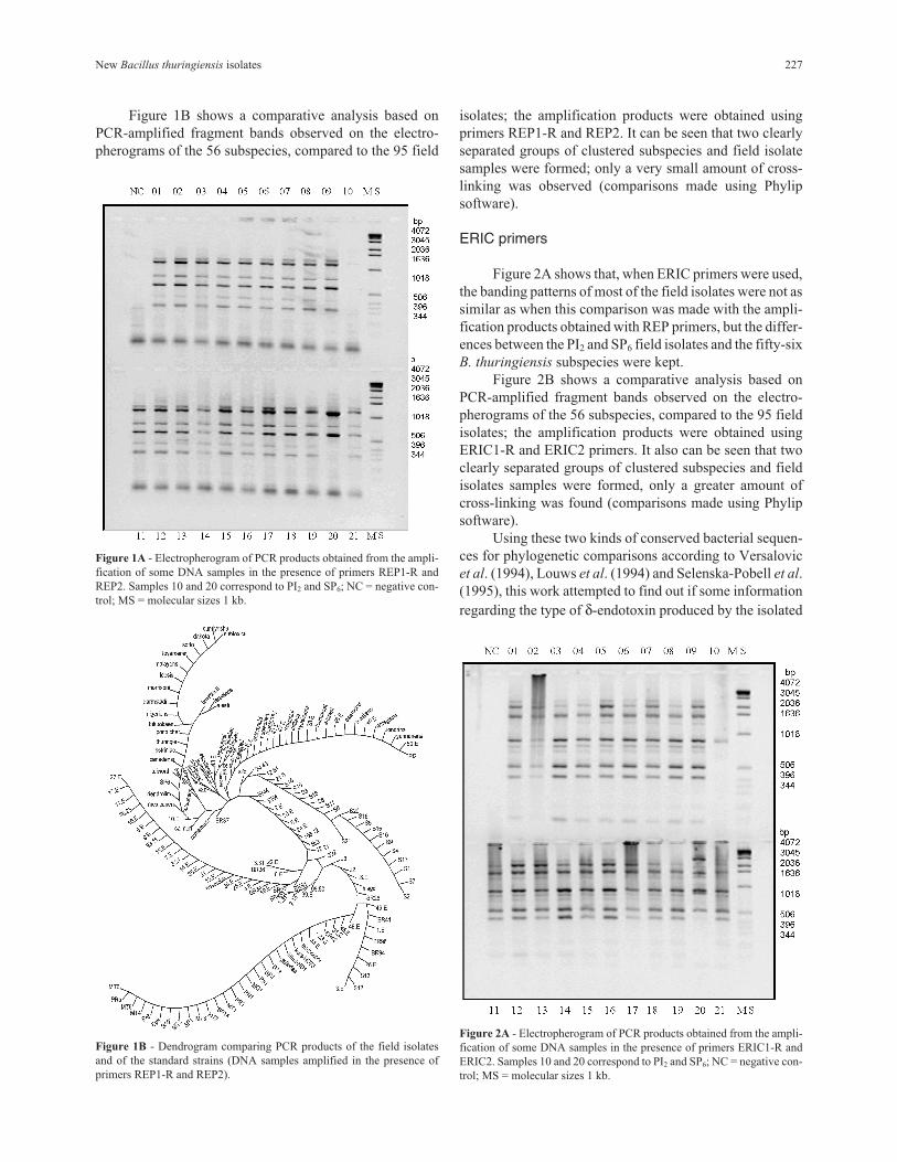
Figure 1B shows a comparative analysis based on
PCR-amplified fragment bands observed on the electro-
pherograms of the 56 subspecies, compared to the 95 field
isolates; the amplification products were obtained using
primers REP1-R and REP2. It can be seen that two clearly
separated groups of clustered subspecies and field isolate
samples were formed; only a very small amount of cross-
linking was observed (comparisons made using Phylip
software).
ERIC primers
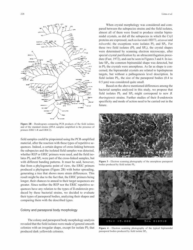
Figure 2A shows that, when ERIC primers were used,
the banding patterns of most of the field isolates were not as
similar as when this comparison was made with the ampli-
fication products obtained with REP primers, but the differ-
ences between the PI2 and SP6 field isolates and the fifty-six
B. thuringiensis subspecies were kept.
Figure 2B shows a comparative analysis based on
PCR-amplified fragment bands observed on the electro-
pherograms of the 56 subspecies, compared to the 95 field
isolates; the amplification products were obtained using
ERIC1-R and ERIC2 primers. It also can be seen that two
clearly separated groups of clustered subspecies and field
isolates samples were formed, only a greater amount of
cross-linking was found (comparisons made using Phylip
software).
Using these two kinds of conserved bacterial sequen-
ces for phylogenetic comparisons according to Versalovic
et al. (1994), Louws et al. (1994) and Selenska-Pobell et al.
(1995), this work attempted to find out if some information
regarding the type of δ-endotoxin produced by the isolated
New Bacillus thuringiensis isolates 227
Figure 1A - Electropherogram of PCR products obtained from the ampli-
fication of some DNA samples in the presence of primers REP1-R and
REP2. Samples 10 and 20 correspond to PI2 and SP6; NC = negative con-
trol; MS = molecular sizes 1 kb.
Figure 1B - Dendrogram comparing PCR products of the field isolates
and of the standard strains (DNA samples amplified in the presence of
primers REP1-R and REP2).
Figure 2A - Electropherogram of PCR products obtained from the ampli-
fication of some DNA samples in the presence of primers ERIC1-R and
ERIC2. Samples 10 and 20 correspond to PI2 and SP6; NC = negative con-
trol; MS = molecular sizes 1 kb.
field samples could be pinpointed using the PCR-amplified
material, after the reaction with these types of repetitive se-
quences. Indeed, a certain degree of cross-linking between
the subspecies and the isolated field samples was detected,
whether REP or ERIC primers were used, and the field iso-
lates PI2 and SP6 were part of the cross-linked samples, but
with different banding patterns. It must be said, however,
that from a phylogenetic point of view, the ERIC primers
produced a phylogram (Figure 2B) with better spreading,
generating a tree that shows more strain differences. This
result might be due to the fact that, the ERIC primers being
longer, their chances to anneal to their target sequences are
greater. Since neither the REP nor the ERIC repetitive se-
quences have any relation to the types of δ-endotoxin pro-
duced by these bacterial strains, we decided to evaluate
their types of parasporal bodies, analyzing their shapes and
comparing them with the described types.
Colony and parasporal body morphology
The colony and parasporal body morphology analysis
revealed that the field isolates were made of general smooth
colonies with an irregular shape, except for isolate PI2 that
produced dark yellowish colonies.
When crystal morphology was considered and com-
pared between the subspecies strains and the field isolates,
almost all of them were found to produce similar bipira-
midal crystals, as did all the subspecies in which the CryI
proteins are expressed, such as kurstaki-HD73, aizawai and
tolworthi; the exceptions were isolates PI2 and SP6. For
these two field isolates (PI2 and SP6), the crystal shapes
were determined by scanning electron microscopy, after
special crystal purification by an ultracentrifugation proce-
dure (Fast, 1972), and can be seen in Figures 3 and 4. In iso-
late SP6, the common bipiramidal shape was detected, but
in PI2 the crystals were amorphous. As far as shape is con-
cerned, the bipiramidal crystals are related to lepidopteran
targets, but without a pathogenesis level description. In
field isolate PI2, the size of the parasporal bodies (0.4 to
0.5 µm) was considered quite small.
Based on the above mentioned differences among the
bacterial samples analyzed in this study, we propose that
field isolates PI2 and SP6 might correspond to new B.
thuringiensis strains. Further studies of their δ-endotoxin
specificity and mode of action need to be carried out in the
future.
228 Lima et al.
Figure 2B - Dendrogram comparing PCR products of the field isolates
and of the standard strains (DNA samples amplified in the presence of
primers ERIC1-R and ERIC2).
Figure 3 - Electron scanning photography of the amorphous parasporal
bodies produced by field isolate PI2.
Figure 4 - Electron scanning photography of the typical bipiramidal
parasporal bodies produced by field isolate SP6.
Acknowledgments
The authors would like to thank Dr. Eliana G.M.
Lemos for her assistance with critical manuscript reading,
and CAPES which supported ASGL with a scholarship.
References
Bravo A, Haendrickx K, Jansens S. and Peferoen M (1992) Immu-
nocytochemical analysis of specific binding of Bacillus
thuringiensis insecticidal crystal proteins to lepidopteran
and coleopteran mid-gut membranes. Journal Invertebrate
Pathology 60:247-253.
Carozzi NB, Kramer VC, Warren GW, Evola S and Koziel M
(1991) Prediction of insecticidal activity Bacillus
thuringiensis strain by polymerase chain reaction product
profiles. Applied Environmental Microbiology 57:353-356.
DE-Bruijn FJ (1992) Use of repetitive (repetitive extragenic pa-
lindromic and enterobacterial repetitive intergenic conse-
sus) sequences and polymerase chain reaction to fingerprint
the genomes of Rhizobium meliloti isolates and other soil
bacteria. Applied Environmental Microbiology
58(7):2180-2187.
Fast PG (1972) The delta endotoxin of Bacillus thuringiensis III: a
rapid method for separating parasporal bodies from spores.
Journal of Invertebrate Pathology 20:189-140.
Ferré J, Real MD, Van Rie J, Jansens S and Peferoen M (1991)
Resistance to the Bacillus thuringiensis bioinsecticide in a
field population of Putella xilostella is due to a change in a
mid-gut membrane receptor. Proc. Natl. Acad. Sci.
88:5119-5123.
Louws FJ, Fulbright DW, Stephens CT and DE-Bruijn FJ (1994)
Specific genomic fingerprint of phytopathogenic Xantho-
monas and Pseudomonas pathovars and strains generated
with repetitive sequence and PCR. Applied Environmental
Microbiology 60(7):2286-2295.
Sambrook J, Frisch EF and Maniatis T (1989) Molecular cloning:
a laboratory manual. 2nd ed., Cold Spring Harbor Laboratory
Press, New York.
Selenska-Pobell S, Gigova L and Petrova N (1995) Strain-specific
fingerprints of Rhizobium galegae generated by PCR with
arbitrary and repetitive primers. Journal of Applied Bacteri-
ology 79:425-431.
Smirnoff WA (1962) A staining method for differentiating spores,
crystal and cell of Bacillus thuringiensis (berliner). Journal
of Insect Pathology 4:384-386.
Van Rie J, Jansens S, Hofte H, Degheele D and Mallaert HV
(1989) Specificity of Bacillus thuringiensis delta-endoto-
xins: importance of specific receptors on the brush border
membrane of the mid-gut of target insects. European Journal
Biochemistry 186:239-247.
Van Rie J, Jansens S, Hofte H, Degheele D and Mellaert HV
(1990a) Receptors on the brush border membrane of the in-
sects’ mid-gut as determinants of the specificity of Bacillus
thuringiensis δ-endotoxins. Applied Environmental Micro-
biology 56(5):1378-1385.
Van Rie J, MacGaughey WH, Johnson DE, Barnett BD and
Mallaert HV (1990b) Mechanism of insect resistance to the
microbial insecticide Bacillus thuringiensis. Science
247:72-74.
Versalovic J, Koeuth T and Lupski JR (1991) Distribution of re-
petitive DNA sequences in eubacteria and application to fin-
gerprinting of bacterial genomes. Nucleic Acids Research
19(24):6823-6831.
Versalovic J, Schneider M, DE-Bruijn FJ and Lupski JR (1994)
Genomic Fingerprint of Bacteria Using repetitive Sequen-
ce-Based Polimerase Chain Reaction. Methods in Molecular
and Cellular Biology 5:25-40.
New Bacillus thuringiensis isolates 229