i na r y Sciec Journal of Veterinary Science & Technology
5
Volume 4 • Issue 1 • 1000129 J Veterinar Sci Technolo ISSN: 2157-7579 JVST, an open access journal Open Access Research Article Autran et al., J Veterinar Sci Technolo 2013, 4:1 DOI: 10.4172/2157-7579.1000129 Keywords: Fatty acid, Mastitis, Prostaglandin F 2α , Staphylococcus aureus Introduction In dairy cattle, Staphylococcus aureus (S. aureus) causes uterine [1] as well as intramammary infections [2]. Staphylococcus aureus remains one of the most important causes of clinical mastitis, and the most frequently isolated pathogen in subclinical mastitis cases worldwide and was found to be the most contagious pathogen present on 43% of dairy operations in 17 of the top producing states in the US [3,4]. Contagious mastitis pathogens cause large economic losses to the dairy industry [5], mainly because of reduced milk production, increased involuntary culling rate, and discarded milk [6,7]. Treatments and immunizations against S. aureus mastitis have been studied for years [8]; however, despite the best possible antimicrobial treatments, bacteriological cure failures are common in S. aureus mastitis, and antimicrobial resistance is considered as one of the reasons for low cure rates [3,9,10,11]. Resistance to various antimicrobials is commonly seen in bovine S. aureus mastitis isolates and the intracellular survival of S. aureus is a significant factor contributing to the difficulty in clearing S. aureus infections following antibiotic therapy [12,13]. Over the years, there have been improvements in reducing contagious mastitis, nevertheless, treatment of S. aureus is quite challenging and a complex interaction between host and pathogen renders the complete eradication of S. aureus especially difficult. Finding an alternative treatment may potentially aid in eradicating mastitis infection caused by S. aureus pathogen. Extensive research has shown that various fatty acids have antimicrobial effects and may be used as an inhibitory agent against bacteria [14-16]. Hogan et al. [15] showed that long chain fatty acids (C12, C14, Polyene C18:2 and C18:3) have bacteriostatic and bactericidal effects on S. aureus *Corresponding author: Amin Ahmadzadeh, University of Idaho, ID 83844- 2330, Moscow, Idaho, USA, Tel: 208 885-7409; Fax: 208-885-6420; E-mail: [email protected] Received November 20, 2012; Accepted January 02, 2013; Published January 04, 2013 Citation: Autran CA, Shafii B, Dalton JC, McGuire MA, Carnahan KG, et al. (2013) Effect of Prostaglandin F 2α on Growth of Staphylococcus aureus Associated with Bovine Mastitis. J Veterinar Sci Technol 4: 129. doi:10.4172/2157-7579.1000129 Copyright: © 2013 Autran CA, et al. This is an open-access article distributed under the terms of the Creative Commons Attribution License, which permits unrestricted use, distribution, and reproduction in any medium, provided the original author and source are credited. Abstract Staphylococcus aureus is a major infectious organism causing mastitis. Certain fatty acids have been shown to inhibit the growth of pathogens including Staphylococcus aureus. An in vitro experiment was conducted to determine the effects of prostaglandin F 2α (PGF 2α ) on growth of S. aureus. Flasks containing tryptic soy broth (TSB) were inoculated with S. aureus Novel, and subsequently treated with PGF 2α (dinoprost tromethamine) at concentrations of 0, 0.3, 0.6, 1.2, and 2.4 mg/ml. Cultures were incubated at 37°C for 24 h and sampled every 3 h. The entire experiment was conducted three times in duplicates. Bacterial growth was assessed by counting colony forming units (CFU) in triplicates. Data were analyzed by repeated measures analysis of variance and reduced and full dummy variable regression models to determine the effect of PGF 2α concentrations on growth patterns of S. aureus over time. There was an effect of treatment and treatment by time interaction (P<0.05) on mean log CFU, indicating that bacterial growth over 24 h was different across treatments. At time of inoculation (0 h), mean log CFU values were not different among treatments; however at 24 h, mean log CFU for each PGF 2α treatment was different from control and decreased (P<0.05) with increasing concentrations of PGF 2α . The predicted growth curve for each treatment was different (P<0.05) when compared with the control, and the rate of bacterial growth was less for 1.2 mg/ml PGF 2α when compared with the control and 0.6 mg/ml. The bacterial growth was completely inhibited during the 24 h period in 2.4 mg/ml PGF 2α treatment. Furthermore, a non-linear regression model was employed to estimate the dose response at the 24 h, and revealed that 1.2 mg/ml was the minimum inhibitory dose. These results provide evidence, for the first time, that PGF 2α , in the form of dinoprost tromethamine, has bacteriostatic and bactericidal effects on S. aureus in vitro. Effect of Prostaglandin F 2α on Growth of Staphylococcus aureus Associated with Bovine Mastitis Chloe A. Autran 1 ������� 2 , Joe. C. Dalton 1 , Mark. A. McGuire 1,3 , Kevin G. Carnahan 1 and Amin Ahmadzadeh 1 * 1 Department of Animal and Veterinary Science, College of Agricultural and Life Sciences, University of Idaho, Moscow, Idaho, USA 2 Statistical Programs, College of Agricultural and Life Sciences, University of Idaho, Moscow, Idaho, USA 3 Institute for Bioinformatics and Evolutionary Studies (IBEST), University of Idaho, Moscow, Idaho, USA and Strep. agalactiae Our laboratory [17] demonstrated that linoleic acid inhibited growth of two different mastitis strains of S. aureus. Interestingly, fatty acids can also be metabolized into various products which can also inhibit bacterial growth. For example, arachidonic acid, a fatty acid derived from linoleic acid [18] inhibited the growth of gram-positive bacteria such as Streptococcus faecalis, Staphylococcus epidermidis, S. aureus [19] and gram-negative H. pylore [20]. Moreover, arachidonic acid is a precursor to prostanoid synthesis when released from membrane phospholipids in response to various stimuli. One prostanoid synthesized from arachidonic acid is prostaglandin F 2α (PGF 2α ), which is readily available for use in estrous synchronization in dairy cattle. Considering that both linoleic and arachidonic acid have been shown to inhibit bacterial growth, we hypothesized that PGF 2α , synthesized from these fatty acids, may have similar antibacterial properties. To our knowledge there is no information on the effect of any prostaglandin on bacterial growth. e objective of this study was to determine the effects of PGF 2α using the commercially available form of dinoprost tromethamine on growth of S. aureus in vitro. J o u r n a l o f V e t e r i n a r y S c i e n c e & T e c h n o l o g y ISSN: 2157-7579 Journal of Veterinary Science & Technology
Transcript of i na r y Sciec Journal of Veterinary Science & Technology
Volume 4 • Issue 1 • 1000129J Veterinar Sci TechnoloISSN: 2157-7579 JVST, an open access journal
Open AccessResearch Article
Autran et al., J Veterinar Sci Technolo 2013, 4:1 DOI: 10.4172/2157-7579.1000129
Keywords: Fatty acid, Mastitis, Prostaglandin F2α, Staphylococcusaureus
IntroductionIn dairy cattle, Staphylococcus aureus (S. aureus) causes uterine [1]
as well as intramammary infections [2]. Staphylococcus aureus remains one of the most important causes of clinical mastitis, and the most frequently isolated pathogen in subclinical mastitis cases worldwide and was found to be the most contagious pathogen present on 43% of dairy operations in 17 of the top producing states in the US [3,4]. Contagious mastitis pathogens cause large economic losses to the dairy industry [5], mainly because of reduced milk production, increased involuntary culling rate, and discarded milk [6,7]. Treatments and immunizations against S. aureus mastitis have been studied for years [8]; however, despite the best possible antimicrobial treatments, bacteriological cure failures are common in S. aureus mastitis, and antimicrobial resistance is considered as one of the reasons for low cure rates [3,9,10,11]. Resistance to various antimicrobials is commonly seen in bovine S. aureus mastitis isolates and the intracellular survival of S. aureus is a significant factor contributing to the difficulty in clearing S. aureus infections following antibiotic therapy [12,13]. Over the years, there have been improvements in reducing contagious mastitis, nevertheless, treatment of S. aureus is quite challenging and a complex interaction between host and pathogen renders the complete eradication of S. aureus especially difficult. Finding an alternative treatment may potentially aid in eradicating mastitis infection caused by S. aureus pathogen. Extensive research has shown that various fatty acids have antimicrobial effects and may be used as an inhibitory agent against bacteria [14-16]. Hogan et al. [15] showed that long chain fatty acids (C12, C14, Polyene C18:2 and C18:3) have bacteriostatic and bactericidal effects on S. aureus
*Corresponding author: Amin Ahmadzadeh, University of Idaho, ID 83844-2330, Moscow, Idaho, USA, Tel: 208 885-7409; Fax: 208-885-6420; E-mail: [email protected]
Received November 20, 2012; Accepted January 02, 2013; Published January 04, 2013
Citation: Autran CA, Shafii B, Dalton JC, McGuire MA, Carnahan KG, et al. (2013) Effect of Prostaglandin F2α on Growth of Staphylococcus aureus Associated with Bovine Mastitis. J Veterinar Sci Technol 4: 129. doi:10.4172/2157-7579.1000129
Copyright: © 2013 Autran CA, et al. This is an open-access article distributed under the terms of the Creative Commons Attribution License, which permits unrestricted use, distribution, and reproduction in any medium, provided the original author and source are credited.
AbstractStaphylococcus aureus is a major infectious organism causing mastitis. Certain fatty acids have been shown to
inhibit the growth of pathogens including Staphylococcus aureus. An in vitro experiment was conducted to determine the effects of prostaglandin F2α (PGF2α) on growth of S. aureus. Flasks containing tryptic soy broth (TSB) were inoculated with S. aureus Novel, and subsequently treated with PGF2α (dinoprost tromethamine) at concentrations of 0, 0.3, 0.6, 1.2, and 2.4 mg/ml. Cultures were incubated at 37°C for 24 h and sampled every 3 h. The entire experiment was conducted three times in duplicates. Bacterial growth was assessed by counting colony forming units (CFU) in triplicates. Data were analyzed by repeated measures analysis of variance and reduced and full dummy variable regression models to determine the effect of PGF2α concentrations on growth patterns of S. aureus over time. There was an effect of treatment and treatment by time interaction (P<0.05) on mean log CFU, indicating that bacterial growth over 24 h was different across treatments. At time of inoculation (0 h), mean log CFU values were not different among treatments; however at 24 h, mean log CFU for each PGF2α treatment was different from control and decreased (P<0.05) with increasing concentrations of PGF2α. The predicted growth curve for each treatment was different (P<0.05) when compared with the control, and the rate of bacterial growth was less for 1.2 mg/ml PGF2α when compared with the control and 0.6 mg/ml. The bacterial growth was completely inhibited during the 24 h period in 2.4 mg/ml PGF2α treatment. Furthermore, a non-linear regression model was employed to estimate the dose response at the 24 h, and revealed that 1.2 mg/ml was the minimum inhibitory dose. These results provide evidence, for the first time, that PGF2α, in the form of dinoprost tromethamine, has bacteriostatic and bactericidal effects on S. aureus in vitro.
Effect of Prostaglandin F2α on Growth of Staphylococcus aureus Associated with Bovine MastitisChloe A. Autran1������� 2, Joe. C. Dalton1, Mark. A. McGuire1,3, Kevin G. Carnahan1 and Amin Ahmadzadeh1*1Department of Animal and Veterinary Science, College of Agricultural and Life Sciences, University of Idaho, Moscow, Idaho, USA2Statistical Programs, College of Agricultural and Life Sciences, University of Idaho, Moscow, Idaho, USA3Institute for Bioinformatics and Evolutionary Studies (IBEST), University of Idaho, Moscow, Idaho, USA
and Strep. agalactiae Our laboratory [17] demonstrated that linoleic acid inhibited growth of two different mastitis strains of S. aureus. Interestingly, fatty acids can also be metabolized into various products which can also inhibit bacterial growth. For example, arachidonic acid, a fatty acid derived from linoleic acid [18] inhibited the growth of gram-positive bacteria such as Streptococcus faecalis, Staphylococcus epidermidis, S. aureus [19] and gram-negative H. pylore [20]. Moreover, arachidonic acid is a precursor to prostanoid synthesis when released from membrane phospholipids in response to various stimuli. One prostanoid synthesized from arachidonic acid is prostaglandin F2α (PGF2α), which is readily available for use in estrous synchronization in dairy cattle. Considering that both linoleic and arachidonic acid have been shown to inhibit bacterial growth, we hypothesized that PGF2α, synthesized from these fatty acids, may have similar antibacterial properties. To our knowledge there is no information on the effect of any prostaglandin on bacterial growth. The objective of this study was to determine the effects of PGF2α using the commercially available form of dinoprost tromethamine on growth of S. aureus in vitro.
Jour
nal o
f Vete
rinary Science & Technology
ISSN: 2157-7579
Journal of Veterinary Science &Technology
Citation: Autran CA, Shafii B, Dalton JC, McGuire MA, Carnahan KG, et al. (2013) Effect of Prostaglandin F2α on Growth of Staphylococcus aureus Associated with Bovine Mastitis. J Veterinar Sci Technol 4: 129. doi:10.4172/2157-7579.1000129
Page 2 of 5
Volume 4 • Issue 1 • 1000129J Veterinar Sci TechnoloISSN: 2157-7579 JVST, an open access journal
Materials and MethodsExperimental Design and Treatment
The bacterial strain used was a clinical bovine mastitis isolate, S. aureus Novel [21]. Cultures were prepared by inoculating a single colony into 3 ml of tryptic soy broth (TSB) (EMD Chemicals Inc., Darmstadt, NJ) followed by overnight incubation at 37°C with shaking at 250 rpm. In order to obtain sufficient culture for the experiment, culture tubes containing 10 ml of fresh TSB were inoculated at 1:100 with 3 ml of overnight S. aureus culture, and once more incubated overnight at 37°C with shaking at 250 rpm. Prostaglandin F2α in the form of dinoprost tromethamine (Lutalyse, Pfizer Animal Health, New York, NY) was added to flasks for a final concentration of 0, 0.3, 0.6, 1.2, and 2.4 mg/ml (2 flasks/treatment). Lutalyse contains 0.9% to 1% benzyl alcohol as a preservative. Consequently, benzyl alcohol (Fischer Scientific, Pittsburgh, PA) was added to the flasks containing 0.3, 0.6, and 1.2 mg/ml of PGF2α, mimicking the total concentration of benzyl alcohol in flasks containing the highest concentration of PGF2α (2.4 mg/ml). This resulted in each flask containing the same final concentration of benzyl alcohol (1%). Sterilized TSB was added to each flask containing the different concentrations of PGF2α, to achieve a final concentration of 3% TSB in a final volume of 50 ml. Two different controls (2 flasks/each) were included in the experiment; the first contained bacteria and TSB alone and the second control contained bacteria, TSB and 1% benzyl alcohol. Flasks, which included both treatment and controls, were inoculated with the 10 ml overnight culture of S. aureus at a concentration of 1:100. Flasks were incubated at 37°C, shaken at 250 rpm for 24 h, and at 0 h, and every 3 h thereafter, 1 ml samples were taken from each flask to assess bacterial growth. The entire experiment was repeated three times, in duplicate, and each experiment started with a new subculture of the bacterium.
Bacteria Counts
To determine colony forming units (CFU), samples of 0.5 ml were taken from flasks for plating and centrifuged twice via a micro centrifuge (International Equipment Company, Needham, MA) to remove benzyl alcohol. The supernatant was removed and the cell pellet was re-suspended with 0.5 ml of fresh TSB. Serial dilutions were performed before samples were placed on agar plates (EMD Chemicals Inc., Darmstadt, NJ). Plate counts were done in triplicate per sample from each flask. Plates were incubated at least 12 h, or until colonies were apparent, at a constant temperature of 37°C [17].
Statistical Analyses
The number of live cells, as measured by log CFU, was determined by averaging the number of cells for the duplicate concentrations of both plates at each 3 h time point. Results for the three growth curves were also pooled by concentration, at each time point. An analysis of variance (repeated measures) was carried out using Mixed procedure of SAS [22], where the model included treatment, time (repeated factor) and their interaction. A full model dummy variable regression procedure was also carried out to analyze the effect of treatments over time on the growth pattern of S. aureus. The coincidence or equality of the estimated regression lines, the rate of bacteria growth over time, and the point at which the inflection of the growth curve occurred (an indication of the time it takes for each treatment to reach maximum bacterial growth) were determined. The estimation of the reduced models for each treatment was carried out using PROC REG procedures of SAS [22], and that of the full model was carried out using PROC GLM procedures of SAS. The fitted reduced model
for each treatment took the form of
Y=β0+β1 x+β2 x2+ε1
where Y was the logarithmic value of the number of live cells (log CFU/ml), x represented time, β0 was the intercept (estimated log CFU/ml at time 0), β1 was the rate of increase for bacterial growth, β2 was the point of inflection (the maximum growth at a specific time), and ε1 represented the random error under the classical regression assumptions. The adequacy of fit was determined by the significance of the parameter estimates (declared at P<0.05), their corresponding magnitudes and signs, and the examination of the estimated residuals. As an ancillary objective, a non-linear regression model was used (PROC NLMixed procedures of SAS [22]) to determine a dose-response and minimum inhibitory concentration for log CFU at 24 h of bacterial growth. The estimation of the model was carried out using a non- linear iterative algorithm (Gauss Newton), where the fitted model took the form of Y = αe– βx + c
where Y was the number of live cells (CFU/ml), x was the concentration of PGF2α, and α represented the intercept, β was the exponential rate of decay for CFU/ml, and c was the lower asymptote. A weighted least squares estimate was used to stabilize residual variance. The adequacy of fit was determined by the significance of the parameter estimates, the structure of correlation among estimated parameters, their corresponding magnitudes and signs, and the examination of the model residuals.
ResultsBacterial growth curves were evaluated in growth media containing
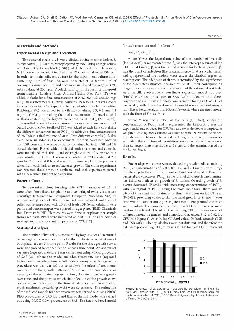
PGF2α at concentrations of 0, 0.3, 0.6, 1.2, and 2.4 mg/ml, with 0 mg/ml referring to the control with and without benzyl alcohol. Based on bacterial growth curves, PGF2α, in the form of dinoprost tromethamine, has inhibitory effects on growth of S. aureus. Overall, growth of S. aureus decreased (P<0.05) with increasing concentrations of PGF2α, with 2.4 mg/ml of PGF2α being the most inhibitory. There was an effect of treatment and treatment by time interaction on log CFU/ml (P<0.05), providing evidence that bacterial growth of S. aureus over time was not similar among PGF2α treatments. Pre-planned contrasts were conducted to compare the mean log CFU/ml values between treatments at 0 and 24 h. At 0 h the mean log CFU/ml values were not different among treatments and control, and averaged 8.12 ± 0.02 log CFU/ml (Figure 1). At 24 h, log CFU/ml values for both controls (TSB and TSB with 1% benzyl alcohol) were not different, and therefore the data were pooled. Log CFU/ml values at 24 h for each PGF2α treatment
0
2
4
6
8
10
0 0.3 0.6 1.2 2.4
Log
CFU
Prostaglandin F2α (mg/mL)
0 h24 h
a
b b
c
d
Figure 1: Growth of S. aureus as measured by log colony forming units (CFU)/mL, treated with PGF2α at 0 h (gray bars) and 24 h (black bars) for each concentration of PGF2α
a, b, c, d Bars designated by different letters are different (P<0.05) at 24 h.
Citation: Autran CA, Shafii B, Dalton JC, McGuire MA, Carnahan KG, et al. (2013) Effect of Prostaglandin F2α on Growth of Staphylococcus aureus Associated with Bovine Mastitis. J Veterinar Sci Technol 4: 129. doi:10.4172/2157-7579.1000129
Page 3 of 5
Volume 4 • Issue 1 • 1000129J Veterinar Sci TechnoloISSN: 2157-7579 JVST, an open access journal
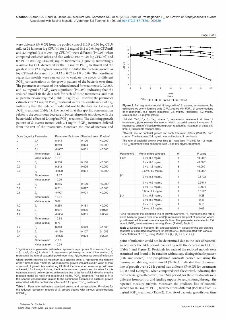
were different (P<0.05) from the pooled control (10.5 ± 0.04 log CFU/ml). At 24 h, mean log CFU/ml for 1.2 mg/ml (8.1 ± 0.04 log CFU/ml) and 2.4 mg/ml (1.8 ± 0.04 log CFU/ml) were different (P<0.05) when compared with each other and also with 0.3 (9.4 ± 0.04 log CFU/ml) and 0.6 (9.0 ± 0.04 log CFU/ml) mg/ml treatments (Figure 1). Interestingly S. aureus log CFU decreased for the 1.2 mg/ml PGF2α treatment and the greatest dose (2.4 mg/ml) completely inhibited the bacteria growth as log CFU/ml decreased from 8.12 ± 0.02 to 1.8 ± 0.04. The non-linear regression models were carried out to evaluate the effects of different PGF2α concentrations on the growth pattern of the bacteria over time. The parameter estimates of the reduced model for treatments 0, 0.3, 0.6, and 1.2 mg/ml of PGF2α were significant (P<0.05), indicating that the reduced model fit the data well for each of those treatments, and that all parameters are required (Table 1, Figure 2). However, the parameter estimates for 2.4 mg/ml PGF2α treatment were non-significant (P>0.05), indicating that the reduced model did not fit the data for 2.4 mg/ml PGF2α treatment (Table 1). The lack of fit at this specific concentration relates to the continuous decrease in bacterial growth associated with the bactericidal effects of 2.4 mg/ml PGF2α treatment. The declining growth pattern of S. aureus treated with 2.4 mg/ml PGF2α treatment differed from the rest of the treatments. Moreover, the rate of increase and
point of inflection could not be determined due to the lack of bacterial growth over the 24 h period, coinciding with the decrease in CFU/ml (Table 1 and Figure 2). Residuals for each of the reduced models were examined and found to be random without any distinguishable pattern (data not shown). The pre-planned contrasts carried out using the dummy variable regression model (Table 2) indicated that the overall line of growth over a 24 h period was different (P<0.05) for treatments 0.3, 0.6 and 1.2 mg/ml, when compared with the control, indicating that the bacterial growth pattern, over 24 h period, for these treatments were different from control and lending support to results found through the repeated measure analysis. Moreover, the predicted line of bacterial growth for 0.6 mg/ml PGF2α treatment was different (P<0.05) from 1.2 mg/ml PGF2α treatment (Table 2). The rate of bacterial growth over time
0
1
2
3
4
5
6
7
8
9
10
11
0 3 6 9 12 15 18 21 24
Log
CFU
Time(Hours)
0 0.3 0.6 1.2 2.4PGF2α
***
Figure 2: Full regression model1 fit to growth of S. aureus, as measured by estimated log colony forming units (CFU),treated with PGF2α at concentrations of 0 (dimonds), 0.3 mg/ml (squares), 0.6 mg/mL (triangles), 1.2 mg/mL (circles) and 2.4 mg/mL (stars). 1Model: Y=β0+β1x+β2x
2+ε1, where β0 represents y-intercept at time of inoculation; β1 represents the rate at which bacterial growth increases; β2 represents point of inflection where growth reached its maximum at a specific time; ε1 represents random error. *Overall line of bacterial growth for each treatment differs (P<0.05) from control. The treatment 2.4 mg/mL was not included in contrasts. The rate of bacterial growth over time (β1) was less (P<0.05) for 1.2 mg/ml PGF2α treatment when compared with 0 and 0.6 mg/mL treatment.
Parameters Pre-planned contrasts df P value Linea 0 vs. 0.3 mg/mL 3 <0.0001
0 vs. 0.6 mg/mL 3 <0.0001 0 vs. 1.2 mg/mL 3 <0.0001
0.6 vs. 1.2 mg/mL 3 <0.0001 β1
b 0 vs. 0.3 mg/mL 1 0.8702
0 vs. 0.6 mg/mL 1 0.3813 0 vs. 1.2 mg/mL 1 0.0004
0.6 vs. 1.2 mg/mL 1 0.0197 β2
c 0 vs. 0.3 mg/mL 1 0.28 0 vs. 0.6 mg/mL 1 0.48 0 vs. 1.2 mg/mL 1 0.10
0.6 vs. 1.2 mg/mL 1 0.05 a Line represents the estimated line of growth over time, bβ1 represents the rate at which bacterial growth over time, and cβ2 represents the point of inflection where growth reached its maximum at a specific time. The parameter estimates for 2.4 mg/mL PGF2α treatment were non-significant (P>0.05; Table 1)Table 2: Degrees of freedom (df), and associated P–values for the pre-planned contrasts of estimated parameters for growth of S. aureus treated with various concentrations of PGF2α using Model: (Y = β0 + β1 x + β2 x
2 + ε1).
a Significance of parameter estimates represents appropriate fit of model (Y = β0 + β1 x +β2 x
2 + ε1) to data. b β0 represents y-intercept at time of inoculation;c β1 represents the rate of bacterial growth over time; d β2 represents point of inflection where growth reached its maximum at a specific time; ε1 represents the random error; e Time to max = time (h) when maximal growth was achieved; f Value at max = amount of growth (estimated log CFU) at the time when maximal growth was achieved; g for 2.4mg/mL dose, the time to maximum growth and its value for this treatment should be interpreted with caution due to the lack of fit indicating that the reduced model did not fit the data for 2.4 mg/mL PGF2α treatment. The lack of fit at this specific concentration relates to the continuous decrease in bacterial growth associated with the bactericidal effects of 2.4 mg/mL PGF2α treatment.Table 1: Parameter estimates, standard errors, and the associated P–values for the reduced regression models of S. aureus treated with various concentrations of PGF2α.
Dose (mg/mL) Parameter Parameter Estimate Standard error P valuea 0 β0
b 8.543 0.126 <0.0001 0 β1
c 0.249 0.024 <0.0001 0 β2
d -0.007 0.001 <0.0001 Time to maxe 18.6 Value at maxf 10.9
0.3 β0 8.358 0.130 <0.0001 0.3 β1 0.242 0.025 <0.0001 0.3 β2 -0.008 0.001 <0.0001
Time to max 14.07 Value at max 10.06
0.6 β0 8.285 0.139 <0.0001 0.6 β1 0.211 0.027 <0.0001 0.6 β2 -0.007 0.001 <0.0001
Time to max 13.31 Value at max 9.69
1.2 β0 8.280 0.181 <0.0001 1.2 β1 0.091 0.036 0.0136 1.2 β2 -0.004 0.001 0.0049
Time to max 10.68 Value at max 8.76
2.4 β0 8.690 0.549 <0.0001 2.4 β1 -0.188 0.107 0.1053 2.4 β2 -0.005 0.004 0.2034
Time to maxg -16.9 Value at maxg 10.28
Citation: Autran CA, Shafii B, Dalton JC, McGuire MA, Carnahan KG, et al. (2013) Effect of Prostaglandin F2α on Growth of Staphylococcus aureus Associated with Bovine Mastitis. J Veterinar Sci Technol 4: 129. doi:10.4172/2157-7579.1000129
Page 4 of 5
Volume 4 • Issue 1 • 1000129J Veterinar Sci TechnoloISSN: 2157-7579 JVST, an open access journal
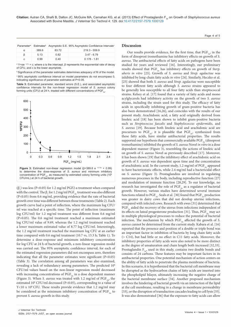
(β1) was less (P<0.05) for 1.2 mg/ml PGF2 α treatment when compared with the control. The β1 for 1.2 mg/ml PGF2α treatment was also different (P<0.05) from 0.6 mg/ml, providing evidence that the rate of bacterial growth over time was different between those treatments (Table 2). Each growth curve had a point of inflection, where the maximum log CFU/ml was reached at a specific time. The point of inflection (maximum log CFU/ml) for 1.2 mg/ml treatment was different from 0.6 mg/ml (P=0.05). The 0.6 mg/ml treatment reached a maximum estimated log CFU/ml value of 9.69, whereas the 1.2 mg/ml treatment reached a lower maximum estimated value of 8.77 log CFU/ml. Interestingly, the 1.2 mg/ml treatment reached the maximum log CFU at an earlier time compared with 0.6 mg/ml treatment (10.7 vs, 13.3 h; Table 1). To determine a dose-response and minimum inhibitory concentration for log CFU at 24 h of bacterial growth, a non-linear regression model was carried out. The 95% asymptotic confidence interval, for each of the estimated regression parameters did not encompass zero, therefore indicating that all the parameter estimates were significant (P<0.05) (Table 3). The correlation among all parameters was also examined, revealing a lack of redundancy among parameters. The predicted 108 CFU/ml values based on the non-linear regression model decreased with increasing concentrations of PGF2α in a dose dependent manner (Figure 3). When S. aureus was treated with 1.2 mg/ml of PGF2α, the estimated 108 CFU/ml decreased (P<0.05), corresponding to a value of Y<10 x 108 CFU. These results provide evidence that 1.2 mg/ml may be considered as the minimum inhibitory concentration of PGF2α to prevent S. aureus growth in this study.
DiscussionOur results provide evidence, for the first time, that PGF2α, in the
form of dinoprost tromethamine has inhibitory effects on growth of S. aureus. The antibacterial effects of fatty acids on pathogens have been studied for years and reviewed [16]. Interestingly, our preliminary results showed that PGF2α has inhibitory effects on growth of Sterp. uberis in vitro [23]. Growth of S. aureus and Strep. agalactiae was inhibited by long-chain fatty acids in vitro [24]. Similarly, Heczko et al. [25] showed that both S. aureus and Strep. agalactiae were susceptible to four different fatty acids although S. aureus strains appeared to be generally less susceptible to all four fatty acids than streptococcal strains. Kelsey et al. [17] found that a variety of fatty acids and mono acylglycerols had inhibitory activity on the growth of two S. aureus strains, including the strain used for this study. The efficacy of fatty acids in specifically inhibiting growth of gram-positive bacteria has also been demonstrated [16,26], and coincides with the results of our present study. Arachidonic acid, a fatty acid originally derived from linoleic acid [18] has been shown to inhibit gram-positive bacteria such as Streptococcus faecalis and Staphylococcus epidermidis, and S. aureus [19]. Because both linoleic acid and arachidonic acid are precursors to PGF2α, it is plausible that PGF2α, synthesized from these fatty acids, have similar antibacterial properties. The results supported our hypothesis that commercially available PGF2α (dinoprost tromethamine) inhibited the growth of S. aureus Novel in vitro in a dose dependent manner (Figure 3), resembling the actions of linoleic acid on growth of S. aureus Novel as previously described [17]. Moreover, it has been shown [19] that the inhibitory effect of arachidonic acid on growth of S. aureus was dependent upon time and the concentration of arachidonic acid. In the current study, 1.2 mg/ml of PGF2α appeared to have bacteriostatic effects, while 2.4 mg/ml had a bactericidal effect on S. aureus (Figure 3). Prostaglandins are involved in regulating numerous processes in the body, including reproductive function [27] and modulation of immune function [28,29]. To our knowledge, no research has investigated the role of PGF2α as a regulator of bacterial growth. However, various studies have determined several immune functions related to PGF2α. Seals et al. [30] found that PGF2α production was greater in dairy cows that did not develop uterine infections, compared with infected cows. Research with ewes [31] determined that PGF2α aided the recovery of the uterus from infections independent of its effects on luteal progesterone production [31]. Thus, PGF2α may act in multiple physiological processes to reduce the potential of bacterial infection. The mechanism by which PGF2α affected the growth of S. aureus cannot be determined from the current study. Kabara et al. [16] reported that the presence and position of a double or triple bond was an important factor in inhibition of bacteria by long chain fatty acids (> C14), but had little or no effect in C11 fatty acids. Moreover, the inhibitory properties of fatty acids were also noted to be more distinct as the degree of unsaturation and chain length both increased [32,33]. Prostaglandin F2α, used in this study, contains two double bonds and consists of 24 carbons. These features may be important factors in its antibacterial properties. One potential mechanism of action centers on the ability of fatty acids to penetrate the plasma membrane of bacteria. In this scenario, it is hypothesized that the bacterial cell membrane may be disrupted as the hydrocarbon chains of fatty acids are inserted into the phospholipid bilayer, ultimately increasing the negative charge of the bacterial membrane surface [34]. Another proposed mechanism involves the hindering of bacterial growth via an interaction of the lipid at the cell membrane, resulting in a change in membrane permeability [33], or the disruption of transduction cascades leading to cell lysis [35]. It was also demonstrated [36] that the exposure to fatty acids can allow
Parametera Estimatesb Asymptotic S.E. 95% Asymptotic Confidence Intervalsc
α 389.6 83.72 219.3 - 559.9 β 5.13 0.82 3.47 - 6.79 c 0.99 0.40 0.178 - 1.81
a Y=αe – β x + c; where α is the intercept, β represents the exponential rate of decay of CFU, and c is the lower asymptote.b Significance of the parameter estimates determines adequacy of fit of the model.c 95% asymptotic confidence interval on model parameters do not encompass 0, indicating significance of parameter estimates at P<0.05.Table 3: Estimated parameter, standard errors (S.E.), and associated asymptotic confidence intervals for the non-linear regression model of S. aureus colony forming units (CFU) at 24 h, treated with different concentrations of PGF2α.
PGF2α (mg/mL)
0 0.3 0.6 0.9 1.2 1.5 1.8 2.1 2.4
600
500
400
300
200
100
0
108 C
FU
Figure 3: Estimated non-linear regression model (ŷ=389.6 e -5.13x + 0.99) to determine the dose-response of S. aureus and minimum inhibitory concentration of PGF2α, as measured by estimated colony forming units (108 CFU/mL) at 24 h of bacterial growth.
Citation: Autran CA, Shafii B, Dalton JC, McGuire MA, Carnahan KG, et al. (2013) Effect of Prostaglandin F2α on Growth of Staphylococcus aureus Associated with Bovine Mastitis. J Veterinar Sci Technol 4: 129. doi:10.4172/2157-7579.1000129
Page 5 of 5
Volume 4 • Issue 1 • 1000129J Veterinar Sci TechnoloISSN: 2157-7579 JVST, an open access journal
the bacteria to stay viable while disabling cell division and resulting in a bacteriostatic effect, similar to that observed in our study when S. aureus was treated with 1.2 mg/ml of PGF2α. Lastly, it has been proposed [19] that the bactericidal effect of arachidonic acid (PGF2α precursor)is mediated through peroxidation of fatty acid catalyzed by bacterialiron and H2O2. Although the mechanism(s) as to how PGF2α inhibitedbacterial growth are currently unknown, we speculate that similarmembrane interactions and multi-stage mechanisms may be employedby PGF2α (dinoprost tromethamine) in limiting or preventing growthof S. aureus. The exact mechanistic actions induced by PGF2α, alongwith its practical application in vivo, still require further investigations.
Acknowledgments
This study was made possible by the support of Pfizer Animal Health, New York, NY, in providing dinoprost tromethamine (Lutalyse®), Idaho Dairymen’s Association, NIH P20 RR15587 and the Idaho Agricultural Experiment Station. The authors would also like to thank Dr. W. J. Price for his help in statistical computations.
References
1. Smith BI, Risco CA (2002) Predisposing factors and potential causes of postpartum metritis in dairy cattle. COMPEL: 24.
2. Bannerman DD, Paape MJ, Lee JW, Zhao X, Hope JC, et al. (2004) Escherichia coli and Staphylococcus aureus elicit differential innate immune responses following intramammary infection. Clin Diagn Lab Immunol 11: 463-472.
3. Saini V, McClure JT, Scholl DT, DeVries TJ, Barkema HW (2012) Herd-level association between antimicrobial use and antimicrobial resistance in bovine mastitis Staphylococcus aureus isolates on Canadian dairy farms. J Dairy Sci 95: 1921-1929.
4. Lombard J, Van Slyke T, Welcome F, Schukken Y, Kopral C (2008) Prevalence of contagious mastitis pathogens on US dairy operations. In: National Mastitis Council 47th Annual Meeting Proceedings, New Orleans, LA, USA.
5. Degraves FJ, Fetrow J (1993) Economics of Mastitis and Mastitis Control. Vet Clin North Am Food Anim Pract 9: 421-434.
6. Zepeda L, Buelow KL, Nordlund KV, Thomas CB, Collins MT, et al. (1998) A linear programming assessment of the profit from strategies to reduce the prevalence of Staphylococcus aureus mastitis. Prev Vet Med 33: 183-193.
7. Heikkila AM, Nousiainen JI, Pyorala S (2012) Costs of clinical mastitis with special reference to premature culling. J Dairy Sci 95: 139-150.
8. Anderson JC (1978) The problem of immunization against staphylococcal mastitis. Br Vet J 134: 412-420.
9. Rajala-Schultz PJ, Smith KL, Hogan JS, Love BC (2004) Antimicrobial susceptibility of mastitis pathogens from first lactation and older cows. Vet Microbiol 102: 33-42.
10. De Vliegher S, Fox LK, Piepers S, McDougall S, Barkema HW (2012) Invited review: Mastitis in dairy heifers: nature of the disease, potential impact, prevention, and control. J Dairy Sci 95: 1025-1040.
11. Barkema HW, Schukken YH, Zadoks RN (2006) Invited Review: The role of cow, pathogen, and treatment regimen in the therapeutic success of bovine Staphylococcus aureus mastitis. J Dairy Sci 89: 1877-1895.
12. Larsen HD, Sloth KH, Elsberg C, Enevoldsen C, Pedersen LH, et al. (2000) The dynamics of Staphylococcus aureus intramammary infection in nine Danish dairy herds. Vet Microbiol 71: 89-101.
13. Sears PM, Guidry AJ, Belschner A (2001) Use of antigen-specific vaccines to enhance antibiotic treatment responses toward Staphylococcus aureus. In: 2nd International Symposium on Mastitis and Milk Quality, Vancouver, BC, Canada.
14. Wang LL, Johnson EA (1992) Inhibition of Listeria monocytogenes by fatty acids and monoglycerides. Appl Environ Microbiol 58: 624-629.
15. Hogan JS, Pankey JW, Duthie AH (1987) Growth inhibition of mastitis pathogens by long-chain fatty acids. J Dairy Sci 70: 927-934.
16. Kabara JJ, Vrable R (1977) Antimicrobial lipids: natural and synthetic fatty acids and monoglycerides. Lipids 12: 753-759.
17. Kelsey JA, Bayles KW, Shafii B, McGuire MA (2006) Fatty acids and monoacylglycerols inhibit growth of Staphylococcus aureus. Lipids 41: 951-961.
18. Marcel YL, Christiansen K, Holman RT (1968) The preferred metabolic pathway from linoleic acid to arachidonic acid in vitro. Biochim Biophys Acta 164: 25-34.
19. Knapp HR, Melly MA (1986) Bactericidal effects of polyunsaturated fatty acids. J Infec Dis 154: 84-94.
20. Khulusi S, Ahmed HA, Patel P, Mendall MA, Northfield TC (1995) The effects of unsaturated fatty acids on Helicobacter pylori in vitro. J Med Microbiol 42: 276-282.
21. Smith TH, Fox LK, Middleton JR (1998) Outbreak of mastitis caused by one strain of Staphylococcus aureus in a closed dairy herd. J Am Vet Med Assoc 212: 553-556.
22. SAS (2000-2004) Statistical Analysis System. SAS 9.1.3 ed. Cary, NC: SAS Instituite Inc. 318 14
23. Autran CA, Shafii B, Dalton JC, McGuire MA, Ahmadzadeh A (2011) Effect of prostaglandin F2α on growth of escherichia coli and streptococcus uberis associated with bovine mastitis. J Dairy Sci 94: 347.
24. Butcher GW, King G, Dyke KG (1976) Sensitivity of Staphylococcus aureus to Unsaturated fatty acids. J Gen Microbiol 94: 290-296.
25. Heczko PB, Lutticken R, Hryniewicz W, Neugebauer M, Pulverer G (1979) Susceptibility of Staphylococcus aureus and group A, B, C, and G streptococci to free fatty acids. J Clin Microbiol 9: 333-335.
26. Kanai K, Kondo E (1979) Antibacterial and cytotoxic aspects of long-chain fatty acids as cell surface events: selected topics. Jpn J Med Sci Biol 32: 135-174.
27. Seguin BE (1980) Role of prostaglandins in bovine reproduction. J Am Vet Med Assoc 176: 1178-1181.
28. Bergstrom S, Carlson LA, Weeks JR (1968) The prostaglandins: a family of biologically active lipids. Pharmacol Rev 20: 1-48.
29. Phipps RP, Stein SH, Roper RL (1991) A new view of prostaglandin E regulation of the immune response. Immunol Today 12: 349-352.
30. Seals RC, Matamoros I, Lewis GS (2002) Relationship between postpartum changes in 13, 14-dihydro-15-keto-PGF2alpha concentrations in Holstein cows and their susceptibility to endometritis. J Anim Sci 80: 1068-1073.
31. Lewis GS, Wulster-Radcliffe MC (2006) Prostaglandin F2alpha upregulates uterine immune defenses in the presence of the immunosuppressive steroid progesterone. Am J Reprod Immunol 56: 102-111.
32. Kabara JJ (1984) Antimicrobial agents derived from fatty acids. J Am Oil Chem Soc 61: 397-403.
33. Nieman C (1954) Influence of trace amounts of fatty acids on the growth of microorganisms. Bacteriol Rev 18: 147-163.
34. Kondo E, Kanai K (1972) The lethal effect of long-chain fatty acids on mycobacteria. Jpn J Med Sci Biol 25: 1-13.
35. Chamberlain NR, Mehrtens BG, Xiong Z, Kapral FA, Boardman JL, et al. (1991) Correlation of carotenoid production, decreased membrane fluidity, and resistance to oleic acid killing in Staphylococcus aureus 18Z. Infect Immun 59: 4332-4337.
36. Sheu CW, Freese E (1972) Effects of Fatty-Acids on Growth and Envelope Proteins of Bacillus-Subti. J Bacteriol 111: 516-524.