Hyperglycemia-like culture conditions induce IL-1B and TNF-α expression and impair autophagy in...
2
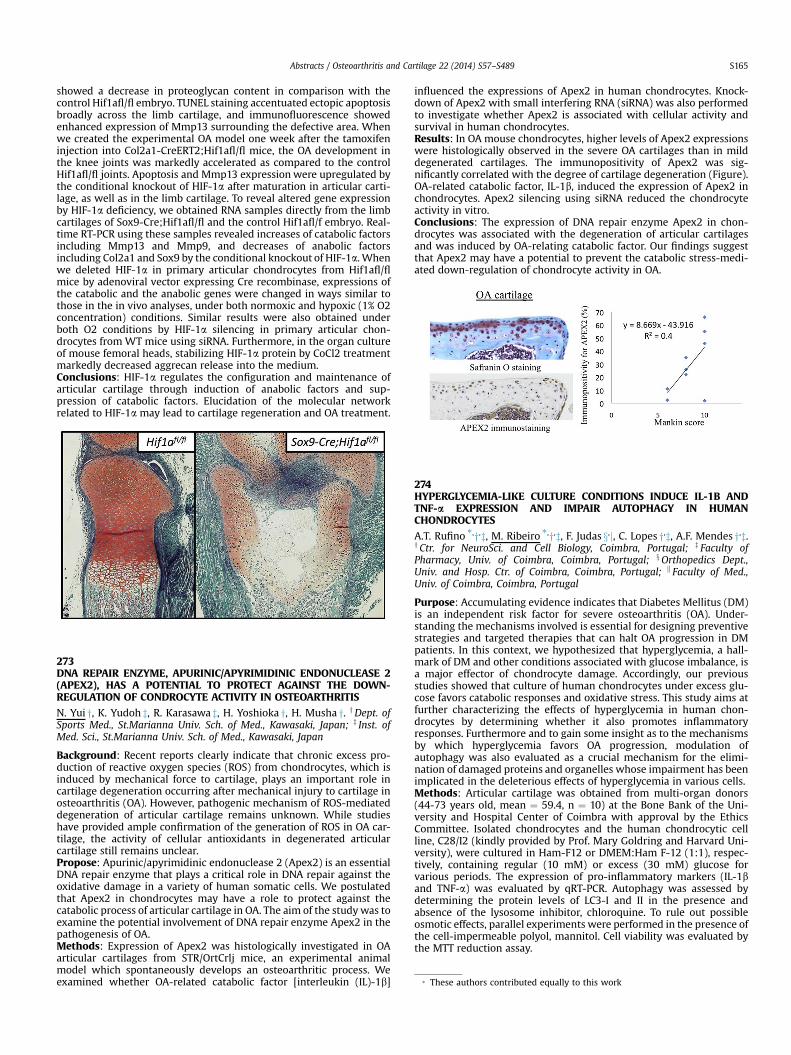
showed a decrease in proteoglycan content in comparison with the control Hif1afl/fl embryo. TUNEL staining accentuated ectopic apoptosis broadly across the limb cartilage, and immunofluorescence showed enhanced expression of Mmp13 surrounding the defective area. When we created the experimental OA model one week after the tamoxifen injection into Col2a1-CreERT2;Hif1afl/fl mice, the OA development in the knee joints was markedly accelerated as compared to the control Hif1afl/fl joints. Apoptosis and Mmp13 expression were upregulated by the conditional knockout of HIF-1a after maturation in articular carti- lage, as well as in the limb cartilage. To reveal altered gene expression by HIF-1a deficiency, we obtained RNA samples directly from the limb cartilages of Sox9-Cre;Hif1afl/fl and the control Hif1afl/f embryo. Real- time RT-PCR using these samples revealed increases of catabolic factors including Mmp13 and Mmp9, and decreases of anabolic factors including Col2a1 and Sox9 by the conditional knockout of HIF-1a. When we deleted HIF-1a in primary articular chondrocytes from Hif1afl/fl mice by adenoviral vector expressing Cre recombinase, expressions of the catabolic and the anabolic genes were changed in ways similar to those in the in vivo analyses, under both normoxic and hypoxic (1% O2 concentration) conditions. Similar results were also obtained under both O2 conditions by HIF-1a silencing in primary articular chon- drocytes from WT mice using siRNA. Furthermore, in the organ culture of mouse femoral heads, stabilizing HIF-1a protein by CoCl2 treatment markedly decreased aggrecan release into the medium. Conclusions: HIF-1a regulates the configuration and maintenance of articular cartilage through induction of anabolic factors and sup- pression of catabolic factors. Elucidation of the molecular network related to HIF-1a may lead to cartilage regeneration and OA treatment. 273 DNA REPAIR ENZYME, APURINIC/APYRIMIDINIC ENDONUCLEASE 2 (APEX2), HAS A POTENTIAL TO PROTECT AGAINST THE DOWN- REGULATION OF CONDROCYTE ACTIVITY IN OSTEOARTHRITIS N. Yui y, K. Yudoh z, R. Karasawa z, H. Yoshioka y, H. Musha y. y Dept. of Sports Med., St.Marianna Univ. Sch. of Med., Kawasaki, Japan; z Inst. of Med. Sci., St.Marianna Univ. Sch. of Med., Kawasaki, Japan Background: Recent reports clearly indicate that chronic excess pro- duction of reactive oxygen species (ROS) from chondrocytes, which is induced by mechanical force to cartilage, plays an important role in cartilage degeneration occurring after mechanical injury to cartilage in osteoarthritis (OA). However, pathogenic mechanism of ROS-mediated degeneration of articular cartilage remains unknown. While studies have provided ample confirmation of the generation of ROS in OA car- tilage, the activity of cellular antioxidants in degenerated articular cartilage still remains unclear. Propose: Apurinic/apyrimidinic endonuclease 2 (Apex2) is an essential DNA repair enzyme that plays a critical role in DNA repair against the oxidative damage in a variety of human somatic cells. We postulated that Apex2 in chondrocytes may have a role to protect against the catabolic process of articular cartilage in OA. The aim of the study was to examine the potential involvement of DNA repair enzyme Apex2 in the pathogenesis of OA. Methods: Expression of Apex2 was histologically investigated in OA articular cartilages from STR/OrtCrlj mice, an experimental animal model which spontaneously develops an osteoarthritic process. We examined whether OA-related catabolic factor [interleukin (IL)-1b] influenced the expressions of Apex2 in human chondrocytes. Knock- down of Apex2 with small interfering RNA (siRNA) was also performed to investigate whether Apex2 is associated with cellular activity and survival in human chondrocytes. Results: In OA mouse chondrocytes, higher levels of Apex2 expressions were histologically observed in the severe OA cartilages than in mild degenerated cartilages. The immunopositivity of Apex2 was sig- nificantly correlated with the degree of cartilage degeneration (Figure). OA-related catabolic factor, IL-1b, induced the expression of Apex2 in chondrocytes. Apex2 silencing using siRNA reduced the chondrocyte activity in vitro. Conclusions: The expression of DNA repair enzyme Apex2 in chon- drocytes was associated with the degeneration of articular cartilages and was induced by OA-relating catabolic factor. Our findings suggest that Apex2 may have a potential to prevent the catabolic stress-medi- ated down-regulation of chondrocyte activity in OA. 274 HYPERGLYCEMIA-LIKE CULTURE CONDITIONS INDUCE IL-1B AND TNF-a EXPRESSION AND IMPAIR AUTOPHAGY IN HUMAN CHONDROCYTES A.T. Rufino *, y , z, M. Ribeiro *, y , z, F. Judas x , j, C. Lopes y , z, A.F. Mendes y , z. y Ctr. for NeuroSci. and Cell Biology, Coimbra, Portugal; z Faculty of Pharmacy, Univ. of Coimbra, Coimbra, Portugal; x Orthopedics Dept., Univ. and Hosp. Ctr. of Coimbra, Coimbra, Portugal; k Faculty of Med., Univ. of Coimbra, Coimbra, Portugal Purpose: Accumulating evidence indicates that Diabetes Mellitus (DM) is an independent risk factor for severe osteoarthritis (OA). Under- standing the mechanisms involved is essential for designing preventive strategies and targeted therapies that can halt OA progression in DM patients. In this context, we hypothesized that hyperglycemia, a hall- mark of DM and other conditions associated with glucose imbalance, is a major effector of chondrocyte damage. Accordingly, our previous studies showed that culture of human chondrocytes under excess glu- cose favors catabolic responses and oxidative stress. This study aims at further characterizing the effects of hyperglycemia in human chon- drocytes by determining whether it also promotes inflammatory responses. Furthermore and to gain some insight as to the mechanisms by which hyperglycemia favors OA progression, modulation of autophagy was also evaluated as a crucial mechanism for the elimi- nation of damaged proteins and organelles whose impairment has been implicated in the deleterious effects of hyperglycemia in various cells. Methods: Articular cartilage was obtained from multi-organ donors (44-73 years old, mean ¼ 59.4, n ¼ 10) at the Bone Bank of the Uni- versity and Hospital Center of Coimbra with approval by the Ethics Committee. Isolated chondrocytes and the human chondrocytic cell line, C28/I2 (kindly provided by Prof. Mary Goldring and Harvard Uni- versity), were cultured in Ham-F12 or DMEM:Ham F-12 (1:1), respec- tively, containing regular (10 mM) or excess (30 mM) glucose for various periods. The expression of pro-inflammatory markers (IL-1b and TNF-a) was evaluated by qRT-PCR. Autophagy was assessed by determining the protein levels of LC3-I and II in the presence and absence of the lysosome inhibitor, chloroquine. To rule out possible osmotic effects, parallel experiments were performed in the presence of the cell-impermeable polyol, mannitol. Cell viability was evaluated by the MTT reduction assay. * These authors contributed equally to this work Abstracts / Osteoarthritis and Cartilage 22 (2014) S57–S489 S165
Transcript of Hyperglycemia-like culture conditions induce IL-1B and TNF-α expression and impair autophagy in...
* These authors contributed equally to this work
Abstracts / Osteoarthritis and Cartilage 22 (2014) S57–S489 S165
showed a decrease in proteoglycan content in comparison with thecontrol Hif1afl/fl embryo. TUNEL staining accentuated ectopic apoptosisbroadly across the limb cartilage, and immunofluorescence showedenhanced expression of Mmp13 surrounding the defective area. Whenwe created the experimental OA model one week after the tamoxifeninjection into Col2a1-CreERT2;Hif1afl/fl mice, the OA development inthe knee joints was markedly accelerated as compared to the controlHif1afl/fl joints. Apoptosis and Mmp13 expression were upregulated bythe conditional knockout of HIF-1a after maturation in articular carti-lage, as well as in the limb cartilage. To reveal altered gene expressionby HIF-1a deficiency, we obtained RNA samples directly from the limbcartilages of Sox9-Cre;Hif1afl/fl and the control Hif1afl/f embryo. Real-time RT-PCR using these samples revealed increases of catabolic factorsincluding Mmp13 and Mmp9, and decreases of anabolic factorsincluding Col2a1 and Sox9 by the conditional knockout of HIF-1a. Whenwe deleted HIF-1a in primary articular chondrocytes from Hif1afl/flmice by adenoviral vector expressing Cre recombinase, expressions ofthe catabolic and the anabolic genes were changed in ways similar tothose in the in vivo analyses, under both normoxic and hypoxic (1% O2concentration) conditions. Similar results were also obtained underboth O2 conditions by HIF-1a silencing in primary articular chon-drocytes from WT mice using siRNA. Furthermore, in the organ cultureof mouse femoral heads, stabilizing HIF-1a protein by CoCl2 treatmentmarkedly decreased aggrecan release into the medium.Conclusions: HIF-1a regulates the configuration and maintenance ofarticular cartilage through induction of anabolic factors and sup-pression of catabolic factors. Elucidation of the molecular networkrelated to HIF-1a may lead to cartilage regeneration and OA treatment.
273DNA REPAIR ENZYME, APURINIC/APYRIMIDINIC ENDONUCLEASE 2(APEX2), HAS A POTENTIAL TO PROTECT AGAINST THE DOWN-REGULATION OF CONDROCYTE ACTIVITY IN OSTEOARTHRITIS
N. Yui y, K. Yudoh z, R. Karasawa z, H. Yoshioka y, H. Musha y. yDept. ofSports Med., St.Marianna Univ. Sch. of Med., Kawasaki, Japan; z Inst. ofMed. Sci., St.Marianna Univ. Sch. of Med., Kawasaki, Japan
Background: Recent reports clearly indicate that chronic excess pro-duction of reactive oxygen species (ROS) from chondrocytes, which isinduced by mechanical force to cartilage, plays an important role incartilage degeneration occurring after mechanical injury to cartilage inosteoarthritis (OA). However, pathogenic mechanism of ROS-mediateddegeneration of articular cartilage remains unknown. While studieshave provided ample confirmation of the generation of ROS in OA car-tilage, the activity of cellular antioxidants in degenerated articularcartilage still remains unclear.Propose: Apurinic/apyrimidinic endonuclease 2 (Apex2) is an essentialDNA repair enzyme that plays a critical role in DNA repair against theoxidative damage in a variety of human somatic cells. We postulatedthat Apex2 in chondrocytes may have a role to protect against thecatabolic process of articular cartilage in OA. The aim of the studywas toexamine the potential involvement of DNA repair enzyme Apex2 in thepathogenesis of OA.Methods: Expression of Apex2 was histologically investigated in OAarticular cartilages from STR/OrtCrlj mice, an experimental animalmodel which spontaneously develops an osteoarthritic process. Weexamined whether OA-related catabolic factor [interleukin (IL)-1b]
influenced the expressions of Apex2 in human chondrocytes. Knock-down of Apex2 with small interfering RNA (siRNA) was also performedto investigate whether Apex2 is associated with cellular activity andsurvival in human chondrocytes.Results: In OA mouse chondrocytes, higher levels of Apex2 expressionswere histologically observed in the severe OA cartilages than in milddegenerated cartilages. The immunopositivity of Apex2 was sig-nificantly correlated with the degree of cartilage degeneration (Figure).OA-related catabolic factor, IL-1b, induced the expression of Apex2 inchondrocytes. Apex2 silencing using siRNA reduced the chondrocyteactivity in vitro.Conclusions: The expression of DNA repair enzyme Apex2 in chon-drocytes was associated with the degeneration of articular cartilagesand was induced by OA-relating catabolic factor. Our findings suggestthat Apex2 may have a potential to prevent the catabolic stress-medi-ated down-regulation of chondrocyte activity in OA.
274HYPERGLYCEMIA-LIKE CULTURE CONDITIONS INDUCE IL-1B ANDTNF-a EXPRESSION AND IMPAIR AUTOPHAGY IN HUMANCHONDROCYTES
A.T. Rufino *,y,z, M. Ribeiro *,y,z, F. Judas x,j, C. Lopes y,z, A.F. Mendes y,z.yCtr. for NeuroSci. and Cell Biology, Coimbra, Portugal; z Faculty ofPharmacy, Univ. of Coimbra, Coimbra, Portugal; xOrthopedics Dept.,Univ. and Hosp. Ctr. of Coimbra, Coimbra, Portugal; k Faculty of Med.,Univ. of Coimbra, Coimbra, Portugal
Purpose: Accumulating evidence indicates that Diabetes Mellitus (DM)is an independent risk factor for severe osteoarthritis (OA). Under-standing the mechanisms involved is essential for designing preventivestrategies and targeted therapies that can halt OA progression in DMpatients. In this context, we hypothesized that hyperglycemia, a hall-mark of DM and other conditions associated with glucose imbalance, isa major effector of chondrocyte damage. Accordingly, our previousstudies showed that culture of human chondrocytes under excess glu-cose favors catabolic responses and oxidative stress. This study aims atfurther characterizing the effects of hyperglycemia in human chon-drocytes by determining whether it also promotes inflammatoryresponses. Furthermore and to gain some insight as to the mechanismsby which hyperglycemia favors OA progression, modulation ofautophagy was also evaluated as a crucial mechanism for the elimi-nation of damaged proteins and organelles whose impairment has beenimplicated in the deleterious effects of hyperglycemia in various cells.Methods: Articular cartilage was obtained from multi-organ donors(44-73 years old, mean ¼ 59.4, n ¼ 10) at the Bone Bank of the Uni-versity and Hospital Center of Coimbra with approval by the EthicsCommittee. Isolated chondrocytes and the human chondrocytic cellline, C28/I2 (kindly provided by Prof. Mary Goldring and Harvard Uni-versity), were cultured in Ham-F12 or DMEM:Ham F-12 (1:1), respec-tively, containing regular (10 mM) or excess (30 mM) glucose forvarious periods. The expression of pro-inflammatory markers (IL-1band TNF-a) was evaluated by qRT-PCR. Autophagy was assessed bydetermining the protein levels of LC3-I and II in the presence andabsence of the lysosome inhibitor, chloroquine. To rule out possibleosmotic effects, parallel experiments were performed in the presence ofthe cell-impermeable polyol, mannitol. Cell viability was evaluated bythe MTT reduction assay.
Abstracts / Osteoarthritis and Cartilage 22 (2014) S57–S489S166
Results: Culture of human chondrocytes in high glucose (30 mM) for upto 48h had no effect on the viability of either primary chondrocytes orC28/I2 chondrocytic cells. Exposure to high glucose (30 mM) for 24hsignificantly increased IL-1b and TNF-a mRNA levels (196.8�33.6%, P ¼0.0066 and 285.4�54.5%, P ¼ 0.0045, respectively). On the other hand,LC3-II levels, as well as the ratio LC3-II/LC3-I, were significantly reducedby culture of C28/I2 cells in high glucose for 24h, remaining low at 48h,either in the presence or absence of chloroquine. This indicates thatLC3-II synthesis was decreased relative to cells maintained in regularglucose medium. Culture in mannitol-containing medium (20 mM inmedium containing 10 mM glucose) for up to 48h had no significanteffect on LC3-I and II levels.Conclusions: Hyperglycemia-like glucose concentrations are sufficientto induce inflammatory responses and impair autophagy in humanchondrocytes which can contribute to the development and pro-gression of OA in patients with DM and other conditions associatedwithimpaired glucose homeostasis.This work was supported by FEDER and the Portuguese Foundation forScience and Technology (FCT) grants PEst-C/SAU/LA0001/2011 andPTDC/EME TME/113039/2009 and PhD fellowships SFRH/BD/47470/2008 and SFRH/BD/78188/2011.
275MCPIP1 REGULATES THE EXPRESSION OF INTERLEUKIN-6 INHUMAN OSTEOARTHRITIS CHONDROCYTES
M.S. Makki, A. Haseeb, T.M. Haqqi. Northeast Ohio Med. Univ.,Rootstown, OH, USA
Purpose: Post-transcriptional regulation of cytokine expression isimportant s for maintaining tissue integrity. MCPIP1 (also known asZC3H12A) was identified as a novel protein, which harbors CCCH-typezinc-finger domain and a PIN-like RNase domain. MCPIP1 destabilizesinflammatory cytokines mRNAs via their 3’ UTR. Osteoarthritis (OA)affects 27 million Americans and is a leading cause of disabilityworldwide in elderly. IL-6 has recently gained attention because of itshigh levels in synovial fluid and ability to induce MMP-13 in OA. In thepresent study we determined whether MCPIP1 regulates IL-6 expres-sion in human OA chondrocytes.Methods Human cartilage samples were obtained from OA patientswho underwent total joint arthroplasty and chondrocytes were pre-pared by the enzymatic digestion. For gene expression analysis totalRNAwas isolated from cultured primary chondrocytes or from damagedor smooth OA cartilage using RNeasy mini kit (Qiagen). RNA fluorescentin-situ hybridization (ISH) for IL-6 and MCPIP1 expression was per-formed using RNAScope (ACD, CA) according to the instructions. Wildtype or mutant MCPIP1 was overexpressed in chondrocytes transfectedwith the cDNA constructs using Amaxa. Knockdown experiments wereperformed using Trisilencer-27 human siRNAs (Origene). For RNAimmunoprecipitation, chondrocytes were stimulated with IL-1b (1ng/ml) for 12 h and then treated with 1% formaldehyde to cross link pro-tein-RNA complexes. After sonication, extracts were centrifuged andlysates were incubated overnight with isotype control IgG or with anti-MCPIP1 antibody (Origene). After multiple stringent washes andreverse cross-linking RNA was purified using RNeasy mini kit. Expres-sion of IL-6 and MCPIP1 was assessed using TaqMan assays (AppliedBiosystems, Carlsbad, CA). Expression of IL-6 targeting miRNAs inchondrocytes was quantified by TaqMan assays.Results: High levels of IL-6 mRNA were induced in chondrocytesstimulated with IL-1b in a time dependent manner and peaked at 8hpost-stimulation and then declined. On the other hand, expression ofMCPIP1 mRNA peaked at 6h post-stimulation with IL-1b. Using multi-plex RNA fluorescent ISH, expression of both IL-6 and MCPIP1 mRNA inIL-1b stimulated human chondrocytes, also showed that IL-6 mRNAexpression was relatively high compared to MCPIP1 mRNA expressionwith distinct speckle patterns. Both IL-6 and MCPIP1 mRNAs werelocalized in the nuclei as well as in the cytoplasm. Overexpression ofwild typeMCPIP1, but not mutant MCPIP1, reduced the expression of IL-6 mRNA. Importantly siRNA-mediated knockdown of MCPIP1 elevatedthe IL-6 mRNA expression in human chondrocytes. Interestingly,alteration in gene expression of other OA markers (MMP3, ACAN orCOL10A1) was not observed, suggesting that these are not regulated byMCPIP1. To confirm the binding of MCPIP1 with IL-6 mRNA, RNAimmunoprecipitation was performed using anti-MCPIP1 antibody.TaqMan analysis of the immunoprecipitated mRNAs showed that anti-MCPIP1 antibody pulled down larger amount of IL-6 mRNA than control
IgG antibody did. To investigate whether there is any indirect effect onIL-6 mRNA stability upon knockdown of MCPIP1 through miRNAs, weinvestigated expression of several reported IL-6 targeting miRNAs (mir-26a, mir-26b, mir-142-3p andmir-146a). No significant difference in theexpression of assayedmiRNAswas observed. Finally, we determined theexpression of MCPIP1 in damaged and smooth cartilage of OA (n¼ 9). Inmajority of samples (n ¼ 7) MCPIP1 expression was down-regulated indamaged cartilage compared to smooth cartilage, suggesting lowerexpression of MCPIP1 may be contributing to the excessive expressionof IL-6 in OA.Conclusions: Expression of MCPIP1 in human cartilage and chon-drocytes is shown for the first time. Differential expression of MCPIP1 indamaged and smooth cartilage and its stimulation by IL-1b, a pro-inflammatory cytokine implicated in the pathogenesis of the disease,suggest a role for MCPIP1 in OA. Interestingly binding of MCPIP1 proteinwith IL-6 mRNA indicated that enzymatic activity of MCPIP1 is impor-tant for the regulation of IL-6mRNA expression in human chondrocytes.Moreover, expression of several reported IL-6 targeting miRNAs wasunchanged upon MCPIP1 knockdown suggesting that MCPIP1 exert itseffect directly on IL-6 mRNA. Furthermore, we also showed down-regulation of MCPIP1 expression in damaged cartilage compare tosmooth cartilage suggesting its contribution in the up-regulation of IL-6and related OA pathophysiology. Taken together, our data suggests thatMCPIP1, a novel RNA modifying enzyme, may be an important player inOA pathogenesis.
276DNA OXIDATIVE DAMAGE AND ITS REPAIR ENZYME (OGG1) INOSTEOARTHRITIC CHONDROCYTES
K. Yudoh y, R. Karasawa y, N. Yui z, H. Yoshioka z. y Institiute of Med. Sci.,St. Marianna Univ. Sch. of Med., Kawasaki City, Japan; zDept. of SportsMed., St. Marianna Univ. Sch. of Med., Kawasaki City, Japan
Background: It is well known that chondrocytes produce excessamounts of reactive oxygen species (ROS) as well as proinflammatorycytokines and chemokines in response to mechanical and chemicalstresses. Studies have provided ample confirmation of the generation ofROS and the depletion of cellular antioxidants in degenerated articularcartilage. 8-Oxoguanine, an oxidized form of guanine, is produced byROS in large amounts in both DNA and nucleotide pools. 8-Oxoguanineis a major causative lesion for mutagenesis by ROS, since it can form astable base pair with adenine as well as with cytosine during DNAreplication. 8-Oxoguanine DNA glycosylase (Ogg1) repairs 8-oxogua-nine, one of the most abundant DNA adducts caused by oxygen freeradicals. Ogg1 is thought to protect against activation of the intrinsicapoptotic pathway in response to oxidative stress by augmenting DNArepair in a variety of cells. We postulated that depletion of cellularantioxidant, Ogg1, in osteoarthritic chondrocytes may participate in thedegeneration of articular cartilage in OA. In the previous studies, wehave focused on nanocarbon molecule, fullerene (C60), which acts astrong free radical scavenger, as an anti-oxidative agent, to protectagainst the OA relating catabolic stress-induced degeneration of artic-ular cartilage both in vitro and in vivo OA models. We postulated thatC60 may have a potential to protect against the catabolic process ofarticular cartilage through the mechanism involving the activation ofOgg1 in osteoarthritic chondrocytes.Purpose: 1) The aim of the study was to examine the potentialinvolvement of accumulation of 8-oxoguanine and impairment of DNArepair enzyme Ogg1 in the pathogenesis of OA.2) Secondly, we studied whether or not C60 induced the expression andactivation of Ogg1 in chondrocytes and protect against the down-reg-ulation of OA chondrocytes and degeneration of articular cartilage invitro and in vivo.Methods: 8-Oxoguanine and Ogg1 expressions were immunohistoro-gically investigated in articular cartilages from patients with OA andfrom OA model rabbits. We studied whether OA-relating catabolicfactors, IL-1beta (10 ng/ml) and H2O2 (100 mM), induced Ogg1 expres-sion in OA chondrocytes. Knockdown of Ogg1 was also performed toinvestigatewhether Ogg1 and C60were associatedwith cellular activityand survival in human chondrocytes in vitro.Results: The increased level of 8-oxoguanine and decreased level ofOgg1 in osteoarthritic chondrocytes of degenerated cartilage wereobserved in comparison with chondrocytes of intact cartilage in animalmodels of OA and in patients with OA. We also found that over-expression of Ogg1 in human osteoarthritis chondrocytes prevented
![Hyperglycemia-induced oxidative stress and heart disease ......and heart failure (HF) in a diabetic state [3, 4]. Chronic hyperglycemia alters the myocardial substrate preference in](https://static.fdocument.org/doc/165x107/60ebf67ef3b32f2f70556515/hyperglycemia-induced-oxidative-stress-and-heart-disease-and-heart-failure.jpg)


![Epigallocatechin-3-O-gallate modulates global microRNA ... › wp-content › uploads › 2020 › ...of hsa-miR-199a-3p expression in stimulated human OA chondrocytes [33]. In the](https://static.fdocument.org/doc/165x107/60d4e7118c05c711a83a6301/epigallocatechin-3-o-gallate-modulates-global-microrna-a-wp-content-a-uploads.jpg)