RESEARCH ARTICLE Open Access Differential expression … · RESEARCH ARTICLE Open Access...
13
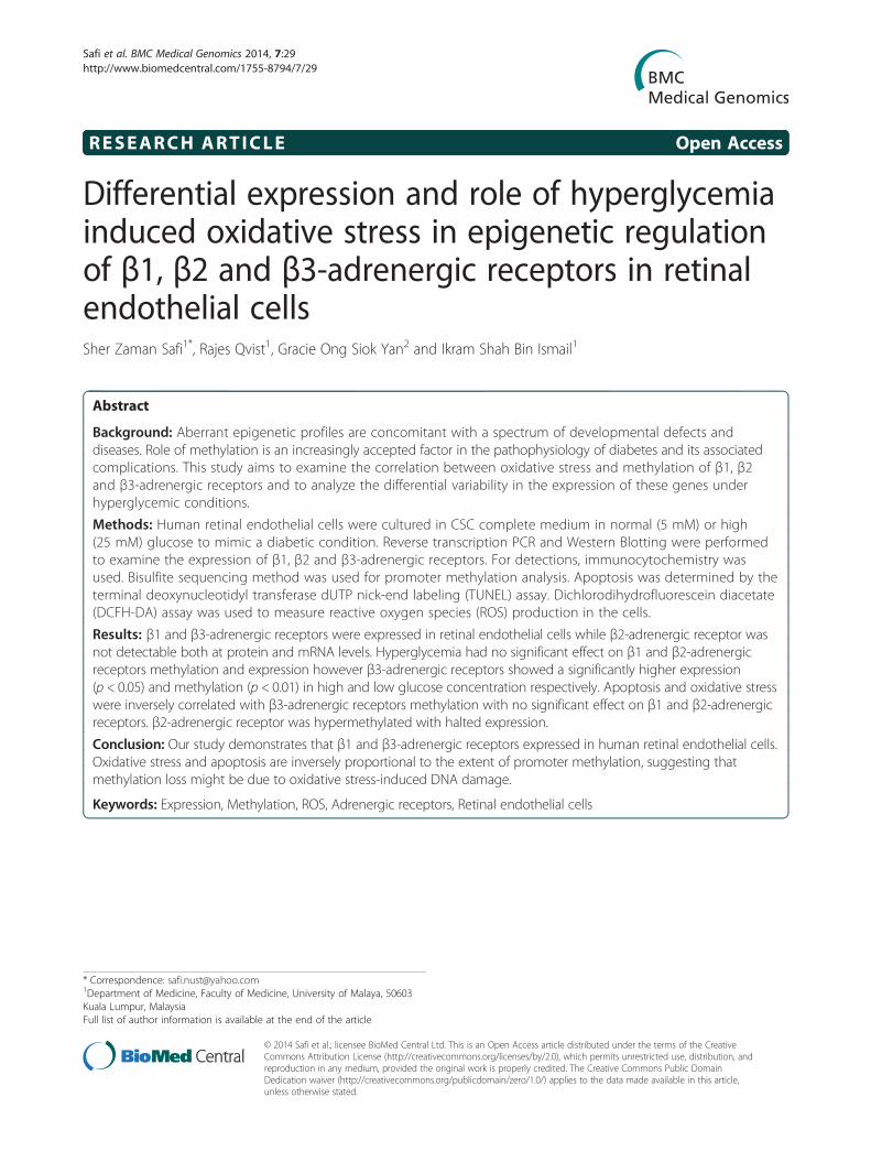
RESEARCH ARTICLE Open Access Differential expression and role of hyperglycemia induced oxidative stress in epigenetic regulation of β1, β2 and β3-adrenergic receptors in retinal endothelial cells Sher Zaman Safi 1* , Rajes Qvist 1 , Gracie Ong Siok Yan 2 and Ikram Shah Bin Ismail 1 Abstract Background: Aberrant epigenetic profiles are concomitant with a spectrum of developmental defects and diseases. Role of methylation is an increasingly accepted factor in the pathophysiology of diabetes and its associated complications. This study aims to examine the correlation between oxidative stress and methylation of β1, β2 and β3-adrenergic receptors and to analyze the differential variability in the expression of these genes under hyperglycemic conditions. Methods: Human retinal endothelial cells were cultured in CSC complete medium in normal (5 mM) or high (25 mM) glucose to mimic a diabetic condition. Reverse transcription PCR and Western Blotting were performed to examine the expression of β1, β2 and β3-adrenergic receptors. For detections, immunocytochemistry was used. Bisulfite sequencing method was used for promoter methylation analysis. Apoptosis was determined by the terminal deoxynucleotidyl transferase dUTP nick-end labeling (TUNEL) assay. Dichlorodihydrofluorescein diacetate (DCFH-DA) assay was used to measure reactive oxygen species (ROS) production in the cells. Results: β1 and β3-adrenergic receptors were expressed in retinal endothelial cells while β2-adrenergic receptor was not detectable both at protein and mRNA levels. Hyperglycemia had no significant effect on β1 and β2-adrenergic receptors methylation and expression however β3-adrenergic receptors showed a significantly higher expression (p< 0.05) and methylation (p< 0.01) in high and low glucose concentration respectively. Apoptosis and oxidative stress were inversely correlated with β3-adrenergic receptors methylation with no significant effect on β1 and β2-adrenergic receptors. β2-adrenergic receptor was hypermethylated with halted expression. Conclusion: Our study demonstrates that β1 and β3-adrenergic receptors expressed in human retinal endothelial cells. Oxidative stress and apoptosis are inversely proportional to the extent of promoter methylation, suggesting that methylation loss might be due to oxidative stress-induced DNA damage. Keywords: Expression, Methylation, ROS, Adrenergic receptors, Retinal endothelial cells * Correspondence: [email protected] 1 Department of Medicine, Faculty of Medicine, University of Malaya, 50603 Kuala Lumpur, Malaysia Full list of author information is available at the end of the article © 2014 Safi et al.; licensee BioMed Central Ltd. This is an Open Access article distributed under the terms of the Creative Commons Attribution License (http://creativecommons.org/licenses/by/2.0), which permits unrestricted use, distribution, and reproduction in any medium, provided the original work is properly credited. The Creative Commons Public Domain Dedication waiver (http://creativecommons.org/publicdomain/zero/1.0/) applies to the data made available in this article, unless otherwise stated. Safi et al. BMC Medical Genomics 2014, 7:29 http://www.biomedcentral.com/1755-8794/7/29
Transcript of RESEARCH ARTICLE Open Access Differential expression … · RESEARCH ARTICLE Open Access...
Safi et al. BMC Medical Genomics 2014, 7:29http://www.biomedcentral.com/1755-8794/7/29
RESEARCH ARTICLE Open Access
Differential expression and role of hyperglycemiainduced oxidative stress in epigenetic regulationof β1, β2 and β3-adrenergic receptors in retinalendothelial cellsSher Zaman Safi1*, Rajes Qvist1, Gracie Ong Siok Yan2 and Ikram Shah Bin Ismail1
Abstract
Background: Aberrant epigenetic profiles are concomitant with a spectrum of developmental defects anddiseases. Role of methylation is an increasingly accepted factor in the pathophysiology of diabetes and its associatedcomplications. This study aims to examine the correlation between oxidative stress and methylation of β1, β2and β3-adrenergic receptors and to analyze the differential variability in the expression of these genes underhyperglycemic conditions.
Methods: Human retinal endothelial cells were cultured in CSC complete medium in normal (5 mM) or high(25 mM) glucose to mimic a diabetic condition. Reverse transcription PCR and Western Blotting were performedto examine the expression of β1, β2 and β3-adrenergic receptors. For detections, immunocytochemistry wasused. Bisulfite sequencing method was used for promoter methylation analysis. Apoptosis was determined by theterminal deoxynucleotidyl transferase dUTP nick-end labeling (TUNEL) assay. Dichlorodihydrofluorescein diacetate(DCFH-DA) assay was used to measure reactive oxygen species (ROS) production in the cells.
Results: β1 and β3-adrenergic receptors were expressed in retinal endothelial cells while β2-adrenergic receptor wasnot detectable both at protein and mRNA levels. Hyperglycemia had no significant effect on β1 and β2-adrenergicreceptors methylation and expression however β3-adrenergic receptors showed a significantly higher expression(p < 0.05) and methylation (p < 0.01) in high and low glucose concentration respectively. Apoptosis and oxidative stresswere inversely correlated with β3-adrenergic receptors methylation with no significant effect on β1 and β2-adrenergicreceptors. β2-adrenergic receptor was hypermethylated with halted expression.
Conclusion: Our study demonstrates that β1 and β3-adrenergic receptors expressed in human retinal endothelial cells.Oxidative stress and apoptosis are inversely proportional to the extent of promoter methylation, suggesting thatmethylation loss might be due to oxidative stress-induced DNA damage.
Keywords: Expression, Methylation, ROS, Adrenergic receptors, Retinal endothelial cells
* Correspondence: [email protected] of Medicine, Faculty of Medicine, University of Malaya, 50603Kuala Lumpur, MalaysiaFull list of author information is available at the end of the article
© 2014 Safi et al.; licensee BioMed Central Ltd. This is an Open Access article distributed under the terms of the CreativeCommons Attribution License (http://creativecommons.org/licenses/by/2.0), which permits unrestricted use, distribution, andreproduction in any medium, provided the original work is properly credited. The Creative Commons Public DomainDedication waiver (http://creativecommons.org/publicdomain/zero/1.0/) applies to the data made available in this article,unless otherwise stated.
Safi et al. BMC Medical Genomics 2014, 7:29 Page 2 of 13http://www.biomedcentral.com/1755-8794/7/29
BackgroundDiabetes is a growing epidemic, caused by excess glucoselevels and body’s inability to produce or regulate insulin[1]. It is predicted that the number of people with dia-betes will increase from 171 million in 2000 to 366 mil-lion by 2030 [2,3]. Diabetes is linked to several vascularpathologies, including severe blindness, atherosclerosis,stroke and renal failure. The growing number of peoplewith diabetes suggests that diabetic retinopathy (DR)and diabetic macular edema (DME) will continue to besight threatening diseases. Distinct morphological abnor-malities in the retinal microvasculature either remainstable or progress to diabetic macular edema or prolifer-ative diabetic retinopathy, the leading causes of severevisual impairment in working-age adults in industrializedcountries [4,5].In vitro and in vivo studies have revealed that hyper-
glycemic environment induces a number of cellularchanges which affect the function and viability of cells[6,7]. Changes in the diabetic retina are due to a varietyof factors including high glucose, oxidative stress andhigh levels of inflammatory markers [8,9]. Reactive oxy-gen species (ROS), especially mitochondrial ROS, play asignificant role in modulating the cellular redox status[10]. Excessive production of ROS and the impairmentof the oxidant/antioxidant balance may in part underliethe pathogenesis of diabetes and its associated compli-cations [11]. Actions of such key pathological mediatorsof diabetes can lead to dysregulated epigenetic mecha-nisms that affect chromatin structure and gene expressionprofiles [12]. DNA methylation is one of the mechanismsfor the epigenetic control of gene expression and aberrantDNA methylation patterns of CpG islands can influencenormal transcriptional regulation in various diseases [13].Beta adrenergic receptors are G protein coupled recep-
tors (GPCRs), initially characterized by Ahlquist in 1948.[14]. Activation of these receptors takes place at thetransmembrane region, which allows ligand binding andelicit a range of cellular actions such as phosphorylationand activation of various signaling pathways [15]. Uponbinding of specific ligands, β1 and β2-adrenergic recep-tors activate the stimulatory G protein resulting in thedissociation of G protein subunits αβ from γ. The αβsubunits are linked to the stimulation of intracellularadenylyl cyclase, followed by the conversion of adeno-sine triphosphate into cyclic adenosine monophosphate(cAMP) and consequently leads to activation of proteinkinase A and phosphorylation of several other substrates[16,17]. However contrary to β1 and β2-adrenergic re-ceptors, β3-adrenergic receptor couple through the in-hibitory G protein (Gi), causing a reduced generation ofcAMP [18]. Work in animal models suggests that ad-renergic receptor agents may promote corneal woundhealing and it is known that cornea has an abundance
of β-adrenergic receptors in both the epithelium and endo-thelium of the cornea [19]. Previous studies demonstratethat β-adrenergic receptors agonists have positive effectson the retina by reducing the inflammatory markers[20-22]. Studies on rats have demonstrated that highly se-lective agonists for β-ARs partially reversed obesity [23]and insulin resistance [24]. Similarly β-adrenergic receptoragonists like isoproterenol inhibits the formation of degen-erative capillaries, prevents apoptosis of cells and reducestumor necrosis factor (TNF) while anatagonists increaseretinal dysfunction [25]. The role of Beta-adrenergic recep-tor agonists and antagonists in the treatments of glaucoma,diabetic retinopathy, and their potential mechanisms of ac-tions, are still under investigation.Type 2 diabetes is a multifactorial disease caused by a
number of genetic, epigenetic and environmental factors[26]. In mammalian cells, DNA methylation takes placeat cytosine in CpG dinucleotides and has been associ-ated with transcriptional silencing [27]. Recent studieshave shown that epigenetic modifications such as DNAmethylation and histone modifications may affect thepathogenesis of type 2 diabetes [28,29]. Differential geneexpressions show dynamic alterations in gene transcrip-tion and mRNA stability that can be influenced by theepigenetic modification of the genome in response tochronic hyperglycemic stress [30,31]. A number of stud-ies have evaluated the epigenetic mechanisms in variousdevelopmental defects and diseases. Oxidative stress andalterations in DNA methylation have also been observedin diabetes, but no clear correlation between these eventshas been demonstrated until now. Very little research hasfocused on the epigenetics of adrenergic receptors in theeye and its relation to diabetic retinopathy. In the presentstudy, we sought to determine which subtypes of Beta-adrenergic receptors that were expressed in retinal endo-thelial cells, and assessed the hyperglycemia induced dif-ferential expression. In addition we analyzed the role ofoxidative stress modulating CpG methylation in the pro-moters of β1, β2 and β3-adrenergic receptors.
MethodsCell cultureHuman retinal endothelial cells (Cell Systems, ACBRI181) were cultured in CSC complete medium (Cell Sys-tems, 4Z0-500) supplemented with 10% serum, 1% Peni-cillin Streptomycin and animal derived growth factors.Cells were grown in attachment factor-coated dishescontaining normal (5 mM, 0.9008 g/L) and high(25 mM, 4.5 g/L) glucose. When the cell reached 80%confluence, they were passaged with the use of a passagereagent group, supplied with the complete medium kit(Cell systems). The cells between 3-6 passages were usedin all experiments and grown in 5% CO2 at 37°C. Mediawas changed every 2 to 3 days.
Safi et al. BMC Medical Genomics 2014, 7:29 Page 3 of 13http://www.biomedcentral.com/1755-8794/7/29
ImmunocytochemistryThe beta-adrenergic receptors were identified as describedearlier [32], with minor changes. Retinal endothelial cellswere seeded (50,000/well) onto 6-well plates with sterilecoverslips in medium containing 5 mM (normal glucose)and 25 mM (high glucose). They were allowed to attachand proliferate in the respective medium for 2 days. Thecells were fixed with cold methanol for 10 minutes at -20°C,rinsed twice with PBS, blocked at room temperature with0.5% Tween (v/v) and 2% BSA (w/v) in PBS (blocking solu-tion) for 1 hour and rinsed again twice with PBS. The wellswere incubated overnight with primary antibodies againstβ1 adrenergic receptor (Santa Cruz, sc-568), β2-adrenergicreceptor (Santa Cruz, sc-569) and β3-adrenergic receptor(Santa Cruz, sc-1472) in blocking solution at a dilutionof 1:100. Anti-β-actin (sc-7210) antibodies were used aspositive control. The following day, the cells werewashed twice with PBS and incubated for 1 hour indark room with secondary antibodies conjugated withfluorescein isothiocyanate (FITC) in blocking solutionat a dilution of 1:200. After rinsing twice with PBS, thecoverslips were mounted with antifading medium (LifeTechnologies, ProLong Gold) and analyzed under fluo-rescence microscope.
RT PCRTotal RNA from Human retinal endothelial cells (HREC)was isolated using RNeasy commercial kit (Qiagen, cat no74104) according to the manufacturer’s protocol and quan-tified spectrophotometrically. First-strand cDNA was syn-thesized with 1 μg of each DNA free total RNA, usingiScript reverse transcription supermix (Biorad USA, cat no170-8841) containing Moloney murine leukemia virus re-verse transcriptase, RNase inhibitor, dNTPs, oligo-(dT),random primers, buffer, MgCl2 and stabilizers. No-RT con-trol with the same amount of RNA but without RT was in-cluded to check genomic DNA carryover in RNA.Reaction conditions for cDNA synthesis were 25°C for5 minutes, 42°C for 50 minutes and 85°C for 5 minutes.Equal amounts of cDNA were subsequently used foramplification in a 50 μl PCR reaction using Emeral-dAmp PCR Master Mix (Takara, Japan), containing Taqpolymerase, dNTPs, buffer and primers targeting β1-adrenergic receptor, β2-adrenergic receptor and β3-adrenergic receptor. PCR was performed with an initialdenaturation step at 94°C for 3 minutes, followed by de-naturation at 94°C for 30 seconds, annealing (60°C for β1-AR; 58°C for β2-AR; 61°C for β3-AR each for 30 seconds)and extension at 72°C for 1 minute. GAPDH was used asquantitative control in each PCR reaction. The PCR pro-ducts were separated on 1.5% agarose gels and visualizedusing ethidium bromide staining under UV transillumin-ation. Primers sequences of β1-AR, β2-AR and β3-AR aregiven in the Table 1.
Western Blot analysisHuman retinal endothelial cells were lysed, centrifugedand proteins were extracted according to the manufac-turer’s protocol (Sigma). Bradford assay (Bio-Rad Labora-tories) was used to quantify the protein concentrations inthe supernatants. 30 μg proteins of the lysate from highglucose and low glucose were loaded in each well and sep-arated on 10% SDS-PAGE (Precast gels, Bio-Rad, cat no456-1093). Gels were electrophoretically transferred tonitrocellulose membranes and blocked for 60 min withTris-buffered saline (TBS) containing 5% nonfat milk(Bio-Rad, USA, cat no 170-6404) and 0.1% Tween-20.Blots were incubated overnight at 4°C with primary anti-bodies (1:3000) against β1-adrenergic receptor (SantaCruz, sc-568), β2-adrenergic receptor (Santa Cruz, sc-569)and β3-adrenergic receptor (Santa Cruz, sc-1472). Anti-β-actin (sc-7210) antibodies were used to ensure the qualityof protein separation and loading contents. After extensivewashes, the membranes were incubated with HRP-conjugated IgG secondary antibodies (Santa Cruz, sc-2004) and visualized with enhanced chemiluminescence(Amersham Life Sciences, UK) using gel imaging system(Biospectrum 410, UVP).
Terminal deoxynucleotidyl transferase dUTP nick endlabeling (TUNEL) assayDNA damage was investigated and quantified with a col-orimetric apoptosis detection kit (Titer TACS; R&D Sys-tem) according to the manufacturer’s protocol. Kitcontained TUNEL staining in a 96-well format. Retinalendothelial cells were cultured in medium containing5 mM (normal) and 25 mM (high) glucose. Cells werethen transferred into a 96-well plate (1 × 10⁵ cells/well)and fixed with 3.7% buffered formaldehyde for 5 minutesfollowed by washing with PBS. Washing was followed bypermeabilization of cells with 100% methanol for 20 mi-nutes (at room temperature) and again washing withPBS. Following the manufacturer’s instructions, cellswere then subjected to labeling procedure and the reactionwas stopped with 0.2 N HCl after 30 minutes. Nucleasetreated control was used to confirm the permeabilizationand labeling reaction. The absorbance was measured at450 nm with microplate reader.
Dichloro-dihydro-fluorescein diacetate (DCFH-DA) assayThe intracellular ROS (reactive oxygen species) was mea-sured by using 2′7′-dichloro-dihydro-fluorescin diacetate(DCFH-DA) reagent. The DCFH-DA enters through thecell membrane and enzymatically hydrolyzed by intracel-lular esterases to non-fluorescent dichlorofluorescin-diacetate (DCFHDA), which is then oxidized to highlyfluorescent dichlorofluorescin (DCF) in the presence ofintracellular reactive oxygen species. Retinal endothelialcells were seeded in 96-well black plates and allowed to
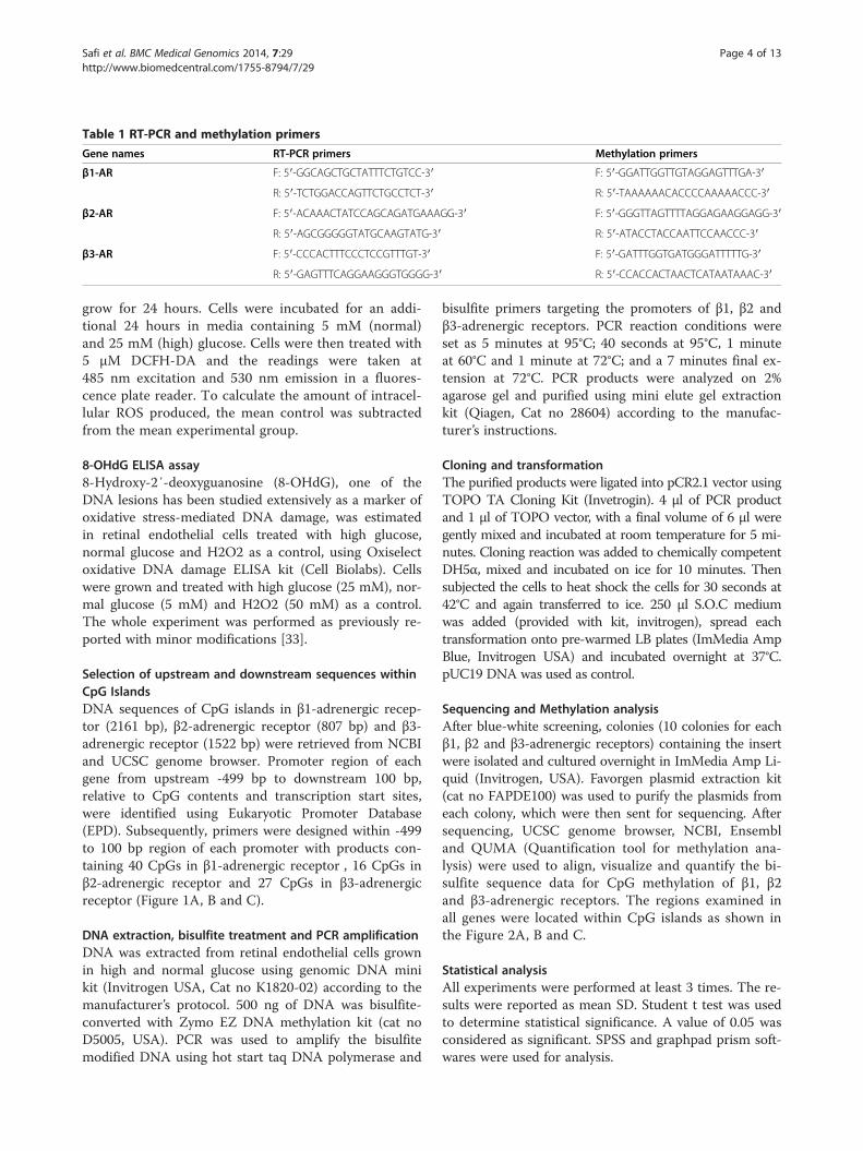
Table 1 RT-PCR and methylation primers
Gene names RT-PCR primers Methylation primers
β1-AR F: 5′-GGCAGCTGCTATTTCTGTCC-3′ F: 5′-GGATTGGTTGTAGGAGTTTGA-3′
R: 5′-TCTGGACCAGTTCTGCCTCT-3′ R: 5′-TAAAAAACACCCCAAAAACCC-3′
β2-AR F: 5′-ACAAACTATCCAGCAGATGAAAGG-3′ F: 5′-GGGTTAGTTTTAGGAGAAGGAGG-3′
R: 5′-AGCGGGGGTATGCAAGTATG-3′ R: 5′-ATACCTACCAATTCCAACCC-3′
β3-AR F: 5′-CCCACTTTCCCTCCGTTTGT-3′ F: 5′-GATTTGGTGATGGGATTTTTG-3′
R: 5′-GAGTTTCAGGAAGGGTGGGG-3′ R: 5′-CCACCACTAACTCATAATAAAC-3′
Safi et al. BMC Medical Genomics 2014, 7:29 Page 4 of 13http://www.biomedcentral.com/1755-8794/7/29
grow for 24 hours. Cells were incubated for an addi-tional 24 hours in media containing 5 mM (normal)and 25 mM (high) glucose. Cells were then treated with5 μM DCFH-DA and the readings were taken at485 nm excitation and 530 nm emission in a fluores-cence plate reader. To calculate the amount of intracel-lular ROS produced, the mean control was subtractedfrom the mean experimental group.
8-OHdG ELISA assay8-Hydroxy-2′-deoxyguanosine (8-OHdG), one of theDNA lesions has been studied extensively as a marker ofoxidative stress-mediated DNA damage, was estimatedin retinal endothelial cells treated with high glucose,normal glucose and H2O2 as a control, using Oxiselectoxidative DNA damage ELISA kit (Cell Biolabs). Cellswere grown and treated with high glucose (25 mM), nor-mal glucose (5 mM) and H2O2 (50 mM) as a control.The whole experiment was performed as previously re-ported with minor modifications [33].
Selection of upstream and downstream sequences withinCpG IslandsDNA sequences of CpG islands in β1-adrenergic recep-tor (2161 bp), β2-adrenergic receptor (807 bp) and β3-adrenergic receptor (1522 bp) were retrieved from NCBIand UCSC genome browser. Promoter region of eachgene from upstream -499 bp to downstream 100 bp,relative to CpG contents and transcription start sites,were identified using Eukaryotic Promoter Database(EPD). Subsequently, primers were designed within -499to 100 bp region of each promoter with products con-taining 40 CpGs in β1-adrenergic receptor , 16 CpGs inβ2-adrenergic receptor and 27 CpGs in β3-adrenergicreceptor (Figure 1A, B and C).
DNA extraction, bisulfite treatment and PCR amplificationDNA was extracted from retinal endothelial cells grownin high and normal glucose using genomic DNA minikit (Invitrogen USA, Cat no K1820-02) according to themanufacturer’s protocol. 500 ng of DNA was bisulfite-converted with Zymo EZ DNA methylation kit (cat noD5005, USA). PCR was used to amplify the bisulfitemodified DNA using hot start taq DNA polymerase and
bisulfite primers targeting the promoters of β1, β2 andβ3-adrenergic receptors. PCR reaction conditions wereset as 5 minutes at 95°C; 40 seconds at 95°C, 1 minuteat 60°C and 1 minute at 72°C; and a 7 minutes final ex-tension at 72°C. PCR products were analyzed on 2%agarose gel and purified using mini elute gel extractionkit (Qiagen, Cat no 28604) according to the manufac-turer’s instructions.
Cloning and transformationThe purified products were ligated into pCR2.1 vector usingTOPO TA Cloning Kit (Invetrogin). 4 μl of PCR productand 1 μl of TOPO vector, with a final volume of 6 μl weregently mixed and incubated at room temperature for 5 mi-nutes. Cloning reaction was added to chemically competentDH5α, mixed and incubated on ice for 10 minutes. Thensubjected the cells to heat shock the cells for 30 seconds at42°C and again transferred to ice. 250 μl S.O.C mediumwas added (provided with kit, invitrogen), spread eachtransformation onto pre-warmed LB plates (ImMedia AmpBlue, Invitrogen USA) and incubated overnight at 37°C.pUC19 DNA was used as control.
Sequencing and Methylation analysisAfter blue-white screening, colonies (10 colonies for eachβ1, β2 and β3-adrenergic receptors) containing the insertwere isolated and cultured overnight in ImMedia Amp Li-quid (Invitrogen, USA). Favorgen plasmid extraction kit(cat no FAPDE100) was used to purify the plasmids fromeach colony, which were then sent for sequencing. Aftersequencing, UCSC genome browser, NCBI, Ensembland QUMA (Quantification tool for methylation ana-lysis) were used to align, visualize and quantify the bi-sulfite sequence data for CpG methylation of β1, β2and β3-adrenergic receptors. The regions examined inall genes were located within CpG islands as shown inthe Figure 2A, B and C.
Statistical analysisAll experiments were performed at least 3 times. The re-sults were reported as mean SD. Student t test was usedto determine statistical significance. A value of 0.05 wasconsidered as significant. SPSS and graphpad prism soft-wares were used for analysis.
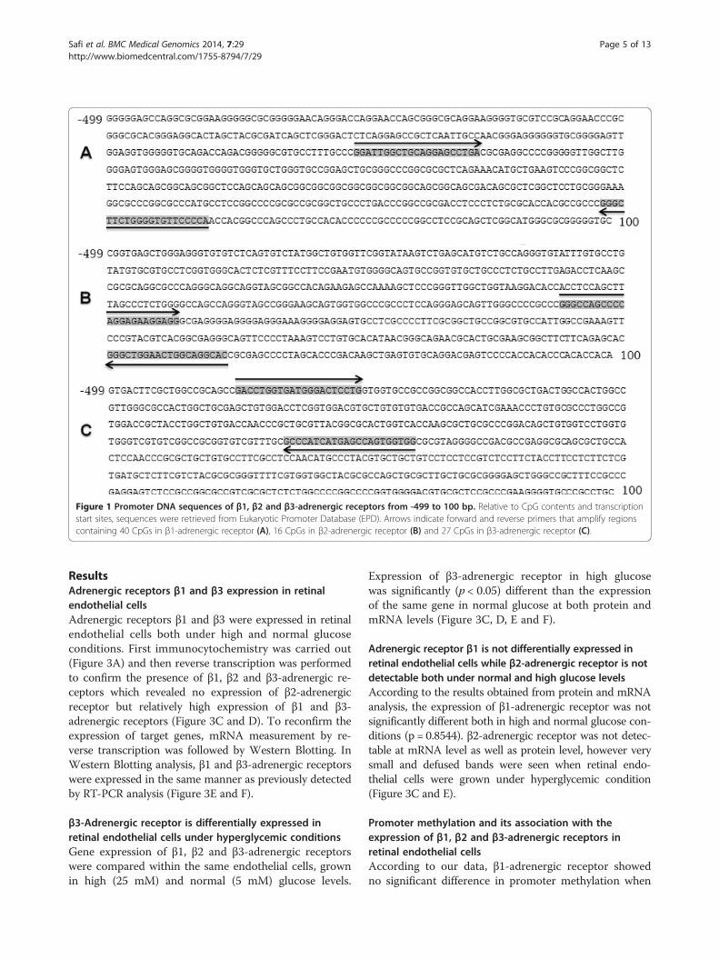
Figure 1 Promoter DNA sequences of β1, β2 and β3-adrenergic receptors from -499 to 100 bp. Relative to CpG contents and transcriptionstart sites, sequences were retrieved from Eukaryotic Promoter Database (EPD). Arrows indicate forward and reverse primers that amplify regionscontaining 40 CpGs in β1-adrenergic receptor (A), 16 CpGs in β2-adrenergic receptor (B) and 27 CpGs in β3-adrenergic receptor (C).
Safi et al. BMC Medical Genomics 2014, 7:29 Page 5 of 13http://www.biomedcentral.com/1755-8794/7/29
ResultsAdrenergic receptors β1 and β3 expression in retinalendothelial cellsAdrenergic receptors β1 and β3 were expressed in retinalendothelial cells both under high and normal glucoseconditions. First immunocytochemistry was carried out(Figure 3A) and then reverse transcription was performedto confirm the presence of β1, β2 and β3-adrenergic re-ceptors which revealed no expression of β2-adrenergicreceptor but relatively high expression of β1 and β3-adrenergic receptors (Figure 3C and D). To reconfirm theexpression of target genes, mRNA measurement by re-verse transcription was followed by Western Blotting. InWestern Blotting analysis, β1 and β3-adrenergic receptorswere expressed in the same manner as previously detectedby RT-PCR analysis (Figure 3E and F).
β3-Adrenergic receptor is differentially expressed inretinal endothelial cells under hyperglycemic conditionsGene expression of β1, β2 and β3-adrenergic receptorswere compared within the same endothelial cells, grownin high (25 mM) and normal (5 mM) glucose levels.
Expression of β3-adrenergic receptor in high glucosewas significantly (p < 0.05) different than the expressionof the same gene in normal glucose at both protein andmRNA levels (Figure 3C, D, E and F).
Adrenergic receptor β1 is not differentially expressed inretinal endothelial cells while β2-adrenergic receptor is notdetectable both under normal and high glucose levelsAccording to the results obtained from protein and mRNAanalysis, the expression of β1-adrenergic receptor was notsignificantly different both in high and normal glucose con-ditions (p = 0.8544). β2-adrenergic receptor was not detec-table at mRNA level as well as protein level, however verysmall and defused bands were seen when retinal endo-thelial cells were grown under hyperglycemic condition(Figure 3C and E).
Promoter methylation and its association with theexpression of β1, β2 and β3-adrenergic receptors inretinal endothelial cellsAccording to our data, β1-adrenergic receptor showedno significant difference in promoter methylation when
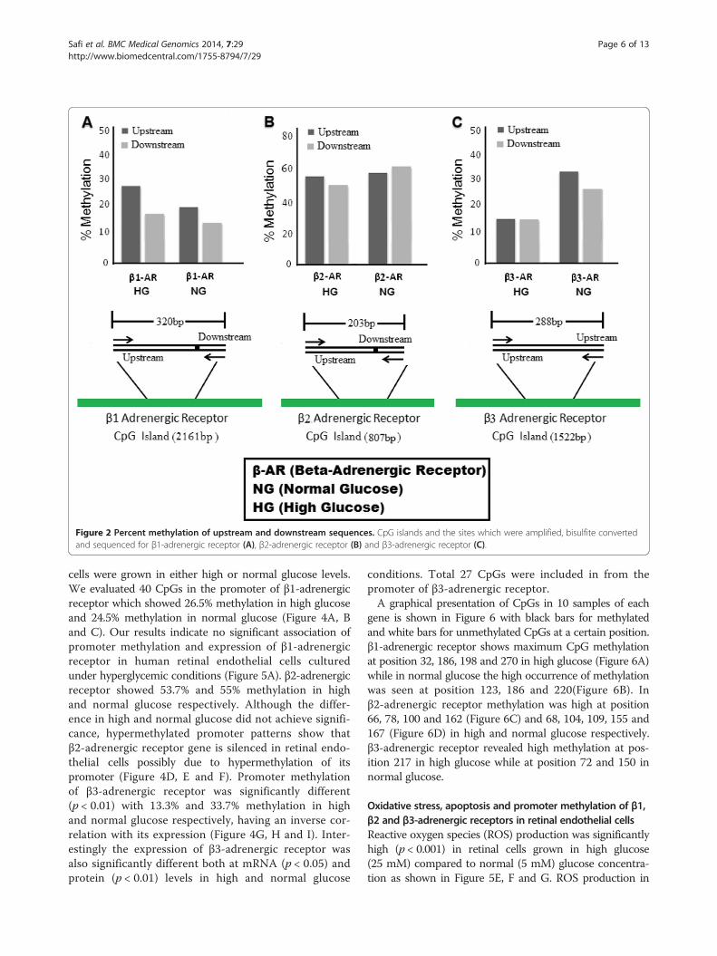
Figure 2 Percent methylation of upstream and downstream sequences. CpG islands and the sites which were amplified, bisulfite convertedand sequenced for β1-adrenergic receptor (A), β2-adrenergic receptor (B) and β3-adrenergic receptor (C).
Safi et al. BMC Medical Genomics 2014, 7:29 Page 6 of 13http://www.biomedcentral.com/1755-8794/7/29
cells were grown in either high or normal glucose levels.We evaluated 40 CpGs in the promoter of β1-adrenergicreceptor which showed 26.5% methylation in high glucoseand 24.5% methylation in normal glucose (Figure 4A, Band C). Our results indicate no significant association ofpromoter methylation and expression of β1-adrenergicreceptor in human retinal endothelial cells culturedunder hyperglycemic conditions (Figure 5A). β2-adrenergicreceptor showed 53.7% and 55% methylation in highand normal glucose respectively. Although the differ-ence in high and normal glucose did not achieve signifi-cance, hypermethylated promoter patterns show thatβ2-adrenergic receptor gene is silenced in retinal endo-thelial cells possibly due to hypermethylation of itspromoter (Figure 4D, E and F). Promoter methylationof β3-adrenergic receptor was significantly different(p < 0.01) with 13.3% and 33.7% methylation in highand normal glucose respectively, having an inverse cor-relation with its expression (Figure 4G, H and I). Inter-estingly the expression of β3-adrenergic receptor wasalso significantly different both at mRNA (p < 0.05) andprotein (p < 0.01) levels in high and normal glucose
conditions. Total 27 CpGs were included in from thepromoter of β3-adrenergic receptor.A graphical presentation of CpGs in 10 samples of each
gene is shown in Figure 6 with black bars for methylatedand white bars for unmethylated CpGs at a certain position.β1-adrenergic receptor shows maximum CpG methylationat position 32, 186, 198 and 270 in high glucose (Figure 6A)while in normal glucose the high occurrence of methylationwas seen at position 123, 186 and 220(Figure 6B). Inβ2-adrenergic receptor methylation was high at position66, 78, 100 and 162 (Figure 6C) and 68, 104, 109, 155 and167 (Figure 6D) in high and normal glucose respectively.β3-adrenergic receptor revealed high methylation at pos-ition 217 in high glucose while at position 72 and 150 innormal glucose.
Oxidative stress, apoptosis and promoter methylation of β1,β2 and β3-adrenergic receptors in retinal endothelial cellsReactive oxygen species (ROS) production was significantlyhigh (p < 0.001) in retinal cells grown in high glucose(25 mM) compared to normal (5 mM) glucose concentra-tion as shown in Figure 5E, F and G. ROS production in
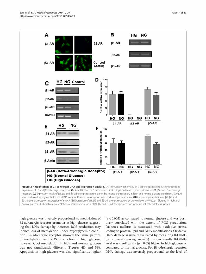
Figure 3 Amplification of CT converted DNA and expression analysis. (A) Immunocytochemistry of β-adrenergic receptors, showing strongexpression of β1and β3-adrenergic receptors. (B) Amplification of CT converted DNA using bisulfite converted primers for β1, β2 and β3-adrenergicreceptors. (C) Expression levels of β1, β2 and β3-adrenergic receptors genes by reverse transcription, in high and normal glucose conditions. GAPDHwas used as a loading control while cDNA without Reverse Transcriptase was used as negative control. (D) Graphical presentation of β1, β2 andβ3-adrenergic receptors expression of mRNA (E) Expression of β1, β2 and β3-adrenergic receptors at protein level by Western Blotting in high andnormal glucose. (F) Graphical presentation of relative expression of β1, β2 and β3-adrenergic receptors genes in retinal endothelial genes.
Safi et al. BMC Medical Genomics 2014, 7:29 Page 7 of 13http://www.biomedcentral.com/1755-8794/7/29
high glucose was inversely proportional to methylation ofβ3-adrenergic receptor promoter in high glucose, suggest-ing that DNA damage by increased ROS production mayinduce loss of methylation under hyperglycemic condi-tion. β2-adrenergic receptor showed the same patternof methylation and ROS production in high glucose;however CpG methylation in high and normal glucosewas not significantly different (Figures 4D and 5B).Apoptosis in high glucose was also significantly higher
(p < 0.005) as compared to normal glucose and was posi-tively correlated with the extent of ROS production.Diabetes mellitus is associated with oxidative stress,leading to protein, lipid and DNA modifications. OxidativeDNA damage is usually evaluated by measuring 8-OHdG(8-hydroxy-2-deoxy-guanosine). In our results 8-OHdGlevel was significantly (p < 0.05) higher in high glucose ascompared to normal glucose. For β3-adrenergic receptor,DNA damage was inversely proportional to the level of
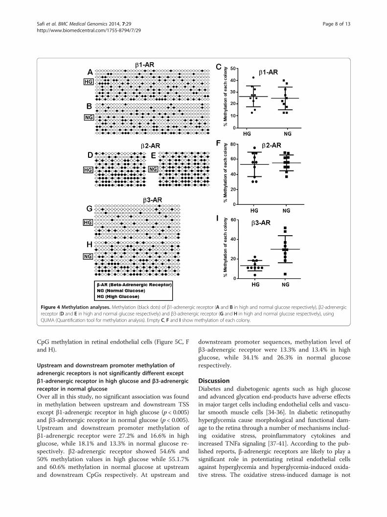
Figure 4 Methylation analyses. Methylation (black dots) of β1-adrenergic receptor (A and B in high and normal glucose respectively), β2-adrenergicreceptor (D and E in high and normal glucose respectively) and β3-adrenergic receptor (G and H in high and normal glucose respectively), usingQUMA (Quantification tool for methylation analysis). Empty C, F and I show methylation of each colony.
Safi et al. BMC Medical Genomics 2014, 7:29 Page 8 of 13http://www.biomedcentral.com/1755-8794/7/29
CpG methylation in retinal endothelial cells (Figure 5C, Fand H).
Upstream and downstream promoter methylation ofadrenergic receptors is not significantly different exceptβ1-adrenergic receptor in high glucose and β3-adrenergicreceptor in normal glucoseOver all in this study, no significant association was foundin methylation between upstream and downstream TSSexcept β1-adrenergic receptor in high glucose (p < 0.005)and β3-adrenergic receptor in normal glucose (p < 0.005).Upstream and downstream promoter methylation ofβ1-adrenergic receptor were 27.2% and 16.6% in highglucose, while 18.1% and 13.3% in normal glucose re-spectively. β2-adrenergic receptor showed 54.6% and50% methylation values in high glucose while 55.1.7%and 60.6% methylation in normal glucose at upstreamand downstream CpGs respectively. At upstream and
downstream promoter sequences, methylation level ofβ3-adrenergic receptor were 13.3% and 13.4% in highglucose, while 34.1% and 26.3% in normal glucoserespectively.
DiscussionDiabetes and diabetogenic agents such as high glucoseand advanced glycation end-products have adverse effectsin major target cells including endothelial cells and vascu-lar smooth muscle cells [34-36]. In diabetic retinopathyhyperglycemia cause morphological and functional dam-age to the retina through a number of mechanisms includ-ing oxidative stress, proinflammatory cytokines andincreased TNFa signaling [37-41]. According to the pub-lished reports, β-adrenergic receptors are likely to play asignificant role in potentiating retinal endothelial cellsagainst hyperglycemia and hyperglycemia-induced oxida-tive stress. The oxidative stress-induced damage is not
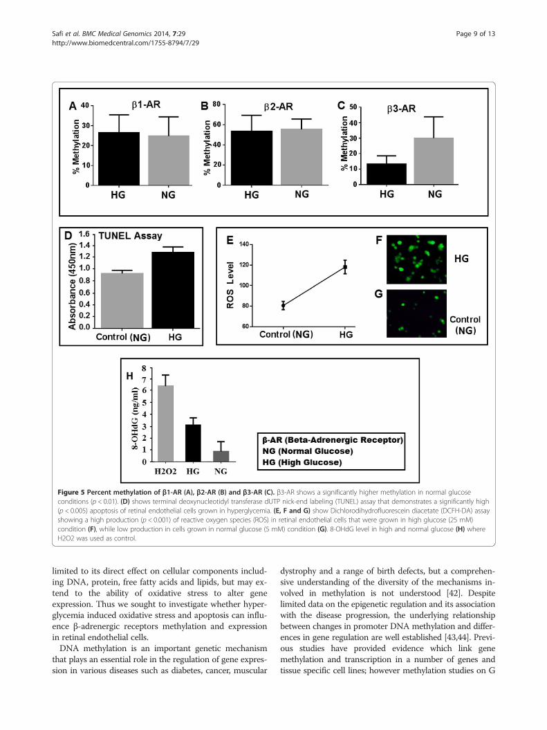
Figure 5 Percent methylation of β1-AR (A), β2-AR (B) and β3-AR (C). β3-AR shows a significantly higher methylation in normal glucoseconditions (p < 0.01). (D) shows terminal deoxynucleotidyl transferase dUTP nick-end labeling (TUNEL) assay that demonstrates a significantly high(p < 0.005) apoptosis of retinal endothelial cells grown in hyperglycemia. (E, F and G) show Dichlorodihydrofluorescein diacetate (DCFH-DA) assayshowing a high production (p < 0.001) of reactive oxygen species (ROS) in retinal endothelial cells that were grown in high glucose (25 mM)condition (F), while low production in cells grown in normal glucose (5 mM) condition (G). 8-OHdG level in high and normal glucose (H) whereH2O2 was used as control.
Safi et al. BMC Medical Genomics 2014, 7:29 Page 9 of 13http://www.biomedcentral.com/1755-8794/7/29
limited to its direct effect on cellular components includ-ing DNA, protein, free fatty acids and lipids, but may ex-tend to the ability of oxidative stress to alter geneexpression. Thus we sought to investigate whether hyper-glycemia induced oxidative stress and apoptosis can influ-ence β-adrenergic receptors methylation and expressionin retinal endothelial cells.DNA methylation is an important genetic mechanism
that plays an essential role in the regulation of gene expres-sion in various diseases such as diabetes, cancer, muscular
dystrophy and a range of birth defects, but a comprehen-sive understanding of the diversity of the mechanisms in-volved in methylation is not understood [42]. Despitelimited data on the epigenetic regulation and its associationwith the disease progression, the underlying relationshipbetween changes in promoter DNA methylation and differ-ences in gene regulation are well established [43,44]. Previ-ous studies have provided evidence which link genemethylation and transcription in a number of genes andtissue specific cell lines; however methylation studies on G
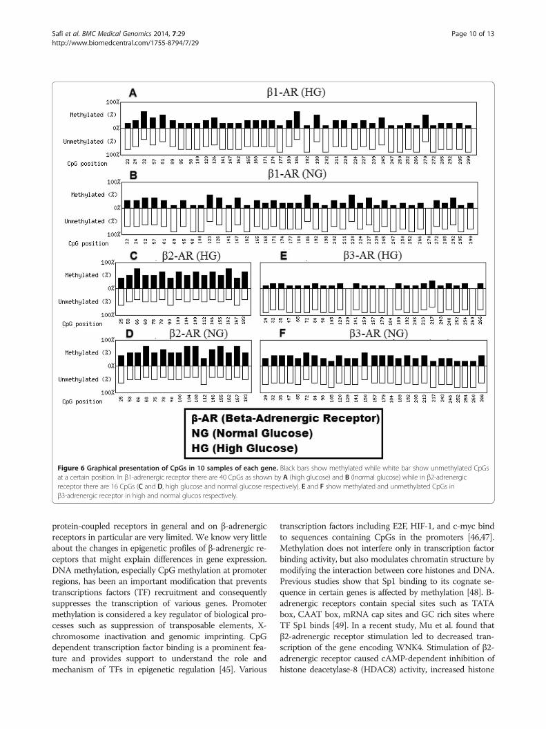
Figure 6 Graphical presentation of CpGs in 10 samples of each gene. Black bars show methylated while white bar show unmethylated CpGsat a certain position. In β1-adrenergic receptor there are 40 CpGs as shown by A (high glucose) and B (lnormal glucose) while in β2-adrenergicreceptor there are 16 CpGs (C and D, high glucose and normal glucose respectively). E and F show methylated and unmethylated CpGs inβ3-adrenergic receptor in high and normal glucos respectively.
Safi et al. BMC Medical Genomics 2014, 7:29 Page 10 of 13http://www.biomedcentral.com/1755-8794/7/29
protein-coupled receptors in general and on β-adrenergicreceptors in particular are very limited. We know very littleabout the changes in epigenetic profiles of β-adrenergic re-ceptors that might explain differences in gene expression.DNA methylation, especially CpG methylation at promoterregions, has been an important modification that preventstranscriptions factors (TF) recruitment and consequentlysuppresses the transcription of various genes. Promotermethylation is considered a key regulator of biological pro-cesses such as suppression of transposable elements, X-chromosome inactivation and genomic imprinting. CpGdependent transcription factor binding is a prominent fea-ture and provides support to understand the role andmechanism of TFs in epigenetic regulation [45]. Various
transcription factors including E2F, HIF-1, and c-myc bindto sequences containing CpGs in the promoters [46,47].Methylation does not interfere only in transcription factorbinding activity, but also modulates chromatin structure bymodifying the interaction between core histones and DNA.Previous studies show that Sp1 binding to its cognate se-quence in certain genes is affected by methylation [48]. Β-adrenergic receptors contain special sites such as TATAbox, CAAT box, mRNA cap sites and GC rich sites whereTF Sp1 binds [49]. In a recent study, Mu et al. found thatβ2-adrenergic receptor stimulation led to decreased tran-scription of the gene encoding WNK4. Stimulation of β2-adrenergic receptor caused cAMP-dependent inhibition ofhistone deacetylase-8 (HDAC8) activity, increased histone
Safi et al. BMC Medical Genomics 2014, 7:29 Page 11 of 13http://www.biomedcentral.com/1755-8794/7/29
acetylation and consequently binding of the glucocorticoidreceptor to a negative glucocorticoid responsive elementin the promoter region [50]. For our results, we alsohypothesize that hypermethylation of β2-adrenergic recep-tor might prevent the core transcription factors such asSp1 to bind with its promoter and hence causing halted ex-pression of β2-adrenergic receptor.In our study, protein and mRNA expression of β1 and
β3-adrenergic receptors were confirmed in retinal endo-thelial cells however β2-adrenergic receptor was hard todetect both at mRNA and protein levels. According toseveral studies including our previous unpublished data,hyperglycemia increases the oxidative stress in several celllines. Kong et al. and Takahata et al. in two different stu-dies found that upregulation of β3-adrenergic receptor isassociated with increased oxidative stress [51] while func-tional expression of β2-adrenergic receptor is responsiblefor protection against oxidative stress through promotionof glutathione synthesis [52]. In 2003, Steinle et al. re-ported that β3-adrenergic receptor mediates migrationand proliferation of retinal endothelial Cells. They UsedBRL37344 agonist to stimulate the β3-adrenergic receptorson retinal endothelial cells and showed that these recep-tors could express in retinal endothelial cells. They re-ported that BRL37344-mediated migration could bealienated by prior inhibitors administration of Akt, PI3K,MEK and MMP-2/MMP-9 and hypothesized that PI3Kmay directly be linked to the β3-adrenergic receptor [53].These studies led us to question whether our hypergly-cemic endothelial cells with no agonist (BRL37344) andincreased oxidative stress would differentially mediate thepromoter methylation and expression of β3, β2 and β1-adrenergic receptors. In our study, hyperglycemic endo-thelial cells had significantly higher oxidative stress andconsequently a higher expression of β3-adrenergic re-ceptor. Methylation in β3-adrenergic receptor was sig-nificantly low in cells with significantly high apoptosisand oxidative stress as per our hypothesis.There is an exquisite interrelationship between methyla-
tion and oxidative stress. Glutathione is depleted by theexcess of oxidative stress; consequently impairing the one-carbon cycle and causes undermethylation. However onthe other hand glutathione, cysteine, and metallothioneinget depleted by undermethylation with oxidative stressoverload. Increased production of ROS can directly or in-directly cause a wide range of DNA lesions including basemodifications, strand breakage, deletions and chromo-somal rearrangements. Such alterations have been shownto impede the ability of DNA to function as a substratefor the DNA methyl transferases (DNMTs), resulting inglobal hypomethylation [54-56]. In our data β3-adrenergicreceptor is hypomethylated with increased expression.The reason behind hyperglycemia led increase in β3-adrenergic receptor is not clear however Walker and
Steinle suggest that the level of norepinephrine are signifi-cantly reduced as early as 1 week after streptozotocin(STZ) treatment, this loss of norepinephrine may conse-quently lead to denervation supersensitivity and increasedexpression of β1-adrenergic receptor in Muller cells [33].Hypermethylation and low expression of β2-adrenergic re-ceptor is another interesting finding in our study. In ourstudy we selected upstream and downstream regions,however high and normal glucose induced no variationand no association was seen in methylation between up-stream and downstream sequences except β1-adrenergicreceptor in high glucose and β3-adrenergic receptor innormal glucose.
ConclusionOur study demonstrates that β1 and β3-adrenergic recep-tors β-ARs expressed in human retinal endothelial cellsHREC. Oxidative stress and apoptosis are inversely propor-tional to the extent of promoter methylation, suggestingthat methylation loss might be due to oxidative stress-induced DNA damage. Collectively this study may help inunderstanding the pathophysiology of diabetic retinopathyand epigenetic regulation of β-adrenergic receptors.
Competing interestsThe authors declare that they have no competing interests.
Authors’ contributionsSZS carried out the experiments and design of study. RQ helped in revisingthe manuscript and data analysis. The entire study was overseen by ISBI. Allauthors contributed to writing the manuscript and approved the finalmanuscript.
AcknowledgementsThis work was supported by UMRG grant no RG528-13HTM, University of Malaya.
Author details1Department of Medicine, Faculty of Medicine, University of Malaya, 50603Kuala Lumpur, Malaysia. 2Department of Anesthesiology, Faculty of Medicine,University of Malaya, 50603 Kuala Lumpur, Malaysia.
Received: 4 October 2013 Accepted: 20 May 2014Published: 30 May 2014
References1. Newman JC, He W, Verdin E: Mitochondrial protein acylation and
intermediary metabolism: regulation by sirtuins and implications formetabolic disease. J Biol Chem 2012, 287:42463–42443.
2. Wild S, Roglic G, Green A, Sicree R, King H: Global prevalence of diabetes:estimates for the year 2000 and projections for 2030. Diabetes Care 2004,27:1047–1053.
3. World Health Organization: Diabetes Action Now, An initiative of the worldhealth organization and the international diabetes federation. Geneva: WorldHealth Organization; 2004.
4. Kempen JH, O’Colmain BJ, Leske MC, Haffner SM, Klein R, Moss SE, TaylorHR, Hamman RF: The prevalence of diabetic retinopathy among adults inthe United States. Arch Ophthalmol 2004, 122:552–563.
5. Williams R, Airey M, Baxter H, Forrester J, Kennedy-Martin T, Girach A: Epidemiologyof diabetic retinopathy and macular oedema: a systematic review.Eye 2004, 18:963–983.
6. Kowluru RA: Diabetic Retinopathy: Mitochondrial Dysfunction and RetinalCapillary Cell Death. Antioxid Redox Signal 2005, 7:1581.
7. Kowluru RA, Kennedy A: Therapeutic potential of anti-oxidants and diabeticretinopathy. Expert Opin Investig Drugs 2001, 10:1665–1676.
Safi et al. BMC Medical Genomics 2014, 7:29 Page 12 of 13http://www.biomedcentral.com/1755-8794/7/29
8. El-Asrar AMA, Dralands L, Missotten L, Al-Jadaan IA, Geboes K: Expression ofApoptosis Markers in the Retinas of Human Subjects with Diabetes.Invest Ophthalmol Vis Sci 2004, 45:2760–2766.
9. Joussen AM, Doehmen S, Le ML: TNF-alpha mediated apoptosis plays animportant role in the development of early diabetic retinopathy andlong-term histopathological alterations. Mol Vis 2009, 15:1418–1428.
10. Cheng X, Siow RC, Mann GE: Impaired redox signaling and antioxidantgene expression in endothelial cells in diabetes: a role for mitochondriaand the nuclear factor-E2-related factor 2-Kelch-like ECH-associatedprotein 1 defense pathway. Antioxid Redox Signal 2011, 14:469–487.
11. Brownlee M: Biochemistry and molecular cell biology of diabeticcomplications. Nature 2001, 414:813–820.
12. Reddy MA, Natarajan R: Epigenetic mechanisms in diabetic vascularcomplications. Cardiovasc Res 2011, 90(3):421–429.
13. Schinke C, Mo Y, Yu Y, Amiri K, Sosman J, Greally J, Verma A: Aberrant DNAmethylation in malignant melanoma. Melanoma Res 2010, 20:253–265.
14. Bylund DB, Eikenberg DC, Hieble JP, Langer SZ, Lefkowitz RJ, Minneman KP,Molinoff PB, Ruffolo RR Jr, Trendelenburg U: International Union ofPharmacology nomenclature of adrenoceptors. Pharmacol Rev 1994,46(2):121–136.
15. Chakraborti S, Chakraborti T, Shaw G: [beta]-adrenergic mechanisms incardiac diseases: A perspective. Cell Signal 2000, 12:499–513.
16. Stiles GL, Caron MG, Lefkowitz RJ: Beta-adrenergic receptors: biochemicalmechanisms of physiological regulation. Physiol Rev 1984, 64:661–743.
17. Wenzel-Seifert K, Liu HY, Seifert R: Similarities and differences in thecoupling of human beta1- and beta2-adrenoceptors to Gs(alpha) splicevariants. Biochem Pharmacol 2002, 64:9–20.
18. Gauthier C, Tavernier G, Charpentier F, Langin D, Le Marec H: Functionalbeta3-adrenoceptor in the human heart. J Clin Invest 1996, 98:556–562.
19. Elena PP, Denis P, Kosina-Boix M, Saraux H, Lapalus P: Beta adrenergicbinding sites in the human eye: an autoradiographic study.J Ocul Pharmacol 1990, 6(2):143–149.
20. Steinle JJ, Chin VC, Williams KP, Panjala SR: Beta-adrenergic receptorstimulation modulates iNOS protein levels through p38 and ERK1/2signaling in human retinal endothelial cells. Exp Eye Res 2008, 87(1):30–34.
21. Walker R, Steinle J: Role of beta-adrenergic receptors in inflammatorymarker expression in muller cells. Invest Ophthalmol Vis Sci 2007,48(11):5276–5281.
22. Williams KP, Steinle JJ: Maintenance of beta-adrenergic receptor signalingcan reduce Fas signaling in human retinal endothelial cells. Exp Eye Res 2009,89(4):448–455.
23. Ghorbani M, Himms-Hagen J: Appearance of brown adipocytes in whiteadipose tissue during CL 316,243-induced reversal of obesity anddiabetes in Zucker fa/fa rats. Int J Obes 1997, 21:465–475.
24. Largis E, Burns M, Muenkel H, Dolan J, Claus T: Antidiabetic and antiobesityeffects of a highly selective ß3-adrenoceptor agonist (CL 316,243).Drug Dev Res 1994, 1994(32):69–76.
25. Jiang Y, Steinle JJ: Systemic propranolol reduces b-wave amplitude inthe ERG and increases IGF-1 receptor phosphorylation in rat retina.Invest Ophthalmol Vis Sci 2010, 51(5):2730–2735.
26. Ling C, Groop L: Epigenetics: a molecular link between environmentalfactors and type 2 diabetes. Diabetes 2009, 58:2718–2725.
27. Lister R, Pelizzola M, Dowen RH, Hawkins RD, Hon G, Tonti-Filippini J, Nery JR,Lee L, Ye Z, Ngo QM, Edsall L, Antosiewicz-Bourget J, Stewart R, Ruotti V,Millar AH, Thomson JA, Ren B, Ecker JR: Human DNA methylomes atbase resolution show widespread epigenomic differences. Nature 2009,462(7271):315–322.
28. Ling C, Del Guerra S, Lupi R, Rönn T, Granhall C, Luthman H, Masiello P,Marchetti P, Groop L, Del Prato S: Epigenetic regulation of PPARGC1A inhuman type 2 diabetic islets and effect on insulin secretion.Diabetologia 2008, 51(4):615–622.
29. Rönn T, Poulsen P, Hansson O, Holmkvist J, Almgren P, Nilsson P, Tuomi T,Isomaa B, Groop L, Vaag A, Ling C: Age influences DNA methylation andgene expression of COX7A1 in human skeletal muscle. Diabetologia 2008,51(7):1159–1168.
30. Ekstrom TJ, Stenvinkel P: The epigenetic conductor: a genomic orchestratorin chronic kidney disease complications? J Nephrol 2009, 22:442–449.
31. Ingrosso D, Perna AF: Epigenetics in hyperhomocysteinemic states. A specialfocus on uremia. Biochim Biophys Acta 2009, 1790:892–899.
32. Castillo C, Albasanz J, Fernandez M, Martin M: Endogenous expression ofadenosine receptors in rat C6 glioma cells. Neurochem Res 2007, 32:1056–1070.
33. Walkernand RJ, Steinle JJ: Role of β-Adrenergic Receptors in InflammatoryMarker Expression in Muller Cells. Invest Ophthalmol Vis Sci 2007,160(1):1–40.
34. Averill MM, Bornfeldt KE: Lipids versus glucose in inflammation and thepathogenesis of macrovascular disease in diabetes. Curr Diab Rep 2009,9:18–25.
35. He Z, King GL: Microvascular complications of diabetes. Endocrinol MetabClin North Am 2004, 33:215–238.
36. Ziyadeh FN, Sharma K: Overview: combating diabetic nephropathy.J Am Soc Nephrol 2003, 14:1355–1357.
37. Ishiyama-Shigemoto S, Yamada K, Yuan X, Ichikawa F, Nonaka K:Association of polymorphisms in the beta2-adrenergic receptor genewith obesity, hypertriglyceridaemia, and diabetes mellitus. Diabetologia1999, 42(1):98–101.
38. Nishio Y, Kashiwagi A, Kida Y, Kodama M, Abe N, Saeki Y, Shigeta Y:Deficiency of cardiac beta-adrenergic receptor in streptozocin-induceddiabetic rats. Diabetes 1988, 37:1181–1187.
39. Kamata K, Miyata N, Kasuya Y: Involvement of endothelial cells inrelaxation and contraction responses of the aorta to isoproterenol innaive and streptozotocin-induced diabetic rats. J Pharmacol Exp Ther 1989,249:890–894.
40. Kawamura T, Egusa G, Okubo M, Imazu M, Yamakido M: Association ofbeta3-adrenergic receptor gene polymorphism with insulin resistance inJapanese-American men. Metabolism 1999, 48:1367–1370.
41. Walston J, Lowe A, Silver K, Yang Y, Bodkin NL, Hansen BC, Shuldiner AR:The beta3-adrenergic receptor in the obesity and diabetes prone rhesusmonkey is very similar to human and contains arginine at codon 64.Gene 1997, 188:207–213.
42. Robertson K: DNA methylation and human disease. Nat Rev Genet 2005,6:597–610.
43. Jaenisch R, Bird A: Epigenetic regulation of gene expression: how thegenome intergrates intrinsic and environmental signals. Nat Genet 2003,33:245–254.
44. Murrell A, Rakyan VK, Beck S: From genome to epigenome. Hum Mol Genet2005, 1:R3–R10.
45. Hu S, Wan J, Su Y, Song Q, Zeng Y, Nguyen HN, Shin J, Cox E, Rho HS,Woodard C, Xia S, Liu S, Lyu H, Ming GL, Wade H, Song H, Qian J, Zhu H:DNA methylation presents distinct binding sites for human transcriptionfactors. Elife 2013, 3(2):e00726.
46. Campanero MR, Armstrong MI, Flemington EK: CpG methylation as amechanism for the regulation of E2F activity. Proc Natl Acad Sci USA 2000,97:6481–6486.
47. Wenger RH, Kvietikova I, Rolfs A, Camenisch G, Gassmann M: Oxygen-regulatederythropoietin gene expression is dependent on a CpG methylation-freehypoxia-inducible factor-1 DNA-binding site. Eur J Biochem 1998,253:771–777.
48. Mulero-Navarro S, Carvajal-Gonzalez JM, Herranz M, Ballestar E, Fraga MF,Ropero S, Esteller M, Fernandez-Salguero PM: The dioxin receptor issilenced by promoter hypermethylation in human acute lymphoblasticleukemia through inhibition of Sp1 binding. Carcinogenesis 2006,27:1099–1104.
49. Emorine LJ, Marullo S, Delavier-Klutchko C, Kaveri SV, Durieu-Trautmann O,Strosberg AD: Structure of the gene for human beta 2-adrenergicreceptor: expression and promoter characterization. Proc Natl AcadSci 1987, 84(20):6995–6999.
50. Mu S, Shimosawa T, Ogura S, Wang H, Uetake Y, Kawakami-Mori F, Marumo T,Yatomi Y, Geller DS, Tanaka H, Fujita T: Epigenetic modulation of the renalβ-adrenergic-WNK4 pathway in salt-sensitive hypertension. Nat Med 2011,17:573–580.
51. Kong YH, Zhang Y, Li N, Zhang L, Gao YH, Xue HJ, Li Y, Li WM: Associationbetween beta3-adrenergic receptor and oxidative stress in chronic heartfailure rats. Zhonghua Xin Xue Guan Bing Za Zhi 2010, 38(5):435–439.
52. Takahata Y, Takarada T, Iemata M, Yamamoto T, Nakamura Y, Kodama A,Yoneda Y: Functional expression of beta2 adrenergic receptorsresponsible for protection against oxidative stress throughpromotion of glutathione synthesis after Nrf2 upregulation inundifferentiated mesenchymal C3H10T1/2 stem cells.J Cell Physiol 2009, 218(2):268–275.
53. Steinle JJ, Booz GW, Meininger CJ, Day JN, Granger HJ: Beta 3-adrenergicreceptors regulate retinal endothelial cell migration andproliferation. J Biol Chem 2003, 278:20681–20686.
Safi et al. BMC Medical Genomics 2014, 7:29 Page 13 of 13http://www.biomedcentral.com/1755-8794/7/29
54. Valko M, Rhodes CJ, Moncol J, Izakovic M, Mazur M: Free radicals, metalsand antioxidants in oxidative stress-induced cancer. Chem Biol Interact2006, 160(1):1–40.
55. Valko M, Izakovic M, Mazur M, Rhodes CJ, Telser J: Role of oxygen radicals inDNA damage and cancer incidence. Mol Cell Biochem 2004, 266(1–2):37–56.
56. Wachsman JT: DNA methylation and the association between geneticand epigenetic changes: relation to carcinogenesis. Mutat Res 1997,375(1):1–8.
doi:10.1186/1755-8794-7-29Cite this article as: Safi et al.: Differential expression and role ofhyperglycemia induced oxidative stress in epigenetic regulation of β1,β2 and β3-adrenergic receptors in retinal endothelial cells. BMC MedicalGenomics 2014 7:29.
Submit your next manuscript to BioMed Centraland take full advantage of:
• Convenient online submission
• Thorough peer review
• No space constraints or color figure charges
• Immediate publication on acceptance
• Inclusion in PubMed, CAS, Scopus and Google Scholar
• Research which is freely available for redistribution
Submit your manuscript at www.biomedcentral.com/submit