Hydrogen peroxide increases extracellular matrix mRNA through TGF-β in human mesangial cells
9
Kidney International, Vol. 59 (2001), pp. 87–95 Hydrogen peroxide increases extracellular matrix mRNA through TGF-b in human mesangial cells M. CARMEN IGLESIAS-DE LA CRUZ,PIEDAD RUIZ-TORRES,JOSE ´ ALCAMI ´ ,LUISA DI ´ EZ-MARQUE ´ S, ROCI ´ O ORTEGA-VELA ´ ZQUEZ,SHELDON CHEN,MANUEL RODRI ´ GUEZ-PUYOL,FUAD N. ZIYADEH, and DIEGO RODRI ´ GUEZ-PUYOL Departments of Physiology and Medicine, Alcala ´ University, Madrid, Spain; Renal-Electrolyte and Hypertension Division and Penn Center for Molecular Studies of Kidney Diseases, University of Pennsylvania, Philadelphia, Pennsylvania, USA; and Centro de Biologı ´a Fundamental, Instituto de Salud Carlos III, and Nephrology Section and Research Unit, Hospital Prı ´ncipe de Asturias, Alcala ´ de Henares, Madrid, Spain Hydrogen peroxide increases extracellular matrix mRNA Reactive oxygen species (ROS) are excessively pro- through TGF-b in human mesangial cells. duced in several disease states, and their injurious effects Background. Reactive oxygen species (ROS) are exces- may contribute to the pathogenesis of many renal dis- sively produced in pathologic states, including many renal dis- eases. The ROS system plays a significant role in the eases. Transforming growth factor-b (TGF-b) may mediate renal pathogenesis of acute and chronic renal diseases [1–5], fibrotic injury, and ROS may act through the TGF-b pathway to exert a profibrotic effect. which are characterized by variable degrees of decreased Methods. The expression of TGF-b1 and extracellular ma- renal function, increased proliferation of infiltrating and trix (ECM) components were assessed in cultured human mes- resident cells, and progressive accumulation of extracel- angial cells (HMCs) incubated with glucose oxidase (GO), an lular matrix (ECM) proteins. Glomerulosclerosis and enzyme that continuously generates hydrogen peroxide from glucose. A neutralizing anti–TGF-b antibody was added to test tubulointerstitial fibrosis represent the final pathological the hypothesis that hydrogen peroxide acts through activation features of these diseases, regardless of the inciting etiol- of the TGF-b pathway to stimulate ECM expression. ogy. The relationship between increased ROS synthesis Results. Northern blot analysis revealed significantly in- and the functional and morphological changes in the creased steady-state levels of TGF-b1 and ECM proteins (colla- kidney has been explored using various experimental ap- gen types I, III, and IV, and fibronectin) by approximately twofold. While no significant effect on mRNA stability after proaches. Several studies have demonstrated that ROS treatment with GO was observed, other studies employing pro- increase the synthesis of arachidonic acid metabolites moter-reporter assays, competitive-quantitative reverse tran- [6], platelet-activating factor [7], endothelin [8], and ni- scription-polymerase chain reaction, mink lung epithelial cell tric oxide [9] in different renal cell types. These vaso- proliferation assay, and TGF-b1 enzyme-linked immunosor- bent assay all demonstrated significant stimulation by GO active mediators modulate cellular proliferation and ECM (.1.5-fold) of TGF-b1 promoter activity, mRNA level, bioac- synthesis in the kidney [10, 11]. Other experiments from tivity, and protein production, respectively. Catalase pretreat- our group have shown that hydrogen peroxide (H 2 O 2 ) ment prevented the GO-induced stimulation of TGF-b1 mRNA. causes mesangial cell contraction [12] and induces cell When incubations were performed with a panselective neu- proliferation [13], suggesting a pathogenic role for ROS tralizing anti–TGF-b antibody, the GO-stimulated expression of ECM molecules was prevented. in the development of renal diseases. Conclusions. GO-induced hydrogen peroxide production In contrast, data supporting a direct relationship be- induces TGF-b1 synthesis and thereby increases ECM gene tween increased ROS and overproduction of ECM pro- expression in cultured HMCs. These cellular responses may teins are scarce, although indirect evidence points to this underlie the development and progression of renal diseases characterized by oxidative stress. association. Transforming growth factor-b (TGF-b) is a ubiquitous fibrogenic cytokine that promotes ECM accumulation [14–16]. We hypothesize that ROS may Key words: oxidative stress, renal fibrosis, glucose oxidase, reactive act through the TGF-b pathway to exert a profibrotic oxygen species, glomerulosclerosis, tubulointerstitial fibrosis, trans- forming growth factor-b. effect. Oxygen derivatives, acting as secondary intracel- lular messengers [17], have been shown to activate tran- Received for publication March 14, 2000 scription factors, such as nuclear factor-kB (NF-kB) and and in revised form July 21, 2000 Accepted for publication July 27, 2000 activated protein-1 (AP-1) [18, 19]. Since the human TGF-b1 promoter contains at least two AP-1 binding 2001 by the International Society of Nephrology 87
Transcript of Hydrogen peroxide increases extracellular matrix mRNA through TGF-β in human mesangial cells

Kidney International, Vol. 59 (2001), pp. 87–95
Hydrogen peroxide increases extracellular matrix mRNAthrough TGF-b in human mesangial cells
M. CARMEN IGLESIAS-DE LA CRUZ, PIEDAD RUIZ-TORRES, JOSE ALCAMI, LUISA DIEZ-MARQUES,ROCIO ORTEGA-VELAZQUEZ, SHELDON CHEN, MANUEL RODRIGUEZ-PUYOL, FUAD N. ZIYADEH,and DIEGO RODRIGUEZ-PUYOL
Departments of Physiology and Medicine, Alcala University, Madrid, Spain; Renal-Electrolyte and Hypertension Division andPenn Center for Molecular Studies of Kidney Diseases, University of Pennsylvania, Philadelphia, Pennsylvania, USA; andCentro de Biologıa Fundamental, Instituto de Salud Carlos III, and Nephrology Section and Research Unit, Hospital Prıncipede Asturias, Alcala de Henares, Madrid, Spain
Hydrogen peroxide increases extracellular matrix mRNA Reactive oxygen species (ROS) are excessively pro-through TGF-b in human mesangial cells. duced in several disease states, and their injurious effects
Background. Reactive oxygen species (ROS) are exces- may contribute to the pathogenesis of many renal dis-sively produced in pathologic states, including many renal dis-eases. The ROS system plays a significant role in theeases. Transforming growth factor-b (TGF-b) may mediate renalpathogenesis of acute and chronic renal diseases [1–5],fibrotic injury, and ROS may act through the TGF-b pathway
to exert a profibrotic effect. which are characterized by variable degrees of decreasedMethods. The expression of TGF-b1 and extracellular ma- renal function, increased proliferation of infiltrating and
trix (ECM) components were assessed in cultured human mes-resident cells, and progressive accumulation of extracel-angial cells (HMCs) incubated with glucose oxidase (GO), anlular matrix (ECM) proteins. Glomerulosclerosis andenzyme that continuously generates hydrogen peroxide from
glucose. A neutralizing anti–TGF-b antibody was added to test tubulointerstitial fibrosis represent the final pathologicalthe hypothesis that hydrogen peroxide acts through activation features of these diseases, regardless of the inciting etiol-of the TGF-b pathway to stimulate ECM expression. ogy. The relationship between increased ROS synthesisResults. Northern blot analysis revealed significantly in-
and the functional and morphological changes in thecreased steady-state levels of TGF-b1 and ECM proteins (colla-kidney has been explored using various experimental ap-gen types I, III, and IV, and fibronectin) by approximately
twofold. While no significant effect on mRNA stability after proaches. Several studies have demonstrated that ROStreatment with GO was observed, other studies employing pro- increase the synthesis of arachidonic acid metabolitesmoter-reporter assays, competitive-quantitative reverse tran-
[6], platelet-activating factor [7], endothelin [8], and ni-scription-polymerase chain reaction, mink lung epithelial celltric oxide [9] in different renal cell types. These vaso-proliferation assay, and TGF-b1 enzyme-linked immunosor-
bent assay all demonstrated significant stimulation by GO active mediators modulate cellular proliferation and ECM(.1.5-fold) of TGF-b1 promoter activity, mRNA level, bioac- synthesis in the kidney [10, 11]. Other experiments fromtivity, and protein production, respectively. Catalase pretreat- our group have shown that hydrogen peroxide (H2O2)ment prevented the GO-induced stimulation of TGF-b1 mRNA.
causes mesangial cell contraction [12] and induces cellWhen incubations were performed with a panselective neu-proliferation [13], suggesting a pathogenic role for ROStralizing anti–TGF-b antibody, the GO-stimulated expression
of ECM molecules was prevented. in the development of renal diseases.Conclusions. GO-induced hydrogen peroxide production In contrast, data supporting a direct relationship be-
induces TGF-b1 synthesis and thereby increases ECM genetween increased ROS and overproduction of ECM pro-expression in cultured HMCs. These cellular responses mayteins are scarce, although indirect evidence points to thisunderlie the development and progression of renal diseases
characterized by oxidative stress. association. Transforming growth factor-b (TGF-b) isa ubiquitous fibrogenic cytokine that promotes ECMaccumulation [14–16]. We hypothesize that ROS may
Key words: oxidative stress, renal fibrosis, glucose oxidase, reactiveact through the TGF-b pathway to exert a profibroticoxygen species, glomerulosclerosis, tubulointerstitial fibrosis, trans-
forming growth factor-b. effect. Oxygen derivatives, acting as secondary intracel-lular messengers [17], have been shown to activate tran-Received for publication March 14, 2000scription factors, such as nuclear factor-kB (NF-kB) andand in revised form July 21, 2000
Accepted for publication July 27, 2000 activated protein-1 (AP-1) [18, 19]. Since the humanTGF-b1 promoter contains at least two AP-1 binding 2001 by the International Society of Nephrology
87
Iglesias-de la Cruz et al: H2O2-induced ECM protein88
sequences [20], ROS may stimulate the transcription and l-glutamine, penicillin, and streptomycin (as describedpreviously in this article) and buffered with HEPES andproduction of TGF-b1 [16]. Some investigators have re-
cently hypothesized [21] that ROS increase the produc- bicarbonate. This concentration of GO produces approx-imately 100 nmol/min H2O2 in the medium and permitstion of both TGF-b1 and ECM proteins in mesangial cells,
but our current study is the first to examine whether the sustained exposure of cells to levels of H2O2 in themmol/L range [23]. In some experiments, 80 U/mL cata-ROS induce ECM proteins through a TGF-b–dependent
pathway. lase (Sigma) was added to the incubation medium 10minutes before GO. Previous studies have documentedThe present experiments were therefore designed to
study the direct effects of H2O2 on the synthesis of that catalase addition eliminates the H2O2 in the mediumthat is generated from GO [22]. In like manner, a neu-TGF-b1 and ECM gene expression by human mesangial
cells (HMCs) and to evaluate the role of TGF-b in medi- tralizing, panselective, monoclonal mouse anti–TGF-bantibody (30 mg/mL; gift of Genentech, S. San Francisco,ating the increment in ECM synthesis by using a panse-
lective neutralizing anti–TGF-b antibody. Our working CA, USA) [14, 15] or an isotype-matched, irrelevantmouse IgG (30 mg/mL; Sigma) was added to the cellshypothesis is that H2O2 increases ECM gene expression
by autocrine activation of the TGF-b system. immediately prior to GO. For the last two conditions,cells were maintained in contact with GO for eight hours.Finally, to assess mRNA stability, GO was added to the
METHODScells in the presence of actinomycin D, and total RNA
HMC culture was extracted at different time intervals. Details regardingreagent concentrations, incubation times, and number ofHuman mesangial cells were cultured according to
previously described procedures [12], with minor modi- experiments are given in the Figure legends. Cells werelysed and denatured in solution D (4 mol/L guanidiniumfications. Portions of macroscopically normal cortical tis-
sue were obtained from a human kidney immediately after isothiocyanate, 25 mmol/L sodium citrate, pH 7, 0.5%sarcosyl, 0.1 mol/L 2-mercaptoethanol) to harvest totalnephrectomy for renal cell carcinoma. The cortex was cut
into slices and washed twice to remove contaminating RNA. To measure the production of TGF-b1, cells wereincubated with 1 mU/mL GO for 24 hours at 378C, andblood. The material was successively pushed through 180
and 105 mm stainless steel sieves and washed to obtain supernatants were frozen at 2808C and later assayed forTGF-b1 protein by bioassay and enzyme-linked immuno-isolated glomeruli free of tubular contamination. Hank’s
balanced salt solution (HBSS; GIBCO BRL, Gaithers- sorbent assay (ELISA). Cell toxicity was evaluated bythe trypan blue exclusion method and by testing for lactateburg, MD, USA) was used in all the steps of glomerular
isolation. Glomeruli were then treated with collagenase dehydrogenase release into the incubation media [9].class IV (Sigma Chemicals, St. Louis, MO, USA), plated
Analysis of mRNA by Northern blot analysisin plastic culture dishes, and maintained in RPMI 1640supplemented with 10% fetal calf serum (FCS), l-gluta- Mesangial cells were homogenized in solution D. Total
RNA was isolated by repeated phenol-chloroform ex-mine (1 mmol/L), penicillin (0.66 mg/mL), and strepto-mycin sulfate (60 mg/mL; GIBCO). Cells were buffered tractions and isopropanol precipitation as described [24].
Twenty micrograms of total RNA from each experimen-with HEPES and bicarbonate, pH 7.4, and grown at 378Cin 5% CO2 atmosphere. Culture medium was changed tal condition were denatured by heating in formamide/
formaldehyde at 1008C for three minutes and electropho-every two days. When cells reached confluence, theywere subcultured at a ratio of 1:4, using the same incuba- resed through a 1% agarose gel containing 0.66 mol/L
formaldehyde. The RNA was transferred to a nitrocellu-tion medium. The identity of HMCs was confirmed bymorphological and functional criteria as previously de- lose membrane (Amersham, Buckinghamshire, UK) by
capillary blotting and was cross-linked by ultraviolet ra-scribed [12, 13]. Experiments were performed on cellsin passages 3–5. diation. Uniform loading and integrity of RNA samples
were assessed by methylene blue staining of the trans-Experimental design ferred RNA [25]. Hybridizations were performed in a
rotating drum in a temperature-controlled oven. TheStudies were performed on HMCs deprived of serumfor 72 hours. Rather than pulsing the culture medium membranes were prehybridized at 428C for 24 hours in
5 3 SSPE [(20 3 SSPE: 3 mol/L NaCl, 0.2 mol/Lwith relatively high doses of exogenous H2O2 [21], whichhas a very short half-life, we added glucose oxidase (GO) NaH2PO4, 0.02 mol/L ethylenediaminetetraacetic acid
(EDTA)], 5 3 Denhardt’s solution [50 3 Denhardt’s:to the cultured cells in order to enzymatically generateH2O2 from glucose and achieve a steady and continuous 1% Ficoll, 1% polyvinylpyrrolidone, 1% bovine serum
albumin (BSA), 50% formamide, 0.1% sodium dodecylsupply of H2O2 in the medium [22]. Cells were incubatedfor variable times (4 to 24 hours) with 1 mU/mL GO sulfate (SDS)] and 0.1 mg/mL denatured salmon sperm
DNA. cDNA inserts were separated from their vectors(Sigma) in fresh serum-free RPMI supplemented with

Iglesias-de la Cruz et al: H2O2-induced ECM protein 89
in low-melting point agarose and radiolabeled with 5 which could easily be distinguished from the 298 bp PCRmCi 32P-deoxyadenosine 59 triphosphate (3000 Ci/mmol; product of TGF-b1 cDNA on an agarose gel. The PCRAmersham) using a radiolabeling system (Ready-to-Go; cycler (MJ Research Inc., Watertown, MA, USA) wasAmersham). Mouse a1(IV) collagen cDNA was kindly programmed for 35 cycles at 558C, and the amplifiedprovided by Dr. Kurkinen (Detroit, MI, USA). The cDNA cDNAs were electrophoresed through 1.5% agaroseprobes encoding mouse TGF-b1, fibronectin, a1(III) col- gels. The resulting ethidium bromide-stained gel waslagen, and a1(I) collagen were synthesized by the poly- imaged with an Image-store Color One Scanner andmerase chain reaction (PCR) using murine kidney cDNA analyzed with the NIH Image 1.55 software. The ratioas templates and specific oligonucleotide primers based of TGF-b1 cDNA to DNA mimic was plotted againston the published cDNA sequences [25, 26]. The PCR the known attomolar concentrations of the DNA mimicproducts were cloned into the pCRII TA cloning system [27]. Data were expressed as attomoles of TGF-b1 per(Invitrogen, La Jolla, CA, USA), and the identity of the microgram of total RNA. Contamination was ruled outprobes was confirmed by nucleotide sequencing. Blots by absence of a PCR product when the reverse transcrip-were hybridized for 24 hours with 106 cpm/mL probe tion step was omitted.at 428C in the same buffer used for prehybridization.Afterward, the membranes were washed twice for five Measurement of TGF-b1 productionminutes each in 2 3 standard saline citrate (SSC; 20 3
Active TGF-b1 protein was measured by bioassay [29].SSC: 3 mol/L NaCl, 0.3 mol/L sodium citrate, pH 7.0)Mink lung epithelial cells CCL-64 were cultured at aat room temperature and then for five minutes in 2 3concentration of 104 cells/well in 24-well plates and main-SSC, 1% SDS at 658C. Autoradiography was performedtained in DMEM (GIBCO) with 10% FCS. Cells werewith intensifying screens (X-OMAT; Kodak, Rochester,incubated in serum-free DMEM for 24 hours to makeNY, USA) at 2808C for five to six hours. Blots werethem quiescent and then in 500 mL of serum-free DMEMstripped for 30 minutes in 1% SDS, 0.1 3 SSC at 1008Cplus 500 mL of conditioned medium for an additional 24and rehybridized with the remaining probes listed. Den-hours. Conditioned medium was acid activated with 10 mLsitometric analysis of the exposed films was performedof 6 mol/L HCl for 20 minutes to convert latent TGF-b1with an Apple scanner and appropriate software (NIHto the active form and then neutralized with 20 mL ofImage 1.55 from the National Institutes of Health).NaOH:HEPES (1:1) for 20 minutes. For the final six hours
Analysis of TGF-b1 mRNA by RT-PCR of incubation in conditioned medium, the CCL-64 cellswere pulsed with 1 mCi/well 3H-thymidine (5 Ci/mmol;Competitive reverse transcription-PCR (RT-PCR) was
performed on cultured HMCs in order to quantitate the Amersham). Afterward, the cells were washed twiceGO-induced TGF-b1 mRNA expression in attomoles in phosphate-buffered saline (PBS) and treated withper mg of total RNA [27]. Total RNA was extracted from 300 mL of 1 mol/L methanol at 48C for one hour. Cellscultured control and GO-treated HMCs (1 mU/mL) as were then washed with the same buffer and maintaineddescribed previously in this article [24], and the quality in 200 mL of 0.1 N NaOH at 48C for 16 hours to lyseand quantity of the RNA were verified by ethidium bro- the cells. Finally, 15 mL of 1 N HCl were added for 15mide staining of rRNA bands on an agarose minigel. minutes, and the cellular homogenate was collected toThe upstream and downstream TGF-b1 primers were measure radioactivity by a scintillation counter.59-CTT CAG CTC CAC AGA GAA GAA CTG C-39 Total TGF-b1 protein (latent 1 active) was also mea-and 39-CAC GAT CAT GTT GGA CAA CTG CTC sured in HMC culture supernatants using a specific sand-C-59, respectively. PCR amplification yielded a single wich ELISA (Promega Corporation, Madison, WI, USA)band corresponding to a 298 bp cDNA fragment [28].
[30] according to the manufacturer’s instructions. HMCsSequence analysis confirmed that the fragment was iden-
(105 cells) were plated onto 24-well plates, rested intical to positions 1266 to 1564 in human TGF-b1 cDNA.serum-free RPMI for 96 hours, and treated with a singleOne microgram of total RNA was reverse transcribeddose of 1 mU/mL GO for the last 24 hours. Cultureat 428C for 30 minutes (RNA PCR kit; Perkin-Elmer,supernatant (250 mL) was collected, treated with 5 mLRoche Molecular Systems Inc., Branchburg, NJ, USA).1 N HCl for 60 minutes to activate latent TGF-b1, andFor quantitation, the RT product and increasing, knownneutralized with 1 N NaOH. A standard curve was con-amounts of a DNA mimic were amplified with the TGF-b1structed using serial dilutions of ultrapure human TGF-b1primers. The DNA mimic, which resembles the TGF-b1(Promega). Cells released from the 24-well plate by tryp-cDNA but competes with it for binding to the oligonucle-sinization were counted in a Neubauer camera. All re-otide primers, was synthesized using a PCR mimic con-sults are normalized to cell number and expressed as pgstruction kit (Clontech Laboratories, Palo Alto, CA,TGF-b1/104 cells. Each sample was measured in dupli-USA) according to the manufacturer’s instructions. The
mimic was designed to produce a 598 bp PCR product, cate from eight independent experiments.
Iglesias-de la Cruz et al: H2O2-induced ECM protein90
Transfection assay
Transfections were performed using TGF-b1 pro-moter fragments derived from either human or mouse.A 550 bp segment of the human TGF-b1 promoter [31]upstream of the transcription start site (Kpn/HindIII in-sert) was recloned into the HindIII site upstream of theluciferase gene as described [32]. The murine TGF-b1promoter construct pA835, containing 835 bp 59 fromthe A transcription start-site, was kindly provided by Dr.Andrew G. Geiser from the National Cancer Institute[33]. This plasmid [34] was recloned into the Hind IIIsite upstream of the luciferase gene of the pXP2 vector[35] to yield the pLA835 reporter plasmid. The b-galac-tosidase–containing plasmid pCMV-bGal (Clontech)was used to control for transfection efficiency. Plasmidswere purified by chromatography columns (Qiagen, Va-lencia, CA, USA) according to the manufacturer’s in-structions, and identities were confirmed by restrictionenzyme analysis.
Semiconfluent NIH-3T3 cells (105 cells per well) weretransfected using either the Lipofectamine Reagent Pluskit (Life Technologies, Gaithersburg, MD, USA) or Su-perFect Transfection Reagent (Qiagen) according to themanufacturers’ instructions. The two reagents yieldedcomparable transfection efficiencies. After 24 hours oftransfection, the media were changed, and GO was
Fig. 1. Expression of transforming growth factor-b1 (TGF-b1) mRNAadded at 1 or 2.5 mU/mL for an additional eight hours.in cultured human mesangial cells (HMCs) treated with glucose oxidaseCells were then washed and lysed in reporter lysis buffer(GO). Cells were incubated for variable times (4 to 24 h) with 1 mU/mL
(Promega). Luciferase activity was measured in a lumi- GO, and the TGF-b1 mRNA expression was analyzed by Northernblot. A representative experiment is shown, with the corresponding 28Snometer and was normalized to b-galactosidase activity.signal used to control for loading (A). The mean 6 SEM (N 5 4) ofEndogenous catalase activity was measured in boththe densitometric ratios of TGF-b1 mRNA to 28S are shown (B), with
HMCs and NIH-3T3 cells to rule out a significant differ- the relative ratio in control cells assigned a value of 1. *P , 0.05 vs.control (C).ence in catalase activity as a cause of the ROS-induced
effects on the TGF-b1 promoter. Both cell types weregrown to confluence and then serum-deprived for 48hours. Catalase activity was measured as previously de-
coxon’s tests). In other cases, nested analysis of variancescribed [36]. Briefly, 2 mL of cell lysate (lysis buffer: 50(ANOVA) was used. P , 0.05 was considered statisti-mmol/L KH2PO4, pH 7.0, 0.2% Triton X-100) werecally significant.added to 1 mL of 30 mmol/L H2O2, and changes in ab-
sorbance at 240 nm were measured for 30 seconds in aspectrophotometer. Catalase activity is expressed in the
RESULTSunit K, derived from the equation, K 5 (1/t2 2 t1) 3Figure 1 demonstrates that incubation with GO sig-ln (A1/A2), where t2 2 t1 is the measured interval in
nificantly increases the expression of TGF-b1 mRNA inseconds, and A1 and A2 are the initial and final absorban-HMCs compared with control. Measured by Northernces at 240 nm, respectively.blot analysis, this effect was maximal after four hours
Statistical analysis of incubation and declined slightly thereafter (Fig. 1).Similar results were obtained when the TGF-b1 mRNAResults shown are the mean 6 SEM, and N 5 numberwas quantitated by RT-PCR. The GO-induced TGF-b1of experiments (Figure legends). The Northern blot den-mRNA was about 2.5-fold higher than control after 24sitometry data were corrected for small variations in gelhours of incubation (control cells, 7.88 6 2.04 attmol/mgloading by accounting for the relative intensities of theRNA; GO-treated cells, 20.36 6 4.28, P , 0.05). Catalase28S band, as assessed by methylene blue staining of theabrogated the GO-induced increase in TGF-b1 mRNA,transfer membrane. When N , 10, nonparametric statis-
tics were used for comparisons (Friedman’s and Wil- suggesting that H2O2 generation was the mechanism un-
Iglesias-de la Cruz et al: H2O2-induced ECM protein 91
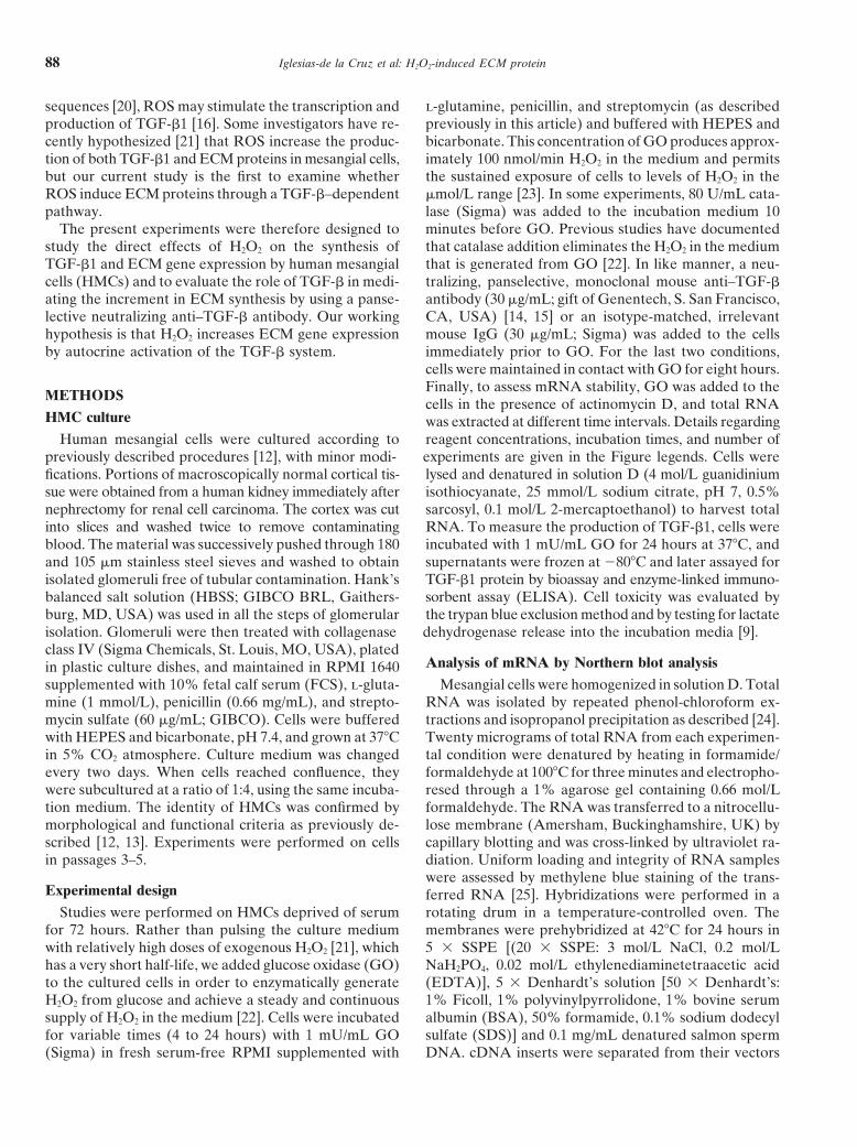
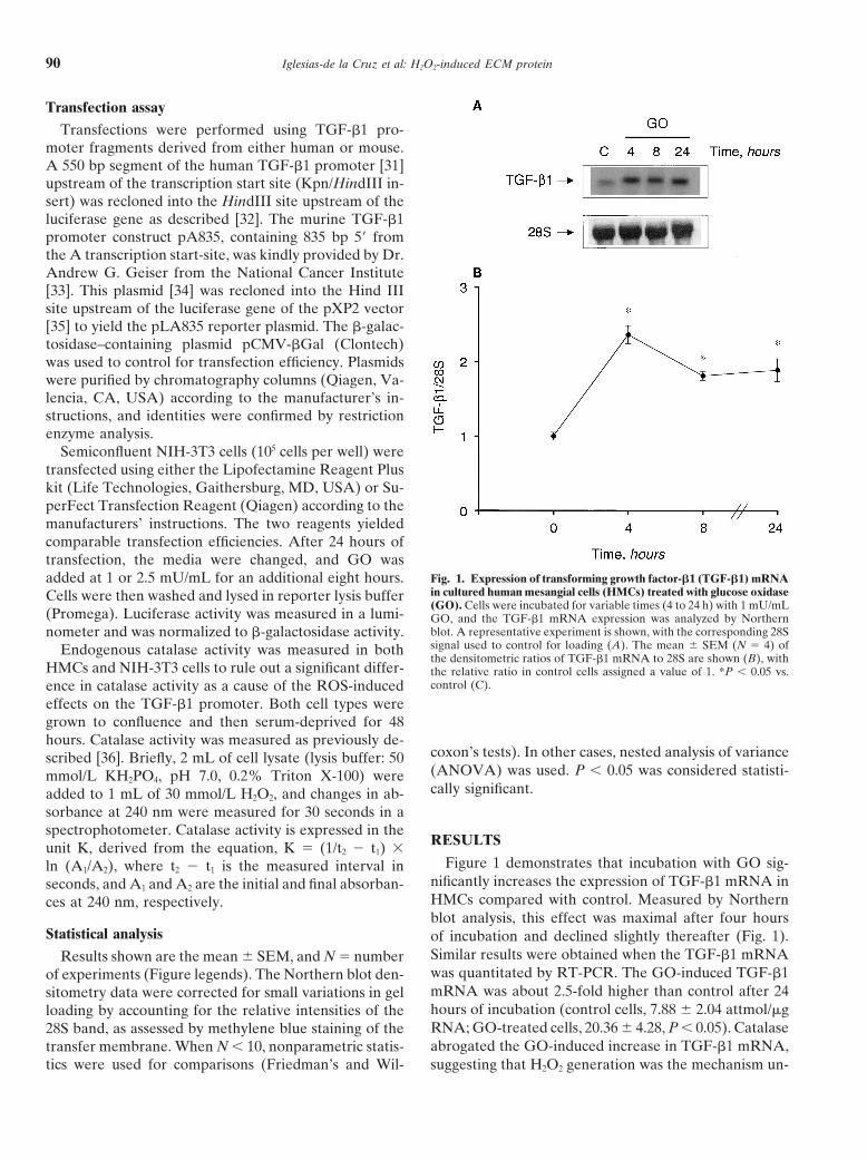
Fig. 2. Expression of TGF-b1 mRNA in cultured HMCs preincubatedwith catalase and treated with GO. Cells were preincubated for 10minutes with 80 U/mL catalase (Cat) and were then incubated for eighthours with 1 mU/mL GO. TGF-b1 mRNA expression was analyzed byNorthern blot with the corresponding 28S signal used to control forloading. The mean 6 SEM of the densitometric ratios of TGF-b1 Fig. 3. Analysis of TGF-b1 mRNA stability in control (j) and GOmRNA to 28S are shown (N 5 4, except N 5 2 for catalase alone), (d)-incubated HMCs. Cultured cells were examined without or withwith the relative ratio in control cells assigned a value of 1. *P , 0.05 exposure to GO (1 mU/mL) for eight hours (initial). Actinomycin Dvs. control. (10 mg/mL) was then added for the designated times in hours. The
decay in TGF-b1 mRNA level was analyzed by Northern blot, with thecorresponding 28S signal used to control for loading. The half-life underthe two experimental conditions was assessed by the hatched lines. Eachpoint represents the mean value of three independent experiments.derlying the effect of GO (Fig. 2). At the concentrations
tested, GO was not toxic to HMCs. More than 95%of the cells excluded the trypan blue dye, and lactatedehydrogenase in the incubation media was similar in mRNA stability (Fig. 3). The slope of the mRNA degra-
dation curve was slightly steeper initially following expo-control and GO-treated cells. Treatment with catalasealone did not affect basal TGF-b1 mRNA expression in sure to GO, but became almost identical to the control
value after two hours. The results in Figure 3 indicateHMCs (Fig. 2).The mechanisms responsible for the increased steady- that the half-life of TGF-b1 mRNA may become short-
ened from approximately six hours in control cells tostate levels of TGF-b1 mRNA in response to GO wereinvestigated using mouse NIH-3T3 cells transfected with three hours in GO-treated cells. Taken together, these
studies suggest that the increase in the steady-state levelthe TGF-b1 promoter linked to a luciferase reportergene. NIH-3T3 cells were employed because of the rela- of TGF-b1 mRNA after GO treatment is most likely
due to an increase in TGF-b1 gene transcription, inde-tive ease of transfection of these cells as compared withHMCs. The catalase activity of NIH-3T3 cells was similar pendent of an effect on TGF-b1 mRNA stability.
To test whether increased TGF-b1 transcription trans-to that of HMCs (HMC, 6.31 6 0.29 mK/104; NIH-3T3cells, 5.27 6 0.57, P 5 NS), indicating that H2O2 levels lates into augmented TGF-b1 translation, TGF-b1 protein
levels were measured by bioassay and ELISA in the super-would be comparable between the two cell types. Theaddition of GO (1 mU/mL) increased human TGF-b1 natants of HMCs incubated with GO. Conditioned media
of HMCs that were incubated with GO significantly in-promoter activity by nearly twofold as demonstrated bythe luciferase assay (control cells, 416 6 114 relative hibited the growth of mink lung epithelial cells (Fig. 4A)
and contained significantly greater amounts of immuno-light units/mg protein; GO-treated cells, 817 6 123; N 56, P , 0.05). GO also stimulated the activity of the mouse assayable TGF-b1 as measured by ELISA (Fig. 4B).
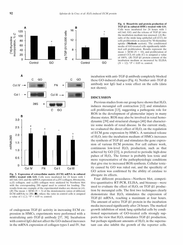
The effect of continuous H2O2 exposure on the mRNATGF-b1 promoter construct pLA835 when this constructwas transfected in NIH-3T3 cells. Transfection efficiency expression of ECM proteins in HMCs is summarized in
Figure 5. Representative Northern blots demonstratewas controlled by cotransfection with a b-galactosidase–expressing vector. At a dose of 1 mU/mL of GO for that H2O2 increases the message levels of different colla-
gen types (I, III, and IV) and fibronectin (Fig. 5A). Oneight hours, the relative promoter activity was increasedto 138 6 11% of control value (N 5 6, P , 0.05); at a average, the cells incubated with GO increased type IV
collagen by 108%, type I collagen by 98%, type III colla-dose of 2.5 mU/mL, this activity was 146 6 7% of control(P , 0.05). gen by 67%, and fibronectin by 96% compared with
control (N 5 3, P , 0.05; Fig. 5B). To assess the roleThe addition of GO did not appreciably affect TGF-b1
Iglesias-de la Cruz et al: H2O2-induced ECM protein92
Fig. 4. Bioactivity and protein production ofTGF-b1 in cultured HMCs treated with GO.Cells were incubated for 24 hours with 1mU/mL GO, and the release of TGF-b1 intothe incubation medium was assessed. (A) Re-sults of the mink lung epithelial cell (CCL-64cell) proliferation as assayed by 3H-thymidineuptake (Methods section). The conditionedmedia of GO-treated cells significantly inhib-ited cell proliferation. Results represent themean 6 SEM (N 5 10), and proliferation ofcontrol CCL-64 cells (C) is assigned a valueof 100%. (B) TGF-b1 protein content of theincubation medium as measured by ELISA(N 5 12). *P , 0.05 vs. control.
incubation with anti–TGF-b antibody completely blockedthese GO-induced changes (Fig. 6). Neither anti–TGF-bantibody nor IgG had a toxic effect on the cells (datanot shown).
DISCUSSION
Previous studies from our group have shown that H2O2
induces mesangial cell contraction [12] and stimulatescell proliferation [13], suggesting a pathogenic role forROS in the development of glomerular injury in manydisease states. ROS may also be involved in renal hemo-dynamic [39] and structural changes [40] that character-ize some models of renal disease. In the current study,we evaluated the direct effect of H2O2 on the regulationof ECM gene expression by HMCs. A sustained releaseof H2O2 into the incubation medium of HMCs increasesthe synthesis of TGF-b1 and stimulates the gene expres-sion of various ECM proteins. For cell culture work,continuous low-level H2O2 production, such as thatachieved by GO [23], is preferred to periodic high-dosepulses of H2O2. The former is probably less toxic andmore representative of the pathophysiologic conditionsthat give rise to increased ROS synthesis. Cellular toxic-ity caused by GO was ruled out, and the specificity ofGO action was confirmed by the ability of catalase to
Fig. 5. Expression of extracellular matrix (ECM) mRNA in cultured abrogate its effects.HMCs treated with GO. Cells were incubated for 24 hours with 1
Four different procedures—Northern blot, competi-mU/mL GO, and the mRNA expression of a1(IV) collagen, fibronectin,a1(I) collagen, and a1(III) collagen were analyzed by Northern blot tive-quantitative RT-PCR, ELISA, and bioassay—werewith the corresponding 28S signal used to control for loading. The used to evaluate the effect of H2O2 on TGF-b1 produc-results from one example of the experimental studies are shown in (A).
tion by mesangial cells. The first two techniques clearly(B) The mean 6 SEM (N 5 3) of the densitometric ratios of differentECM mRNAs to 28S (j), where the ratio in control cells is assigned demonstrate that H2O2 stimulates the expression ofa value of 1 (h). *P , 0.05 vs. control. TGF-b1 mRNA, reaching a maximum at four hours.
The amount of active TGF-b1 protein in the incubationmedia increased significantly after 24 hours. The markedgrowth inhibition of mink lung epithelial cells by condi-of endogenous TGF-b1 activity in increasing ECM ex-tioned supernatants of GO-treated cells strongly sup-pression in HMCs, experiments were performed with aports the view that H2O2 stimulates TGF-b1 production,neutralizing anti–TGF-b antibody [37, 38]. Incubationbut perhaps significant amounts of H2O2 in the superna-with control IgG did not affect the GO-induced increases
in the mRNA expression of collagen types I and IV, but tant can also inhibit the growth of the reporter cells.

Iglesias-de la Cruz et al: H2O2-induced ECM protein 93
evidence that the observed effects are specifically dueto H2O2 since pretreatment with the antioxidant catalase,which detoxifies H2O2, inhibits the GO-induced expres-sion of TGF-b1.
The mechanisms responsible for the increased steady-state levels of TGF-b1 mRNA after H2O2 treatment werealso analyzed. H2O2 did not stabilize the TGF-b1 mRNAlevel but did increase the transcription rate of the TGF-b1gene, as evidenced by the enhanced TGF-b1 promoteractivity in the transfection experiments. The level ofTGF-b1 promoter activity correlated well with the quan-tity of TGF-b1 mRNA, as assessed by Northern blotand competitive-quantitative RT-PCR. The predominantcontrol of TGF-b1 production in response to H2O2 there-fore occurs at the transcriptional level.
The message expression of every ECM protein testedincreased after GO incubation. Two native mesangialECM components (type IV collagen and fibronectin)and two interstitial collagens (types I and III) were ana-lyzed, and in every case, the mRNA levels increasedafter cells were exposed to GO. H2O2 may have stimu-lated ECM expression by a direct effect or an indirecteffect through activation of the TGF-b system. The latterpossibility seems likely because experiments employingthe panselective anti–TGF-b antibody disclosed com-plete prevention of the GO-induced increase in ECMexpression. Thus, one or more of the TGF-b isoformscan be implicated in mediating of the effect of GO onFig. 6. Expression of collagen mRNA in cultured HMCs treated with
GO in the presence of an anti–TGF-b blocking antibody. Cells were collagen or fibronectin expression. The TGF-b1 isoformincubated for 24 hours without (C) or with 1 mU/mL GO plus 30 mg/mL
is known to increase the synthesis and decrease the deg-anti–TGF-b antibody (Ab 1 GO) or 30 mg/mL nonspecific IgG (IgG 1radation of ECM proteins [16, 37]. The control IgG didGO). (A) A representative Northern assay of a1(IV) collagen and a1(I)
collagen mRNAs with the corresponding 28S signal used to control for not reverse the GO-induced increases in ECM expres-loading. (B) The mean 6 SEM (N 5 4) of the densitometric ratios of
sion, suggesting that the effect of the anti–TGF-b anti-collagen mRNAs to 28S, where the ratio in control cells ( ) is assignedbody is not due to a direct antioxidant effect.a value of 1. The stimulation of collagen mRNA levels by GO ( )
was prevented by anti–TGF-b antibody ( ), but not by IgG (j). *P , The present results provide new insights into the patho-0.05 vs. control. physiologic mechanisms that underlie certain diseases
characterized by progressive renal sclerosis. IncreasedROS synthesis has been implicated in some glomerulo-nephritides [1–5], the remnant kidney model of renalAlthough the mink lung epithelial cell bioassay measures
the activity of all three isoforms of TGF-b, the ELISA sclerosis [41], chronic renal rejection [39], cyclosporineA-related nephropathy [42], and age-related renal dys-specific for TGF-b1 confirmed that the release of TGF-b1
isoform into the media of mesangial cells is augmented function [43, 44]. All of these situations are characterizedby the relentless accumulation of ECM proteins in thein response to H2O2 (Fig. 4). In a previous report by
Nath et al [21], addition of a pulse of H2O2 was able to renal parenchyma, and ROS may initiate and maintainthis harmful morphological process. ROS have also beeninduce directly increases in the mRNA levels of TGF-b1,
type III collagen, and type IV collagen in cultured NRK- linked to fibrotic processes other than glomerulosclero-sis, and they have been associated with several cytokines49F fibroblasts and in rat mesangial cells. In the present
studies, we demonstrate not only that TGF-b1 mRNA that up-regulate ECM proteins. In vascular smooth cells,the synthesis of insulin-like growth factor-I is increasedexpression is up-regulated, but also that TGF-b1 protein
synthesis is augmented in the presence of continuous by exposure to ROS, suggesting that the autocrine insu-lin-like growth factor-I system plays an important roleexposure to H2O2. Moreover, we show that the effect of
H2O2 on ECM production is mediated by increased TGF-b in vascular smooth muscle cell growth in response toROS [45]. Oxygen derivatives have been shown to pro-activity, since treatment with a panselective anti–TGF-b
antibody completely abrogates the GO-induced in- duce progressive pulmonary inflammation and fibrosisthrough activation of MEK-ERK pathways [46]. ROScreases in ECM gene expression. We provide further

Iglesias-de la Cruz et al: H2O2-induced ECM protein94
of somatostatin on cultured human mesangial cells. Endocrinologymay also activate the STAT family of transcription factors136:3444–3451, 1995
in response to platelet-derived growth factor (PDGF). 13. Duque I, Rodriguez-Puyol M, Ruiz-Torres MP, et al: Calciumchannel blockers inhibit hydrogen peroxide-induced proliferationThese findings indicate that the JAK-STAT pathwayof cultured rat mesangial cells. J Pharmacol Exp Ther 267:612–616,responds to intracellular ROS and that PDGF uses ROS1993
as a second messenger to regulate STAT activation [47]. 14. Sharma K, Jin Y, Guo J, et al: Neutralization of TGF-beta byanti-TGF-beta antibody attenuates kidney hypertrophy and theWe conclude that hydrogen peroxide stimulates extra-enhanced extracellular matrix gene expression in STZ-inducedcellular matrix protein synthesis in cultured human mes-diabetic mice. Diabetes 45:522–530, 1996
angial cells, an effect that is largely dependent on activation 15. Ziyadeh FN, Hoffman BB, Han DC, et al: Long-term preventionof renal insufficiency, excess matrix gene expression and glomeru-of TGF-b1. We propose that the previously mentionedlar mesangial matrix expression by treatment with monoclonalmolecular changes induced by oxidative stress may have anti-TGF-b antibody in db/db diabetic mice. Proc Natl Acad Sci
important consequences in the development and pro- USA 97:8015–8020, 200016. Ketteler M, Noble NA, Border WA: Transforming growth fac-gression of many renal diseases. Furthermore, therapeu-
tor-beta and angiotensin II: The missing link from glomerulartic interventions to reduce reactive oxygen species pro- hyperfiltration to glomerulosclerosis? Annu Rev Physiol 57:279–duction may delay or even prevent the development of 295, 1995
17. Sundaresan M, Yu ZX, Ferrans VJ, et al: Requirement for gener-such diseases.ation of H2O2 for platelet-derived growth factor signal transduction.Science 270:296–299, 1995
18. Schreck R, Rieber P, Baeuerle PA: Reactive oxygen intermedi-ACKNOWLEDGMENTSates as apparently widely used messengers in the activation of the
This study was supported by the CICYT, SAF 96/0186, and FIS 98/ NF-kappa B transcription factor and HIV-1. EMBO J 10:2247–0337. M.C. Iglesias-de la Cruz and M.P. Ruiz-Torres are postdoctoral 2258, 1991fellows of the Ministerio de Educacion y Cultura of Spain. F.N. Ziyadeh 19. Meyer M, Schreck R, Baeuerle PA: H2O2 and antioxidants haveand S. Chen are supported by the Juvenile Diabetes Foundation Inter- opposite effects on activation of NF-kappa B and AP-1 in intactnational. The authors thank Ms. Susana Comendador for her technical cells: AP-1 as secondary antioxidant-responsive factor. EMBO Jsupport. 12:2005–2015, 1993
20. Kim SJ, Angel P, Lafyatis R, et al: Autoinduction of transformingReprint requests to Carmen Iglesias-de la Cruz, Ph.D., Renal-Electro- growth factor beta 1 is mediated by the AP-1 complex. Mol Cell
lyte and Hypertension Division, 700 Clinical Research Building, Univer- Biol 10:1492–1497, 1990sity of Pennsylvania, 415 Curie Boulevard, Philadelphia, Pennsylvania 21. Nath KA, Grande J, Croatt A, et al: Redox regulation of renal19104-6144, USA. DNA synthesis, transforming growth factor-beta1 and collagen
gene expression. Kidney Int 53:367–381, 1998E-mail: [email protected]. Gow AJ, Branco F, Christofidou-Salomidou M, et al: Immuno-
targeting of glucose oxidase: Intracellular production of H2O2 andREFERENCES endothelial oxidative stress. Am J Physiol 277:L271–L281, 1999
23. Miura K, Ishii T, Sugita Y, et al: Cystine uptake and glutathione1. Shah SV: Role of reactive oxygen metabolites in experimentallevel in endothelial cells exposed to oxidative stress. Am J Physiolglomerular disease. Kidney Int 35:1093–1106, 1989 262:C50–C58, 19922. Adler S, Baker PJ, Johnson RJ, et al: Complement membrane 24. Chomczynski P, Sacchi N: Single-step method of RNA isolation by
attack complex stimulates production of reactive oxygen metabo- acid guanidinium thiocyanate-phenol-chloroform extraction. Anallites by cultured rat mesangial cells. J Clin Invest 77:762–767, 1986 Biochem 162:156–159, 1987
3. Diamond JR, Bonventre JV, Karnovsky MJ: A role for oxygen 25. Sharma K, Ziyadeh FN: Renal hypertrophy is associated withfree radicals in aminonucleoside nephrosis. Kidney Int 29:478–483, upregulation of TGF-b1 gene expression in diabetic BB rat and1986 NOD mouse. Am J Physiol 267:F1094–F1101, 1994
4. McCord JM: Oxygen-derived free radicals in postischemic tissue 26. Rocco MV, Neilson EG, Hoyer JR, et al: Attenuated expressioninjury. N Engl J Med 312:159–163, 1985 of epithelial cell adhesion molecules in murine polycystic kidney
5. Paller MS, Hoidal JR, Ferris TF: Oxygen free radicals in ische- disease. Am J Physiol 262:F679–F686, 1992mic acute renal failure in the rat. J Clin Invest 74:1156–1164, 1984 27. Ruiz-Torres MP, Bosch RJ, O’Valle F, et al: Age-related in-
6. Baud L, Nivez MP, Chansel D, et al: Stimulation by oxygen crease in expression of TGF-beta1 in the rat kidney: Relationshipradicals of prostaglandin production by rat renal glomeruli. Kidney to morphologic changes. J Am Soc Nephrol 9:782–791, 1998Int 20:332–339, 1981 28. Qian SW, Kondaiah P, Roberts AB, et al: cDNA cloning by PCR
7. Lewis MS, Whatley RE, Cain P, et al: Hydrogen peroxide stimu- of rat transforming growth factor beta-1. Nucleic Acids Res 18:3059,lates the synthesis of platelet-activating factor by endothelium and 1990induces endothelial cell-dependent neutrophil adhesion. J Clin In- 29. Meager A: Assays for transforming growth factor beta. J Immunolvest 82:2045–2055, 1988 Methods 141:1–14, 1991
8. Hughes AK, Stricklett PK, Padilla E, et al: Effect of reactive 30. Wolf G, Ziyadeh FN, Zahner G, et al: Angiotensin II-stimulatedoxygen species on endothelin-1 production by human mesangial expression of transforming growth factor beta in renal proximalcells. Kidney Int 49:181–189, 1996 tubular cells: Attenuation after stable transfection with the c-mas
9. Lopez-Ongil S, Hernandez-Perera O, Navarro-Antolin J, et oncogene. Kidney Int 48:1818–1827, 1995al: Role of reactive oxygen species in the signalling cascade of 31. Kim SJ, Glick A, Sporn MB, et al: Characterization of the promotercyclosporine A-mediated up-regulation of eNOS in vascular endo- region of the human transforming growth factor-beta 1 gene. J Biolthelial cells. Br J Pharmacol 124:447–454, 1998 Chem 264:402–408, 1989
10. Scharschmidt LA, Dunn MJ: Prostaglandin synthesis by rat glo- 32. Michelson S, Alcami J, Kim SJ, et al: Human cytomegalovirusmerular mesangial cells in culture: Effects of angiotensin II and infection induces transcription and secretion of transformingarginine vasopressin. J Clin Invest 71:1756–1764, 1983 growth factor beta 1. J Virol 68:5730–5737, 1994
11. Trachtman H, Futterweit S, Singhal P: Nitric oxide modulates 33. Geiser AG, Kim S-J, Roberts AB, et al: Characterization of thethe synthesis of extracellular matrix proteins in cultured rat mesan- mouse transforming growth factor-b1 promoter and activation bygial cells. Biochem Biophys Res Commun 207:120–125, 1995 the Ha-ras oncogene. Mol Cell Biol 11:84–92, 1991
34. Hoffman BB, Sharma K, Zhu Y, et al: Transcriptional activation12. Diez-Marques ML, Garcia-Escribano C, Medina J, et al: Effects
Iglesias-de la Cruz et al: H2O2-induced ECM protein 95
of transforming growth factor-b1 in mesangial cell culture by high tubular iron accumulation after partial nephrectomy. Kidney Int45:1006–1013, 1994glucose concentration. Kidney Int 54:1107–1116, 1998
42. Parra T, de Arriba G, Arribas I, et al: Cyclosporine A nephrotox-35. Nordeen SK: Luciferase reporter gene vectors for analysis of pro-icity: Role of thromboxane and reactive oxygen species. J Labmoters and enhancers. Biotechniques 6:454–458, 1988Clin Med 131:63–70, 199836. Aebi HE: Catalase. Basel, Verlag-Chemie, 1983
43. Iglesias-de la Cruz MC, Ruiz-Torres MP, Garcia-del Moral37. Ziyadeh FN, Sharma K, Ericksen M, et al: Stimulation of collagen R, et al: Age-related progressive renal fibrosis in rats and its preven-gene expression and protein synthesis in murine mesangial cells tion with ACE inhibitors and taurine. Am J Physiol 278:F122–F129,by high glucose is mediated by autocrine activation of transforming 2000growth factor-beta. J Clin Invest 93:536–542, 1994 44. Ruiz-Torres P, Gonzalez-Rubio M, Lucio-Cazana FJ, et al: Re-
38. Han DC, Isono M, Hoffman BB, et al: High glucose stimulates active oxygen species and platelet-activating factor synthesis inproliferation and collagen type I synthesis in renal cortical fibro- age-related glomerulosclerosis. J Lab Clin Med 124:489–495, 1994
45. Delafontaine P, Ku L: Reactive oxygen species stimulate insulin-blasts: Mediation by autocrine activation of TGF-b. J Am Soclike growth factor I synthesis in vascular smooth muscle cells.Nephrol 10:1891–1899, 1999Cardiovasc Res 33:216–222, 199739. Yoshioka T, Ichikawa I: Glomerular dysfunction induced by poly-
46. Cho YJ, Seo MS, Kim JK, et al: Silica-induced generation of reactivemorphonuclear leukocyte-derived reactive oxygen species. Am Joxygen species in Rat2 fibroblast: Role in activation of mitogen-Physiol 257:F53–F59, 1989 activated protein kinase. Biochem Biophys Res Commun 262:708–
40. Clayton L, Hiley C, Davies S, et al: Glomerular injury induced 712, 1999by hydrogen peroxide: Modifying influence of ACE inhibitors. 47. Simon AR, Rai U, Fanburg BL, et al: Activation of the JAK-Free Radic Res Commun 17:271–278, 1992 STAT pathway by reactive oxygen species. Am J Physiol
275:C1640–C1652, 199841. Nankivell BJ, Tay YC, Boadle RA, et al: Dietary protein alters