Hydrodynamic properties of D-β-hydroxybutyrate dehydrogenase, a lipid-requiring enzyme
9
HYDRODYNAMICS OF BDH VOL. 17, NO. 20, 1978 4169 Hydrodynamic Properties of D-P-Hydroxybutyrate Dehydrogenase, a Lipid-Requiring Enzyme? J. Oliver McIntyre,t Leslie A. Holladay, Murray Smigel,s David Puett,l and Sidney Fleischer* ABSTRACT: The hydrodynamic properties of purified D-0- hydroxybutyrate dehydrogenase (BDH), a lecithin-requiring enzyme isolated from bovine heart mitochondria, have been studied using the active-enzyme sedimentation technique. An active complex of BDH with dioctanoyl-L-a-phosphatidyl- choline, PC(8:0), was sedimented under assay conditions, and the sedimenting boundary was followed by observing the re- duction of NAD+. Between 2.4 and 30 pg of BDH/mL, the ~ 2 0 , ~ increased and was inversely related to the enzymatic activity. The most active species has the smallest apparent $20,~ (3.3 f 0.2 S). The diffusion coefficient of the BDH-PC(8:O) complex at 2.4 pg/mL, measured by the free diffusion method, was found to be 4.9 f 0.5 X cm2/s; at higher protein concentrations, D ~O,~ decreased. The hydrodynamic studies both of the active BDH-PC(8:O) complex and of the apoen- Biological membranes are macromolecular complexes consisting of proteins and phospholipids. D-0-Hydroxybutyrate dehydrogenase, normally bound to the mitochondrial inner membrane, has been purified from bovine heart mitochondria to homogeneity as a soluble, phospholipid-free protein, apo- BDH.' It has a subunit molecular weight of 31 000 estimated by NaDodS04-polyacrylamide gel electrophoresis (Bock and Fleischer, 1974, 1975). Apo-BDH is inactive in the absence of lipid but forms an active complex (BDH-PC) upon the ad- dition of aqueous dispersions of lecithin or phospholipids containing lecithin (Gazzotti et al., 1975). This paper presents a study of the hydrodynamic properties of the active enzyme. A preliminary communication has appeared (McIntyre et al., 1976). + From the Departments of Molecular Biology (J.O.M., M.S., and S.F.) and Biochemistry (L.A.H. and D.P.), Vanderbilt University, Nashville, Tennessee 37235. Receiaed April 4, 1978. This research was supported in large part by the National Institutes of Health (Research Grant AM- 14632; Diabetes-Endocrinology Center Grant AM- 17026 also provided useful services). In addition, L.A.H. and D.P. thank the Vanderbilt Re- search Council for partial support and D.P. acknowledges a grant from the Dreyfus Foundation (BRSG RR-05424). J.O.M. thanks Vanderbilt University for a special graduate scholarship. A preliminary communi- cation of portions of this work has been presented [McIntyre, O., Holladay, L., Smigel, M., and Fleischer, S. (1976), Fed, Proc., Fed. Am. Soc. Exp. Biol. 35, 14361. *This work was in partial fulfillment of the Ph.D. dissertation re- quirements of J. Oliver McIntyre, Vanderbilt University, 1978. 8 Present address: Department of Pharmacology, University of Virginia, Charlottesville, Va. 22903. 7 Recipient of a Research Career Development Award (AM-00055). I Abbreviations used: BDH, D-P-hydroxybutyrate dehydrogenase; apo-BDH, lecithin-free and enzymatically inactive BDH; BSA, bovine plasma albumin; CD, circular dichroic; CMC, critical micelle concen- tration; DTT, dithiothreitol; GdmC1, guanidinium chloride; Hepes, 4- (2-hydroxyethy1)- 1 -piperazineethanesulfonic acid; MPL, mitochondrial phospholipid; PC, phosphatidylcholine; PC(8:O) and PC( 18:l cisA9), dioctanoyl- and dioleoyl-L-a-phosphatidylcholine, respectively; EDTA, (ethy1enedinitrilo)tetraacetic acid; Tris-CI, 2-amino-2-hydroxyrnethyl- 1,3-propanediol chloride. 0006-2960/78/04 17-4169%0 1 .OO/O zyme demonstrate that the protein undergoes concentration- dependent self-association. At the lowest protein concentration at which meaningful measurements could be made (the layered concentration of 2.4 Fg/mL), the apparent molecular weight of the active BDH-PC(8:O) complex was calculated from ~20,~ and D ~o,~ to be 61 600, which corresponds to a dimer. Using these values, the Stokes radius and frictional ratio (flfo) of the BDH-PC(8:O) complex were estimated to be 28 %, and 1.56, respectively, implying an axial ratio (a/b) of 9 for a prolate ellipsoid or 11 for an oblate ellipsoid. The circular dichroic spectrum of apo-BDH between 205 and 240 nm suggests the presence of nearly 30% a helicity but only limited 0 structure. The addition of mitochondrial phospholipid had no significant effect on the circular dichroic spectrum between 21 5 and 240 nm. Materials and Methods Solutions were prepared with deionized water. Chemicals were of reagent grade unless otherwise specified. Sucrose, special enzyme grade, ultrapure, was obtained from Schwarz/Mann Co. (Orangeburg, N.Y.). DL-@-hydroxy- butyric acid (sodium salt), bovine serum albumin (fraction V, powder), equine cytochrome c (grade III), ovalbumin (grade VI), and dithiothreitol (DTT) were obtained from Sigma Chemical Co. (St. Louis, Mo.). Equine y-globulin was ob- tained from Mann Research Laboratories (New York, N.Y.). Bovine plasma albumin solution, used as the protein standard, and crystalline plasma albumin were obtained from Armour Pharmaceutical Co. (Chicago, Ill.). NAD+ was obtained from P-L Biochemicals Inc. (Milwaukee, Wis.). LiBr, obtained from Matheson, Coleman and Bell (Norwood, Ohio), was prepared as a 4 M stock solution which was purified by filtration through an activated-carbon column and a 0.22-pm filter (Millipore Corp., Bedford, Mass.). Preparation and Measurement of BDH and Lipids. The apo-BDH used in these studies was purified to homogeneity as described by Bock and Fleischer (1974, 1975) and was stored frozen in a liquid nitrogen refrigerator. The lipids were prepared as described previously (Gazzotti et a]., 1975), except that PC(8:O) was stored at -20 OC as a 1 mM stock solution in water and was maintained at 40 "C for the studies described below. ['4C]PC(8:O), synthesized from [ l-'4C]octanoic acid as described previously (Gazzotti et al., 1975), was used, after dilution with unlabeled PC(8:0), for measurement of the concentration of unbound PC(8:O) in the presence of BDH (see below). Protein was determined by the procedure of Lowry et al. (1 95 1) with a BSA standard. The assay was carried out as described by Ross and Schatz (1973) using iodoacetate to carboxymethylate the DTT which would otherwise interfere with the assay for protein. Phosphorus was measured using a modification (Rouser and Fleischer, 1964) of Chen et al. (1956). 0 1978 American Chemical Society
Transcript of Hydrodynamic properties of D-β-hydroxybutyrate dehydrogenase, a lipid-requiring enzyme
H Y D R O D Y N A M I C S O F B D H V O L . 1 7 , N O . 2 0 , 1 9 7 8 4169
Hydrodynamic Properties of D-P-Hydroxybutyrate Dehydrogenase, a Lipid-Requiring Enzyme?
J. Oliver McIntyre,t Leslie A. Holladay, Murray Smigel,s David Puett,l and Sidney Fleischer*
ABSTRACT: The hydrodynamic properties of purified D-0- hydroxybutyrate dehydrogenase (BDH), a lecithin-requiring enzyme isolated from bovine heart mitochondria, have been studied using the active-enzyme sedimentation technique. An active complex of BDH with dioctanoyl-L-a-phosphatidyl- choline, PC(8:0), was sedimented under assay conditions, and the sedimenting boundary was followed by observing the re- duction of NAD+. Between 2.4 and 30 pg of BDH/mL, the ~ 2 0 , ~ increased and was inversely related to the enzymatic activity. The most active species has the smallest apparent $ 2 0 , ~ (3.3 f 0.2 S). The diffusion coefficient of the BDH-PC(8:O) complex at 2.4 pg/mL, measured by the free diffusion method, was found to be 4.9 f 0.5 X cm2/s; at higher protein concentrations, D ~ O , ~ decreased. The hydrodynamic studies both of the active BDH-PC(8:O) complex and of the apoen-
B i o l o g i c a l membranes are macromolecular complexes consisting of proteins and phospholipids. D-0-Hydroxybutyrate dehydrogenase, normally bound to the mitochondrial inner membrane, has been purified from bovine heart mitochondria to homogeneity as a soluble, phospholipid-free protein, apo- BDH.' I t has a subunit molecular weight of 31 000 estimated by NaDodS04-polyacrylamide gel electrophoresis (Bock and Fleischer, 1974, 1975). Apo-BDH is inactive in the absence of lipid but forms an active complex (BDH-PC) upon the ad- dition of aqueous dispersions of lecithin or phospholipids containing lecithin (Gazzotti et al., 1975). This paper presents a study of the hydrodynamic properties of the active enzyme. A preliminary communication has appeared (McIntyre et al., 1976).
+ From the Departments of Molecular Biology (J.O.M., M.S., and S.F.) and Biochemistry (L.A.H. and D.P.), Vanderbilt University, Nashville, Tennessee 37235. Receiaed April 4, 1978. This research was supported in large part by the National Institutes of Health (Research Grant AM- 14632; Diabetes-Endocrinology Center Grant AM- 17026 also provided useful services). In addition, L.A.H. and D.P. thank the Vanderbilt Re- search Council for partial support and D.P. acknowledges a grant from the Dreyfus Foundation (BRSG RR-05424). J.O.M. thanks Vanderbilt University for a special graduate scholarship. A preliminary communi- cation of portions of this work has been presented [McIntyre, O., Holladay, L., Smigel, M., and Fleischer, S. (1976), Fed, Proc., Fed. Am. Soc. Exp . Biol. 35, 14361.
*This work was in partial fulfillment of the Ph.D. dissertation re- quirements of J . Oliver McIntyre, Vanderbilt University, 1978.
8 Present address: Department of Pharmacology, University of Virginia, Charlottesville, Va. 22903.
7 Recipient of a Research Career Development Award (AM-00055). I Abbreviations used: BDH, D-P-hydroxybutyrate dehydrogenase;
apo-BDH, lecithin-free and enzymatically inactive BDH; BSA, bovine plasma albumin; CD, circular dichroic; CMC, critical micelle concen- tration; DTT, dithiothreitol; GdmC1, guanidinium chloride; Hepes, 4- (2- hydroxyethy1)- 1 -piperazineethanesulfonic acid; MPL, mitochondrial phospholipid; PC, phosphatidylcholine; PC(8:O) and PC( 18:l cisA9), dioctanoyl- and dioleoyl-L-a-phosphatidylcholine, respectively; EDTA, (ethy1enedinitrilo)tetraacetic acid; Tris-CI, 2-amino-2-hydroxyrnethyl- 1,3-propanediol chloride.
0006-2960/78/04 17-4 169%0 1 .OO/O
zyme demonstrate that the protein undergoes concentration- dependent self-association. At the lowest protein concentration at which meaningful measurements could be made (the layered concentration of 2.4 Fg/mL), the apparent molecular weight of the active BDH-PC(8:O) complex was calculated from ~ 2 0 , ~
and D ~ o , ~ to be 61 600, which corresponds to a dimer. Using these values, the Stokes radius and frictional ratio (flfo) of the BDH-PC(8:O) complex were estimated to be 28 %, and 1.56, respectively, implying an axial ratio ( a / b ) of 9 for a prolate ellipsoid or 11 for an oblate ellipsoid. The circular dichroic spectrum of apo-BDH between 205 and 240 nm suggests the presence of nearly 30% a helicity but only limited 0 structure. The addition of mitochondrial phospholipid had no significant effect on the circular dichroic spectrum between 21 5 and 240 nm.
Materials and Methods Solutions were prepared with deionized water. Chemicals
were of reagent grade unless otherwise specified. Sucrose, special enzyme grade, ultrapure, was obtained from Schwarz/Mann Co. (Orangeburg, N.Y.). DL-@-hydroxy- butyric acid (sodium salt), bovine serum albumin (fraction V, powder), equine cytochrome c (grade III) , ovalbumin (grade VI), and dithiothreitol (DTT) were obtained from Sigma Chemical Co. (St. Louis, Mo.). Equine y-globulin was ob- tained from Mann Research Laboratories (New York, N.Y.). Bovine plasma albumin solution, used as the protein standard, and crystalline plasma albumin were obtained from Armour Pharmaceutical Co. (Chicago, Ill.). NAD+ was obtained from P-L Biochemicals Inc. (Milwaukee, Wis.). LiBr, obtained from Matheson, Coleman and Bell (Norwood, Ohio), was prepared as a 4 M stock solution which was purified by filtration through an activated-carbon column and a 0.22-pm filter (Millipore Corp., Bedford, Mass.).
Preparation and Measurement of BDH and Lipids. The apo-BDH used in these studies was purified to homogeneity as described by Bock and Fleischer (1974, 1975) and was stored frozen in a liquid nitrogen refrigerator. The lipids were prepared as described previously (Gazzotti et a]., 1975), except that PC(8:O) was stored at -20 OC as a 1 mM stock solution in water and was maintained at 40 "C for the studies described below. ['4C]PC(8:O), synthesized from [ l-'4C]octanoic acid as described previously (Gazzotti et al., 1975), was used, after dilution with unlabeled PC(8:0), for measurement of the concentration of unbound PC(8:O) in the presence of BDH (see below). Protein was determined by the procedure of Lowry et al. (1 95 1) with a BSA standard. The assay was carried out as described by Ross and Schatz (1973) using iodoacetate to carboxymethylate the DTT which would otherwise interfere with the assay for protein. Phosphorus was measured using a modification (Rouser and Fleischer, 1964) of Chen et al. (1956).
0 1978 American Chemical Society
4 170 B I O c H E M I S T R Y
Enzymatic Assay. BDH activity was measured as described previously (Bock and Fleischer, 1974). With PC(8:O) activa- tion of apo-BDH, the assay was modified as follows. The assay was carried out a t 25 'C, and both BSA and ethanol were omitted from the assay mixture. All components except for PC(8:0), enzyme, and substrate were incubated at 25 O C for 5 min and then an aliquot of a 1 m M PC(8:0), stock solu- tion, maintained at 40 OC, was added; after a 5-min incubation, the enzyme was added. Following a further 15-min incubation to form the active BDH-PC(8:O) complex, the reaction was started by the addition 20 p L of 1.0 M DL-0-hydroxy- butyrate.
Measurement of the Concentration of Unbound PC(8.0) in the Presence of BDH. The free concentration of PC(8:O) in the presence of various concentrations of BDH was measured both by equilibrium dialysis and rapid filtration. Equilibrium dialysis was carried out as described previously (Gazzotti et al., 1974) using chambers which contained 100 p L of sample on each side of the dialysis membrane. BDH was diluted to 40, 120, or 390 pg/mL in 100 rnM sodium phosphate buffer, pH 7.35, containing 0.5 m M EDTA, 5 m M DTT, and 125 pM [ 14C]PC(8:O) (specific radioactivity 660 cpm/nmol) and was dialyzed at 4 'C for 14 h against the same solution but without BDH; the enzyme maintained greater than 80% of its activity during dialysis. Aliquots (35 pL) were removed and counted in a Packard Tri-Carb scintillation counter to measure the concentration of PC(8:O) on both sides of the dialysis mem- brane and thus determine whether, in solution, there is an ex- cess of free (unbound) PC(8:O) available for activation of the enzyme. The concentration of PC(8:O) not bound to BDH was also measured using the rapid-filtration method described by Paulus (1969). For this technique, BDH was diluted to final concentrations of protein, varying from 1.6 to 160 pg/mL, in 0.5 mL of 100 m M potassium phosphate (pH 7.35), 0.5 mM EDTA, 0.2 mM DTT, and 125 pM [14C]PC(8:O) (specific radioactivity 1 I O cpm/nmol). The enzyme was incubated with the PC(8:O) for 15 min at room temperature (ca. 25 "C) to obtain optimum activation. Each sample was then filtered a t room temperature through a PM- I O ultrafiltration membrane (Amicon Corp., Lexington, Mass.); the filtration time for each sample was about 10 min. The filtrate from each sample was collected i n two fractions and the PC(8:O) concentration de- termined by counting 50-pL aliquots as described above. There was no detectable difference between the concentration of PC(8:O) in the two fractions of filtrate collected from each sample. The filters were also counted to measure the retention
Sedimentation Velocity. The sedimentation coefficient of apo-BDH was measured using a Model E ultracentrifuge with scanner optics. On the day before each sedimentation run, the stock apo-BDH (-2 mg/mL) was centrifuged at 130 OOOg for 1 h. The supernatant was recovered and an aliquot diluted to the appropriate protein concentration for the sedimentation velocity run . The diluted sample was stored frozen overnight in a liquid nitrogen refrigerator and was thawed just prior to loading. The protein was loaded into one side of a 1.2-cm path-length double-sector cell and centrifuged a t 52 000 rpm in an AN-H rotor equilibrated a t 20 O C . The cell was scanned every 250 s, monitoring protein absorbance at 280 nm vs. buffer blank, until the principal boundary had sedimented two-thirds of the distance down the cell. A control sample of apo-BDH, which was maintained a t room temperature during the course of the sedimentation run, lost less than 10% of its enzymatic activity. I n addition to the protein which gives rise to the principal boundary, some protein sediments more rapidly and as a separate heterogeneous species. The amount of protein
of PC(8:O).
M C I N T Y R E E T A L .
present in this rapidly sedirnenting species was estimated from the difference between the initial absorbance and the absorb- ance at the principal boundary after the two species had sep- arated. The rapidly sedimenting species is probably denatured protein and is not in rapid equilibrium with the majority of the protein, which gives rise to the principal sedimentation boundary. The radial position (rb) of the midpoint of the principal boundary was determined for each time that the cell was scanned, and the apparent s20 was determined from the plot of In rb vs. time; such plots were nearly linear, although some slight curvature was noted. This is expected with this self-associating system due to radial dilution as the protein sediments (Cox, 1969). The apparent s20 of the rapidly sedi- menting protein was estimated i n a similar manner. The sed- imentation coefficient of apo-BDH a t various initial layered concentrations was also measured by the sucrose density gra- dient centrifugation method described by Martin and Ames (1961).
The sedimentation coefficient of the BDH-PC(8:O) complex was determined using the active-enzyme sedimentation method of Cohen and Mire ( 197 1 ). A Vinograd-type double-sector centerpiece (Beckman part no. 33 1359) was used in an A N - H rotor with the Model E ultracentrifuge. The enzyme was ac- tivated with PC(8:O) below its C M C , i n a solution containing I O m M potassium phosphate (pH 7.35) , 2 m M DTT, 0.5 m M EDTA, 125 pM PC(R:O), and 2-30 pg/mL apo-BDH: 10 p L of this BDH-PC(8:O) complex was placed in the sample well in the centerpiece of the cell and the rotor equilibrated at 20 "C inside the centrifuge. As the rotor accelerated to 52 000 rpm, the active-enzyme complex layered onto the surface of an assay solution in the main cell compartment; the assay so- lution contained 0.1 M potassium phosphate (pH 7.35), 2 m M DTT, 0.5 mM EDTA, 125 FM PC(8:0), 20 m M DL-0-hy- droxybutyrate, and 2 mM NAD+. The sedimentation of the enzyme band was monitored by the production of NADH from the enzymatic reaction, by observing the 340-nm absorption of this product. The cell was scanned at 250-s intervals, and the radial position ( r ) of the scanner trace half-height (the midpoint of the reaction boundary) was determined for each scan. The data were analyzed using an approximation method (Kemper and Everse, 1973) which computes s20 from the plot of In r vs. time; these plots were linear within experimental error. The enzymatic activity of a control sample of the BDH-PC(8:O) complex, maintained at room temperature during the course of each sedimentation run, remained con- stant when assayed in the presence of either MPL or 125 pM PC( 8:O).
Partial Specific Volume Determinations. The partial spe- cific volume (i;) of the protein was calculated to be 0.735 mL/g from the apparent specific volumes of the component amino acids (Cohn and Edsall, 1943; McMeekin and Marshall, 1952) and the amino acid composition of apo-BDH (Bock and Fleischer, 1975). Although i; does not equal V of the active BDH-PC(8:O) complex, the effective partial specific volume of the complex (4') in the 0.1 M potassium phosphate buffer used for the active enzyme sedimentation experiments is close to this value. This is demonstrated by using Cassassa and Eisenberg's (1964) equation
M ( 1 - $ ' p ) = - j p ) f 6D(1 -vnp)] ( I )
where p is the solution density, ?ID the lipid to protein weight ratio, V D the partial specific volume of the lipid, and 4' the "effective partial specific volume" of the complex. Since V D = 0.987 for PC(8:O) below the C M C (Tausk et al., 1974), ( I - jDp) = 0.0029. Thus, if 1 g of PC(8:O) was bound/g of apo-BDH, 4' would equal 0.732 as compared to 0.735, which
H Y D R O D Y N A M I C S O F B D H
is the partial specific volume of apo-BDH. The amount of PC(8:O) bound to the apo-BDH was estimated by Gazzotti et al. (1975) to be about 10 mol/mol of subunit (0.17 g/g of apo-BDH), using equilibrium binding with gel-exclusion chromatography. Therefore, the contribution of the lipid to the effective partial specific volume of the BDH-PC(8:O) complex in the buffer solution used is considered negligible for purposes of the calculations.
The densities of the solutions used for the sedimentation experiments were determined using a Mettler-Parr DM02A density meter. The viscosities of the solutions were measured relative to water in a U-tube viscometer a t 20 OC. The apparent ~ 2 0 , ~ was calculated from the measured s20 using the formu- la:
V O L . 1 7 , N O . 2 0 , 1 9 7 8 4171 cantly modified by the diffusion of the sucrose or other inter- acting flows.
The diffusion constants (Dobsd) were corrected to D ~ O , ~ by two methods. One method uses the relationship
(4) Tsvobsd
D20,w = Dobsd - ( T o b s d v s )
where T , is the standard temperature (293.16 K), Tobsd the temperature of the measurement, vs the viscosity of water a t Ts and assuming the solvent viscosity (vobsd) to be the viscosity of the solvent a t the average sucrose concentration in which Dobsd was measured. Calculated D ~ o , ~ values for the standard proteins agreed with the literature values within experimental error. The other method utilizes standard proteins of known diffusion constants to determine the correction factor for temperature and viscosity. For each experiment, Dobsd vs. D ~ O , ~ for the standard proteins was plotted and Dobsd for the BDH-PC(8:O) complexes corrected to D ~ o , ~ using the rela- tionship so obtained. The following D ~ o , ~ values were used (all values are X107 in units of cm2/s): equine cytochrome c (1 3.0), human hemoglobin (6.9), BSA (5.94), and equine y-globulins (4.8).
Circular Dichroism. The C D spectra were obtained with a Cary 60 spectropolarimeter equipped with a Model 6002 C D attachment. A thermostated cell holder was used with a cir- culating water bath to control temperatures to within f 0 . 2 O C . Cells of path length 0.5 and 1 .O mm were used to record spectra between ca. 205 and 240 nm under a variety of experimental conditions. The instrument was operated with a time constant of 3 s and sensitivities 2 and 4 mdeg/in. Several scans of sample and appropriate blanks were taken and averages are reported. A mean residue molecular weight of 110 was used to obtain the reduced mean-residue ellipticity, [ e ] , in units of deg- cm2/dmol.
Results Activation of Apo-BDH with Lecithin. The maximum
specific activity which could be obtained upon activation of apo-BDH with M P L varied between 90 and 100 pmol of NAD+ reduced min-l mg-I a t 37 OC; this is equivalent to a specific activity of between 36 and 40 a t 25 O C . Since natural lecithins are not soluble in aqueous solution and exist as mac- romolecular arrays (liposomes), hydrodynamic studies of the enzyme activated by them would reflect the properties of such lipid vesicles.
However, activation of apo-BDH can be achieved with the soluble lecithin PC(8:0), thus permitting characterization of the hydrodynamic properties of the active enzyme. PC(8:0), a t 200 KM, activated apo-BDH (4.8 pg/mL) to the same ex- tent as PC( 18:l cisA9), giving a maximum specific activity of 32 pmol of NAD+ reduced min-l mg-l a t 25 OC in the pres- ence of both 1.27% ethanol and 0.4 mg/mL BSA (data not shown). At concentrations of PC(8:O) greater than 200 p M , inhibition of BDH activity occurred due to PC(8:O) micelle formation (Gazzotti et al., 1975; Tausk et al., 1974). When the concentrations, in the standard assay medium, of either po- tassium phosphate (pH 7.35) or ethanol are varied, the PC(8:O) concentration which gives maximum activation of BDH is altered. For example, in the absence of both BSA and ethanol, in 10 or 100 m M buffer, the C M C of PC(8:O) is reduced so that optimum activation of BDH is obtained with 120 or 130 p M PC(8:0), respectively; in 100 m M buffer with either 1.27 or 9% ethanol (v/v) and in the absence of BSA, optimum ac- tivation of BDH is obtained with 170 or 175 KM PC(8:0), re- spectively. These differences seem to reflect changes in the C M C of the PC(8:O) with the conditions of the medium. For
with i; = 0.735. Diffusion Methods. Diffusion coefficients were determined
using the free diffusion method (Gosting, 1956). Apo-BDH was activated with PC(8:O) in a solution containing 0.1 M sodium phosphate (pH 7.35), 5 m M DTT, 0.5 m M EDTA, 125 p M PC(8:0), and 5% (w/v) ultrapure sucrose. The solution was saturated with nitrogen gas prior to the addition of the PC(8:0), and finally the apo-BDH was added. This solution (2.5 mL), which contains the BDH-PC(8:O) complex, was pipetted into a 2 X 0.5 in. cellulose nitrate centrifuge tube (Beckman, part no. 305050) which was fixed vertically in a test-tube rack. An equal volume of a ‘‘light’’ solution of the same composition, but without the protein or the sucrose, was carefully overlayed to form a sharp initial boundary. After diffusion for between 20 and 30 h a t 25 f 0.5 “C, fractions of equal volume (by drop counting) were collected a t 4 O C using a Beckman “fraction- recovery system” to expel the contents through the top of the tube. The BDH activity in each fraction was measured using the standard assay with M P L activation to determine the distribution profile of active enzyme. The sucrose distribution was measured by using a refractometer. A series of standard proteins, run in separate tubes, was analyzed as follows: cy- tochrome c and human hemoglobin by light absorbance measured a t 41 5 nm; ovalbumin, bovine plasma albumin, and equine y-globulins by protein analysis using the modified method of Lowry et al. (1951).
The distribution profile, a t time = t , of a single species dif- fusing from a semiinfinite region terminated by a sharp initial boundary at X = 0 is described by the equation
where CO is the initial concentration, CX the concentration X cm from the initial boundary, t the diffusion time in seconds and D the diffusion coefficient. This equation is derived by a n integration of Fick’s second law of diffusion as described by Gosting (1956). The form of eq 3 is obtained directly from eq 70 ,72 and 73 of Gosting (1956) with the lower concentration C, being zero for our experimental conditions. The initial boundary position was taken as the value of X a t which CX = 0.5 CO; when this was determined from both the sucrose and protein distributions, the same initial boundary position was found within experimental error. For each CO and t , CX vs. X was computed for various values of D. For several values of X, on both sides of the initial boundary position, CX was measured for the experimental data and D determined by interpolation from the computer-simulated values of CX. Dobsd was taken as the average of these D values determined a t various posi- tions. I t was assumed that the protein diffusion is not signifi-
41 72 B I O C H E M I S T R Y M C I N T Y R E E T A L
MOLES PC(8:O) / MOLE BOH 1,500 200 100 70
24 1 A -DIRECTION OF SEDIMENTATION 1
0- 0 1020x)405060
BDH CONC, ( IJG/ML)
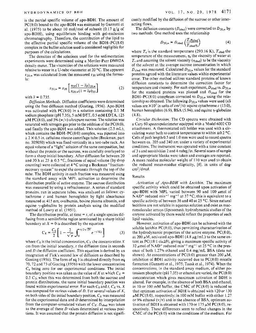
FiGLRE 1 : The effect of BDH concentration on the specific activity of the enzyme activated with PC(8:O). The activity obtained with different amounts of BDH, as indicated, and 125 pM PC(8:O) was measured at 25 "C, as described in the text in a medium containing 100 mM potassium phosphate (pH 7 . 3 5 ) , 2 mM NAD+, 20 mM DL-P-hydroxybutyrate, 0.5 mM EDTA and 0.3 mM DTT. BDH specific activity is expressed as pmol of NAD+ reduced min-' (mg of BDH)-'. The molar ratio of PC(8:O) to BDH subunit with increasing BDH concentration was calculated (upper ordinate).
the active-enzyme sedimentation of BDH-PC(8:0), the assay medium in the main compartment of the cell contained 125 pM PC(8:O) and 100 m M potassium phosphate, p H 7.35. Under these conditions, the specific activity of BDH a t 4.8 pg of protein/mL is 16 pmol of NAD+ reduced min-' mg-I a t 25 O C . This salt concentration minimizes nonspecific electrostatic interactions and also provides a sufficient density difference between the assay medium and the enzyme sample, which is overlayed, so that a sharp boundary forms. In this assay me- dium, 125 pM PC(8:O) is below the apparent CMC of this lipid.
The specific activity of BDH, activated with PC(8:0), is dependent on the BDH concentration (Figure 1). The observed decrease in BDH activity with increasing protein concentration is related to the self-association of the protein (see below) and is not due to limiting amounts of PC(8:O) for activation of the enzyme. The free concentration of PC(8:O) in the presence of either 40,120, or 390 pg of B D H / m L was measured by equi- librium dialysis. At 40 pg/mL, the Concentration of PC(8:O) on both sides of the dialysis membrane was the same (1 25 pM) within experimental error. At 390 pg/mL, the concentration of PC(8:O) in the protein-free chamber was diminished but to only about 94 pM. The free concentration of PC(8:0), in the presence of from 2 to 160 pg of B D H / m L was also measured using rapid filtration (Paulus, 1969). Although the concen- tration of PC(8:O) in the effluent was diminished to approxi- mately 100 pM by nonspecific absorption of between 10 and 15 nmol of PC(8:O) to each membrane filter, the presence of BDH (from 2 to 160 pg/mL) did not alter the free concen- tration of PC(8:O). Therefore, when variable amounts of BDH are activated with PC(8:0), although the molar ratio of PC(8:O) to BDH decreases rapidly with increasing protein concentration (Figure l ) , the concentration of free PC(8:O) is not significantly altered, within experimental error, by the presence of from 2 to 60 pg of BDH/mL. Since the amount of PC(8:O) bound to BDH is dependent only on the dissociation constant for the interaction and the free PC(8:O) concentration, we conclude that the decrease in BDH activity with increasing protein concentration is not due to an insufficiency of PC(8: 0 ) .
Sedimentation Velocity of Apo-BDH and of BDH-PC(8:O). The sedimentation coefficient of apo-BDH was measured using conventional sedimentation velocity, and several typical scans
J-
2550 SECONDS I
4590 SECONDS T
I n n l
I A u 4 5 9 0 SECONDS 1
I I
1' -DIRECTION OF SEDIMENTATION
950 SECONDS 0 0 4
1550 SECONDS !L 1 1 1 SECOND;
n
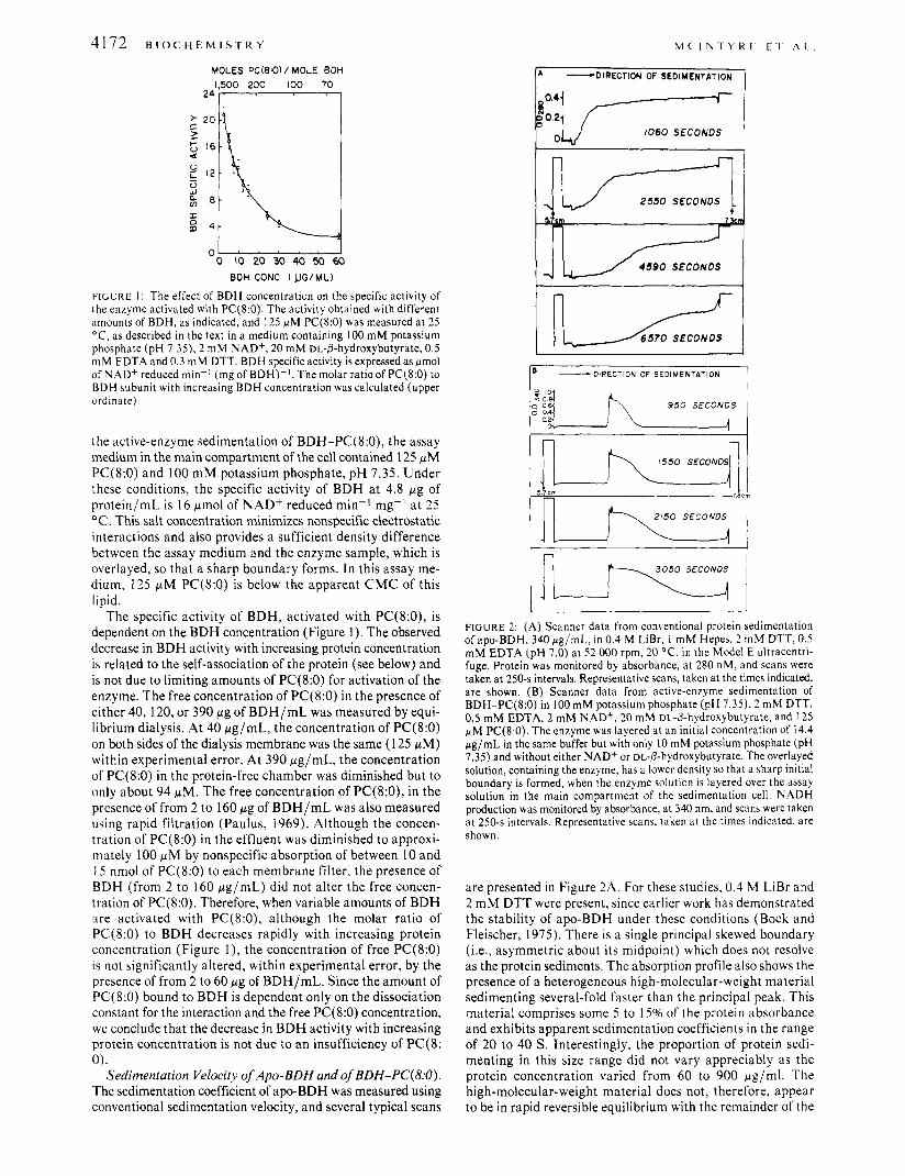
FIGURE 2: (A) Scanner data from conventional protein sedimentation of apo-BDH, 340 pg/mL, in 0.4 M LiBr, 1 mM Hepes, 2 mM DTT, 0.5 mM EDTA (pH 7.0) at 5 2 000 rpm, 20 "C, in the Model E ultracentri- fuge. Protein was monitored by absorbance, at 280 nM, and scans were taken at 250-s intervals. Representative scans, taken at the times indicated, are shown. (B) Scanner data from active-enzyme sedimentation of BDH-PC(8:O) in 100 mM potassium phosphate (pH 7.35), 2 mM DTT, 0.5 mM EDTA, 2 mM NAD+, 20 mM DL-/3-hydroxybutyrate, and 125 pM PC(8:O). The enzyme was layered at an initial concentration of 14.4 pg/mL in the same buffer but with only I O mM potassium phosphate (pH 7.35) and without either NAD+ or DL-P-hydroxybutyrate. The overlayed solution, containing the enzyme, has a lower density so that a sharp initial boundary is formed, when the enzyme solution is layered over the assay solution in the main compartment of the sedimentation cell. NADH production was monitored by absorbance, at 340 nm, and scans were taken at 250-s intervals. Representative scans, taken at the times indicated, are shown
are presented in Figure 2A. For these studies, 0.4 M LiBr and 2 m M DTT were present, since earlier work has demonstrated the stability of apo-BDH under these conditions (Bock and Fleischer, 1975). There is a single principal skewed boundary (i.e., asymmetric about its midpoint) which does not resolve as the protein sediments. The absorption profile also shows the presence of a heterogeneous high-molecular-weight material sedimenting several-fold faster than the principal peak. This material comprises some 5 to 15% of the protein absorbance and exhibits apparent sedimentation coefficients in the range of 20 to 40 S . Interestingly, the proportion of protein sedi- menting in this size range did not vary appreciably as the protein concentration varied from 60 to 900 pg/ml. The high-molecular-weight material does not, therefore, appear to be in rapid reversible equilibrium with the remainder of the
H Y D R O D Y N A M I C S O F B D H V O L . 1 7 , N O . 2 0 , 1 9 7 8 4173
m:4,0F31,;DALT;s SPHERICAL PROTEIN , , , , I 3.0
0 200 400 600 800 IO00 1200
BDH CONC. ( JJGIML)
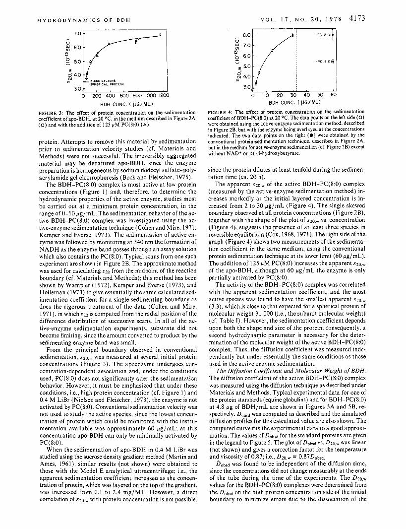
FIGURE 3: The effect of protein concentration on the sedimentation coefficient of apo-BDH, at 20 OC, in the medium described in Figure 2A (0) and with the addition of 125 FM PC(8:O) (A).
protein. Attempts to remove this material by sedimentation prior to sedimentation velocity studies (cf. Materials and Methods) were not successful. The irreversibly aggregated material may be denatured apo-BDH, since the enzyme preparation is homogeneous by sodium dodecyl sulfate-poly- acrylamide gel electrophoresis (Bock and Fleischer, 1975).
The BDH-PC(8:O) complex is most active at low protein concentrations (Figure 1) and, therefore, to determine the hydrodynamic properties of the active enzyme, studies must be carried out a t a minimum protein concentration, in the range of 0-10 pg/mL. The sedimentation behavior of the ac- tive BDH-PC(8:O) complex was investigated using the ac- tive-enzyme sedimentation technique (Cohen and Mire, 197 1; Kemper and Everse, 1973). The sedimentation of active en- zyme was followed by monitoring a t 340 nm the formation of N A D H as the enzyme band passes through an assay solution which also contains the PC(8:O). Typical scans from one such experiment are shown in Figure 2B. The approximate method was used for calculating s20 from the midpoint of the reaction boundary (cf. Materials and Methods); this method has been shown by Wampler (1972), Kemper and Everse (1973), and Holleman (1973) to give essentially the same calculated sed- imentation coefficient for a single sedimenting boundary as does the rigorous treatment of the data (Cohen and Mire, 1971), in which s20 is computed from the radial position of the difference distribution of successive scans. In all of the ac- tive-enzyme sedimentation experiments, substrate did not become limiting, since the amount converted to product by the sedimenting enzyme band was small.
From the principal boundary observed in conventional sedimentation, 3 2 0 , ~ was measured a t several initial protein concentrations (Figure 3). The apoenzyme undergoes con- centration-dependent association and, under the conditions used, PC(8:O) does not significantly alter the sedimentation behavior. However, it must be emphasized that under these conditions, Le., high protein concentration (cf. Figure 1) and 0.4 M LiBr (Nielsen and Fleischer, 1973), the enzyme is not activated by PC(8:O). Conventional sedimentation velocity was not used to study the active species, since the lowest concen- tration of protein which could be monitored with the instru- mentation available was approximately 60 pg/mL; a t this concentration apo-BDH can only be minimally activated by PC ( 8 :0) .
When the sedimentation of apo-BDH in 0.4 M LiBr was studied using the sucrose density gradient method (Martin and Ames, 1961), similar results (not shown) were obtained to those with the Model E analytical ultracentrifuge; i.e., the apparent sedimentation coefficient increased as the concen- tration of protein, which was layered on the top of the gradient, was increased from 0.1 to 2.4 mg/ML. However, a direct correlation of szo,,., with protein concentration is not possible,
BDH CONC. ( JJG/ML)
FIGURE 4: The effect of protein concentration on the sedimentation coefficient of BDH-PC(8:O) at 20 OC. The data points on the left side (0) were obtained using the active-enzyme sedimentation method, described in Figure 2B, but with the enzyme being overlayed at the concentrations indicated. The two data points on the right ( 0 ) were obtained by the conventional protein-sedimentation technique, described in Figure 2A, but in the medium for active-enzyme sedimentation (cf. Figure 2B) except without NAD+ or DL-p-hydroxybutyrate.
since the protein dilutes a t least tenfold during the sedimen- tation time (ca. 20 h).
The apparent $ 2 0 , ~ of the active BDH-PC(8:O) complex (measured by the active-enzyme sedimentation method) in- creases markedly as the initial layered concentration is in- creased from 2 to 30 pg/mL (Figure 4). The single skewed boundary observed at all protein concentrations (Figure 2B), together with the shape of the plot of s~o,,., vs. concentration (Figure 4), suggests the presence of a t least three species in reversible equilibrium (Cox, 1968, 1971). The right side of the graph (Figure 4) shows two measurements of the sedimenta- tion coefficient in the same medium, using the conventional protein sedimentation technique a t its lower limit (60 pg/mL). The addition of 125 p M PC(8:O) increases the apparent ~ 2 0 , ~
of the apo-BDH, although a t 60 pg/mL the enzyme is only partially activated by PC(8:O).
The activity of the BDH-PC(8:O) complex was correlated with the apparent sedimentation coefficient, and the most active species was found to have the smallest apparent ~ 2 0 , ~
(3.3), which is close to that expected for a spherical protein of molecular weight 3 1 000 (Le., the subunit molecular weight) (cf. Table I). However, the sedimentation coefficient depends upon both the shape and size of ihe protein; consequently, a second hydrodynamic parameter is necessary for the deter- mination of the molecular weight of the active BDH-PC(8:O) complex. Thus, the diffusion coefficient was measured inde- pendently but under essentially the same conditions as those used in the active enzyme sedimentation.
The Diffusion Coefficient and Molecular Weight of BDH. The diffusion coefficient of the active BDH-PC(8:O) complex was measured using the diffusion technique as described under Materials and Methods. Typical experimental data for one of the protein standards (equine globulins) and for BDH-PC(8:O) a t 4.8 pg of BDH/mL are shown in Figures 5A and 5B, re- spectively. Dobsd was computed as described and the simulated diffusion profiles for this calculated value are also shown. The computed curve fits the experimental data to a good approxi- mation. The values of D&sd for the standard proteins are given in the legend to Figure 5 . The plot of Dobsd vs. D Z O , ~ was h e a r (not shown) and gives a correction factor for the temperature and viscosity of 0.87; Le., D ~ O , ~ = o.87D,bsd
Dobsd was found to be independent of the diffusion time, since the concentrations did not change measurably at the ends of the tube during the time of the experiments. The D ~ o , ~ values for the BDH-PC(8:O) complexes were determined from the Dobsd on the high protein concentration side of the initial boundary to minimize errors due to the dissociation of the
4 1 7 4 B I o c H E M I s T R Y M ( . ’ l N l Y K ~ I-1. , A I
I ~ .__. - .
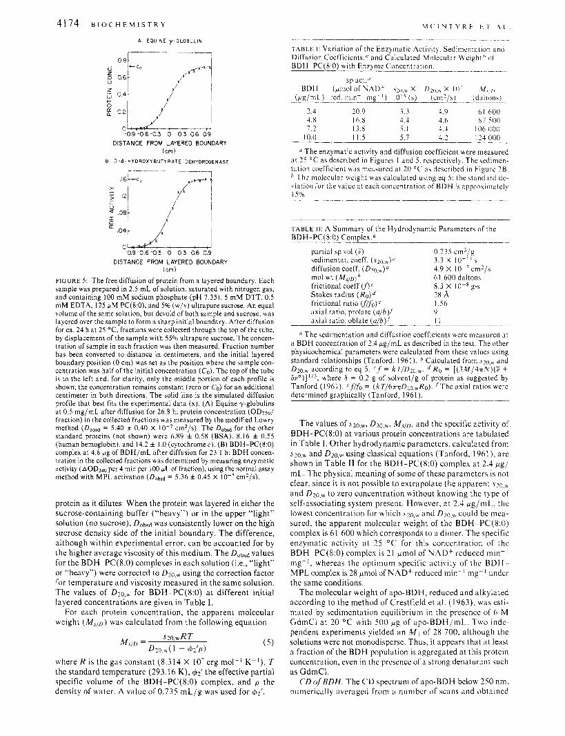
TAB1.E 1: Variation of the Enzymatic Activity, Sedimentation a n d Diffusion Coefficicnts.O and Calculated Molecular Weight /’ of BDH-PC(8:O) with Enzyme Concentration, __
sp act .“ BDH (pl11ol of NAD’ s?(J,, X /)2(1,,+ X IO’ .M,/,
(pg /n iL) red. i n i n - ’ _ _ ~ _ mg-I) 10’ ‘ (a ) (cm!/s) (daltons)
2.4 20.9 3.3 4.9 61 hOO 4.8 16.8 4.4 4.6 81 500
IO6 000 7 . 2 13.8 5 . 1 4.4 10.0 I 1.5 5.1 4.2 I24 000
A EQUINE 7 - G L O B U L I N
1 I
DISTANCE FROM LAYERED BOUNDARY (cm)
B D - A - H Y D R O X Y B U T Y R A T E DEHYDRCGENASE
0 -0.9 0 6 -0.3 0 0 3 0.6 0.9
DISTANCE FROM LAYERED BOUNDARY ( c m i
FIGURE 5: The free diffusion of protein from a layered boundary. Each sample was prepared in 2.5 mL of solution, saturated with nitrogen gas, and containing 100 mM sodium phosphate (pH 7.35), 5 mM DTT, 0.5 mM EDTA, 125 r M PC(8:0), and 5% (w/v) ultrapure sucrose. An equal volume of the same solution, but devoid of both sample and sucrose, was layered over the sample to form a sharp initial boundary. After diffusion for ca. 24 h at 25 O C , fractions were collected through the top of the tube. by displacement of the sample with 55% ultrapure sucrose. The concen- tration of sample in each fraction was then measured. Fraction number has been converted to distance in centimeters, and the initial layered boundary position (0 cm) was set as the position where the sample con- centration was half of the initial concentration (CO). The top of the tube is to the left and. for clarity, only the middle portion of each profile is shown; the concentration remains constant (zero or CO) for an additional centimeter in both directions. The solid line is the simulated diffusion profile that best fits the experimental data (x). (A) Equine y-globulins at 0.5 mg/mL after diffusion for 26.8 h; protein concentration (OD75o/ fraction) in the collected fractions was measured by the modified Lowry method (Dobsd = 5.40 f 0.40 X cm2/S). The Dobsd for the other standard proteins (not shown) were 6.89 f 0.58 (BSA): 8.16 f 0.55 (human hemoglobin), and 14.2 f I .O (cytochrome c). (B) BDH-PC(8:O) complex at 4.8 f ig of BDH/mL after diffusion for 23.1 h: BDH concen- tration in the collected fractions was determined by measuring enzymatic activity (aOD340 per 4 min per 100 JLL of fraction), using the normal assay method with MPL activation (Dobsd = 5.36 f 0.45 X IO-’ cm2/s).
protein as it dilutes. When the protein was layered in either the sucrose-containing buffer (“heavy”) or in the upper ‘‘light’’ solution (no sucrose), Dobsd was consistently lower on the high sucrose density side of the initial boundary. The difference, although within experimental error, can be accounted for by the higher average viscosity of this medium. The Dobsd values for the BDH -PC(8:0) complexes in each solution (;.e., ‘‘light’’ o r “heavy”) were corrected to D ~ , o , ~ using the correction factor for temperature and viscosity measured in the same solution. The values of D~o, , , for BDH-PC(8:O) a t different initial layered concentrations are given in Table I .
For each protein concentration, the apparent molecular weight (M.>,D) was calculated from the following equation
(5)
where R is the gas constant (8.314 X I O 7 erg mol-’ K-’), T the standard temperature (293.16 K), &’ the effective partial 5pecific volume of the BDH-PC(8:O) complex, and p the density of water. A v;ilue of 0.735 mL/g was used for $2‘.
The enzymatic activity and diffusion coefficient were measured at 25 ‘ C as described in Figures 1 and 5. respectively. The sedimcn- tation coefficient was measured at 20 “C’ as described in Fipurc ’H. ’’ The molecular weight was calculated using eq 5 : the htandard de- viation for the value at each concentration of BDH is approximately IS%. ___^__I_.__ --.__
T A B L E 11: A Summary of the Hydrodynamic Parameters of the BDH-PC(8:O) Complex.” ______ __ ._
partial sp vol (.) sedimentat. coeff. ( ~ 2 0 , ~ ) ~ ~
diffusion coeff. ( D ~ o , , ) ~ mol wt ( h f , p ) b
Stokes radius (Ro) 28 A frictional ratio ((/fo) e 1 .S6 axial ratio, prolate (u /b ) i 9 axial ratio, oblate (a lb l i I I
0 .735 cm’/g 3.3 x 10⁻¹³ 5 4.9 x IO-’cm?/s 61 600 daltons
frictional coeff (f)‘ 8.3 X I O - ’ ~ . S
The sedimentation and diffustion coefficients were measured at a BDH concentration of 2.4 pg/mL as described in the text. The other physicochemical parameters were calculated from these values using standard relationships (Tanford, 1961). Calculated from ~ 2 0 . ~ and D~o., , according to eq 5. l ’ f = kT /D20 ,w . RO = [ ( 3 M / 4 x N ) ( Z + b ~ ’ ) ] ’ / ~ , where 6 = 0.2 g of solvent/g of protein as suggested by Tanford (1961). ‘flfo = (kT/67rqD2o,,Ro). / T h e axial ratios were determined graohicallv (Tanford. 1961 ) .
The values of S ~ O , ~ , D ~ o , ~ , M,/o, and the specific activity of BDH-PC(8:O) at various protein concentrations are tabulated in Table I . Other hydrodynamic parameters, calculated from s ~ o . , , and D ~ o . ~ using classical equations (Tanford, 1961), are shown in Table I1 for the BDH-PC(8:O) complex at 2.4 p g / mL. The physical meaning of some of these parameters is not clear, since it is not possible to extrapolate the apparent s~o,, and D ~ o , ~ to zero concentration without knowing the type of self-associating system present. However, at 2.4 pg/mL, the lowest concentration for which ~ 2 0 , ~ and D ~ O , , ~ could be mea- sured, the apparent molecular weight of the BDH- PC(8:O) complex is 61 600 which corresponds to a dimer. The specific enzymatic activity a t 25 “C for this concentration of the BDH-PC(8:O) complex is 21 pmol of NAD+ reduced min-l mg-I, whereas the optimum specific activity of the BDIi MPL complex is 28 pmol of NAD+ reduced min- I mg-’ undcr the same conditions.
The molecular weight of apo-BDH, reduced and alkylated according to the method of Crestfield et al. ( 1 963), was esti- mated by sedimentation equilibrium in the presence of 6 M GdmCl at 20 “C wlith 500 p g of apo-BDH/mL.. Two inde- pendent experiments yielded an M I of 28 700, although the solutions were not monodisperse. Thus, it appears that a t least a fraction of the BDH population is aggregated at this protein concentration, even in the presence of a strong denaturant such as GdmCI.
CD of BDH. The C D spectrum of apo-RDH below 250 nm, numerically averaged from a number of scans and obtained
H Y D R O D Y N A M I C S O F B D H V O L . 1 7 , N O . 2 0 , 1 9 7 8 4175
-15- 200 210 220 230 240
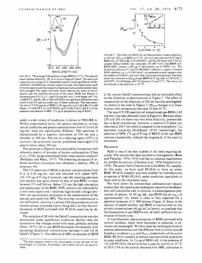
WAVELENGTH (nm) FIGURE 6: The average CD spectrum of apo-BDH at 25 "C. The reduced mean residue ellipticity, [e], is in units of deg-cm2/dmol. The spectrum represents an average of six independent spectra involving different buffer conditions and different enzyme concentrations and preparations; each of the six spectra and the respective baselines were scanned several times and averaged. The upper and lower limits indicate the mean of the six spectra and the standard deviation of the mean. BDH was diluted to concentrations of 0.13,0.17, and 0.26 mg/mL with I mM Hepes (pH 7.0), 2 mM DTT, and 80, 100, or 500 mM NaCI; no significant differences were noted in the CD spectra under any of these conditions. The inset shows the near-UV C D spectra of BDH (1.85 mg/mL) in 0.2 M LiBr, 0.5 mM Hepes, 2.5 mM DTT, 0.5 mM EDTA, and 10 mM Tris-CI (pH 8.1) in the presence and absence of MPL (73 pg of phosphorus/mg of BDH).
under a wide variety of conditions, is shown in FIGURE 6/ Within experimental error, the spectra obtained a t various solvent conditions and protein concentrations from 0.13 to 0.26 mg/mL were not significantly different. The spectrum is characterized by a negative extremum at 210 nm and a shoulder a t 209 nm. The use of a reducing agent (DTT) to maintain the enzymatic activity prohibited meaningful C D spectra below about 205 nm.
The spectrum in Figure 6 was analyzed by comparison with reference spectra of proteins of known conformation (Chen et al., 1974), using constrained linear least-squares analysis2 (Holladay and Puett, 1977). The following estimates of or- dered secondary structures were obtained: (Y helicity, 29%; 0 structure, 6%.
The C D spectrum of BDH a t protein concentrations from 0.13 to 0.26 mg/mL was also obtained with added M P L (30-170 pg of P/mg of protein), and the resulting spectrum (not shown) was quite similar to that of apo-BDH, a t least between 21 5 and 240 nm. Below 215 nm, the light absorption and opalescence of the BDH-MPL solution (as indicated by a very noisy signal and a relatively high dynode voltage) pro- hibit any quantitative estimation of the conformation of the enzyme activated with MPL. The existing instrumentation is not sufficiently sensitive to permit C D measurements a t low concentrations of protein (up to 10 pg/mL), so that the con- formation of BDH activated with PC(8:O) could not be ob- tained.
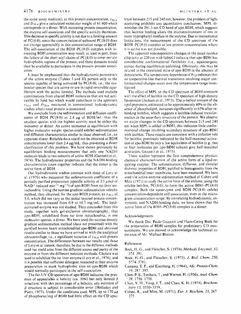
The variation of [ B ] with the GdmCl concentration was also determined under equilibrium conditions. Rather than the cooperative-like changes noted with many globular proteins (Pace, 1975), [ e ] of apo-BDH decreased continuously with increasing denaturant concentration between 0 and 5.6 M GdmCl (Figure 7). The addition of M P L to solutions of BDH
The least-squares analysis was constrained so that the sum of the percentages of LY helicity, p structure, and random coil was set equal to 100%.
" 2 4 6
GUANIDINE.HCI [MI oh ' ' ' ' ' '
FIGURE 7: The effect of GdmCl on the reduced mean-residue ellipticity at 220 nm, [0]220, of BDH at 25 "C. All solutions contained 0.25 mg of BDH/mL, 27 niM LiBr, 0.34 mM DTT, and 68 FM Hepes (pH 7.0); the sample without GdmCl also contained 150 mM NaCI: Apo-BDH (0); BDH-MPL complex (100 fig of phosphorus/mg of BDH) (0). The dashed line between 0.3 and 0.8 M GdmCl indicates a region of limited BDH solubility; e.g., at 0.5 M GdmCI, an opalescent solution appears after the addition of GdmCI, and with time, the protein precipitates. The inset shows the variation in [e1220 of apo-BDH (0.25 mg/mL, 0.2 M NaCI, 2 mM DTT, I O mM Hepes, pH 7.0) between 10 and 35 OC. The values are normalized to the ellipticity at 25 OC.
a t the various GdmCl concentrations had no noticeable effect on the ellipticity as demonstrated in Figure 7. The effect of temperature on the ellipticity a t 220 nm was also investigated. As shown in the inset to Figure 7, [0]220 changes in a linear fashion with temperature between 10 and 35 O C .
The near-UV C D spectrum of concentrated apo-BDH (1.85 mg/mL) was also obtained (inset in Figure 6). Between about 250 and 285 nm there was very little ellipticity, presumably due to band cancellation. However, a positive C D band was observed at 291.5 nm which is assigned to the tryptophanyl ' Lb electronic transition (Strickland, 1974). Interestingly, the addition of M P L (73 pg of P /mg of BDH) to the apo-BDH solution considerably reduced the magnitude of the positive extremum.
Discussion BDH is one of the best studied of the lipid-requiring en-
zymes. The enzyme has been purified to homogeneity (Bock and Fleischer, 1974, 1975) and has an absolute requirement for lecithin for activity (Fleischer et al., 1974; Gazzotti et al., 1975). The active form of the enzyme is the BDH-PL complex. In this study, we have used PC(8:O) to form an active BDH-PC(8:O) complex and have studied the hydrodynamic properties of BDH-PC(8:O) under conditions equivalent to those used in the enzymatic assay.
W e have shown by conventional sedimentation velocity studies that the apoenzyme undergoes concentration-depen- dent self-association and, in solution, is a heterogeneous pop- ulation of species. At 60 pg/mL, apo-BDH has an $ 2 0 , ~ of approximately 4.0, which is close to that expected for a spherical monomer of 31 000 daltons (Figure 3). Since, in the absence of added lecithin, apo-BDH is inactive even at low protein concentrations (60 pg/mL or lower), we conclude that the dissociation of apo-BDH is not, of itself, sufficient for ac- tivation of the enzyme.
A hydrodynamic characterization of BDH activated with natural lecithins, which form liposomes in solutions, is ob- viously not meaningful. W e have used the techniques of active enzyme sedimentation and free diffusion from an initial layered boundary to obtain ~ 2 0 , ~ and D ~ o , ~ , respectively, of the active BDH-PC(8:O) complex at protein concentrations equivalent to assay conditions. At 2.4 pg of BDH/mL in the presence of 125 p M PC(S:O), the enzyme has a specific activity a t 25 OC of 20.9 (75% of the activity obtained with MPL activation in
41 76 B I o c H E M I S T R Y M C I N T Y R E E T A t .
the same assay medium); a t this protein concentration, ~ 2 0 , ~ and &o,, give a calculated molecular weight of 61 600 which corresponds to a dimer. With increasing protein concentration, the enzyme self-associates and the specific activity decreases. This decrease in specific activity is not due to a limiting amount of PC(8:0), since the concentration of unbound PC(8:O) does not change appreciably in this concentration range of BDH. The self-association of the BDH-PC(8:O) complex with in - creasing BDH concentration may result, a t least in part, from the failure of the short acyl chains of PC(8:O) to cover certain hydrophobic regions of the protein, and these domains would then be available to participate in the protein-protein associ- ation.
It must be emphasized that the hydrodynamic parameters of the active enzyme (Tables I and 11) pertain only to the species capable of being activated by PC(8:0), i.e., the mo- lecular species that are active or are in rapid reversible equi- librium with the active form(s). The methods used exclude contributions from altered BDH molecules that are not acti- vatible by lipid but which would contribute to the apparent
and Dlo,w measured in conventional hydrodynamic studies where total protein is monitored.
W e conclude from the sedimentation and diffusion coeffi- cients of BDH-PC(8:O) at 2.4 pg of BDH/mL that the smallest species with the highest activity must be either the monomer or dimer. An active monomer in equilibrium with higher molecular weight species could exhibit sedimentation and diffusion characteristics similar to those observed, Le., an apparent dimer. Reliable data could not be obtained a t BDH concentrations lower than 2.4 pg/mL, thus preventing a direct clarification of this problem. W e have shown previously by equilibrium binding measurements that only one N A D H molecule binds to two subunits of active BDH (Gazzotti et al.. 1974). The hydrodynamic properties and the NADH-binding characteristics taken together indicate that the dimer is the active form of BDH.
Our hydrodynamic studies contrast with those of Levy et al. ( 1 976) who measured the sedimentation coefficient of a partially purified preparation (specific activity of 3.1 pmol of NADf reduced min-' mg-I) of apo-BDH from rat liver mi- tochondria. Using the sucrose gradient sedimentation velocity method, they obtained, for the apo-BDH protein, an ~ 2 0 . ~ ' of 5.0, which did not vary as the initial layered protein concen- tration was increased from 0.9 to 10.7 mg/mL. The lipid- activated enzyme was not studied. They concluded from this study, together with gel-exclusion chromatography, that apo-BDH, solubilized from rat liver mitochondria, is one molecular species, a dimer. W e have used the sucrose density gradient sedimentation method (data not presented) with the purified bovine heart mitochondrial apo-BDH and obtained results similar to those we have arrived at with the analytical ultracentrifuge, i.e., a significant variation of ~ 2 0 , ~ with protein concentration. The differences between our results and those of Levy et al. cannot, therefore, be due to the different methods used but could arise from the different source and purity of the enzyme or from the different isolation methods. Cholate was used to solubilize the rat liver enzyme (Levy et al., 1976), and it is possible that sufficient detergent remained in their enzyme preparation to mask hydrophobic sites on apo-BDH which would normally participate in the self-association.
The far-UV C D spectrum of apo-BDH indicates the pres- ence of appreciable cy helicity (ca. 30%) but only limited 0 structure; with this percentage of cy helicity, any estimate of 0 structure is subject to considerable error (Holladay and Puett, 1977). Under the conditions used, M P L up to 170 pg of phosphorus/mg of BDH had little effect on the C D spec-
trum between 21 5 and 240 nm; however, the problem of light scattering prohibits any quantitative conclusions. M P L di- minishes the 291.5-nm C D band of apo-BDH, which suggests that lecithin binding alters the microenvironment of one or more tryptophanyl residues in the enzyme. Due to instrumental limitations. the measurement of the C D spectrum of the BDH-PC(8:O) complex at low protein concentrations where it is active was not possible.
The apparent noncooperative changes in the mean residue ellipticity a t 220 nm with GdmCl indicate that apo-BDH has considerable conformational flexibility (i.e., equienergetic states) during equilibrium unfolding. Obviously, this may be related to the associated state of apo-BDH in the absence of denaturants. The temperature dependence of 0220 indicates that no cooperative-like thermal transitions involving major con- formational changes occur over the temperature range inves- tigated.
The effect of MPL on the C D spectrum of BDH contrasts with the effect of lecithin on the C D spectrum of high-density lipoprotein (Jackson et al., 1973). The a-helical content of the apolipoprotein, estimated to be approximately 49% in the ab- sence of phospholipid, increases significantly to 65% after re- binding lecithin, which suggests a lecithin-induced transfor- mation in the secondary structure of the protein. We observe no major changes in the C D spectrum between 21 5 and 240 nm when M P L is added to BDH; this indicates little confor- mational change involving secondary structure of apo-BDH with lecithin. These results are consistent with a cofactor role for lecithin, previously indicated by the half-maximal activa- tion of apo-BDH by only a few equivalents of lecithin (e.g., two to four molecules per apo-BDH subunit give half-maximal activation, Gazzotti et al., 1975).
These studies represent the first comprehensive physico- chemical characterization of the active form of a lipid-re- quiring enzyme. The sedimentation, diffusion, and circular dichroic properties of BDH, purified to homogeneity from the mitochondrial inner membrane, have been measured. We have used the active-enzyme sedimentation method of Cohen and Mire ( 1 97 1) to study the active form of the enzyme, using the soluble lecithin, PC(8:0), to form the active BDH-PC(8:O) complex. Both the apoenzyme and BDH-PC(8:O) exhibit concentration-dependent self-association, even in the micro- gram concentration range. By correlating hydrodynamic, en- zymatic, and NADH-binding data, we have shown that the active form of the BDH--PC(8:0) complex is a dimer.
Acknowledgments W e thank Drs. Paolo Gazzotti and Hans-Georg Bock for
the preparation of BDH samples for preliminary C D mea- surements. W e are pleased to acknowledge the technical as- sistance of Mr. Michael Blotzer.
References Bock, H.-G., and Fleischer, S. (1974), Methods Enzymol. 32,
374--39 I . Bock, H.-G., and Fleischer, S . (1975), J . Biol. Chem. 250,
5774-5781. Cassassa, E. F.. and Eisenberg, H. (1964), Ado. Protein Chem.
Chen, P. S., Toribara, T., and Warner, H. (1956), Anal. Chem.
Chen, Y . H. , Yang, J . T., and Chau, K . H. ( 1 974), Biochem-
Cohen, R., and Mire, M. (1971), Eur. J . Biochem. 23, 261-
19, 287-395.
28, 1756-1 758.
istry 13, 3350-3359.
275.
H Y D R O D Y N A M I C S O F B D H
Cohn, E. J., and Edsall, J . T. (1943), in Proteins, Amino Acids and Peptides, New York, N.Y., Reinhold, pp 157-161 and 370-375.
Cox, D. J . (1969), Arch. Biochem. Biophys. 129, 106-123. Cox, D. J . (1971), Arch. Biochem. Biophys. 142, 514-526. Crestfield A. M., Moore, S., and Stein, W. W. (1963), J . Biol.
Chem. 238, 622-627. Fleischer, S., Bock, H.-G. , and Gazzotti, P. (1974), in Mem-
brane Proteins in Transport and Phosphorylation, Azzone, G. F., Klingengerg, M. E., Quaqliariello, E., and Siliprandi, N. , Ed., Amsterdam, North-Holland Publishing Co., pp
Gazzotti, P., Bock, H.-G., and Fleischer, S. (1974), Biochem.
Gazzotti, P., Bock, H.-G., and Fleischer, S. (1 975), J . Biol.
Gosting, L. J . (1956), Ado. Protein Chem. 11, 429-554. Holladay, L. A,, and Puett, D. (1977), Int. J . Pept. Protein
Holleman, W. H . (1973), Biochim. Biophys. Acta 327,
Isaacson, I . , Deroo, P. W.. Rosenthal, A. F., Bittman, R., McIntyre, J . O., Bock, H.-G., Gazzotti, P., and Fleischer, S. ( 1 977), Biophys. J . 17, 72a.
Jackson, R. L., Gotto, A. M., Lux, S. E., John, K. M., and Fleischer, S. (1 973), J . Biol. Chem. 248, 8449-8456.
Kemper, D . L., and Everse, J . (1973), Methods Enzymol. 27,
125-1 36.
Biophys. Res. Commun. 58, 309-3 15.
Chem. 250, 5782-5790.
Res. 10, 363-368.
176-1 85.
67-82.
V O L . 1 7 , N O . 2 0 , 1 9 7 8 4177 Levy, M., Joncourt, M., and Thiessard, J. (1976), Biochim.
Biophys. Acta 424, 57-65. Lowry, 0. H., Rosebrough, N . J., Farr, A. L., and Randall, R.
J. (1951), J . Biol. Chem. 193, 265-275. Martin, R. G., and Ames, B. N . (1961), J . Biol. Chem. 236,
1372-1379. McIntyre, O., Holladay, L., Smigel, M., and Fleischer, S.
(1976), Fed. Proc., Fed. Am. Soc. Exp. Biol. 35, 1436. McMeekin, T. L., and Marshall, K. (1952), Science 116,
Nielsen, N . C., and Fleischer, S. (1973), J . Biol. Chem. 248,
Nielsen, N . C., Zahler, W. L., and Fleischer, S. (1 973), J . Biol.
Pace, N. (1975), CRC Crit. Reu. Biochem. 3, 1-43. Paulus, H . (1 969), Anal. Biochem. 32, 9 1 - 100. Ross, E., and Schatz, G. (1973), Anal. Biochem. 54, 304-
Rouser, G., and Fleischer, S. (1964), Methods Enzymol. 10,
Strickland, E. H. (1974) CRC Crit. Rev. Biochem. 2, 113- 175.
Tanford, C . ( 1 96 l ) , Physical Chemistry of Macromolecules, New York, N.Y., Wiley, Chapter 6, pp 317-456.
Tausk, R. J . M., Van Esch, J . , Karmiggelt, J., Voordouw, G., and Overbeek, J . Th. G. (1974) Biophys. Chem. 1, 184- 203.
142-143.
2549-2555.
Chem. 248, 2556-2562.
306.
385 -406.
Wampler, D. E. (1972), Biochemistry 11, 4428-4435.