David R. Boyer HHS Public Access 1,† Binsen Li2,† Chuanqi ...
Identification of human presequence protease (hPreP) agonists for the treatment of Alzheimer’s disease
Jhansi Rani Vangavaragu, Koteswara Rao Valasani, Xueqi Gan, and Shirley ShiDu YanDepartment of Pharmacology and Toxicology, and Higuchi Bioscience Center, School of Pharmacy, University of Kansas, Lawrence, KS 66047, USA
Abstract
Amyloid-β (Aβ), a neurotoxic peptide, is linked to the onset of Alzheimer’s disease (AD).
Increased Aβ content within neuronal cell mitochondria is a pathological feature in both human
and mouse models with AD. This accumulation of Aβ within the mitochondrial landscape
perpetuates increased free radical production and activation of the apoptotic pathway. Human
Presequence Protease (hPreP) is responsible for the degradation of mitochondrial amyloid-β
peptide in human neuronal cells, and is thus an attractive target to increase the proteolysis of Aβ.
Therefore, it offers a potential target for Alzheimer’s drug design, by identifying potential
activators of hPreP. We applied structure-based drug design, combined with experimental
methodologies to investigate the ability of various compounds to enhance hPreP proteolytic
activity. Compounds 3c & 4c enhanced hPreP-mediated proteolysis of Aβ (1–42), pF1β (2–54) and
fluorogenic-substrate V. These results suggest that activation of hPreP by small benzimidazole
derivatives provide a promising avenue for AD treatment.
Keywords
Amyloid beta; Alzheimer’s disease; enzyme activators; benzimidazole derivatives; hPreP
1. Introduction
Mitochondrial dysfunction is an early pathologic feature of Alzheimer’s disease (AD). AD-
affected brain shows abnormalities in mitochondrial structure and function including
hypometabolism, increased production of reactive oxygen species (ROS) and decreased
respiration potential [1–8]. Amyloid-β (Aβ) is a major component of amyloid pathology
contributing to the pathogenesis of AD. Recent studies from our and other independent
groups demonstrate accumulation of Aβ in mitochondria in aged and AD brain [6, 9–17]. Aβ
enters mitochondria through the protein translocase of the outer membrane (TOM)
machinery [15]. The receptor for advanced glycation end product (RAGE) also aids in
Corresponding author: Dr. Shirley ShiDu Yan, Departments of Pharmacology and Toxicology and Higuchi Bioscience Center, School of Pharmacy, University of Kansas, 2099 Constant Ave., Lawrence, KS 66047, USA, [email protected], Tel: 1-785-864-3637.
Publisher's Disclaimer: This is a PDF file of an unedited manuscript that has been accepted for publication. As a service to our customers we are providing this early version of the manuscript. The manuscript will undergo copyediting, typesetting, and review of the resulting proof before it is published in its final citable form. Please note that during the production process errors may be discovered which could affect the content, and all legal disclaimers that apply to the journal pertain.
HHS Public AccessAuthor manuscriptEur J Med Chem. Author manuscript; available in PMC 2015 April 09.
Published in final edited form as:Eur J Med Chem. 2014 April 9; 76: 506–516. doi:10.1016/j.ejmech.2014.02.046.
Author M
anuscriptA
uthor Manuscript
Author M
anuscriptA
uthor Manuscript
localization and transport of mitochondrial Aβ. Neurons lacking RAGE exhibit lowered
mitochondrial Aβ accumulation and are protected from Aβ-induced mitochondrial
dysfunction [11]. The degree of mitochondrial Aβ accumulation correlates to mitochondrial
and synaptic dysfunction as well as cognitive decline observed in AD mouse models. Excess
Aβ disrupts normal mitochondrial function thereby inducing oxidative stress, lowered ATP
content, and a rise in intercellular Ca2+ [14, 16, 18, 19] [20]concentration. Subsequently, an
increase in mitochondrial Aβ intensifies its interaction with mitochondrial protein including
amyloid binding alcohol dehydrogenase (ABAD)[14, 21] and cyclophilin-D (CypD)[16, 22],
exaggerating mitochondrial and neuronal stress. Thus, mitochondria provide a direct site for
Aβ-induced mitochondrial damage and reducing Aβ accumulation or increasing Aβ
clearance in mitochondria may be a therapeutic strategy for prevention and treatment of AD
in particular during early stage.
Human Presequence protease (hPreP), a 114kDa human zinc metalloprotease 1, (hMP1),
[23] is located in the mitochondrial matrix [24] and belongs to the pitrilysin M16C family of
peptidases containing an inverted zinc-binding motif, HXXEH [25]. hPreP is an ATP-
independent protease that consists of 1,037 amino acids (AAH05025) and is encoded by the
PITRM1 gene located on chromosome 10 [23]. The presence of human PreP (hPreP) in
mitochondrial matrix is confirmed by proteomics 17.31% amino acid sequence [28] and is
involved in the degradation of small-unstructured peptides. Importantly, hPreP was found to
be the novel protease responsible for the degradation of mitochondrial Aβ in the human AD-
affected brain [15, 26, 27].
Although it is a functional analogue to insulin degrading enzyme (IDE), unlike IDE, PreP
cannot degrade insulin [24, 28]. It does however degrade Aβ, but the degradation of Aβ
peptides by recombinant hPreP results in several fragments that are unique only to hPreP.
Despite the fact that the three dimensional structures of PreP [29] and IDE [30] are very
similar, IDE harbors an exosite in the catalytic chamber, which is hypothesized to unfold
small proteins. The corresponding site is absent in PreP, thereby preventing the degradation
of small folded proteins. This makes PreP a better candidate than IDE for clear Aβ since it
does not degrade the important insulin protein [24, 28]. The available literature suggests that
decreased PreP activity in AD-affected human and mouse neuronal mitochondrial matrixes
leads to Aβ aggregation followed by apoptotic cell death. Introduction of hPreP allows for
the degradation of Aβ and could prove vital to the understanding of AD pathology.
The literature suggests that increased ROS production [31] is an early indicator of AD
pathogenesis and that increased ROS negatively impacts the ability of hPreP to degrade Aβ.
In a study, using five to twelve month-old mice [24], PreP proteolytic activity is decreased
with age and Aβ-rich mitochondria and AD-affected brain, indicating that there may be a
link between overall Prep activity, Aβ accumulation, and age.
We built a three dimensional structural homologous model of hPreP based on the 2.1Å
crystal structure of AtPreP [28], in which two cysteines are in close proximity to each other
allowing for the possible formation of a disulfide bond under oxidizing conditions inhibiting
hPreP activity. A recent study demonstrated that hPreP inactivation by H2O2 is not due to
the formation of a disulfide bridge, but is instead due to the methionine residues that are
Vangavaragu et al. Page 2
Eur J Med Chem. Author manuscript; available in PMC 2015 April 09.
Author M
anuscriptA
uthor Manuscript
Author M
anuscriptA
uthor Manuscript

readily oxidized to produce methionine sulfoxide ROS. Conserved methionine residues play
an important role in protecting proteins from oxidative inactivation by scavenging oxidizing
agents and limiting the damage to catalytically essential residues [32]. This data strongly
supports the potential of hPreP to act as a target in the development of AD treatments.
The goal of the present study was to develop small molecules that regulate hPreP function to
enhance degradation and clearance of mitochondrial small unstructured peptides including
Aβ. We utilized structure based virtual screening to identify novel compounds that bind to
the hPreP active site, with the goal of enhancing proteolysis to specific substrates.
2. Results and Discussion
2.1. PreP activators design
It is also well known from the literature that benzimidazole derivatives have antioxidant
activity. [34–36] It is also possible that our benzimidazole derivatives could potentially
reduce ROS levels. A series of compounds were designed and tested to determine their
potential use in AD treatment. Each novel compound was subjected to a structure based
virtual screening approach to identify their potential to bind with the hPreP active site. The
drug like properties of newly designed compounds was predicted by QSAR and molecular
docking studies. The QSAR analysis helped to derive highly applicable models that allowed
for the modification of novel reactive molecules. Preliminary structure–activity relationship
(SAR) studies indicated that benzimadazole moiety is required for the activation of hPreP
activity. This is consistent with the benzimadazole derivative described in the first examples
of computer-aided exploration into IDE regulators which can be used for the treatment of
AD.[33] The newly designed compounds proposed in this study could serve as initiators for
lead optimization studies, which could ultimately lead towards the design of compounds for
the treatment of Alzheimer’s disease.
2.2. Synthesis
We synthesized a series of novel benzimidazole derivatives (3a–e & 4a–e, shown in Scheme
1), with various aromatic aldehydes, 4-nitrobenzene-1,2-diamine (2) by mixing with
trimethylsilyl chloride (TMSCL, 1mol %) in water (5mL). The reaction mixture was stirred
at room temperature for 8–12 h. TMSCl, an extremely efficient catalyst, was used in the
preparation of compounds 4a–e. All reactions were carried out following this general
procedure. Compounds 3a–e were prepared through the reaction of various aromatic
aldehydes with 4-nitrobenzene-1,2-diamine in trifluoroethanol (TFE) at room temperature.
The chemical structures of the new compounds were confirmed using: IR, 1H NMR, 13C
NMR spectral data and HRMS, the data for which are presented in the Experimental
Protocols section. We observed characteristic IR absorption readings in regions at: 3475–
3371 cm−1 and 3342–3330−1, for: N–H [37, 38], O–H [39, 40], respectively. In the 1H NMR
spectra of compounds 3a–e & 4a–e, the chemical shifts of aromatic hydrogen’s on the
phenyl ring appeared as doublets in region and multiplets of 6.96–7.92 and 6.75–7.94
respectively [41, 42]. The O–H hydrogen resonated as a broad singlet in region 11.59–10.97.
In 13C NMR, we observed chemical shifts for compounds 3a–e & 4a–e in expected regions.
Vangavaragu et al. Page 3
Eur J Med Chem. Author manuscript; available in PMC 2015 April 09.
Author M
anuscriptA
uthor Manuscript
Author M
anuscriptA
uthor Manuscript
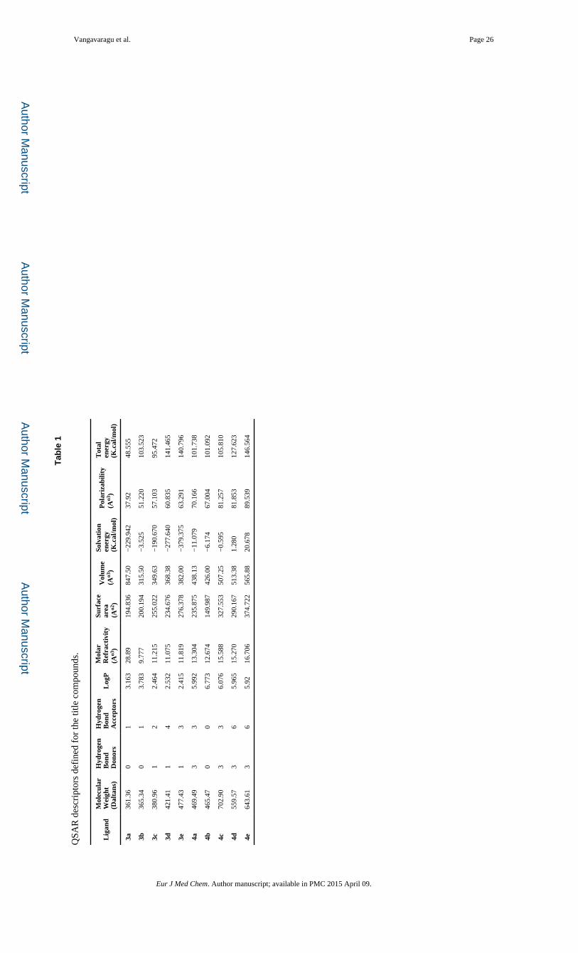
2.3. Quantitative structure-activity relationship (QSAR) descriptors
The three dimensional structures of our compounds (3a–e & 4a–e) were constructed using
the builder interface of the molecular operating environment (MOE) program and subjected
to energy minimization using the Merck molecular Force Field 94x (MMFF94x)[43, 44]. All
items were set to default with an RMS gradient of 0.01 kcal/mol and an RMS distance of 0.1
A°. We subjected the resultant models to a systematic conformational search. Using the
above conformations, we studied QSAR descriptors, meeting the Lipinski criteria for safe
drugs [45, 46]. Our designed compounds have a molecular weight less than 500 Da with
exception of compounds 4c, 4d and 4e. Compound 3a had the lowest molecular weight
(361.35 Da), while compound 4c had the highest molecular weight (702.90 Da). There were
less than 5 numbers of hydrogen bond donors and less than 10 hydrogen bond acceptors for
all compounds. LogP values were below 5, indicating that all are non-toxic to the host
system. Molar refractivity was in-between 40–150, the optimal range. Characterization of
remaining descriptors such as surface area, volume, hydration energy, polarizability and
energy levels were also encouraging showing suitable feature in binding domains of the
targets and inhibitors suggesting the potential safety and efficacy of these targets (Table 1).
2.4. Molecular modeling [47, 48]
Molecular modeling studies has often been proven to be a powerful tool for rationalizing
ligand-receptor interactions and for making this information available to virtual screening
techniques. The ligand database that was developed from the total set of ten compounds was
docked into the specified binding domain of the hPreP receptor. As we know that PreP is
present in Arabidopsis thaliana, and is responsible for the degradation of mitochondrial
presequences and elimination of chloroplast transit peptides generated after organelle
precursor protein import and processing [49–51], we identified hPreP binding sites using the
crystal structure of AtPreP (http://www.rcsb.org/pdb, PDB ID: 2FGE), with residues:
Asn136, Ala137, Phe138, Thr139, Ala140, Glu205, Try906, Arg900, Gln222. A total of 30
conformations were generated for each ligand-receptor complex and among them, the
conformation with least docking score was considered for further analysis. The MOE
interaction of all ligand molecules in the binding domain cavity was analyzed. The ligand-
receptor complexes were analyzed by both London G free energy approximations and E
interaction energies. Compounds 3c had the best docking score of −8.6 kcal/mole, followed
by docking scores for compound 4c (−8.3 kcal/mole). Interestingly, compound 3c formed an
arene-hydrogen bond with Gly121 (Figure 1). These two ligands had the best docking scores
with stable ligand interactions and are composed of primarily non-polar pi-stacking
interactions between Phe 123, Phe124, Try906, Try921, with minimal H-bonding and Zn+2
coordination (Figure 1).
2.5. Pharmacophore Model: PreP activators 5 Feature Pharmacophore Model[28, 47, 48]
Using the Pharmacophore five-feature model, shown in Figure 3, we selected 4 out of 10
compounds as ‘hit’s, namely compounds 3c, 3d, 4c and 4d. Compounds 3c and 4c exhibited
strong binding against hPreP. Based on the number of hits, the predictive capacity of active
hits is at least 50% (Figure 2).
Vangavaragu et al. Page 4
Eur J Med Chem. Author manuscript; available in PMC 2015 April 09.
Author M
anuscriptA
uthor Manuscript
Author M
anuscriptA
uthor Manuscript
2.6. Evaluation of enzymatic activity of hPreP
Compounds with small molecular weights are necessary to boost hPreP activity and provide
therapeutic protection against AD. The literature suggests that hPreP degrades mitochondrial
targeting peptides that are cleaved off by mitochondrial processing peptidases following
import to mitochondria. PreP’s critical role in the degradation of mitochondrial Aβ is
significant as accumulation of mitochondrial Aβ is directly linked to Aβ-induced
mitochondrial toxicity. Further, hPreP also degrades non-Aβ substrates such as pF1β, a 53
amino acid long mitochondrial presequence peptide, and substrate V, a 9 amino acid
fluorogenic peptide. Even though these peptides have slightly different properties (Aβ
peptides are negatively charged, and pF1β are positively charged), hPreP degrades both with
similar efficiency. To our knowledge, compound 3c & 4c are the first drug-like compounds
shown to stimulate hPreP-mediated proteolysis of various substrates.
To study the regulatory effect of our target compounds (3a–e & 4a–e) on the proteolytic
activity of hPreP, we selected three substrates of different lengths and properties for use in
an in vitro degradation assay: 1)Aβ (1–42); 2) the presequence of ATP synthase F1β subunit
pF1β (2–54); 3) and a fluorescent peptide known as substrate V. Activity assays were
initially set at a concentration of 100 μM for all studied molecules in order to identify the
effect of those compounds on overall hPreP activity. We quantified hPreP activity by
monitoring the change in fluorescence for proteolysis of fluorogenic-substrate V and
monitored the efficacy of degradation of Aβ (1–42) & pF1β, using immunoblotting for Aβ
with NuPAGE 12% Bis-tris gel assay.
Analysis of compounds showed that 3c & 4c increased proteolytic activity of hPreP against
biotin labeled Aβ (1–42), significant up to a 1μM concentration when compared to other
analogs. Compounds 3c & 4c effectively facilitated the degradation of Aβ at more efficient
levels than hPreP alone (Figure 3A & 4A). We observed degenerated and dose dependent
immunoreactive biotin Aβ bands due to the enhanced proteolytic activity of hPreP at various
concentrations of compound 3c & 4c (Figure 3C, D & 4C, D), with no Aβ immunoreactive
bands at 100μM (Figure 3D & 4D, lane 5), compared to hPreP alone. Analysis for the effect
of compounds on hPreP activity showed that 3c increased the hPreP -mediated Aβ
degradation by 1.7 (42%) & 4c by 2.1 folds (54%) (Figure 9A).
Next, we analyzed an EC50 value of each molecule that enhanced hPreP activity using a
dose response assay, where specific activity of hPreP was measured with different
concentrations of the lead compounds and then the plot was fit to prism nonlinear
regression, a log (agonist) vs. normalized response equation. Calculated EC50 values for 3c & 4c were 0.713 μM and 0.402 μM, respectively (Figure 3B & 4B).
In the case of non-Aβ substrates, hPreP also degraded pF1β and compounds 3c & 4c showed
significant proteolytic activity of hPreP in degrading pF1β even at 1μM concentration.
Significant changes in pF1β degradation were observed with compounds 3c & 4c when
compared to hPreP alone (Figure 5A & 6A). In figures 5C & 6C the presence of degenerated
& dose dependent immunoreactive biotin pF1β bands, indicate enhanced proteolytic activity
of hPreP with different concentrations of compounds 3c & 4c. Compounds 3c & 4c increased hPreP-mediated pF1β degradation by 1.8 (45%) & 2.2 folds (55%) respectively
Vangavaragu et al. Page 5
Eur J Med Chem. Author manuscript; available in PMC 2015 April 09.
Author M
anuscriptA
uthor Manuscript
Author M
anuscriptA
uthor Manuscript
(Figure 9B). EC50 values for 3c & 4c were measured at 0.012 & 0.601μM, respectively
(Figure 5B & 6B).
Further, we performed kinetic studies using Substrate V, a fluorogenic peptide 9 amino acids
long containing the fluorescent group 7-methoxycoumarin and the quencher group 2, 4-
dinitrophenyl, thus, providing light emission upon cleavage of the peptide bond between
these two groups. Compounds, 3c & 4c increased the initial rate of proteolysis activity of
hPreP the most, specifically by 1.5 and 1.8 folds, respectively at 1μM (Figures 7A & 8A).
Further, the EC50 values for 3c & 4c were 0.152 & 0.057 μM, respectively (Figures 7B &
8B).
Interestingly, condensation of 5-bromo-2-hydroxy benzaldehyde with nitro o-phenelene
diamine plays a crucial role in bioactivity of our compounds 3c & 4c. Introduction of a 5-
bromo-2-hydroxy group (3c into the benzimidazole ring) enhanced the proteolytic activity of
hPreP against biotin Aβ (1–42), pF1β (2–54) and substrate V. The same correlation was also
observed in compound 4c against these three aforementioned substrates. It is clear that the
presence of a bromo group on the aromatic ring is essential for improving hydrophilic
interactions between the compound and hPreP. Substitution of the fluoro, methoxy &
hydroxy in the aromatic aldehyde caused a substantial loss of proteolytic activity of hPreP.
These results suggest that the compounds with halogen atoms (bromo) that underwent
hydroxyl substitution on their phenyl ring(s) (3c & 4c) were the most potent and selective
activators for the proteolytic activity of hPreP. Our results clearly indicate that
benzimidazole analogues shed new light on the design and understanding of selective
proteolytic enzyme activators, which will enable the synthesis of our scaffold into clinically
useful anti-Alzheimer’s drugs.
3. Conclusion
In the present study, we designed novel benzimidazole derivatives according to their binding
modes inside the hPreP active site. Amongst them, we selected compounds that obeyed the
Lipinski’s “rule of five.” This is extensively used to screen for the drug-like properties. Our
novel compounds behaved like hPreP agonists, with compounds 3c & 4c being most active.
Introduction of a 5-bromo-2-hydroxy benzaldehyde group increased potency resulting in the
4-bromo-2-(1-(5-bromo-2-hydroxybenzyl)-5-nitro-1H-benzo[d]imidazol-2-yl) phenol &
2,2′-((2-(5-bromo-2-hydroxyphenyl)-5-nitro-1H-benzo[d]imidazole-1,3(2H)
diyl)bis(methylene))bis(4-bromophenol), which are the most active compounds (at 100 μM)
and seem to be good agonists of hPreP with EC50 in the low μM range.
Together with biological results (indicating that the synthesized benzimidazole-based
compounds possess agonist activity with hPreP) and the results of the docking studies and
regulating activities, we conclude that compounds 3c & 4c are appropriate scaffolds for the
development of new hPreP regulators. Further investigation of the effect of these
benzimadazole derivatives on hPreP agonist activity will move the development of new
agents for the treatment of AD forward towards translation to clinical use.
Vangavaragu et al. Page 6
Eur J Med Chem. Author manuscript; available in PMC 2015 April 09.
Author M
anuscriptA
uthor Manuscript
Author M
anuscriptA
uthor Manuscript
4. Materials and Methods
4.2. Chemistry
4.2.1. General—All reagents were commercially available and used without further
purification. Melting points were determined in open capillary tubes using a Laboratory
Devices Mel-Temp apparatus and are uncorrected. 1H and 13C NMR spectra were recorded
in d6-DMSO on a Bruker DRX-500 spectrometer operating at 500 MHz, and 125 MHz,
respectively, and calibrated to the solvent peak. Abbreviations used for the split patterns of
proton NMR signals are: singlet (s), doublet (d), triplet (t), quartet (q), quintet (qui),
multiplet (m) and broad signal (br). High-resolution mass spectrometry (HRMS) was
recorded on a LCT Premier Spectrometer.
4.2.2. General procedure for the preparation of compounds (3a–3e)—A mixture
of aromatic aldehyde (2 mmol), 4-nitrobenzene-1, 2-diamine (1 mmol, 2 equiv), and TFE
(1mL) was stirred magnetically at room temperature for 1h. The progress of the reaction was
monitored using TLC (hexane: ethyl acetate, 1:1 v/v). After completion of the reaction, the
reaction mixture was dissolved in EtOAc (3 mL), adsorbed on silica gel (0.5 g, 230–400
mesh), and concentrated under rotary vacuum evaporation. The resultant solid mass was
charged onto a flash chromatography column and eluted with hexane-EtOAc (85:15) to
afford the title compounds.
4.2.3. 4-(1-(4-Hydroxybenzyl)-5-nitro-1H-benzo[d]imidazol-2-yl) phenol (3a)—Light color solid, 77% yield; mp: 185–186 °C; Rf 0.22; (hexane: ethyl acetate, 1:1 v/v). IR:
3234, 3050, 2902, 2864, 2362, 1712, 1668, 1529, 1431, 1350, 1257, 1091, 1031, 748
cm−1; 1H NMR (DMSO-d6) ™ 10.15 (s, 1H), 9.46 (s, 1H), 8.45 (d, J = 3.75 Hz, 1H),
8.15-8.13 (m, 1H) 7.84 (d, J = 3.75 Hz, 1H), 7.67-7.63 (m, 2H), 6.94-6.91 (m, 2H),
6.69-6.66 (m, 2H), 5.61 (s, 2H). 13C NMR (DMSO-d6)™ 159.6, 158.6, 156.8, 147.6, 142.6,
135.4, 130.9, 127.5, 126.4, 119.6, 119.0, 117.8, 115.7 (J = 15.0 Hz), 47.6. HRMS cald for
C20H16N3O54 (M+H) 362.1132; found 362.1141 (TOF MS ES+).
4.2.4. 1-(4-Fluorobenzyl)-2-(4-fluorophenyl)-5-nitro-1H-benzo[d]imidazole (3b)—Yellow solid, 88% yield; mp: 201–203 °C; Rf 0.32; (hexane: ethyl acetate, 1:1 v/v). IR:
3076, 3055, 2948, 2765, 1897, 1610, 1523, 1483, 1329, 1265, 1172, 1110, 954, 823, 748
cm−1; 1H NMR (DMSO-d6)™ 8.63-8.61 (m, 1H), 8.22-8.18 (s, 1H), 7.86-7.79 (m, 3H)
7.45-7.40 (t, 2H), 7.15-7.02 (m, 4H), 5.76-5.68 (m, 2H). 13C NMR (DMSO-d6) ™ 164.3(d,
J = 7.5 Hz), 162.4, 160.4(d, J = 3.75 Hz), 157.2, 156.1, 146.9, 143.2, 142.9, 141.6, 140.2,
135.3, 132.4(d, J = 3.75 Hz), 132.2(d, J = 2.5 Hz), 131.7, 128.3(d, J = 8.75 Hz), 119.6,
118.4, 118.0, 116.1, 115.9, 115.7, 115.6, 115.4, 111.7, 108.1, 47.2 (d, J = 15 Hz). HRMS
cald for C20H14F2N3O2 (M+H) 366.1052; found 366.1054 (TOF MS ES+).
4.2.5. 4-Bromo-2-(1-(5-bromo-2-hydroxybenzyl)-5-nitro-1H-benzo[d]imidazol-2-yl) phenol (3c)—White crystals, 78% yield; mp: 226–228 °C; Rf 0.32; (hexane: ethyl
acetate, 1:1 v/v). IR: 3475, 3342, 3055, 2989, 2893, 2304, 1903, 1629, 1610, 1579, 1488,
1434, 1348, 1222, 1176, 977, 896, 748 cm−1; 1H NMR (DMSO-d6) ™ 11.60 (s, 2H), 8.92 (s,
2H), 8.12 (d, 1H) 7.96-7.93 (m, 2H), 7.56-7.53 (m, 2H), 6.96 (d, J = 10.0 Hz, 2H), 6.81-6.78
Vangavaragu et al. Page 7
Eur J Med Chem. Author manuscript; available in PMC 2015 April 09.
Author M
anuscriptA
uthor Manuscript
Author M
anuscriptA
uthor Manuscript
(m, 2H). 13C NMR (DMSO-d6) ™ 159.1, 158.3, 150.3, 136.0, 135.5, 133.6, 132.5, 124.4,
122.6, 199.0, 144.1, 113.1, 110.4. HRMS cald for C20H13Br2N3O4 (M+H); 517.9351, found
517.9352 (TOF MS ES+).
4.2.6. Methyl 2-hydroxy-5-(1-(4-hydroxy-3-(methoxycarbonyl)benzyl)-5-nitro-1H-benzo[d]imidazol-2-yl)benzoate (3e)—Yellow solid, 86% yield; mp: 265–
267 °C; Rf 0.20; (hexane: ethyl acetate, 1:1 v/v). IR: 3371, 3055, 2950, 2767, 1903, 1627,
1612, 1487, 1265, 1176, 977, 896, 817, 738 cm−1; 1H NMR (DMSO-d6) ™ 11.06 (s, 2H),
8.88-8.68(m, 2H), 8.45-8.38 (m, 5H), 8.15-8.13 (m, 1H) 7.78 (d, J = 5.0 Hz, 2H), 7.24 (d, J
= 10.0 Hz, 2H), 3.96 (s, 6H). 13C NMR (DMSO-d6) ™ 167.8, 162.0, 154.0, 143.1, 141.2,
137.1, 133.8, 130.0, 118.7, 118.5, 114.7, 114.4, 111.1, 52.6. HRMS cald for C24H20N3O8
(M+H) 478.1250; found 478.1259 (TOF MS ES+).
4.2.7. General procedure for the preparation of compounds (4a–4e)—To a
stirred solution aromatic aldehyde (3 mmol), 4-nitrobenzene-1, 2-diamine (1 mmol, 2
equiv), was added in the presence of trimethylsilylchloride dissolved in water (5 mL). The
reaction mixture was stirred for 8–12 h at room temperature. The progress of the reaction
was monitored by TLC (hexane: ethylacetate1:1). After completion of the reaction, a solid
was formed. The solid was filtered off, washed with water, and dried. It was purified by
silica gel column chromatography eluting with hexane: ethyl acetate (95:5) mixture to afford
the title compounds.
4.2.8. 4,4′-((2-(4-Hydroxyphenyl)-5-nitro-1H-benzo[d]imidazole-1,3(2H)-diyl)bis(methylene)) diphenol (4a)—Light orange color solid; mp: 224–226 °C; Rf
0.24; (hexane: ethyl acetate, 1:1 v/v). IR: 3234, 3103, 2902, 2796, 2362, 1712, 1668, 1529,
1350, 1091, 1031, 881, 738 cm−1; 1H NMR (DMSO-d6) δ 10.23 (s, 1H), 9.45 (s, 2H), 8.48
(d, J = 3.75 Hz, 2H), 8.15-8.13 (m, 2H) 7.68-7.62 (m, 4H), 7.01–7.06 (m, 3H), 6.82–6.89
(m, 3H), 6.71-6.69 (m, 2H), 5.56 (s, 4H). 13C NMR (DMSO-d6) δ 160.3, 159.2, 158.0,
148.1, 142.8, 136.1, 131.2, 128.6, 127.5, 120.0, 119.4, 118.5, 116.8 (J = 15.0 Hz), 48.1.
HRMS cald for C27H24N3O5 (M+H) 470.1716; found 470.1761(TOF MS ES+).
4.2.9. 1,3-Bis(4-fluorobenzyl)-2-(4-fluorophenyl)-5-nitro-2,3-dihydro-1H-benzo[d]imidazole (4b)—Yellow solid, 92% yield; mp: 196–198 °C; Rf 0.30; (hexane:
ethyl acetate, 1:1 v/v). IR: 3099, 3055, 2927, 2761, 1895, 1730, 1610, 1579, 1483, 1344,
1265, 1174, 1110, 896, 748 cm−1; 1H NMR (DMSO-d6) δ 8.68-8.59 (m, 2H), 8.28-8.22 (s,
2H), 7.70-7.59 (m, 5H) 7.48-7.41 (m, 4H), 7.16-7.01(m, 4H), 5.80-5.71 (m, 4H). 13C NMR
(DMSO-d6) δ 165.2, 163.2, 161.1(d, J = 3.75 Hz), 158.1, 158.1, 142.1, 140.5, 139.8, 138.8,
138.2, 136.3, 131.4(d, J = 3.75 Hz), 131.1, 129.2, 127.3, 120.6, 119.3, 118.2, 116.8, 115.9,
115.2, 114.2, 113.2, 110.1, 107.1, 48.3 (d, J = 15 Hz). HRMS cald for C27H21F3N3O2 (M
+H) 476.1586; found 476.1591 (TOF MS ES+).
4.2.10. 2,2′-((2-(5-Bromo-2-hydroxyphenyl)-5-nitro-1H-benzo[d]imidazole-1,3(2H) diyl)bis(methylene))bis(4-bromophenol) (4c)—White
crystals, 82% yield; mp: 256–257 °C; Rf 0.28; (hexane: ethyl acetate, 1:1 v/v). IR: 3330,
3236, 2927, 2883, 1924, 1714, 1668, 1568, 1525, 1346, 1253, 1091, 1051, 881, 738, 698
cm−1; 1H NMR (DMSO-d6) ™ 11.59 (s, 2H), 8.94 (s, 2H), 8.12 (d, 2H) 7.96-7.94 (m, 4H),
Vangavaragu et al. Page 8
Eur J Med Chem. Author manuscript; available in PMC 2015 April 09.
Author M
anuscriptA
uthor Manuscript
Author M
anuscriptA
uthor Manuscript
7.56-7.54 (m, 2H), 6.96 (d, J = 10.0 Hz, 2H), 6.80-6.78 (m, 6H). 13C NMR (DMSO-d6) ™
159.1, 158.3, 150.3, 136.0, 135.5, 133.6, 132.5, 124.4, 122.6, 199.0, 144.1, 113.1, 110.4.
HRMS cald for C27H20Br3N3O5 (M+H) 702.8953; found 702.8922 (TOF MS ES+).
4.2.11. Dimethyl 5,5′-((2-(4-hydroxy-3-(methoxycarbonyl)phenyl)-5-nitro-1H-benzo[d]imidazole-1,3(2H)-diyl)bis(methylene))bis(2-hydroxybenzoate) (4e)—White crystals, 72% yield; mp: 286–288 °C; Rf 0.18; (hexane: ethyl acetate, 1:1 v/v). IR:
3055, 2989, 2950, 1629, 1579, 1587, 1534, 1487,1222, 1176, 977, 817, 750 cm−1; 1H NMR
(DMSO-d6) ™ 10.97 (s, 3H), 8.81 (d, J = 5.0 Hz, 3H), 8.41 (m, 2H), 8.32-8.30 (m, 1H),
8.15-8.13 (m, 1H) 8.00 (d, J = 5.0 Hz, 3H), 7.92 (d, J = 5.0 Hz, 1H), 7.14-7.11 (m, 3H),
6.77-6.75 (m, 4H), 3.94 (s, 9H). 13C NMR (DMSO-d6) ™ 168.5, 162.2, 158.6, 150.9, 135.9,
135.0, 134.2, 132.4, 127.8, 124.1, 118.0, 113.8, 113.0, 112.5, 52.6. HRMS cald for
C33H30N3O11 (M+H) 644.1880; found 644.1852 (TOF MS ES+).
4.1.1. Preparation of human PreP homology model [28]—A Blast (NCBI,
www.ncbi.nih.gov) database search of the PDB Databank revealed the optimal starting
template structure for human PreP as a zinc metalloprotease ((PDB:http://www.rcsb.org/
pdb, PDB ID: 2FGE). Sequence homology construction of the human PMP1 enzyme
sequence based on 2FGE structure was carried out using a MOE protein homology
algorithm. Hydrogens were added to the resulting 3D structure based on a pH of 7 and a salt
concentration of 0.1 M. pH of 7. The implicit born solvation model and a non-bonded cut off
value of 10–12 were incorporated in the MMFF94x force field. The complete structure was
energy minimized to a gradient cut off value of 0.05. Molecular dynamic simulations were
carried out at a constant temperature of 300° K for a heating time of 10 pico seconds.
Simulations were carried out for 10 nano seconds. The time step was considered as 0.001
pico seconds and the temperature relaxation time was set to 0.2 pico seconds. Position,
velocity and acceleration were saved every 0.5 pico seconds.
4.1.2. Prediction of Binding site for Ligands—The binding site of hPreP was
elucidated through the crystal structure of AtPreP (PDB:http://www.rcsb.org/pdb, PDB ID:
2FGE). In our case, we used a proximity radius of r </= 10 Ao. A confirmatory approach for
determining the binding site of these novel drug-like candidates involved use of Alpha Site
Finder methodology [Edelsbrunner, et al. “Measuring Proteins and Voids in Proteins”,.
Proceedings from the 28th International Conference on Systems Science, 256–264(1995)],
incorporated into the Chemical Computing Group’s the molecular modeling software. This
algorithm calculates and displays potential regions of tight atomic packing on a protein
surface.
4.1.3. Molecular Docking [24, 25]—A conformational set was generated for each of our
ligand molecules using the LowModeMD Search algorithm within the MOE program. This
involves a short molecular dynamics simulation using velocities with low kinetic energy on
high-frequency vibrational modes. An iteration limit of 5000 (maximum number of attempts
to generate a new conformation) and a minimization limit of 250 (maximum number of
minimization steps) were set to control the number of conformations generated. Each
generated conformation was docked into the specified binding domain of the human PreP
Vangavaragu et al. Page 9
Eur J Med Chem. Author manuscript; available in PMC 2015 April 09.
Author M
anuscriptA
uthor Manuscript
Author M
anuscriptA
uthor Manuscript

receptor. We analyzed interaction of all ligand molecules in the binding domain cavity using
ligand interaction study in the MOE software. The ligand-receptor complexes were analyzed
using the sum of the following two scoring functions: (i) London ⊗G free energy
approximations and (ii) interaction energies, ⊗E. Ligands that bound with the lowest
docking score were considered for further analysis.
4.1.4. Pharmacophore Model—A pharmacophore defines features as well as locations
of important binding interactions between a ligand and its receptor. Our five-feature
pharmacophore model was constructed by overlapping the top two ligand candidates
(compounds 3c and 4c) that had the strongest binding affinities. The Unified scheme within
MOE was used to define the unique features (2 H-bond [Don/Acc] and 3 aromatic centers
[Aro]). The locations of these features were determined by inspection of strong interactions
of these two compounds at the PreP active site.
4.3. Degradation assays
For the analysis of the novel compounds as chemical regulators for hPreP activity, we
performed degradation assays with biotin-labeled Aβ (1–42) and F1β (2–54) [36]
presequence peptides in degradation buffer (20mM HEPES-KOH pH 8.0) with10mM
MgCl2, 1μg of purified hPreP and 0.02μg of a substrate at various concentrations (0.00001–
100μM). Compounds were incubated for 2.5 h at 373C. Reactions were stopped by the
addition of 4× sample buffer, and then analyzed on NuPAGE 12% Bis-Tris gel (Invitrogen,
CA), and run in 1×MES buffer. Proteins were electrophoretically transferred to
nitrocellulose membrane HybondTM (Amersham Bioscience) for 1 h at 100V. For pF1β
identification, the nitrocellulose membrane was blocked overnight in 5% milk-PBS followed
by incubation with pF1β antibody (1:2000) and detected using horseradish peroxidase-
conjugated anti-rabbit secondary antibody (1:2500) and enhanced chemiluminescence (ECL,
GE Healthcare). For analyzing the degradation of biotin labeled Aβ (1–42) (biotin- LC-
Aβ1–40, biotin-LC- Aβ1–42), the nitrocellulose membrane was dried overnight at 253C
followed by blocking with 2% milk-PBS for 1 h. Immunoblotting was performed with
ExtraAvidin Peroxidase Conjugate 1:3000 (Sigma) and detection by ECL.
4.4. Degradation of Substrate V by hPrep
To determine the efficiency of these novel compounds as chemical regulators for hPreP
activity, we used a fluorescence assay with fluorogenic substrate V (7-methoxycoumarin-4-
yl-acetyl-NPPGFSAFK-2, 4-dinitrophenyl, R&D Systems) and measured the kinetics of
proteolysis. Reaction was carried out in the presence of 1μg hPreP in 20mM HEPES, pH 8.0
with 10mM MgCl2 mixed with 0.1μg Substrate V and various concentrations (0.00001–
1μM) of our compounds in a final volume of 250μl. The hydrolysis of substrate V was
measured for 10 minutes using a fluorometer (SpectraMax Gemini) with excitation and
emission wavelengths set at 320 and 405 nm, respectively. Results are shown as the
Substrate V degradation rate and averaged over three independent experiments (Figure 7)
4.5. Statistical analysis
Statistical analyses were performed using STATVIEW software. One-way ANOVA was
used for repeated measures followed by Bonferroni/Dunn Protected Least Significant
Vangavaragu et al. Page 10
Eur J Med Chem. Author manuscript; available in PMC 2015 April 09.
Author M
anuscriptA
uthor Manuscript
Author M
anuscriptA
uthor Manuscript
Difference analysis for post-hoc comparisons. Results are expressed as mean ± Standard
Error Mean (SEM). Significance was set at p < 0.05.
Supplementary Material
Refer to Web version on PubMed Central for supplementary material.
Acknowledgments
This study was supported by grant awards (R01GM095355, R37AG037319, and RO1NS65482) from the National Institute of General Medical Sciences, the National Institute on Aging, and the National Institute of Neurological Disease and Stroke.
References
1. Maurer I, Zierz S, Moller HJ. A selective defect of cytochrome c oxidase is present in brain of Alzheimer disease patients. Neurobiology of aging. 2000; 21:455–462. [PubMed: 10858595]
2. Sheehan JP, Swerdlow RH, Miller SW, Davis RE, Parks JK, Parker WD, Tuttle JB. Calcium homeostasis and reactive oxygen species production in cells transformed by mitochondria from individuals with sporadic Alzheimer’s disease. The Journal of neuroscience : the official journal of the Society for Neuroscience. 1997; 17:4612–4622. [PubMed: 9169522]
3. Swerdlow RH, Khan SM. A “mitochondrial cascade hypothesis” for sporadic Alzheimer’s disease. Medical hypotheses. 2004; 63:8–20. [PubMed: 15193340]
4. Du H, Yan SS. Mitochondrial medicine for neurodegenerative diseases. The international journal of biochemistry & cell biology. 2010; 42:560–572. [PubMed: 20067840]
5. Lin MT, Beal MF. Alzheimer’s APP mangles mitochondria. Nature medicine. 2006; 12:1241–1243.
6. Eckert A, Hauptmann S, Scherping I, Rhein V, Muller-Spahn F, Gotz J, Muller WE. Soluble beta-amyloid leads to mitochondrial defects in amyloid precursor protein and tau transgenic mice. Neuro-degenerative diseases. 2008; 5:157–159. [PubMed: 18322377]
7. Yao J, Irwin RW, Zhao L, Nilsen J, Hamilton RT, Brinton RD. Mitochondrial bioenergetic deficit precedes Alzheimer’s pathology in female mouse model of Alzheimer’s disease. Proceedings of the National Academy of Sciences of the United States of America. 2009; 106:14670–14675. [PubMed: 19667196]
8. Caspersen C, Wang N, Yao J, Sosunov A, Chen X, Lustbader JW, Xu HW, Stern D, McKhann G, Yan SD. Mitochondrial Abeta: a potential focal point for neuronal metabolic dysfunction in Alzheimer’s disease. The FASEB journal : official publication of the Federation of American Societies for Experimental Biology. 2005; 19:2040–2041.
9. Yao J, Irwin RW, Zhao L, Nilsen J, Hamilton RT, Brinton RD. Mitochondrial bioenergetic deficit precedes Alzheimer’s pathology in female mouse model of Alzheimer’s disease. Proc Natl Acad Sci U S A. 2009
10. Yao J, Du H, Yan S, Fang F, Wang C, Lue LF, Guo L, Chen D, Stern DM, Gunn Moore FJ, Xi Chen J, Arancio O, Yan SS. Inhibition of amyloid-beta (Abeta) peptide-binding alcohol dehydrogenase-Abeta interaction reduces Abeta accumulation and improves mitochondrial function in a mouse model of Alzheimer’s disease. J Neurosci. 2011; 31:2313–2320. [PubMed: 21307267]
11. Takuma K, Fang F, Zhang W, Yan S, Fukuzaki E, Du H, Sosunov A, McKhann G, Funatsu Y, Nakamichi N, Nagai T, Mizoguchi H, Ibi D, Hori O, Ogawa S, Stern DM, Yamada K, Yan SS. RAGE-mediated signaling contributes to intraneuronal transport of amyloid-beta and neuronal dysfunction. Proceedings of the National Academy of Sciences of the United States of America. 2009; 106:20021–20026. [PubMed: 19901339]
12. Pavlov PF, Petersen CH, Glaser E, Ankarcrona M. Mitochondrial accumulation of APP and Abeta: Significance for Alzheimer disease pathogenesis. J Cell Mol Med. 2009
13. Manczak M, Anekonda TS, Henson E, Park BS, Quinn J, Reddy PH. Mitochondria are a direct site of A beta accumulation in Alzheimer’s disease neurons: implications for free radical generation
Vangavaragu et al. Page 11
Eur J Med Chem. Author manuscript; available in PMC 2015 April 09.
Author M
anuscriptA
uthor Manuscript
Author M
anuscriptA
uthor Manuscript
and oxidative damage in disease progression. Hum Mol Genet. 2006; 15:1437–1449. [PubMed: 16551656]
14. Lustbader JW, Cirilli M, Lin C, Xu HW, Takuma K, Wang N, Caspersen C, Chen X, Pollak S, Chaney M, Trinchese F, Liu S, Gunn-Moore F, Lue LF, Walker DG, Kuppusamy P, Zewier ZL, Arancio O, Stern D, Yan SS, Wu H. ABAD directly links Abeta to mitochondrial toxicity in Alzheimer’s disease. Science. 2004; 304:448–452. [PubMed: 15087549]
15. Hansson Petersen CA, Alikhani N, Behbahani H, Wiehager B, Pavlov PF, Alafuzoff I, Leinonen V, Ito A, Winblad B, Glaser E, Ankarcrona M. The amyloid beta-peptide is imported into mitochondria via the TOM import machinery and localized to mitochondrial cristae. Proc Natl Acad Sci U S A. 2008; 105:13145–13150. [PubMed: 18757748]
16. Du H, Guo L, Fang F, Chen D, Sosunov AA, McKhann GM, Yan Y, Wang C, Zhang H, Molkentin JD, Gunn-Moore FJ, Vonsattel JP, Arancio O, Chen JX, Yan SD. Cyclophilin D deficiency attenuates mitochondrial and neuronal perturbation and ameliorates learning and memory in Alzheimer’s disease. Nat Med. 2008; 14:1097–1105. [PubMed: 18806802]
17. Dragicevic N, Mamcarz M, Zhu Y, Buzzeo R, Tan J, Arendash GW, Bradshaw PC. Mitochondrial amyloid-beta levels are associated with the extent of mitochondrial dysfunction in different brain regions and the degree of cognitive impairment in Alzheimer’s transgenic mice. J Alzheimers Dis. 2010; 20(Suppl 2):S535–550. [PubMed: 20463404]
18. Butterfield DA, Hensley K, Harris M, Mattson M, Carney J. beta-Amyloid peptide free radical fragments initiate synaptosomal lipoperoxidation in a sequence-specific fashion: implications to Alzheimer’s disease. Biochemical and biophysical research communications. 1994; 200:710–715. [PubMed: 8179604]
19. Cardoso SM, Santana I, Swerdlow RH, Oliveira CR. Mitochondria dysfunction of Alzheimer’s disease cybrids enhances Abeta toxicity. Journal of neurochemistry. 2004; 89:1417–1426. [PubMed: 15189344]
20. Cakir B, Dagliyan O, Dagyildiz E, Baris I, Kavakli IH, Kizilel S, Turkay M. Structure based discovery of small molecules to regulate the activity of human insulin degrading enzyme. Plos One. 2012; 7:e31787. [PubMed: 22355395]
21. Takuma K, Yao J, Huang J, Xu H, Chen X, Luddy J, Trillat AC, Stern DM, Arancio O, Yan SS. ABAD enhances Abeta-induced cell stress via mitochondrial dysfunction. Faseb J. 2005; 19:597–598. [PubMed: 15665036]
22. Du H, Guo L, Zhang W, Rydzewska M, Yan S. Cyclophilin D deficiency improves mitochondrial function and learning/memory in aging Alzheimer disease mouse model. Neurobiology of aging. 2011; 32:398–406. [PubMed: 19362755]
23. Mzhavia N, Berman YL, Qian Y, Yan L, Devi LA. Cloning, expression, and characterization of human metalloprotease 1: a novel member of the pitrilysin family of metalloendoproteases. DNA and cell biology. 1999; 18:369–380. [PubMed: 10360838]
24. Falkevall A, Alikhani N, Bhushan S, Pavlov PF, Busch K, Johnson KA, Eneqvist T, Tjernberg L, Ankarcrona M, Glaser E. Degradation of the amyloid beta-protein by the novel mitochondrial peptidasome, PreP. J Biol Chem. 2006; 281:29096–29104. [PubMed: 16849325]
25. Becker AB, Roth RA. An unusual active site identified in a family of zinc metalloendopeptidases. Proceedings of the National Academy of Sciences of the United States of America. 1992; 89:3835–3839. [PubMed: 1570301]
26. Taylor SW, Fahy E, Zhang B, Glenn GM, Warnock DE, Wiley S, Murphy AN, Gaucher SP, Capaldi RA, Gibson BW, Ghosh SS. Characterization of the human heart mitochondrial proteome. Nature biotechnology. 2003; 21:281–286.
27. Alikhani N, Ankarcrona M, Glaser E. Mitochondria and Alzheimer’s disease: amyloid-beta peptide uptake and degradation by the presequence protease, hPreP. Journal of bioenergetics and biomembranes. 2009; 41:447–451. [PubMed: 19798557]
28. Johnson KA, Bhushan S, Stahl A, Hallberg BM, Frohn A, Glaser E, Eneqvist T. The closed structure of presequence protease PreP forms a unique 10,000 Angstroms3 chamber for proteolysis. The EMBO journal. 2006; 25:1977–1986. [PubMed: 16601675]
29. Maduke M, Roise D. Import of a mitochondrial presequence into protein-free phospholipid vesicles. Science. 1993; 260:364–367. [PubMed: 8385804]
Vangavaragu et al. Page 12
Eur J Med Chem. Author manuscript; available in PMC 2015 April 09.
Author M
anuscriptA
uthor Manuscript
Author M
anuscriptA
uthor Manuscript
30. Shen Y, Joachimiak A, Rosner MR, Tang WJ. Structures of human insulin-degrading enzyme reveal a new substrate recognition mechanism. Nature. 2006; 443:870–874. [PubMed: 17051221]
31. Butterfield DA. Amyloid beta-peptide (1–42)-induced oxidative stress and neurotoxicity: implications for neurodegeneration in Alzheimer’s disease brain. A review. Free radical research. 2002; 36:1307–1313. [PubMed: 12607822]
32. Teixeira PF, Pinho CM, Branca RM, Lehtio J, Levine RL, Glaser E. In vitro oxidative inactivation of human presequence protease (hPreP). Free radical biology & medicine. 2012; 53:2188–2195. [PubMed: 23041349]
33. Cakir B, Dagliyan O, Dagyildiz E, Baris I, Kavakli IH, Kizilel S, Turkay M. Structure Based Discovery of Small Molecules to Regulate the Activity of Human Insulin Degrading Enzyme. Plos One. 2012; 7
34. Nile SH, Kumar B, Park SW. In Vitro Evaluation of Selected Benzimidazole Derivatives as an Antioxidant and Xanthine Oxidase Inhibitors. Chemical biology & drug design. 2013; 82:290–295. [PubMed: 23581708]
35. Ayhan-Kilcigil G, Kus C, Ozdamar ED, Can-Eke B, Iscan M. Synthesis and antioxidant capacities of some new benzimidazole derivatives. Arch Pharm. 2007; 340:607–611.
36. Gurer-Orhan H, Orhan H, Suzen S, Puskullu MO, Buyukbingol E. Synthesis and evaluation of in vitro antioxidant capacities of some benzimidazole derivatives. J Enzym Inhib Med Ch. 2006; 21:241–247.
37. Koteswara Rao V, Janardhan Rao A, Subba Reddy S, Naga Raju C, Visweswara Rao P, Ghosh SK. Synthesis, spectral characterization and biological evaluation of phosphorylated derivatives of galanthamine. European journal of medicinal chemistry. 2010; 45:203–209. [PubMed: 19853328]
38. Rao VK, Reddy SS, Krishna BS, Reddy CS, Reddy NP, Reddy TCM, Raju CN, Ghosh SK. Design, Synthesis and Anti Colon Cancer Activity Evaluation of Phosphorylated Derivatives of Lamivudine (3TC). Lett Drug Des Discov. 2011; 8:59–64.
39. Jhansi Rani V, Raghavendra A, Kishore P, Nanda Kumar Y, Hema Kumar K, Jagadeeswarareddy K. Synthesis and biological activity evaluation of cytidine-5′-deoxy-5-fluoro-N-[(alkoxy/aryloxy)] carbonyl-cyclic 2′,3′-carbonates. European journal of medicinal chemistry. 2012; 54:690–696. [PubMed: 22796042]
40. Rani VJ, Aminedi R, Polireddy K, Jagadeeswarareddy K. Synthesis and spectral characterization of new bis(2-(pyrimidin-2-yl)ethoxy)alkanes and their pharmacological activity. Archiv der Pharmazie. 2012; 345:663–669. [PubMed: 22592977]
41. Rao VK, Babu BH, Babu KR, Srinivasulu D, Raju CN. Ecofriendly Synthesis of Tetrahydropyrimidine Derivatives in Aqueous Medium under Ultrasonic Irradiation. Synthetic Commun. 2012; 42:3368–3376.
42. Rao VK, Reddy SS, Krishna BS, Naidu KRM, Raju CN, Ghosh SK. Synthesis of Schiff’s bases in aqueous medium: a green alternative approach with effective mass yield and high reaction rates. Green Chem Lett Rev. 2010; 3:217–223.
43. Halgren TA. Merck molecular force field .1. Basis, form, scope, parameterization, and performance of MMFF94. Journal of computational chemistry. 1996; 17:490–519.
44. Halgren TA, Nachbar RB. Merck molecular force field .4. Conformational energies and geometries for MMFF94. Journal of computational chemistry. 1996; 17:587–615.
45. Lipinski CA, Lombardo F, Dominy BW, Feeney PJ. Experimental and computational approaches to estimate solubility and permeability in drug discovery and development settings. Adv Drug Deliver Rev. 1997; 23:3–25.
46. Lipinski CA, Lombardo F, Dominy BW, Feeney PJ. Experimental and computational approaches to estimate solubility and permeability in drug discovery and development settings. Adv Drug Deliver Rev. 2001; 46:3–26.
47. Valasani KR, Chaney MO, Day VW, Shidu Yan S. Acetylcholinesterase Inhibitors: Structure Based Design, Synthesis, Pharmacophore Modeling and Virtual Screening. Journal of chemical information and modeling. 2013
48. Valasani KR, Hu G, Chaney MO, Yan SS. Structure-based design and synthesis of benzothiazole phosphonate analogues with inhibitors of human ABAD-Abeta for treatment of Alzheimer’s disease. Chemical biology & drug design. 2013; 81:238–249. [PubMed: 23039767]
Vangavaragu et al. Page 13
Eur J Med Chem. Author manuscript; available in PMC 2015 April 09.
Author M
anuscriptA
uthor Manuscript
Author M
anuscriptA
uthor Manuscript
49. Moberg P, Stahl A, Bhushan S, Wright SJ, Eriksson A, Bruce BD, Glaser E. Characterization of a novel zinc metalloprotease involved in degrading targeting peptides in mitochondria and chloroplasts. The Plant journal : for cell and molecular biology. 2003; 36:616–628. [PubMed: 14617063]
50. Stahl A, Moberg P, Ytterberg J, Panfilov O, Brockenhuus Von Lowenhielm H, Nilsson F, Glaser E. Isolation and identification of a novel mitochondrial metalloprotease (PreP) that degrades targeting presequences in plants. The Journal of biological chemistry. 2002; 277:41931–41939. [PubMed: 12138166]
51. Stahl A, Pavlov PF, Szigyarto C, Glaser E. Rapid degradation of the presequence of the f1beta precursor of the ATP synthase inside mitochondria. The Biochemical journal. 2000; 349(Pt 3):703–707. [PubMed: 10903130]
Vangavaragu et al. Page 14
Eur J Med Chem. Author manuscript; available in PMC 2015 April 09.
Author M
anuscriptA
uthor Manuscript
Author M
anuscriptA
uthor Manuscript
• We have deigned and synthesized hPreP antagonists for treatment of
Alzheimer’s Disease.
• Title compounds enhanced hPreP-mediated proteolysis of amyloid beta (Aβ).
• Aβ (1–42), pF1β (2–54) and fluorogenic-substrate V were used as substrates of
hPreP.
• 3c & 4c showed potent enhancement of hPreP-mediated proteolysis various
substrates in vitro.
Vangavaragu et al. Page 15
Eur J Med Chem. Author manuscript; available in PMC 2015 April 09.
Author M
anuscriptA
uthor Manuscript
Author M
anuscriptA
uthor Manuscript
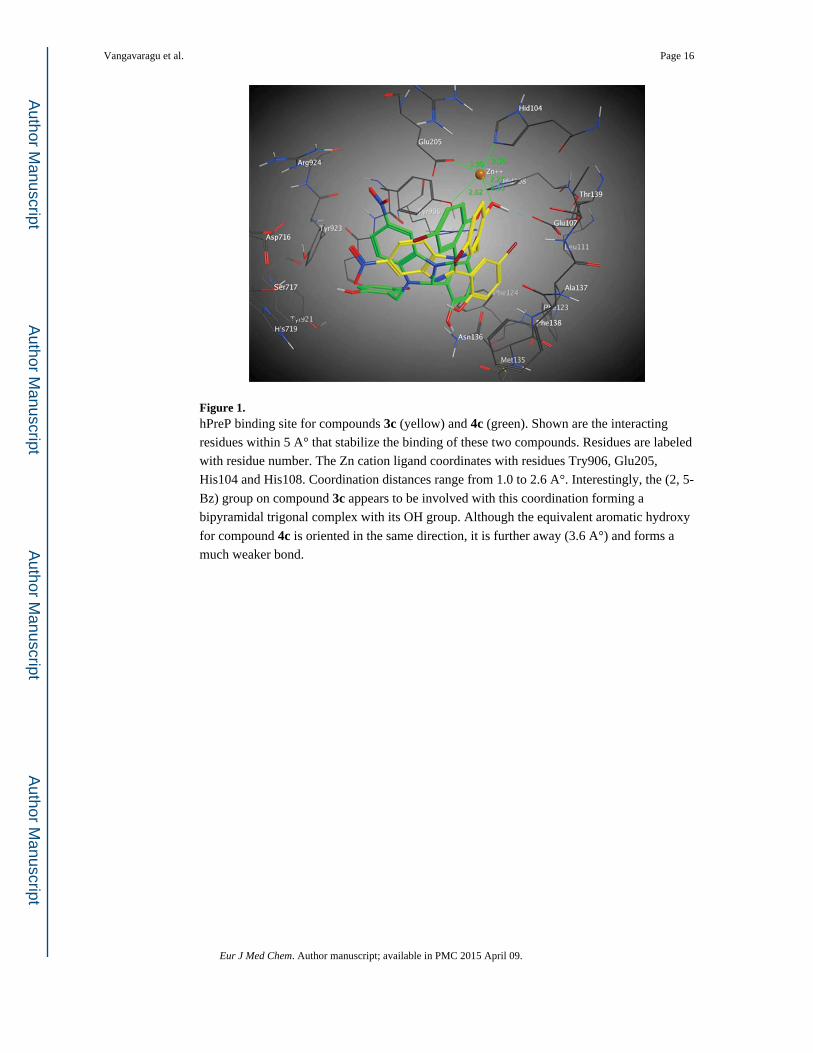
Figure 1. hPreP binding site for compounds 3c (yellow) and 4c (green). Shown are the interacting
residues within 5 A° that stabilize the binding of these two compounds. Residues are labeled
with residue number. The Zn cation ligand coordinates with residues Try906, Glu205,
His104 and His108. Coordination distances range from 1.0 to 2.6 A°. Interestingly, the (2, 5-
Bz) group on compound 3c appears to be involved with this coordination forming a
bipyramidal trigonal complex with its OH group. Although the equivalent aromatic hydroxy
for compound 4c is oriented in the same direction, it is further away (3.6 A°) and forms a
much weaker bond.
Vangavaragu et al. Page 16
Eur J Med Chem. Author manuscript; available in PMC 2015 April 09.
Author M
anuscriptA
uthor Manuscript
Author M
anuscriptA
uthor Manuscript
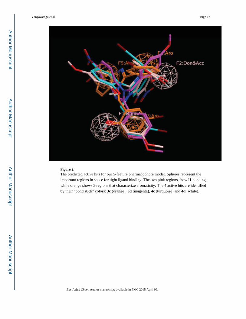
Figure 2. The predicted active hits for our 5-feature pharmacophore model. Spheres represent the
important regions in space for tight ligand binding. The two pink regions show H-bonding,
while orange shows 3 regions that characterize aromaticity. The 4 active hits are identified
by their “bond stick” colors: 3c (orange), 3d (magenta), 4c (turquoise) and 4d (white).
Vangavaragu et al. Page 17
Eur J Med Chem. Author manuscript; available in PMC 2015 April 09.
Author M
anuscriptA
uthor Manuscript
Author M
anuscriptA
uthor Manuscript
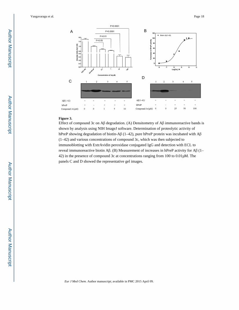
Figure 3. Effect of compound 3c on Aβ degradation. (A) Densitometry of Aβ immunoreactive bands is
shown by analysis using NIH ImageJ software. Determination of proteolytic activity of
hPreP showing degradation of biotin-Aβ (1–42), pure hPreP protein was incubated with Aβ
(1–42) and various concentrations of compound 3c, which was then subjected to
immunoblotting with ExtrAvidin peroxidase conjugated IgG and detection with ECL to
reveal immunoreactive biotin Aβ. (B) Measurement of increases in hPreP activity for Aβ (1–
42) in the presence of compound 3c at concentrations ranging from 100 to 0.01μM. The
panels C and D showed the representative gel images.
Vangavaragu et al. Page 18
Eur J Med Chem. Author manuscript; available in PMC 2015 April 09.
Author M
anuscriptA
uthor Manuscript
Author M
anuscriptA
uthor Manuscript
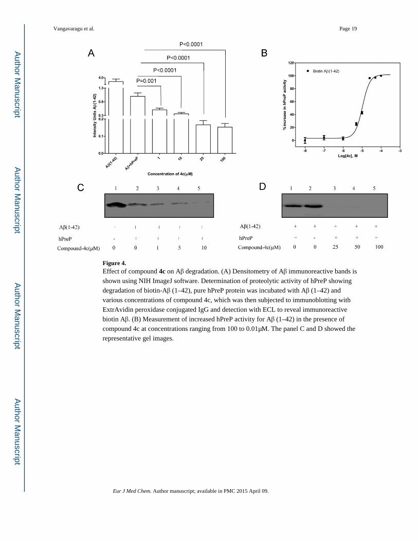
Figure 4. Effect of compound 4c on Aβ degradation. (A) Densitometry of Aβ immunoreactive bands is
shown using NIH ImageJ software. Determination of proteolytic activity of hPreP showing
degradation of biotin-Aβ (1–42), pure hPreP protein was incubated with Aβ (1–42) and
various concentrations of compound 4c, which was then subjected to immunoblotting with
ExtrAvidin peroxidase conjugated IgG and detection with ECL to reveal immunoreactive
biotin Aβ. (B) Measurement of increased hPreP activity for Aβ (1–42) in the presence of
compound 4c at concentrations ranging from 100 to 0.01μM. The panel C and D showed the
representative gel images.
Vangavaragu et al. Page 19
Eur J Med Chem. Author manuscript; available in PMC 2015 April 09.
Author M
anuscriptA
uthor Manuscript
Author M
anuscriptA
uthor Manuscript
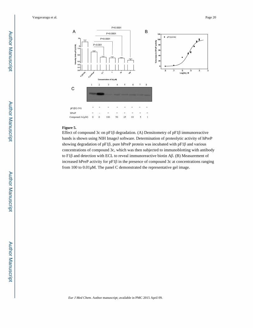
Figure 5. Effect of compound 3c on pF1β degradation. (A) Densitometry of pF1β immunoreactive
bands is shown using NIH ImageJ software. Determination of proteolytic activity of hPreP
showing degradation of pF1β, pure hPreP protein was incubated with pF1β and various
concentrations of compound 3c, which was then subjected to immunoblotting with antibody
to F1β and detection with ECL to reveal immunoreactive biotin Aβ. (B) Measurement of
increased hPreP activity for pF1β in the presence of compound 3c at concentrations ranging
from 100 to 0.01μM. The panel C demonstrated the representative gel image.
Vangavaragu et al. Page 20
Eur J Med Chem. Author manuscript; available in PMC 2015 April 09.
Author M
anuscriptA
uthor Manuscript
Author M
anuscriptA
uthor Manuscript
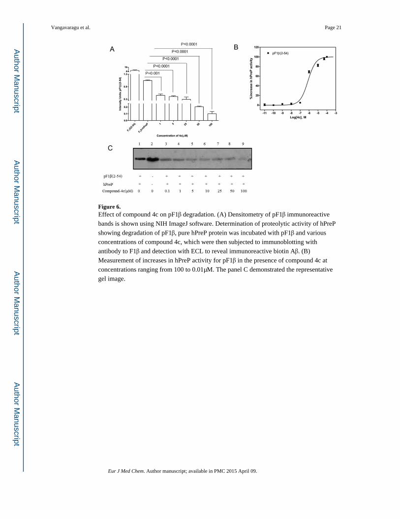
Figure 6. Effect of compound 4c on pF1β degradation. (A) Densitometry of pF1β immunoreactive
bands is shown using NIH ImageJ software. Determination of proteolytic activity of hPreP
showing degradation of pF1β, pure hPreP protein was incubated with pF1β and various
concentrations of compound 4c, which were then subjected to immunoblotting with
antibody to F1β and detection with ECL to reveal immunoreactive biotin Aβ. (B)
Measurement of increases in hPreP activity for pF1β in the presence of compound 4c at
concentrations ranging from 100 to 0.01μM. The panel C demonstrated the representative
gel image.
Vangavaragu et al. Page 21
Eur J Med Chem. Author manuscript; available in PMC 2015 April 09.
Author M
anuscriptA
uthor Manuscript
Author M
anuscriptA
uthor Manuscript
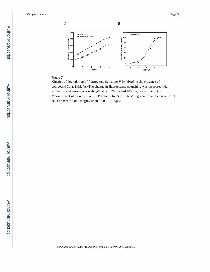
Figure 7. Kinetics of degradation of fluorogenic Substrate V by hPreP in the presence of
compound-3c at 1μM. (A) The change in fluorescence quenching was measured with
excitation and emission wavelength set at 320 nm and 405 nm, respectively. (B)
Measurement of increases in hPreP activity for Substrate V degradation in the presence of
3c at concentrations ranging from 0.00001 to 5μM.
Vangavaragu et al. Page 22
Eur J Med Chem. Author manuscript; available in PMC 2015 April 09.
Author M
anuscriptA
uthor Manuscript
Author M
anuscriptA
uthor Manuscript

Figure 8. Kinetics of degradation of the fluorogenic Substrate V by hPreP in the presence of
compound-4c at 1μM. (A) The change in fluorescence quenching was measured with
excitation and emission wavelength set at 320 nm and 405 nm, respectively. (B)
Measurement of increases in hPreP activity for Substrate V degradation in the presence of
4c at concentrations ranging from 0.00001 to 5μM.
Vangavaragu et al. Page 23
Eur J Med Chem. Author manuscript; available in PMC 2015 April 09.
Author M
anuscriptA
uthor Manuscript
Author M
anuscriptA
uthor Manuscript
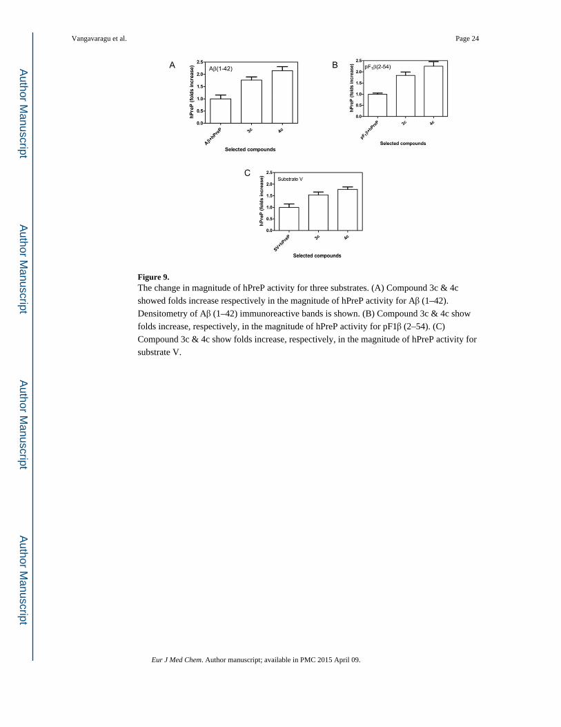
Figure 9. The change in magnitude of hPreP activity for three substrates. (A) Compound 3c & 4c
showed folds increase respectively in the magnitude of hPreP activity for Aβ (1–42).
Densitometry of Aβ (1–42) immunoreactive bands is shown. (B) Compound 3c & 4c show
folds increase, respectively, in the magnitude of hPreP activity for pF1β (2–54). (C)
Compound 3c & 4c show folds increase, respectively, in the magnitude of hPreP activity for
substrate V.
Vangavaragu et al. Page 24
Eur J Med Chem. Author manuscript; available in PMC 2015 April 09.
Author M
anuscriptA
uthor Manuscript
Author M
anuscriptA
uthor Manuscript
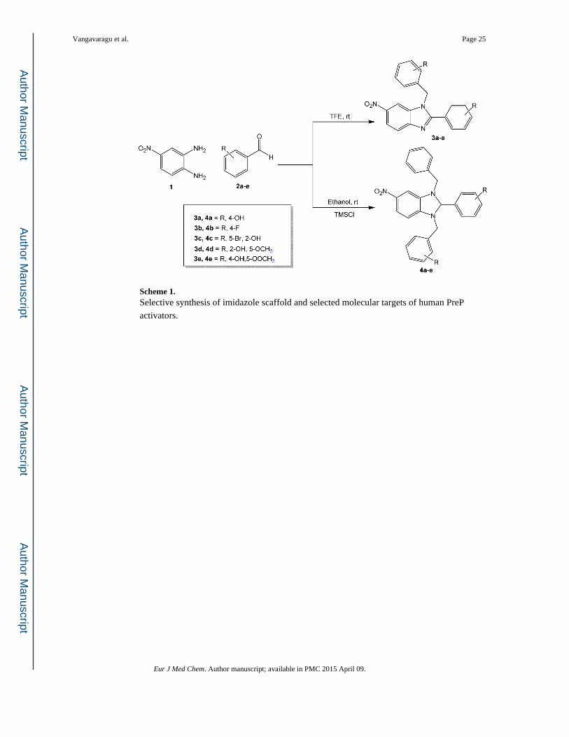
Scheme 1. Selective synthesis of imidazole scaffold and selected molecular targets of human PreP
activators.
Vangavaragu et al. Page 25
Eur J Med Chem. Author manuscript; available in PMC 2015 April 09.
Author M
anuscriptA
uthor Manuscript
Author M
anuscriptA
uthor Manuscript
Author M
anuscriptA
uthor Manuscript
Author M
anuscriptA
uthor Manuscript
Vangavaragu et al. Page 26
Tab
le 1
QSA
R d
escr
ipto
rs d
efin
ed f
or th
e tit
le c
ompo
unds
.
Lig
and
Mol
ecul
arW
eigh
t(D
alta
ns)
Hyd
roge
nB
ond
Don
ors
Hyd
roge
nB
ond
Acc
epto
rsL
ogP
Mol
arR
efra
ctiv
ity
(Ao3
)
Surf
ace
area
(Ao2
)
Vol
ume
(Ao3
)
Solv
atio
nen
ergy
(K.c
al/m
ol)
Pol
ariz
abili
ty(A
o3)
Tot
alen
ergy
(K.c
al/m
ol)
3a36
1.36
01
3.16
328
.89
194.
836
847.
50−
229.
942
37.9
248
.555
3b36
5.34
01
3.78
39.
777
200.
194
315.
50−
3.52
551
.220
103.
523
3c38
0.96
12
2.46
411
.215
255.
022
349.
63−
190.
670
57.1
0395
.472
3d42
1.41
14
2.53
211
.075
234.
676
368.
38−
277.
640
60.8
3514
1.46
5
3e47
7.43
13
2.41
511
.819
276.
378
382.
00−
379.
375
63.2
9114
0.79
6
4a46
9.49
33
5.99
213
.304
235.
875
438.
13−
11.0
7970
.166
101.
738
4b46
5.47
00
6.77
312
.674
149.
987
426.
00−
6.17
467
.004
101.
092
4c70
2.90
33
6.07
615
.588
327.
553
507.
25−
0.59
581
.257
105.
810
4d55
9.57
36
5.96
515
.270
290.
167
513.
381.
280
81.8
5312
7.62
3
4e64
3.61
36
5.92
16.7
0637
4.72
256
5.88
20.6
7889
.539
146.
564
Eur J Med Chem. Author manuscript; available in PMC 2015 April 09.


![High optical and structural quality of GaN epilayers grown ...projects.itn.pt/marco_fct/[4]High optical and structural quality of GaN... · High optical and structural quality of](https://static.fdocument.org/doc/165x107/5e880c2016bca472f2564feb/high-optical-and-structural-quality-of-gan-epilayers-grown-4high-optical-and.jpg)

![Nootaree Niljianskul HHS Public Access Shaolin Zhu, and ... · attractive as starting materials for the synthesis of α-aminosilanes.[10] The intermolecular hydroamination of vinylsilanes](https://static.fdocument.org/doc/165x107/5ee1d576ad6a402d666c91d2/nootaree-niljianskul-hhs-public-access-shaolin-zhu-and-attractive-as-starting.jpg)