
Heat-Induced Redistribution of Disulfide Bonds in Milk Proteins. 1. Bovine β-Lactoglobulin
9
Heat-Induced Redistribution of Disulfide Bonds in Milk Proteins. 1. Bovine -Lactoglobulin LAWRENCE K. CREAMER,* ,² ANNIE BIENVENUE, ‡ HANNA NILSSON, § MARIE PAULSSON, § MIRIAM VAN WANROIJ, # EDWIN K. LOWE, ² SKELTE G. ANEMA, ² MICHAEL J. BOLAND, ² AND RAFAEL JIME Ä NEZ-FLORES ‡ Fonterra Research Centre, Private Bag 11-029, Palmerston North, New Zealand; Food Engineering, Lund University, Box 117, SE-221 00 Lund, Sweden; Dairy Product Technology Center, California Polytechnic State University, San Luis Obispo, California 93407; and Department of Dairy Technology, Wageningen University and Research, Wageningen, The Netherlands Changes in the structure and chemistry of -lactoglobulin (-LG) play an important role in the processing and functionality of milk products. In model -LG systems, there is evidence that the aggregates of heated -LG are held together by a mixture of intermolecular non-covalent association and heat-induced non-native disulfide bonds. Although a number of non-native disulfide bonds have been identified, little is known about the initial inter- and intramolecular disulfide bond rearrangements that occur as a result of heating. These interchange reactions were explored by examining the products of heat treatment to determine the novel disulfide bonds that form in the heated -LG aggregates. The native protein and heat-induced aggregates were hydrolyzed by trypsin, and the resulting peptides, before and after reduction with dithiothreitol, were separated by high-performance liquid chromatog- raphy and their identities confirmed by electrospray ionization mass spectrometry. Comparisons of these peptide patterns showed that some of the Cys160 was in the reduced form in heated -LG aggregates, indicating that the Cys160-Cys66 disulfide bond had been broken during heating. This finding suggests that disulfide bond interchange reactions between -LG non-native monomers, or polymers, and other proteins could occur largely via Cys160. KEYWORDS: -Lactoglobulin; disulfide bonding; heat-induced change; mass spectroscopy INTRODUCTION The effect of heat on -lactoglobulin (-LG) and other milk proteins has received considerable attention, as heat is known to have an impact on dairy processing, such as fouling of equipment and prevention of milk coagulation during cheese- making, because of the reaction of the denaturing whey protein with κ-casein on the casein micelle (1-4). In contrast, whey protein isolates and concentrates, with -LG content of at least 50% of the protein, readily form heat- or pressure-induced gels and can be used as functional ingredients in food formulations. It has long been recognized that the heat-induced aggregation of -LG involves both disulfide bond interchange and modifica- tion to the hydrophobic interactions that are intramolecular in the native whey proteins and become intermolecular in the aggregates and gels (5, 6). To study the disulfide bond interchange phenomena, it was necessary to work with more dilute solutions so that intermediate species could be identified and at lower temperatures to slow the reaction so that the intermediates in the pathway could be identified. The structure of native -LG is now well-known from both X-ray crystallographic studies (7-9) and high-resolution nuclear magnetic resonance studies (10-12). -LG has nine strands, eight of which fold into two sheets that face each other. There is a three-turn R helix that sits above one of the sheets. Three (Cys106, Cys119, and Cys121) of the five Cys residues sit within a very hydrophobic pocket between one side of the helix and segments of the G and H strands (Figure 1)(8). The Cys106-Cys119 disulfide bond is separated from Cys121 by the phenyl ring of Phe136. It is generally recognized that the first effect of heat is the reversible dissociation of the native -LG dimer into monomers (5, 13). The second change is the partial unfolding of the -LG monomer with a loss of helical structure (14, 15), allowing the free sulfhydryl group on Cys121 to interact with the Cys106- Cys119 disulfide bond and, presumably, to reversibly create a Cys106-Cys121 disulfide bond and a free-thiol-containing Cys119 (16). This may be the “activated” monomer postulated (5, 13) as the starting point for the aggregation reactions leading to larger polymers with other disulfide-bond-containing proteins, such as R-lactalbumin. It is at least equally possible that the * Corresponding author (telephone 64-6-350-4649; fax 64-6-356-1476; e-mail [email protected]). ² Fonterra. ‡ California Polytechnic State University. § Lund University. # Wageningen University and Research. 7660 J. Agric. Food Chem. 2004, 52, 7660-7668 10.1021/jf049388y CCC: $27.50 © 2004 American Chemical Society Published on Web 11/16/2004
Transcript of Heat-Induced Redistribution of Disulfide Bonds in Milk Proteins. 1. Bovine β-Lactoglobulin
Heat-Induced Redistribution of Disulfide Bonds in Milk Proteins.1. Bovine â-Lactoglobulin
LAWRENCE K. CREAMER,*,† ANNIE BIENVENUE,‡ HANNA NILSSON,§
MARIE PAULSSON,§ MIRIAM VAN WANROIJ,# EDWIN K. LOWE,†
SKELTE G. ANEMA,† MICHAEL J. BOLAND,† AND RAFAEL JIMEÄ NEZ-FLORES‡
Fonterra Research Centre, Private Bag 11-029, Palmerston North, New Zealand; Food Engineering,Lund University, Box 117, SE-221 00 Lund, Sweden; Dairy Product Technology Center, California
Polytechnic State University, San Luis Obispo, California 93407; and Department of DairyTechnology, Wageningen University and Research, Wageningen, The Netherlands
Changes in the structure and chemistry of â-lactoglobulin (â-LG) play an important role in theprocessing and functionality of milk products. In model â-LG systems, there is evidence that theaggregates of heated â-LG are held together by a mixture of intermolecular non-covalent associationand heat-induced non-native disulfide bonds. Although a number of non-native disulfide bonds havebeen identified, little is known about the initial inter- and intramolecular disulfide bond rearrangementsthat occur as a result of heating. These interchange reactions were explored by examining the productsof heat treatment to determine the novel disulfide bonds that form in the heated â-LG aggregates.The native protein and heat-induced aggregates were hydrolyzed by trypsin, and the resulting peptides,before and after reduction with dithiothreitol, were separated by high-performance liquid chromatog-raphy and their identities confirmed by electrospray ionization mass spectrometry. Comparisons ofthese peptide patterns showed that some of the Cys160 was in the reduced form in heated â-LGaggregates, indicating that the Cys160-Cys66 disulfide bond had been broken during heating. Thisfinding suggests that disulfide bond interchange reactions between â-LG non-native monomers, orpolymers, and other proteins could occur largely via Cys160.
KEYWORDS: â-Lactoglobulin; disulfide bonding; heat-induced change; mass spectroscopy
INTRODUCTION
The effect of heat onâ-lactoglobulin (â-LG) and other milkproteins has received considerable attention, as heat is knownto have an impact on dairy processing, such as fouling ofequipment and prevention of milk coagulation during cheese-making, because of the reaction of the denaturing whey proteinwith κ-casein on the casein micelle (1-4). In contrast, wheyprotein isolates and concentrates, withâ-LG content of at least50% of the protein, readily form heat- or pressure-induced gelsand can be used as functional ingredients in food formulations.
It has long been recognized that the heat-induced aggregationof â-LG involves both disulfide bond interchange and modifica-tion to the hydrophobic interactions that are intramolecular inthe native whey proteins and become intermolecular in theaggregates and gels (5, 6). To study the disulfide bondinterchange phenomena, it was necessary to work with moredilute solutions so that intermediate species could be identified
and at lower temperatures to slow the reaction so that theintermediates in the pathway could be identified.
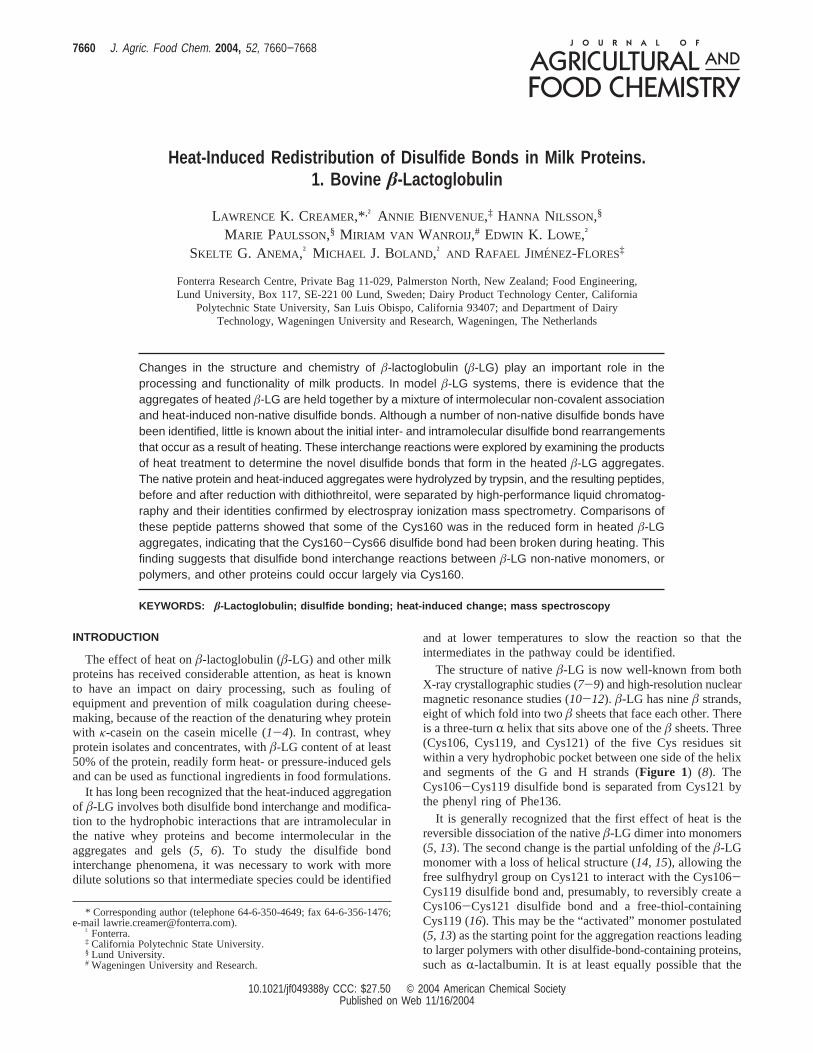
The structure of nativeâ-LG is now well-known from bothX-ray crystallographic studies (7-9) and high-resolution nuclearmagnetic resonance studies (10-12). â-LG has nineâ strands,eight of which fold into twoâ sheets that face each other. Thereis a three-turnR helix that sits above one of theâ sheets. Three(Cys106, Cys119, and Cys121) of the five Cys residues sitwithin a very hydrophobic pocket between one side of the helixand segments of the G and H strands (Figure 1) (8). TheCys106-Cys119 disulfide bond is separated from Cys121 bythe phenyl ring of Phe136.
It is generally recognized that the first effect of heat is thereversible dissociation of the nativeâ-LG dimer into monomers(5, 13). The second change is the partial unfolding of theâ-LGmonomer with a loss of helical structure (14, 15), allowing thefree sulfhydryl group on Cys121 to interact with the Cys106-Cys119 disulfide bond and, presumably, to reversibly create aCys106-Cys121 disulfide bond and a free-thiol-containingCys119 (16). This may be the “activated” monomer postulated(5, 13) as the starting point for the aggregation reactions leadingto larger polymers with other disulfide-bond-containing proteins,such asR-lactalbumin. It is at least equally possible that the
* Corresponding author (telephone 64-6-350-4649; fax 64-6-356-1476;e-mail [email protected]).
† Fonterra.‡ California Polytechnic State University.§ Lund University.# Wageningen University and Research.
7660 J. Agric. Food Chem. 2004, 52, 7660−7668
10.1021/jf049388y CCC: $27.50 © 2004 American Chemical SocietyPublished on Web 11/16/2004
non-native protein with an exposed Cys121 (16) is the “acti-vated” monomer postulated.
Both Surroca et al. (17) and Livney et al. (18) used high-resolution matrix-assisted laser desorption ionization time-of-flight mass spectrometry (MALDI-TOF MS) analysis of peptidemixtures from tryptic digests of heatedâ-LG. Surroca et al.(17) analyzed the hydrolysates of the complete mixture and thepolyacrylamide gel electrophoresis (PAGE)-separated mono-meric, dimeric, and trimericâ-LG AB species, whereas Livneyet al. (18) digested heat-treatedâ-LG B, separated the peptidemixture into fractions using high-performance liquid chroma-tography (HPLC), and analyzed selected HPLC fractions. Bothgroups identified that the peptide containing Cys106, Cys119,and Cys121 (Tyr102-Arg124) formed disulfide-bonded com-plexes with the peptides containing either Cys66 (Tyr61-Lys69or Lys70) or Cys160 (Ala149-Ile162). Surroca et al. (17) alsoshowed that the A variant of the Cys66 peptide could be boundto the B variant of the Tyr102-Arg124 peptide. Livney et al.(18) also reported peptide species that contained two or threeof the Tyr102-Arg124 peptides linked by one or more disulfidebonds.
Manderson et al. (19) reported the presence of non-nativemonomers in heatedâ-LG solutions, supporting the sizeexclusion result of Iametti et al. (20) and confirmed by Schokkeret al. (21). These were stable entities and did not appear to bethe reactive intermediates postulated earlier (5, 13). It has beensuggested (19) that these species are in equilibrium with thenative protein and with the non-native dimers (22), althoughothers have considered them to be intermediates (21).
In this paper, we present the characterization of peptides froma series of tryptic hydrolysates of heat-denaturedâ-LG B andthe identity of the peptides recovered by using liquid chroma-tography electrospray ionization mass spectrometry (LC-ESI-MS). Some of the preliminary material that formed a basis forthis paper was presented as a poster (23) to show that theC-terminal peptides (Ala142-Arg148 and Ala149-Leu162,which contains Cys160 in the reduced form) form early in thereaction.
MATERIALS AND METHODS
Isolation of â-LG. â-LG was isolated from fresh milk of cows thatwere homozygous forâ-LG A or B using the method described byManderson et al. (19) and based on that of Mailliart and Ribadeau-
Dumas (24). The â-LG solution (∼50 mg/mL) was stored frozen.Aliquots containing 120 mg ofâ-LG were chromatographed at 4°Con a column (50× 600 mm) of Superdex 75 (Pharmacia, Uppsala,Sweden) in a dilute phosphate buffer at pH 6.0. After selection of thepurest fractions, determined by native PAGE, these were bulked anddialyzed for 12 h.
The electrophoresis chemicals were obtained from Bio-Rad Labo-ratories (Hercules, CA). dl-Dithiothreitol (DTT), 2-mercaptoethanol,trypsin inhibitor (type I-S), TPCK-treated trypsin (catalog no. T 1426),and trifluoroacetic acid (TFA) were obtained from Sigma ChemicalCo. (St. Louis, MO). The HPLC solvents, which were of “far-UV”grade, and all other chemicals were obtained from BDH.
Heat Treatment. Three milliliter solutions of purified samples ofâ-LG A and B in a 26 mM sodium phosphate/68 mM NaCl bufferadjusted to pH 6.7, at a concentration of 1.15 mg/mL, were heated ina preheated test tube in a temperature-controlled water bath. The tubeswere then cooled in ice water. The sample ofâ-LG B heated at 80°Cfor 15 min was used for the data reported.
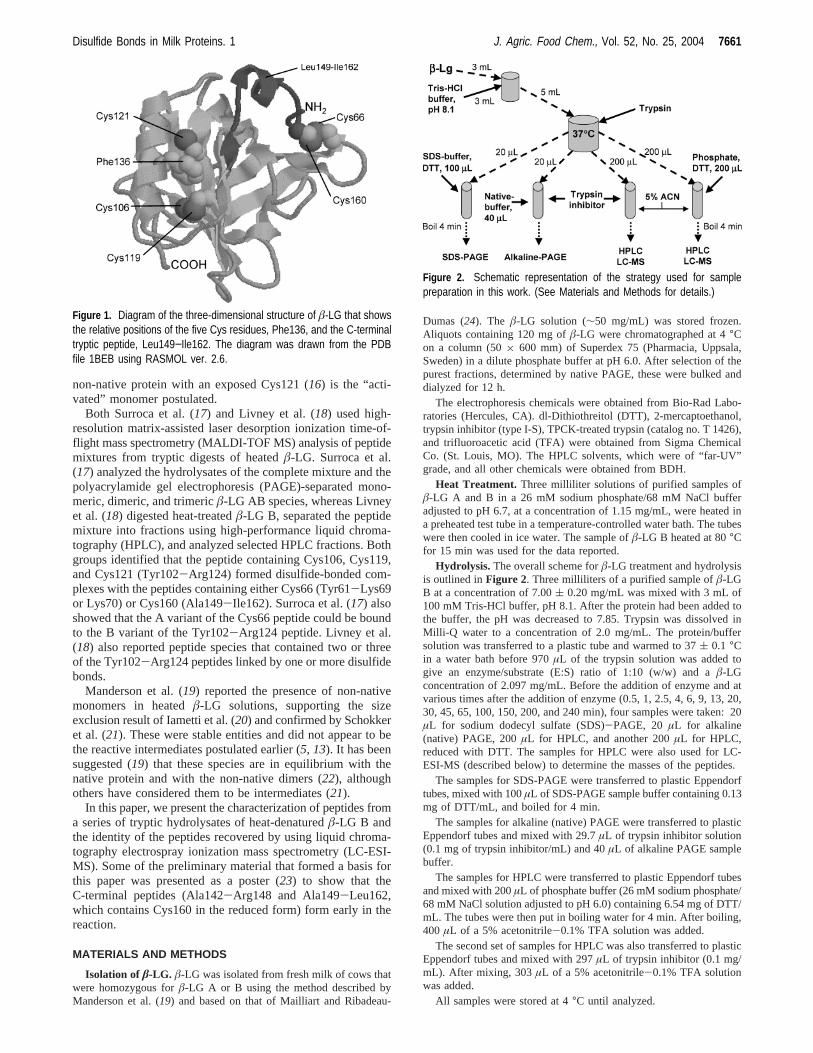
Hydrolysis. The overall scheme forâ-LG treatment and hydrolysisis outlined inFigure 2. Three milliliters of a purified sample ofâ-LGB at a concentration of 7.00( 0.20 mg/mL was mixed with 3 mL of100 mM Tris-HCl buffer, pH 8.1. After the protein had been added tothe buffer, the pH was decreased to 7.85. Trypsin was dissolved inMilli-Q water to a concentration of 2.0 mg/mL. The protein/buffersolution was transferred to a plastic tube and warmed to 37( 0.1 °Cin a water bath before 970µL of the trypsin solution was added togive an enzyme/substrate (E:S) ratio of 1:10 (w/w) and aâ-LGconcentration of 2.097 mg/mL. Before the addition of enzyme and atvarious times after the addition of enzyme (0.5, 1, 2.5, 4, 6, 9, 13, 20,30, 45, 65, 100, 150, 200, and 240 min), four samples were taken: 20µL for sodium dodecyl sulfate (SDS)-PAGE, 20 µL for alkaline(native) PAGE, 200µL for HPLC, and another 200µL for HPLC,reduced with DTT. The samples for HPLC were also used for LC-ESI-MS (described below) to determine the masses of the peptides.
The samples for SDS-PAGE were transferred to plastic Eppendorftubes, mixed with 100µL of SDS-PAGE sample buffer containing 0.13mg of DTT/mL, and boiled for 4 min.
The samples for alkaline (native) PAGE were transferred to plasticEppendorf tubes and mixed with 29.7µL of trypsin inhibitor solution(0.1 mg of trypsin inhibitor/mL) and 40µL of alkaline PAGE samplebuffer.
The samples for HPLC were transferred to plastic Eppendorf tubesand mixed with 200µL of phosphate buffer (26 mM sodium phosphate/68 mM NaCl solution adjusted to pH 6.0) containing 6.54 mg of DTT/mL. The tubes were then put in boiling water for 4 min. After boiling,400 µL of a 5% acetonitrile-0.1% TFA solution was added.
The second set of samples for HPLC was also transferred to plasticEppendorf tubes and mixed with 297µL of trypsin inhibitor (0.1 mg/mL). After mixing, 303µL of a 5% acetonitrile-0.1% TFA solutionwas added.
All samples were stored at 4°C until analyzed.
Figure 1. Diagram of the three-dimensional structure of â-LG that showsthe relative positions of the five Cys residues, Phe136, and the C-terminaltryptic peptide, Leu149−Ile162. The diagram was drawn from the PDBfile 1BEB using RASMOL ver. 2.6.
Figure 2. Schematic representation of the strategy used for samplepreparation in this work. (See Materials and Methods for details.)
Disulfide Bonds in Milk Proteins. 1 J. Agric. Food Chem., Vol. 52, No. 25, 2004 7661

PAGE Analysis.Protein samples were separated by electrophoresisusing Mini Protean II equipment (Bio-Rad Laboratories, Hercules, CA).One-dimensional alkaline (native) PAGE and SDS-PAGE were usedas described by Manderson et al. (19). The Coomassie blue R250 stainedgels were scanned using a Molecular Dynamics scanner (MolecularDynamics Inc., Sunnyvale, CA). The gel scans were analyzed withImageQuant 5.0 software to calculate the quantity ofâ-LG present [asa percentage of that in the control sample (not hydrolyzed)]. Theseresults were then used to estimate the hydrolysis rate, which was doneusing Microsoft Excel 97.
Reversed-Phase (RP) HPLC Analysis.The hydrolysates wereprepared in reducing sample buffer (molar ratioâ-LG/DTT 1:1500)and nonreducing sample buffer so that the formation of new disulfidebonds could be followed. The samples for hydrolysis contained a finalconcentration of 1 mg/mL ofâ-LG and hydrolysis products, TFA 0.1%and acetonitrile 5%.
An Agilent 1100 HPLC system (Agilent Technologies, Waldbronn,Germany), which comprised an Agilent 1100 series quaternary pumpand an Agilent 1100 series thermostated autosampler, was used. TwoHPLC columns, PharmaciaµRPC C2/C18 ST (column size) 4.6 mm× 100 mm) (Pharmacia Biotech, Uppsala, Sweden; code 17-5057-01),were connected in series. The temperature was controlled with anAgilent 1100 series thermostated column compartment and set to25 °C. An aliquot of 100µL of each sample and blank was injectedinto the system. The flow rate was 0.70 mL/min. The following elutionprotocol was used: (a) 5 min of 100% solvent A, (b) 0-100% solventB over 70 min, (c) 5 min of 100% solvent B, and (d) 15 min of 100%solvent A. Solvent A was an aqueous solution containing 5% acetonitrileand 0.1% TFA. Solvent B was an aqueous solution containing 60%acetonitrile and 0.1% TFA. Absorbance data were collected at 1 nmintervals using a Hewlett-Packard 1040A multiwavelength diode arraydetection system (Hewlett-Packard Co., Camas, WA). The signal wasmonitored at 205, 210, 220, 280, and 295 nm. The data were transferredand further processed using the HP ChemStation software (1990-1998;rev. A. 06.03.509).
LC-ESI-MS. The peptides were separated by RP HPLC on a WatersHPLC system (Waters Associates, Millipore Corp., Waters Chroma-tography Division, Milford, MA) Alliance 2690 separations module,which comprised a Waters 486 MS tunable absorbance detector and aWaters 996 photodiode array detector. Signals at 205 and 280 nm weremonitored. The same HPLC columns and method previously describedwere used in the Waters system. The software used was WatersMillennium version 3.05.01 within a Windows operating system.
The eluting peptides were characterized by mass spectrometry usinga Perkin-Elmer Sciex (Thornhill, ON, Canada) triple-quadrupole API300 LC/MS/MS system. The eluent stream from the Waters HPLCsystem was split such that∼10-15 µL/min was introduced into themass spectrometer using an electrospray Perkin-Elmer Sciex API ionspray inlet interface. Ions were generated and focused using a positiveion spray voltage of 5600 V with detection betweenm/z values of 200and 3000 amu. Parameters for operation of the mass spectrometer wereas follows: step size, 0.2 amu; dwell time, 0.5 ms; scan time, 7 s; and25 V. The RNG and IQ2 voltages were ramped from 140 to 280 Vand from -15 to -90 V, respectively. The mass spectrometer wascalibrated using a polypropylene glycol standard as outlined in thePerkin-Elmer Sciex API 300 manual, using MassChrom 1.0 software.The data were analyzed using BioMultiView 1.3.1 (Perkin-ElmerSciex), and the reference mass library used was Peptide Map 2.2(Perkin-Elmer Sciex).
RESULTS
Preliminary results showed that, whereas the trypsin treatmentof native protein apparently hydrolyzed a large number of bondsessentially simultaneously, hydrolysis of the heat-modifiedproteins rapidly produced a number of peptides followed by aslower release of further peptides. Comparison of the hydrolysisof the A and B variant proteins showed that there was a greaterdifference between the initial hydrolysis rates of the nativeprotein and the heated protein forâ-LG B. These experiments
also showed that, to encompass a wide range of rates of releaseof the tryptic peptides, the time intervals needed to be closertogether early in the reaction and more widely spaced later inthe reaction.
Hydrolysis of Heatedâ-LG B. The alkaline PAGE patternof â-LG B heated at 80°C for 15 min in a phosphate buffer isshown in the zero-time lane ofFigure 3. Figure 3a shows thepattern obtained at an E:S ratio of 1:30, andFigure 3b showsthe pattern for the samples from heatedâ-LG B treated at anE:S ratio of 1:10, which were analyzed by HPLC. There werea number of protein bands of lower mobility than the nativeâ-LG B, which were identified as the non-native monomers,dimers, trimers, etc. by comparison with earlier results (19).There was also material caught at the top of the stacking gel,within the stacking gel, and at the interface between the stackingand resolving gels. About half of the original protein remainedin the native state.
After 1 min of hydrolysis at the 1:10 ratio, almost all of thesmaller non-nativeâ-LG aggregates were absent from the PAGEpattern (Figure 3b, lane 2), and, after 65 min, the native proteinwas no longer visible (Figure 3b, lane 7). Thus, much of theheat-modified â-LG B was hydrolyzed very early in thehydrolysis reaction, whereas virtually all of the nativeâ-LG Bwas hydrolyzed in the first 60 min.
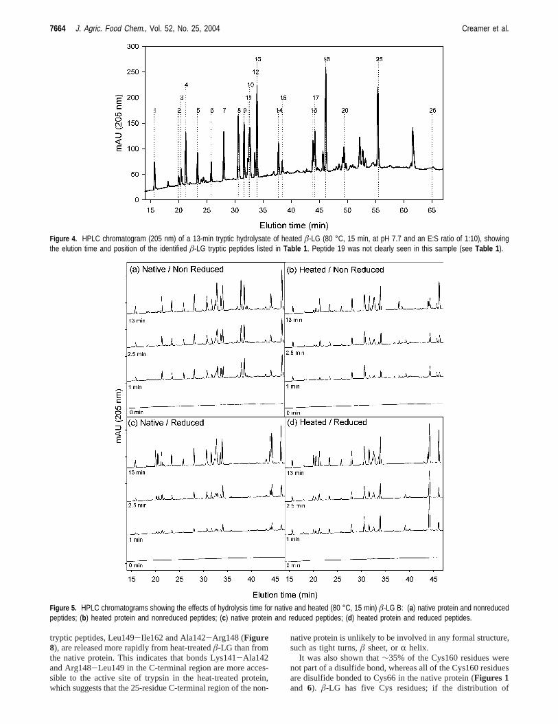
Identification of the Peptides. The HPLC traces obtainedfrom a nativeâ-LG B hydrolysate were comparable with thoseobtained earlier (25) or previously published (26-29), andpreliminary assignments could be made. The use of 205 nmabsorbance in the present study meant that the method was moresensitive to peptide bonds and relatively less sensitive to someside chains than measurements made at 214 or 220 nm. Thesimultaneous absorbance measurements made at 280 and 295nm allowed the identification of the peptides containing Tyr(e.g., Val41-Lys60), Trp (e.g., Trp61-Lys70), and both (e.g.,Val15-Tyr20) (Table 1). Analysis of some mixtures with anLC-ESI-MS instrument confirmed the identity of many of thepeptides in the HPLC peaks produced from the hydrolysismixtures (Table 1). Reduction of the hydrolysate with DTT priorto HPLC separation allowed the identification of disulfide-bonded peptides by the changes in peak intensity, for example,peptides 2 (Trp61-Lys69), 14 (Trp61-Lys70:Leu149-Ile162),and 17 (Leu149-Ile162).Figure 4 shows a typical chromato-gram of a hydrolysate of a heated sample ofâ-LG B, andTable1 shows the probable identity of each of the peptides. Thenumbering was based on the elution order found earlier (25),but the major peptides in peaks 10 and 11 were eluted in reverseorder in this study (Figure 4), that is, Thr76-Lys83 (peptide/peak 11) eluted before Val15-Tyr20 (peptide/peak 10).
Figure 3. Alkaline-PAGE patterns of heated â-LG B (80 °C, 15 min):(a) hydrolyzed for 0, 1, 2.5, 4, 6, 9, 13, 20, 30, 45, 65, 100, 150, 200,and 240 min (lanes 1−15) at 37 °C, pH 7.85, and an E:S ratio of 1:30;(b) hydrolyzed for 0, 1, 2.5, 4, 6, (blank), 65, and 100 min (lanes 1−8) at37 °C, pH 7.85, and an E:S ratio of 1:10.
7662 J. Agric. Food Chem., Vol. 52, No. 25, 2004 Creamer et al.
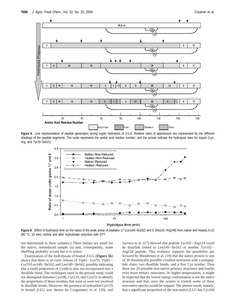
Relative Release Rates of the Peptides.HPLC analysis ofthe 15 hydrolysates per hydrolysis run of the nativeâ-LG Bshowed that a number of peptide peaks appeared to be initiallyreleased at the same rate and that a number appeared moreslowly. A selection of chromatograms is shown inFigure 5.The zero-time chromatograms of nativeâ-LG B (Figure 5a,c)showed very little absorbance between 15 and 50 min and apeak at∼67 min. The heated samples (Figure 5b,d) had a peakbroadly centered on∼66 min (not shown), corresponding tothe whole protein. Theseâ-LG B peaks were essentially absentafter 30 min of hydrolysis.
Hydrolysis Patterns of Native â-LG. Figure 5a showschromatograms of the zero-time sample and the 1, 2.5, and 13min hydrolysates of nativeâ-LG B. Clearly the patterns werevery similar qualitatively, but the sizes of some of the peaksincreased more rapidly than others with hydrolysis time. Peaksof peptides 2 (Trp61-Lys70), 3 (Trp61-Lys69), and 17(Leu149-Ile162) were not apparent, but peptides 14 (Trp61-Lys70:Leu149-Ile162) and 15 (Trp61-Lys69:Leu149-Ile162)were prominent and increased with time, showing that thedisulfide bond between Cys66 and Cys160 (Figures 1 and6)remained intact during the hydrolysis and the hydrolysateanalysis. Reduction of the hydrolysates prior to HPLC analysischanged the chromatographic pattern (Figure 5c) so thatpeptides 2, 3, and 17 were present and peptides 14 and 15 werenot present, showing that DTT treatment of the hydrolysatesreduced peptides 14 (Trp61-Lys70:Leu149-Ile162) and 15(Trp61-Lys69:Leu149-Ile162) to peptides 2 (Trp61-Lys70)plus 17 (Leu149-Ile162) and 3 (Trp61-Lys69) plus 17(Leu149-Ile162), respectively. This comparison of nativeâ-LGB hydrolysates is shown inFigure 7 as chromatogramsa andc with the chromatographic peaks labeled appropriately.
Hydrolysis Patterns of Heatedâ-LG B. When the heatedâ-LG B was hydrolyzed and analyzed by HPLC, peptides 2, 3,14, 15, and 17 were present (Figures 4and5b). Peptides 8 and17 gave the largest peaks in the 1-min chromatogram and, incontrast to peptides 2, 3, 14, and 15, the peak from peptide 17
did not increase in size with hydrolysis time. This showed thatthe 148-149 peptide bond was very labile and that a proportionof the Cys160 of peptide 17 was not disulfide bonded to Cys66,or any other Cys residue, in the heatedâ-LG B. Analysis ofthe reduced hydrolysate (Figure 5d) showed that a considerablequantity of peptide 17 was released after 1 min of hydrolysisand that its concentration did not increase after 2.5 min ofhydrolysis. In contrast, the peaks from peptides 4 (Ile84-Lys91)and 18 (Val41-Lys60) did increase (Figure 5b,d). After 13min of hydrolysis (Figure 5d), peak 17 increased, probablybecause the nativeâ-LG B was being hydrolyzed. Thiscomparison of heatedâ-LG B hydrolysates is also shown inFigure 7 as chromatogramsb andd.
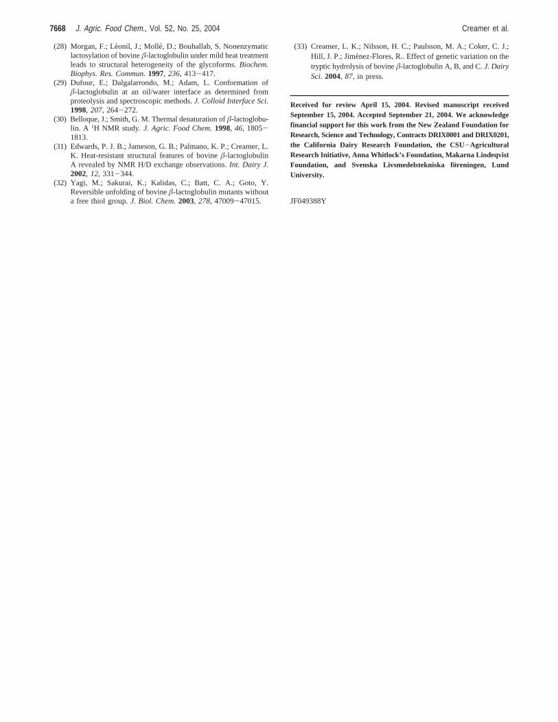
Disulfide Bonding of Cys160 after Heat Treatment ofâ-LG B. Comparison of patterns obtained using heated (Figure5b) instead of native (Figure 5a) â-LG B showed that the 1-minhydrolysate from heatedâ-LG B contained two significantpeaks, namely, peaks 8 (Ala142-Arg148) and 17 (Leu149-Ile162); these peptides are adjacent in theâ-LG B sequence(Figures 6and8). However, these peptides appeared to be lesssignificant in the hydrolysates of nativeâ-LG B. Consequently,a further comparison was made to show the significant effectof heat treatment on the release of the C-terminal peptides fromnative and heat-treatedâ-LG. In this instance, the ratios of theintegrated areas of peak 17 to peak 8 were calculated and plottedagainst hydrolysis time (Figure 9). Thus, peptide 8 acted as an“internal standard” for the quantitative appearance of peptide17, which was dictated by the existence of disulfide bondingof Cys160 to other Cys residues.
The top two lines ofFigure 9 show that the peak area ratiosof peptides 17 to 8 were between 1.7 and 2.0 for the reducedhydrolysates for the first 65 min of hydrolysis. (After that time,nonspecific hydrolysis diminished the quantity of peptide 8 inthe mixture.) The lower two lines had a much lower ratio ofpeptide 17 to peptide 8, showing that a considerable proportionof peptide 17 was disulfide bonded to another peptide. For thenative protein this would have been peptides 2 and 3 to givepeptides 14 and 15, respectively [cf.Figure 7a (native) andFigure 7b]. Comparison of the peptide ratios (Figure 9) of thenonreduced samples during hydrolysis showed a high proportion(∼35%) of the available peptide 17 was released initially. Thereduction in this ratio during the first 65 min suggested thatsome disulfide shuffling could have occurred.
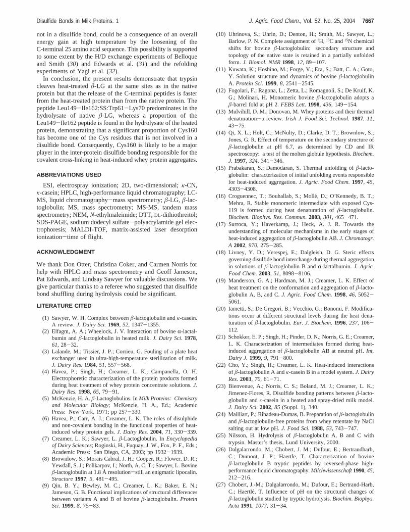
Figure 8 summarizes our results of the tryptic hydrolysis ofheatedâ-LG. The top bar of the diagram represents the linearbackbone ofâ-LG and would be identified as peak 26 in ourchromatograms. The short vertical arrows above the bar showthe very fastest cleavages that give rise to peptides 7, 8, and17, which are shown in the second bar of the diagram. Thesepeptides are released during the digestion and are detected atthe earliest possible time points of analysis, before 1 min ofreaction, and are therefore shaded as “very fast”. Subsequentquantitative detection of the peaks during the course of thehydrolysis allowed us to classify them subjectively on their orderof appearance.
The arrows throughout the diagram also show the tryptic sitesin the protein and intermediate peptides, which are labeled, thatgive rise to the final peptide mix. In all of the representations,the disulfide sites have been left as found in the native protein.
DISCUSSION
The present study on the release of tryptic peptides fromnative and heat-treatedâ-LG has shown that the two C-terminal
Table 1. Identity and Approximate HPLC Elution Times of the MajorTryptic Peptides of â-LG B
peptide/peak no.
elutiontimea (min) peptide
1 15.6 Ile71−Lys752 19.9 Trp61−Lys703 20.4 Trp61−Lys694 21.2 Ile84−Lys915 23.3 Gly9−Lys146 25.8 Thr125−Lys1357 28.0 Leu1−Lys88 30.6 Ala142−Arg1489 31.6 Val92−Lys10110 32.6 Val15−Tyr2011 32.3 Thr76−Lys8312 33.5 Val92−Lys10013 33.9 Thr125−Lys13814 37.7 Trp61−Lys70:Leu149−Ile16215 38.4 Trp61−Lys69:Leu149−Ile16216 43.85 Ser21−Arg4017 44.2 Leu149−Ile16218 46.1 Val41−Lys6019 Lys101-Arg12420 49.4 Tyr102−Arg12425 55.5 Val15−Arg4026 65.0 intact â-LG
a Times taken from the data used in Figure 4. Peak 19 normally elutes 0.5 minprior to peak 20. It is commonly seen when native â-LG A is hydrolyzed but is atlower concentrations in â-LG B hydrolysates (33).
Disulfide Bonds in Milk Proteins. 1 J. Agric. Food Chem., Vol. 52, No. 25, 2004 7663
tryptic peptides, Leu149-Ile162 and Ala142-Arg148 (Figure8), are released more rapidly from heat-treatedâ-LG than fromthe native protein. This indicates that bonds Lys141-Ala142and Arg148-Leu149 in the C-terminal region are more acces-sible to the active site of trypsin in the heat-treated protein,which suggests that the 25-residue C-terminal region of the non-
native protein is unlikely to be involved in any formal structure,such as tight turns,â sheet, orR helix.
It was also shown that∼35% of the Cys160 residues werenot part of a disulfide bond, whereas all of the Cys160 residuesare disulfide bonded to Cys66 in the native protein (Figures 1and 6). â-LG has five Cys residues; if the distribution of
Figure 4. HPLC chromatogram (205 nm) of a 13-min tryptic hydrolysate of heated â-LG (80 °C, 15 min, at pH 7.7 and an E:S ratio of 1:10), showingthe elution time and position of the identified â-LG tryptic peptides listed in Table 1 . Peptide 19 was not clearly seen in this sample (see Table 1 ).
Figure 5. HPLC chromatograms showing the effects of hydrolysis time for native and heated (80 °C, 15 min) â-LG B: (a) native protein and nonreducedpeptides; (b) heated protein and nonreduced peptides; (c) native protein and reduced peptides; (d) heated protein and reduced peptides.
7664 J. Agric. Food Chem., Vol. 52, No. 25, 2004 Creamer et al.
unbonded Cys residues (CysH) was evenly divided among thesesites, then it might be expected that 20% of Cys160 would beunbonded. The data inFigures 7and9 suggest that the initialproportion is close to 35%, showing that about one-third of theCys160 residues could be available to interchange with adisulfide bond in anotherâ-LG molecule or in some otherprotein. The gradual decrease of CysH160 during tryptichydrolysis (Figure 9) could have occurred because of disulfide
bond shuffling at pH 7.85 or a gradual oxidation of all the thiols.Nevertheless, and regardless of subsequent reactions, there isan initial burst of free CysH160 in heat-treatedâ-LG. It seemslikely that this reaction occurs simultaneously with, or shortlyafter, the CysH121-CysH119 interchange (16).
The patterns inFigures 5 and7 show that there are bumpseluting where peaks 3 (Trp61-Lys69) and 17 (Leu149-Ile162)normally elute. (The identity of the peptides in these peaks was
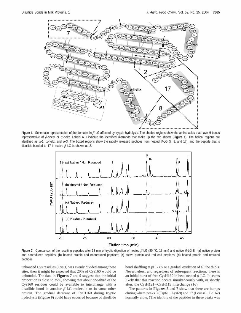
Figure 6. Schematic representation of the domains in â-LG affected by trypsin hydrolysis. The shaded regions show the amino acids that have H-bondsrepresentative of â-sheet or R-helix. Labels A−I indicate the identified â-strands that make up the two sheets (Figure 1 ). The helical regions areidentified as R-1, R-helix, and R-3. The boxed regions show the rapidly released peptides from heated â-LG (7, 8, and 17), and the peptide that isdisulfide-bonded to 17 in native â-LG is shown as 2.
Figure 7. Comparison of the resulting peptides after 13 min of tryptic digestion of heated â-LG (80 °C, 15 min) and native â-LG B: (a) native proteinand nonreduced peptides; (b) heated protein and nonreduced peptides; (c) native protein and reduced peptides; (d) heated protein and reducedpeptides.
Disulfide Bonds in Milk Proteins. 1 J. Agric. Food Chem., Vol. 52, No. 25, 2004 7665
not determined in these samples.) These bumps are small forthe native, nonreduced sample (a) and, consequently, someshuffling probably occurs but it is minor.
Examination of the hydrolysate of heatedâ-LG (Figure 5b)shows that there is an early release of Trp61-Lys70, Trp61-Lys70:Leu149-Ile162, and Leu149-Ile162, possibly indicatingthat a small proportion of Cys66 is also not incorporated into adisulfide bond. The techniques used in the present study couldnot distinguish between Cys106, Cys119, and Cys121 to identifythe proportions of these residues that were or were not involvedin disulfide bonds. However, the presence of unbonded Cys119in heatedâ-LG was shown by Croguennec et al. (16), and
Surroca et al. (17) showed that peptide Tyr102-Arg124 couldbe disulfide linked to Leu149-Ile162 or another Tyr102-Arg124 peptide. This evidence supports the possibility putforward by Manderson et al. (19) that the native protein is oneof 30 theoretically possible covalent structures with a polypep-tide chain, two disulfide bonds, and a free Cys residue. Thusthere are 29 possible non-native primary structures and maybeeven more tertiary structures. At higher temperatures, it mightbe expected that the lowest energy conformation is not the nativestructure and that, once the system is cooled, some of thesenon-native species would be trapped. The present result, namely,that a significant proportion of the non-nativeâ-LG has Cys160
Figure 8. Line representation of peptide generation during tryptic hydrolysis of â-LG. Relative rates of appearance are represented by the differentshadings of the peptide segments. The scale represents the amino acid residue number, and the arrows indicate the hydrolysis sites for trypsin (Lys,Arg, and Tyr20−Ser21).
Figure 9. Effect of hydrolysis time on the ratios of the peak areas of peptides 17 (Leu149−Ile162) and 8 (Ala142−Arg148) from native and heated â-LG(80 °C, 15 min), before and after hydrolysate reduction with DTT.
7666 J. Agric. Food Chem., Vol. 52, No. 25, 2004 Creamer et al.
not in a disulfide bond, could be a consequence of an overallenergy gain at high temperature by the loosening of theC-terminal 25 amino acid sequence. This possibility is supportedto some extent by the H/D exchange experiments of Belloqueand Smith (30) and Edwards et al. (31) and the refoldingexperiments of Yagi et al. (32).
In conclusion, the present results demonstrate that trypsincleaves heat-treatedâ-LG at the same sites as in the nativeprotein but that the release of the C-terminal peptides is fasterfrom the heat-treated protein than from the native protein. Thepeptide Leu149-Ile162:SS:Trp61-Lys70 predominates in thehydrolysate of nativeâ-LG, whereas a proportion of theLeu149-Ile162 peptide is found in the hydrolysate of the heatedprotein, demonstrating that a significant proportion of Cys160has become one of the Cys residues that is not involved in adisulfide bond. Consequently, Cys160 is likely to be a majorplayer in the inter-protein disulfide bonding responsible for thecovalent cross-linking in heat-induced whey protein aggregates.
ABBREVIATIONS USED
ESI, electrospray ionization; 2D, two-dimensional;κ-CN,κ-casein; HPLC, high-performance liquid chromatography; LC-MS, liquid chromatography-mass spectrometry;â-LG, â-lac-toglobulin; MS, mass spectrometry; MS-MS, tandem massspectrometry; NEM,N-ethylmaleimide; DTT,DL-dithiothreitol;SDS-PAGE, sodium dodecyl sulfate-polyacrylamide gel elec-trophoresis; MALDI-TOF, matrix-assisted laser desorptionionization-time of flight.
ACKNOWLEDGMENT
We thank Don Otter, Christina Coker, and Carmen Norris forhelp with HPLC and mass spectrometry and Geoff Jameson,Pat Edwards, and Lindsay Sawyer for valuable discussions. Wegive particular thanks to a referee who suggested that disulfidebond shuffling during hydrolysis could be significant.
LITERATURE CITED
(1) Sawyer, W. H. Complex betweenâ-lactoglobulin andκ-casein.A review. J. Dairy Sci.1969, 52, 1347-1355.
(2) Elfagm, A. A.; Wheelock, J. V. Interaction of bovineR-lactal-bumin andâ-lactoglobulin in heated milk.J. Dairy Sci.1978,61, 28-32.
(3) Lalande, M.; Tissier, J. P.; Corrieu, G. Fouling of a plate heatexchanger used in ultra-high-temperature sterilization of milk.J. Dairy Res.1984, 51, 557-568.
(4) Havea, P.; Singh, H.; Creamer, L. K.; Campanella, O. H.Electrophoretic characterization of the protein products formedduring heat treatment of whey protein concentrate solutions.J.Dairy Res.1998, 65, 79-91.
(5) McKenzie, H. A.â-Lactoglobulins. InMilk Proteins: Chemistryand Molecular Biology; McKenzie, H. A., Ed.; AcademicPress: New York, 1971; pp 257-330.
(6) Havea, P.; Carr, A. J.; Creamer, L. K. The roles of disulphideand non-covalent bonding in the functional properties of heat-induced whey protein gels.J. Dairy Res.2004, 71, 330-339.
(7) Creamer, L. K.; Sawyer, L.â-Lactoglobulin. InEncyclopediaof Dairy Sciences; Roginski, H., Fuquay, J. W., Fox, P. F., Eds.;Academic Press: San Diego, CA, 2003; pp 1932-1939.
(8) Brownlow, S.; Morais Cabral, J. H.; Cooper, R.; Flower, D. R.;Yewdall, S. J.; Polikarpov, I.; North, A. C. T.; Sawyer, L. Bovineâ-lactoglobulin at 1.8 Å resolutionsstill an enigmatic lipocalin.Structure1997, 5, 481-495.
(9) Qin, B. Y.; Bewley, M. C.; Creamer, L. K.; Baker, E. N.;Jameson, G. B. Functional implications of structural differencesbetween variants A and B of bovineâ-lactoglobulin.ProteinSci.1999, 8, 75-83.
(10) Uhrinova, S.; Uhrin, D.; Denton, H.; Smith, M.; Sawyer, L.;Barlow, P. N. Complete assignment of1H, 13C and15N chemicalshifts for bovine â-lactoglobulin: secondary structure andtopology of the native state is retained in a partially unfoldedform. J. Biomol. NMR1998, 12, 89-107.
(11) Kuwata, K.; Hoshino, M.; Forge, V.; Era, S.; Batt, C. A.; Goto,Y. Solution structure and dynamics of bovineâ-lactoglobulinA. Protein Sci.1999, 8, 2541-2545.
(12) Fogolari, F.; Ragona, L.; Zetta, L.; Romagnoli, S.; De Kruif, K.G.; Molinari, H. Monomeric bovineâ-lactoglobulin adopts aâ-barrel fold at pH 2.FEBS Lett.1998, 436, 149-154.
(13) Mulvihill, D. M.; Donovan, M. Whey proteins and their thermaldenaturationsa review. Irish J. Food Sci. Technol.1987, 11,43-75.
(14) Qi, X. L.; Holt, C.; McNulty, D.; Clarke, D. T.; Brownlow, S.;Jones, G. R. Effect of temperature on the secondary structure ofâ-lactoglobulin at pH 6.7, as determined by CD and IRspectroscopy: a test of the molten globule hypothesis.Biochem.J. 1997, 324, 341-346.
(15) Prabakaran, S.; Damodaran, S. Thermal unfolding ofâ-lacto-globulin: characterization of initial unfolding events responsiblefor heat-induced aggregation.J. Agric. Food Chem.1997, 45,4303-4308.
(16) Croguennec, T.; Bouhallab, S.; Molle´, D.; O’Kennedy, B. T.;Mehra, R. Stable monomeric intermediate with exposed Cys-119 is formed during heat denaturation ofâ-lactoglobulin.Biochem. Biophys. Res. Commun.2003, 301, 465-471.
(17) Surroca, Y.; Haverkamp, J.; Heck, A. J. R. Towards theunderstanding of molecular mechanisms in the early stages ofheat-induced aggregation ofâ-lactoglobulin AB.J. Chromatogr.A 2002, 970, 275-285.
(18) Livney, Y. D.; Verespej, E.; Dalgleish, D. G. Steric effectsgoverning disulfide bond interchange during thermal aggregationin solutions ofâ-lactoglobulin B andR-lactalbumin.J. Agric.Food Chem.2003, 51, 8098-8106.
(19) Manderson, G. A.; Hardman, M. J.; Creamer, L. K. Effect ofheat treatment on the conformation and aggregation ofâ-lacto-globulin A, B, and C.J. Agric. Food Chem.1998, 46, 5052-5061.
(20) Iametti, S.; De Gregori, B.; Vecchio, G.; Bonomi, F. Modifica-tions occur at different structural levels during the heat dena-turation ofâ-lactoglobulin.Eur. J. Biochem.1996, 237, 106-112.
(21) Schokker, E. P.; Singh, H.; Pinder, D. N.; Norris, G. E.; Creamer,L. K. Characterization of intermediates formed during heat-induced aggregation ofâ-lactoglobulin AB at neutral pH.Int.Dairy J. 1999, 9, 791-800.
(22) Cho, Y.; Singh, H.; Creamer, L. K. Heat-induced interactionsof â-lactoglobulin A andκ-casein B in a model system.J. DairyRes.2003, 70, 61-71.
(23) Bienvenue, A.; Norris, C. S.; Boland, M. J.; Creamer, L. K.;Jimenez-Flores, R. Disulfide bonding patterns betweenâ-lacto-globulin andκ-casein in a heated and spray-dried milk model.J. Dairy Sci.2002, 85 (Suppl. 1), 340.
(24) Mailliart, P.; Ribadeau-Dumas, B. Preparation ofâ-lactoglobulinandâ-lactoglobulin-free proteins from whey retentate by NaClsalting out at low pH.J. Food Sci.1988, 53, 743-747.
(25) Nilsson, H. Hydrolysis ofâ-lactoglobulin A, B and C withtrypsin. Master’s thesis, Lund University, 2000.
(26) Dalgalarrondo, M.; Chobert, J. M.; Dufour, E.; Bertrandharb,C.; Dumont, J. P.; Haertle, T. Characterization of bovineâ-lactoglobulin B tryptic peptides by reversed-phase high-performance liquid chromatography.Milchwissenschaft1990, 45,212-216.
(27) Chobert, J.-M.; Dalgalarrondo, M.; Dufour, E.; Bertrand-Harb,C.; Haertle, T. Influence of pH on the structural changes ofâ-lactoglobulin studied by tryptic hydrolysis.Biochim. Biophys.Acta 1991, 1077, 31-34.
Disulfide Bonds in Milk Proteins. 1 J. Agric. Food Chem., Vol. 52, No. 25, 2004 7667
(28) Morgan, F.; Le´onil, J.; Molle, D.; Bouhallab, S. Nonenzymaticlactosylation of bovineâ-lactoglobulin under mild heat treatmentleads to structural heterogeneity of the glycoforms.Biochem.Biophys. Res. Commun.1997, 236, 413-417.
(29) Dufour, E.; Dalgalarrondo, M.; Adam, L. Conformation ofâ-lactoglobulin at an oil/water interface as determined fromproteolysis and spectroscopic methods.J. Colloid Interface Sci.1998, 207, 264-272.
(30) Belloque, J.; Smith, G. M. Thermal denaturation ofâ-lactoglobu-lin. A 1H NMR study.J. Agric. Food Chem.1998, 46, 1805-1813.
(31) Edwards, P. J. B.; Jameson, G. B.; Palmano, K. P.; Creamer, L.K. Heat-resistant structural features of bovineâ-lactoglobulinA revealed by NMR H/D exchange observations.Int. Dairy J.2002, 12, 331-344.
(32) Yagi, M.; Sakurai, K.; Kalidas, C.; Batt, C. A.; Goto, Y.Reversible unfolding of bovineâ-lactoglobulin mutants withouta free thiol group.J. Biol. Chem.2003, 278, 47009-47015.
(33) Creamer, L. K.; Nilsson, H. C.; Paulsson, M. A.; Coker, C. J.;Hill, J. P.; Jimenez-Flores, R.. Effect of genetic variation on thetryptic hydrolysis of bovineâ-lactoglobulin A, B, and C.J. DairySci.2004, 87, in press.
Received for review April 15, 2004. Revised manuscript receivedSeptember 15, 2004. Accepted September 21, 2004. We acknowledgefinancial support for this work from the New Zealand Foundation forResearch, Science and Technology, Contracts DRIX0001 and DRIX0201,the California Dairy Research Foundation, the CSU-AgriculturalResearch Initiative, Anna Whitlock’s Foundation, Makarna LindeqvistFoundation, and Svenska Livsmedelstekniska fo1reningen, LundUniversity.
JF049388Y
7668 J. Agric. Food Chem., Vol. 52, No. 25, 2004 Creamer et al.


![Fibronectin Fibronectin exists as a dimer, consisting of two nearly identical polypeptide chains linked by a pair of C-terminal disulfide bonds. [3] Each.](https://static.fdocument.org/doc/165x107/56649d4e5503460f94a2e7cf/fibronectin-fibronectin-exists-as-a-dimer-consisting-of-two-nearly-identical.jpg)
![Adsorption of Milk Proteins (-Casein and -Lactoglobulin ... · protein with a random coil conformation in solution, but recent studies have challenged this view [16]. On the contrary,](https://static.fdocument.org/doc/165x107/5fa3935da2da091e9e210d6e/adsorption-of-milk-proteins-casein-and-lactoglobulin-protein-with-a-random.jpg)