Mechanistic Study and Identification of Essential residues of Family 3 b -Glucosidase
Upload
johanna-alessandra-floresCategory
view
8download
1description
A
OiatRcpCα
©
K
1
ls(apaf
aucwafb
h2
Available online at www.sciencedirect.com
ScienceDirectHOSTED BY
Food Science and Human Wellness 3 (2014) 136–174
α-Glucosidase inhibitors isolated from medicinal plants
Zhenhua Yin a, Wei Zhang a, Fajin Feng a,b, Yong Zhang a, Wenyi Kang a,∗a Huanghe Science and Technology College, Zhengzhou 450063, China
b College of Chemistry and Chemical Engineering, Guizhou University, Guiyang 550025, China
Received 5 November 2014; accepted 20 November 2014
bstract
bjective: α-Glucosidase inhibitors can be used as a new class of antidiabetic drug. By competitively inhibiting glycosidase activity, thesenhibitors help to prevent the fast breakdown of sugars and thereby control the blood sugar level. This study provides a wealth of informationbout α-glucosidase inhibitors isolated from medicinal plants; this knowledge will be useful in finding more potent antidiabetic candidates fromhe natural resources for the clinical development of antidiabetic therapeutics.esults: 411 compounds exhibiting α-glucosidase inhibitory activity were summarized and isolated them from medicinal plants. The compoundlasses isolated include: terpenes (61) from 14 genus, alkaloids (37) from 11 genus, quinines (49) from 4 genus, flavonoids (103) from 24 genus,henols (37) from 9 genus, phenylpropanoids (73) from 20 genus, sterides (8) from 5 genus, and other types of compounds (43).onclusion: Compounds with α-glucosidase inhibitory activity are abundant in nature and can be obtained from several sources. They have high-glucosidase inhibitory potential, and can be clinically developed for treating diabetes mellitus.
2015 Beijing Academy of Food Sciences. Production and hosting by Elsevier B.V. All rights reserved.
eywords: α-Glucosidase inhibitor; Inhibitory activity; Medicinal plants
. Introduction
Diabetes mellitus is a well-known metabolic disorder, which is characterized by an abnormal postprandial increase of blood glucoseevel. The control of postprandial hyperglycemia is believed to be important in the treatment of diabetes mellitus. α-Glucosidaseecreted from intestinal chorionic epithelium is responsible for the degradation of carbohydrates. In the 1980s, α-glucosidaseEC 3.2.1.20) inhibitors became a new class of antidiabetic drug. α-Glucosidase inhibitors slow down the process of digestionnd absorption of carbohydrates by competitively blocking the activity of glucosidase. Consequently, the peak concentration ofostprandial blood glucose is reduced and the blood sugar level comes under control. α-Glucosidase inhibitors can offer severaldvantages and has been recommend by the Third Asia-Pacific Region Diabetes Treatment Guidelines as the first-line of treatmentor lowering postprandial hyperglycemia [1].
α-Glucosidase inhibitors fall under the third category of oral hypoglycemic agents [2]. Several α-glucosidase inhibitors, such ascarbose and voglibose obtained from natural sources, can effectively control blood glucose levels after food intake and have beensed clinically in the treatment of diabetes mellitus [3]. Only a few α-glucosidase inhibitors are commercially available. All of themontain sugar moieties and their synthesis involves tedious multistep procedures. Moreover, clinically they have been associatedith serious gastrointestinal side effects. Therefore, it is necessary to search for alternatives that can display α-glucosidase inhibitory
ctivity but without side reactions. In recent years, projects undertaken to discover potent non-sugar based α-glucosidase inhibitorsrom natural sources have received tremendous attention because of the highly abundant compounds in nature and their promisingiological activities [4].
∗ Corresponding author at: Huanghe Science and Technology College, Zhengzhou 450063, China.E-mail address: [email protected] (W. Kang).Peer review under responsibility of Beijing Academy of Food Sciences.
ttp://dx.doi.org/10.1016/j.fshw.2014.11.003213-4530/© 2015 Beijing Academy of Food Sciences. Production and hosting by Elsevier B.V. All rights reserved.

2
2
asBigmnip
2
m7riav
r(t
g
dα
oe
da
f3
(t2t1
w(
fi(14α
Z. Yin et al. / Food Science and Human Wellness 3 (2014) 136–174 137
. Research development
.1. α-Glucosidase inhibitory compounds from medicinal plants
α-Glucosidase inhibitors were isolated from natural resources including microorganisms and medicinal plants. A review of liter-ture reveals that α-glucosidase enzyme from yeast, rat intestine, and mouse intestine have been widely used for pharmacologicalcreenings. In addition, coffee-glucosidase, amylase mammal glucosidase, rat intestinal isomaltase, α-glucosidase type IV fromacillus stearothermophilus, and glucosidase II from rat liver microsomes have been used as the sources of enzymes for enzyme
nhibition assays. Acarbose has been frequently used as positive control in screenings, and a few researchers have also appliedenistein, quercitrin, and 1-deoxynojirinmycin as positive controls. On the basis of literatures published worldwide, we have sum-arized a list of 411 natural products isolated from medicinal plants that showed α-glucosidase inhibitory activity. Structurally these
atural product inhibitors incorporate terpene, alkaloid, quinine, flavonoid, phenol, phenylpropanoid, and steride frameworks richn organic acid, ester, alcohol, and allyl functional groups. A majority of the compounds reported contain flavonoid, terpene, andhenylpropanoid ring structures.
.2. Terpenes
Sixty-one terpenoids isolated from plants have shown α-glucosidase inhibitory activity. Compound 1 was isolated from theethanol extracts of stem and bark of Fagara tessmannii (Rutaceae). It showed strong inhibitory activity with an IC50 value of
.6 �mol/L [5]. Compounds 2–4 were isolated from Luculia pinceana Hook using bioactivity-guided method. The IC50 valueseported for these compounds were 18.48, 3.30, and 2.88 �g/mL, respectively. Compound 2 (Ki = 3.36 �g/mL) showed competitivenhibitory profile for α-glucosidase activity and the profile closely resembled the inhibitory activity of acarbose, which was useds positive control. The inhibitory activity of compounds 3 and 4 fitted the profile of a non-competitive inhibition model with Ki
alues—195.04 and 3.36 �g/mL, respectively [6].Triterpenoid saponins (5–11) and (IC50 = 23.1, 65.5, 15.2, 78.5, 59.7, 98.2, and 85.2 �mol/L, respectively), isolated from the
oots of Gypsophila oldhamiana, showed stronger inhibitory potency than acarbose (388.0 ± 9.6 �mol/L) [7]. Triterpenoid saponins12–13) (IC50 = 908.5 and 819.7 �mol/L, respectively), isolated from the leaves of Acanthopanax senticosus, showed higher inhibi-ion compared with acarbose (788.6 ± 53.66 �mol/L) [8].
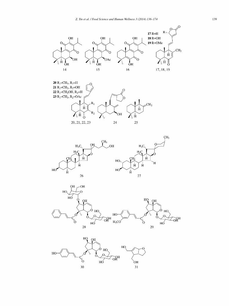
Three abietane diterpenoids (14–16) were isolated from methanol extract of Plectranthus madagascariensis and exhibited α-lucosidase inhibitory activity with IC50 values of 274.9 ± 12.3, 108.2 ± 1.3, and 142.7 ± 1.4 �mol/L, respectively [9].
Phytochemical investigation of antihyperglycemic extract from the rhizomes of Hedychium spicatum led to the isolation ofiterpenes (17–25). Compounds 17–24 displayed varying degree of intestinal α-glucosidase inhibitory potentials. The presence of,β-unsaturated γ-lactone (17–19) and furan (20–23) ring system was essential to inhibit α-glucosidase activity because the absencef these structural motifs in 25 rendered the compound inactive. Hydroxy group at C-16 in the lactone ring (18) strongly increasednzyme inhibitory potential in comparison with methoxy group [10].
Compounds (25S)-5α-furastan-3β,22,26-triol (26) and gitogenin (27) isolated from the plant of Tribulus longipetalus showedecent α-glucosidase inhibitory activity with IC50 values, i.e., 33.5 ± 0.22 and 37.2 ± 0.18 �mol/L, respectively, compared withcarbose (IC50 value = 38.3 ± 0.12 �mol/L) [11].
D-Galactopyranosyl harpagoside (28), 8-O-feruloyl harpagide (29), 8-O-(coumaroyl)harpagide (30), and ninpogenin (31) isolatedrom the roots of Scrophularia ningpoensis exhibited moderate α-glucosidase inhibitory activity (IC50 = 2.16 ± 0.13, 3.02 ± 0.16,.09 ± 0.16, and 1.54 ± 0.31 mmol/L, respectively) compared with acarbose (IC50 = 0.37 ± 0.01 mM) [12].
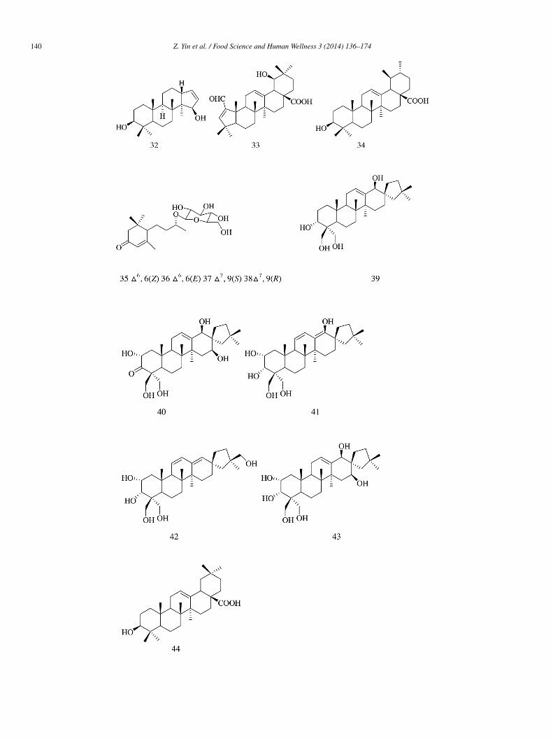
Octanordammarane triterpene—3β,15a-dihydroxymansumbinol (32) and A-ring contracted oleanane triterpenoid—2-formyl-A)1–19a-hydroxy-1-norolean-2,12-dien-28-oic acid (33), ursolic acid (34), and sesquiterpene glucoside (35–38) were isolated fromhe root extract of Rosa rugosa. The IC50 values recorded for compounds 32–36 were 40.03 ± 3.25%, 32.39 ± 5.40%, 31.96 ± 4.47%,8.26 ± 5.83%, and 28.63 ± 2.51%, respectively; the inhibitory sucrose activity was moderate for these compounds compared withhat of acarbose (50.96 ± 2.97%). Compounds 37 and 38 showed weak inhibitory activity with IC50 values of 13.98 ± 2.92% and2.51 ± 4.06%, respectively [13].
Compounds 39–44 (IC50 = 38.0 ± 0.2, 315.8 ± 0.2, 355.5 ± 0.9, 318.5 ± 0.3, 305.3 ± 1.0, and 231.3 ± 0.1 �mol/L, respectively)ere isolated from the entire plant extract of Phlomis stewartii and showed α-glucosidase inhibitory activity with acarbose
IC50 = 38.0 ± 0.1 �mol/L) [14].Six triterpenesaponins, including one new glinoside C (45), were isolated from the aerial parts of Glinus oppositifolius and the
ve known constituents included 3-O-(β-D-xylopyranosyl)-spergulagenin A (46), spergulacin (47), spergulin A (48), spergulacin A
49), and spergulin B (50). Among them, compound 45 exhibited the greatest inhibitory potency toward α-glucosidase with IC50 of27 ± 30 �mol/L and demonstrated an inhibitory profile similar to that of a mixed-type inhibitor (Ki = 157.9 �mol/L). Compounds6–50 (IC50 = 1654 ± 170, 628 ± 80, 143 ± 20, 694 ± 60, and 1783 ± 290 �mol/L) demonstrated weaker inhibitory activity toward-glucosidase compared with acarbose (IC50 = 15 ± 3 �mol/L) [15].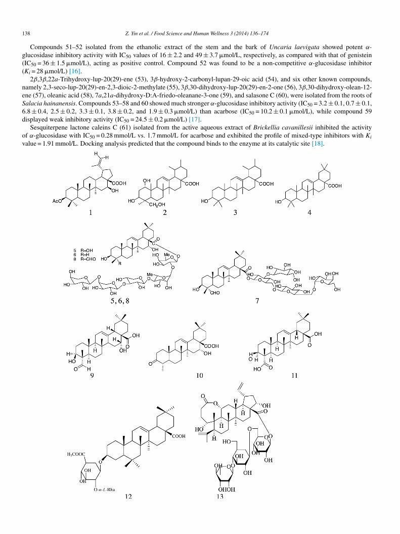
1
g((
neS6d
ov
38 Z. Yin et al. / Food Science and Human Wellness 3 (2014) 136–174
Compounds 51–52 isolated from the ethanolic extract of the stem and the bark of Uncaria laevigata showed potent α-lucosidase inhibitory activity with IC50 values of 16 ± 2.2 and 49 ± 3.7 �mol/L, respectively, as compared with that of genisteinIC50 = 36 ± 1.5 �mol/L), acting as positive control. Compound 52 was found to be a non-competitive α-glucosidase inhibitorKi = 28 �mol/L) [16].
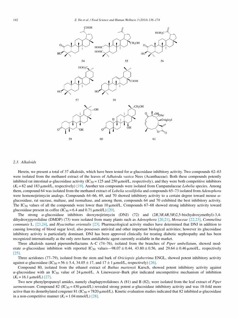
2β,3β,22a-Trihydroxy-lup-20(29)-ene (53), 3β-hydroxy-2-carbonyl-lupan-29-oic acid (54), and six other known compounds,amely 2,3-seco-lup-20(29)-en-2,3-dioic-2-methylate (55), 3β,30-dihydroxy-lup-20(29)-en-2-one (56), 3β,30-dihydroxy-olean-12-ne (57), oleanic acid (58), 7a,21a-dihydroxy-D:A-friedo-oleanane-3-one (59), and salasone C (60), were isolated from the roots ofalacia hainanensis. Compounds 53–58 and 60 showed much stronger α-glucosidase inhibitory activity (IC50 = 3.2 ± 0.1, 0.7 ± 0.1,.8 ± 0.4, 2.5 ± 0.2, 3.3 ± 0.1, 3.8 ± 0.2, and 1.9 ± 0.3 �mol/L) than acarbose (IC50 = 10.2 ± 0.1 �mol/L), while compound 59isplayed weak inhibitory activity (IC50 = 24.5 ± 0.2 �mol/L) [17].
Sesquiterpene lactone caleins C (61) isolated from the active aqueous extract of Brickellia cavanillesii inhibited the activityf α-glucosidase with IC50 = 0.28 mmol/L vs. 1.7 mmol/L for acarbose and exhibited the profile of mixed-type inhibitors with Ki
alue = 1.91 mmol/L. Docking analysis predicted that the compound binds to the enzyme at its catalytic site [18].
Z. Yin et al. / Food Science and Human Wellness 3 (2014) 136–174 139
1
40 Z. Yin et al. / Food Science and Human Wellness 3 (2014) 136–174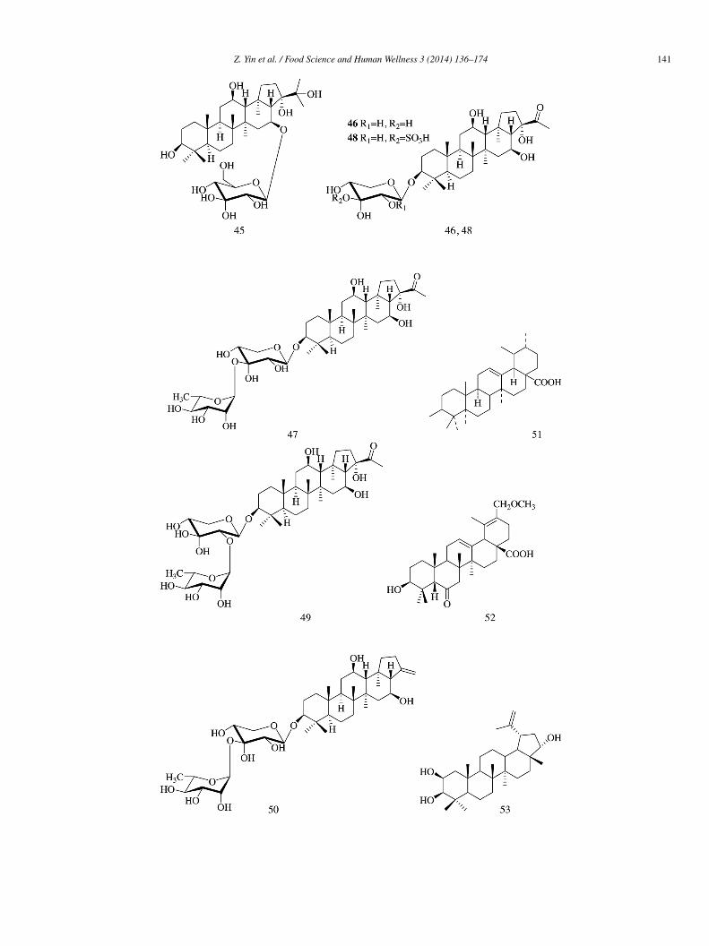
Z. Yin et al. / Food Science and Human Wellness 3 (2014) 136–174 141
1
2
wi(twgTg
dccir
e[
a
α
(
sai
42 Z. Yin et al. / Food Science and Human Wellness 3 (2014) 136–174
.3. Alkaloids
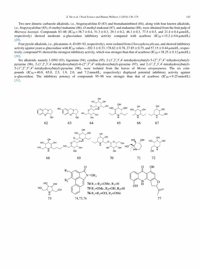
Herein, we present a total of 37 alkaloids, which have been tested for α-glucosidase inhibitory activity. Two compounds 62–63ere isolated from the methanol extract of the leaves of Adhatoda vasica Nees (Acanthaceae). Both these compounds potently
nhibited rat intestinal α-glucosidase activity (IC50 = 125 and 250 �mol/L, respectively), and they were both competitive inhibitorsKi = 82 and 183 �mol/L, respectively) [19]. Another ten compounds were isolated from Campanulaceae Lobelia species. Amonghem, compound 64 was isolated from the methanol extract of Lobelia sessilifolia and compounds 65–73 isolated from Adenophoraere homonojirimycin analogs. Compounds 64–66, 69, and 70 showed inhibitory activity to a certain degree toward mouse α-lucosidase, rat sucrase, maltase, and isomaltase, and among them, compounds 64 and 70 exhibited the best inhibitory activity.he IC50 values of all the compounds were lower than 10 �mol/L. Compounds 67–68 showed strong inhibitory activity towardlucosidase present in coffee (IC50 = 6.4 and 0.71 �mol/L) [20].
The strong α-glucosidase inhibitors deoxynojirimycin (DNJ) (72) and (2R,3R,4R,5R)2,5-bis(hydroxymethyl)-3,4-ihydroxypyrrolidine (DMDP) (73) were isolated from many plants such as Adenophora [20,21], Moraceae [22,23], Commelinaommunis L. [23,24], and Hyacinthus orientalis [23]. Pharmacological activity studies have determined that DNJ in addition toausing lowering of blood sugar level, also possesses antiviral and other important biological activities; however its glucosidasenhibitory activity is particularly dominant. DNJ has been approved clinically for treating diabetic nephropathy and has beenecognized internationally as the only zero harm antidiabetic agent currently available in the market.
Three alkaloids named piperumbellactams A–C (74–76), isolated from the branches of Piper umbellatum, showed mod-rate α-glucosidase inhibition with reported IC50 values—98.07 ± 0.44, 43.80 ± 0.56, and 29.64 ± 0.46 �mol/L, respectively25].
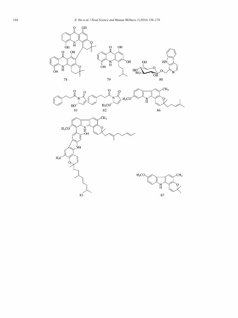
Three acridones (77–79), isolated from the stem and bark of Oriciopsis glaberrima ENGL, showed potent inhibitory activitygainst α-glucosidase (IC50 = 56 ± 5.4, 34.05 ± 17, and 17 ± 1 �mol/L, respectively) [26].
Compound 80, isolated from the ethanol extract of Buthus martensii Karsch, showed potent inhibitory activity against-glucosidase with an IC50 value of 24 �mol/L. A Lineweaver–Burk plot indicated uncompetitive mechanism of inhibitionKi = 16.1 �mol/L) [27].
Two new phenylpropanoyl amides, namely chaplupyrrolidones A (81) and B (82), were isolated from the leaf extract of Piperarmentosum. Compound 82 (IC50 = 430 �mol/L) revealed strong potent α-glucosidase inhibitory activity and was 18-fold morective than its demethylated congener 81 (IC50 = 7820 �mol/L). Kinetic evaluation studies indicated that 82 inhibited α-glucosidasen a non-competitive manner (Ki = 1.04 mmol/L) [28].
iMr[
at[
p5pα
[
Z. Yin et al. / Food Science and Human Wellness 3 (2014) 136–174 143
Two new dimeric carbazole alkaloids, i.e., bisgerayafoline D (83) and bismahanimbinol (84), along with four known alkaloids,.e., bispyrayafoline (85), O-methyl mahanine (86), O-methyl mukonal (87), and mahanine (88), were obtained from the fruit pulp of
urraya koenigii. Compounds 83–88 (IC50 = 38.7 ± 0.4, 51.3 ± 0.3, 29.1 ± 0.2, 46.1 ± 0.3, 77.5 ± 0.5, and 21.4 ± 0.4 �mol/L,espectively) showed moderate α-glucosidase inhibitory activity compared with acarbose (IC50 = 15.2 ± 0.6 �mol/L)29].
Four pyrole alkaloids, i.e., plicatanins A–D (89–92, respectively), were isolated from Chrozophora plicata, and showed inhibitoryctivity against yeast α-glucosidase with IC50 values—202.3 ± 0.33, 178.62 ± 0.78, 27.85 ± 0.75, and 57.15 ± 0.44 �mol/L, respec-ively; compound 91 showed the strongest inhibitory activity, which was stronger than that of acarbose (IC50 = 38.25 ± 0.12 �mol/L)30].
Six alkaloids, namely 1-DNJ (93), fagomine (94), cytidine (95), 2-(1′,2′,3′,4′-tetrahydroxybutyl)-5-(2′′,3′′,4′′-trihydroxybutyl)-yrazine (96), 2-(1′,2′,3′,4′-tetrahydroxybutyl)-6-(2′′,3′′,4′′-trihydroxybutyl)-pyrazine (97), and 2-(1′,2′,3′,4′-tetrahydroxybutyl)--(1′′,2′′,3′′,4′′-tetrahydroxybutyl)-pyrazine (98), were isolated from the leaves of Morus atropurpurea. The six com-ounds (IC50 = 40.0, 65.0, 2.5, 1.9, 2.0, and 7.2 mmol/L, respectively) displayed potential inhibitory activity against-glucosidase. The inhibitory potency of compounds 95–98 was stronger than that of acarbose (IC50 = 9.25 mmol/L)31].
1
44 Z. Yin et al. / Food Science and Human Wellness 3 (2014) 136–174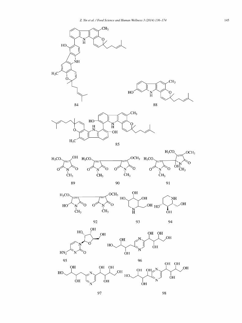
Z. Yin et al. / Food Science and Human Wellness 3 (2014) 136–174 145
1
2
tibfmm
c11a1
1tt87ewl
46 Z. Yin et al. / Food Science and Human Wellness 3 (2014) 136–174
.4. Quinones
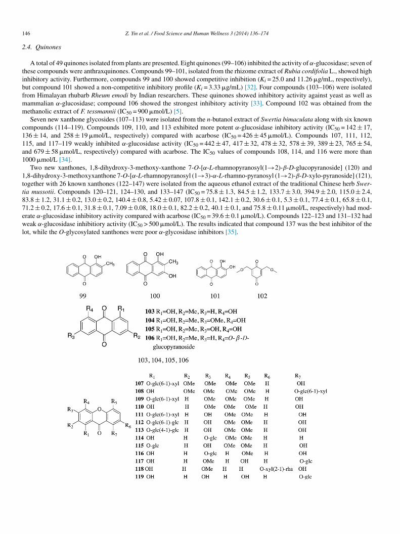
A total of 49 quinones isolated from plants are presented. Eight quinones (99–106) inhibited the activity of α-glucosidase; seven ofhese compounds were anthraxquinones. Compounds 99–101, isolated from the rhizome extract of Rubia cordifolia L., showed highnhibitory activity. Furthermore, compounds 99 and 100 showed competitive inhibition (Ki = 25.0 and 11.26 �g/mL, respectively),ut compound 101 showed a non-competitive inhibitory profile (Ki = 3.33 �g/mL) [32]. Four compounds (103–106) were isolatedrom Himalayan rhubarb Rheum emodi by Indian researchers. These quinones showed inhibitory activity against yeast as well asammalian α-glucosidase; compound 106 showed the strongest inhibitory activity [33]. Compound 102 was obtained from theethanolic extract of F. tessmannii (IC50 = 900 �mol/L) [5].Seven new xanthone glycosides (107–113) were isolated from the n-butanol extract of Swertia bimaculata along with six known
ompounds (114–119). Compounds 109, 110, and 113 exhibited more potent α-glucosidase inhibitory activity (IC50 = 142 ± 17,36 ± 14, and 258 ± 19 �mol/L, respectively) compared with acarbose (IC50 = 426 ± 45 �mol/L). Compounds 107, 111, 112,15, and 117–119 weakly inhibited α-glucosidase activity (IC50 = 442 ± 47, 417 ± 32, 478 ± 32, 578 ± 39, 389 ± 23, 765 ± 54,nd 679 ± 58 �mol/L, respectively) compared with acarbose. The IC50 values of compounds 108, 114, and 116 were more than000 �mol/L [34].
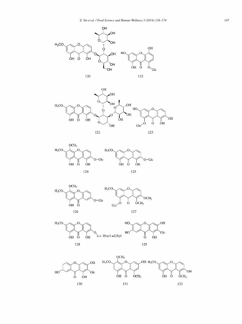
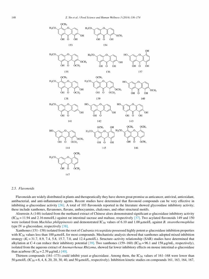
Two new xanthones, 1,8-dihydroxy-3-methoxy-xanthone 7-O-[α-L-rhamnopyranosyl(1→2)-β-D-glucopyranoside] (120) and,8-dihydroxy-3-methoxyxanthone 7-O-[α-L-rhamnopyranosyl (1→3)-α-L-rhamno-pyranosyl (1→2)-β-D-xylo-pyranoside] (121),ogether with 26 known xanthones (122–147) were isolated from the aqueous ethanol extract of the traditional Chinese herb Swer-ia mussotii. Compounds 120–121, 124–130, and 133–147 (IC50 = 75.8 ± 1.3, 84.5 ± 1.2, 133.7 ± 3.0, 394.9 ± 2.0, 115.0 ± 2.4,3.8 ± 1.2, 31.1 ± 0.2, 13.0 ± 0.2, 140.4 ± 0.8, 5.42 ± 0.07, 107.8 ± 0.1, 142.1 ± 0.2, 30.6 ± 0.1, 5.3 ± 0.1, 77.4 ± 0.1, 65.8 ± 0.1,1.2 ± 0.2, 17.6 ± 0.1, 31.8 ± 0.1, 7.09 ± 0.08, 18.0 ± 0.1, 82.2 ± 0.2, 40.1 ± 0.1, and 75.8 ± 0.11 �mol/L, respectively) had mod-rate α-glucosidase inhibitory activity compared with acarbose (IC50 = 39.6 ± 0.1 �mol/L). Compounds 122–123 and 131–132 hadeak α-glucosidase inhibitiory activity (IC50 > 500 �mol/L). The results indicated that compound 137 was the best inhibitor of the
ot, while the O-glycosylated xanthones were poor α-glycosidase inhibitors [35].
Z. Yin et al. / Food Science and Human Wellness 3 (2014) 136–174 147
1
2
ait
(wt
wsait
5
48 Z. Yin et al. / Food Science and Human Wellness 3 (2014) 136–174
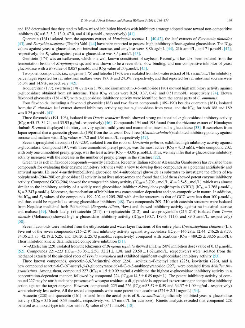
.5. Flavonoids
Flavonoids are widely distributed in plants and therapeutically they have shown great promise as anticancer, antiviral, antioxidant,ntibacterial, and anti-inflammatory agents. Recent studies have determined that flavonoid compounds can be very effective innhibiting α-glucosidase activity [36]. A total of 103 flavonoids reported in the literature showed glycosidase inhibitory activity;hese include xanthones, flavanones, flavans, anthocyamins, chalcones, and other structural motifs.
Aloeresin A (148) isolated from the methanol extract of Chinese aloes demonstrated significant α-glucosidase inhibitory activityIC50 = 11.94 and 2.16 mmol/L) against rat intestinal sucrase and maltase, respectively [37]. Two acylated flavonoids 149 and 150ere isolated from Machilus philippinensis and demonstrated IC50 values of 6.10 and 1.00 �mol/L against B. stearothermophilus
ype IV α-glucosidase, respectively [38].Xanthones (151–158) isolated from the root of Cudrania tricuspidata possessed highly potent α-glucosidase inhibition properties
ith IC50 values less than 100 �mol/L for most compounds. Mechanistic analysis showed that xanthones adopted mixed inhibitiontrategy (Ki = 31.7, 8.9, 7.4, 5.8, 15.7, 7.0, and 12.4 �mol/L). Structure–activity relationship (SAR) studies have determined thatlkylation at C-4 can reduce their inhibitory potential [39]. Two xanthones (159–160) (IC50 = 96.1 and 158 �g/mL, respectively),
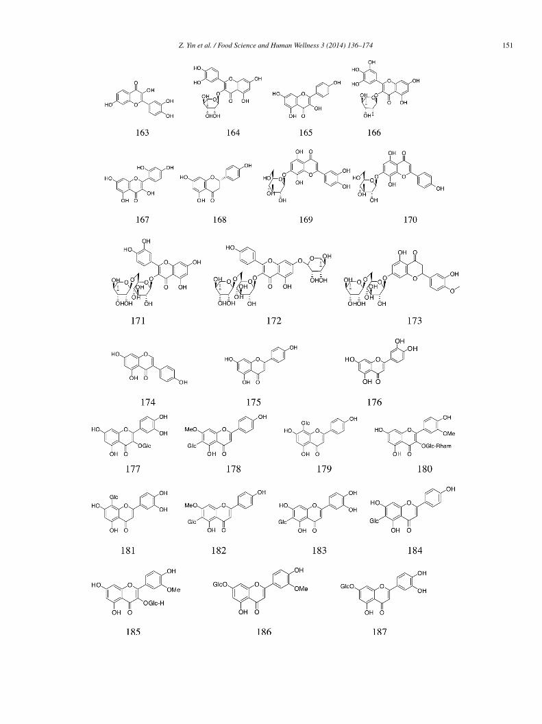
solated from the aqueous extract of Anemarrhenae Rhizoma, showed far lower inhibitory effects on mouse intestinal α-glucosidasehan acarbose (IC50 = 2.39 �g/mL) [40].Thirteen compounds (161–173) could inhibit yeast α-glucosidase. Among them, the IC50 values of 161–168 were lower than0 �mol/L (IC50 = 8, 4, 8, 20, 20, 30, 40, and 50 �mol/L, respectively). Inhibition kinetic studies on compounds 161, 163, 164, 167,
ai
[vr
fg
p3
α
fl
fw
(rJs
α
wa
capasKtafas[
F7T
[m
ngcpaw
ab
Z. Yin et al. / Food Science and Human Wellness 3 (2014) 136–174 149
nd 168 determined that they tend to follow mixed inhibition kinetics with inhibitory strategy adopted more toward non-competitivenhibitors (Ki = 6.2, 3.2, 13.0, 47.0, and 41.0 �mol/L, respectively) [41].
Quercetin (161) isolated from the aqueous extract of Matricaria recutita L. [40,42], the leaf extracts of Eucommia ulmoides43], and Forsythia suspensa (Thunb) Vahl. [44] have been reported to possess high inhibitory effects against glucosidase. The IC50alues against yeast α-glucosidase, rat intestinal sucrose, and amylase were 8.86 �g/mL [44], 216 �mol/L, and 71 �mol/L [42],espectively; the Ki value against yeast α-glucosidase was 8.5 �mol/L [43].
Genistein (174) was an isoflavone, which is a well-known constituent of soybean. Recently, it has also been isolated from theermentation broths of Streptomyces sp. and was shown to be a reversible, slow binding, and non-competitive inhibitor of yeastlucosidase with a Ki value of 0.057 �mol/L and IC50 value of 50 �mol/L [45].
Two potent compounds, i.e., apigenin (175) and luteolin (176), were isolated from hot water extract of M. recutita L. The inhibitoryercentages reported for rat intestinal maltase were 18.0% and 24.3%, respectively, and that reported for rat intestinal sucrase were5.3% and 14.9%, respectively [42].
Isoquercitrin (177), swertisin (178), vitexin (179), and isorhamnetin-3-O-rutinoside (180) showed high inhibitory activity against-glucosidase obtained from rat intestine. Their IC50 values were 0.24, 0.37, 0.42, and 0.51 mmol/L, respectively [24]. Elevenavonoid glycosides (181–187) with α-glucosidase inhibitory activity were identified from the aerial parts of C. ommunis.
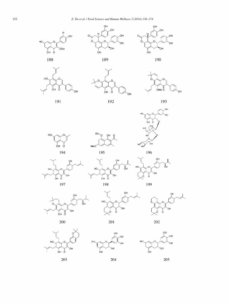
Four flavonoids, including a flavonoid glycoside (188) and two flavan compounds (189–190) besides quercetin (161), isolatedrom the E. ulmoides leaf extract showed inhibitory activity against α-glucosidase from yeast, and the IC50 for both 188 and 189as 0.25 �mol/L [43].Three flavonoids (191–193), isolated from Derris scandens Benth, showed strong rat intestinal α-glucosidase inhibitory activity
IC50 = 45.17, 34.74, and 33.93 �g/mL respectively) [46]. Compounds 194 and 195 found from the rhizome extract of Himalayanhubarb R. emodi displayed inhibitory activity against mild yeast and mammalian intestinal α-glucosidase [33]. Researchers fromapan reported that a quercetin glycoside (196) from the leaves of Devil tree (Alstonia scholaris) exhibited inhibitory potency againstucrase and maltase with IC50 values = 17.2 and 1.96 mmol/L, respectively [47].
Seven triprenylated flavonols (197–203), isolated from the roots of Dorstenia psilurus, exhibited high inhibitory activity against-glucosidase. Compound 197, with three unmodified prenyl groups, was the most active (IC50 = 4.13 mM), while compound 202,ith only one unmodified prenyl group, was the least active (IC50 = 43.95 �mol/L). Thus, we may infer that α-glucosidase inhibitory
ctivity increases with the increase in the number of prenyl groups in the structure [22].Green tea is rich in flavonol compounds—mostly catechins. Recently, Italian scholar Alessandra Gamberucci has revisited these
ompounds for evaluating their enzyme inhibitory activities with a view to develop these compounds as a potential antidiabetic andntiviral agents. He used 4-methylumbelliferyl glucoside and 4-nitrophenyl glucoside as substrates to investigate the effects of teaolyphenols (204–208) on glucosidase II activity in rat liver microsomes and found that all of them showed potent enzyme inhibitoryctivity. Compound GCG (204) showed the strongest inhibitory activity (IC50 = 3.702 �mol/L, Ki = 2.545 �mol/L), which was almostimilar to the inhibitory activity of a widely used glucosidase inhibitor N-butyldeoxynojirimycin (NBDJ) (IC50 = 3.268 �mol/L,i = 2.247 �mol/L). Moreover, the mechanism of inhibition was concentration dependent and non-competitive in nature. In addition,
he IC50 and Ki values of the compounds 199–202 and 205–208 with similar structure as that of GCG were less than 100 �mol/L,nd thus could be regarded as strong glucosidase inhibitors [48]. Two compounds 209–210 with catechin structure were isolatedrom Nepalese medicinal herb Pakhanbhed (Bergenia ciliata, Haw.) and showed inhibitory activity against rat intestinal sucrasend maltase [49]. Much lately, (+)-catechin (211), (−)-epicatechin (212), and two procyanidin (213–214) isolated from Toonainensis (Meliaceae) showed high α-glucosidase inhibitory activity (IC50 = 190.7, 189.0, 111.0, and 89.0 �mol/L, respectively)50].
Seven flavonoids were isolated from the ethylacetate and water layer fractions of the entire plant Crossostephium chinense (L.).ive out of the seven compounds (215–219) had inhibitory activity against α-glucosidase (IC50 = 146.28 ± 12.44, 246.26 ± 8.73,4.06 ± 3.83, 42.19 ± 5.25, and 136.20 ± 25.73 �mol/L, respectively) compared with acarbose (IC50 = 489.25 ± 38.55 �mol/L).heir inhibition kinetic data indicated competitive inhibition [51].
(+)-Afzelechin (220) isolated from the Rhizomes of Bergenia ligulata showed an ID50 (50% inhibition dose) value of 0.13 �mol/L52]. Compounds 221–223 (IC50 = 56.06 ± 2.56, 32.21 ± 1.38, and 20.50 ± 1.62 �mol/L, respectively) were isolated from theethanol extracts of the air-dried roots of Ferula mongolica and exhibited significant α-glucosidase inhibitory activity [53].Three known compounds, quercetin-3,6,7-trimethyl ether (224), isovitexin-4′-methyl ether (225), isovitexin (226), and a
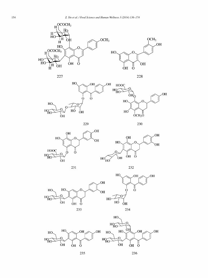
ew compound acaetin-6-C-(6′′-acetyl-β-D-glucopyranoside)-8-C-α-L-arabinopyranoside (227), were obtained from Achillea fra-rantissima. Among them, compound 227 (IC50 = 1.5 ± 0.09 mg/mL) exhibited the highest α-glucosidase inhibitory activity in aoncentration-dependent manner, followed by compound 224 (IC50 = 14.5 ± 0.89 mg/mL). The potent inhibitory activity of com-ound 227 may be attributed to the presence of two sugar residues; a di-glycoside is supposed to exert stronger competitive inhibitoryction against the target enzyme. However, compounds 225 and 226 (IC50 = 83.57 ± 0.59 and 34.37 ± 1.09 mg/mL, respectively)
ere relatively less active. All the tested compounds were more potent than acarbose (224 ± 2.31 mg/mL) [54].Acacetin (228) and quercetin (161) isolated from the aerial parts of B. cavanillesii significantly inhibited yeast α-glucosidasectivity (IC50 = 0.16 and 0.53 mmol/L, respectively, vs. 1.7 mmol/L for acarbose). Kinetic analysis revealed that compound 228ehaved as a mixed-type inhibitor with a Ki value of 0.41 mmol/L [18].

1
ti3ii
ni(t
fa
in
(
50 Z. Yin et al. / Food Science and Human Wellness 3 (2014) 136–174
Four new compounds, i.e., aquilarisinin (229), aquilarisin (230), hypolaetin 5-O-β-D-glucuronopyranoside (231), and aquilarixan-hone (232) (IC50 = 273.7 ± 39.8, 634.7 ± 45.7, 298.9 ± 27.9, and 678.1 ± 137.4 �mol/L, respectively), and four known compounds,.e., mangiferin (233), iriflophenone 2-O-α-L-rhamnopyranoside (234), iriflophenone 3-C-β-D-glucoside (235), and iriflophenone,5-C-β-D-diglucopyranoside (236) (IC50 = 299.7 ± 42.3, 366.7 ± 27.0, 404.7 ± 27.7, and 454.4 ± 24.0 �mol/L, respectively), weresolated from 70% aqueous ethanolic leaf extracts of Aquilaria sinensis (Lour.) Gilg b. and showed more significant α-glucosidasenhibitory activity than acarbose (IC50 = 576.2 ± 58.5 �mol/L) [55].
(2R)-3α,7,4′-Trihydroxy-5-methoxy-8-(γ ,γ-dimethylallyl)-flavanone (237), isoxanthohumol (238), norkurarinone (239), kurari-one (240), (2S)-2′-methoxy-kurarinone (241), kushenol T (242), norkurarinol (243), kuraridin (244), and formononetin (245)solated from the dried stem and bark of Sophora flavescens had moderate but less significant α-glucosidase inhibitory effectsIC50 = 107.8 ± 5.7, 86.9 ± 6.1, 17.7 ± 2.6, 14.5 ± 1.3, 14.0 ± 1.2, 16.7 ± 2.1, 18.0 ± 1.5, 3.6 ± 0.8, and 119.6 ± 9.4 �mol/L, respec-ively) than acarbose (IC50 = 2.9 ± 0.2 �mol/L) [56].
Three flavonoid glycosides vitexin (246), isovitexin (184), and isorhamnetin 3-O-β-D-rutinoside (180) obtained from Microctisolium exerted satisfactory α-glucosidase inhibitory effects (IC50 = 244.0, 266.2, and 275.4 �mol/L, respectively) compared withcarbose (IC50 = 1007 �mol/L) [57].
Two flavonoid compounds plicatanoside (247) and apigenin-5-O-β-D-glucopyranoside (248), isolated from C. plicata, showednhibitory activity against yeast α-glucosidase (IC50 = 111.23 ± 0.65 and 287.12 ± 0.75 �mol/L, respectively), but the inhibition isot as strong as that of acarbose (IC50 = 38.25 ± 0.12 �mol/L) [30].
Coryfolin (249) and daidzein (250) isolated from Psoralea corylifolia displayed slightly weaker α-glucosidase inhibitory activityIC50 = 45.73 and 49.44 mmol/L, respectively) than acarbose (IC50 = 38.25 ± 0.12 mmol/L) [58].
Z. Yin et al. / Food Science and Human Wellness 3 (2014) 136–174 151
1
52 Z. Yin et al. / Food Science and Human Wellness 3 (2014) 136–174
Z. Yin et al. / Food Science and Human Wellness 3 (2014) 136–174 153
1
54 Z. Yin et al. / Food Science and Human Wellness 3 (2014) 136–174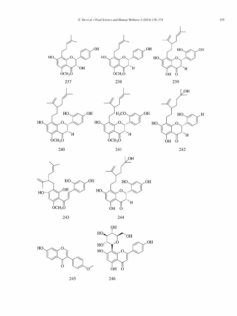
Z. Yin et al. / Food Science and Human Wellness 3 (2014) 136–174 155

1
2
cvs(
g
(o
hα
wi0
t2a1aag
56 Z. Yin et al. / Food Science and Human Wellness 3 (2014) 136–174
.6. Phenols
Thirty-seven polyphenols from plants have shown promising α-glucosidase inhibitory activity. Gallic acid (251), an importantonstituent of many plants species [50,59], showed strong inhibitory activity against glucosidase both in vitro and in vivo; its IC50alue (24.3 �mol/L) was lower than that of acarbose (59.5 �mol/L) [50]. Moreover, methyl gallate (252) obtained from the driedtem and bark extracts of Terminalia superb (IC50 = 11.5 �mol/L) [59] and propyl gallate (253) isolated from green tea extractsIC50 = 11.5 �mol/L, Ki = 43.12 �mol/L) [48] showed strong α-glucosidase inhibitory activity.
Rosmarinic acid (254) isolated from the methanol extract of P. madagascariensis exhibited inhibitory activity against α-lucosidase (IC50 = 33.0 ± 4.6 �mol/L) [9].
Three compounds trans-N-p-coumaroyltyramine (255) (IC50 = 0.40 �mol/L), 1,7-bis(4-hydroxyphenyl)heptane-3,5-diol (256)IC50 = 0.38 mmol/L), and 6-hydroxy-2,4,7-trimethoxyphe-nanth-rene (257) (IC50 = 0.77 mmol/L) isolated from the air-dried slicesf fresh tuberous rhizomes of Dioscorea opposita showed yeast α-glucosidase inhibitory activity [60].
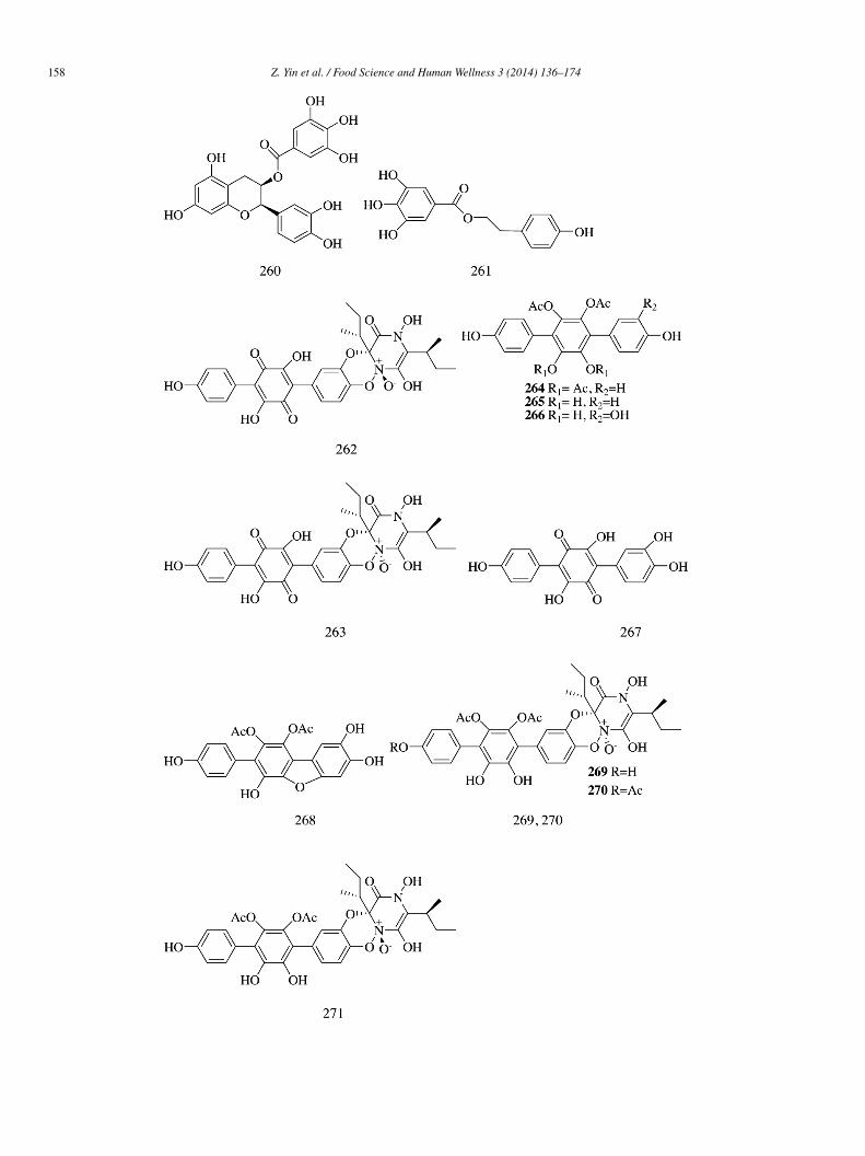
Epicatechin (EC, 258), epicatechin-(4β,8)-epicatechingallate (B 2-3′-O-gallate, 259), epicatechin gallate (ECG, 260), and 2-(4-ydroxyphenyl)ethyl 3,4,5-trihydroxybenzoate (HETB, 261) isolated from the roots of Rhodiola crenulata demonstrated significant-glucosidase inhibitory activity (IC50 = 29.85 ± 2.20, 0.31 ± 0.01, 0.71 ± 0.01, and 4.77 ± 0.22 �mol/L, respectively). Comparedith the known α-glucosidase inhibitor quercetin (IC50 = 5.30 ± 0.11 �mol/L), compounds 259–261 showed strong α-glucosidase
nhibitory effect. The inhibition kinetics study indicated that 259 and 260 were mixed competitive inhibitors (Ki = 0.30 ± 0.03 and.21 ± 0.04 �mol/L), while 261 was a competitive inhibitor (Ki = 3.10 ± 0.09 �mol/L) [61].
Ten compounds (262–271) were isolated from the ethyl acetate extract of the edible mushroom Sarcodon leucopus. Of allhe compounds tested, sarcoviolin β (262) showed the strongest inhibition (IC50 = 0.58 ± 0.01 �mol/L). Compounds 263 and66–271 exhibited moderate inhibitory activity (IC50 = 1.07 ± 0.04, 3.35 ± 0.09, 3.53 ± 0.03, 6.22 ± 0.03, 3.62 ± 0.06, 4.20 ± 0.02,nd 1.23 ± 0.06 �mol/L, respectively). Compounds 264–265 showed relatively weak inhibitory activity (IC50 = 35 ± 0.1 and
9 ± 0.1 �mol/L). The configuration at N-1β and C-2β greatly influenced the α-glucosidase inhibitory activity of sarcodoninsnd sarcoviolins. Compounds 262 and 271 with cis configuration at N-1β and C-2β showed stronger activity than compounds 263nd 269 with trans configuration. For the p-terphenyl derivatives (264–268), the number of phenolic hydroxyl groups in the structurereatly contributed to their α-glucosidase inhibitory activity [62].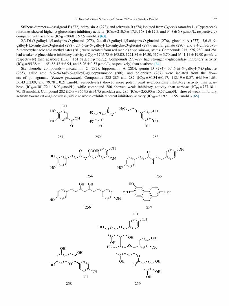
rc
g5hr(
(e5b7a
Z. Yin et al. / Food Science and Human Wellness 3 (2014) 136–174 157
Stilbene dimmers—cassigarol E (272), scirpusin A (273), and scirpusin B (274) isolated from Cyperus rotundus L. (Cyperaceae)hizomes showed higher α-glucosidase inhibitory activity (IC50 = 210.5 ± 17.3, 168.1 ± 12.5, and 94.3 ± 6.8 �mol/L, respectively)ompared with acarbose (IC50 = 2060 ± 97.5 �mol/L) [63].
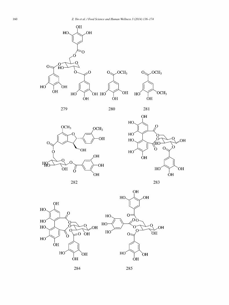
2,3-Di-O-galloyl-1,5-anhydro-D-glucitol (275), 2,4-di-O-galloyl-1,5-anhydro-D-glucitol (276), ginnalin A (277), 3,6-di-O-alloyl-1,5-anhydro-D-glucitol (278), 2,4,6-tri-O-galloyl-1,5-anhydro-D-glucitol (279), methyl gallate (280), and 3,4-dihydroxy--methoxybenzoic acid methyl ester (281) were isolated from red maple (Acer rubrum) stems. Compounds 275, 276, 280, and 281ad weaker α-glucosidase inhibitory activity (IC50 = 1745.78 ± 168.05, 1221.84 ± 16.30, 317 ± 3.70, and 6541.11 ± 19.90 �mol/L,espectively) than acarbose (IC50 = 161.38 ± 5.5 �mol/L). Compounds 277–279 had stronger α-glucosidase inhibitory activityIC50 = 95.38 ± 11.65, 88.42 ± 6.94, and 8.26 ± 0.37 �mol/L, respectively) than acarbose [64].
Six phenolic compounds—unicatannin C (282), hippomanin A (283), gemin D (284), 3,4,6-tri-O-galloyl-β-D-glucose285), gallic acid 3-O-β-D-(6′-O-galloyl)-glucopyranoside (286), and phloridzin (287) were isolated from the flow-rs of pomegranate (Punica granatum). Compounds 282–285 and 287 (IC50 = 80.34 ± 0.17, 118.19 ± 0.57, 64.19 ± 1.63,6.43 ± 2.09, and 79.78 ± 0.21 �mol/L, respectively) showed more potent yeast α-glucosidase inhibitory activity than acar-ose (IC50 = 301.72 ± 18.93 �mol/L), while compound 286 showed weak inhibitory activity than acrbose (IC50 = 737.18 ±0.18 �mol/L). Compound 282 (IC50 = 366.95 ± 54.75 �mol/L) and 285 (IC50 = 255.90 ± 15.57 �mol/L) showed weak inhibitoryctivity toward rat α-glucosidase, while acarbose exhibited potent inhibitory activity (IC50 = 21.92 ± 1.55 �mol/L) [65].
1
58 Z. Yin et al. / Food Science and Human Wellness 3 (2014) 136–174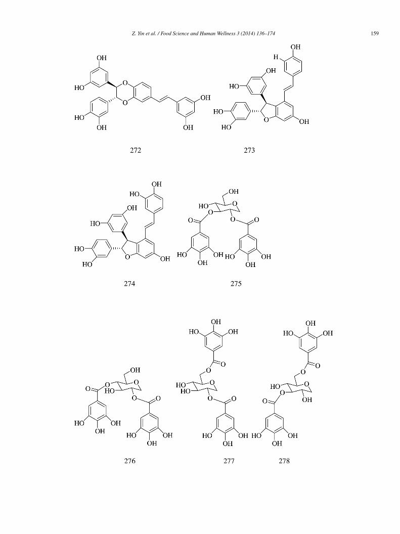
Z. Yin et al. / Food Science and Human Wellness 3 (2014) 136–174 159
1
60 Z. Yin et al. / Food Science and Human Wellness 3 (2014) 136–174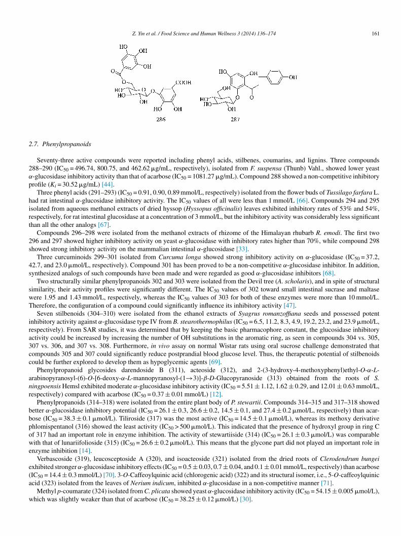
2
2α
p
hirt
2s
4s
swT
ira3cc
anr
bbpowe
e(a
w
Z. Yin et al. / Food Science and Human Wellness 3 (2014) 136–174 161
.7. Phenylpropanoids
Seventy-three active compounds were reported including phenyl acids, stilbenes, coumarins, and lignins. Three compounds88–290 (IC50 = 496.74, 800.75, and 462.62 �g/mL, respectively), isolated from F. suspensa (Thunb) Vahl., showed lower yeast-glucosidase inhibitory activity than that of acarbose (IC50 = 1081.27 �g/mL). Compound 288 showed a non-competitive inhibitoryrofile (Ki = 30.52 �g/mL) [44].
Three phenyl acids (291–293) (IC50 = 0.91, 0.90, 0.89 mmol/L, respectively) isolated from the flower buds of Tussilago farfara L.ad rat intestinal α-glucosidase inhibitory activity. The IC50 values of all were less than 1 mmol/L [66]. Compounds 294 and 295solated from aqueous methanol extracts of dried hyssop (Hyssopus officinalis) leaves exhibited inhibitory rates of 53% and 54%,espectively, for rat intestinal glucosidase at a concentration of 3 mmol/L, but the inhibitory activity was considerably less significanthan all the other analogs [67].
Compounds 296–298 were isolated from the methanol extracts of rhizome of the Himalayan rhubarb R. emodi. The first two96 and 297 showed higher inhibitory activity on yeast α-glucosidase with inhibitory rates higher than 70%, while compound 298howed strong inhibitory activity on the mammalian intestinal α-glucosidase [33].
Three curcuminoids 299–301 isolated from Curcuma longa showed strong inhibitory activity on α-glucosidase (IC50 = 37.2,2.7, and 23.0 �mol/L, respectively). Compound 301 has been proved to be a non-competitive α-glucosidase inhibitor. In addition,ynthesized analogs of such compounds have been made and were regarded as good α-glucosidase inhibitors [68].
Two structurally similar phenylpropanoids 302 and 303 were isolated from the Devil tree (A. scholaris), and in spite of structuralimilarity, their activity profiles were significantly different. The IC50 values of 302 toward small intestinal sucrase and maltaseere 1.95 and 1.43 mmol/L, respectively, whereas the IC50 values of 303 for both of these enzymes were more than 10 mmol/L.herefore, the configuration of a compound could significantly influence its inhibitory activity [47].
Seven stilbenoids (304–310) were isolated from the ethanol extracts of Syagrus romanzoffiana seeds and possessed potentnhibitory activity against α-glucosidase type IV from B. stearothermophilus (IC50 = 6.5, 11.2, 8.3, 4.9, 19.2, 23.2, and 23.9 �mol/L,espectively). From SAR studies, it was determined that by keeping the basic pharmacophore constant, the glucosidase inhibitoryctivity could be increased by increasing the number of OH substitutions in the aromatic ring, as seen in compounds 304 vs. 305,07 vs. 306, and 307 vs. 308. Furthermore, in vivo assay on normal Wistar rats using oral sucrose challenge demonstrated thatompounds 305 and 307 could significantly reduce postprandial blood glucose level. Thus, the therapeutic potential of stilbenoidsould be further explored to develop them as hypoglycemic agents [69].
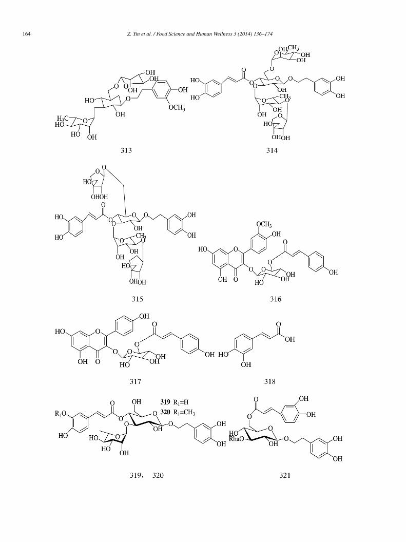
Phenylpropanoid glycosides darendoside B (311), acteoside (312), and 2-(3-hydroxy-4-methoxyphenyl)ethyl-O-α-L-rabinopyranosyl-(6)-O-[6-deoxy-α-L-mannopyranosyl-(1→3)]-β-D-Glucopyranoside (313) obtained from the roots of S.ingpoensis Hemsl exhibited moderate α-glucosidase inhibitory activity (IC50 = 5.51 ± 1.12, 1.62 ± 0.29, and 12.01 ± 0.63 mmol/L,espectively) compared with acarbose (IC50 = 0.37 ± 0.01 mmol/L) [12].
Phenylpropanoids (314–318) were isolated from the entire plant body of P. stewartii. Compounds 314–315 and 317–318 showedetter α-glucosidase inhibitory potential (IC50 = 26.1 ± 0.3, 26.6 ± 0.2, 14.5 ± 0.1, and 27.4 ± 0.2 �mol/L, respectively) than acar-ose (IC50 = 38.3 ± 0.1 �mol/L). Tiliroside (317) was the most active (IC50 = 14.5 ± 0.1 �mol/L), whereas its methoxy derivativehlomispentanol (316) showed the least activity (IC50 > 500 �mol/L). This indicated that the presence of hydroxyl group in ring Cf 317 had an important role in enzyme inhibition. The activity of stewartiiside (314) (IC50 = 26.1 ± 0.3 �mol/L) was comparableith that of lunariifolioside (315) (IC50 = 26.6 ± 0.2 �mol/L). This means that the glycone part did not played an important role in
nzyme inhibition [14].Verbascoside (319), leucosceptoside A (320), and isoacteoside (321) isolated from the dried roots of Clerodendrum bungei
xhibited stronger α-glucosidase inhibitory effects (IC50 = 0.5 ± 0.03, 0.7 ± 0.04, and 0.1 ± 0.01 mmol/L, respectively) than acarbose
IC50 = 14.4 ± 0.3 mmol/L) [70]. 3-O-Caffeoylquinic acid (chlorogenic acid) (322) and its structural isomer, i.e., 5-O-caffeoylquiniccid (323) isolated from the leaves of Nerium indicum, inhibited α-glucosidase in a non-competitive manner [71].Methyl p-coumarate (324) isolated from C. plicata showed yeast α-glucosidase inhibitory activity (IC50 = 54.15 ± 0.005 �mol/L),hich was slightly weaker than that of acarbose (IC50 = 38.25 ± 0.12 �mol/L) [30].
1
(t
3w
c9dltta
dsα
mrI
slsa
t
(0t
62 Z. Yin et al. / Food Science and Human Wellness 3 (2014) 136–174
p-Coumarica (325) (IC50 > 30 mmol/L), ferulic acid (326) (IC50 = 4.9 ± 0.3 mmol/L), and sinapic acid (327)IC50 = 6.1 ± 0.8 mmol/L) isolated from the sprouts of Triticum aestivum L. had lower α-glucosidase inhibitory activityhan acarbose (IC50 = 1.7 ± 0.1 mmol/L) [72].
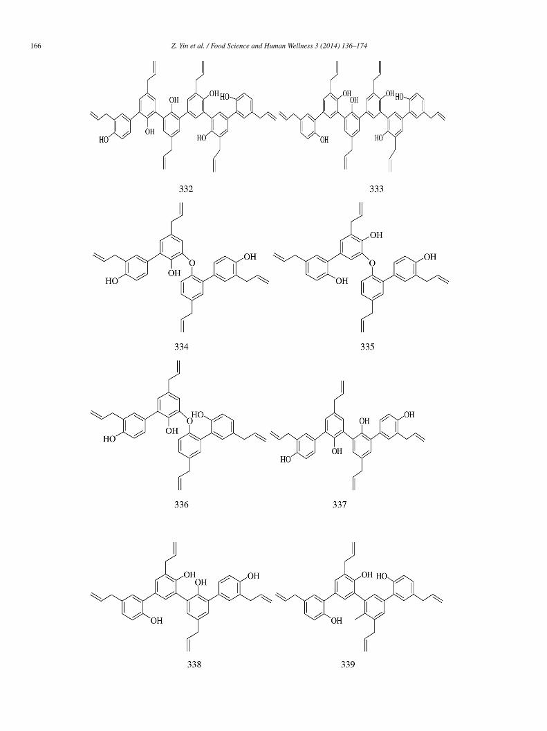
Epi-mukulin (328) and diasesartemin (329) were isolated from guggul, the oleogum resin of Commiphora wightii. Compound29 (IC50 = 60.6 ± 0.01 �mol/L) was found to be more potent than acarbose (IC50 = 92.94 ± 0.01 �mol/L). The IC50 value of 328as found to be 159.33 ± 0.04 �mol/L [73].Ten honokiol oligomers (330–339), including four trimers (330–333) and four dimers (334–337), were obtained from Momordica
harantia. All the honokioltrimers and dimers had good inhibitory effects on α-glucosidase with IC50 values ranging from 1.38 to5.31 �mol/L. The potency of honokiol trimers (331–332) were found to be higher than those of dimers (334–339), and among the siximers, the potency of the carbon oxygen linkage dimers (334–336) appeared to be much stronger than those of the carbon acarboninkage ones (337–339). Among all the tested compounds, the compound 331 (IC50 = 1.38 �mol/L), i.e., the honokioltrimer, exhibitedhe most significant inhibitory activity toward α-glucosidase in a concentration-dependent manner and was 128-fold more potenthan that of honokiol (IC50 = 177.03 �mol/L). Compared with genistein (IC50 = 15.31 �mol/L) and 1-DNJ (IC50 = 239.17 �mol/L)s positive controls, compound 331 was 11 and 173 times more active [74].
Extracts isolated from plants containing coumarin compounds also showed good α-glucosidase inhibitory activity. Two coumarinerivatives 340 and 341 (IC50 = 35.03 and 69.26 �g/mL, respectively), isolated from the branch extract of L. pinceana Hook, showedtrong α-glucosidase inhibitory activity. Kinetic analysis revealed that both the compounds were non-competitive inhibitors of-glucosidase (Ki = 2.44 and 167.83 �g/mL, respectively) [6].
Four coumarins (342–345), isolated from the aqueous extract of M. recutita L., showed inhibitory activity against rat intestinalaltase and sucrose at the concentration of 400 �mol/L; compound 344 recorded the highest activity (IC50 = 534 and 72 �mol/L,
espectively) [42]. Scandenin A (346), isolated from D. scandens Benth, showed rat intestinal α-glucosidase inhibitory activity withC50 = 25.17 �g/mL [46].
Three compounds 347–349 with a coumarin nucleus isolated from the fruit extract of Terminalia chebula Retz. (Combretaceae)howed strong inhibitory activity (IC50 = 960, 97 and 36 �mol/L, respectively) on α-glucosidase. Chebulagic acid (348) and chebu-inic acid (349) belong to the class of non-competitive α-glucosidase inhibitors (Ki = 208 and 24 �mol/L, respectively) [75]. Thetem and bark extract of another plant named T. superba contained three coumarins (350–352), with IC50 values of 194.1, 184.6,nd 118.7 �mol/L, respectively, on yeast α-glucosidase [59].
Psoralidin (353) isolated from P. corylifolia had α-glucosidase inhibitory activity (IC50 = 40.74 mmol/L), which was strongerhan that of acarbose (IC50 = 38.25 ± 0.12 mg/L) [58].
Coumarins compounds (354–360) isolated from the root extract of R. rugosa showed potent sucrase inhibitory activity61.88 ± 3.19% to 84.70 ± 3.07%) at a concentration of 1.0 mmol/L (IC50 = 0.25 ± 0.04, 0.48 ± 0.12, 0.43 ± 0.11, 0.40 ± 0.18,.52 ± 0.01, and 0.31 ± 0.02 mmol/L, respectively), and the activity was comparable with that of acarbose (50.96 ± 2.97% inhibi-ion, IC50 < 0.5 mmol/L) [13].

Z. Yin et al. / Food Science and Human Wellness 3 (2014) 136–174 163
1
64 Z. Yin et al. / Food Science and Human Wellness 3 (2014) 136–174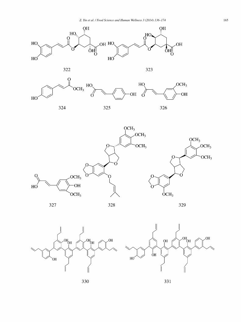
Z. Yin et al. / Food Science and Human Wellness 3 (2014) 136–174 165
1
66 Z. Yin et al. / Food Science and Human Wellness 3 (2014) 136–174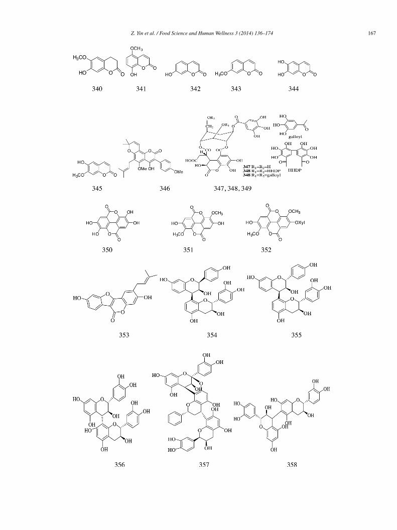
Z. Yin et al. / Food Science and Human Wellness 3 (2014) 136–174 167
1
2
i
f(
α
i
i(
(
68 Z. Yin et al. / Food Science and Human Wellness 3 (2014) 136–174
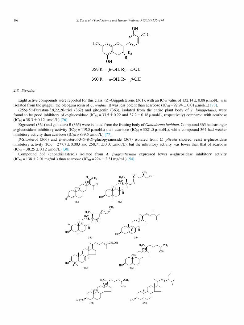
.8. Sterides
Eight active compounds were reported for this class. (Z)-Guggulsterone (361), with an IC50 value of 132.14 ± 0.08 �mol/L, wassolated from the guggul, the oleogum resin of C. wightii. It was less potent than acarbose (IC50 = 92.94 ± 0.01 �mol/L) [73].
(25S)-5a-Furastan-3β,22,26-triol (362) and gitogenin (363), isolated from the entire plant body of T. longipetalus, wereound to be good inhibitors of α-glucosidase (IC50 = 33.5 ± 0.22 and 37.2 ± 0.18 �mol/L, respectively) compared with acarboseIC50 = 38.3 ± 0.12 �mol/L) [76].
Ergosterol (364) and ganodero B (365) were isolated from the fruiting body of Ganoderma lucidum. Compound 365 had stronger-glucosidase inhibitory activity (IC50 = 119.8 �mol/L) than acarbose (IC50 = 3521.5 �mol/L), while compound 364 had weaker
nhibitory activity than acarbose (IC50 > 839.5 �mol/L) [77].β-Sitosterol (366) and β-sitosterol-3-O-β-D-glucopyranoside (367) isolated from C. plicata showed yeast α-glucosidase
nhibitory activity (IC50 = 277.7 ± 0.003 and 258.71 ± 0.07 �mol/L), but the inhibitory activity was lower than that of acarboseIC50 = 38.25 ± 0.12 �mol/L) [30].
Compound 368 (chondrillasterol) isolated from A. fragrantissima expressed lower α-glucosidase inhibitory activityIC50 = 138 ± 2.01 mg/mL) than acarbose (IC50 = 224 ± 2.31 mg/mL) [54].
2
gv(sth
h(ciy
a2α
na
ia((
fa1
ai4m
Z. Yin et al. / Food Science and Human Wellness 3 (2014) 136–174 169
.9. Other compound types
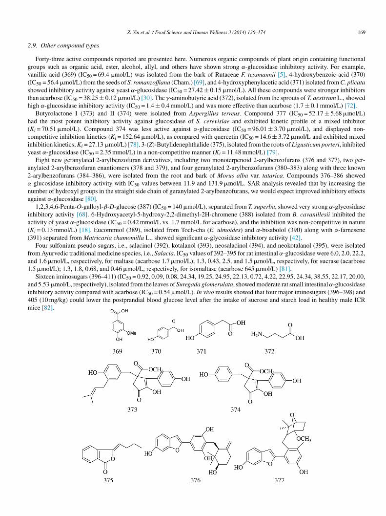
Forty-three active compounds reported are presented here. Numerous organic compounds of plant origin containing functionalroups such as organic acid, ester, alcohol, allyl, and others have shown strong α-glucosidase inhibitory activity. For example,anillic acid (369) (IC50 = 69.4 �mol/L) was isolated from the bark of Rutaceae F. tessmannii [5], 4-hydroxybenzoic acid (370)IC50 = 56.4 �mol/L) from the seeds of S. romanzoffiana (Cham.) [69], and 4-hydroxyphenylacetic acid (371) isolated from C. plicatahowed inhibitory activity against yeast α-glucosidase (IC50 = 27.42 ± 0.15 �mol/L). All these compounds were stronger inhibitorshan acarbose (IC50 = 38.25 ± 0.12 �mol/L) [30]. The γ-aminobutyric acid (372), isolated from the sprouts of T. aestivum L., showedigh α-glucosidase inhibitory activity (IC50 = 1.4 ± 0.4 mmol/L) and was more effective than acarbose (1.7 ± 0.1 mmol/L) [72].
Butyrolactone I (373) and II (374) were isolated from Aspergillus terreus. Compound 377 (IC50 = 52.17 ± 5.68 �mol/L)ad the most potent inhibitory activity against glucosidase of S. cerevisiae and exhibited kinetic profile of a mixed inhibitorKi = 70.51 �mol/L). Compound 374 was less active against α-glucosidase (IC50 = 96.01 ± 3.70 �mol/L), and displayed non-ompetitive inhibition kinetics (Ki = 152.64 �mol/L), as compared with quercetin (IC50 = 14.6 ± 3.72 �mol/L and exhibited mixednhibition kinetics; Ki = 27.13 �mol/L) [78]. 3-(Z)-Butylidenephthalide (375), isolated from the roots of Ligusticum porteri, inhibitedeast α-glucosidase (IC50 = 2.35 mmol/L) in a non-competitive manner (Ki = 11.48 mmol/L) [79].
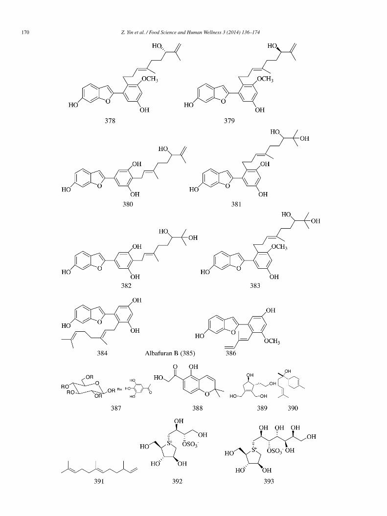
Eight new geranylated 2-arylbenzofuran derivatives, including two monoterpenoid 2-arylbenzofurans (376 and 377), two ger-nylated 2-arylbenzofuran enantiomers (378 and 379), and four geranylated 2-arylbenzofurans (380–383) along with three known-arylbenzofurans (384–386), were isolated from the root and bark of Morus alba var. tatarica. Compounds 376–386 showed-glucosidase inhibitory activity with IC50 values between 11.9 and 131.9 �mol/L. SAR analysis revealed that by increasing theumber of hydroxyl groups in the straight side chain of geranylated 2-arylbenzofurans, we would expect improved inhibitory effectsgainst α-glucosidase [80].
1,2,3,4,6-Penta-O-galloyl-β-D-glucose (387) (IC50 = 140 �mol/L), separated from T. superba, showed very strong α-glycosidasenhibitory activity [68]. 6-Hydroxyacetyl-5-hydroxy-2,2-dimethyl-2H-chromene (388) isolated from B. cavanillesii inhibited thectivity of yeast α-glucosidase (IC50 = 0.42 mmol/L vs. 1.7 mmol/L for acarbose), and the inhibition was non-competitive in natureKi = 0.13 mmol/L) [18]. Eucommiol (389), isolated from Toch-cha (E. ulmoides) and α-bisabolol (390) along with α-farnesene391) separated from Matricaria chamomilla L., showed significant α-glycosidase inhibitory activity [42].
Four sulfonium pseudo-sugars, i.e., salacinol (392), kotalanol (393), neosalacinol (394), and neokotalanol (395), were isolatedrom Ayurvedic traditional medicine species, i.e., Salacia. IC50 values of 392–395 for rat intestinal α-glucosidase were 6.0, 2.0, 22.2,nd 1.6 �mol/L, respectively, for maltase (acarbose 1.7 �mol/L); 1.3, 0.43, 2.5, and 1.5 �mol/L, respectively, for sucrase (acarbose.5 �mol/L); 1.3, 1.8, 0.68, and 0.46 �mol/L, respectively, for isomaltase (acarbose 645 �mol/L) [81].
Sixteen iminosugars (396–411) (IC50 = 0.92, 0.09, 0.08, 24.34, 19.25, 24.95, 22.13, 0.72, 4.22, 22.95, 24.34, 38.55, 22.17, 20.00,nd 5.53 �mol/L, respectively), isolated from the leaves of Suregada glomerulata, showed moderate rat small intestinal α-glucosidasenhibitory activity compared with acarbose (IC50 = 0.54 �mol/L). In vivo results showed that four major iminosugars (396–398) and05 (10 mg/kg) could lower the postprandial blood glucose level after the intake of sucrose and starch load in healthy male ICRice [82].
1
70 Z. Yin et al. / Food Science and Human Wellness 3 (2014) 136–174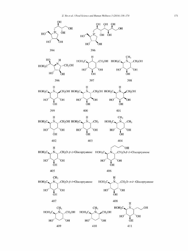
Z. Yin et al. / Food Science and Human Wellness 3 (2014) 136–174 171
1
3
psmtm
ma
dd
C
A
1HT
R
[
[[
[
[
[
[[[[[
[[
[
72 Z. Yin et al. / Food Science and Human Wellness 3 (2014) 136–174
. Conclusion
Natural products are still considered as potential resources for drug discovery and play an important role in drug developmentrograms. Moreover, many medicinal herbs are a rich source of bioactive chemicals that are remarkably free from undesirableide-effects and display powerful pharmacological actions. Compounds having α-glucosidase inhibitory activity are ubiquitous inedicinal plants. A systematic review of literature revealed that 411 compounds belonging to different structural frameworks, i.e.,
erpenes, alkaloids, quinines, flavonoids, phenols, phenylpropanoids, sterides, and compounds with other structural and functionalotifs isolated from medicinal plants, showed potent inhibitory activity toward α-glucosidase.With the future rise in diabetic population worldwide, the search foractive compounds with α-glucosidase inhibitory activity from
edicinal plants has become a very meaningful task. Furthermore, these compounds can be evaluated for potential pharmacologicalctivity against other metabolic diseases.
In short, efforts should be made to optimize the procedure of screening natural products isolated from different plants for theiscovery of new natural herbal antidiabetic drugs. These natural products can be used as alternatives to synthetic oral hypoglycemicrugs with less or even no prominent side effects.
onflict of interest
The authors declare that they have no conflict of interest to disclose.
cknowledgments
This research was supported by Key Project in Science and Technology Agency of Henan Province (Nos. 132102310261 and42102310147) and Natural Science Project in Department of Education of Henan Province (Nos. 13B360981 and 14B360011),enan Province Department of Education Teachers, the backbone of Youth Fund (2013GGJS-220), and Key Project in Science andechnology Agency of Zhengzhou City (No. 120140790).
eferences
[1] Y.P. Li, B. Bai, J. Ye, et al., Reviews on preparation and determination of α-glucosidase inhibitor, Food Sci. 29 (2008) 617–619.[2] W.Q. Du, X.F. Shi, M.Y. Qiu, et al., Progress in treatment of diabetes drugs, Chin. Hosp. Pharm. J. 25 (2005) 67–69.[3] R.J. Playford, C. Pither, R. Gao, et al., Use of the α-glucosidase inhibitor acarbose in patients with ‘Middleton syndrome’: normal gastric anatomy but with
accelerated gastric emptying causing postprandial reactive hypoglycemia and diarrhea, Can. J. Gastroenterol. 27 (2013) 403–404.[4] R. Mata, S. Cristians, S. Escandón-Rivera, et al., Mexican antidiabetic herbs: valuable sources of inhibitors of α-glucosidases, J. Nat. Prod. 76 (2013) 468–483.[5] L.M. Mbaze, H.M.P. Poumale, J.D. Wansi, et al., α-Glucosidase inhibitory pentacyclic triterpenes from the stem bark of Fagara tessmannii (Rutaceae),
Phytochemistry 68 (2007) 591–595.[6] W.Y. Kang, L. Zhang, Y.L. Song, α-Glucosidase inhibitors from Luculia pinciana, China J. Chin. Med. 34 (2009) 406–409.[7] J.G. Luo, L. Ma, L.Y. Kong, New triterpenoid saponins with strong α-glucosidase inhibitory activity from the roots of Gypsophila oldhamiana, Bioorg. Med.
Chem. 16 (2008) 2912–2920.[8] B.Z. Wang, H. Jiang, Y.G. Xia, et al., α-Glucosidase inhibitory constituents from Acanthopanax senticosus harm leaves, Molecules 17 (2012) 6269–6276.[9] R. Kubínová, R. Porízková, A. Navrátilová, et al., Antimicrobial and enzyme inhibitory activities of the constituents of Plectranthus madagascariensis (Pers.)
Benth, J. Enzyme Inhib. Med. Chem. 29 (2014) 1–4.10] P. Prabhakar Reddy, A.K. Tiwari, R. Ranga Rao, et al., New Labdanediterpenes as intestinal alpha-glucosidase inhibitor from antihyperglycemic extract of
Hedychium spicatum (Ham. Ex Smith) rhizomes, Bioorg. Med. Chem. Lett. 19 (2009) 2562–2565.11] M.A. Naveed, N. Riaz, M. Saleema, et al., Longipetalosides A–C, new steroidal saponins from Tribulus longipetalus, Steroids 83 (2014) 45–51.12] J. Hua, J. Qi, B.Y. Yu, Iridoid and phenylpropanoid glycosides from Scrophularia ningpoensis Hemsl. and their α-glucosidaseinhibitory activities, Fitoterapia
93 (2014) 67–73.13] N.P. Thao, B.T. Luyen, B.H. Tai, et al., Rat intestinal sucrase inhibition of constituents from the roots of Rosa rugosa Thunb, Bioorg. Med. Chem. Lett. 24
(2014) 1192–1196.14] B. Jabeen, N. Riaz, M. Saleem, et al., Isolation of natural compounds from Phlomis stewartii showing α-glucosidase inhibitory activity, Phytochemistry 96
(2013) 443–448.15] D. Kumar, V. Shah, R. Ghosh, et al., A new triterpenoidsaponin from Glinus oppositifolius with α-glucosidase inhibitory activity, Nat. Prod. Res. 27 (2013)
624–630.16] Z.W. Wang, J.S. Wang, J. Luo, et al., α-Glucosidase inhibitory triterpenoids from the stem barks of Uncaria laevigata, Fitoterapia 90 (2013) 30–37.17] Z.H. Guo, J. Huang, G.S. Wan, et al., New inhibitors of a-glucosidase in Salacia hainanensis Chun et How, J. Nat. Med. 67 (2013) 844–849.18] S. Escandón-Rivera, M. González-Andrade, R. Bye, et al., α-Glucosidase inhibitors from Brickellia cavanillesii, J. Nat. Prod. 75 (2012) 968–974.19] H. Gao, Y.N. Huang, B. Gao, et al., Inhibitory effect on �-glucosidase by Adhatoda vasica Nees, Food Chem. 108 (2008) 965–972.20] K. Ikeda, M. Takahashi, M. Nishida, et al., Homonojirimycin analogues and their glucosides from Lobelia sessilifolia and Adenophora spp. (Campanulaceae),
Carbohydr. Res. 323 (2000) 73–80.
21] H.X. Yang, R.X. Zhu, Research progress of DNJ, Bull. Ser. 34 (2003) 6–10.22] T.K. Tabopda, J. Ngoupayo, P.K. Awoussong, et al., Triprenylated flavonoids from Dorstenia psilurus and their α-glucosidase inhibition properties, J. Nat. Prod.71 (2008) 2068–2072.23] J. Ma, S.J. Liu, Research progress of DNJ in mulberry twig, Food Sci. Technol. 9 (2006) 112–114.
[
[[
[[
[
[[
[[
[[[[[
[
[
[[[
[[[
[[[
[[
[[
[[
[[
[
[
[[[
[[[
[[[[[
Z. Yin et al. / Food Science and Human Wellness 3 (2014) 136–174 173
24] M. Shibano, K. Kakutani, M. Taniguchi, et al., Antioxidant constituents in the dayflower (Commelina communis L.) and their α-glucosidase-inhibitory activity,J. Nat. Med. 62 (2008) 349–353.
25] T.K. Tabopda, J. Ngoupayo, J. Liu, et al., Bioactive aristolactams from Piper umbellatum, Phytochemistry 69 (2008) 1726–1731.26] J.D. Wansi, J. Wandji, L.M. Meváa, et al., α-Glucosidase inhibitory and antioxidant acridone alkaloids from the stem bark of Oriciopsis glaberrima ENGL.
(Rutaceae), Chem. Pharm. Bull. 54 (2006) 292–296.27] S.D. Kim, α-Glucosidase inhibitor from Buthus martensi Karsch, Food Chem. 136 (2013) 297–300.28] T. Damsud, S. Adisakwattana, P. Phuwapraisirisan, Three new phenylpropanoyl amides from the leaves of Piper sarmentosu and their a-glucosidase inhibitory
activities, Phytochem. Lett. 6 (2013) 350–354.29] C. Uvarani, N. Jaivel, M. Sankaran, et al., Axially chiral biscarbazoles and biological evaluation of the constituents from Murraya koenigii, Fitoterapia 94 (2014)
10–20.30] A. Tabussuma, N. Riaz, M. Saleema, et al., a-Glucosidase inhibitory constituents from Chrozophora plicata, Phytochem. Lett. 6 (2013) 614–619.31] B.Q. Tang, T.T. Yang, W.Q. Yang, et al., Chemical constituents in leaves of Morus atropurpurea and their α-glucosidase activity, Chin. Traditi. Herb. Drugs 44
(2013) 3109–3113.32] W.Y. Kang, L. Zhang, Y.L. Song, α-Glucosidase inhibitors from Rubia cordifolia L., China J. Chin. Med. 34 (2009) 1104–1107.33] K.S. Babu, A.K. Tiwari, P.V. Srinivas, et al., Yeast and mammalian α-glucosidase inhibitory constituents from Himalayan rhubarb Rheum emodi Wall.ex
Meisson, Bioorg. Med. Chem. Lett. 14 (2004) 3841–3845.34] Y.D. Yue, Y.T. Zhang, Z.X. Liu, et al., Xanthone glycosides from swertiabimaculata with α-glucosidase inhibitory activity, Planta Med. 80 (2014) 502–508.35] C.T. Luo, H.H. Zheng, S.S. Mao, et al., Xanthones from Swertia mussotii and the α-glycosidase inhibitory activities, Planta Med. 80 (2014) 201–208.36] H.Y. Zhang, L. Yan, Research progress in anti-microbial of flavonoids, Anti Infect. Pharm. 6 (2009) 92–95.37] N. Jong-Anurakkun, M.R. Bhandari, G. Hong, et al., α-Glucosidase inhibitor from Chinese aloes, Fitoterapia 79 (2008) 456–457.38] S.S. Lee, H.C. Lin, C.K. Chen, Acylated flavonol monorhamnosides, α-glucosidase inhibitors, from Machilus philippinensis, Phytochemistry 69 (2008)
2347–2353.39] E.J. Seo, M.J. Curtis-Long, B.W. Lee, et al., Xanthones from Cudrania tricuspidata displaying potent α-glucosidase inhibition, Bioorg. Med. Chem. Lett. 17
(2007) 6421–6424.40] H. Ichiki, O. Takeda, I. Sakakibara, et al., Inhibitory effects of compounds from Anemarrhenae Rhizoma on α-glucosidase and aldose reductase and its contents
by drying conditions, J. Nat. Med. 61 (2007) 146–153.41] M. Iio, A. Yoshioka, Y. Imayoshi, et al., Effect of flavonoids on α-glucosidase and β-fructosidase from yeast, Agric. Biol. Chem. 48 (1984) 1559–1563.42] A. Kato, Y. Minoshima, J. Yamamoto, et al., Protective effects of dietary chamomile tea on diabetic complications, J. Agric. Food Chem. 56 (2008) 8206–8211.43] J. Watanabe, J. Kawabata, H. Kurihara, et al., Isolation and identification of α-glucosidase inhibitors from Tochu-cha, Biosci. Biotechnol. Biochem. 61 (1997)
177–178.44] W.Y. Kang, J.M. Wang, L. Zhang, α-Glucosidase inhibitors from Forsythia suspense (Thunb) Vahl, China J. Chin. Mater. Med. 35 (2010) 1156–1159.45] D.S. Lee, S.H. Lee, Genistein, a soy isoflavone, is a potent �-glucosidase inhibitor, FEBS Lett. 501 (2001) 84–86.46] S.A. Raoa, P.V. Srinivas, A.K. Tiwari, et al., Isolation, characterization and chemobiological quantification of α-glucosidase enzyme inhibitory and free radical
scavenging constituents from Derris scandens Benth, J. Chromatogr. B 855 (2007) 166–172.47] N. Jong-Anurakkun, M.R. Bhandari, J. Kawabata, α-Glucosidase inhibitors from Devil tree (Alstonia scholaris), Food Chem. 103 (2007) 1319–1323.48] A. Gamberucci, L. Konta, A. Colucci, et al., Green tea flavonols inhibit glucosidase II, Biochem. Pharmacol. 72 (2006) 640–646.49] M.R. Bhandari, N. Jong-Anurakkun, G. Hong, et al., α-Glucosidase and α-amylase inhibitory activities of Nepalese medicinal herb Pakhanbhed (Bergenia
ciliata, Haw.), Food Chem. 106 (2008) 247–252.50] J. Zhao, X.W. Zhou, X.B. Chen, et al., α-Glucosidase inhibitory constituents from Toona sinensis, Chem. Nat. Comp. 45 (2009) 244–246.51] Q. Wu, X.W. Yang, L. Zou, et al., Bioactivity guided isolation of alpha-glucosidase inhibitor from whole herbs of Crossostephium chinense, China J. Chin.
Mater. Med. 34 (2009) 2206–2211.52] J. Saijyo, Y. Suzuki, Y. Okuno, et al., α-Glucosidase Inhibitor from Bergenia ligulata, J. Oleo Sci. 57 (2008) 431–435.53] M.I. Choudhary, I. Baig, M. Nur-e-Alam, et al., New α-glucosidase inhibitors from the Mongolian medicinal plant Ferula mongolica, Helv. Chim. Acta 84
(2001) 2409–2416.54] M. Shahira, M.M. Salama, A new α-glucosidase inhibitor from Achillea fragrantissima (Forssk.) Sch. Bip. growing in Egypt, Nat. Prod. Res. 28 (2014) 812–818.55] J. Feng, X.W. Yang, R.F. Wang, Bio-assay guided isolation and identification of a-glucosidase inhibitors from the leaves of Aquilaria sinensis, Phytochemistry
72 (2011) 242–247.56] T.H. Quang, N.T. Thanh, C.V. Minh, et al., α-Glucosidase inhibitors from the roots of Sophora flavescens, Bull. Korean Chem. Soc. 33 (2012) 1791–1793.57] Y.G. Chen, P. Li, P. Li, et al., α-Glucosidase inhibitory effect and simultaneous quantification of three major flavonoid glycosides in Microctis folium, Molecules
18 (2013) 4221–4232.58] T.X. Wang, Z.H. Yin, W. Zhang, et al., Chemical constituents from Psoralea corylifolia and their antioxidant α-glucosidase inhibitory and antimicrobial activities,
China J. Chin. Mater. Med. 38 (2013) 2328–2333.59] J.D. Wansi, M.C. Lallemand, D.D. Chiozem, et al., α-Glucosidase inhibitory constituents from stem bark of Terminalia superba (Combretaceae), Phytochemistry
68 (2007) 2096–2100.60] L. Zhang, B. Bai, X.H. Liu, et al., α-Glucosidase inhibitors from Chinese Yam (Dioscorea opposita Thunb.), Food Chem. 126 (2011) 203–206.61] Y.H. Chu, S.H. Wu, J.F. Hsieh, Isolation and characterization of α-glucosidase inhibitory constituents from Rhodio lacrenulata, Food Res. Int. 57 (2014) 8–14.62] K. Ma, J.J. Han, L. Bao, et al., Two sarcoviolins with antioxidative and α-glucosidase inhibitory activity from the edible mushroom Sarcodon leucopus collected
in Tibet, J. Nat. Prod. 77 (2014) 942–947.63] H.H.T. Tran, M.C. Nguyen, H.T. Le, et al., Inhibitors of a-glucosidase and a-amylase from Cyperus rotundus, Pharm. Biol. 52 (2014) 74–77.64] C. Wan, T. Yuan, L. Li, et al., Maplexins, new a-glucosidase inhibitors from red maple (Acer rubrum) stems, Bioorg. Med. Chem. Lett. 22 (2012) 597–600.65] T. Yuan, C.P. Wan, H. Ma, et al., New phenolics from the flowers of Punica granatum and their in vitro α-glucosidase inhibitory activities, Planta Med. 79
(2013) 1674–1679.66] H. Gao, Y.N. Huang, B. Gao, et al., α-Glucosidase inhibitory effect by the flower buds of Tussilago farfara L., Food Chem. 106 (2008) 1195–1201.67] H. Matsuuraa, H. Miyazakia, C. Asakawaa, et al., Isolation of α-glucosidase inhibitors from hyssop (Hyssopus officinalis), Phytochemistry 65 (2004) 91–97.
68] Z.Y. Du, R.R. Liu, W.Y. Shao, et al., α-Glucosidase inhibition of natural curcuminoids and curcumin analogs, Eur. J. Med. Chem. 41 (2006) 213–218.69] S.H. Lam, J.M. Chen, C.J. Kang, et al., α-Glucosidase inhibitors from the seeds of Syagrus romanzoffiana, Phytochemistry 69 (2008) 1173–1178.70] Q. Liu, H.J. Hu, P.F. Li, et al., Diterpenoids and phenylethanoid glycosides from the roots of Clerodendrum bungei and their inhibitory effects against angiotensinconverting enzyme and α-glucosidase, Phytochemistry 103 (2014) 196–202.
1
[
[
[
[
[[[
[
[[
[
[
74 Z. Yin et al. / Food Science and Human Wellness 3 (2014) 136–174
71] A. Ishikawa, H. Yamashita, M. Hiemori, et al., Characterization of inhibitors of postprandial hyperglycemia from the leaves of Nerium indicum, J. Nutr. Sci.Vitaminol. (Tokyo) 53 (2007) 166–173.
72] E.Y. Jeong, K.S. Cho, H.S. Lee, α-Amylase and α-glucosidase inhibitors isolated from Triticum aestivum L. sprouts, J. Korean Soc. Appl. Biol. Chem. 55 (2012)47–51.
73] S. El-Mekkawy, M.R. Meselhy, N. Nkobole, et al., Three new a-glucosidase inhibitors from guggul, the oleogum resin of Commiphora wightii, Nat. Prod. Res.27 (2013) 146–154.
74] Y. He, X.B. Wang, B.Y. Fan, et al., Honokioltrimers and dimers via biotransformation catalyzed by Momordica charantia peroxidase: novel and potenta-glucosidase inhibitors, Bioorg. Med. Chem. 22 (2014) 762–771.
75] H. Gao, Y.N. Huang, P.Y. Xu, et al., Inhibitory effect on α-glucosidase by the fruits of Terminalia chebula Retz, Food Chem. 105 (2007) 628–634.76] M.A. Naveed, N. Riaz, M. Saleem, et al., Longipetalosides A–C, new steroidal saponins from Tribulus longipetalus, Steroids 83 (2014) 45–51.77] S. Fatmawati, K. Shimizua, S. Kondo, Ganoderol B: a potent α-glucosidase inhibitor isolated from the fruiting body of Ganoderma lucidum, Phytomedicine 18
(2011) 1053–1055.78] R.T. Dewi, S. Tachibana, A. Darmawan, Effect on a-glucosidase inhibition and antioxidant activities of butyrolactone derivatives from Aspergillus terreus
MC751, Med. Chem. Res. 23 (2014) 454–460.79] F. Brindis, R. Rodríguez, R. Bye, et al., (Z)-3-Butylidenephthalide from Ligusticum porteri, and α-glucosidase inhibitor, J. Nat. Prod. 74 (2010) 314–320.80] Y.Y. Zhang, J.G. Luo, C.X. Wan, et al., Geranylated 2-arylbenzofurans from Morus alba var. tatarica and their α-glucosidase and protein tyrosine phosphatase
1B inhibitory activities, Fitoterapia 92 (2014) 116–126.81] O. Muraoka, T. Morikawa, S. Miyake, et al., Quantitative analysis of neosalacinol and neokotalanol, another two potent α-glucosidase inhibitors from Salacia
species, by LC-MS with ion pair chromatography, J. Nat. Med. 65 (2010) 142–148.82] R.Y. Yan, H.Q. Wang, C. Liu, et al., α-Glucosidase-inhibitory iminosugars from the leaves of Suregada glomerulata, Bioorg. Med. Chem. 21 (2013) 6796–6803.

![Wellness 05[1]](https://static.fdocument.org/doc/165x107/552d707c5503466f768b470c/wellness-051.jpg)