Functional assessment of cell entry and receptor …...10% OptiPrep cushion in PBS (Sigma) and...
26
1 Functional assessment of cell entry and receptor usage for lineage B β- coronaviruses, including 2019-nCoV Michael Letko # and Vincent Munster # Laboratory of Virology, Division of Intramural Research, National Institute of Allergy and Infectious Diseases, National Institutes of Health, Hamilton, MT, 59840, USA # Corresponding authors: Drs. Vincent Munster and Michael Letko Rocky Mountain Laboratories NIAID/NIH 903S 4th Street Hamilton, MT 59840 Tel: (406) 363-9357 Email: [email protected] Email: [email protected] author/funder. All rights reserved. No reuse allowed without permission. The copyright holder for this preprint (which was not peer-reviewed) is the . https://doi.org/10.1101/2020.01.22.915660 doi: bioRxiv preprint
Transcript of Functional assessment of cell entry and receptor …...10% OptiPrep cushion in PBS (Sigma) and...
1
Functional assessment of cell entry and receptor usage for lineage B β-coronaviruses, including 2019-nCoV Michael Letko# and Vincent Munster# Laboratory of Virology, Division of Intramural Research, National Institute of Allergy and
Infectious Diseases, National Institutes of Health, Hamilton, MT, 59840, USA
# Corresponding authors:
Drs. Vincent Munster and Michael Letko
Rocky Mountain Laboratories
NIAID/NIH
903S 4th Street
Hamilton, MT 59840
Tel: (406) 363-9357
Email: [email protected]
Email: [email protected]
author/funder. All rights reserved. No reuse allowed without permission. The copyright holder for this preprint (which was not peer-reviewed) is the. https://doi.org/10.1101/2020.01.22.915660doi: bioRxiv preprint
2
Abstract Over the past 20 years, several coronaviruses have crossed the species barrier into
humans, causing outbreaks of severe, and often fatal, respiratory illness. Since SARS-
CoV was first identified in animal markets, global viromics projects have discovered
thousands of coronavirus sequences in diverse animals and geographic regions.
Unfortunately, there are few tools available to functionally test these novel viruses for their
ability to infect humans, which has severely hampered efforts to predict the next zoonotic
viral outbreak. Here we developed an approach to rapidly screen lineage B
betacoronaviruses, such as SARS-CoV and the recent 2019-nCoV, for receptor usage
and their ability to infect cell types from different species. We show that host protease
processing during viral entry is a significant barrier for several lineage B viruses and that
bypassing this barrier allows several lineage B viruses to enter human cells through an
unknown receptor. We also demonstrate how different lineage B viruses can recombine
to gain entry into human cells and confirm that human ACE2 is the receptor for the
recently emerging 2019-nCoV.
author/funder. All rights reserved. No reuse allowed without permission. The copyright holder for this preprint (which was not peer-reviewed) is the. https://doi.org/10.1101/2020.01.22.915660doi: bioRxiv preprint
3
Introduction
Severe Acute Respiratory Syndrome Coronavirus (SARS-CoV) first emerged in
humans in 2003 after transmitting from animals in open air markets in China1,2. Shortly
thereafter, several genetically related viruses were identified in Chinese Horseshoes bats
(Rhinolophus sinicus)3-7. At the same time, improvements in next generation sequencing
technology lead to a boom of virus discovery, uncovering thousands of novel virus
sequences in wild animal populations around the world. While most of these viruses have
never been found in humans, many are genetically similar to known human viruses within
the betacoronaviruses (β-CoV) genus. The β-CoVs are further divided into four lineages:
lineage B, which includes SARS-CoV and the newly emerging 2019-nCoV, has
approximately 200 published virus sequences whereas lineage C, which includes MERS-
CoV, has over 500 viral sequences.
Every year, additional novel CoV sequences are discovered. However, there is a
massive knowledge gap in the field as very little work is performed after the viral
sequences are published. Therefore, it is unknown whether these novel viruses have the
potential to emerge in human populations.
Current methods for studying novel β-CoVs are technically demanding. Viral
isolation from field samples is rarely successful and reverse genetics recovery of
recombinant virus is labor-intensive, and expensive as synthesis of a single genome can
cost upwards of $15,000. These limitations are prohibitive to studying novel CoVs at the
scale in which they are discovered.
Cell entry is an essential component of cross-species transmission, especially for
the β-CoVs. All CoVs encode a surface glycoprotein, spike, which binds to the host-cell
author/funder. All rights reserved. No reuse allowed without permission. The copyright holder for this preprint (which was not peer-reviewed) is the. https://doi.org/10.1101/2020.01.22.915660doi: bioRxiv preprint
4
receptor and mediates viral entry8. For β-CoVs, a single region of the spike protein called
the receptor binding domain (RBD) mediates the interaction with the host cell receptor.
After binding the receptor, a nearby host protease cleaves the spike, which releases the
spike fusion peptide, facilitating virus entry9-12. Known host receptors for β-CoVs include
angiotensin converting enzyme 2 (ACE2) for SARS-CoV and dipeptidyl peptidase 4
(DPP4) for MERS-CoV13,14.
Structural studies of coronaviruses have shown that the spike RBD is capable of
folding independently from the rest of the spike protein and contains all of the structural
information for host receptor binding15. Additionally, a previous study showed that
replacing the RBD of the lineage B bat virus, Rp3, allowed the virus to enter cells
expressing human ACE2 (hACE2)16. We therefore developed a method to functionally
test the RBDs from novel lineage B β-CoVs in place of the SARS-CoV spike RBD (figure
1). Synthesizing just the RBD of spike is much faster and cost-effective than conventional
pseudotyping methods that rely on synthesis of the full ~4kb spike sequence for novel
CoVs: a process that can take weeks and is cost-prohibitive for large panels of spike
sequences. The short turnaround time for our approach allowed us to test the receptor
usage of all published, unique RBD sequences in lineage B, and also rapidly confirm the
ACE2 receptor usage of the 2019-nCoV spike, which emerged in China in January 2020
as our study was ongoing.
We show that lineage B RBDs divide into functionally distinct clades and that
several previously-unappreciated viruses exhibit compatibility with an unknown receptor
on human cells. We also show that these clades are capable of recombining to impart
human host-cell entry phenotypes, and that, beyond the RDB-receptor interaction, host
author/funder. All rights reserved. No reuse allowed without permission. The copyright holder for this preprint (which was not peer-reviewed) is the. https://doi.org/10.1101/2020.01.22.915660doi: bioRxiv preprint

5
protease processing is another species barrier encountered by lineage B β-CoVs during
cell entry.
author/funder. All rights reserved. No reuse allowed without permission. The copyright holder for this preprint (which was not peer-reviewed) is the. https://doi.org/10.1101/2020.01.22.915660doi: bioRxiv preprint
6
Methods
Cells
293T, A549, BHK, Caco-2, Huh-7.5, PK-15, and Vero cells were maintained in
DMEM (Sigma) supplemented with 10% FBS, penicillin/streptomycin, and L-glutamine.
RhiNi/40.1, AJ-primary, AJi, HypNi, RaKSM-2.5i, RhiLu, and RhiNi cells were maintained
in DMEM/F12 (Gibco) supplemented with 12% FBS, penicillin/streptomycin, non-
essential amino acids, sodium pyruvate and L-glutamine. AJ-primary cells were
immortalized with a lentiviral vector expressing SV40 T-antigen following the
manufacturer’s instructions to generate AJi cells (abm; #G203). RaKSM-2.5 primary cells
have been previously described and were immortalized in this study similar to AJi cells17.
Plasmids
The spike coding sequences for SARS-CoV Urbani, As6526, and BM48-31 were
codon optimized for human cells, appended with a 5’ kozak expression sequence
(GCCACC) and 3’ tetra-glycine linker followed by nucleotides encoding a FLAG tag
sequence (DYKDDDDK). For SARS-CoV spike, silent mutations were introduced around
codons 308 and 519 to form KpnI and XhoI digest sites. For As6526 spike, silent
mutations were introduced around codons 290 and 501 to form AflII and HindIII digest
sites. For BM48-31 spike, silent mutations were introduced around codons 295 and 501
to form AflII and HindIII digest sites. These engineered spike sequences were
synthesized and cloned into pcDNA3.1+ (GenScript).
Spike RBDs were first codon-optimized for human cells, appended with regions of
the target spike backbone to facilitate Infusion cloning and synthesized as double
author/funder. All rights reserved. No reuse allowed without permission. The copyright holder for this preprint (which was not peer-reviewed) is the. https://doi.org/10.1101/2020.01.22.915660doi: bioRxiv preprint
7
stranded DNA fragments (IDT DNA). SARS-CoV, As6526 or BM48-31 engineered spike
plasmids were digested with their corresponding restriction enzymes and gel purified.
RBD inserts were resuspended in water and Infusion cloned into gel purified, digested
spike backbone vectors (Takara).
Human ACE2 (Q9BYF1.2), DPP4 (XM_005246371.3), or APN (NP_001141.2)
were synthesized and cloned into pcDNA3.1+ (GenScript). All DNA constructs were
verified by Sanger sequencing (ACGT Inc.).
Receptor transfection
BHK cells were seeded in black 96-well plates and transfected the next day with
100ng plasmid DNA encoding human ACE2, DPP4, APN or empty vector, using
polyethyleneimine (Polysciences). All downstream experiments were performed 24 hours
post-transfection.
Pseudotype production
Pseudotypes were produced as previously described18. 293T cells were seeded
onto 6-well plates pre-coated with poly-L-lysine (Sigma) and transfected the next day with
1200ng empty plasmid and 400ng of plasmid encoding coronavirus spike or GFP as a no
pseudotype control. Twenty-four hours later, transfected cells were infected with VSVΔG
particles pseudotyped with VSV-g as previously described19. After one hour of incubating
at 37°C, cells were washed three times and incubated in 2mL DMEM supplemented with
2% FBS, penicillin/streptomycin, and L-glutamine for 48 hours. Supernatants were
collected, centrifuged at 500xG for 5 minutes, aliquoted and stored at -80.
author/funder. All rights reserved. No reuse allowed without permission. The copyright holder for this preprint (which was not peer-reviewed) is the. https://doi.org/10.1101/2020.01.22.915660doi: bioRxiv preprint
8
Luciferase-based cell entry assay
Target cells were seeded in black 96-well plates and inoculated, in triplicate, with
equivalent volumes of pseudotype stocks. For trypsin experiments, pseudotype stocks
were diluted 1:1 in DMEM without FBS, trypsin was added to a final concentration of
2500μg/mL and samples were incubated at 37°C for 15 minutes. Samples were then
diluted again 1:1 in cold DMEM supplemented with 2% FBS and added to cells. Inoculated
plates were centrifuged at 1200xG, 4°C, for 1 hour and incubated over night at 37°C.
Approximately 18-20 hours post-infection, Bright-Glo luciferase reagent (Promega) was
added to each well, 1:1, without removing culture media and luciferase was measured.
Relative entry was calculated by normalizing the relative light unit (RLU) for spike
pseudotypes to the plate RLU average for the “no pseudotype control.”
Western blot
Producer cells (spike-transfected 293T) were lysed in 1%SDS, 150mM NaCl,
50mM Tris-HCl, 5mM EDTA and clarified by centrifugation at 14000xG for 20 minutes.
Pseudotyped particles were concentrated from producer cell lysates that were overlaid a
10% OptiPrep cushion in PBS (Sigma) and centrifuged at 20,000× g for 2 hours at 4 °C.
Lysates and concentrated particles were analyzed for FLAG, GAPDH and/or VSV-m
expression on 10% Bis-Tris PAGE gel (ThermoFisher).
Accession numbers
Accession numbers for all spike sequences used here can be found in figure s1b.
author/funder. All rights reserved. No reuse allowed without permission. The copyright holder for this preprint (which was not peer-reviewed) is the. https://doi.org/10.1101/2020.01.22.915660doi: bioRxiv preprint
9
Results
ACE2 entry is lineage B clade 1-specific
The receptor binding domain (RBD) of lineage B β-CoVs is a single, continuous
domain that contains all structural information necessary to interact with the host receptor
(figure 1a, b). We introduced silent mutations in the codon optimized coding sequence for
SARS-CoV to facilitate replacing the SARS RBD with the RBD from other lineage B
viruses (figure 1b). All lineage B sequences were downloaded from online repositories
and parsed to 29 unique RBD sequences, representing all published variations of the
lineage B RBD (supp. fig 1a, b). The panel of 29 RBDs phylogenetically cluster into 3
clades, as previously described5, but these RBD clades were not apparent in phylogenetic
analysis of other viral sequences, such as the RNA-dependent RNA polymerase (supp.
fig 1c). All 29 RBDs were codon optimized, synthesized and cloned in place of the SARS
RBD, effectively generating chimeric spike expression constructs. We then generated
VSV-luciferase reporter particles pseudotyped with the chimeric spikes (figure 1c). We
chose VSV over lentiviruses as our pseudotype platform because a lentiviral pseudotypes
have failed to accurately reflect viral entry with novel bat coronavirus spike protein7. All
constructs exhibited similar levels of expression in producer cells and incorporation into
VSV pseudotypes, except the chimera with BM48-31 which displayed somewhat reduced
expression compared to WT SARS spike (figure 1d). We then infected BHK cells
expressing the receptor for SARS-CoV or empty vector (figure 1e) and observed only
clade 1, which includes SARS-CoV, WIV1 and SHC014, could enter cells transfected with
human ACE2 (figure 1e).
author/funder. All rights reserved. No reuse allowed without permission. The copyright holder for this preprint (which was not peer-reviewed) is the. https://doi.org/10.1101/2020.01.22.915660doi: bioRxiv preprint
10
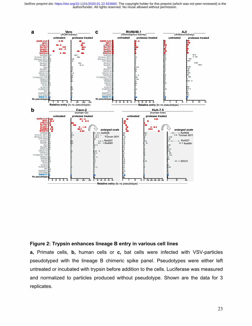
Protease enhances clade 2 entry
After binding the host receptor, host-cell protease cleaves spike, releasing the
fusion peptide and allowing for host cell entry 20. Previous studies have shown that
absence of the host protease or incompatibility between the host protease and viral spike
can block viral entry 21-24. To circumvent host-cell protease incompatibility or absence, we
treated our lineage B pseudotype panel and infected a wide variety of cell types from
different host species (figure 2, supp. fig. 2). In the absence of exogenous protease, only
clade 1 infected cells from African green monkey kidney, human gastrointestinal tract,
human liver, and porcine kidney, in agreement with previous studies (figure 2; supp. fig.
2a, b). Surprisingly, exogenous protease enhanced entry of a subset of clade 2 spike
chimeras in nonhuman primate, bat and human cells (figure 2). Importantly, VSV-g
pseudotyped particles were able to produce luciferase signal in all cell lines tested in this
study (supp. fig. 2c).
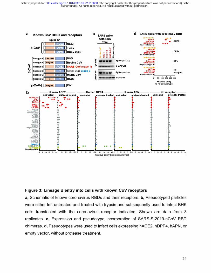
Clade 2 entry is receptor-dependent
We next tested human variants of known β-CoV receptors for their ability to
mediate cell entry of clade 2 and 3 spike chimeras. We also tested human
aminopeptidase N (APN), a receptor for alphacoronaviruses, which have been shown to
utilize either human ACE2 or human APN for cell entry (figure 3a). Protease treatment
only enhanced entry of clade 1 RBDs on cells expressing human ACE2, but not human
DPP4 or APN. No entry was observed with clade 2 or 3 spikes, regardless of receptor or
protease addition. Human dipeptidyl peptidase IV (DPP4), the receptor for the lineage C
β-CoVs, MERS-CoV, only mediated entry of MERS-CoV (figure 2b, middle panels).
author/funder. All rights reserved. No reuse allowed without permission. The copyright holder for this preprint (which was not peer-reviewed) is the. https://doi.org/10.1101/2020.01.22.915660doi: bioRxiv preprint
11
Importantly, in the absence of receptor, no entry was observed for any of the
pseudotypes, suggesting that protease-mediated entry is receptor-dependent (figure 2b,
right panels).
Receptor usage of 2019-nCoV
While our study was ongoing, a novel lineage B virus tentatively named 2019-
nCoV was identified as the cause of a pneumonia outbreak in Hubei, China. Once the
sequence was publicly available, we synthesized, cloned and tested the RBD from 2019-
nCoV in our assay with human variants of known coronavirus receptors. The chimeric
SARS-2019-nCoV spike protein expressed and was incorporated into particles similarly
to other clade 1 chimeric spikes (figure 3c). The 2019-nCoV RBD was capable of entering
cells expressing human ACE2, but not any of the other receptors tested (figure 3d; s3).
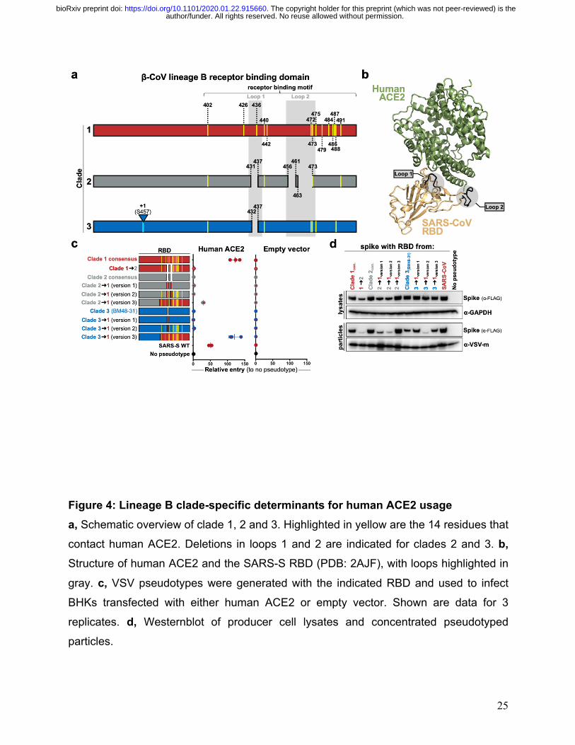
Clade determinants for ACE2 usage
Consensus sequences of the three lineage B clades showed several key
differences between these groups. Only clade 1 RBDs contain all 14 residues that have
been shown through crystallography, to interact with human ACE2 (figure 4a; s4). The
majority of these residues are absent from clades 2 and 3, which contain additional
deletions in surface exposed loops that cluster at the interface with ACE2 (figure 4 a, b).
We generated a series of clade consensus RBD variants to determine the minimum
number of mutations needed to impart ACE2 function on clade 2 and 3 RBDs (figure 4c).
Introducing the two loop deletions from clade 1 in clade 2 results in a reduced spike
expression, impaired pseudotype incorporation and loss of cell entry (figure 4c, d).
author/funder. All rights reserved. No reuse allowed without permission. The copyright holder for this preprint (which was not peer-reviewed) is the. https://doi.org/10.1101/2020.01.22.915660doi: bioRxiv preprint
12
Restoring these loops in clade 2 and 3 from the loops found in clade 1 did not enhance
entry with ACE2 (figure 4c; 2→1 and 3→1 version 1). Introducing all 14 ACE2 contact
points in clade 2 or 3 also failed to restore ACE2 entry (figure 4c; 2→1 and 3→1 version
2). Only replacing all 14 contact points and the surrounding amino acids (also known as
the receptor binding motif, RBM) lead to increased ACE2 entry with clade 2 and 3 RBDs
(figure 4c; 2→1 version 3 = clade 2 residues 322-400 + clade 1 residues 400-501; 3→1
version 3 = clade 3 residues 322-385 + clade 1 residues 386-501). Taken together, these
results show that the entire RBM from clade 1 is needed for ACE2 entry.
Full-spike and RBD chimeras are comparable
We next synthesized full-length clade 2 and 3 spikes to compare to our RBD
chimeras. We selected the clade 2 spike, As6526, because it consistently gave strong
entry signal in human cells following protease-treatment (figure 2b) as well as BM48-31,
the only clade 3 spike in our panel. As we did for SARS-CoV spike, clade 2 and 3 spikes
were codon optimized, FLAG-tagged and silent mutations were introduced to facilitate
replacing their RBD with the consensus RBD from clade 1 (figure 5a). All chimeric
constructs expressed similarly, with the exception for the SARS-BM48-31 RBD chimera,
which exhibited reduced expression and incorporation (figure 5b). Protease treatment
enhanced entry of both the As6526 clade 2 RBD chimera and full-length spike entry into
Huh cells (figure 5c). Protease treatment had no effect on either the BM48-31 clade 3
chimera or full-length spike (figure 5c). Taken together, these findings show that SARS-
lineage B RBD chimeras reflect the entry phenotype of full-length lineage B spikes.
author/funder. All rights reserved. No reuse allowed without permission. The copyright holder for this preprint (which was not peer-reviewed) is the. https://doi.org/10.1101/2020.01.22.915660doi: bioRxiv preprint
13
Finally, we tested if receptor-binding and protease processing are coupled. We
replaced the RBD of full-length clade 2 and 3 spike with the consensus RBD from clade
1 and tested pseudotypes on cells expressing the clade 1 receptor. The clade 1
consensus RBD efficiently facilitated entry of both As6526 and BM48-31 spike only
following protease treatment. These findings show that even though BHK-hACE2 cells
support full-length clade 1 spike entry, just having the RBD from a clade 1 virus is
insufficient to mediate entry. As seen in our previous experiments, protease treatment did
not enhance pseudotype entry in the absence of host receptor (figure 5).
author/funder. All rights reserved. No reuse allowed without permission. The copyright holder for this preprint (which was not peer-reviewed) is the. https://doi.org/10.1101/2020.01.22.915660doi: bioRxiv preprint
14
Discussion
Despite significant advances in next generation sequencing technologies, which
have facilitated the discovery of thousands of novel animal-derived viruses, tools for
downstream functional assessment of these novel sequences are lacking. To gain
traction on this ever-growing problem, we took a reductionist approach to coronavirus
entry and developed a scalable, BSL2-compatible method for testing only the minimal
region of the virus essential for interacting with the host receptor (figure 1, figure s1a).
Because most of these viruses have never been isolated, we resorted to synthetic biology
and molecular engineering to reduce the burden of gene synthesis to just a small
fragment. Thus, the cost and synthesis production time for testing several spikes for entry
in our system is dramatically reduced (figure s1d). In theory, this approach to functional
viromics should be applicable to a wide variety of virus-host proteins and interactions.
Coronavirus entry is a multi-step process involving multiple, distinct domains in
spike that mediate virus attachment to the cell surface, receptor engagement, protease
processing and membrane fusion8. While the RBD:receptor interaction is most studied in
this process, recent studies have highlighted the major role host protease processing
plays as a species barrier22,25-27. Lineage C coronaviruses include MERS-CoV as well as
distantly related viruses such as HKU4, HKU5 and PDF-2180 28,29. Studies have shown
that HKU4 can bind human DPP4 but requires addition of exogenous trypsin to facilitate
cell entry and that HKU5 and PDF-2180 spikes can enter human cells through an
unidentified receptor with protease treatment22,26. Analogous to these earlier studies of
lineage C CoVs, we observed protease-enhanced entry of lineage B CoVs (figure 2, 3,
5). While it has been shown that host proteases cleave spike, allowing for downstream
author/funder. All rights reserved. No reuse allowed without permission. The copyright holder for this preprint (which was not peer-reviewed) is the. https://doi.org/10.1101/2020.01.22.915660doi: bioRxiv preprint
15
membrane fusion, other evidence suggests that protease may act on the receptor as well
to activate it30. Addition of protease during the course of SARS-CoV infection facilitated
entry in cells with low-expression of ACE2 that is normally insufficient to support virus
entry30. Indeed, we saw evidence of residual trypsin activity on the cells after infection in
our studies as the cell monolayer was loose compared to the untreated condition.
Similarly, Menachery et al. also observed cell rounding during their trypsin infections26.
Therefore, further studies are needed to assess where trypsin is enhancing entry of
coronaviruses: at the level of spike, the receptor, or both.
In the absence of exogenous protease, only clade 1 RBDs entered nonhuman
primate, human and porcine cell lines (figure 2a, b, s2a, b). These findings are in strong
agreement with previous studies that have either isolated virus (WIV1) or rescued
recombinant chimeric viruses (SHC014, Rs4231, Rs7237)5,7,31. However, with trypsin, a
subset of genetically-similar clade 2 RBDs gained entry in these cells, suggesting their
barrier is at the level of protease processing (figure 2a, b). The other spike from clade 2
and 3 did not enter the cells we tested, regardless of protease addition, suggesting an
absent or incompatible receptor. Surprisingly, the protease-dependent entry phenotype
was consistent in the reverse spike chimeras in which we replaced the RBD in clade 2 or
3 spike with a clade 1 RBD (figure 5d), suggesting that either the protease site between
S1/S2 is not compatible with the chimeric spike backbone or the protease is not
expressed in these cells (figure s5). Because clade 1 spikes enter cells expressing human
ACE2 without addition of protease but clade2-clade1 chimeras require protease, our data
suggests the spike protease cleavage site is adapted to the protease environment of the
receptor-bound RBD (figure s5).
author/funder. All rights reserved. No reuse allowed without permission. The copyright holder for this preprint (which was not peer-reviewed) is the. https://doi.org/10.1101/2020.01.22.915660doi: bioRxiv preprint
16
None of the spike pseudotypes efficiently entered Rhinolophus cells, which has been
observed in previous studies using these cells32,33 (figure 2c). Surprisingly, AJi cells were
selectively permissive for only clade 2 entry following protease treatment, which suggests
that clade 2 RBDs interact with a receptor that is distinct from clade 1 (figure 2c).
Our results show that, despite all being classified as the same virus species, most
lineage B β-CoVs do not use currently known coronavirus receptors (figure 1e, 3a, b).
Critically, we did not observe any pseudotype entry in the presence of protease and
absence of receptor, suggesting that lineage B cell entry is still receptor-dependent
following protease treatment (figure 3b). While our study was ongoing, a novel lineage B
β-CoV was identified as the etiological agent behind an outbreak of pneumonia in Wuhan,
Hubei, China (2019-nCoV). The RBD for 2019-nCoV has residues and motifs found in all
3 clades but forms a distinct clade, so we tested it for receptor usage and observed entry
only with human ACE2 but not other known coronavirus receptors (figure 3d).
Interestingly, within the backbone of SARS-CoV spike, cell entry of 2019-nCoV was
similar to the other clade 1 spikes tested, including SARS-CoV. These finding suggests
2019-nCoV is capable of using human ACE2 as efficiently as SARS-CoV, which may help
explain the human-to-human transmissibility of this virus. More studies are needed with
the full spike sequence and, ideally, a viral isolate.
The receptor binding motif (RBM) is a small region in the C-terminal half of the RBD
and contains all the residues that interface with the host receptor (figure 3a)15. The 14
contact points in the co-structure of the SARS-RBD bound to human ACE2 are largely
absent from clade 2 and 3 RBDs, which also contain deletions compared to clade 1 RBMs
(figure 4a, b, s4a). Simply mutating clade 2 and 3 to have the 14 contact points was
author/funder. All rights reserved. No reuse allowed without permission. The copyright holder for this preprint (which was not peer-reviewed) is the. https://doi.org/10.1101/2020.01.22.915660doi: bioRxiv preprint
17
insufficient to impart human ACE2 usage (figure 4c). This is likely because the non-
contact residues in the RBM play a supportive and structural role for these contact points,
and indeed, these non-contact residues are different between the clades (figure s4).
In contrast to changing individual amino acids, our chimeric RBD constructs show that
clade 2 and 3 RBD containing the clade 1 RBM are compatible with human ACE2.
Coronaviruses frequently undergo recombination, gaining large swaths of genetic
material at once 34,35. Taken together with our data, it is possible that recombination with
a clade 1 virus will impart compatibility with human ACE2. Interestingly, the 2019-nCoV
RBD forms a clade that is distinct from the other 3 clades (figure s1c). However, the 2019-
nCoV RBD contains most of the contact points with human ACE2 that are found in clade
1 as well as some amino acid variations that are unique to clade 2 and 3 (figure s4b).
Taken together with our receptor assay results, it may be possible that 2019-nCoV arose
from recombination between clade 1 and the other clades.
As we saw with the SARS-As6526 RBD (clade 2) spike chimera, full length As6526
spike entered cells following protease treatment, but BM48-31 (clade 3) spike did not
(figure 2, 5c). These data show that the chimeric spikes generally reproduce the entry
phenotypes of full-length spikes. Notably, the full length As6526 spike did not enter cells
as efficiently as the SARS-As6526 chimera, suggesting that other human-cell adaptations
are likely needed in As6526 spike.
The capacity to predict the zoonotic potential of newly detected viruses has been
severely hindered by a lack of functional data for these novel animal virus sequences.
Here, we have developed a rapid and cost-effective platform to functionally test large
groups of related viruses for zoonotic potential. We found that several other lineage B
author/funder. All rights reserved. No reuse allowed without permission. The copyright holder for this preprint (which was not peer-reviewed) is the. https://doi.org/10.1101/2020.01.22.915660doi: bioRxiv preprint
18
coronaviruses are capable of entering human cells through an unknown receptor and that
lineage B spike proteins can recombine to gain entry with a known host-receptor. Taken
together with the latest outbreak of 2019-nCoV in humans, these findings underscore the
importance of continued surveillance of coronaviruses at the sequence and functional
levels in order to better prepare for the next emerging virus.
Acknowledgements
We would like to thank Dr. Anthony Schountz for generously sharing primary artibeus
cells, as well as Dr. Marcel Muller for providing the Rhinolophus cells used in this study.
We would also like to acknowledge Wu,F., Zhao,S., Yu,B., Chen,Y.-M., Wang,W., Hu,Y.,
Song,Z.-G., Tao,Z.-W., Tian,J.-H., Pei,Y.-Y., Yuan,M.L., Zhang,Y.-L., Dai,F.-H., Liu,Y.,
Wang,Q.-M., Zheng,J.-J., Xu,L., Holmes,E.C. and Zhang,Y.-Z, the Wuhan Institute of
Virology, Chinese Academy of Medical Sciences & Peking Union Medical College,
Chinese Center for Disease Control and Prevention, and Wuhan Jinyintan Hospitar for
providing the genome sequence for 2019-nCoV and other essential outbreak information
to the scientific community. This work was supported by the Intramural Research Program
of the National Institute of Allergy and Infectious Diseases.
Declarations of Interests
The authors declare no competing interests.
author/funder. All rights reserved. No reuse allowed without permission. The copyright holder for this preprint (which was not peer-reviewed) is the. https://doi.org/10.1101/2020.01.22.915660doi: bioRxiv preprint
19
References 1 Wang, M. et al. SARS-CoV infection in a restaurant from palm civet. Emerg Infect Dis 11,
1860-1865, doi:10.3201/eid1112.041293 (2005). 2 Kuiken, T. et al. Newly discovered coronavirus as the primary cause of severe acute
respiratory syndrome. Lancet 362, 263-270, doi:10.1016/S0140-6736(03)13967-0 (2003).
3 Li, W. et al. Bats are natural reservoirs of SARS-like coronaviruses. Science 310, 676-679, doi:10.1126/science.1118391 (2005).
4 Ge, X. Y. et al. Isolation and characterization of a bat SARS-like coronavirus that uses the ACE2 receptor. Nature 503, 535-538, doi:10.1038/nature12711 (2013).
5 Hu, B. et al. Discovery of a rich gene pool of bat SARS-related coronaviruses provides new insights into the origin of SARS coronavirus. PLoS Pathog 13, e1006698, doi:10.1371/journal.ppat.1006698 (2017).
6 Lau, S. K. et al. Severe acute respiratory syndrome coronavirus-like virus in Chinese horseshoe bats. Proc Natl Acad Sci U S A 102, 14040-14045, doi:10.1073/pnas.0506735102 (2005).
7 Menachery, V. D. et al. A SARS-like cluster of circulating bat coronaviruses shows potential for human emergence. Nat Med 21, 1508-1513, doi:10.1038/nm.3985 (2015).
8 Li, F. Structure, Function, and Evolution of Coronavirus Spike Proteins. Annu Rev Virol 3, 237-261, doi:10.1146/annurev-virology-110615-042301 (2016).
9 Simmons, G., Zmora, P., Gierer, S., Heurich, A. & Pohlmann, S. Proteolytic activation of the SARS-coronavirus spike protein: cutting enzymes at the cutting edge of antiviral research. Antiviral Res 100, 605-614, doi:10.1016/j.antiviral.2013.09.028 (2013).
10 Matsuyama, S. et al. Efficient activation of the severe acute respiratory syndrome coronavirus spike protein by the transmembrane protease TMPRSS2. J Virol 84, 12658-12664, doi:10.1128/JVI.01542-10 (2010).
11 Bertram, S. et al. Cleavage and activation of the severe acute respiratory syndrome coronavirus spike protein by human airway trypsin-like protease. J Virol 85, 13363-13372, doi:10.1128/JVI.05300-11 (2011).
12 Belouzard, S., Chu, V. C. & Whittaker, G. R. Activation of the SARS coronavirus spike protein via sequential proteolytic cleavage at two distinct sites. Proc Natl Acad Sci U S A 106, 5871-5876, doi:10.1073/pnas.0809524106 (2009).
13 Raj, V. S. et al. Dipeptidyl peptidase 4 is a functional receptor for the emerging human coronavirus-EMC. Nature 495, 251-254, doi:10.1038/nature12005 (2013).
14 Li, W. et al. Angiotensin-converting enzyme 2 is a functional receptor for the SARS coronavirus. Nature 426, 450-454, doi:10.1038/nature02145 (2003).
15 Li, F., Li, W., Farzan, M. & Harrison, S. C. Structure of SARS coronavirus spike receptor-binding domain complexed with receptor. Science 309, 1864-1868, doi:10.1126/science.1116480 (2005).
16 Becker, M. M. et al. Synthetic recombinant bat SARS-like coronavirus is infectious in cultured cells and in mice. Proc Natl Acad Sci U S A 105, 19944-19949, doi:10.1073/pnas.0808116105 (2008).
author/funder. All rights reserved. No reuse allowed without permission. The copyright holder for this preprint (which was not peer-reviewed) is the. https://doi.org/10.1101/2020.01.22.915660doi: bioRxiv preprint
20
17 Seifert, S. N. et al. Rousettus aegyptiacus Bats Do Not Support Productive Nipah Virus Replication. J Infect Dis, doi:10.1093/infdis/jiz429 (2019).
18 Letko, M. et al. Adaptive Evolution of MERS-CoV to Species Variation in DPP4. Cell Rep 24, 1730-1737, doi:10.1016/j.celrep.2018.07.045 (2018).
19 Takada, A. et al. A system for functional analysis of Ebola virus glycoprotein. Proc Natl Acad Sci U S A 94, 14764-14769, doi:10.1073/pnas.94.26.14764 (1997).
20 Millet, J. K. & Whittaker, G. R. Host cell proteases: Critical determinants of coronavirus tropism and pathogenesis. Virus Res 202, 120-134, doi:10.1016/j.virusres.2014.11.021 (2015).
21 Menachery, V. D. et al. Trypsin treatment unlocks barrier for zoonotic coronaviruses infection. bioRxiv (2019).
22 Yang, Y. et al. Receptor usage and cell entry of bat coronavirus HKU4 provide insight into bat-to-human transmission of MERS coronavirus. Proc Natl Acad Sci U S A 111, 12516-12521, doi:10.1073/pnas.1405889111 (2014).
23 Matsuyama, S., Ujike, M., Morikawa, S., Tashiro, M. & Taguchi, F. Protease-mediated enhancement of severe acute respiratory syndrome coronavirus infection. Proc Natl Acad Sci U S A 102, 12543-12547, doi:10.1073/pnas.0503203102 (2005).
24 Kam, Y. W. et al. Cleavage of the SARS coronavirus spike glycoprotein by airway proteases enhances virus entry into human bronchial epithelial cells in vitro. PLoS One 4, e7870, doi:10.1371/journal.pone.0007870 (2009).
25 Yang, Y. et al. Two Mutations Were Critical for Bat-to-Human Transmission of Middle East Respiratory Syndrome Coronavirus. J Virol 89, 9119-9123, doi:10.1128/JVI.01279-15 (2015).
26 Menachery, V. D. et al. Trypsin treatment unlocks barrier for zoonotic bat coronaviruses infection. J Virol, doi:10.1128/JVI.01774-19 (2019).
27 Beniac, D. R., deVarennes, S. L., Andonov, A., He, R. & Booth, T. F. Conformational reorganization of the SARS coronavirus spike following receptor binding: implications for membrane fusion. PLoS One 2, e1082, doi:10.1371/journal.pone.0001082 (2007).
28 Woo, P. C. et al. Molecular diversity of coronaviruses in bats. Virology 351, 180-187, doi:10.1016/j.virol.2006.02.041 (2006).
29 Anthony, S. J. et al. Further Evidence for Bats as the Evolutionary Source of Middle East Respiratory Syndrome Coronavirus. mBio 8, doi:10.1128/mBio.00373-17 (2017).
30 Shulla, A. et al. A transmembrane serine protease is linked to the severe acute respiratory syndrome coronavirus receptor and activates virus entry. J Virol 85, 873-882, doi:10.1128/JVI.02062-10 (2011).
31 Adney, D. R. et al. Replication and shedding of MERS-CoV in upper respiratory tract of inoculated dromedary camels. Emerg Infect Dis 20, 1999-2005, doi:10.3201/eid2012.141280 (2014).
32 Muth, D. et al. Attenuation of replication by a 29 nucleotide deletion in SARS-coronavirus acquired during the early stages of human-to-human transmission. Sci Rep 8, 15177, doi:10.1038/s41598-018-33487-8 (2018).
33 Hoffmann, M. et al. Differential sensitivity of bat cells to infection by enveloped RNA viruses: coronaviruses, paramyxoviruses, filoviruses, and influenza viruses. PLoS One 8, e72942, doi:10.1371/journal.pone.0072942 (2013).
author/funder. All rights reserved. No reuse allowed without permission. The copyright holder for this preprint (which was not peer-reviewed) is the. https://doi.org/10.1101/2020.01.22.915660doi: bioRxiv preprint
21
34 Liao, C. L. & Lai, M. M. RNA recombination in a coronavirus: recombination between viral genomic RNA and transfected RNA fragments. J Virol 66, 6117-6124 (1992).
35 Graham, R. L. & Baric, R. S. Recombination, reservoirs, and the modular spike: mechanisms of coronavirus cross-species transmission. J Virol 84, 3134-3146, doi:10.1128/JVI.01394-09 (2010).
author/funder. All rights reserved. No reuse allowed without permission. The copyright holder for this preprint (which was not peer-reviewed) is the. https://doi.org/10.1101/2020.01.22.915660doi: bioRxiv preprint
22
Figures
Figure 1: β-Coronavirus lineage B entry with human ACE2 is clade-specific a, β-Coronaviruses, including SARS-CoV, interact with the host cell receptor via the
Receptor Binding Domain (RBD) in spike (PDB: 5X5B, 2AJF) . b, SARS-CoV spike was
engineered with silent mutations to facilitate cloning novel RBD sequences in place of the
SARS spike RBD. SARS spike amino acid numbers are indicated for silent cloning sites
and the RBD in grey and orange, respectively. c, outline of experimental workflow. d, Westernblot of producer cell lysates and concentrated reporter particles. e, BHK cells
were transfected with either human ACE2 or empty vector, subsequently infected with
VSV-reporter particles pseudotyped with chimeric spikes, luciferase was measured and
normalized to no pseudotype as a readout for cell entry. Shown are the data for 3
replicates.
author/funder. All rights reserved. No reuse allowed without permission. The copyright holder for this preprint (which was not peer-reviewed) is the. https://doi.org/10.1101/2020.01.22.915660doi: bioRxiv preprint
23
Figure 2: Trypsin enhances lineage B entry in various cell lines
a, Primate cells, b, human cells or c, bat cells were infected with VSV-particles
pseudotyped with the lineage B chimeric spike panel. Pseudotypes were either left
untreated or incubated with trypsin before addition to the cells. Luciferase was measured
and normalized to particles produced without pseudotype. Shown are the data for 3
replicates.
author/funder. All rights reserved. No reuse allowed without permission. The copyright holder for this preprint (which was not peer-reviewed) is the. https://doi.org/10.1101/2020.01.22.915660doi: bioRxiv preprint
24
Figure 3: Lineage B entry into cells with known CoV receptors
a, Schematic of known coronavirus RBDs and their receptors. b, Pseudotyped particles
were either left untreated and treated with trypsin and subsequently used to infect BHK
cells transfected with the coronavirus receptor indicated. Shown are data from 3
replicates. c, Expression and pseudotype incorporation of SARS-S-2019-nCoV RBD
chimeras. d, Pseudotypes were used to infect cells expressing hACE2, hDPP4, hAPN, or
empty vector, without protease treatment.
author/funder. All rights reserved. No reuse allowed without permission. The copyright holder for this preprint (which was not peer-reviewed) is the. https://doi.org/10.1101/2020.01.22.915660doi: bioRxiv preprint
25
Figure 4: Lineage B clade-specific determinants for human ACE2 usage
a, Schematic overview of clade 1, 2 and 3. Highlighted in yellow are the 14 residues that
contact human ACE2. Deletions in loops 1 and 2 are indicated for clades 2 and 3. b, Structure of human ACE2 and the SARS-S RBD (PDB: 2AJF), with loops highlighted in
gray. c, VSV pseudotypes were generated with the indicated RBD and used to infect
BHKs transfected with either human ACE2 or empty vector. Shown are data for 3
replicates. d, Westernblot of producer cell lysates and concentrated pseudotyped
particles.
author/funder. All rights reserved. No reuse allowed without permission. The copyright holder for this preprint (which was not peer-reviewed) is the. https://doi.org/10.1101/2020.01.22.915660doi: bioRxiv preprint
26
Figure 5: Comparison of chimeric and full-length clade 2 and 3 spikes a, Full length spike sequences from As6526 (clade 2) and BM48-31 (clade 3) were codon
optimized, FLAG tagged and synthesized. Silent mutations flanking the RBD facilitated
replacing the native RBD with the clade 1 consensus RBD. b, Westernblot of producer
cell lysates and concentrated pseudotypes particles. c, Pseudotypes with indicated spike
constructs were left untreated or treated with trypsin and subsequently used to infect Huh-
7.5 cells. Shown are data for 3 replicates. d, Pseudotypes with indicated spike constructs
were left untreated or treated with trypsin and subsequently used to infect BHK cells
transfected with human ACE2. Shown are data for 3 replicates.
author/funder. All rights reserved. No reuse allowed without permission. The copyright holder for this preprint (which was not peer-reviewed) is the. https://doi.org/10.1101/2020.01.22.915660doi: bioRxiv preprint