For Peer Review · For Peer Review Professor Ian C. McGrath Editor-in-Chief British Journal of...
53
For Peer Review FM19G11 reverses endothelial dysfunction in rat and human arteries through stimulation of the PI3K/Akt/eNOS pathway, independently of mTOR/HIF-1α activation Journal: British Journal of Pharmacology Manuscript ID: 2014-BJP-0951-RP.R2 Manuscript Type: Research Paper Date Submitted by the Author: n/a Complete List of Authors: El Assar, Mariam; Hospital Universitario de Getafe, Fundación para la Investigación Biomédica Sánchez-Puelles, José; Centro de Investigaciones Biológicas, Consejo Superior de Investigaciones Científicas, 2. Molecular Pharmacology Group, Cellular and Molecular Medicine Dept Royo, Inmaculada; Centro de Investigaciones Biológicas, Consejo Superior de Investigaciones Científicas, 2. Molecular Pharmacology Group, Cellular and Molecular Medicine Dept López-Hernández, Eva; Centro de Investigaciones Biológicas, Consejo Superior de Investigaciones Científicas, 2. Molecular Pharmacology Group, Cellular and Molecular Medicine Dept Sanchez-Ferrer, Alberto; Hospital Universitario de Getafe, Fundación para la Investigación Biomédica Aceña, José; Universidad del País Vasco UPV/EHU, 3. Departamento de Química Orgánica Rodríguez-Mañas, Leocadio; Hospital Universitario de Getafe, Fundación para la Investigación Biomédica Angulo, Javier; Hospital Ramón y Cajal, Investigación Major area of pharmacology: Vascular pharmacology Cross-cutting area: Intracellular signaling, Diabetes Additional area(s): Endothelium, Kinases, Nitric oxide, Post-translational modification, Nitric oxide synthase, Translational Pharmacology British Pharmacological Society British Journal of Pharmacology
Transcript of For Peer Review · For Peer Review Professor Ian C. McGrath Editor-in-Chief British Journal of...
For Peer Review
FM19G11 reverses endothelial dysfunction in rat and human
arteries through stimulation of the PI3K/Akt/eNOS pathway, independently of mTOR/HIF-1α activation
Journal: British Journal of Pharmacology
Manuscript ID: 2014-BJP-0951-RP.R2
Manuscript Type: Research Paper
Date Submitted by the Author: n/a
Complete List of Authors: El Assar, Mariam; Hospital Universitario de Getafe, Fundación para la Investigación Biomédica Sánchez-Puelles, José; Centro de Investigaciones Biológicas, Consejo Superior de Investigaciones Científicas, 2. Molecular Pharmacology Group, Cellular and Molecular Medicine Dept Royo, Inmaculada; Centro de Investigaciones Biológicas, Consejo Superior de Investigaciones Científicas, 2. Molecular Pharmacology Group, Cellular and Molecular Medicine Dept López-Hernández, Eva; Centro de Investigaciones Biológicas, Consejo Superior de Investigaciones Científicas, 2. Molecular Pharmacology Group, Cellular and Molecular Medicine Dept Sanchez-Ferrer, Alberto; Hospital Universitario de Getafe, Fundación para la Investigación Biomédica Aceña, José; Universidad del País Vasco UPV/EHU, 3. Departamento de Química Orgánica Rodríguez-Mañas, Leocadio; Hospital Universitario de Getafe, Fundación para la Investigación Biomédica Angulo, Javier; Hospital Ramón y Cajal, Investigación
Major area of pharmacology: Vascular pharmacology
Cross-cutting area: Intracellular signaling, Diabetes
Additional area(s): Endothelium, Kinases, Nitric oxide, Post-translational modification, Nitric oxide synthase, Translational Pharmacology
British Pharmacological Society
British Journal of Pharmacology
For Peer Review
Professor Ian C. McGrath
Editor-in-Chief
British Journal of Pharmacology
October 23rd, 2014
Dear Editor,
We are submitting the revised version of the manuscript 2014-BJP-0951-RP entitled
“FM19G11 reverses endothelial dysfunction in rat and human arteries through
stimulation of the PI3K/Akt/eNOS pathway, independently of mTOR/HIF-1α
activation” by El Assar, Royo, López-Hernández, Sánchez-Ferrer, Aceña, Rodríguez-
Mañas, Angulo, and myself to be considered for publication in the British Journal of
Pharmacology.
After having fulfilled most of the questions raised by the additional reviewer in the
previous revision, we have now generated a point by point response to the new
comments raised by the Editor and the reviewer and modified the manuscript
accordingly (changes are highlighted). We think we have reasonably addressed the
comments of the reviewer and the Editor and we hope that our work would be now
adequate for publication in the British Journal of Pharmacology.
Sincerely yours,
José M. Sánchez-Puelles, PhD
Molecular Pharmacology Group, Cellular and Molecular Medicine Dept., Centro de
Investigaciones Biológicas, Consejo Superior de Investigaciones Científicas.
c/ Ramiro de Maeztu 9
28040 Madrid, Spain.
Page 1 of 50
British Pharmacological Society
British Journal of Pharmacology
For Peer Review
Answers to Reviewer and Editor – Manuscript BJP-0951-RP.R1
Editor's Comments to Author:
The revised manuscript is now much improved but there are some important points
raised by the final reviewer that still require attention. Addressing these matters may not
require further experimentation if the authors a) decide change their stated conclusions
to match the data and b) discuss the points raised with respect to the data presented in
figure 5. However, doing so would change the conclusions of the paper somewhat and
such changes may not be acceptable to the reviewer/editor. Of course, within the scope
of a revision, it is possible to include additional data to address the points raised by the
reviewer should the authors decide to do so.
Although we were confident about the accuracy of the results presented in previous
figure 5, in order to expedite acceptance, we have performed new determinations using
immunofluorescence techniques to further support our conclusions. As expected, these
new results presented in Figure 5 are confirmatory of our previous data and overcome
the criticisms raised by the reviewer regarding previous Figure 5. Moreover, we have to
note that our conclusions are not merely based in results shown in Figure 5; at this
respect, we have provided Western blot and functional data with selective inhibitors
(Figures 3 and 4) supporting that phosphorylation of Akt and eNOS is increased by
FM19G11 and this effect is required for the improvement driven by this drug on
endothelial vasodilation under endothelial dysfunction conditions.
Please ensure that your drug/molecular target nomenclature (e.g. receptors, ion channels
and so on) conforms to BJP's Concise Guide to Pharmacology, and add a comment to
this effect in the Methods section, citing the source 'Concise Guide to
PHARMACOLOGY citation', e.g. Alexander SPH, Benson HE, Faccenda E, Pawson
AJ, Sharman JL, Spedding M, Peters JA and Harmar AJ, CGTP Collaborators. (2013)
The Concise Guide to PHARMACOLOGY 2013/14: G Protein-Coupled Receptors. Br
J Pharmacol. 170: 1459-1581.
We have ensured that nomenclature used for designating drugs and molecular targets
conforms to BJP's Concise Guide to Pharmacology and we have introduced a sentence
in this sense in the Methods section (page 14, lines 11-12).
Reviewers' Comments to Author:
Reviewer: 2
Comments to the Author
The authors have adequately addressed most of the points raised.
However, it is still not clear that phosphorylation of eNOS and Akt were actually
increased in FM19G11-treated aorta (Fig. 5). In particular:-
1. Fig. 5A: The nuclei of endothelial cells are too faint. Only two or three nuclei can be
seen. Moreover, phospho-Akt-positive nuclei cannot be discriminated from Akt-
positive ones.
To further confirm our data, we have performed new determinations using
immunofluorescence techniques to confirm the increase in phosphorylation of Akt and
eNOS in aortae treated with FM19G11 either in vivo or ex vivo. These additional
results support those originally provided by using immunohistochemistry approach
Page 2 of 50
British Pharmacological Society
British Journal of Pharmacology
For Peer Review
since almost complete absence of red fluorescence signal is obtained in untreated IRR
aortae while a clear fluorescence is observed in FM19G11 treated aortae. Nuclei are
clearly visible after counterstaining with DAPI in all samples. Due to inclusion of the
new method for detection phospho-Akt and phospho-eNOS as shown in new Figure 5,
we have modified accordingly Methods section (page 12, first paragraph) and
corresponding figure legend.
Moreover, we would like to note that the results shown in Figure 5 reinforce those we
have provided by Western blot and functional data with selective inhibitors (Figures 3
and 4) supporting that phosphorylation of Akt and eNOS is increased by FM19G11 in
aorta from IRR and this effect is required for the improvement driven by this drug on
endothelial vasodilation under endothelial dysfunction conditions. Taking all this data
as a whole, we do feel that there is strong experimental support sustaining our
hypothesis.
2. Fig. 5B: Although the authors indicated phospho-eNOS positive cells in the
endothelial lining, there is no difference between phospho-eNOS positive and negative
cells. If phospho-eNOS form is stained in the cytosol as in Fig5D, there seem to be no
phospho-eNOS positive cells in Fig. 5B.
New immunofluorescence assays show a clear line of positive endothelial cells only in
FM19G11-treated aortae (Figure 5C and 5E). In fact, the difference with an untreated
aorta from IRR (Figure 5A) is now more evident.
3. Fig. 5E: The phospho-Akt staining was too faint to be discriminated as positive
staining.
In the same sense, we think that the new immunofluorescence assays shown in the new
Figure 5 provide a better support to the conclusions than the previously shown figure.
Although positive signal for p-Akt is less intense and localized than that of p-eNOS,
there is no doubt that only after treatment with FM19G11 a clear fluorescence signal is
appreciated in the aortae from IRR (Figure 5D and 5F). Although, more intense in
endothelium, positive signal is also observed in smooth muscle cells. This is not
unexpected since Akt is also expressed in smooth muscle cells. However, the increase in
p-Akt translates into eNOS phosphorylation only in the endothelium where it is
expressed (Figure 5C and 5E).
4. Fig. 5G: why did the authors not present the quantification of phospho-Akt data in
Fig. 5A, 5C, 5E?
Following reviewer’s suggestion we also provide quantification of the percentage of
endothelial cells positive for phospho-Akt (New Figure 5G).
Page 3 of 50
British Pharmacological Society
British Journal of Pharmacology
For Peer Review
1
FM19G11 reverses endothelial dysfunction in rat and human arteries through
stimulation of the PI3K/Akt/eNOS pathway, independently of mTOR/HIF-1α
activation.
M El Assar* 1, J M Sánchez-Puelles*
1,2, I Royo
2, E López-Hernández
2, A Sánchez-
Ferrer 1, J L Aceña
3, L Rodríguez-Mañas
1,4, J Angulo
5
1. Fundación para la Investigación Biomédica del Hospital Universitario de Getafe.
Getafe, Madrid, Spain.
2. Molecular Pharmacology Group, Cellular and Molecular Medicine Dept., Centro
de Investigaciones Biológicas, Consejo Superior de Investigaciones Científicas.
Madrid, Spain.
3. Departamento de Química Orgánica Facultad de Química, Universidad del País
Vasco UPV/EHU. San Sebastián, Spain.
4. Servicio de Geriatría. Hospital Universitario de Getafe. Getafe, Madrid, Spain.
5. Instituto Ramón y Cajal de Investigación Sanitaria (IRYCIS). Hospital
Universitario Ramón y Cajal. Madrid, Spain.
* Both authors contributed equally
Running title: endothelial function recovery by FM19G11
Correspondence:
José M. Sánchez-Puelles
Page 4 of 50
British Pharmacological Society
British Journal of Pharmacology
For Peer Review
2
Molecular Pharmacology Group, Cellular and Molecular Medicine Dept., Centro de
Investigaciones Biológicas, Consejo Superior de Investigaciones Científicas, c/ Ramiro
de Maeztu 9, 28040 Madrid, Spain.
Page 5 of 50
British Pharmacological Society
British Journal of Pharmacology
For Peer Review
3
Summary
BACKGROUND AND PURPOSE
FM19G11 up-regulates mTOR/HIF-1α and PI3K/Akt pathways which are involved in
endothelial function. We evaluated the effects of FM19G11 on defective endothelial
vasodilatation in arteries from rats and humans and investigated the contributing
mechanism.
EXPERIMENTAL APPROACH:
The effects of chronic in vivo administration of FM19G11 on aortic endothelial
vasodilatation were evaluated together with ex vivo treatment in aortic and mesenteric
arteries from control (CR) and insulin-resistant rats (IRR) as well as in human penile
arteries (HPRA) and corpus cavernosum (HCC) from men with vasculogenic erectile
dysfunction (ED) as a model of human endothelial dysfunction. Vascular expression of
phosphorylated-endothelial NO synthase (p-eNOS), phosphorylated-Akt (p-Akt) and
HIF-1α was determined by immunodetection while cGMP was monitored by ELISA.
KEY RESULTS:
Chronic administration of FM19G11 reversed the impairment of endothelial
vasodilatation in IRR. Ex vivo treatment with FM19G11 also significantly improved
endothelium-dependent vasodilatation in aorta and mesenteric arteries from IRR. These
effects were accompanied by the recovery of p-eNOS and cGMP levels in IRR aorta
and were prevented by either NOS or PI3K inhibition. p-Akt and p-eNOS contents were
increased by FM19G11 in aortic endothelium of IRR. Improvement of endothelial
vasodilatation by FM19G11 remained despite mTOR/HIF-1α inhibition. Finally,
FM19G11 completely recovered endothelial vasodilatation in HPRA and HCC from ED
patients.
Page 6 of 50
British Pharmacological Society
British Journal of Pharmacology
For Peer Review
4
CONCLUSIONS AND IMPLICATIONS:
Stimulation of the PI3K/Akt/eNOS pathway with FM19G11 relieves impaired NO-
mediated endothelial vasodilatation in rat and human arteries independently of
mTOR/HIF-1α activation. This pharmacological strategy could be beneficial for
managing pathological conditions associated with endothelial dysfunction, such as
erectile dysfunction.
Keywords: endothelial dysfunction – insulin resistance – FM19G11 – endothelial nitric
oxide synthase – phosphatidylinositol-3 kinase/Akt pathway – hypoxia inducible factor-
1 – human penile resistance arteries – erectile dysfunction
Abbreviations
ACh, acetylcholine; cGMP, cyclic guanosine monophosphate; CR, control rats; ED,
erectile dysfunction; eNOS, endothelial NO synthase; HCC, human corpus cavernsoum;
HIF-1α, hypoxia inducible factor-1α; HPRA, human penile resistance arteries; IRR,
insulin-resistant rats; L-NAME, NG-nitro-L-arginine methyl ester; mTOR, mammalian
target of rapamycin; p-eNOS, phosphorylated eNOS; p-Akt, phosphorylated Akt; PI3K,
phosphatidylinositol-3 kinase; SNP, sodium nitroprusside.
Page 7 of 50
British Pharmacological Society
British Journal of Pharmacology
For Peer Review
5
Introduction
Endothelial dysfunction is a key process in the pathogenesis of cardiovascular disease.
It precedes the disease’s clinical manifestations and predicts future cardiovascular
events (Green et al., 2011). Situations increasing cardiovascular disease risk, such as
diabetes, hypertension and aging, are clearly associated with the presence of endothelial
dysfunction. Therapeutic strategies targeted at preserving or recovering endothelial
function are key to the prevention of cardiovascular disease. Nitric oxide (NO)
participates in many functions of the endothelium and is a key mediator of endothelium-
dependent vasodilatation. In fact, the main risk factors for cardiovascular disease in
humans share the characteristic of defective NO-mediated vasodilation (Paniagua et al.,
2001; Rodríguez-Mañas et al., 2009; Angulo et al., 2010). Thus, the recovery of NO
signaling is an adequate strategy for overcoming endothelial dysfunction and preventing
cardiovascular disease.
The enzyme responsible for NO generation from the endothelium is endothelial NO
synthase (eNOS). Its activity is triggered by a rise in intracellular calcium in response to
various stimuli (acetylcholine, bradykinin, thrombin, shear stress, etc.) and is also
regulated by post-translational mechanisms such as acylation, S-nitrosylation and
phosphorylation (Michel and Vanhoutte, 2010). Among these, phosphatidyl-inositol-3-
kinase (PI3K)/Akt-dependent phosphorylation at Ser1177 is an important mechanism
for enhancing NO synthesis by eNOS (Fisslthaler et al., 2000; Hisamoto et al., 2001;
Symons et al., 2009). Furthermore, defective PI3K/Akt-dependent phosphorylation of
eNOS contributes to the impairment of NO-mediated vasodilatation in pathological
circumstances, including insulin resistance (Kobayashi et al., 2004; Li et al., 2010;
Zhang et al., 2012).
Page 8 of 50
British Pharmacological Society
British Journal of Pharmacology
For Peer Review
6
Insulin resistance is a pathological situation defined by a defective action of insulin in
target organs, compensated for by increased insulin secretion to maintain circulating
glucose concentrations in normal range. In addition to predicting type-2 diabetes,
insulin resistance represents an important risk for the development of cardiovascular
disease (Hanley et al., 2002; Thacker et al., 2011; Reaven, 2012). This is probably
related to the impact of insulin resistance on endothelial function, since impairment of
endothelial vasodilatation has been demonstrated in different vascular areas in humans
with this condition (Lteif et al., 2005; Suzuki et al., 2007; Fujii et al., 2008). In fact,
induction of insulin resistance by feeding rats with high fructose results in defective
endothelium-dependent relaxation in both macro- and micro-vessels (Shinozaki et al.,
1999; Katakam et al., 1999), providing a well-characterized model of endothelial
dysfunction (Tran et al., 2009).
Erectile dysfunction (ED) is an indicator of the existence of several peripheral
vasculopathies (Goksu et al., 2014) and even predicts the presence of subclinical
coronary artery disease (Jackson, 2013). In fact, ED is actually considered an indicator
of systemic endothelial dysfunction and a sentinel symptom of silent generalized
cardiovascular disease (Gandaglia et al., 2014). Vascular ED in men is indeed
associated to a defective endothelium-dependent relaxation in key vascular structures
involved in penile erection: penile arteries and corpus cavernsoum (Angulo et al.,
2010), representing an appropriate model of human endothelial dysfunction.
Hypoxia transcription factor (HIF) has a prevalent position in the context of
angiogenesis and pathological processes of cancer, inflammation and cardiovascular and
neurodegenerative diseases (Majmundar et al., 2010; Royo et al., 2011). HIF-1α is
thought to mediate cardio- and neuro-protection, in part by reprogramming cell
metabolism. Therefore, it is conceivable that HIF-1α therapies could treat vascular and
Page 9 of 50
British Pharmacological Society
British Journal of Pharmacology
For Peer Review
7
metabolic diseases. Growing evidence supports a role for HIF-1α activity in the
neoangiogenic response to tissue ischemia (Bosch-Marce et al., 2007).
Therapies targeted to overcome the endothelial dysfunction associated with aging and
other cardiovascular risk factors are definitely needed. FM19G11 is a novel compound
that modulates transcriptional activity of HIF-α proteins under hypoxic conditions
(Moreno-Manzano et al., 2010), while in normoxia it up-regulates protein expression of
HIF-1α through rapid activation of the transcription factor, mammalian target of
rapamycin (mTOR) (Rodriguez-Jimenez et al., 2010; 2012). Mechanistic analysis
revealed that FM19G11 facilitates glucose availability and metabolism in ependymal
stem cells by activating the PI3K/Akt pathway (Rodríguez-Jiménez et al., 2012).
The aim of the present study was to evaluate the capacity of FM19G11 to reverse the
impairment of endothelium-dependent vasodilatation in insulin-resistant rats,
determining the involvement of PI3K/Akt/eNOS and mTOR/HIF-1α pathways. The
validity of this pharmacological strategy to overcome endothelial dysfunction in a
human model of endothelial dysfunction was evaluated in penile resistance arteries
(HPRA) and corpus cavernosum (HCC) from patients with vasculogenic ED.
Page 10 of 50
British Pharmacological Society
British Journal of Pharmacology
For Peer Review
8
Methods
Animal model for insulin resistance
Studies were performed in accordance with the Declaration of Helsinki and with the
Guide for the Care and Use of Laboratory Animals, as adopted and promulgated by
National Institutes of Health, and were approved by the local Ethics Committee for
Animal Experimentation of the Hospital Universitario de Getafe. All studies involving
animals are reported in accordance with the ARRIVE guidelines for reporting
experiments involving animals (McGrath et al., 2010). A total number of 90 male
Wistar rats (Harlan, Barcelona, Spain) under 12 h light/dark cycles with free access to
food and water were used. Fructose-fed rats were used as a model of insulin resistance.
This is a widely characterized model (Tran et al., 2009) that has been shown to be
associated with impairment of endothelial vasodilatation (Shinozaki et al., 1999). Four
to five week-old rats were fed with fructose (20%) in drinking water for 8 weeks. Age-
matched rats maintained in the same conditions but not receiving fructose in drinking
water were used as controls (non-insulin-resistant). Insulin-resistant rats continuing on
high-fructose diet were treated intraperitoneally (1 ml·kg-1
) with FM19G11 (10 mg·kg-
1·day
-1) or vehicle (25% dimethylsulfoxide) for 7 days. This duration of treatment was
chosen based on previously reported functional benefits in spinal cord injury
regeneration in rats after one week of FM19G11 administration (Rodríguez-Jiménez et
al. 2012). Then, rats were killed and aortae and mesenteric arteries were obtained for
vascular reactivity experiments. Rats were fasted overnight before death and serum was
obtained for determination of glucose and insulin concentrations.
Serum glucose and insulin determinations
Page 11 of 50
British Pharmacological Society
British Journal of Pharmacology
For Peer Review
9
Circulating levels of insulin were determined in rat serum by ELISA, following the
manufacturer’s instructions (Mercodia AB, Sweden, cat. # 10-1250-01). Serum glucose
concentrations were determined by a colorimetric commercial kit (Biolabo SA, Maizy,
France, cat. # LP80209). All samples were assessed in duplicate. Homeostasis Model
Assessment of Insulin Resistance (HOMA-IR) index was calculated as described by
Mathews et al. (1985) and normalized to the value obtained in control rats.
Relaxation of aortic segments
Rats were anesthetized with ketamine (50 mg·kg-1
) and diazepam (4 mg·kg-1
) and killed
by bleeding. The thoracic aorta was carefully excised, cleaned of surrounding fat and
connective tissue and placed in a Petri dish with Krebs-Henseleit solution (KHS) at 4ºC.
Composition of KHS was (in mM): NaCl 119, KCl 4.6, CaCl2 1.5, MgCl2 1.2, NaHCO3
24.9, glucose 11, KH2PO4 1.2 and EDTA 0.027. Aortae were cut into 4 to 5 mm-long
cylindrical segments. For circular isometric tension recording, each vascular cylinder
was set up in an organ bath containing KHS at 37ºC continuously bubbled with 95% O2/
5% CO2 mixture, which gave a pH of 7.4, according to the method described elsewhere
(Angulo et al., 1996). Tension was continuously recorded in a data acquisition system
(MP100A BIOPAC System, Santa Barbara, CA, USA). Aortic segments were
contracted with norepinephrine (NE, 10-30 nM) and, when a stable plateau was reached,
increasing concentrations of acetylcholine (ACh, 0.01 to 10 µM), insulin (0.01 nM to 1
µM) or sodium nitroprusside (SNP; 1 nM to 10 µM) were added and vasodilatory
responses were determined. For evaluation of the acute effects of experimental
treatments on endothelial vasodilatation, treatments were randomly assigned to different
segments from the same animal and the experiments were systematically performed in
Page 12 of 50
British Pharmacological Society
British Journal of Pharmacology
For Peer Review
10
parallel for each series of experiments. Each vascular segment received only one of the
treatments.
Determination of Cyclic Guanosine Monophosphate Content in Rat Aorta
Rat aortic segments treated for 30 min with vehicle, FM19G11 (1 µM), L-NAME (100
µM) or FM19G11 plus L-NAME, were exposed for 5 min to 10 µM ACh, immediately
frozen in liquid nitrogen and then stored at -80ºC till extraction for cyclic nucleotide
assay. Cyclic nucleotides were extracted by homogenization in 6% trichloroacetic acid,
followed by ether (H2O-saturated) extraction and lyophilization. cGMP concentration
was determined by enzyme-linked immunosorbent assay, using a kit from Cayman
Chemical Company (Ann Arbor, MI, USA, cat. # 581021) (Angulo et al., 2010).
Vascular reactivity of rat mesenteric arteries
Second to third order branches of mesenteric arterial tree (lumen diameter 200-400 µm)
were obtained from omentum specimens and dissected by carefully removing the
adhering fat tissue. Arterial ring segments (2 mm long) were subsequently mounted on
microvascular wire myographs (J.P. Trading, Aarhus, Denmark) for circular isometric
tension recordings, as described elsewhere (Rodríguez-Mañas et al., 2003). The vessels
were allowed to equilibrate for 30 min in KHS continuously bubbled with 95% O2/5%
CO2 mixture to maintain a pH of 7.4. The passive tension and internal circumference of
vascular segments when relaxed in situ under a transmural pressure of 100 mmHg
(L100), were determined. The arteries were then set to an internal circumference
equivalent to 90% of L100, at which the force development is close to maximal
(Mulvany and Halpern, 1977). Preparations were then exposed to 125 mM K+ (KKHS,
equimolar substitution of NaCl for KCl in PSS) and the contractile response was
measured. After a stabilization period, rat arteries were contracted with 1-3 µM NE
Page 13 of 50
British Pharmacological Society
British Journal of Pharmacology

For Peer Review
11
(80% of KKHS-induced contraction, approximately) and relaxation responses were
evaluated by cumulative additions of ACh (1 nM to 10 µM) to the chambers.
Experiments were run in parallel. Concentration-response curves to the agents in arterial
segments from the same animal that previously received only vehicle (0.01% DMSO)
were considered as controls for the evaluation of the effects of the different treatments.
Western Blot analysis
Total protein extracts were obtained by homogenization of aortic tissue with a MagNA
Laser electric homogenizer using T-PER extraction reagent (Pierce Biotechnology, Inc.,
Rockford, IL) according to the manufacturer’s recommendations, with the addition of
1x Protease Inhibitor Cocktail and 1x Phosphatase Inhibitor Cocktail (Roche
Diagnostics, IN). Equal amounts of protein extracts (20 µg) were loaded onto a 10%
SDS-polyacrylamide gel and resolved by standard SDS-PAGE. Proteins were
electrophoretically transferred onto PVDF membranes. Membranes were blocked with
5% skimmed milk in phosphate-buffered saline containing 0.1% Tween 20 for 60 min
and tested overnight with specific antibodies at 1:500 dilution against eNOS, phospho-
eNOS (Ser1177) (Abcam, Cambridge, UK, cat # ab66127 and ab75639, respectively),
HIF-1α, HIF-2α (Novus, Littleton, CO, USA, cat. # NB100-105 and NB-100-122,
respectively), Akt, phospho-Akt (Ser473) (Cell Signaling, Danvers, MA, cat # 2920S
and 4060S, respectively) and at 1:5,000 dilution against β-actin (Sigma, St. Louis, MO,
cat. # A1978), which was used as loading control. Subsequently, membranes were
incubated with rabbit anti-mouse or goat anti-rabbit horseradish peroxidase-conjugated
secondary antibody (1:5,000) (Sigma). Blots were visualized by the ECL detection
system (Amersham Biosciences, Piscataway, NJ). Results were quantified by
densitometry, using QuantityOne/Chemi-Doc Software (Bio-Rad, Barcelona, Spain).
Page 14 of 50
British Pharmacological Society
British Journal of Pharmacology
For Peer Review
12
Immunofluorescence
Rat aortic segments were fixed in 4% paraformaldehyde and included in paraffin
blocks. Antigen retrieval was achieved by heating deparaffinized tissue sections (5 µm)
in citrate buffer (pH 6). Following blockade with 5% bovine serum albumin (BSA) plus
0.3% Triton X-100 in phosphate-buffered saline (PBS) for 1 hour at 37ºC, sections were
incubated with antibodies against phospho-Akt (1:100 dilution) or against phospho-
eNOS (1:200 dilution) overnight at 4 ºC. After washout in PBS plus 0.3% Triton X-100,
the sections were incubated with a secondary Alexa Fluor 546-conjugated goat anti-
rabbit antibody (dilution 1:250; Life Technologies, Alcobendas, Spain) and with
diamidino-2-phenylindole (DAPI, Life Technologies) to counterstain nuclei for 1 hour
at room temperature. Sections were mounted and viewed by fluorescence microscopy
(Olympus BX51, Japan). Controls without primary antibodies showed no unspecific
reactivity (data not shown). Images from each aortic specimen were captured and the
total number of endothelial cells as well as the number of those positive for p-eNOS and
p-Akt were determined and the percentages of endothelial cells positive for p-eNOS and
p-Akt were calculated.
Human tissues
Human penile tissue biopsies were obtained from 9 men with erectile dysfunction (ED)
who gave informed consent at the time of penile prosthesis insertion. The patients had
an average age of 59.0±2.3 years (range 46-71). Five were hypertensive, three had
dyslipidemia, two had type 2 diabetes, two had manifested cardiovascular disease, one
suffered from atrial fibrillation, one was obese and two had a smoking habit. The
etiology of ED was considered as vascular in all patients. Healthy cavernosal specimens
were obtained from 10 organ donors (without history of ED or vascular risk factors) at
Page 15 of 50
British Pharmacological Society
British Journal of Pharmacology
For Peer Review
13
the time of organ extraction for transplantation (average age 48.7±5.3 years, range 23-
62). The study was approved by the Ethics Committee of Hospital Santo Antonio, Porto
(081/10(059-DEFI/077-CES)), where the samples were collected. Tissues were
maintained at 4º to 6ºC in M-400 solution (composition in mM: mannitol, 230; KH2PO4,
15; K2HPO4·3H2O, 43; KCl, 15; NaHCO3, 10) until used, which was between 16 and 24
hours after extraction (Angulo et al., 2002; Angulo et al., 2010; González-Corrochano
et al., 2013).
Experiments with human penile resistance arteries
Small penile arteries — helicine arteries (lumen diameter 150 µm to 400 µm), which are
the terminal branches of deep penile arteries — were dissected by carefully removing
the adhering trabecular tissue. Then arterial ring segments (2-mm long) were mounted
on microvascular wire myographs for circular isometric tension recordings, as described
elsewhere (González-Corrochano et al., 2013). The arteries were then set to an internal
circumference equivalent to 90% in the same way as described above for rat mesenteric
arteries. The preparations were then exposed to 125 mM K+ (KKHS) and the contractile
response was measured. The arteries were contracted with 1 to 3 µM of NE (80% of
KKHS-induced contraction, approximately), and relaxation response was evaluated by
cumulative additions of ACh to the chambers. After extensive washout and
equilibration period, FM19G11 (1 µM) or vehicle (0.01% DMSO) were added 30 min
before contraction with NE for re-evaluating ACh-induced responses.
Experiments with human corpus cavernosum tissue
Strips of corpus cavernosum tissue (3 x 3 x 7 mm) obtained from human penile tissue
specimens were immersed in 8 ml organ chambers containing PSS, maintained at 37ºC
Page 16 of 50
British Pharmacological Society
British Journal of Pharmacology
For Peer Review
14
and aerated with 5% CO2/95% O2, pH 7.4. Each tissue strip was incrementally
stretched to optimal isometric tension, as determined by maximal contractile response to
1 µM phenylephrine (PE). The preparations were then exposed to KKHS and the
contractile response was measured. After an equilibration period, tissues were
contracted with 1 - 3 µM PE (80% of KKHS induced contraction) and relaxation
responses were evaluated by cumulative additions of ACh to the chambers. After
extensive washout and equilibration period, FM19G11 (1 µM) or vehicle (0.01%
DMSO) were added 30 min before contraction with PE for re-evaluating ACh-induced
responses.
Drugs and Materials
Nomenclature used for designating drugs and molecular targets conforms to BJP's
Concise Guide to Pharmacology (Alexander et al. 2013). Norepinephrine (arterenol),
phenylephrine, acetylcholine, sodium nitroprusside, NG-nitro-L-arginine methyl ester
(L-NAME), wortmannin and rapamycin were obtained from Sigma Chemical Co. (St.
Louis, MO). Human insulin (Humulin®
) was obtained from Lilly España (Alcobendas,
Madrid, Spain). FM19G11 (3-[(2,4-dinitrobenzoyl)amino]-benzoic acid 2-(4-
methylphenyl)-2-oxoethyl ester) was synthesized at Departamento de Química
Orgánica, Centro de Investigación Príncipe Felipe, Valencia, Spain. For in vitro
experiments, all drugs were dissolved in deionized water, except for FM19G11,
wortmannin and rapamycin which were dissolved at 10 mM concentration in
dimethylsulfoxide (DMSO). The subsequent dilutions were made in deionized water.
Final DMSO concentration was 0.01% or lower.
Statistical analysis
Page 17 of 50
British Pharmacological Society
British Journal of Pharmacology
For Peer Review
15
Two-factors analysis of variance (ANOVA) was applied to analyse the effects of the
treatments on the complete concentration-response curves. Expression data and cGMP
values were compared by one-factor ANOVA followed by Student-Newmann-Keuls
test. pD2 was defined as the –log M of the concentration required to obtain 50%
relaxation. Emax is the maximal relaxation response expressed as percentage. pD2 and
Emax data given in the text were compared by Student t-test except those involving more
than two groups where one-factor ANOVA followed by Student-Newmann-Keuls test
was used.
Page 18 of 50
British Pharmacological Society
British Journal of Pharmacology
For Peer Review
16
Results
Systemic administration of FM19G11 recovers endothelium-dependent vasodilatation
in insulin-resistant rats.
Fructose-fed rats developed insulin resistance, as confirmed by the significant increase
in the HOMA-IR score (3.15±0.44-fold increase; p < 0.001). The increase in HOMA-IR
was reduced to 1.70±0.42 after 7 days of intraperitoneal (i.p.) administration of
FM19G11 (10 mg·kg-1
·day-1
) (p < 0.05 vs. untreated fructose-fed rats). Aortic segments
from vehicle-treated insulin-resistant rats (IRR) displayed significantly diminished
vasodilatation in response to ACh (10 nM to 10 µM) than segments from control rats
(CR). Administration of FM19G11 completely reversed the impairment of endothelial
vasodilatation, since ACh-induced responses in aortic segments from IRR treated with
FM19G11 were not significantly different from those obtained in aortae from control
rats (Figure 1A). Vasodilatation caused by insulin (0.01 nM to 1 µM) in aorta was also
blunted in IRR. Treatment of IRR with FM19G11 resulted in the recovery of insulin-
induced dilations to the level of control rats (Figure 1B). Endothelium-independent
vasodilatations caused by the nitric oxide donor, sodium nitroprusside (SNP; 1 nM to 10
µM), were not altered by the presence of insulin resistance and were not modified by the
treatment with FM19G11 (pD2 8.22±0.04, 8.31±0.13 and 8.42±0.04 for CR, IRR and
IRR+FM19G11; n.s.).
Acute preincubation of arteries from IRR with FM19G11 improves endothelium-
dependent vasodilatation
Page 19 of 50
British Pharmacological Society
British Journal of Pharmacology
For Peer Review
17
Preincubation for 30 minutes with FM19G11 (1 µM) did not significantly modify
endothelium-dependent relaxation of aortic segments from CR (Figure 2B). At 0.3 µM
concentration, FM19G11 induced a modest but significant increase in ACh-induced
vasodilatation in IRR aorta (Figure 2C), but, when FM19G11 concentration was
increased to 1 µM, a marked improvement in endothelial vasodilatation in IRR aorta
was seen (Figure 2A and 2D). At this concentration, FM19G11 also enhanced
vasodilatation driven by insulin in IRR aorta (Emax 54.1±4.0% vs 70.7±5.3%, p < 0.05).
This improvement of endothelium-dependent vasodilatation by FM19G11 (1 µM) was
confirmed in small mesenteric arteries from IRR rats (Figure 2E). Similar to that
observed in aorta, ACh-induced responses were not significantly modified by FM19G11
(1 µM) in mesenteric arteries from CR (pD2 7.48±0.10 vs 7.73±0.25; n.s.).
Enhancement of endothelial vasodilation by FM19G11 is unlikely to be due to
destabilization of adrenergic tone. This is supported by the fact that the treatment with
FM19G11 did not influence the NE-induced tone in aorta from IRR when vehicle was
administered instead of ACh whereas potentiation of ACh-induced vasodilation is
clearly observed (Figure 2A). In addition, contraction data provided in figure legends
demonstrate the lack of potential interference of contractile tone on the effects exerted
by FM19G11 on vascular relaxation.
Enhancing effects of FM19G11 on endothelial function are mediated by the
NO/cGMP pathway
ACh-induced relaxation of aortic segments in IRR is mainly mediated by NO, since
inhibition of NOS with NG-nitro-L-arginine methyl ester (L-NAME; 100 µM) almost
Page 20 of 50
British Pharmacological Society
British Journal of Pharmacology
For Peer Review
18
abolished endothelium-dependent vasodilatation. When the NOS inhibitor was present,
FM19G11 (1 µM) failed to exert its enhancing effect on ACh-induced responses (Figure
3A). After exposure of aortic tissue to ACh, the intracellular content of the second
messenger of NO, cyclic guanosine monophospate (cGMP), was reduced in IRR. In
ACh-stimulated aortic tissue from IRR, the treatment with FM19G11 significantly
increased the cGMP content, while a non-significant increment was observed in aortic
tissue from CR (Figure 3B). The requirement of NO synthesis for these effects on
cGMP accumulation was confirmed by the fact that L-NAME reduced cGMP level in
ACh-stimulated aortic tissue and prevented the increase in cGMP content driven by
FM19G11 (Figure 3B). Protein amounts in the phosphorylated form of eNOS at
Ser1177 (p-eNOS) relative to total eNOS content were significantly reduced in IRR
aortae. Treatment with FM19G11 (1 µM) for 30 minutes caused a significant increase in
the aortic p-eNOS/eNOS ratio in these rats (Figure 3C). FM19G11 (1 µM) did not
significantly modify aortic content of total eNOS in CR or IRR (eNOS/ß-actin ratios
were 0.796±0.118 and 0.805±0.107 for vehicle- and FM19G11-treated aortae from CR,
and 0.756±0.135 and 0.732±0.110 for vehicle- and FM19G11-treated aortae from IRR).
Improvement of endothelial vasodilatation in IRR aorta by FM19G11 involves
activation of the PI3K/Akt/eNOS pathway.
Inhibition of phosphatidyl inositol-3 kinase (PI3K) by wortmannin (500 nM) did not
modify ACh-induced vasodilatation in IRR aorta significantly, but completely
prevented the enhancing effects induced by FM19G11 (1 µM) on these responses
(Figure 4A). This suggests the involvement of the PI3K/Akt pathway in FM19G11-
induced improvement in endothelial vasodilatation in IRR. In this sense, although Akt
Page 21 of 50
British Pharmacological Society
British Journal of Pharmacology
For Peer Review
19
expression was not significantly modified by FM19G11 (1 µM) (0.897±0.170 and
0.863±0.152 for vehicle- and FM19G11-treated aortae from IRR), increased
phosphorylation of Akt was observed after 30 min incubation with FM19G11 (1 µM) in
IRR aortae, an effect that was prevented by co-treatment with wortmannin (500 nM)
(Figure 4B). Furthermore, increased amounts of the phosphorylated forms of Akt and
eNOS were immunodetected in aortic endothelium from IRR after either chronic (7
days) or acute (30 min) exposure to FM19G11 (10 mg·kg-1
·day-1
; i.p. and 1 µM,
respectively) (Figure 5). Quantification of endothelial cells (ECs) positive for p-Akt in
aortae from IRR yielded a significant increase in the percentage of ECs expressing p-
Akt after acute or chronic exposure to FM19G11 (Figure 5G). Consistently, the
percentage of ECs positive for p-eNOS increased in aorta from IRR after treatment with
FM19G11 (Figure 5H).
Improvement of endothelial vasodilatation caused by FM19G11 is not dependent on
upregulation of HIF-1α.
Consistent with the previously reported ability of FM19G11 to upregulate mammalian
target of rapamycin (mTOR)/HIF-1α signaling under normoxic conditions in different
cell types (Rodríguez-Jiménez et al., 2012), treatment for 30 min with this compound
resulted in increased aortic expression of HIF-1α protein in both CR and IRR (Figure
6A). However, this upregulation of HIF-1α driven by FM19G11 seems not to be
responsible for the improving effects of this molecule on endothelial vasodilatation in
IRR, since the inhibition of mTOR with rapamycin (20 µM) did not prevent the
improvement induced by FM19G11 on ACh-induced vasodilatation in IRR aorta
(Figure 6B). Western blot example in figure 6C clearly shows that mTOR inhibition
Page 22 of 50
British Pharmacological Society
British Journal of Pharmacology
For Peer Review
20
with rapamycin (20 µM) prevented HIF-1α upregulation induced by FM19G11 in aorta
from IRR while it did not preclude FM19G11-induced enhancement of Akt
phosphorylation. On the other hand, HIF-2α/ß-actin ratio was not significantly modified
by the treatment with FM19G11 (1 µM) in aortae from CR (0.877±0.064 vs.
0.703±0.187 for vehicle and FM19G11, respectively, n=3) or IRR (0.706±0.095 vs.
0.739±0.175 for vehicle and FM19G11, respectively, n=3).
FM19G11 reverses impairment of endothelial vasodilation in human penile arteries
and corpus cavernosum from patients with vascular erectile dysfunction
Endothelium-dependent vasodilatation elicited by ACh (1 nM to 10 µM) in HPRA from
patients with vascular erectile dysfunction was not modified after treatment with the
vehicle (0.01% DMSO) (pD2 5.93±0.34 vs. 6.08±0.32; n.s.). Impaired endothelial
vasodilatation was observed in HPRA from ED patients when compared to HPRA from
No ED subjects but this impairment was reversed by treating arterial segments with
FM19G11 (1 µM), as can be clearly observed in tracings in Figure 7A. Measurement of
the results showed a significant potentiation of ACh-induced vasodilation of HPRA by
FM19G11 (Figure 7B). This resulted in the recovery of endothelial vasodilatation in
HPRA from patients with vasculogenic ED, since endothelial vasodilatory responses
were comparable to those obtained in arteries from healthy penile tissues. Similar
results were obtained when the effects of FM19G11 on endothelium-dependent
relaxation of human corpus cavernosum (HCC) were evaluated. FM19G11 (1 µM)
improved relaxations induced by ACh (1 nM to 10 µM) in HCC from patients with
vascular ED to a relaxation response similar to that obtained in HCC from patients
without ED (Figure 7C).
Page 23 of 50
British Pharmacological Society
British Journal of Pharmacology
For Peer Review
21
Discussion
Present results show that continued systemic administration of FM19G11 reverses the
impairment of endothelium-dependent vasodilatation caused by insulin resistance in rat
aorta. In fact, short-term incubation with FM19G11 improved endothelial vasodilatation
in aorta and mesenteric arteries of insulin-resistant rats. FM19G11 exerted its protective
effect through the PI3K/Akt/eNOS/cGMP pathway, since first, it was abolished by NO
synthesis inhibition, second, it was prevented by PI3K inhibition and, finally, it was
associated with an increment in the phosphorylated forms of Akt and eNOS, and a
cGMP increase in aortic tissue from IR rats. Although FM19G11-induced actions were
paralleled by an increase in HIF-1α protein content in aortic tissue, HIF-1α did not
significantly contribute to the positive effects of FM19G11 on endothelial
vasodilatation. The ability of FM19G11 to recover defective endothelium-dependent
vasodilatation was confirmed in human penile arteries and corpus cavernosum from
patients with endothelial dysfunction.
Aging and metabolic disorders lead to abnormal endothelial function that implies
vascular damage and, ultimately, cardiovascular disease. Although there are portfolios
of safe drugs to treat metabolic diseases, few of them, if any, are oriented to the
endothelial function, with a view to avoiding the long-term undesired side-effects of
metabolic disorders, such as cardiovascular disease. Therefore, there is an urgent
clinical need to identify novel mechanisms of action focusing on impeding endothelium
deterioration. The rationale for evaluating the effects of FM19G11 on endothelial
vasodilatation is based on evidence demonstrating its ability to activate PI3K/Akt and
mTOR/HIF-1α in ependymal stem cells under normoxic conditions (Rodríguez-Jiménez
et al., 2012). The PI3K/Akt pathway regulates the phosphorylation of eNOS at Ser1177,
Page 24 of 50
British Pharmacological Society
British Journal of Pharmacology
For Peer Review
22
which results in increased synthesis of NO by this enzyme and enhances vasodilatatory
response, a process that might be compromised in pathological situations associated
with endothelial dysfunction (Kobayashi et al., 2004; Li et al., 2010; Zhang et al.,
2012).
Results confirm the hypothesis that FM19G11 exerts positive effects on endothelial
vasodilatation when endothelial dysfunction is present, as demonstrated by the data
obtained after administration of the drug in a well-accepted rat insulin-resistance model
with endothelial dysfunction. The impairment of endothelial vasodilatation caused by
insulin resistance in rats was reversed by chronic in vivo treatment with FM19G11. This
beneficial in vivo effect on vasodilatation was also produced by short-term
preincubation of the arteries ex vivo, suggesting that FM19G11 acts on vascular tissue.
This effect was not limited to a specific vascular area, since it occurred in both large
(aorta) and resistance (mesenteric) arteries, which differ in blood flow regulatory
functions. The improvement of vasodilatation by FM19G11 was mediated by the NO
pathway in IRR aorta, as this effect was prevented by the NOS inhibitor, L-NAME. NO
generation by endothelium stimulates soluble guanylyl cyclase in vascular smooth
muscle, promoting cGMP formation that triggers intracellular processes leading to
smooth muscle relaxation and vasodilatation (Moro et al., 1996). Accumulation of
cGMP in aortic tissue in response to endothelial stimulation was reduced in IRR aorta,
but was recovered by exposure to FM19G11, demonstrating that this compound
strengthens the NO/cGMP pathway. This is probably accomplished by the observed
increase in eNOS phosphorylation driven by FM19G11 in aortic tissue from IRR. In
fact, FM19G11 treatment recovered the amount of phospho-eNOS in IRR aorta, which
was lower than in CR. Therefore, phosphorylation at Ser1177 confers increased activity
Page 25 of 50
British Pharmacological Society
British Journal of Pharmacology

For Peer Review
23
on eNOS, leading to greater NO synthesis and enhanced vasodilatation. Thus,
functional recovery of vasodilatation in IRR by systemic administration of FM19G11
could be triggered at the molecular level by adequate phosphorylation of eNOS. This
result clearly suggests that pharmacological interventions leading to eNOS activation
could be a reasonable way of overcoming endothelial dysfunction and thus preventing
vascular damage.
Overactivation of PI3K/Akt is likely the mechanism leading to phospho-eNOS increase
after treatment with FM19G11, since inhibition of PI3K with wortmannin completely
prevented the positive effects of FM19G11 on aortic vasodilation in IRR. PI3K/Akt
involvement in FM19G11-induced effects is further supported by the results obtained
with an additional PI3K inhibitor. LY294002 does not inhibit myosin light chain kinase
(MLCK) at concentration used (Yano et al. 1995), but, similarly to wortmannin, it
prevented the improvement of vasodilatation exerted by FM19G11 in aorta from IRR.
In confirmation of these results, immunodetermination experiments done after exposure
to FM19G11 showed increased phosphorylation of Akt in aortic tissue from IRR, which
was prevented by inhibition of PI3K. Although immunodetection of phosphorylated
proteins, phospho-eNOS and phospho-Akt, in aortic tissue homogenates do not allow
for determining if endothelium or smooth muscle, which is the major component of
aortic tissue, are the source of phosphorylated proteins, immunolocalization assays
show that both chronic and acute treatments with FM19G11 increased phospho-eNOS
and phospho-Akt in aortic endothelium from IRR. This evidence strongly suggests that
FM19G11 activates the PI3K/Akt pathway in endothelial cells, promoting increased
phosphorylation and therefore activation of eNOS, the latter increasing NO availability
and producing larger quantities of cGMP that cause enhanced vasodilatation. This
Page 26 of 50
British Pharmacological Society
British Journal of Pharmacology
For Peer Review
24
concept is further supported by the ability of in vivo as well as in vitro treatment with
FM19G11 to improve insulin-mediated vasodilatation, since this response has been
firmly postulated as produced by eNOS phosphorylation via the PI3K/Akt pathway
(Montagnani et al., 2002; Kobayashi et al., 2005; Gentile et al., 2008). It is worth
mentioning here that previous publications showed that FM19G11 also activates the
serine/threonine-protein kinase ATR, a protein structurally related to PI3K (Rodríguez-
Jiménez et al., 2010, 2012)
FM19G11 has been described as enhancing glucose metabolism in ependymal stem
cells (Rodríguez-Jiménez et al., 2012). This is compatible with the lowering effect
exerted by FM19G11 on HOMA-IR in fructose-fed rats. In fact, since insulin action
involves activation of PI3K/Akt signalling, the reduction in HOMA-IR would be
consistent with the ability by FM19G11 to enhance this pathway. The reduction in
HOMA-IR is an important feature confers potential relevance to FM19G11 for
increasing insulin sensitivity. However, although improved glucose disposal at the
systemic level could contribute to the beneficial effects of chronic FM19G11 on
endothelial function, it cannot explain the improving effect exerted by acute FM19G11,
which is probably mediated by acting on endothelial cells.
It is relevant to note that FM19G11, in addition to activating the PI3K/Akt pathway, has
also been reported to activate the mTOR/HIF-1α pathway and promote HIF-1α
upregulation in stem cells under normoxic conditions (Rodríguez-Jiménez et al., 2010;
2012). This was also the case in rat vascular tissue, since FM19G11 induced a
significant increase in HIF-1α protein content in aorta from both CR and IRR. Although
upregulation of HIF-1α reverses the inability of aged mice to recover perfusion and
motor function in ischemic hindlimbs (Bosch-Marce et al., 2007; Di et al., 2013), it
Page 27 of 50
British Pharmacological Society
British Journal of Pharmacology
For Peer Review
25
does restore the blunted inotropic response of hearts from old rats (Tan et al., 2010) and
contributes to cardiac repair by cell therapy (Cerrada et al., 2013), HIF-1α elevation
triggered by FM19G11 does not seem to be related to its capacity to overcome
endothelial dysfunction in IRR. This is demonstrated by the fact that inhibition of
mTOR with rapamycin prevented the increase in HIF-1α expression induced by
FM19G11 but without affecting the increment in Akt phosphorylation and,
consequently, did not impede the improvement of endothelial vasodilation driven by
FM19G11 in IRR aorta. mTOR-mediated up-regulation of HIF-1α may be a
consequence of PI3K/Akt pathway activation in normoxia (Agani and Jiang, 2013).
Whether this is so for FM19G11 or it is produced by an independent mechanism is
irrelevant to the improvement of endothelial vasodilatation by FM19G11, since the
switching off of HIF-1α expression did not alter such an effect. Although it has been
suggested that HIF-2α is involved in endothelial homeostasis (Ahmad et al., 2013) and
it was reported that FM19G11 inhibited HIF-2α expression under hypoxic conditions in
cancer cells and embryonic stem cells (Moreno-Manzano et al., 2010), FM19G11 failed
to significantly modify HIF-2α content in aortic tissue in our oxygen abundance
conditions. HIF-2α expression is controlled by mTOR complex 2 (mTORC2) rather
than mTORC1 (Toschi et al., 2008). It is assumed that mTORC2 is less sensitive to
rapamycin than mTORC1 but the high concentration of rapamycin (20 µM) used in our
study has been demonstrated to block mTORC2 signalling in vascular tissue (Gao et al.
2011). Thus, the lack of inhibition by rapamycin on FM19G11-induced improvement of
endothelial vasodilatation in IRR suggests that the recovery of endothelial function by
FM19G11 is not contributed by any interference of FM19G11 with either HIF-1α or
HIF-2α expression.
Page 28 of 50
British Pharmacological Society
British Journal of Pharmacology
For Peer Review
26
Endothelial dysfunction associated to the presence of cardiovascular risk factors is
assumed to play a key role in ED of vascular aetiology (Gratzke et al., 2010). In fact,
human penile resistance arteries (HPRA) and corpus cavernosum (HCC) from patients
with ED show an impaired endothelial vasodilatation, which is associated with a
defective NO/cGMP pathway in penile tissue (Angulo et al., 2010). The presence of a
broad spectrum of cardiovascular risk factors, including elevated age, could contribute
to endothelial dysfunction in our ED patients. In this context, treatment with FM19G11
improved endothelium-dependent vasodilation in HPRA and HCC from patients with
vascular ED, which may reflect the presence of systemic endothelial dysfunction
(Gandaglia et al., 2014). Thus, consistent with the results obtained in rats, FM19G11 is
able to enhance endothelial vasodilatation in human vasculature characterized for
having endothelial dysfunction and defective NO pathway. This demonstrates that the
mechanism triggered by FM19G11 is efficacious in improving endothelial function in
rat and human vasculature. It is not limited to the reversion of endothelial impairment
specifically caused by insulin resistance, but seems to improve vasodilatation in a
broader spectrum of pathological conditions associated with endothelial dysfunction.
In conclusion, FM19G11 improves endothelial dysfunction due to an effect on the NO-
mediated responses by a mechanism linked to the activation of the PI3K/Akt pathway
but not to mTOR activation/HIF-1α expression. This effect is not restricted to a unique
pathological mechanism of endothelial dysfunction and seems to be present in both
animal models of disease and human conditions.
Page 29 of 50
British Pharmacological Society
British Journal of Pharmacology
For Peer Review
27
Acknowledgements
We thank Argentina Fernández for her excellent technical assistance. This research
work was supported by grants from the Ministerio de Economía y Competitividad
(Instituto de Salud Carlos III, PI10/02781, PI11/01068, PI12/01628, S2010/BMD-2353,
RETICEF RD12/0043), Spanish Government, and the Fundación Mutua Madrileña
(AP103152012).
Author contributions
MEA, JMSP, LRM and JA were responsible for conception and design of the study.
MEA, IR, ELH and ASF acquired the data.
JLA synthesized FM19G11.
MEA, JMSP, IR, ELH, ASF, JLA, LRM and JA analysed and interpreted the data.
MEA, JMSP and JA drafted the manuscript.
MEA, JMSP, IR, ELH, JLA, LRM and JA reviewed the manuscript for intellectual
content.
All authors revised and approved final version of the manuscript.
Conflict of interests
None of the authors have any conflict of interests
Page 30 of 50
British Pharmacological Society
British Journal of Pharmacology
For Peer Review
28
References
Agani F, Jiang BH (2013). Oxygen-independent regulation of HIF-1: novel involvement
of PI3K/AKT/mTOR pathway in cancer. Curr Cancer Drug Targets 13: 245-251.
Ahmad A, Ahmad S, Malcolm KC, Miller SM, Hendry-Hofer T, Schaack JB, White
CW (2013). Differential regulation of pulmonary vascular cell growth by hypoxia-
inducible transcription factor-1α and hypoxia-inducible transcription factor-2α. Am J
Respir Cell Mol Biol 49: 78-85.
Alexander SP, Benson HE, Faccenda E, Pawson AJ, Sharman JL, Spedding M, et al.
(2013) The Concise Guide to Pharmacology 2013/14: G protein-coupled receptors. Br J
Pharmacol 170: 1459-1581.
Angulo J, Cuevas P, La Fuente JM, Pomerol JM, Ruiz-Castañé E, Puigvert A et al.
(2002). Regulation of human penile smooth muscle tone by prostanoid receptors. Br J
Pharmacol 136: 23-30.
Angulo J, González-Corrochano R, Cuevas P, Fernández A, La Fuente JM, Rolo F et al.
(2010). Diabetes exacerbates the functional deficiency of NO/cGMP pathway associated
with erectile dysfunction in human corpus cavernosum and penile arteries. J Sex Med 7:
758-768.
Angulo J, Sánchez-Ferrer CF, Peiró C, Marín J, Rodríguez-Mańas L (1996). Impairment
of endothelium-dependent relaxation by increasing percentages of glycosylated human
hemoglobin. Possible mechanisms involved. Hypertension 28: 583-592.
Page 31 of 50
British Pharmacological Society
British Journal of Pharmacology
For Peer Review
29
Bosch-Marce M, Okuyama H, Wesley JB, Sarkar K, Kimura H, Liu YV et al. (2007).
Effects of aging and hypoxia-inducible factor-1 activity on angiogenic cell mobilization
and recovery of perfusion after limb ischemia. Circ Res 101:1310-1318.
Cerrada I, Ruiz-Saurí A, Carrero R, Trigueros C, Dorronsoro A, Sánchez-Puelles JM et
al. (2013). Hypoxia-inducible factor 1 alpha contributes to cardiac healing in
mesenchymal stem cells mediated cardiac repair. Stem Cells Dev 22: 501-511
Di Q, Cheng Z, Kim W, Liu Z, Song H, Li X et al. (2013) Impaired cross-activation of
beta integrin and VEGFR-2 on endothelial progenitor cells with aging decreases
angiogenesis in response to hypoxia. Int J Cardiol 168: 2167-2176.
Fisslthaler B, Dimmeler S, Hermann C, Busse R, Fleming I (2000). Phosphorylation
and activation of the endothelial nitric oxide synthase by fluid shear stress. Acta Physiol
Scand 168: 81-88.
Fujii N, Tsuchihashi K, Sasao H, Eguchi M, Miurakami H, Hase M et al. (2008).
Insulin resistance functionally limits endothelium-dependent coronary vasodilation in
nondiabetic patients. Heart Vessels 23: 9-15.
Gandaglia G, Briganti A, Jackson G, Kloner RA, Montorsi F, Montorsi P, Vlachopoulos
C (2014). A systematic review of the association between erectile dysfunction and
cardiovascular disease. Eur Urol 65: 968-978.
Gao G, Li JJ, Li Y, Li D, Wang Y, Wang L et al. (2011). Rapamycin inhibits hydrogen
peroxide-induced loss of vascular contractility. Am J Physiol Heart Circ Physiol 300:
H1583-H1594.
Page 32 of 50
British Pharmacological Society
British Journal of Pharmacology
For Peer Review
30
Gentile MT, Vecchione C, Marino G, Aretini A, Di Pardo A, Antenucci G et al. (2008).
Resistin impairs insulin-evoked vasodilation. Diabetes 57: 577-583.
Goksu C, Deveer M, Sivrioglu AK, Goksu P, Cucen B, Parlak S et al. (2014).
Peripheral atherosclerosis in patients with arterial erectile dysfunction. Int J Impot Res
26: 55-60.
González-Corrochano R, La Fuente J, Cuevas P, Fernández A, Chen M, Sáenz de
Tejada I, Angulo J (2013). Ca2+
-activated K+ channel (KCa) stimulation improves
relaxant capacity of PDE5 inhibitors in human penile arteries and recovers the reduced
efficacy of PDE5 inhibition in diabetic erectile dysfunction. Br J Pharmacol 169: 449-
461.
Gratzke C, Angulo J, Chitaley K, Dai YT, Kim NN, Paick JS et al. (2010). Anatomy,
physiology, and pathophysiology of erectile dysfunction. J Sex Med 7:445-475.
Green DJ, Jones H, Thijssen D, Cable NT, Atkinson G (2011). Flow mediated dilation
and cardiovascular event prediction. Does nitric oxide matter? Hypertension 57: 363-
369.
Hanley AJ, Williams K, Stern MP, Haffner SM (2002). Homeostasis model assessment
of insulin resistance in relation to the incidence of cardiovascular disease: the San
Antonio Heart Study. Diabetes Care 25: 1177-1184.
Hisamoto K, Ohmichi M, Kurachi H, Hayakawa J, Kanda Y, Nishio Y et al. (2001).
Estrogen induces the Akt-dependent activation of endothelial nitric-oxide synthase in
vascular endothelial cells. J Biol Chem 276: 3459-3467.
Page 33 of 50
British Pharmacological Society
British Journal of Pharmacology
For Peer Review
31
Jackson G (2013). Erectile dysfunction and asymptomatic coronary artery disease:
frequently detected by computed tomography coronary angiography but not by exercise
electrocardiography. Int J Clin Pract 67: 1159-1162.
Katakam PV, Ujhelyi MR, Miller AW (1999). EDHF-mediated relaxation is impaired in
fructose-fed rats. J Cardiovasc Pharmacol 34: 461-467.
Kobayashi T, Matsumoto T, Kamata K (2005). The PI3K/Akt pathway: roles related to
alterations in vasomotor responses in diabetic models. J Smooth Muscle Res 41: 283-
302.
Kobayashi T, Taguchi K, Yasuhiro T, Matsumoto T, Kamata K (2004). Impairment of
PI3-K/Akt pathway underlies attenuated endothelial function in aorta of type 2 diabetic
mouse model. Hypertension 44: 956-962.
Li R, Zhang H, Wang W, Wang X, Huang Y, Huang C, Gao F (2010). Vascular insulin
resistance in prehypertensive rats: role of PI3-kinase/Akt/eNOS signaling. Eur J
Pharmacol 628:140-147.
Lteif AA, Han K, Mather KJ (2005). Obesity, insulin resistance, and the metabolic
syndrome: determinants of endothelial dysfunction in whites and blacks. Circulation
112: 32-38.
Majmundar AJ, Wong WJ, Simon CS (2010). Hypoxia-inducible factors and the
response to hypoxic stress. Mol Cell 40: 294-309.
Matthews DR, Hosker JP, Rudenski AS, Naylor BA, Treacher DF, Turner RC (1985).
Homeostasis model assessment: insulin resistance and beta-cell function from fasting
plasma glucose and insulin concentrations in man. Diabetologia 28: 412-419.
Page 34 of 50
British Pharmacological Society
British Journal of Pharmacology
For Peer Review
32
McGrath JC, Drummond GB, McLachlan EM, Kilkenny C, Wainwright CL (2010).
Guidelines for reporting experiments involving animals: the ARRIVE guidelines. Br J
Pharmacol 160:1573-1576.
Michel T, Vanhoutte PM (2010). Cellular signaling and NO production. Pflugers Arch
459: 807-816.
Montagnani M, Ravichandran LV, Chen H, Esposito DL, Quon MJ (2002). Insulin
receptor substrate-1 and phosphoinositide-dependent kinase-1 are required for insulin-
stimulated production of nitric oxide in endothelial cells. Mol Endocrinol 16: 1931-
1942.
Moreno-Manzano V, Rodríguez-Jiménez FJ, Aceña-Bonilla JL, Fustero-Lardíes S,
Erceg S, Dopazo J et al. (2010). FM19G11, a new hypoxia-inducible factor (HIF)
modulator, affects stem cell differentiation status. J Biol Chem 285: 1333-1342.
Moro MA, Russel RJ, Cellek S, Lizasoain I, Su Y, Darley-Usmar VM et al. (1996).
cGMP mediates the vascular and platelet actions of nitric oxide: confirmation using an
inhibitor of the soluble guanylyl cyclase. Proc Natl Acad Sci USA 93: 1480-1485.
Mulvany MJ, Halpern W (1977). Contractile properties of small resistance arteries in
spontaneously hypertensive and normotensive rats. Circ Res 41: 19-26.
Paniagua OA, Bryant MB, Panza JA (2001). Role of endotelial nitric oxide in shear
stress-induced vasodilation in human vasculature. Diminished activity in hypertensive
and hypercholesterolemic patients. Circulation 103: 1752-1758.
Reaven G (2012). Insulin resistance and coronary heart disease in nondiabetic
individuals. Arterioscler Thromb Vasc Biol 32: 1754-1759.
Page 35 of 50
British Pharmacological Society
British Journal of Pharmacology
For Peer Review
33
Rodríguez-Jiménez FJ, Alastrue-Agudo A, Erceg S, Stojkovic M, Moreno-Manzano V
(2012). FM19G11 favors spinal cord injury regeneration and stem cell self-renewal by
mitochondrial uncoupling and glucose metabolism induction. Stem Cells 30: 2221-
2233.
Rodríguez-Jiménez FJ, Moreno-Manzano V, Mateos-Gregorio P, Royo I, Erceg S,
Murguia JR, Sánchez-Puelles JM (2010). FM19G11: A new modulator of HIF that links
mTOR activation with the DNA damage checkpoint pathways. Cell Cycle 9: 2803-
2813.
Rodríguez-Mañas L, Angulo J, Vallejo S, Peiró C, Sánchez-Ferrer A, Cercas E et al.
(2003). Early and intermediate Amadori glycosylation adducts, oxidative stress, and
endothelial dysfunction in the streptozotocin-induced diabetic rats vasculature.
Diabetologia. 46: 556-566.
Rodríguez-Mañas L, El-Assar M, Vallejo S, López-Dóriga P, Solís J, Petidier R et al.
(2009). Endothelial dysfunction in aged humans is related with oxidative stress and
vascular inflammation. Aging Cell 8: 226-238.
Royo I, Moreno-Manzano V, Rodríguez-Jimenez FJ, Sepúlveda P, Sánchez-Puelles JM
(2011). The biology of HIFα proteins in cell differentiation and disease, Vitam Horm
87: 368-379.
Shinozaki K, Kashiwagi A, Nishio Y, Okamura T, Yoshida Y, Masada M et al. (1999).
Abnormal biopterin metabolism is a major cause of impaired endothelium-dependent
relaxation through nitric oxide/O2- imbalance in insulin-resistant rat aorta. Diabetes 48:
2437-2445.
Page 36 of 50
British Pharmacological Society
British Journal of Pharmacology
For Peer Review
34
Suzuki M, Takamisawa I, Yoshimasa Y, Harano Y (2007). Association between insulin
resistance and endothelial dysfunction in type 2 diabetes and the effects of pioglitazone.
Diabetes Res Clin Pract 2007, 76: 12-17.
Symons JD, McMillin SL, Riehle C, Tanner J, Palionyte M, Hillas E et al. (2009).
Contribution of insulin and Akt1 signaling to endothelial nitric oxide synthase in the
regulation of endothelial function and blood pressure. Circ Res 104: 1085-1094.
Tan T, Marin-Garcia J, Damle S, Weiss HR (2010). Hypoxia-inducible factor-1
improves inotropic responses of cardiac myocytes in ageing heart without affecting
mitochondrial activity. Exp Physiol 95:712-722.
Thacker EL, Psaty BM, McKnight B, Heckbert SR, Longstreth WT Jr, Mukamal KJ et
al. (2011). Fasting and post-glucose load measures of insulin resistance and risk of
ischemic stroke in older adults. Stroke 42: 3347-3351.
Toschi A, Lee E, Gadir N, Ohh M, Foster DA (2008). Differential dependence of
hypoxia-inducible factors 1α and 2α on mTORC1 and mTORC2. J Biol Chem 283:
34495-34499.
Tran LT, Yuen VG, McNeill JH (2009). The fructose-fed rat: a review on the
mechanisms of fructose-induced insulin resistance and hypertension. Mol Cell Biochem
332: 145-159.
Yano H, Agatsuma T, Nakanishi S, Saitoh Y, Fukui Y, Nonomura Y, Matsuda Y
(1995). Biochemical and pharmacological studies with KT7692 and LY294002 on the
role of phosphatidylinositol 3-kinase in FcεRI-mediated signal transduction. Biochem J
312:145-150.
Page 37 of 50
British Pharmacological Society
British Journal of Pharmacology
For Peer Review
35
Zhang QJ, Holland WL, Wilson L, Tanner JM, Kearns D, Cahoon JM et al. (2012).
Ceramide mediates vascular dysfunction in diet-induced obesity by PP2A-mediated
dephosphorylation of the eNOS-Akt complex. Diabetes 61:1848-1859.
Page 38 of 50
British Pharmacological Society
British Journal of Pharmacology
For Peer Review
36
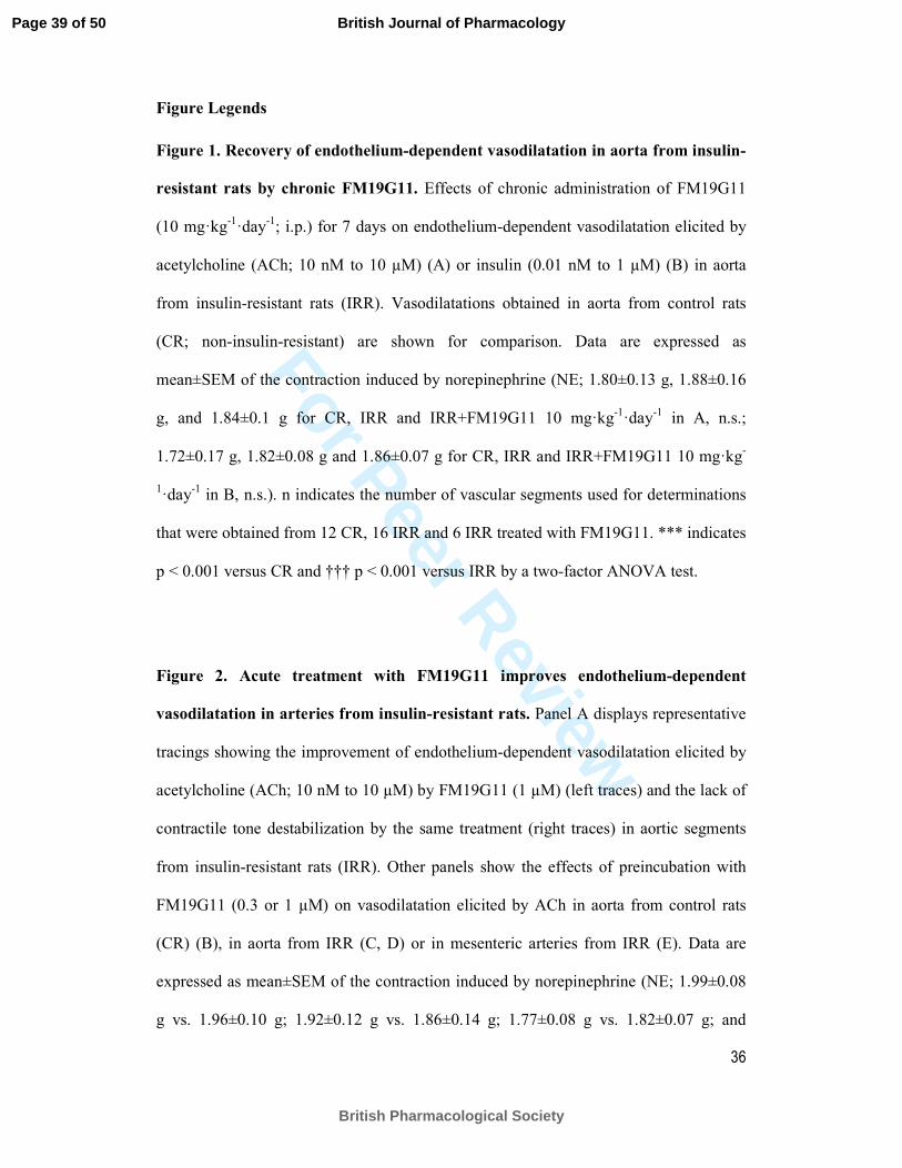
Figure Legends
Figure 1. Recovery of endothelium-dependent vasodilatation in aorta from insulin-
resistant rats by chronic FM19G11. Effects of chronic administration of FM19G11
(10 mg·kg-1
·day-1
; i.p.) for 7 days on endothelium-dependent vasodilatation elicited by
acetylcholine (ACh; 10 nM to 10 µM) (A) or insulin (0.01 nM to 1 µM) (B) in aorta
from insulin-resistant rats (IRR). Vasodilatations obtained in aorta from control rats
(CR; non-insulin-resistant) are shown for comparison. Data are expressed as
mean±SEM of the contraction induced by norepinephrine (NE; 1.80±0.13 g, 1.88±0.16
g, and 1.84±0.1 g for CR, IRR and IRR+FM19G11 10 mg·kg-1
·day-1
in A, n.s.;
1.72±0.17 g, 1.82±0.08 g and 1.86±0.07 g for CR, IRR and IRR+FM19G11 10 mg·kg-
1·day
-1 in B, n.s.). n indicates the number of vascular segments used for determinations
that were obtained from 12 CR, 16 IRR and 6 IRR treated with FM19G11. *** indicates
p < 0.001 versus CR and ††† p < 0.001 versus IRR by a two-factor ANOVA test.
Figure 2. Acute treatment with FM19G11 improves endothelium-dependent
vasodilatation in arteries from insulin-resistant rats. Panel A displays representative
tracings showing the improvement of endothelium-dependent vasodilatation elicited by
acetylcholine (ACh; 10 nM to 10 µM) by FM19G11 (1 µM) (left traces) and the lack of
contractile tone destabilization by the same treatment (right traces) in aortic segments
from insulin-resistant rats (IRR). Other panels show the effects of preincubation with
FM19G11 (0.3 or 1 µM) on vasodilatation elicited by ACh in aorta from control rats
(CR) (B), in aorta from IRR (C, D) or in mesenteric arteries from IRR (E). Data are
expressed as mean±SEM of the contraction induced by norepinephrine (NE; 1.99±0.08
g vs. 1.96±0.10 g; 1.92±0.12 g vs. 1.86±0.14 g; 1.77±0.08 g vs. 1.82±0.07 g; and
Page 39 of 50
British Pharmacological Society
British Journal of Pharmacology
For Peer Review
37
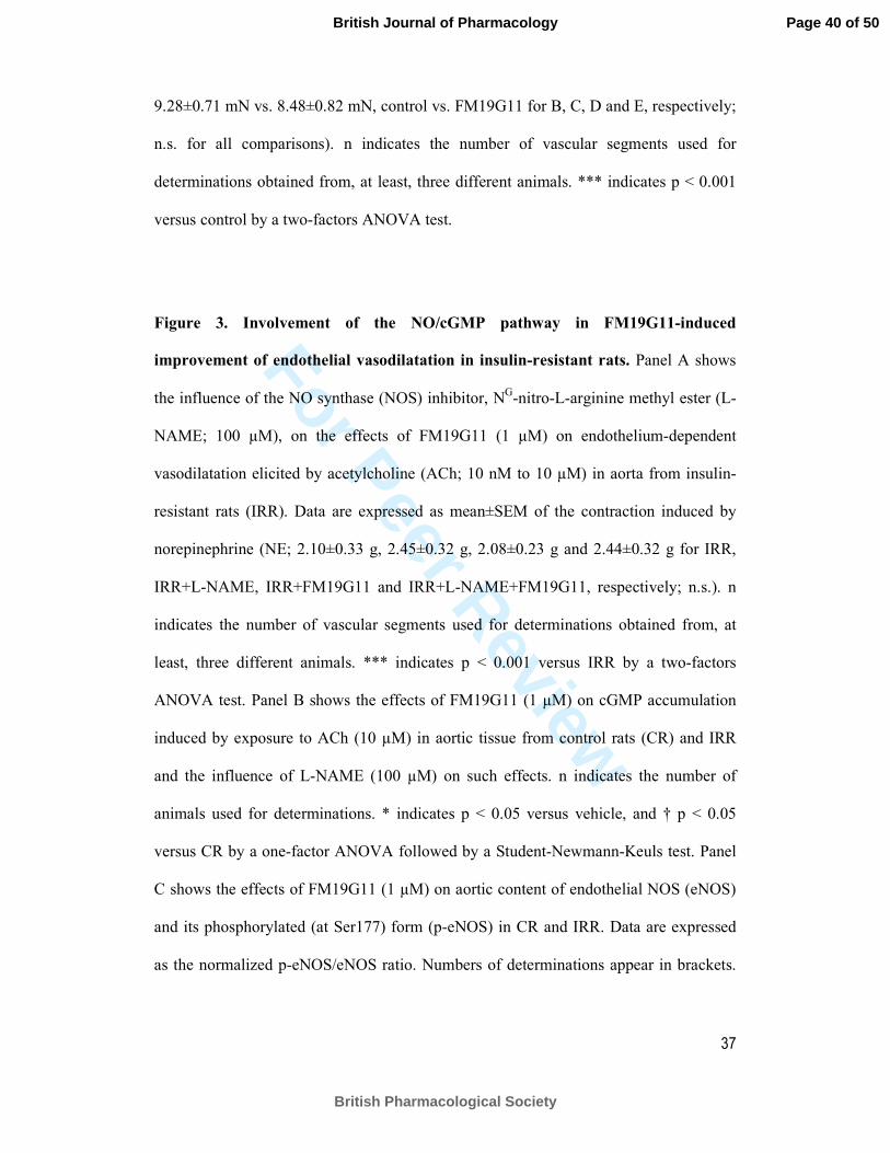
9.28±0.71 mN vs. 8.48±0.82 mN, control vs. FM19G11 for B, C, D and E, respectively;
n.s. for all comparisons). n indicates the number of vascular segments used for
determinations obtained from, at least, three different animals. *** indicates p < 0.001
versus control by a two-factors ANOVA test.
Figure 3. Involvement of the NO/cGMP pathway in FM19G11-induced
improvement of endothelial vasodilatation in insulin-resistant rats. Panel A shows
the influence of the NO synthase (NOS) inhibitor, NG-nitro-L-arginine methyl ester (L-
NAME; 100 µM), on the effects of FM19G11 (1 µM) on endothelium-dependent
vasodilatation elicited by acetylcholine (ACh; 10 nM to 10 µM) in aorta from insulin-
resistant rats (IRR). Data are expressed as mean±SEM of the contraction induced by
norepinephrine (NE; 2.10±0.33 g, 2.45±0.32 g, 2.08±0.23 g and 2.44±0.32 g for IRR,
IRR+L-NAME, IRR+FM19G11 and IRR+L-NAME+FM19G11, respectively; n.s.). n
indicates the number of vascular segments used for determinations obtained from, at
least, three different animals. *** indicates p < 0.001 versus IRR by a two-factors
ANOVA test. Panel B shows the effects of FM19G11 (1 µM) on cGMP accumulation
induced by exposure to ACh (10 µM) in aortic tissue from control rats (CR) and IRR
and the influence of L-NAME (100 µM) on such effects. n indicates the number of
animals used for determinations. * indicates p < 0.05 versus vehicle, and † p < 0.05
versus CR by a one-factor ANOVA followed by a Student-Newmann-Keuls test. Panel
C shows the effects of FM19G11 (1 µM) on aortic content of endothelial NOS (eNOS)
and its phosphorylated (at Ser177) form (p-eNOS) in CR and IRR. Data are expressed
as the normalized p-eNOS/eNOS ratio. Numbers of determinations appear in brackets.
Page 40 of 50
British Pharmacological Society
British Journal of Pharmacology
For Peer Review
38
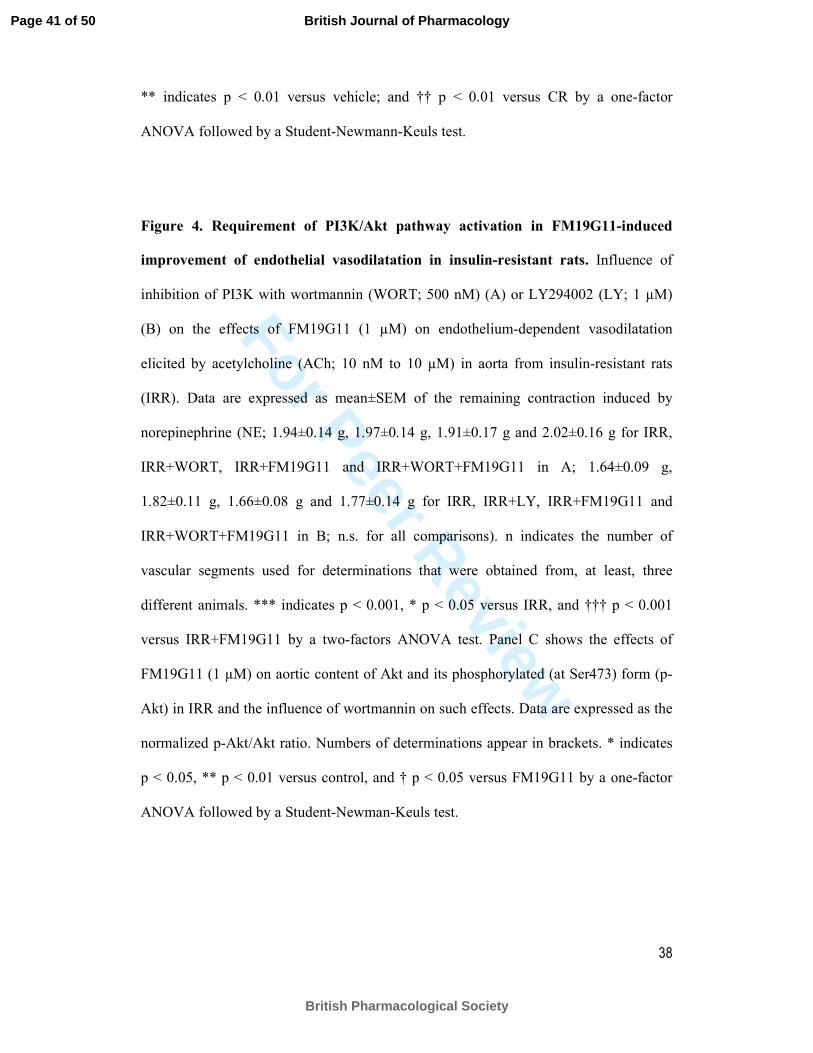
** indicates p < 0.01 versus vehicle; and †† p < 0.01 versus CR by a one-factor
ANOVA followed by a Student-Newmann-Keuls test.
Figure 4. Requirement of PI3K/Akt pathway activation in FM19G11-induced
improvement of endothelial vasodilatation in insulin-resistant rats. Influence of
inhibition of PI3K with wortmannin (WORT; 500 nM) (A) or LY294002 (LY; 1 µM)
(B) on the effects of FM19G11 (1 µM) on endothelium-dependent vasodilatation
elicited by acetylcholine (ACh; 10 nM to 10 µM) in aorta from insulin-resistant rats
(IRR). Data are expressed as mean±SEM of the remaining contraction induced by
norepinephrine (NE; 1.94±0.14 g, 1.97±0.14 g, 1.91±0.17 g and 2.02±0.16 g for IRR,
IRR+WORT, IRR+FM19G11 and IRR+WORT+FM19G11 in A; 1.64±0.09 g,
1.82±0.11 g, 1.66±0.08 g and 1.77±0.14 g for IRR, IRR+LY, IRR+FM19G11 and
IRR+WORT+FM19G11 in B; n.s. for all comparisons). n indicates the number of
vascular segments used for determinations that were obtained from, at least, three
different animals. *** indicates p < 0.001, * p < 0.05 versus IRR, and ††† p < 0.001
versus IRR+FM19G11 by a two-factors ANOVA test. Panel C shows the effects of
FM19G11 (1 µM) on aortic content of Akt and its phosphorylated (at Ser473) form (p-
Akt) in IRR and the influence of wortmannin on such effects. Data are expressed as the
normalized p-Akt/Akt ratio. Numbers of determinations appear in brackets. * indicates
p < 0.05, ** p < 0.01 versus control, and † p < 0.05 versus FM19G11 by a one-factor
ANOVA followed by a Student-Newman-Keuls test.
Page 41 of 50
British Pharmacological Society
British Journal of Pharmacology
For Peer Review
39
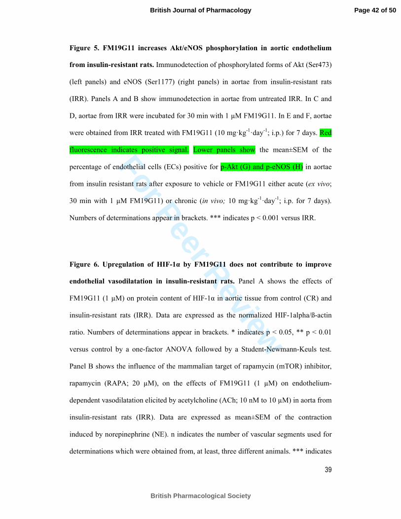
Figure 5. FM19G11 increases Akt/eNOS phosphorylation in aortic endothelium
from insulin-resistant rats. Immunodetection of phosphorylated forms of Akt (Ser473)
(left panels) and eNOS (Ser1177) (right panels) in aortae from insulin-resistant rats
(IRR). Panels A and B show immunodetection in aortae from untreated IRR. In C and
D, aortae from IRR were incubated for 30 min with 1 µM FM19G11. In E and F, aortae
were obtained from IRR treated with FM19G11 (10 mg·kg-1
·day-1
; i.p.) for 7 days. Red
fluorescence indicates positive signal. Lower panels show the mean±SEM of the
percentage of endothelial cells (ECs) positive for p-Akt (G) and p-eNOS (H) in aortae
from insulin resistant rats after exposure to vehicle or FM19G11 either acute (ex vivo;
30 min with 1 µM FM19G11) or chronic (in vivo; 10 mg·kg-1
·day-1
; i.p. for 7 days).
Numbers of determinations appear in brackets. *** indicates p < 0.001 versus IRR.
Figure 6. Upregulation of HIF-1α by FM19G11 does not contribute to improve
endothelial vasodilatation in insulin-resistant rats. Panel A shows the effects of
FM19G11 (1 µM) on protein content of HIF-1α in aortic tissue from control (CR) and
insulin-resistant rats (IRR). Data are expressed as the normalized HIF-1alpha/ß-actin
ratio. Numbers of determinations appear in brackets. * indicates p < 0.05, ** p < 0.01
versus control by a one-factor ANOVA followed by a Student-Newmann-Keuls test.
Panel B shows the influence of the mammalian target of rapamycin (mTOR) inhibitor,
rapamycin (RAPA; 20 µM), on the effects of FM19G11 (1 µM) on endothelium-
dependent vasodilatation elicited by acetylcholine (ACh; 10 nM to 10 µM) in aorta from
insulin-resistant rats (IRR). Data are expressed as mean±SEM of the contraction
induced by norepinephrine (NE). n indicates the number of vascular segments used for
determinations which were obtained from, at least, three different animals. *** indicates
Page 42 of 50
British Pharmacological Society
British Journal of Pharmacology
For Peer Review
40
p < 0.001 versus IRR by a two-factors ANOVA test. Panel C shows a representative
blotting showing that treatment with RAPA (20 µM) prevents FM19G11-induced HIF-
1α upregulation in aorta of IRR while does not precludes FM19G11-induced Akt
phosphorylation at Ser473.
Figure 7. FM19G11 recovers endothelium-dependent vasodilatation in human
penile resistance arteries and corpus cavernsoum. Panel A shows representative
tracings of the vasodilatation elicited by acetylcholine (ACh; 1 nM to 10 µM) in penile
resistance arteries from a subject without known erectile dysfunction (No ED) (upper
tracing) and from a patient with erectile dysfunction (ED) (lower tracings) before (left)
and after (right) incubation with FM19G11 (1 µM) for 30 min. Other panels show the
effects of FM19G11 (1 µM) on endothelium-dependent vasodilatation elicited by ACh
in human penile resistance arteries (HPRA) (B) and corpus cavernosum (HCC) (C)
from patients with ED. ACh-induced vasodilation in HPRA and HCC from No ED
subjects is shown for comparison. Data are expressed as mean±SEM of the contraction
induced by norepinephrine (NE; 6.73±0.68 mN, 6.26±1.73 mN and 6.33±1.51 mN for
No ED, ED and ED+FM19G11, respectively; n.s. for all comparisons) and
phenylephrine (PE; 1.20±0.29 g, 0.99±0.22 g and 1.05±0.20 g for No ED, ED and
ED+FM19G11, respectively; n.s. for all comparisons), respectively. n indicates the
number of patients from whom the tissues were collected. *** indicates p < 0.001
versus No ED, ††† p < 0.001 versus ED by a two-factors ANOVA test.
Page 43 of 50
British Pharmacological Society
British Journal of Pharmacology
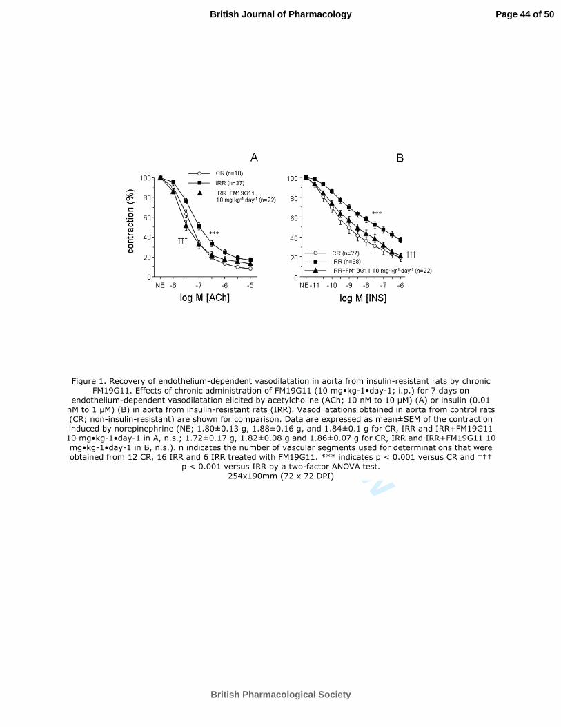
For Peer Review
Figure 1. Recovery of endothelium-dependent vasodilatation in aorta from insulin-resistant rats by chronic FM19G11. Effects of chronic administration of FM19G11 (10 mg•kg-1•day-1; i.p.) for 7 days on
endothelium-dependent vasodilatation elicited by acetylcholine (ACh; 10 nM to 10 µM) (A) or insulin (0.01
nM to 1 µM) (B) in aorta from insulin-resistant rats (IRR). Vasodilatations obtained in aorta from control rats (CR; non-insulin-resistant) are shown for comparison. Data are expressed as mean±SEM of the contraction induced by norepinephrine (NE; 1.80±0.13 g, 1.88±0.16 g, and 1.84±0.1 g for CR, IRR and IRR+FM19G11
10 mg•kg-1•day-1 in A, n.s.; 1.72±0.17 g, 1.82±0.08 g and 1.86±0.07 g for CR, IRR and IRR+FM19G11 10 mg•kg-1•day-1 in B, n.s.). n indicates the number of vascular segments used for determinations that were obtained from 12 CR, 16 IRR and 6 IRR treated with FM19G11. *** indicates p < 0.001 versus CR and †††
p < 0.001 versus IRR by a two-factor ANOVA test. 254x190mm (72 x 72 DPI)
Page 44 of 50
British Pharmacological Society
British Journal of Pharmacology
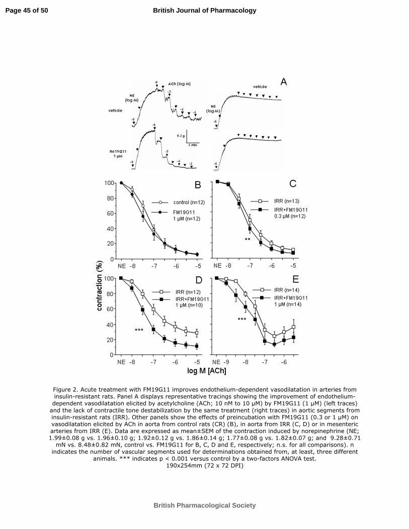
For Peer Review
Figure 2. Acute treatment with FM19G11 improves endothelium-dependent vasodilatation in arteries from insulin-resistant rats. Panel A displays representative tracings showing the improvement of endothelium-dependent vasodilatation elicited by acetylcholine (ACh; 10 nM to 10 µM) by FM19G11 (1 µM) (left traces)
and the lack of contractile tone destabilization by the same treatment (right traces) in aortic segments from insulin-resistant rats (IRR). Other panels show the effects of preincubation with FM19G11 (0.3 or 1 µM) on vasodilatation elicited by ACh in aorta from control rats (CR) (B), in aorta from IRR (C, D) or in mesenteric arteries from IRR (E). Data are expressed as mean±SEM of the contraction induced by norepinephrine (NE; 1.99±0.08 g vs. 1.96±0.10 g; 1.92±0.12 g vs. 1.86±0.14 g; 1.77±0.08 g vs. 1.82±0.07 g; and 9.28±0.71
mN vs. 8.48±0.82 mN, control vs. FM19G11 for B, C, D and E, respectively; n.s. for all comparisons). n indicates the number of vascular segments used for determinations obtained from, at least, three different
animals. *** indicates p < 0.001 versus control by a two-factors ANOVA test. 190x254mm (72 x 72 DPI)
Page 45 of 50
British Pharmacological Society
British Journal of Pharmacology
For Peer Review
Page 46 of 50
British Pharmacological Society
British Journal of Pharmacology
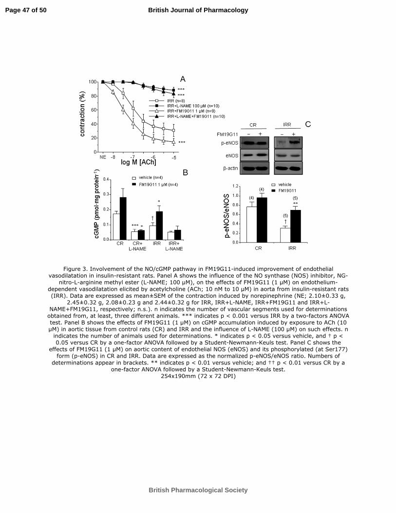
For Peer Review
Figure 3. Involvement of the NO/cGMP pathway in FM19G11-induced improvement of endothelial vasodilatation in insulin-resistant rats. Panel A shows the influence of the NO synthase (NOS) inhibitor, NG-
nitro-L-arginine methyl ester (L-NAME; 100 µM), on the effects of FM19G11 (1 µM) on endothelium-
dependent vasodilatation elicited by acetylcholine (ACh; 10 nM to 10 µM) in aorta from insulin-resistant rats (IRR). Data are expressed as mean±SEM of the contraction induced by norepinephrine (NE; 2.10±0.33 g,
2.45±0.32 g, 2.08±0.23 g and 2.44±0.32 g for IRR, IRR+L-NAME, IRR+FM19G11 and IRR+L-NAME+FM19G11, respectively; n.s.). n indicates the number of vascular segments used for determinations
obtained from, at least, three different animals. *** indicates p < 0.001 versus IRR by a two-factors ANOVA test. Panel B shows the effects of FM19G11 (1 µM) on cGMP accumulation induced by exposure to ACh (10 µM) in aortic tissue from control rats (CR) and IRR and the influence of L-NAME (100 µM) on such effects. n
indicates the number of animals used for determinations. * indicates p < 0.05 versus vehicle, and † p < 0.05 versus CR by a one-factor ANOVA followed by a Student-Newmann-Keuls test. Panel C shows the
effects of FM19G11 (1 µM) on aortic content of endothelial NOS (eNOS) and its phosphorylated (at Ser177) form (p-eNOS) in CR and IRR. Data are expressed as the normalized p-eNOS/eNOS ratio. Numbers of
determinations appear in brackets. ** indicates p < 0.01 versus vehicle; and †† p < 0.01 versus CR by a one-factor ANOVA followed by a Student-Newmann-Keuls test.
254x190mm (72 x 72 DPI)
Page 47 of 50
British Pharmacological Society
British Journal of Pharmacology
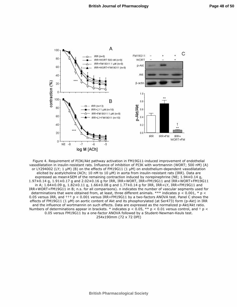
For Peer Review
Figure 4. Requirement of PI3K/Akt pathway activation in FM19G11-induced improvement of endothelial vasodilatation in insulin-resistant rats. Influence of inhibition of PI3K with wortmannin (WORT; 500 nM) (A)
or LY294002 (LY; 1 µM) (B) on the effects of FM19G11 (1 µM) on endothelium-dependent vasodilatation
elicited by acetylcholine (ACh; 10 nM to 10 µM) in aorta from insulin-resistant rats (IRR). Data are expressed as mean±SEM of the remaining contraction induced by norepinephrine (NE; 1.94±0.14 g,
1.97±0.14 g, 1.91±0.17 g and 2.02±0.16 g for IRR, IRR+WORT, IRR+FM19G11 and IRR+WORT+FM19G11 in A; 1.64±0.09 g, 1.82±0.11 g, 1.66±0.08 g and 1.77±0.14 g for IRR, IRR+LY, IRR+FM19G11 and
IRR+WORT+FM19G11 in B; n.s. for all comparisons). n indicates the number of vascular segments used for determinations that were obtained from, at least, three different animals. *** indicates p < 0.001, * p <
0.05 versus IRR, and ††† p < 0.001 versus IRR+FM19G11 by a two-factors ANOVA test. Panel C shows the effects of FM19G11 (1 µM) on aortic content of Akt and its phosphorylated (at Ser473) form (p-Akt) in IRR
and the influence of wortmannin on such effects. Data are expressed as the normalized p-Akt/Akt ratio. Numbers of determinations appear in brackets. * indicates p < 0.05, ** p < 0.01 versus control, and † p <
0.05 versus FM19G11 by a one-factor ANOVA followed by a Student-Newman-Keuls test.
254x190mm (72 x 72 DPI)
Page 48 of 50
British Pharmacological Society
British Journal of Pharmacology
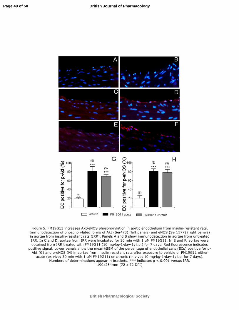
For Peer Review
Figure 5. FM19G11 increases Akt/eNOS phosphorylation in aortic endothelium from insulin-resistant rats. Immunodetection of phosphorylated forms of Akt (Ser473) (left panels) and eNOS (Ser1177) (right panels) in aortae from insulin-resistant rats (IRR). Panels A and B show immunodetection in aortae from untreated
IRR. In C and D, aortae from IRR were incubated for 30 min with 1 µM FM19G11. In E and F, aortae were obtained from IRR treated with FM19G11 (10 mg·kg-1·day-1; i.p.) for 7 days. Red fluorescence indicates
positive signal. Lower panels show the mean±SEM of the percentage of endothelial cells (ECs) positive for p-Akt (G) and p-eNOS (H) in aortae from insulin resistant rats after exposure to vehicle or FM19G11 either
acute (ex vivo; 30 min with 1 µM FM19G11) or chronic (in vivo; 10 mg·kg-1·day-1; i.p. for 7 days). Numbers of determinations appear in brackets. *** indicates p < 0.001 versus IRR.
190x254mm (72 x 72 DPI)
Page 49 of 50
British Pharmacological Society
British Journal of Pharmacology
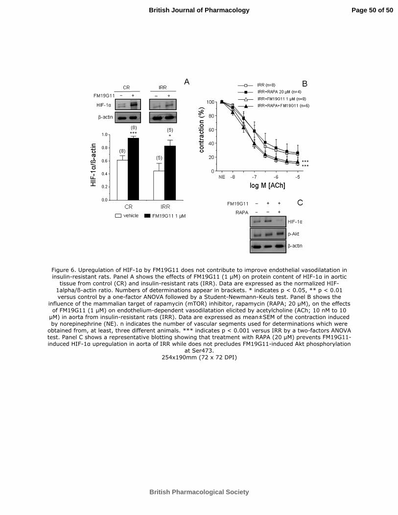
For Peer Review
Figure 6. Upregulation of HIF-1α by FM19G11 does not contribute to improve endothelial vasodilatation in insulin-resistant rats. Panel A shows the effects of FM19G11 (1 µM) on protein content of HIF-1α in aortic
tissue from control (CR) and insulin-resistant rats (IRR). Data are expressed as the normalized HIF-
1alpha/ß-actin ratio. Numbers of determinations appear in brackets. * indicates p < 0.05, ** p < 0.01 versus control by a one-factor ANOVA followed by a Student-Newmann-Keuls test. Panel B shows the
influence of the mammalian target of rapamycin (mTOR) inhibitor, rapamycin (RAPA; 20 µM), on the effects of FM19G11 (1 µM) on endothelium-dependent vasodilatation elicited by acetylcholine (ACh; 10 nM to 10
µM) in aorta from insulin-resistant rats (IRR). Data are expressed as mean±SEM of the contraction induced by norepinephrine (NE). n indicates the number of vascular segments used for determinations which were
obtained from, at least, three different animals. *** indicates p < 0.001 versus IRR by a two-factors ANOVA test. Panel C shows a representative blotting showing that treatment with RAPA (20 µM) prevents FM19G11-induced HIF-1α upregulation in aorta of IRR while does not precludes FM19G11-induced Akt phosphorylation
at Ser473. 254x190mm (72 x 72 DPI)
Page 50 of 50
British Pharmacological Society
British Journal of Pharmacology
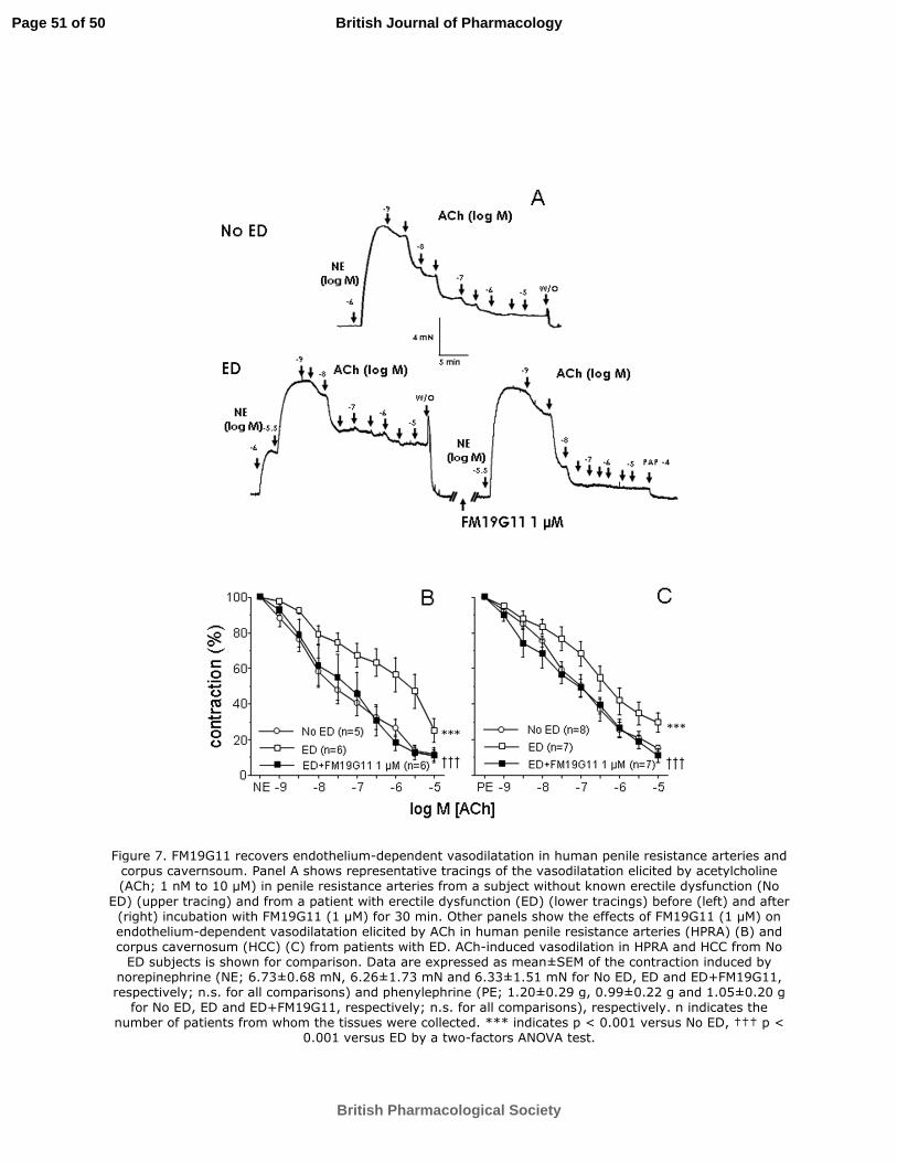
For Peer Review
Figure 7. FM19G11 recovers endothelium-dependent vasodilatation in human penile resistance arteries and corpus cavernsoum. Panel A shows representative tracings of the vasodilatation elicited by acetylcholine (ACh; 1 nM to 10 µM) in penile resistance arteries from a subject without known erectile dysfunction (No
ED) (upper tracing) and from a patient with erectile dysfunction (ED) (lower tracings) before (left) and after (right) incubation with FM19G11 (1 µM) for 30 min. Other panels show the effects of FM19G11 (1 µM) on endothelium-dependent vasodilatation elicited by ACh in human penile resistance arteries (HPRA) (B) and corpus cavernosum (HCC) (C) from patients with ED. ACh-induced vasodilation in HPRA and HCC from No
ED subjects is shown for comparison. Data are expressed as mean±SEM of the contraction induced by norepinephrine (NE; 6.73±0.68 mN, 6.26±1.73 mN and 6.33±1.51 mN for No ED, ED and ED+FM19G11, respectively; n.s. for all comparisons) and phenylephrine (PE; 1.20±0.29 g, 0.99±0.22 g and 1.05±0.20 g
for No ED, ED and ED+FM19G11, respectively; n.s. for all comparisons), respectively. n indicates the number of patients from whom the tissues were collected. *** indicates p < 0.001 versus No ED, ††† p <
0.001 versus ED by a two-factors ANOVA test.
Page 51 of 50
British Pharmacological Society
British Journal of Pharmacology
For Peer Review
190x254mm (72 x 72 DPI)
Page 52 of 50
British Pharmacological Society
British Journal of Pharmacology


![Review Article Bioactive Peptides: A Review - BASclbme.bas.bg/bioautomation/2011/vol_15.4/files/15.4_02.pdf · Review Article Bioactive Peptides: A Review ... casein [145]. Other](https://static.fdocument.org/doc/165x107/5acd360f7f8b9a93268d5e73/review-article-bioactive-peptides-a-review-article-bioactive-peptides-a-review.jpg)