Fluorescence Quenching Studies of -Butyrolactone Binding …pradeep/assets/29.pdf · Fluorescence...
21
Fluorescence Quenching Studies of γ‑Butyrolactone Binding Protein (CprB) from Streptomyces coelicolor A3(2) Anwesha Biswas, † Ravi K. Swarnkar, † Bhukya Hussain, †,‡ Suraj K. Sahoo, † P. I. Pradeepkumar, † G. Naresh Patwari,* ,† and Ruchi Anand* ,† † Department of Chemistry, Indian Institute of Technology Bombay, Mumbai, Maharashtra 400076, India * S Supporting Information ABSTRACT: Quorum sensing is a cell density dependent phenomenon that utilizes small molecule inducers like γ-butyrolactones (GBLs) and their receptor proteins for adaptation to the environment. The cognate GBLs that bind to several of this GBL receptor family of proteins remain elusive. Here, using CprB protein from Streptomyces coelicolor A3(2) as a model system, we devise a method suited for ligand screening that would be applicable to the entire family of GBL receptors. Docking studies were performed to confirm the identity of the ligand binding pocket, and it was ascertained that the common γ-butyrolactone moiety interacts with the conserved tryptophan residue (W127) residing in the ligand binding pocket. The presence of W127 in the cavity was exploited to monitor its fluorescence quenching on the addition of two chemically synthesized GBLs. Analysis of the data with both the native and W185L mutant versions of the protein confirmed that the compounds used as quenchers reside in the ligand binding pocket. Furthermore, fluorescence lifetime and potassium iodide (KI) quenching studies established that the quenching is static in nature and that the tryptophan residue is buried and inaccessible to surface quenchers. Additionally, a combination of concentration dependent fluorescence quenching and dynamic light scattering experiments revealed that the binding properties of the protein are concentration dependent and it was concluded that the most efficient binding of the ligand is evoked by working at the lowest concentration of protein, providing a sufficient signal, where the aggregation effects are negligible. ■ INTRODUCTION Bacteria employ a response mechanism termed quorum sensing to regulate certain transcription and gene expression processes that in several instances lead to activation of secondary metabolic pathways. 1−3 Quorum sensing involves small signal- ing molecules called autoinducers, exuded by the bacteria, which trigger a response at a threshold concentration by binding to their receptor proteins. 4 The response is therefore cell density dependent and is elicited as a consequence of the protein−autoinducer complex formation. Extensive work on such systems has unearthed numerous classes of these signaling molecules like the N-acylhomoserine lactones (AHLs), 3,5,6 4- hydroxy-2-alkyl quinolines (HAQs), 7,8 γ-butyrolactones (GBLs), 9,10 cyclic and linear oligopeptides, 11,12 etc. A GBL from Streptomyces griseus named A-factor (2-isocaprylolyl-3R- hydroxymethyl-butyrolactone) was first among the butanolides that have been identified. 13 It was established in 1994 by Horinouchi and co-workers that A-factor effectively triggers morphological differentiation and streptomycin production in this species by binding to its receptor protein ArpA. 14−17 Subsequently, other GBLs and the transcription factors they regulate have been identified in several species of Streptomy- ces. 18−21 There is, however, a paucity of X-ray structural information available in the GBL receptor family of proteins. CprB from S. coelicolor A3(2), 22 predicted to be a homologue of ArpA, is the only member for which the structure has been elucidated in the apo form (Figure 1). 23,24 The structure confirmed the presence of two domains: an N-terminal DNA binding domain, similar to the tetracycline receptor family of antibiotic resistance proteins, 25 and a divergent C-terminal ligand binding domain (the regulatory domain) that is proposed to bind the cognate quorum sensing molecule. The protein exists in a dimeric state with the two units being related by a pseudo-2-fold axis. 24 The regulatory domain which is of the most interest for our study is composed of an antiparallel bundle of five helices (α5−α10) with helix α6 forming the base of the cavity. The large cavity thus created that has a depth of approximately 20 Å and a diameter of 5 Å is lined by hydrophobic residues. Comparison of GBL binding proteins showed that most of the residues in the pocket although hydrophobic in nature are not strictly conserved. Interestingly, the pocket contains a tryptophan residue at the 127 position (Figure 1) which is conserved among all the members of this family. 15 Mutation of the corresponding tryptophan residue W119 in ArpA (W127 for CprB) abolished the A-factor binding properties of the protein completely, thereby signifying its importance in GBL binding. 15 This also indicated that, apart from the few conserved residues within the pocket, the rest have been fine- tuned in each member of the family of proteins to recognize Received: April 12, 2014 Revised: July 9, 2014 Published: July 30, 2014 Article pubs.acs.org/JPCB © 2014 American Chemical Society 10035 dx.doi.org/10.1021/jp503589h | J. Phys. Chem. B 2014, 118, 10035−10042
Transcript of Fluorescence Quenching Studies of -Butyrolactone Binding …pradeep/assets/29.pdf · Fluorescence...
Fluorescence Quenching Studies of γ‑Butyrolactone Binding Protein(CprB) from Streptomyces coelicolor A3(2)Anwesha Biswas,† Ravi K. Swarnkar,† Bhukya Hussain,†,‡ Suraj K. Sahoo,† P. I. Pradeepkumar,†
G. Naresh Patwari,*,† and Ruchi Anand*,†
†Department of Chemistry, Indian Institute of Technology Bombay, Mumbai, Maharashtra 400076, India
*S Supporting Information
ABSTRACT: Quorum sensing is a cell density dependent phenomenon that utilizessmall molecule inducers like γ-butyrolactones (GBLs) and their receptor proteins foradaptation to the environment. The cognate GBLs that bind to several of this GBLreceptor family of proteins remain elusive. Here, using CprB protein from Streptomycescoelicolor A3(2) as a model system, we devise a method suited for ligand screening thatwould be applicable to the entire family of GBL receptors. Docking studies wereperformed to confirm the identity of the ligand binding pocket, and it was ascertainedthat the common γ-butyrolactone moiety interacts with the conserved tryptophan residue(W127) residing in the ligand binding pocket. The presence of W127 in the cavity wasexploited to monitor its fluorescence quenching on the addition of two chemicallysynthesized GBLs. Analysis of the data with both the native and W185L mutant versions of the protein confirmed that thecompounds used as quenchers reside in the ligand binding pocket. Furthermore, fluorescence lifetime and potassium iodide (KI)quenching studies established that the quenching is static in nature and that the tryptophan residue is buried and inaccessible tosurface quenchers. Additionally, a combination of concentration dependent fluorescence quenching and dynamic light scatteringexperiments revealed that the binding properties of the protein are concentration dependent and it was concluded that the mostefficient binding of the ligand is evoked by working at the lowest concentration of protein, providing a sufficient signal, where theaggregation effects are negligible.
■ INTRODUCTION
Bacteria employ a response mechanism termed quorum sensingto regulate certain transcription and gene expression processesthat in several instances lead to activation of secondarymetabolic pathways.1−3 Quorum sensing involves small signal-ing molecules called autoinducers, exuded by the bacteria,which trigger a response at a threshold concentration bybinding to their receptor proteins.4 The response is thereforecell density dependent and is elicited as a consequence of theprotein−autoinducer complex formation. Extensive work onsuch systems has unearthed numerous classes of these signalingmolecules like the N-acylhomoserine lactones (AHLs),3,5,6 4-hydroxy-2-alkyl quinolines (HAQs),7,8 γ-butyrolactones(GBLs),9,10 cyclic and linear oligopeptides,11,12 etc. A GBLfrom Streptomyces griseus named A-factor (2-isocaprylolyl-3R-hydroxymethyl-butyrolactone) was first among the butanolidesthat have been identified.13 It was established in 1994 byHorinouchi and co-workers that A-factor effectively triggersmorphological differentiation and streptomycin production inthis species by binding to its receptor protein ArpA.14−17
Subsequently, other GBLs and the transcription factors theyregulate have been identified in several species of Streptomy-ces.18−21
There is, however, a paucity of X-ray structural informationavailable in the GBL receptor family of proteins. CprB from S.coelicolor A3(2),22 predicted to be a homologue of ArpA, is theonly member for which the structure has been elucidated in the
apo form (Figure 1).23,24 The structure confirmed the presenceof two domains: an N-terminal DNA binding domain, similarto the tetracycline receptor family of antibiotic resistanceproteins,25 and a divergent C-terminal ligand binding domain(the regulatory domain) that is proposed to bind the cognatequorum sensing molecule. The protein exists in a dimeric statewith the two units being related by a pseudo-2-fold axis.24 Theregulatory domain which is of the most interest for our study iscomposed of an antiparallel bundle of five helices (α5−α10)with helix α6 forming the base of the cavity. The large cavitythus created that has a depth of approximately 20 Å and adiameter of 5 Å is lined by hydrophobic residues.Comparison of GBL binding proteins showed that most of
the residues in the pocket although hydrophobic in nature arenot strictly conserved. Interestingly, the pocket contains atryptophan residue at the 127 position (Figure 1) which isconserved among all the members of this family.15 Mutation ofthe corresponding tryptophan residue W119 in ArpA (W127for CprB) abolished the A-factor binding properties of theprotein completely, thereby signifying its importance in GBLbinding.15 This also indicated that, apart from the fewconserved residues within the pocket, the rest have been fine-tuned in each member of the family of proteins to recognize
Received: April 12, 2014Revised: July 9, 2014Published: July 30, 2014
Article
pubs.acs.org/JPCB
© 2014 American Chemical Society 10035 dx.doi.org/10.1021/jp503589h | J. Phys. Chem. B 2014, 118, 10035−10042
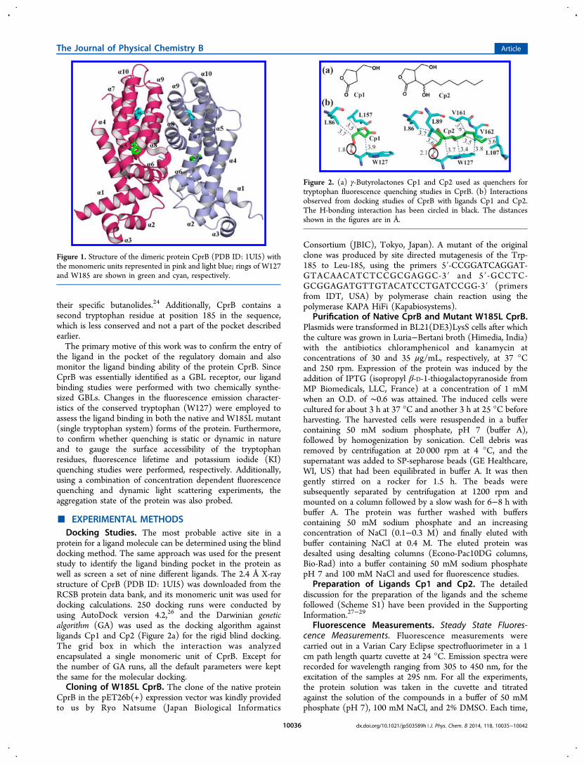
their specific butanolides.24 Additionally, CprB contains asecond tryptophan residue at position 185 in the sequence,which is less conserved and not a part of the pocket describedearlier.The primary motive of this work was to confirm the entry of
the ligand in the pocket of the regulatory domain and alsomonitor the ligand binding ability of the protein CprB. SinceCprB was essentially identified as a GBL receptor, our ligandbinding studies were performed with two chemically synthe-sized GBLs. Changes in the fluorescence emission character-istics of the conserved tryptophan (W127) were employed toassess the ligand binding in both the native and W185L mutant(single tryptophan system) forms of the protein. Furthermore,to confirm whether quenching is static or dynamic in natureand to gauge the surface accessibility of the tryptophanresidues, fluorescence lifetime and potassium iodide (KI)quenching studies were performed, respectively. Additionally,using a combination of concentration dependent fluorescencequenching and dynamic light scattering experiments, theaggregation state of the protein was also probed.
■ EXPERIMENTAL METHODSDocking Studies. The most probable active site in a
protein for a ligand molecule can be determined using the blinddocking method. The same approach was used for the presentstudy to identify the ligand binding pocket in the protein aswell as screen a set of nine different ligands. The 2.4 Å X-raystructure of CprB (PDB ID: 1UI5) was downloaded from theRCSB protein data bank, and its monomeric unit was used fordocking calculations. 250 docking runs were conducted byusing AutoDock version 4.2,26 and the Darwinian geneticalgorithm (GA) was used as the docking algorithm againstligands Cp1 and Cp2 (Figure 2a) for the rigid blind docking.The grid box in which the interaction was analyzedencapsulated a single monomeric unit of CprB. Except forthe number of GA runs, all the default parameters were keptthe same for the molecular docking.Cloning of W185L CprB. The clone of the native protein
CprB in the pET26b(+) expression vector was kindly providedto us by Ryo Natsume (Japan Biological Informatics
Consortium (JBIC), Tokyo, Japan). A mutant of the originalclone was produced by site directed mutagenesis of the Trp-185 to Leu-185, using the primers 5′-CCGGATCAGGAT-GTACAACATCTCCGCGAGGC-3′ and 5′-GCCTC-GCGGAGATGTTGTACATCCTGATCCGG-3′ (primersfrom IDT, USA) by polymerase chain reaction using thepolymerase KAPA HiFi (Kapabiosystems).
Purification of Native CprB and Mutant W185L CprB.Plasmids were transformed in BL21(DE3)LysS cells after whichthe culture was grown in Luria−Bertani broth (Himedia, India)with the antibiotics chloramphenicol and kanamycin atconcentrations of 30 and 35 μg/mL, respectively, at 37 °Cand 250 rpm. Expression of the protein was induced by theaddition of IPTG (isopropyl β-D-1-thiogalactopyranoside fromMP Biomedicals, LLC, France) at a concentration of 1 mMwhen an O.D. of ∼0.6 was attained. The induced cells werecultured for about 3 h at 37 °C and another 3 h at 25 °C beforeharvesting. The harvested cells were resuspended in a buffercontaining 50 mM sodium phosphate, pH 7 (buffer A),followed by homogenization by sonication. Cell debris wasremoved by centrifugation at 20 000 rpm at 4 °C, and thesupernatant was added to SP-sepharose beads (GE Healthcare,WI, US) that had been equilibrated in buffer A. It was thengently stirred on a rocker for 1.5 h. The beads weresubsequently separated by centrifugation at 1200 rpm andmounted on a column followed by a slow wash for 6−8 h withbuffer A. The protein was further washed with bufferscontaining 50 mM sodium phosphate and an increasingconcentration of NaCl (0.1−0.3 M) and finally eluted withbuffer containing NaCl at 0.4 M. The eluted protein wasdesalted using desalting columns (Econo-Pac10DG columns,Bio-Rad) into a buffer containing 50 mM sodium phosphatepH 7 and 100 mM NaCl and used for fluorescence studies.
Preparation of Ligands Cp1 and Cp2. The detaileddiscussion for the preparation of the ligands and the schemefollowed (Scheme S1) have been provided in the SupportingInformation.27−29
Fluorescence Measurements. Steady State Fluores-cence Measurements. Fluorescence measurements werecarried out in a Varian Cary Eclipse spectrofluorimeter in a 1cm path length quartz cuvette at 24 °C. Emission spectra wererecorded for wavelength ranging from 305 to 450 nm, for theexcitation of the samples at 295 nm. For all the experiments,the protein solution was taken in the cuvette and titratedagainst the solution of the compounds in a buffer of 50 mMphosphate (pH 7), 100 mM NaCl, and 2% DMSO. Each time,
Figure 1. Structure of the dimeric protein CprB (PDB ID: 1UI5) withthe monomeric units represented in pink and light blue; rings of W127and W185 are shown in green and cyan, respectively.
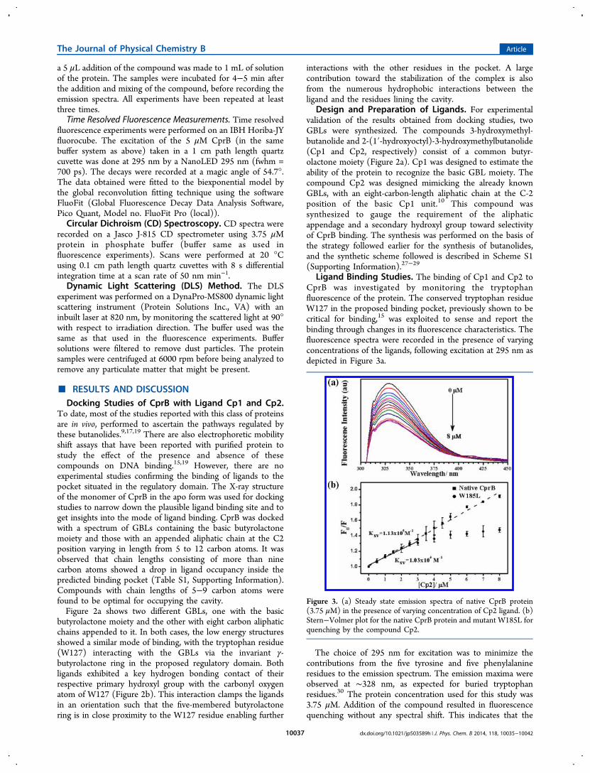
Figure 2. (a) γ-Butyrolactones Cp1 and Cp2 used as quenchers fortryptophan fluorescence quenching studies in CprB. (b) Interactionsobserved from docking studies of CprB with ligands Cp1 and Cp2.The H-bonding interaction has been circled in black. The distancesshown in the figures are in Å.
The Journal of Physical Chemistry B Article
dx.doi.org/10.1021/jp503589h | J. Phys. Chem. B 2014, 118, 10035−1004210036
a 5 μL addition of the compound was made to 1 mL of solutionof the protein. The samples were incubated for 4−5 min afterthe addition and mixing of the compound, before recording theemission spectra. All experiments have been repeated at leastthree times.Time Resolved Fluorescence Measurements. Time resolved
fluorescence experiments were performed on an IBH Horiba-JYfluorocube. The excitation of the 5 μM CprB (in the samebuffer system as above) taken in a 1 cm path length quartzcuvette was done at 295 nm by a NanoLED 295 nm (fwhm =700 ps). The decays were recorded at a magic angle of 54.7°.The data obtained were fitted to the biexponential model bythe global reconvolution fitting technique using the softwareFluoFit (Global Fluorescence Decay Data Analysis Software,Pico Quant, Model no. FluoFit Pro (local)).Circular Dichroism (CD) Spectroscopy. CD spectra were
recorded on a Jasco J-815 CD spectrometer using 3.75 μMprotein in phosphate buffer (buffer same as used influorescence experiments). Scans were performed at 20 °Cusing 0.1 cm path length quartz cuvettes with 8 s differentialintegration time at a scan rate of 50 nm min−1.Dynamic Light Scattering (DLS) Method. The DLS
experiment was performed on a DynaPro-MS800 dynamic lightscattering instrument (Protein Solutions Inc., VA) with aninbuilt laser at 820 nm, by monitoring the scattered light at 90°with respect to irradiation direction. The buffer used was thesame as that used in the fluorescence experiments. Buffersolutions were filtered to remove dust particles. The proteinsamples were centrifuged at 6000 rpm before being analyzed toremove any particulate matter that might be present.
■ RESULTS AND DISCUSSIONDocking Studies of CprB with Ligand Cp1 and Cp2.
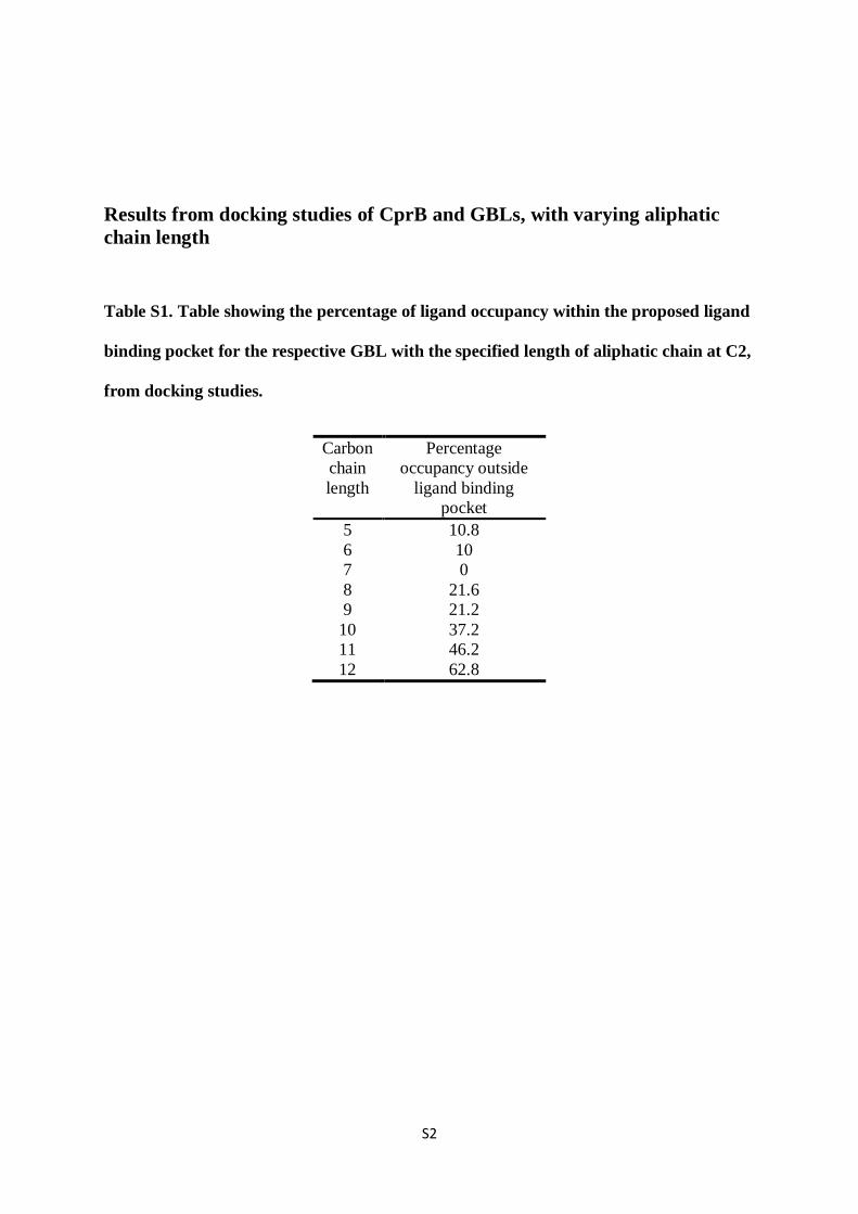
To date, most of the studies reported with this class of proteinsare in vivo, performed to ascertain the pathways regulated bythese butanolides.9,17,19 There are also electrophoretic mobilityshift assays that have been reported with purified protein tostudy the effect of the presence and absence of thesecompounds on DNA binding.15,19 However, there are noexperimental studies confirming the binding of ligands to thepocket situated in the regulatory domain. The X-ray structureof the monomer of CprB in the apo form was used for dockingstudies to narrow down the plausible ligand binding site and toget insights into the mode of ligand binding. CprB was dockedwith a spectrum of GBLs containing the basic butyrolactonemoiety and those with an appended aliphatic chain at the C2position varying in length from 5 to 12 carbon atoms. It wasobserved that chain lengths consisting of more than ninecarbon atoms showed a drop in ligand occupancy inside thepredicted binding pocket (Table S1, Supporting Information).Compounds with chain lengths of 5−9 carbon atoms werefound to be optimal for occupying the cavity.Figure 2a shows two different GBLs, one with the basic
butyrolactone moiety and the other with eight carbon aliphaticchains appended to it. In both cases, the low energy structuresshowed a similar mode of binding, with the tryptophan residue(W127) interacting with the GBLs via the invariant γ-butyrolactone ring in the proposed regulatory domain. Bothligands exhibited a key hydrogen bonding contact of theirrespective primary hydroxyl group with the carbonyl oxygenatom of W127 (Figure 2b). This interaction clamps the ligandsin an orientation such that the five-membered butyrolactonering is in close proximity to the W127 residue enabling further
interactions with the other residues in the pocket. A largecontribution toward the stabilization of the complex is alsofrom the numerous hydrophobic interactions between theligand and the residues lining the cavity.
Design and Preparation of Ligands. For experimentalvalidation of the results obtained from docking studies, twoGBLs were synthesized. The compounds 3-hydroxymethyl-butanolide and 2-(1′-hydroxyoctyl)-3-hydroxymethylbutanolide(Cp1 and Cp2, respectively) consist of a common butyr-olactone moiety (Figure 2a). Cp1 was designed to estimate theability of the protein to recognize the basic GBL moiety. Thecompound Cp2 was designed mimicking the already knownGBLs, with an eight-carbon-length aliphatic chain at the C-2position of the basic Cp1 unit.10 This compound wassynthesized to gauge the requirement of the aliphaticappendage and a secondary hydroxyl group toward selectivityof CprB binding. The synthesis was performed on the basis ofthe strategy followed earlier for the synthesis of butanolides,and the synthetic scheme followed is described in Scheme S1(Supporting Information).27−29
Ligand Binding Studies. The binding of Cp1 and Cp2 toCprB was investigated by monitoring the tryptophanfluorescence of the protein. The conserved tryptophan residueW127 in the proposed binding pocket, previously shown to becritical for binding,15 was exploited to sense and report thebinding through changes in its fluorescence characteristics. Thefluorescence spectra were recorded in the presence of varyingconcentrations of the ligands, following excitation at 295 nm asdepicted in Figure 3a.
The choice of 295 nm for excitation was to minimize thecontributions from the five tyrosine and five phenylalanineresidues to the emission spectrum. The emission maxima wereobserved at ∼328 nm, as expected for buried tryptophanresidues.30 The protein concentration used for this study was3.75 μM. Addition of the compound resulted in fluorescencequenching without any spectral shift. This indicates that the
Figure 3. (a) Steady state emission spectra of native CprB protein(3.75 μM) in the presence of varying concentration of Cp2 ligand. (b)Stern−Volmer plot for the native CprB protein and mutant W185L forquenching by the compound Cp2.
The Journal of Physical Chemistry B Article
dx.doi.org/10.1021/jp503589h | J. Phys. Chem. B 2014, 118, 10035−1004210037
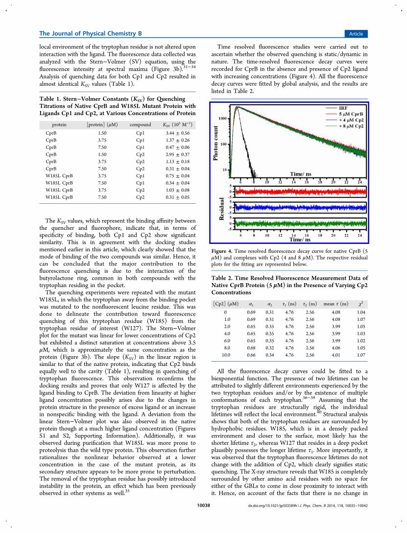
local environment of the tryptophan residue is not altered uponinteraction with the ligand. The fluorescence data collected wasanalyzed with the Stern−Volmer (SV) equation, using thefluorescence intensity at spectral maxima (Figure 3b).31−34
Analysis of quenching data for both Cp1 and Cp2 resulted inalmost identical KSV values (Table 1).
The KSV values, which represent the binding affinity betweenthe quencher and fluorophore, indicate that, in terms ofspecificity of binding, both Cp1 and Cp2 show significantsimilarity. This is in agreement with the docking studiesmentioned earlier in this article, which clearly showed that themode of binding of the two compounds was similar. Hence, itcan be concluded that the major contribution to thefluorescence quenching is due to the interaction of thebutyrolactone ring, common in both compounds with thetryptophan residing in the pocket.The quenching experiments were repeated with the mutant
W185L, in which the tryptophan away from the binding pocketwas mutated to the nonfluorescent leucine residue. This wasdone to delineate the contribution toward fluorescencequenching of this tryptophan residue (W185) from thetryptophan residue of interest (W127). The Stern−Volmerplot for the mutant was linear for lower concentrations of Cp2but exhibited a distinct saturation at concentrations above 3.5μM, which is approximately the same concentration as theprotein (Figure 3b). The slope (KSV) in the linear region issimilar to that of the native protein, indicating that Cp2 bindsequally well to the cavity (Table 1), resulting in quenching oftryptophan fluorescence. This observation reconfirms thedocking results and proves that only W127 is affected by theligand binding to CprB. The deviation from linearity at higherligand concentration possibly arises due to the changes inprotein structure in the presence of excess ligand or an increasein nonspecific binding with the ligand. A deviation from thelinear Stern−Volmer plot was also observed in the nativeprotein though at a much higher ligand concentration (FiguresS1 and S2, Supporting Information). Additionally, it wasobserved during purification that W185L was more prone toproteolysis than the wild type protein. This observation furtherrationalizes the nonlinear behavior observed at a lowerconcentration in the case of the mutant protein, as itssecondary structure appears to be more prone to perturbation.The removal of the tryptophan residue has possibly introducedinstability in the protein, an effect which has been previouslyobserved in other systems as well.35
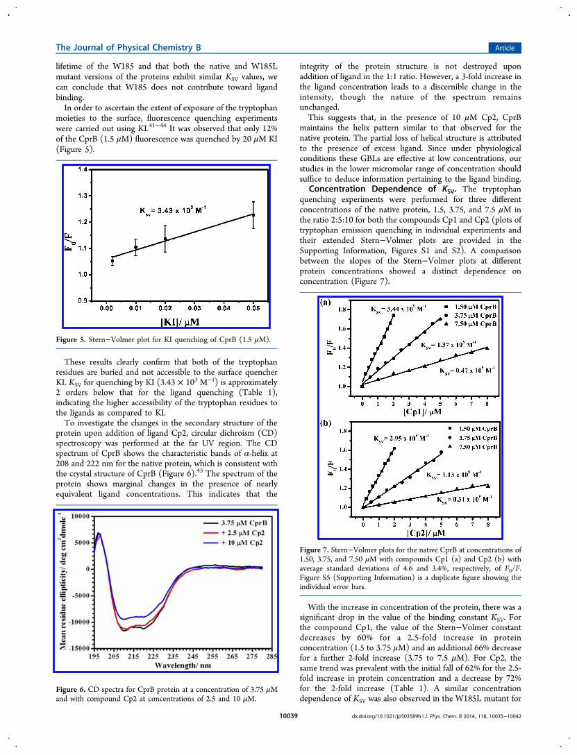
Time resolved fluorescence studies were carried out toascertain whether the observed quenching is static/dynamic innature. The time-resolved fluorescence decay curves wererecorded for CprB in the absence and presence of Cp2 ligandwith increasing concentrations (Figure 4). All the fluorescencedecay curves were fitted by global analysis, and the results arelisted in Table 2.
All the fluorescence decay curves could be fitted to abiexponential function. The presence of two lifetimes can beattributed to slightly different environments experienced by thetwo tryptophan residues and/or by the existence of multipleconformations of each tryptophan.36−39 Assuming that thetryptophan residues are structurally rigid, the individuallifetimes will reflect the local environment.40 Structural analysisshows that both of the tryptophan residues are surrounded byhydrophobic residues. W185, which is in a densely packedenvironment and closer to the surface, most likely has theshorter lifetime τ2, whereas W127 that resides in a deep pocketplausibly possesses the longer lifetime τ1. More importantly, itwas observed that the tryptophan fluorescence lifetimes do notchange with the addition of Cp2, which clearly signifies staticquenching. The X-ray structure reveals that W185 is completelysurrounded by other amino acid residues with no space foreither of the GBLs to come in close proximity to interact withit. Hence, on account of the facts that there is no change in
Table 1. Stern−Volmer Constants (KSV) for QuenchingTitrations of Native CprB and W185L Mutant Protein withLigands Cp1 and Cp2, at Various Concentrations of Protein
protein [protein] (μM) compound KSV (105 M−1)
CprB 1.50 Cp1 3.44 ± 0.56CprB 3.75 Cp1 1.37 ± 0.26CprB 7.50 Cp1 0.47 ± 0.06CprB 1.50 Cp2 2.95 ± 0.37CprB 3.75 Cp2 1.13 ± 0.18CprB 7.50 Cp2 0.31 ± 0.04W185L CprB 3.75 Cp1 0.75 ± 0.04W185L CprB 7.50 Cp1 0.34 ± 0.04W185L CprB 3.75 Cp2 1.03 ± 0.08W185L CprB 7.50 Cp2 0.31 ± 0.05
Figure 4. Time resolved fluorescence decay curve for native CprB (5μM) and complexes with Cp2 (4 and 8 μM). The respective residualplots for the fitting are represented below.
Table 2. Time Resolved Fluorescence Measurement Data ofNative CprB Protein (5 μM) in the Presence of Varying Cp2Concentrations
[Cp2] (μM) α1 α2 τ1 (ns) τ2 (ns) mean τ (ns) χ2
0 0.69 0.31 4.76 2.56 4.08 1.041.0 0.69 0.31 4.76 2.56 4.08 1.072.0 0.65 0.35 4.76 2.56 3.99 1.054.0 0.65 0.35 4.76 2.56 3.99 1.036.0 0.65 0.35 4.76 2.56 3.99 1.028.0 0.68 0.32 4.76 2.56 4.06 1.0510.0 0.66 0.34 4.76 2.56 4.01 1.07
The Journal of Physical Chemistry B Article
dx.doi.org/10.1021/jp503589h | J. Phys. Chem. B 2014, 118, 10035−1004210038
lifetime of the W185 and that both the native and W185Lmutant versions of the proteins exhibit similar KSV values, wecan conclude that W185 does not contribute toward ligandbinding.In order to ascertain the extent of exposure of the tryptophan
moieties to the surface, fluorescence quenching experimentswere carried out using KI.41−44 It was observed that only 12%of the CprB (1.5 μM) fluorescence was quenched by 20 μM KI(Figure 5).
These results clearly confirm that both of the tryptophanresidues are buried and not accessible to the surface quencherKI. KSV for quenching by KI (3.43 × 10³ M−1) is approximately2 orders below that for the ligand quenching (Table 1),indicating the higher accessibility of the tryptophan residues tothe ligands as compared to KI.To investigate the changes in the secondary structure of the
protein upon addition of ligand Cp2, circular dichroism (CD)spectroscopy was performed at the far UV region. The CDspectrum of CprB shows the characteristic bands of α-helix at208 and 222 nm for the native protein, which is consistent withthe crystal structure of CprB (Figure 6).45 The spectrum of theprotein shows marginal changes in the presence of nearlyequivalent ligand concentrations. This indicates that the
integrity of the protein structure is not destroyed uponaddition of ligand in the 1:1 ratio. However, a 3-fold increase inthe ligand concentration leads to a discernible change in theintensity, though the nature of the spectrum remainsunchanged.This suggests that, in the presence of 10 μM Cp2, CprB
maintains the helix pattern similar to that observed for thenative protein. The partial loss of helical structure is attributedto the presence of excess ligand. Since under physiologicalconditions these GBLs are effective at low concentrations, ourstudies in the lower micromolar range of concentration shouldsuffice to deduce information pertaining to the ligand binding.
Concentration Dependence of KSV. The tryptophanquenching experiments were performed for three differentconcentrations of the native protein, 1.5, 3.75, and 7.5 μM inthe ratio 2:5:10 for both the compounds Cp1 and Cp2 (plots oftryptophan emission quenching in individual experiments andtheir extended Stern−Volmer plots are provided in theSupporting Information, Figures S1 and S2). A comparisonbetween the slopes of the Stern−Volmer plots at differentprotein concentrations showed a distinct dependence onconcentration (Figure 7).
With the increase in concentration of the protein, there was asignificant drop in the value of the binding constant KSV. Forthe compound Cp1, the value of the Stern−Volmer constantdecreases by 60% for a 2.5-fold increase in proteinconcentration (1.5 to 3.75 μM) and an additional 66% decreasefor a further 2-fold increase (3.75 to 7.5 μM). For Cp2, thesame trend was prevalent with the initial fall of 62% for the 2.5-fold increase in protein concentration and a decrease by 72%for the 2-fold increase (Table 1). A similar concentrationdependence of KSV was also observed in the W185L mutant for
Figure 5. Stern−Volmer plot for KI quenching of CprB (1.5 μM).
Figure 6. CD spectra for CprB protein at a concentration of 3.75 μMand with compound Cp2 at concentrations of 2.5 and 10 μM.
Figure 7. Stern−Volmer plots for the native CprB at concentrations of1.50, 3.75, and 7.50 μM with compounds Cp1 (a) and Cp2 (b) withaverage standard deviations of 4.6 and 3.4%, respectively, of F0/F.Figure S5 (Supporting Information) is a duplicate figure showing theindividual error bars.
The Journal of Physical Chemistry B Article
dx.doi.org/10.1021/jp503589h | J. Phys. Chem. B 2014, 118, 10035−1004210039
both compounds (Table 1; Figures S3 and S4, SupportingInformation). The cause of this trend could be attributed to thetendency of the protein to oligomerize.23 The property ofoligomerization, which is known to be a concentrationdependent effect, could be the reason behind the inefficientbinding of the compounds with the increased proteinconcentration, thereby resulting in a decreased bindingconstant as depicted in Figure 7.To confirm the oligmerization tendency, dynamic light
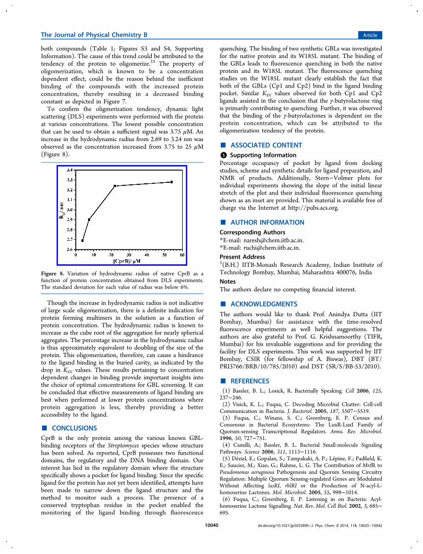
scattering (DLS) experiments were performed with the proteinat various concentrations. The lowest possible concentrationthat can be used to obtain a sufficient signal was 3.75 μM. Anincrease in the hydrodynamic radius from 2.69 to 3.24 nm wasobserved as the concentration increased from 3.75 to 25 μM(Figure 8).
Though the increase in hydrodynamic radius is not indicativeof large scale oligomerization, there is a definite indication forprotein forming multimers in the solution as a function ofprotein concentration. The hydrodynamic radius is known toincrease as the cube root of the aggregation for nearly sphericalaggregates. The percentage increase in the hydrodynamic radiusis thus approximately equivalent to doubling of the size of theprotein. This oligomerization, therefore, can cause a hindranceto the ligand binding in the buried cavity, as indicated by thedrop in KSV values. These results pertaining to concentrationdependent changes in binding provide important insights intothe choice of optimal concentrations for GBL screening. It canbe concluded that effective measurements of ligand binding arebest when performed at lower protein concentrations whereprotein aggregation is less, thereby providing a betteraccessibility to the ligand.
■ CONCLUSIONSCprB is the only protein among the various known GBL-binding receptors of the Streptomyces species whose structurehas been solved. As reported, CprB possesses two functionaldomains, the regulatory and the DNA binding domain. Ourinterest has lied in the regulatory domain where the structurespecifically shows a pocket for ligand binding. Since the specificligand for the protein has not yet been identified, attempts havebeen made to narrow down the ligand structure and themethod to monitor such a process. The presence of aconserved tryptophan residue in the pocket enabled themonitoring of the ligand binding through fluorescence
quenching. The binding of two synthetic GBLs was investigatedfor the native protein and its W185L mutant. The binding ofthe GBLs leads to fluorescence quenching in both the nativeprotein and its W185L mutant. The fluorescence quenchingstudies on the W185L mutant clearly establish the fact thatboth of the GBLs (Cp1 and Cp2) bind in the ligand bindingpocket. Similar KSV values observed for both Cp1 and Cp2ligands assisted in the conclusion that the γ-butyrolactone ringis primarily contributing to quenching. Further, it was observedthat the binding of the γ-butyrolactones is dependent on theprotein concentration, which can be attributed to theoligomerization tendency of the protein.
■ ASSOCIATED CONTENT*S Supporting InformationPercentage occupancy of pocket by ligand from dockingstudies, scheme and synthetic details for ligand preparation, andNMR of products. Additionally, Stern−Volmer plots forindividual experiments showing the slope of the initial linearstretch of the plot and their individual fluorescence quenchingshown as an inset are provided. This material is available free ofcharge via the Internet at http://pubs.acs.org.
■ AUTHOR INFORMATIONCorresponding Authors*E-mail: [email protected].*E-mail: [email protected] Address‡(B.H.) IITB-Monash Research Academy, Indian Institute ofTechnology Bombay, Mumbai, Maharashtra 400076, IndiaNotesThe authors declare no competing financial interest.
■ ACKNOWLEDGMENTSThe authors would like to thank Prof. Anindya Dutta (IITBombay, Mumbai) for assistance with the time-resolvedfluorescence experiments as well helpful suggestions. Theauthors are also grateful to Prof. G. Krishnamoorthy (TIFR,Mumbai) for his invaluable suggestions and for providing thefacility for DLS experiments. This work was supported by IITBombay, CSIR (for fellowship of A. Biswas), DBT (BT/PRI3766/BRB/10/785/2010) and DST (SR/S/BB-53/2010).
■ REFERENCES(1) Bassler, B. L.; Losick, R. Bacterially Speaking. Cell 2006, 125,237−246.(2) Visick, K. L.; Fuqua, C. Decoding Microbial Chatter: Cell-cellCommunication in Bacteria. J. Bacteriol. 2005, 187, 5507−5519.(3) Fuqua, C.; Winans, S. C.; Greenberg, E. P. Census andConsensus in Bacterial Ecosystems: The LuxR-LuxI Family ofQuorum-sensing Transcriptional Regulators. Annu. Rev. Microbiol.1996, 50, 727−751.(4) Camilli, A.; Bassler, B. L. Bacterial Small-molecule SignalingPathways. Science 2006, 311, 1113−1116.(5) Deziel, E.; Gopalan, S.; Tampakaki, A. P.; Lepine, F.; Padfield, K.E.; Saucier, M.; Xiao, G.; Rahme, L. G. The Contribution of MvfR toPseudomonas aeruginosa Pathogenesis and Quorum Sensing CircuitryRegulation: Multiple Quorum Sensing-regulated Genes are ModulatedWithout Affecting lasRI, rhlRI or the Production of N-acyl-L-homoserine Lactones. Mol. Microbiol. 2005, 55, 998−1014.(6) Fuqua, C.; Greenberg, E. P. Listening in on Bacteria: Acyl-homoserine Lactone Signalling. Nat. Rev. Mol. Cell Biol. 2002, 3, 685−695.
Figure 8. Variation of hydrodynamic radius of native CprB as afunction of protein concentration obtained from DLS experiments.The standard deviation for each value of radius was below 6%.
The Journal of Physical Chemistry B Article
dx.doi.org/10.1021/jp503589h | J. Phys. Chem. B 2014, 118, 10035−1004210040
(7) Deziel, E.; Lepine, F.; Milot, S.; He, J.; Mindrinos, M. N.;Tompkins, R. G.; Rahme, L. G. Analysis of Pseudomonas aeruginosa 4-hydroxy-2-alkylquinolines (HAQs) Reveals a Role for 4-hydroxy-2-heptylquinoline in Cell-to-cell Communication. Proc. Natl. Acad. Sci.U. S. A. 2004, 101, 1339−1344.(8) Pesci, E. C.; Milbank, J. B. J.; Pearson, J. P.; McKnight, S.; Kende,A. S.; Greenberg, E. P.; Iglewski, B. H. Quinolone Signaling in theCell-to-cell Communication System of Pseudomonas aeruginosa. Proc.Natl. Acad. Sci. U. S. A. 1999, 96, 11229−11234.(9) Hsiao, N. H.; Nakayama, S.; Merlo, M. E.; de Vries, M.; Bunet,R.; Kitani, S.; Nihira, T.; Takano, E. Analysis of Two AdditionalSignaling Molecules in Streptomyces coelicolor and the Development ofa Butyrolactone-specific Reporter System. Chem. Biol. 2009, 16, 951−960.(10) Takano, E. Gamma-butyrolactones: Streptomyces SignallingMolecules Regulating Antibiotic Production and Differentiation. Curr.Opin. Microbiol. 2006, 9, 287−294.(11) Pottathil, M.; Lazazzera, B. A. The Extracellular Phr Peptide-RapPhosphatase Signaling Circuit of Bacillus subtilis. Front. Biosci. 2003, 8,d32−d45.(12) Holden, M. T.; Ram, C. S.; de Nys, R.; Stead, P.; Bainton, N. J.;Hill, P. J.; Manefield, M.; Kumar, N.; Labatte, M.; England, D.; et al.Quorum-sensing Cross Talk: Isolation and Chemical Characterizationof Cyclic Dipeptides from Pseudomonas aeruginosa and other Gram-negative Bacteria. Mol. Microbiol. 1999, 33, 1254−1266.(13) Khokhlov, A. S.; Tovarova, I. I.; Borisova, L. N.; Pliner, S. A.;Shevchenko, L. N.; Kornitskaia, E. I.; Ivkina, N. S.; Rapoport, I. A. TheA-factor, Responsible for Streptomycin Biosynthesis by Mutant Strainsof Actinomyces streptomycini. Dokl. Akad. Nauk SSSR 1967, 177, 232−235.(14) Horinouchi, S. A Microbial Hormone, A-factor, as a MasterSwitch for Morphological Differentiation and Secondary Metabolismin Streptomyces griseus. Front. Biosci. 2002, 7, d2045−d2057.(15) Sugiyama, M.; Onaka, H.; Nakagawa, T.; Horinouchi, S. Site-directed Mutagenesis of the A-factor Receptor Protein: Val-41Important for DNA-binding and Trp-119 Important for Ligand-binding. Gene 1998, 222, 133−144.(16) Onaka, H.; Ando, N.; Nihira, T.; Yamada, Y.; Beppu, T.;Horinouchi, S. Cloning and Characterization of the A-factor ReceptorGene from Streptomyces griseus. J. Bacteriol. 1995, 177, 6083−6092.(17) Horinouchi, S.; Beppu, T. A-factor as a Microbial Hormone thatControls Cellular Differentiation and Secondary Metabolism inStreptomyces griseus. Mol. Microbiol. 1994, 12, 859−864.(18) Kinoshita, H.; Ipposhi, H.; Okamoto, S.; Nakano, H.; Nihira, T.;Yamada, Y. Butyrolactone Autoregulator Receptor Protein (BarA) as aTranscriptional Regulator in Streptomyces virginiae. J. Bacteriol. 1997,179, 6986−6993.(19) Takano, E.; Chakraburtty, R.; Nihira, T.; Yamada, Y.; Bibb, M. J.A Complex Role for the γ-butyrolactone SCB1 in RegulatingAntibiotic Production in Streptomyces coelicolor A3(2). Mol. Microbiol.2001, 41, 1015−1028.(20) Waki, M.; Nihira, T.; Yamada, Y. Cloning and Characterizationof the Gene (farA) Encoding the Receptor for an ExtracellularRegulatory Factor (IM-2) from Streptomyces sp. Strain FRI-5. J.Bacteriol. 1997, 179, 5131−5137.(21) Kitani, S.; Kinoshita, H.; Nihira, T.; Yamada, Y. In Vitro Analysisof the Butyrolactone Autoregulator Receptor Protein (FarA) ofStreptomyces lavendulae FRI-5 Reveals that FarA Acts as a DNA-Binding Transcriptional Regulator that Controls its Own Synthesis. J.Bacteriol. 1999, 181, 5081−5084.(22) Onaka, H.; Nakagawa, T.; Horinouchi, S. Involvement of TwoA-factor Receptor Homologues in Streptomyces coelicolor A3(2) in theRegulation of Secondary Metabolism and Morphogenesis. Mol.Microbiol. 1998, 28, 743−753.(23) Natsume, R.; Takeshita, R.; Sugiyama, M.; Ohnishi, Y.; Senda,T.; Horinouchi, S. Crystallization of CprB, an Autoregulator-receptorProtein from Streptomyces coelicolor A3(2). Acta Crystallogr., Sect. D2003, 59, 2313−2315.
(24) Natsume, R.; Ohnishi, Y.; Senda, T.; Horinouchi, S. CrystalStructure of a Gamma-butyrolactone Autoregulator Receptor Proteinin Streptomyces coelicolor A3(2). J. Mol. Biol. 2004, 336, 409−419.(25) Ramos, J. L.; Martinez-Bueno, M.; Molina-Henares, A. J.; Teran,W.; Watanabe, K.; Zhang, X.; Gallegos, M. T.; Brennan, R.; Tobes, R.The TetR Family of Transcriptional Repressors. Microbiol. Mol. Biol.Rev. 2005, 69, 326−356.(26) Morris, G. M.; Huey, R.; Lindstrom, W.; Sanner, M. F.; Belew,R. K.; Goodsell, D. S.; Olson, A. J. AutoDock4 and AutoDockTools4:Automated Docking with Selective Receptor Flexibility. J. Comput.Chem. 2009, 30, 2785−2791.(27) Weber, W.; Schoenmakers, R.; Spielmann, M.; El-Baba, M. D.;Folcher, M.; Keller, B.; Weber, C. C.; Link, N.; van de Wetering, P.;Heinzen, C.; et al. Streptomyces-derived Quorum-sensing SystemsEngineered for Adjustable Transgene Expression in Mammalian Cellsand Mice. Nucleic Acids Res. 2003, 31, e71.(28) Takano, E.; Nihira, T.; Hara, Y.; Jones, J. J.; Gershater, C. J.;Yamada, Y.; Bibb, M. Purification and Structural Determination ofSCB1, a Gamma-butyrolactone that Elicits Antibiotic Production inStreptomyces coelicolor A3(2). J. Biol. Chem. 2000, 275, 11010−11016.(29) Yamada, Y.; Sugamura, K.; Kondo, K.; Yanagimoto, M.; Okada,H. The Structure of Inducing Factors for Virginiamycin Production inStreptomyces virginiae. J. Antibiot.(Tokyo) 1987, 40, 496−504.(30) Konev, S. V. Fluorescence and Phosphorescence of Proteins andNucleic Acids; Plenum Press: New York, 1967.(31) Papadopoulou, A.; Green, R. J.; Frazier, R. A. Interaction ofFlavonoids with Bovine Serum Albumin: A Fluorescence QuenchingStudy. J. Agric. Food Chem. 2004, 53, 158−163.(32) Sukowska, A. Interaction of Drugs with Bovine and HumanSerum Albumin. J. Mol. Struct. 2002, 614, 227−232.(33) Sonveaux, N.; Vigano, C.; Shapiro, A. B.; Ling, V.; Ruysschaert,J. M. Ligand-mediated Tertiary Structure Changes of Reconstituted P-glycoprotein: A Tryptophan Fluorescence Quenching Analysis. J. Biol.Chem. 1999, 274, 17649−17654.(34) Chadborn, N.; Bryant, J.; Bain, A. J.; O’Shea, P. Ligand-dependent Conformational Equilibria of Serum Albumin Revealed byTryptophan Fluorescence Quenching. Biophys. J. 1999, 76, 2198−2207.(35) James, N. G.; Byrne, S. L.; Steere, A. N.; Smith, V. C.; MacGillivray, R. T.; Mason, A. B. Inequivalent Contribution of the FiveTryptophan Residues in the C-Lobe of Human Serum Transferrin tothe Fluorescence Increase when Iron is Released. Biochemistry 2009,48, 2858−2867.(36) Beechem, J. M.; Brand, L. Time-Resolved Fluorescence ofProteins. Annu. Rev. Biochem. 1985, 54, 43−71.(37) Engh, R. A.; Chen, L. X. Q.; Fleming, G. R. ConformationalDynamics of Tryptophan: A Proposal for the Origin of the Non-exponential Fluorescence Decay. Chem. Phys. Lett. 1986, 126, 365−372.(38) Petrich, J. W.; Chang, M. C.; McDonald, D. B.; Fleming, G. R.On the Origin of Nonexponential Fluorescence Decay in Tryptophanand its Derivatives. J. Am. Chem. Soc. 1983, 105, 3824−3832.(39) Chang, M. C.; Petrich, J. W.; McDonald, D. B.; Fleming, G. R.Nonexponential Fluorescence Decay of Tryptophan, Tryptophylgly-cine, and Glycyltryptophan. J. Am. Chem. Soc. 1983, 105, 3819−3824.(40) Rouviere, N.; Vincent, M.; Craescu, C. T.; Gallay, J.Immunosuppressor Binding to the Immunophilin FKBP59 Affectsthe Local Structural Dynamics of a Surface β-strand: Time-resolvedFluorescence Study. Biochemistry 1997, 36, 7339−7352.(41) Lehrer, S. S. Solute Perturbation of Protein Fluorescence.Quenching of the Tryptophyl Fluorescence of Model Compounds andof Lysozyme by Iodide Ion. Biochemistry 1971, 10, 3254−3263.(42) Lakshmikanth, G. S.; Krishnamoorthy, G. Solvent-exposedTryptophans Probe the Dynamics at Protein Surfaces. Biophys. J. 1999,77, 1100−1106.(43) Pawagi, A. B.; Deber, C. M. Ligand-dependent Quenching ofTryptophan Fluorescence in Human Erythrocyte Hexose TransportProtein. Biochemistry 1990, 29, 950−955.
The Journal of Physical Chemistry B Article
dx.doi.org/10.1021/jp503589h | J. Phys. Chem. B 2014, 118, 10035−1004210041

(44) Hansen, D.; Altschmied, L.; Hillen, W. Engineered TetRepressor Mutants with Single Tryptophan Residues as FluorescentProbes. Solvent Accessibilities of DNA and Inducer Binding Sites andInteraction with Tetracycline. J. Biol. Chem. 1987, 262, 14030−14035.(45) Holzwarth, F.; Doty, P. The Ultraviolet Circular Dichroism ofPolypeptides. J. Am. Chem. Soc. 1965, 87, 218−228.
The Journal of Physical Chemistry B Article
dx.doi.org/10.1021/jp503589h | J. Phys. Chem. B 2014, 118, 10035−1004210042
S1
Supporting Information
Fluorescence Quenching Studies of γ-Butyrolactone Binding Protein (CprB) from
Streptomyces coelicolor A3(2)
Anwesha Biswas, Ravi K. Swarnkar, Bhukya Hussain, Suraj K. Sahoo, P. I. Pradeepkumar, G.
Naresh Patwari,* Ruchi Anand*
Department of Chemistry, Indian Institute of Technology Bombay, Mumbai, Maharashtra 400076,
India
Details of docking studies in terms of percentage occupancy of pocket by ligand have been
provided. The scheme and synthetic details for ligand preparation and NMR of respective
products have also been included. Additionally, Stern-Volmer plots for individual
experiments showing slope of the initial linear stretch of the plot and their individual
fluorescence quenching (inset) have been provided. Stern-Volmer plots of the main paper
have also been shown here along with the error bars.
S2
Results from docking studies of CprB and GBLs, with varying aliphatic
chain length
Table S1. Table showing the percentage of ligand occupancy within the proposed ligand
binding pocket for the respective GBL with the specified length of aliphatic chain at C2,
from docking studies.
Carbon
chain
length
Percentage
occupancy outside
ligand binding
5 10.8
6 10
7 0
8 21.6
9 21.2
10
11
12
37.2
46.2
62.8
S3
Preparation of ligands Cp1 and Cp2
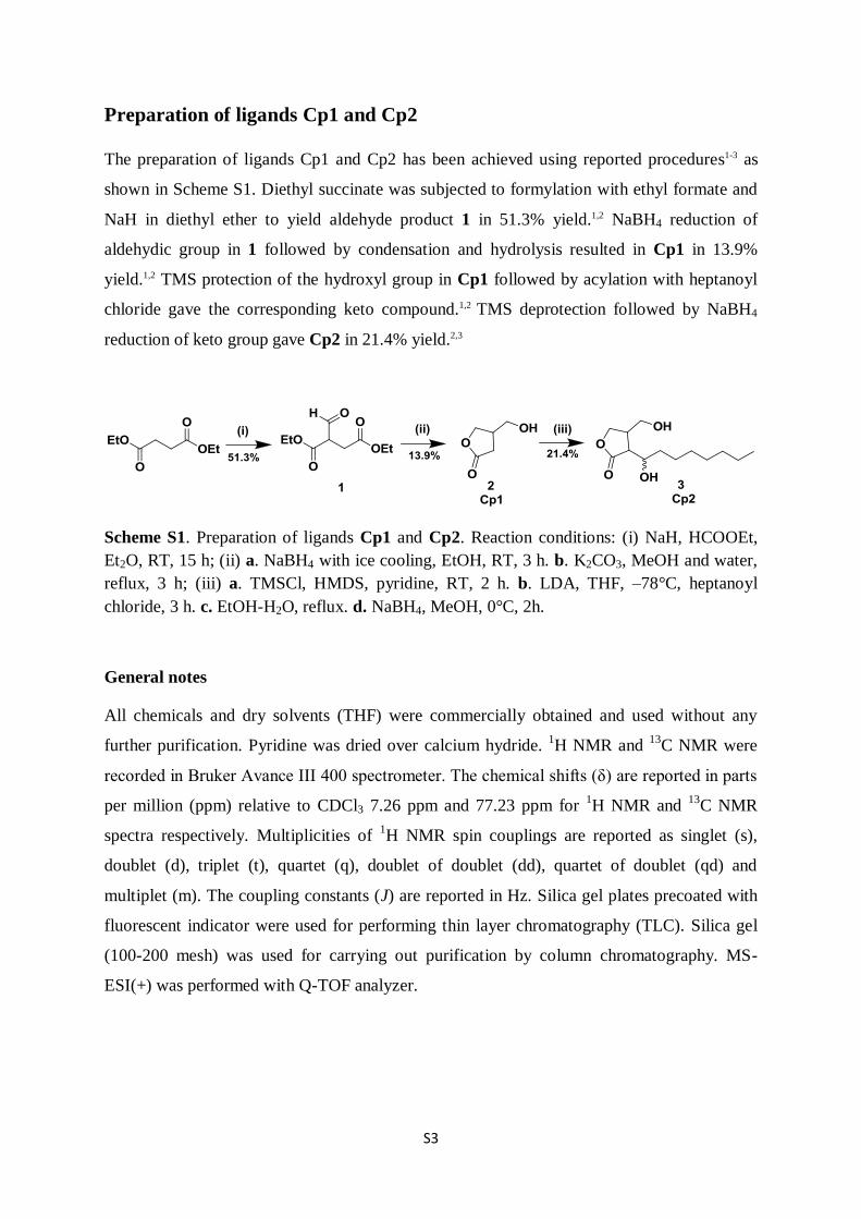
The preparation of ligands Cp1 and Cp2 has been achieved using reported procedures1-3 as
shown in Scheme S1. Diethyl succinate was subjected to formylation with ethyl formate and
NaH in diethyl ether to yield aldehyde product 1 in 51.3% yield.1,2 NaBH4 reduction of
aldehydic group in 1 followed by condensation and hydrolysis resulted in Cp1 in 13.9%
yield.1,2 TMS protection of the hydroxyl group in Cp1 followed by acylation with heptanoyl
chloride gave the corresponding keto compound.1,2 TMS deprotection followed by NaBH4
reduction of keto group gave Cp2 in 21.4% yield.2,3
Scheme S1. Preparation of ligands Cp1 and Cp2. Reaction conditions: (i) NaH, HCOOEt,
Et2O, RT, 15 h; (ii) a. NaBH4 with ice cooling, EtOH, RT, 3 h. b. K2CO3, MeOH and water,
reflux, 3 h; (iii) a. TMSCl, HMDS, pyridine, RT, 2 h. b. LDA, THF, ‒78°C, heptanoyl
chloride, 3 h. c. EtOH-H2O, reflux. d. NaBH4, MeOH, 0°C, 2h.
General notes
All chemicals and dry solvents (THF) were commercially obtained and used without any
further purification. Pyridine was dried over calcium hydride. 1H NMR and
13C NMR were
recorded in Bruker Avance III 400 spectrometer. The chemical shifts (δ) are reported in parts
per million (ppm) relative to CDCl3 7.26 ppm and 77.23 ppm for 1H NMR and
13C NMR
spectra respectively. Multiplicities of 1H NMR spin couplings are reported as singlet (s),
doublet (d), triplet (t), quartet (q), doublet of doublet (dd), quartet of doublet (qd) and
multiplet (m). The coupling constants (J) are reported in Hz. Silica gel plates precoated with
fluorescent indicator were used for performing thin layer chromatography (TLC). Silica gel
(100-200 mesh) was used for carrying out purification by column chromatography. MS-
ESI(+) was performed with Q-TOF analyzer.
S4
Experimental details
Diethyl formyl succinate (1)
To a stirred solution of NaH (2.75 g, 114 mmol) in 75 mL of diethyl ether, a mixture of
diethyl succinate (20 g, 114 mmol), ethyl formate (10.2 g, 138 mmol) and 20 mL ethanol was
added at room temperature and stirring was continued for 5 h. The resulting reaction mixture
was left overnight. Then, 50 mL water was added to the reaction mixture and the aqueous
phase was acidified using 2 M HCl followed by washing with brine solution and subsequent
extraction with diethyl ether (3 × 150 mL). The ether extract was then dried over Na2SO4,
filtered and concentrated. The remaining liquid was distilled under vacuum (bp 76 °C/6
mbar), resulting in a colorless oil (12 g, 51.3%) to yield compound 1. Rƒ = 0.3 (33% ethyl
acetate in hexane); 1H NMR (400 MHz, CDCl3), : 11.52 (d, J = 12.6 Hz, 1H), 7.08 (d, J =
12.7 Hz, 1H), 4.29-4.20 (m, 4H), 2.89 (qd, J = 14.7, 6.1 Hz, 2H), 1.30 (t, J = 7.1 Hz, 6H);
MS-ESI(+): Calculated for C9H15O5 [M+H]+ 203.09; found 203.09.
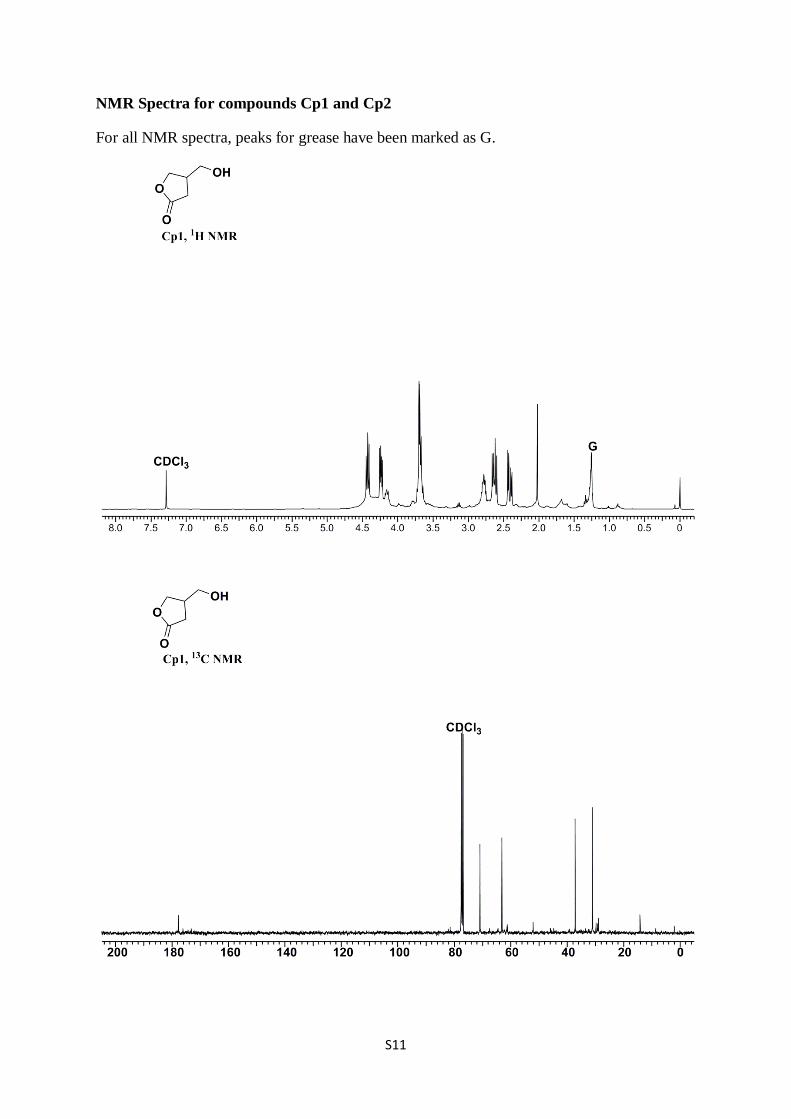
3-hydroxymethylbutanolide (Cp1)
NaBH4 (1.01 g, 26 mmol) was added stepwise to an ice cold solution of 1 (5 g, 24 mmol) in
20 mL ethanol under stirring. Then the reaction mixture was stirred for 3 h at room
temperature. 4 M aqueous HCl solution was added and the solution was stirred further for 1
h. The precipitate formed was filtered off. The filtrate was concentrated and the residue was
extracted with ethyl acetate. The organic solution was washed with brine, dried over Na2SO4
and concentrated to give 2 g of yellowish oil. The oil was dissolved in a mixture of 12 mL
methanol and 4 mL water and then K2CO3 (1.5 g) was added to it in small portions. The
mixture was then refluxed for 3 h followed by acidification with 4 M HCl and concentrated.
The residue was extracted with ethyl acetate (3 × 125 mL) and the organic solution was
washed with brine, dried over Na2SO4 and concentrated. The product was purified by column
chromatography (10% DCM in methanol) resulting in the pure lactone Cp2 (0.4 g, 21%). Rƒ
= 0.53 (5% ethanol in ethyl acetate); 1H NMR (400 MHz, CDCl3), δ: 4.41 (dd, J = 9.3, 7.5
Hz, 1H), 4.22 (dd, J = 9.3, 5.3 Hz, 1H), 3.71-3.62 (m, 2H), 2.80-2.73 (m, 1H), 2.61 (dd, J =
17.7, 8.9 Hz, 1H), 2.39 (dd, J = 17.7, 5.9 Hz, 1H); 13
C-NMR (100 MHz; CDCl3), δ: 177.8,
70.9, 63.1, 37.2, 31.1; MS-ESI(+): Calculated for C5H9O3 [M+H]+ 117.0547; found
117.0552.
S5
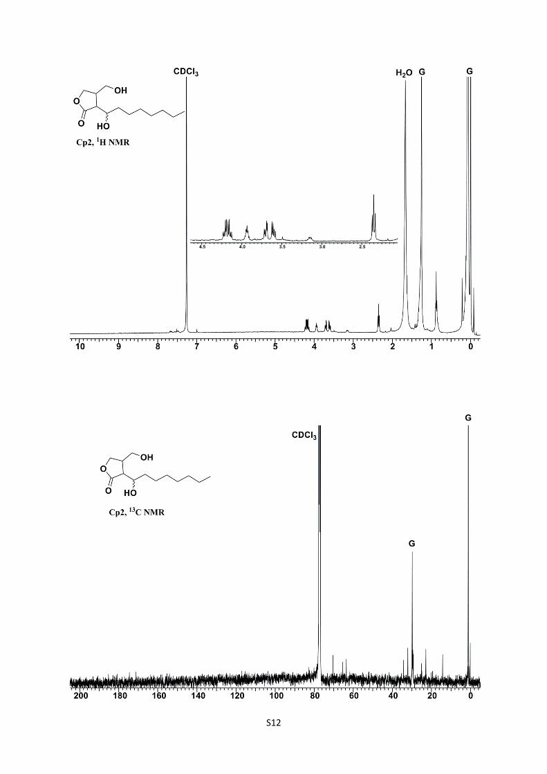
2-(1'-hydroxyoctyl)-3-hydroxymethylbutanolide (Cp2)
Hexamethyldisilazane (0.09 mL, 0.40 mmol) and trimethylsilyl chloride (0.08 mL, 0.62
mmol) were added to an ice-cooled solution of Cp1 (60 mg, 0.52 mmol) in 2 mL pyridine,
and the reaction mixture was and stirred for 2 h at room temperature under a nitrogen
atmosphere resulting in the formation of white precipitate. Then, 5 mL of benzene/hexane
(1:1 v/v) was added to the reaction mixture and the precipitate was removed by filtration. The
filtrate was concentrated and the crude material subjected to column chromatography (33%
ethyl acetate in hexane) to produce a yellowish oily product (78 mg).
The oily compound was dissolved in 3 mL of THF and added to LDA solution [prepared by
using N,N-diisopropylamine (0.145 mL, 1.06 mmol) and n-butyl lithium (0.65 mL, 1.06
mmol, 1.6 M in hexane) at –78 °C followed by stirring for 15 min]. To this mixture heptanoyl
chloride (60 mg, 0.41 mmol), was added and the reaction was stirred for another 1.5 h, at –78
°C. The reaction mixture was allowed to warm up to 0 °C followed by the addition of 10 mL
ice-cold water with 1.5 mL acetic acid. The product formed was subsequently extracted with
dichloromethane (3 × 100 mL) and concentrated. The residue was dissolved in 200 mL
dichloromethane, washed with 100 mL saturated aq. NaHCO3 and brine. After removal of the
solvents, the silyl protected compound was obtained as yellowish oil. The trimethylsilyl
group was removed by refluxing with 10 mL of ethanol and 4 mL of water for 30 min. After
concentrating, the resulting mixture was purified by column chromatography (20-33% ethyl
acetate in hexane) to yield 2-(octanoyl)-3-hydroxymethylbutanolide (50 mg, 0.2 mmol) as an
oily liquid.
The compound was dissolved in 8 mL of ethanol and NaBH4 (10.5 mg, 0.3 mol) was added at
0 °C with stirring for 3 h followed by the addition of 3 M aqueous HCl solution. The
precipitate formed was removed by filtration and the aqueous solution was extracted with
ethyl acetate (3 × 50 mL) and the organic extract was dried over Na2SO4. The compound was
purified by HPLC to obtain Cp2 in 21.4% yield (27.73 mg). Rƒ = 0.32 (33% ethyl acetate in
hexane); 1H NMR (400 MHz, CDCl3), δ: 4.23-4.13 (m, 2H), 3.96-3.91 (m, 1H), 3.70 (dd, J =
11.4, 4.0 Hz, 1H), 3.60 (dd, J = 11.5, 5.7 Hz, 1H), 3.19-3.11 (m, 1H), 2.35 (t, J = 7.6 Hz,
1H), 1.67 (m), 1.25 (m), 0.88 (t, J = 6.8 Hz, 3H); 13
C-NMR (100 MHz; CDCl3), δ: 70.5, 65.4,
63.6, 34.4, 32.2, 29.67, 29.58, 29.47, 29.35, 25.1, 22.9, 14.3; MS-ESI(+): Calculated for
C13H25O4 [M+H]+ 245.1753; found 245.1754.
S6
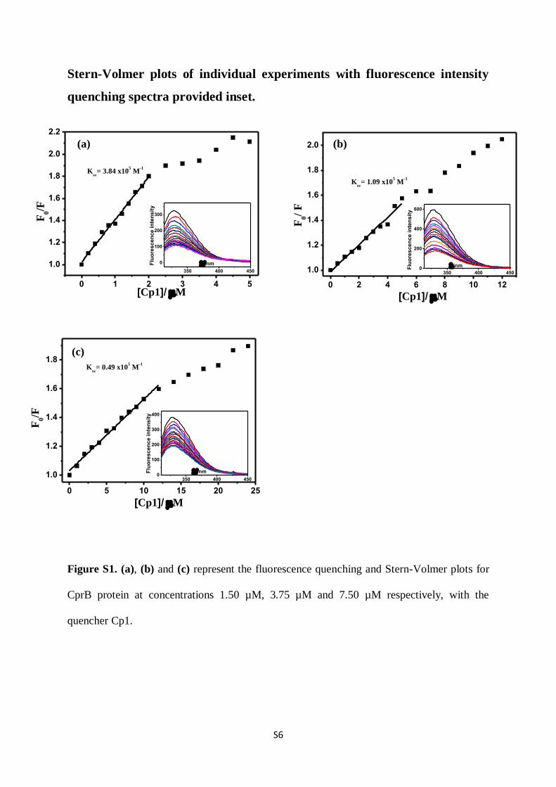
Stern-Volmer plots of individual experiments with fluorescence intensity
quenching spectra provided inset.
Figure S1. (a), (b) and (c) represent the fluorescence quenching and Stern-Volmer plots for
CprB protein at concentrations 1.50 µM, 3.75 µM and 7.50 µM respectively, with the
quencher Cp1.
0 1 2 3 4 5
1.0
1.2
1.4
1.6
1.8
2.0
2.2
Ksv
= 3.84 x10⁵ M
-1
F0/F
[Cp1]/ M
350 400 450
0
100
200
300
Flu
ore
sc
en
ce
in
ten
sit
y
nm
350 400 4500
200
400
600
Flu
ore
sc
en
ce
in
ten
sit
y
/nm
350 400 4500
100
200
300
400
Flu
ore
sc
en
ce
in
ten
sit
y
nm
0 5 10 15 20 25
1.0
1.2
1.4
1.6
1.8K
sv= 0.49 x10
5 M
-1
F0/F
[Cp1]/ M
(c)
(b) (a)
0 2 4 6 8 10 12
1.0
1.2
1.4
1.6
1.8
2.0
F0/
F[Cp1]/ M
Ksv
= 1.09 x10⁵ M
-1
S7
350 400 450
0
100
200
300
Flu
ore
sc
en
ce
in
ten
sit
y
nm
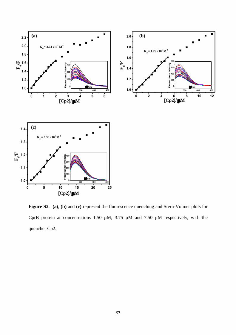
Figure S2. (a), (b) and (c) represent the fluorescence quenching and Stern-Volmer plots for
CprB protein at concentrations 1.50 µM, 3.75 µM and 7.50 µM respectively, with the
quencher Cp2.
350 400 4500
100
200
300
400
Flu
ore
sce
nce
in
ten
sit
y
nm
0 1 2 3 4 5 6
1.0
1.2
1.4
1.6
1.8
2.0
2.2
Ksv
= 3.24 x10⁵ M
-1
F0/F
[Cp2]/ M
350 400 450
0
200
400
600
800
Flu
ore
sc
en
ce
in
ten
sit
y
nm
0 5 10 15 20 25
1.0
1.1
1.2
1.3
1.4
Ksv
= 0.30 x10⁵ M
-1
F0/F
[Cp2]/ M
(c)
(a) (b)
0 2 4 6 8 10 12
1.0
1.2
1.4
1.6
1.8
2.0
F0/F
[Cp2]/ M
Ksv
= 1.26 x10⁵ M
-1
(a)
S8
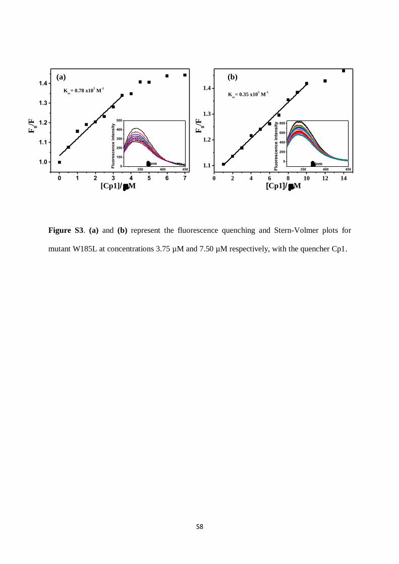
Figure S3. (a) and (b) represent the fluorescence quenching and Stern-Volmer plots for
mutant W185L at concentrations 3.75 µM and 7.50 µM respectively, with the quencher Cp1.
0 1 2 3 4 5 6 7
1.0
1.1
1.2
1.3
1.4K
sv= 0.78 x10
5 M
-1
F0/F
[Cp1]/ M
350 400 4500
100
200
300
400
500
Flu
ore
sce
nce
in
ten
sit
y
/nm
350 400 450
0
200
400
600
800
Flu
ore
sc
en
ce
in
ten
sit
y
/nm
0 2 4 6 8 10 12 14
1.1
1.2
1.3
1.4K
sv= 0.35 x10
5 M
-1
F0/F
[Cp1]/ M
(a) (b)
S9
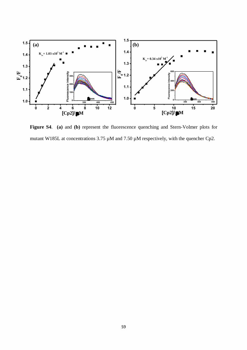
Figure S4. (a) and (b) represent the fluorescence quenching and Stern-Volmer plots for
mutant W185L at concentrations 3.75 µM and 7.50 µM respectively, with the quencher Cp2.
350 400 4500
200
400
600
Flu
ore
scen
ce i
nte
nsit
y
/nm
0 2 4 6 8 10 12
1.0
1.1
1.2
1.3
1.4
1.5
Ksv
= 1.03 x10⁵ M
-1
F0/F
[Cp2]/M
350 400 4500
100
200
300
Flu
ore
scen
ce in
ten
sit
y
/nm
(a) (b)
0 5 10 15 20
1.0
1.1
1.2
1.3
1.4
1.5
F0/F
Ksv
= 0.34 x10⁵ M
-1
[Cp2]/ M
S10
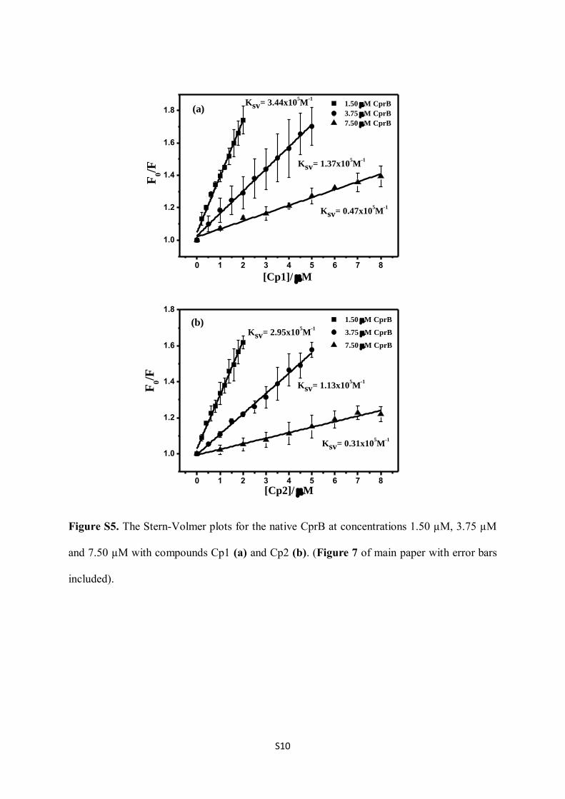
Figure S5. The Stern-Volmer plots for the native CprB at concentrations 1.50 µM, 3.75 µM
and 7.50 µM with compounds Cp1 (a) and Cp2 (b). (Figure 7 of main paper with error bars
included).
0 1 2 3 4 5 6 7 8
1.0
1.2
1.4
1.6
1.8
F0/F
[Cp1]/ M
Ksv= 0.47x10⁵M
-1
Ksv= 1.37x10⁵M
-1
Ksv= 3.44x10⁵M
-1
1.50 M CprB
3.75 M CprB
7.50 M CprB
0 1 2 3 4 5 6 7 8
1.0
1.2
1.4
1.6
1.8
Ksv= 0.31x10⁵M
-1
Ksv= 1.13x10⁵M
-1
Ksv= 2.95x10⁵M
-1
F0/F
1.50 M CprB
3.75 M CprB
7.50 M CprB
[Cp2]/ M
(a)
(b)
S11
NMR Spectra for compounds Cp1 and Cp2
For all NMR spectra, peaks for grease have been marked as G.
S12

S13
References:
(1) Yamada, Y.; Sugamura, K.; Kondo, K.; Yanagimoto, M.; Okada, H. The Structure of
Inducing Factors for Virginiamycin Production in Streptomyces virginiae. J. Antibiot. 1987,
40, 496‒504.
(2) Weber, W.; Schoenmakers, R.; Spielmann, M.; El-Baba, M. D.; Folcher, M.; Keller,
B.; Weber, C. C.; Link, N.; van de Wetering, P.; Heinzen, C.; Jolivet, B.; Sequin, U.; Aubel,
D.; Thompson, C. J.; Fussenegger, M. Streptomyces-derived Quorum-sensing Systems
Engineered for Adjustable Transgene Expression in Mammalian Cells and Mice. Nucleic.
Acids Res. 2003, 31, e71.
(3) Takano, E.; Nihira, T.; Hara, Y.; Jones, J. J.; Gershater, C. J.; Yamada, Y.; Bibb, M.
Purification and Structural Determination of SCB1, a Gamma-butyrolactone that Elicits
Antibiotic Production in Streptomyces coelicolor A3(2). J. Biol. Chem. 2000, 275,
11010‒11016.
References from main paper with more than 10 authors:
(12) Holden, M. T.; Ram, C. S.; de Nys, R.; Stead, P.; Bainton, N. J.; Hill, P. J.;
Manefield, M.; Kumar, N.; Labatte, M.; England, D.; Rice, S.; Givskov, M.; Salmond, G. P.;
Stewart, G. S.; Bycroft, B. W.; Kjelleberg, S.; Williams, P. Quorum-sensing Cross Talk:
Isolation and Chemical Characterization of Cyclic Dipeptides from Pseudomonas aeruginosa
and Other Gram-negative Bacteria. Mol. Microbiol. 1999, 33, 1254‒1266.
(27) Weber, W.; Schoenmakers, R.; Spielmann, M.; El-Baba, M. D.; Folcher, M.; Keller,
B.; Weber, C. C.; Link, N.; van de Wetering, P.; Heinzen, C.; Jolivet, B.; Sequin, U.; Aubel,
D.; Thompson, C. J.; Fussenegger, M. Streptomyces-derived Quorum-sensing Systems
Engineered for Adjustable Transgene Expression in Mammalian Cells and Mice. Nucleic.
Acids Res. 2003, 31, e71.
![Efficient construction of highly functionalizedS1 Efficient construction of highly functionalized spiro[γ-butyrolactone-pyrrolidin-3,3′-oxindole] tricyclic skeletons via an organocatalytic](https://static.fdocument.org/doc/165x107/60fac77bcf8dba3437692a22/efficient-construction-of-highly-s1-efficient-construction-of-highly-functionalized.jpg)


![GRAZING INCIDENCE X-RAY FLUORESCENCE OF MAGNETIC … · 2001. 10. 9. · Analyzing crystal 2 θ Fluorescence 2φ In ... based on the layered model of Vidal & Vincent [6], to calculate](https://static.fdocument.org/doc/165x107/60d08a535057142e751bd384/grazing-incidence-x-ray-fluorescence-of-magnetic-2001-10-9-analyzing-crystal.jpg)