Expression and regulation of ATF6α in the mouse uterus ...
15
RESEARCH Open Access Expression and regulation of ATF6α in the mouse uterus during embryo implantation Yongjie Xiong 1,2† , Wenzhe Li 1,2† , Pengfei Lin 1,2 , Lei Wang 1,2 , Nan Wang 1,2 , Fenglei Chen 1,2 , Xiao Li 1,2 , Aihua Wang 2 and Yaping Jin 1,2* Abstract Background: ATF6α, one of the sensor proteins in the stress signaling pathway of the endoplasmic reticulum, is located in the membrane of the endoplasmic reticulum. To date, the physiological function of ATF6α in the process of embryo implantation has not been reported. Methods: In this study, the expression pattern of ATF6α in the mouse uterus during peri-implantation and the estrous cycle was detected by real-time PCR, western blot and immunohistochemistry. Results: ATF6α mRNA and protein levels were higher in the uterus near the implantation site on day 5 and were intensely expressed in the secondary decidual zone (SDZ) on days 7–8. In the uteri of pseudopregnant mice, ATF6α mRNA and protein levels were lower on day 5 than on other days. The activating blastocyst and artificial decidualization had an obvious effect of increasing the expression of ATF6α. In addition, the expression of ATF6α was affected by progesterone (P 4 ) and estrogen (E 2 ) in ovariectomized mice. This finding is further supported by evidence from mice during the estrous cycle. Conclusions: Thus, we have concluded that ATF6α may play an important role during embryo implantation and decidualization. Keywords: ATF6α, Progesterone, Estrogen, Uterus, Implantation, Decidualization Background Embryo implantation is a complex process for the suc- cessful mammalian pregnancy and is affected by various factors [1–3]. The precise coordination between blasto- cyst activation and maternal endometrium receptivity is the most important step [4]. Once the apposition of the activated blastocyst is complete, the processes of adhe- sion and invasion must be initiated successively. A bat- tery of physiological changes occurs in the cells and tissues of the uterus during early pregnancy, including morphological changes and the secretion of cytokines or proteins related to embryonic development and uterine receptivity [5, 6]. Implantation failure is one of the main causes of infertility. Although many molecular modula- tors have been identified as important for embryo implantation, the complete molecular mechanism is still unclear [7, 8]. Activating transcription factor 6 (ATF6) is a sensor protein in the membrane of the endoplasmic reticulum [9]. ATF6 includes two isoforms, known as ATF6α and ATF6β, which are members of the activating transcrip- tion factor (ATF)/cyclic adenosine monophosphate re- sponse element-binding protein (CREB) family [10]. Under the conditions of endoplasmic reticulum stress, the two isoforms are transported to the Golgi complex, where they are hydrolyzed into their cleaved forms, ATF6α (P50-ATF6α/N-ATF6α) and ATF6β (P50-ATF6β/ N-ATF6β), by site-1 protease (S1P) and site-2 protease (S2P), respectively [11]. The cleaved ATF6 then translo- cates to the nucleus where it induces the expression of its downstream target genes [12, 13]. ATF6α plays a dominant role in inducing the expression of ATF6 target genes, while ATF6β plays a compensatory role in the ab- sence of ATF6α [14–16]. * Correspondence: [email protected] † Equal contributors 1 Key Laboratory of Animal Biotechnology of the Ministry of Agriculture, Northwest A&F University, Yangling, Shaanxi 712100, China 2 College of Veterinary Medicine, Northwest A&F University, Yangling, Shaanxi 712100, China © 2016 The Author(s). Open Access This article is distributed under the terms of the Creative Commons Attribution 4.0 International License (http://creativecommons.org/licenses/by/4.0/), which permits unrestricted use, distribution, and reproduction in any medium, provided you give appropriate credit to the original author(s) and the source, provide a link to the Creative Commons license, and indicate if changes were made. The Creative Commons Public Domain Dedication waiver (http://creativecommons.org/publicdomain/zero/1.0/) applies to the data made available in this article, unless otherwise stated. Xiong et al. Reproductive Biology and Endocrinology (2016) 14:65 DOI 10.1186/s12958-016-0199-0
Transcript of Expression and regulation of ATF6α in the mouse uterus ...
RESEARCH Open Access
Expression and regulation of ATF6α in themouse uterus during embryo implantationYongjie Xiong1,2†, Wenzhe Li1,2†, Pengfei Lin1,2, Lei Wang1,2, Nan Wang1,2, Fenglei Chen1,2, Xiao Li1,2,Aihua Wang2 and Yaping Jin1,2*
Abstract
Background: ATF6α, one of the sensor proteins in the stress signaling pathway of the endoplasmic reticulum, islocated in the membrane of the endoplasmic reticulum. To date, the physiological function of ATF6α in the processof embryo implantation has not been reported.
Methods: In this study, the expression pattern of ATF6α in the mouse uterus during peri-implantation and theestrous cycle was detected by real-time PCR, western blot and immunohistochemistry.
Results: ATF6α mRNA and protein levels were higher in the uterus near the implantation site on day 5 and wereintensely expressed in the secondary decidual zone (SDZ) on days 7–8. In the uteri of pseudopregnant mice, ATF6αmRNA and protein levels were lower on day 5 than on other days. The activating blastocyst and artificial decidualizationhad an obvious effect of increasing the expression of ATF6α. In addition, the expression of ATF6α was affected byprogesterone (P4) and estrogen (E2) in ovariectomized mice. This finding is further supported by evidence from miceduring the estrous cycle.
Conclusions: Thus, we have concluded that ATF6α may play an important role during embryo implantation anddecidualization.
Keywords: ATF6α, Progesterone, Estrogen, Uterus, Implantation, Decidualization
BackgroundEmbryo implantation is a complex process for the suc-cessful mammalian pregnancy and is affected by variousfactors [1–3]. The precise coordination between blasto-cyst activation and maternal endometrium receptivity isthe most important step [4]. Once the apposition of theactivated blastocyst is complete, the processes of adhe-sion and invasion must be initiated successively. A bat-tery of physiological changes occurs in the cells andtissues of the uterus during early pregnancy, includingmorphological changes and the secretion of cytokines orproteins related to embryonic development and uterinereceptivity [5, 6]. Implantation failure is one of the maincauses of infertility. Although many molecular modula-tors have been identified as important for embryo
implantation, the complete molecular mechanism is stillunclear [7, 8].Activating transcription factor 6 (ATF6) is a sensor
protein in the membrane of the endoplasmic reticulum[9]. ATF6 includes two isoforms, known as ATF6α andATF6β, which are members of the activating transcrip-tion factor (ATF)/cyclic adenosine monophosphate re-sponse element-binding protein (CREB) family [10].Under the conditions of endoplasmic reticulum stress,the two isoforms are transported to the Golgi complex,where they are hydrolyzed into their cleaved forms,ATF6α (P50-ATF6α/N-ATF6α) and ATF6β (P50-ATF6β/N-ATF6β), by site-1 protease (S1P) and site-2 protease(S2P), respectively [11]. The cleaved ATF6 then translo-cates to the nucleus where it induces the expression ofits downstream target genes [12, 13]. ATF6α plays adominant role in inducing the expression of ATF6 targetgenes, while ATF6β plays a compensatory role in the ab-sence of ATF6α [14–16].
* Correspondence: [email protected]†Equal contributors1Key Laboratory of Animal Biotechnology of the Ministry of Agriculture,Northwest A&F University, Yangling, Shaanxi 712100, China2College of Veterinary Medicine, Northwest A&F University, Yangling, Shaanxi712100, China
© 2016 The Author(s). Open Access This article is distributed under the terms of the Creative Commons Attribution 4.0International License (http://creativecommons.org/licenses/by/4.0/), which permits unrestricted use, distribution, andreproduction in any medium, provided you give appropriate credit to the original author(s) and the source, provide a link tothe Creative Commons license, and indicate if changes were made. The Creative Commons Public Domain Dedication waiver(http://creativecommons.org/publicdomain/zero/1.0/) applies to the data made available in this article, unless otherwise stated.
Xiong et al. Reproductive Biology and Endocrinology (2016) 14:65 DOI 10.1186/s12958-016-0199-0
In eukaryotes, the endoplasmic reticulum (ER) playsan important role in the synthesis and modification ofproteins. The ER can maintain the self-homeostatic sta-tus by refolding or degrading misfolded or unfolded pro-teins [17]. Once the self-homeostatic status in the ER isdisrupted by the overload of misfolded or unfolded pro-teins resulting from a variety of physiological or patho-logical factors, the ER stress response is initiated toreestablish self-homeostasis [18]. Previous studies havedemonstrated that ER stress has an important effect onthe reproductive processes in female mammals [19].During embryo implantation, many genes involved inthe ER stress response are up-regulated. For example,the expression level of Grp78, a marker of ER stress, isincreased in the pregnant mouse uterus on day 5, andthe positive immunoreactivity of Grp78 is present in theuterine luminal epithelia [20]; these results also implythat ER stress has an important effect on embryo im-plantation by regulating the expression of the ER stressresponse genes. The ATF6 protein, as a signal trans-ducer, is essential in transmitting the stress signals dur-ing the ER stress response [21]. It has been proven thatATF6 plays various roles in a variety of physiological andpathological process in mammals. Deletion of ATF6αcan result in an aggravation of Parkinson’s disease (PD)by accelerating neuronal degeneration and promotingubiquitin accumulation [22]. The activation of ER stresssignals mediated by ATF6 in mouse Leydig cells reducesthe production of testosterone by regulating steroido-genic enzyme expression [23]. Moreover, a high level ofcleaved ATF6 is predominantly maintained during thefunctional stage of the corpus luteum in mouse, imply-ing that ATF6 is involved in sustaining the normalfunction of the corpus luteum [24]. The decreased ex-pression of ATF6 in the placenta may be responsible forthe pathophysiology of late-onset pre-eclampsia (PE)that is caused by the incomplete attachment of theblastocyst and the abnormal vascular remodeling of theplacenta [25]. Additionally, several studies also haveshown that ATF6 plays an important role in the femalereproductive process; ATF6 has been detected in theembryonic extravillous trophoblast cells and contributesto blastocyst attachment, and knockout of both of theATF6 isoforms causes embryonic lethality in mice[16, 25, 26]. To date, ATF6 and ER stress have been im-plicated in a variety of roles in the reproductive pro-cesses. However, an in-depth study of the expressionand regulation of ATF6 in the mouse uterus during theearly stages of pregnancy has not been performed. Fur-thermore, whether ATF6, as an important participant inthe ER stress response, is involved in the successful im-plantation of the embryo remains largely elusive. There-fore, the aim of our study is to examine the expressionand regulation of ATF6α in the mouse uterus during
peri-implantation and to provide a basis for exploringthe role of ATF6 in female reproduction. In this study,the main finding is that an increased level of ATF6α ispresent in the uterus with an implantation site on preg-nancy day 5, a crucial stage of embryo implantation, in-dicating that up-regulation of ATF6α may have a closeassociation with the success of embryo implantation.
MethodsAnimals and treatmentsMature male and female mice (Kunming White outbredstrain) were purchased from the laboratory animal cen-ter of Xi’An JiaoTong University. The mice were housedin a 25 ± 1 °C environmentally controlled and artificiallylighted room (12 h of light: 12 h of darkness) with foodand water provided ad libitum. All procedures were ap-proved by the Committee for the Ethics on Animal Careand Experiments of Northwest A&F University.We established the experimental mouse models of
normal pregnancy at days 1–8, pseudopregnancy at days1–5, stromal-cell induced decidualization, delayed andactivation-implantation, and steroid hormone regulationusing previously described procedures [27, 28]. Themouse models of normal pregnancy were prepared byfollowing methods. In brief, adult female and male micewere mated at 5:00 PM. At 9:00 AM on the morning ofthe second day, those mice presenting with a vaginalplug were considered as pregnant day 1. The pregnantmice from day 1 to 4 were further confirmed by flushingthe oviducts and uteri to confirm the presence of an em-bryo. For the pregnant mice on days 5–8, trypan blue(0.2 ml/mouse, 1 % in normal saline; Sigma-AldrichCorp., St. Louis, MO, USA) was injected intravenouslybefore the mice were euthanized to identify the implant-ation site. The pregnant mice on days 1–8 were sacri-ficed to collect the uteri at 9:00 AM for further analysis.Vasectomized and proven sterile male mice were used
to produce pseudopregnant mice. Using a similar ap-proach, the female mice presenting with a vaginal plugwere considered as pseudopregnant day 1. The uteriwere collected from pseudopregnant mice on days 1–5.For the pseudopregnant mice on day 4, one of the uter-ine horns was infused with 30 μl sesame oil to induceartificial decidualization, while the non-injected contra-lateral uterine horn served as the non-decidualizationcontrol. The uteri that were injected and non-injectedsesame oil were collected on day 8 of pseudopregnancy.The induced decidualization was confirmed by weighingthe uterine horn and through the histomorphologicalexamination of the uterine sections.The delayed implantation model was made by ovariec-
tomizing the pregnant mice on day 4 at 8:00–9:00 AMunder anesthesia, followed by subcutaneous injection ofprogesterone (P4, 1 mg/mouse/day; Sigma-Aldrich Corp.,
Xiong et al. Reproductive Biology and Endocrinology (2016) 14:65 Page 2 of 15
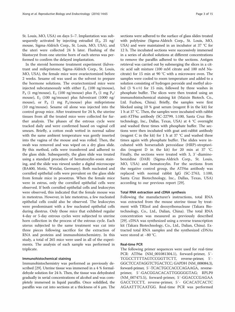
St. Louis, MO, USA) on days 5–7. Implantation was sub-sequently activated by injecting estradiol (E2, 25 ng/mouse, Sigma-Aldrich Corp., St. Louis, MO, USA), andthe uteri were collected 24 h later. Flushing of theblastocyst from one uterine horn of each uterus was per-formed to confirm the delayed implantation.In the steroid hormone treatment experiment (fulves-
trant and mifepristone; Sigma-Aldrich Corp., St. Louis,MO, USA), the female mice were ovariectomized before2 weeks. Sesame oil was used as the solvent to preparethe hormone solutions. The ovariectomized mice wereinjected subcutaneously with either E2 (100 ng/mouse),P4 (1 mg/mouse), E2 (100 ng/mouse) plus P4 (1 mg P4/mouse), E2 (100 ng/mouse) plus fulvestrant (1000 ng/mouse), or P4 (1 mg P4/mouse) plus mifepristone(10 mg/mouse). Sesame oil alone was injected into thecontrol group mice. After treatment for 24 h, the uterinetissues from all the treated mice were collected for fur-ther analysis. The phases of the estrous cycle weretracked daily and were confirmed based on vaginal cellsmears. Briefly, a cotton swab wetted in normal salinewith the same ambient temperature was gently insertedinto the vagina of the mouse and was rolled. Then, theswab was removed and was wiped on a dry glass slide.By this method, cells were transferred and adhered tothe glass slide. Subsequently, the glass slide was treatedusing a standard procedure of hematoxylin-eosin stain-ing, and the slide was viewed under a digital microscope(BA400, Motic, Wetzlar, Germany). Both nucleated andcornified epithelial cells were prevalent on the glass slidefrom female mice in proestrus. When the female micewere in estrus, only the cornified epithelial cells wereobserved. If both cornified epithelial cells and leukocyteswere observed, this indicated that the female mouse wasin metestrus. However, in late metestrus, a few nucleatedepithelial cells could also be observed. The leukocyteswere predominant with a few nucleated epithelial cellsduring diestrus. Only those mice that exhibited regular4-day or 5-day estrous cycles were subjected to uterinehorn collection in the process of the estrous cycle. Eachuterus subjected to the same treatment was cut intothree pieces following sacrifice for the extraction ofRNA and proteins and immunohistochemistry. In thisstudy, a total of 265 mice were used in all of the experi-ments. The analysis of each sample was performed intriplicate.
Immunohistochemical stainingImmunohistochemistry was performed as previously de-scribed [29]. Uterine tissue was immersed in a 4 % formal-dehyde solution for 24 h. Then, the tissue was dehydratedgradually in serial concentrations of alcohol and was com-pletely immersed in liquid paraffin. Once solidified, theparaffin was cut into sections at a thickness of 6 μm. The
sections were adhered to the surface of glass slides treatedwith polylysine (Sigma-Aldrich Corp., St. Louis, MO,USA) and were maintained in an incubator at 37 °C for12 h. The incubated sections were successively immersedin a series of alcohol solutions at different concentrationsto remove the paraffin adhered to the sections. Antigenretrieval was carried out by submerging the slices in a cit-ric acid salt mixture (100 mM citrate and 100 mM Na-citrate) for 15 min at 90 °C with a microwave oven. Thesamples were cooled to room temperature and added to asolution consisting of hydrogen peroxide and methyl alco-hol (3 % v/v) for 15 min, followed by three washes inphosphate buffer. The slices were then treated using animmunohistochemical staining kit (Maixin Biotech. Co.,Ltd. Fuzhou, China). Briefly, the samples were firstblocked using 10 % goat serum (reagent B in the kit) for1 h at 37 °C. Then, the samples were incubated with rabbitanti-ATF6α antibody (SC-22799, 1:100, Santa Cruz Bio-technology, Inc., Dallas, Texas, USA) at 4 °C overnightand washed three times with phosphate buffer. The sec-tions were then incubated with goat anti-rabbit antibody(reagent C in the kit) for 1 h at 37 °C and washed threetimes again with phosphate buffer. The slides were in-cubated with horseradish peroxidase (HRP)-streptavi-din (reagent D in the kit) for 20 min at 37 °C.Finally, the sections were treated with 3, 3′-diamino-benzidine (DAB) (Sigma-Aldrich Corp., St. Louis,MO, USA) and hematoxylin. For the sections fromthe negative control group, the ATF6α antibody wasreplaced with normal rabbit IgG (SC-2763, 1:100,Santa Cruz Biotechnology, Inc., Dallas, Texas, USA)according to our previous report [29].
Total RNA extraction and cDNA synthesisFollowing the manufacturer’s instructions, total RNAwas extracted from the mouse uterine tissue by treat-ment with TRIzol and deoxyribonuclease (Takara Bio-technology, Co., Ltd., Dalian, China). The total RNAconcentration was measured as previously described[29]. cDNA was synthesized using a reverse transcriptionkit (Takara Biotechnology, Co., Ltd., Dalian, China). Ex-tracted total RNA samples and the synthesized cDNAswere stored at −80 °C.
Real-time PCRThe following primer sequences were used for real-timePCR: ATF6α (NM_001081304.1), forward-primer, 5′-TCGCCTTTTAGTCCGGTTCTT, reverse-primer, 5′-GGCTCCATAGGTCTGACTCC; GAPDH (NM_008084.3),forward-primer, 5′-TCACTGCCACCCAGAAGA, reverse-primer, 5′-GACGGACACATTGGGGGTAG; RPLP0(NM_007475.5), forward-primer, 5′-GGACCCGAGAAGACCTCCTT, reverse-primer, 5′- GCACATCACTCAGAATTTCAATGG. Real-time PCR was performed
Xiong et al. Reproductive Biology and Endocrinology (2016) 14:65 Page 3 of 15
using SYBR® Premix Ex Taq™ (Perfect Real Time)(Takara Biotechnology, Co., Ltd., Dalian, China), andthe data were analyzed using the Bio-Rad iQ5 system(Bio-Rad Laboratories, Inc., Hercules, California, USA)following the manufacturer’s instructions. Each PCR reac-tion contained the following reagents: 2.0 μl reverse tran-scription products (containing 20 ng cDNA), 1.6 μl primermixture, 10.0 μl SYBR Premix Ex TaqTM II and 6.4 μlribonuclease-free water. The PCR cycling conditions wereas follows: denaturation at 95 °C for 30 s, followed by40 cycles at 95 °C for 5 s and 60 °C for 20 s. To ascertainthe specificity of the PCR amplification, a melting curveanalysis was performed including the negative controlgroup (no template cDNA). The relative mRNA
expression levels were determined using the 2-△△Ct
method; GAPDH and RPLP0 were used as the internalreference genes and the geometric averaging of these tworeference genes were used to normalize the relativemRNA expression according to previous reports [30–32].
Western blottingThe extraction, concentration, and detection of proteinsfrom uterine tissues were completed using a protein ex-traction and examination kit (Jiangsu Keygen BiologicalTechnology Co., Ltd, Nanjing, China), according to themanufacturer’s instructions. Protein samples (30 μg)were resolved by electrophoresis on a 12 % sodium do-decyl sulfate-polyacrylamide gel. After electrophoresis,
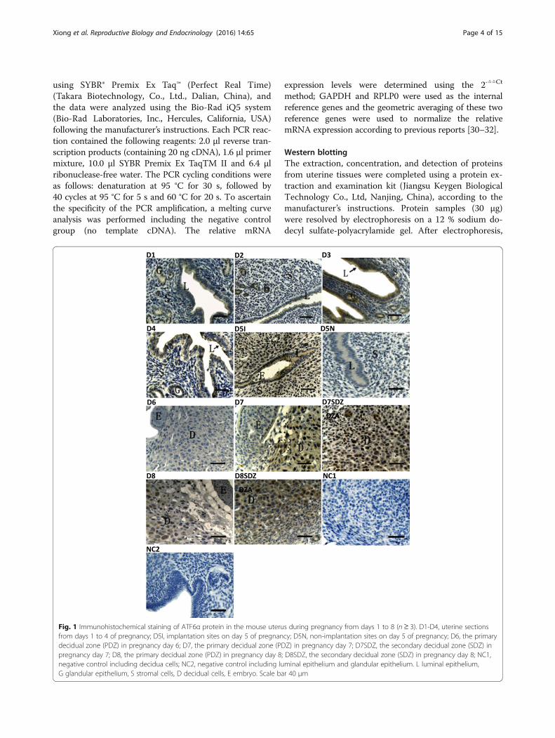
Fig. 1 Immunohistochemical staining of ATF6α protein in the mouse uterus during pregnancy from days 1 to 8 (n ≥ 3). D1-D4, uterine sectionsfrom days 1 to 4 of pregnancy; D5I, implantation sites on day 5 of pregnancy; D5N, non-implantation sites on day 5 of pregnancy; D6, the primarydecidual zone (PDZ) in pregnancy day 6; D7, the primary decidual zone (PDZ) in pregnancy day 7; D7SDZ, the secondary decidual zone (SDZ) inpregnancy day 7; D8, the primary decidual zone (PDZ) in pregnancy day 8; D8SDZ, the secondary decidual zone (SDZ) in pregnancy day 8; NC1,negative control including decidua cells; NC2, negative control including luminal epithelium and glandular epithelium. L luminal epithelium,G glandular epithelium, S stromal cells, D decidual cells, E embryo. Scale bar 40 μm
Xiong et al. Reproductive Biology and Endocrinology (2016) 14:65 Page 4 of 15
the resolved proteins were transferred to a polyvinyli-dene fluoride membrane (Millipore, Co. Ltd., Shanghai,China). The membrane was then blocked by immersionin Tris-buffered saline (TBS) containing 10 % nonfat-dried milk for 60 min at room temperature (RT). Then,the membranes were incubated with anti-ATF6α
antibody (1:200, SC-22799; Santa Cruz Biotechnology,Inc., Dallas, Texas, USA) or β-actin as a loading control(CW0096, 1:1000, CWBIO Biological Technology Co.Ltd. Beijing, China) for 12 h at 4 °C. After washing withTBS, the membranes were incubated with horseradishperoxidase conjugated anti-rabbit IgG antibody (1:6000;
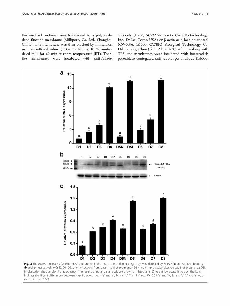
Fig. 2 The expression levels of ATF6α mRNA and protein in the mouse uterus during pregnancy were detected by RT-PCR (a) and western blotting(b and c), respectively (n ≥ 3). D1–D8, uterine sections from days 1 to 8 of pregnancy; D5N, non-implantation sites on day 5 of pregnancy; D5I,implantation sites on day 5 of pregnancy. The results of statistical analysis are shown as histograms. Different lowercase letters on the barsindicate significant differences between specific two groups (‘a’ and ‘a’, ‘b’ and ‘b’, ‘f’ and ‘f’, etc., P < 0.05; ‘a’ and ‘b’, ‘b’ and ‘c’, ‘c’ and ‘e’, etc.,P < 0.05 or P < 0.01)
Xiong et al. Reproductive Biology and Endocrinology (2016) 14:65 Page 5 of 15
SUNGENE Biotech Co. Ltd., Tianjin, China) at RT for60 min. After washing, the immunoblotting signals wereanalyzed using the Gel Photograph System (TanonTechnology Co. Ltd., Shanghai, China). According to themanufacturer’s instructions, western blot analysis of thecleaved ATF6α expression in rat liver extract (SC-2395,1:200, Santa Cruz Biotechnology, Inc., Dallas, Texas,USA) was performed as a positive control. The normalrabbit IgG was used as the negative non-relevant IgGcontrol.
Data statistics and analysisThroughout this study, all experiments were replicatedat least three times for each group or each treatment(n ≥ 3). The data are presented as the mean ± S.E.M.ANOVA, Fisher LSD and independent sample T-testswere conducted using SPSS statistical software (version19.0, Chicago, IL, USA). Differences were consideredsignificant if P < 0.05.
ResultsATF6α expression in the uteri of pregnant mice duringdays 1–8To elucidate the spatial and temporal distribution ofATF6α in the mouse uterus early in pregnancy, we ana-lyzed the expression pattern of ATF6α in mouse uteriduring peri-implantation by immunohistochemical stain-ing, real-time PCR, and western blot. It was found thatATF6α proteins were mainly localized in the endomet-rium luminal cells and glandular epithelial cells duringdays 1 to 4 of pregnancy (Fig. 1, D1–D4). Moreover, theexpression levels of ATF6α mRNA and protein were sig-nificantly increased from day 1 to day 5, with the highestlevels detected in the pregnant uterus on day 5 withinthe implantation site (P < 0.01, Fig. 2a–c). Notably, on
pregnancy day 5, a large number of endometrium lu-minal, glandular epithelial and stromal cells with immu-nostaining of ATF6α were present in the uterus with animplantation site, whereas the positive immunostainingof ATF6α was hardly observed in these cells in uteriwithout an implantation site (Fig. 1, D5I–D5N). Addition-ally, as decidualization proceeded during days 6–8, immu-nohistochemistry demonstrated that the ATF6α proteinwas present in the primary decidualization zone (PDZ)and the secondary decidualization zone (SDZ) (Fig. 1, D6-D8SDZ). Simultaneously, although the results from real-time PCR and western blot indicated that the expressionlevels of ATF6α mRNA and protein were relatively low atthe beginning of decidualization on day 6, the levels ofATF6α mRNA and protein were progressively increasedover the whole process of decidualization from days 6–8(P < 0.05 or P < 0.01, Fig. 2a–c). Western blot analysis ofcleaved ATF6α expression in rat liver extract was per-formed as a positive control. The normal rabbit IgG wasused as the negative non-relevant IgG control. The resultsshown are representative of 3 independent experiments(Additional file 1).
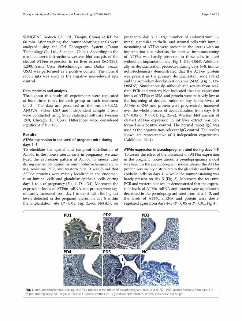
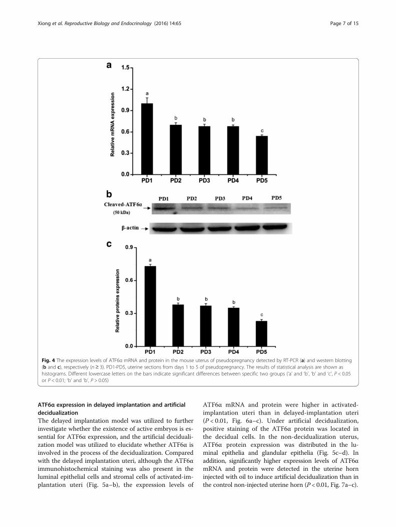
ATF6α expression in pseudopregnant uteri during days 1–5To assess the effect of the blastocyst on ATF6α expressionin the pregnant mouse uterus, a pseudopregnancy modelwas used. In the pseudopregnant mouse uterus, the ATF6αprotein was mainly distributed in the glandular and luminalepithelial cells on days 1–4, while the immunostaining wasbarely present on day 5 (Fig. 3). Moreover, the real-timePCR and western blot results demonstrated that the expres-sion levels of ATF6α mRNA and protein were significantlydecreased in the pseudopregnant uteri from days 1–2, andthe levels of ATF6α mRNA and protein were down-regulated again from days 4–5 (P < 0.05 or P < 0.01, Fig. 4).
Fig. 3 Immunohistochemical staining of ATF6α protein in the uterus of pseudopregnant mice (n≥ 3). PD1–PD5, uterine sections from days 1–5of pseudopregnancy; NC, negative control. L luminal epithelium, G glandular epithelium, S stromal cells, Scale bar 40 μm
Xiong et al. Reproductive Biology and Endocrinology (2016) 14:65 Page 6 of 15
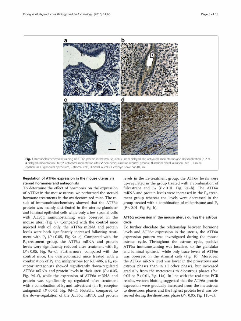
ATF6α expression in delayed implantation and artificialdecidualizationThe delayed implantation model was utilized to furtherinvestigate whether the existence of active embryos is es-sential for ATF6α expression, and the artificial deciduali-zation model was utilized to elucidate whether ATF6α isinvolved in the process of the decidualization. Comparedwith the delayed implantation uteri, although the ATF6αimmunohistochemical staining was also present in theluminal epithelial cells and stromal cells of activated-im-plantation uteri (Fig. 5a–b), the expression levels of
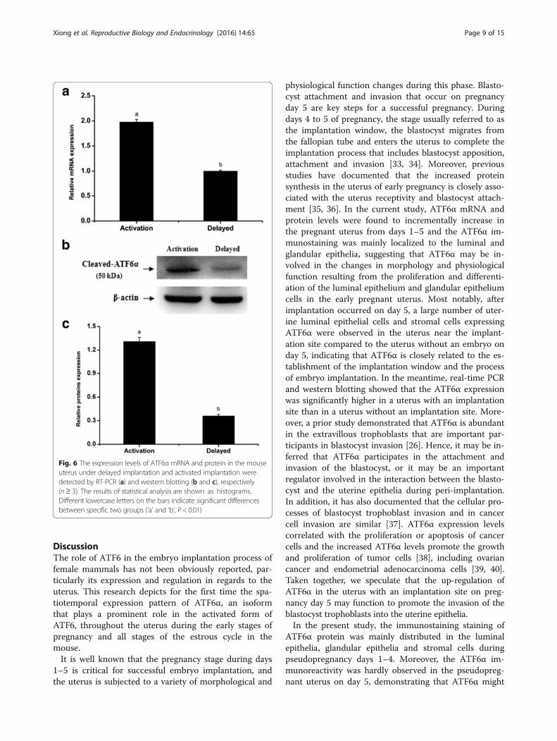
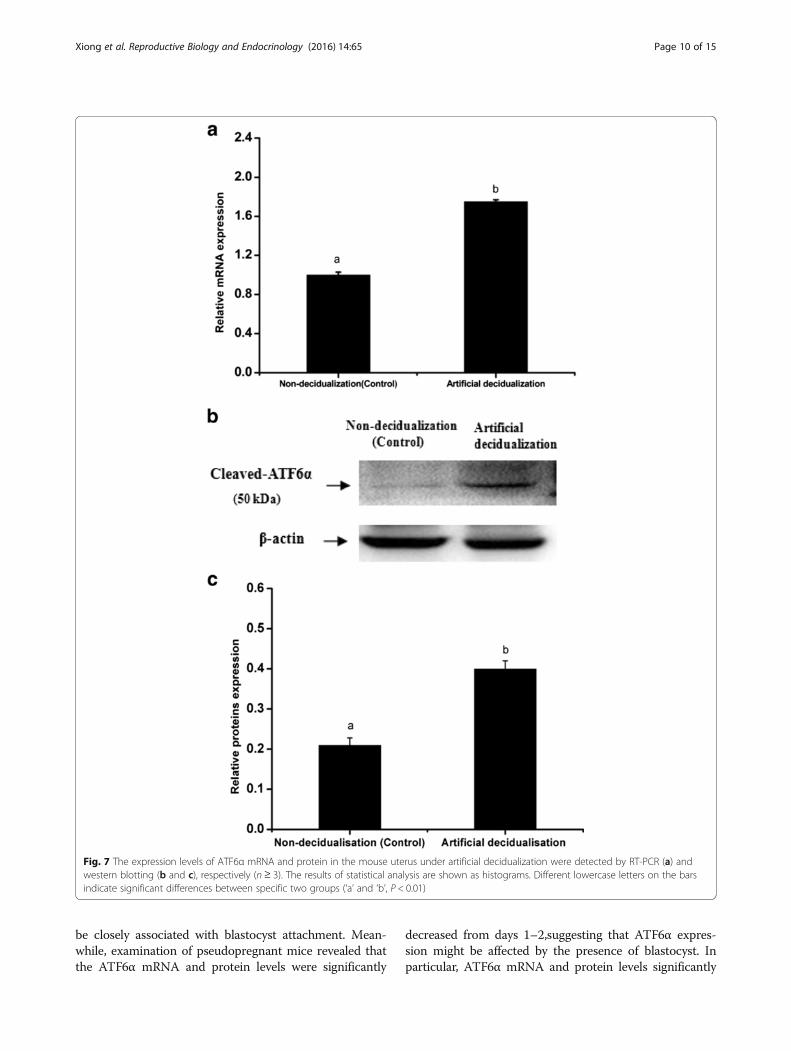
ATF6α mRNA and protein were higher in activated-implantation uteri than in delayed-implantation uteri(P < 0.01, Fig. 6a–c). Under artificial decidualization,positive staining of the ATF6α protein was located inthe decidual cells. In the non-decidualization uterus,ATF6α protein expression was distributed in the lu-minal epithelia and glandular epithelia (Fig. 5c–d). Inaddition, significantly higher expression levels of ATF6αmRNA and protein were detected in the uterine horninjected with oil to induce artificial decidualization than inthe control non-injected uterine horn (P < 0.01, Fig. 7a–c).
Fig. 4 The expression levels of ATF6α mRNA and protein in the mouse uterus of pseudopregnancy detected by RT-PCR (a) and western blotting(b and c), respectively (n ≥ 3). PD1-PD5, uterine sections from days 1 to 5 of pseudopregnancy. The results of statistical analysis are shown ashistograms. Different lowercase letters on the bars indicate significant differences between specific two groups (‘a’ and ‘b’, ‘b’ and ‘c’, P < 0.05or P < 0.01; ‘b’ and ‘b’, P > 0.05)
Xiong et al. Reproductive Biology and Endocrinology (2016) 14:65 Page 7 of 15
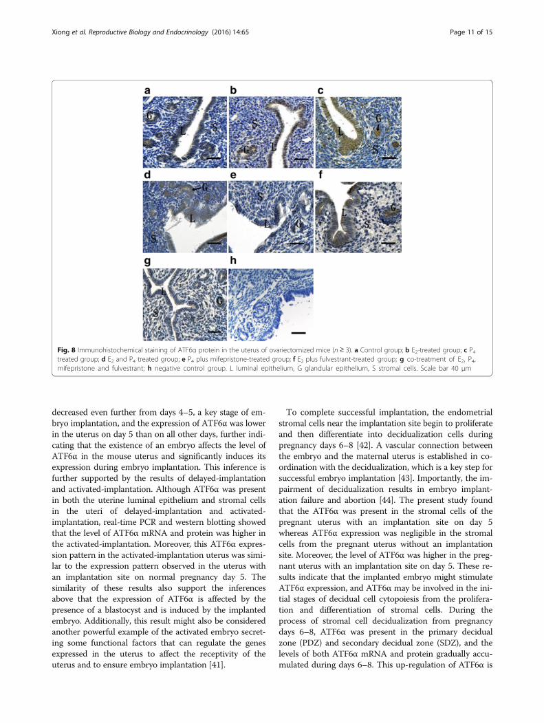
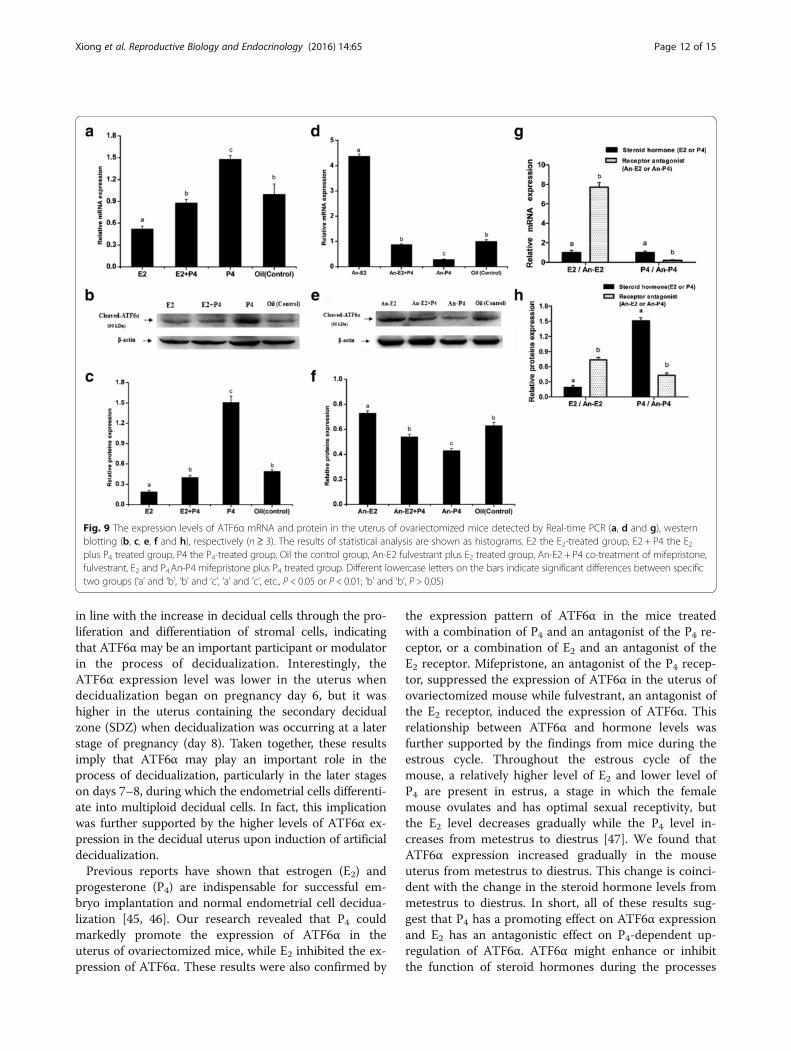
Regulation of ATF6α expression in the mouse uterus viasteroid hormones and antagonistsTo determine the effect of hormones on the expressionof ATF6α in the mouse uterus, we performed the steroidhormone treatments in the ovariectomized mice. The re-sult of immunohistochemistry showed that the ATF6αprotein was mainly distributed in the uterine glandularand luminal epithelial cells while only a few stromal cellswith ATF6α immunostaining were observed in themouse uteri (Fig. 8). Compared with the control miceinjected with oil only, the ATF6α mRNA and proteinlevels were both significantly increased following treat-ment with P4 (P < 0.05, Fig. 9a–c). Compared with theP4-treatment group, the ATF6α mRNA and proteinlevels were significantly reduced after treatment with E2(P < 0.05, Fig. 9a–c). Furthermore, compared with thecontrol mice, the ovariectomized mice treated with acombination of P4 and mifepristone (or RU-486, a P4 re-ceptor antagonist) showed significantly down-regulatedATF6α mRNA and protein levels in their uteri (P < 0.05,Fig. 9d–f ), while the expression of ATF6α mRNA andprotein was significantly up-regulated after treatmentwith a combination of E2 and fulvestrant (an E2 receptorantagonist) (P < 0.05, Fig. 9d–f ). Notably, compared tothe down-regulation of the ATF6α mRNA and protein
levels in the E2-treatment group, the ATF6α levels wereup-regulated in the group treated with a combination offulvestrant and E2 (P < 0.01, Fig. 9g–h). The ATF6αmRNA and protein levels were increased in the P4-treat-ment group whereas the levels were decreased in thegroup treated with a combination of mifepristone and P4(P < 0.01, Fig. 9g–h).
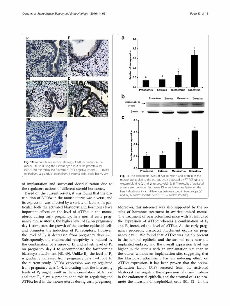
ATF6α expression in the mouse uterus during the estrouscycleTo further elucidate the relationship between hormonelevels and ATF6α expression in the uterus, the ATF6αexpression pattern was investigated during the mouseestrous cycle. Throughout the estrous cycle, positiveATF6α immunostaining was localized to the glandularand luminal epithelia, while only trace levels of ATF6αwas observed in the stromal cells (Fig. 10). Moreover,the ATF6α mRNA level was lower in the proestrous andestrous phases than in all other phases, but increasedgradually from the metestrous to dioestrous phases (P <0.05 or P < 0.01, Fig. 11a). In line with the real-time PCRresults, western blotting suggested that the ATF6α proteinexpression were gradually increased from the metestrousto dioestrous phases and the highest protein level was ob-served during the dioestrous phase (P < 0.05, Fig. 11b–c).
Fig. 5 Immunohistochemical staining of ATF6α protein in the mouse uterus under delayed and activated implantation and decidualization (n≥ 3).a delayed-implantation uteri; b activated-implantation uteri; c non-decidualization (control groups); d artificial decidualization uteri. L luminalepithelium, G glandular epithelium, S stromal cells, D decidual cells, E embryo. Scale bar 40 μm
Xiong et al. Reproductive Biology and Endocrinology (2016) 14:65 Page 8 of 15
DiscussionThe role of ATF6 in the embryo implantation process offemale mammals has not been obviously reported, par-ticularly its expression and regulation in regards to theuterus. This research depicts for the first time the spa-tiotemporal expression pattern of ATF6α, an isoformthat plays a prominent role in the activated form ofATF6, throughout the uterus during the early stages ofpregnancy and all stages of the estrous cycle in themouse.It is well known that the pregnancy stage during days
1–5 is critical for successful embryo implantation, andthe uterus is subjected to a variety of morphological and
physiological function changes during this phase. Blasto-cyst attachment and invasion that occur on pregnancyday 5 are key steps for a successful pregnancy. Duringdays 4 to 5 of pregnancy, the stage usually referred to asthe implantation window, the blastocyst migrates fromthe fallopian tube and enters the uterus to complete theimplantation process that includes blastocyst apposition,attachment and invasion [33, 34]. Moreover, previousstudies have documented that the increased proteinsynthesis in the uterus of early pregnancy is closely asso-ciated with the uterus receptivity and blastocyst attach-ment [35, 36]. In the current study, ATF6α mRNA andprotein levels were found to incrementally increase inthe pregnant uterus from days 1–5 and the ATF6α im-munostaining was mainly localized to the luminal andglandular epithelia, suggesting that ATF6α may be in-volved in the changes in morphology and physiologicalfunction resulting from the proliferation and differenti-ation of the luminal epithelium and glandular epitheliumcells in the early pregnant uterus. Most notably, afterimplantation occurred on day 5, a large number of uter-ine luminal epithelial cells and stromal cells expressingATF6α were observed in the uterus near the implant-ation site compared to the uterus without an embryo onday 5, indicating that ATF6α is closely related to the es-tablishment of the implantation window and the processof embryo implantation. In the meantime, real-time PCRand western blotting showed that the ATF6α expressionwas significantly higher in a uterus with an implantationsite than in a uterus without an implantation site. More-over, a prior study demonstrated that ATF6α is abundantin the extravillous trophoblasts that are important par-ticipants in blastocyst invasion [26]. Hence, it may be in-ferred that ATF6α participates in the attachment andinvasion of the blastocyst, or it may be an importantregulator involved in the interaction between the blasto-cyst and the uterine epithelia during peri-implantation.In addition, it has also documented that the cellular pro-cesses of blastocyst trophoblast invasion and in cancercell invasion are similar [37]. ATF6α expression levelscorrelated with the proliferation or apoptosis of cancercells and the increased ATF6α levels promote the growthand proliferation of tumor cells [38], including ovariancancer and endometrial adenocarcinoma cells [39, 40].Taken together, we speculate that the up-regulation ofATF6α in the uterus with an implantation site on preg-nancy day 5 may function to promote the invasion of theblastocyst trophoblasts into the uterine epithelia.In the present study, the immunostaining staining of
ATF6α protein was mainly distributed in the luminalepithelia, glandular epithelia and stromal cells duringpseudopregnancy days 1–4. Moreover, the ATF6α im-munoreactivity was hardly observed in the pseudopreg-nant uterus on day 5, demonstrating that ATF6α might
Fig. 6 The expression levels of ATF6α mRNA and protein in the mouseuterus under delayed implantation and activated implantation weredetected by RT-PCR (a) and western blotting (b and c), respectively(n≥ 3). The results of statistical analysis are shown as histograms.Different lowercase letters on the bars indicate significant differencesbetween specific two groups (‘a’ and ‘b’, P < 0.01)
Xiong et al. Reproductive Biology and Endocrinology (2016) 14:65 Page 9 of 15
be closely associated with blastocyst attachment. Mean-while, examination of pseudopregnant mice revealed thatthe ATF6α mRNA and protein levels were significantly
decreased from days 1–2,suggesting that ATF6α expres-sion might be affected by the presence of blastocyst. Inparticular, ATF6α mRNA and protein levels significantly
Fig. 7 The expression levels of ATF6α mRNA and protein in the mouse uterus under artificial decidualization were detected by RT-PCR (a) andwestern blotting (b and c), respectively (n ≥ 3). The results of statistical analysis are shown as histograms. Different lowercase letters on the barsindicate significant differences between specific two groups (‘a’ and ‘b’, P < 0.01)
Xiong et al. Reproductive Biology and Endocrinology (2016) 14:65 Page 10 of 15
decreased even further from days 4–5, a key stage of em-bryo implantation, and the expression of ATF6α was lowerin the uterus on day 5 than on all other days, further indi-cating that the existence of an embryo affects the level ofATF6α in the mouse uterus and significantly induces itsexpression during embryo implantation. This inference isfurther supported by the results of delayed-implantationand activated-implantation. Although ATF6α was presentin both the uterine luminal epithelium and stromal cellsin the uteri of delayed-implantation and activated-implantation, real-time PCR and western blotting showedthat the level of ATF6α mRNA and protein was higher inthe activated-implantation. Moreover, this ATF6α expres-sion pattern in the activated-implantation uterus was simi-lar to the expression pattern observed in the uterus withan implantation site on normal pregnancy day 5. Thesimilarity of these results also support the inferencesabove that the expression of ATF6α is affected by thepresence of a blastocyst and is induced by the implantedembryo. Additionally, this result might also be consideredanother powerful example of the activated embryo secret-ing some functional factors that can regulate the genesexpressed in the uterus to affect the receptivity of theuterus and to ensure embryo implantation [41].
To complete successful implantation, the endometrialstromal cells near the implantation site begin to proliferateand then differentiate into decidualization cells duringpregnancy days 6–8 [42]. A vascular connection betweenthe embryo and the maternal uterus is established in co-ordination with the decidualization, which is a key step forsuccessful embryo implantation [43]. Importantly, the im-pairment of decidualization results in embryo implant-ation failure and abortion [44]. The present study foundthat the ATF6α was present in the stromal cells of thepregnant uterus with an implantation site on day 5whereas ATF6α expression was negligible in the stromalcells from the pregnant uterus without an implantationsite. Moreover, the level of ATF6α was higher in the preg-nant uterus with an implantation site on day 5. These re-sults indicate that the implanted embryo might stimulateATF6α expression, and ATF6α may be involved in the ini-tial stages of decidual cell cytopoiesis from the prolifera-tion and differentiation of stromal cells. During theprocess of stromal cell decidualization from pregnancydays 6–8, ATF6α was present in the primary decidualzone (PDZ) and secondary decidual zone (SDZ), and thelevels of both ATF6α mRNA and protein gradually accu-mulated during days 6–8. This up-regulation of ATF6α is
Fig. 8 Immunohistochemical staining of ATF6α protein in the uterus of ovariectomized mice (n ≥ 3). a Control group; b E2-treated group; c P4treated group; d E2 and P4 treated group; e P4 plus mifepristone-treated group; f E2 plus fulvestrant-treated group; g co-treatment of E2, P4,mifepristone and fulvestrant; h negative control group. L luminal epithelium, G glandular epithelium, S stromal cells. Scale bar 40 μm
Xiong et al. Reproductive Biology and Endocrinology (2016) 14:65 Page 11 of 15
in line with the increase in decidual cells through the pro-liferation and differentiation of stromal cells, indicatingthat ATF6α may be an important participant or modulatorin the process of decidualization. Interestingly, theATF6α expression level was lower in the uterus whendecidualization began on pregnancy day 6, but it washigher in the uterus containing the secondary decidualzone (SDZ) when decidualization was occurring at a laterstage of pregnancy (day 8). Taken together, these resultsimply that ATF6α may play an important role in theprocess of decidualization, particularly in the later stageson days 7–8, during which the endometrial cells differenti-ate into multiploid decidual cells. In fact, this implicationwas further supported by the higher levels of ATF6α ex-pression in the decidual uterus upon induction of artificialdecidualization.Previous reports have shown that estrogen (E2) and
progesterone (P4) are indispensable for successful em-bryo implantation and normal endometrial cell decidua-lization [45, 46]. Our research revealed that P4 couldmarkedly promote the expression of ATF6α in theuterus of ovariectomized mice, while E2 inhibited the ex-pression of ATF6α. These results were also confirmed by
the expression pattern of ATF6α in the mice treatedwith a combination of P4 and an antagonist of the P4 re-ceptor, or a combination of E2 and an antagonist of theE2 receptor. Mifepristone, an antagonist of the P4 recep-tor, suppressed the expression of ATF6α in the uterus ofovariectomized mouse while fulvestrant, an antagonist ofthe E2 receptor, induced the expression of ATF6α. Thisrelationship between ATF6α and hormone levels wasfurther supported by the findings from mice during theestrous cycle. Throughout the estrous cycle of themouse, a relatively higher level of E2 and lower level ofP4 are present in estrus, a stage in which the femalemouse ovulates and has optimal sexual receptivity, butthe E2 level decreases gradually while the P4 level in-creases from metestrus to diestrus [47]. We found thatATF6α expression increased gradually in the mouseuterus from metestrus to diestrus. This change is coinci-dent with the change in the steroid hormone levels frommetestrus to diestrus. In short, all of these results sug-gest that P4 has a promoting effect on ATF6α expressionand E2 has an antagonistic effect on P4-dependent up-regulation of ATF6α. ATF6α might enhance or inhibitthe function of steroid hormones during the processes
Fig. 9 The expression levels of ATF6α mRNA and protein in the uterus of ovariectomized mice detected by Real-time PCR (a, d and g), westernblotting (b, c, e, f and h), respectively (n ≥ 3). The results of statistical analysis are shown as histograms. E2 the E2-treated group, E2 + P4 the E2plus P4 treated group, P4 the P4-treated group, Oil the control group, An-E2 fulvestrant plus E2 treated group, An-E2 + P4 co-treatment of mifepristone,fulvestrant, E2 and P4,An-P4 mifepristone plus P4 treated group. Different lowercase letters on the bars indicate significant differences between specifictwo groups (‘a’ and ‘b’, ‘b’ and ‘c’, ‘a’ and ‘c’, etc., P < 0.05 or P < 0.01; ‘b’ and ‘b’, P > 0.05)
Xiong et al. Reproductive Biology and Endocrinology (2016) 14:65 Page 12 of 15
of implantation and successful decidualization due tothe regulatory actions of different steroid hormones.Based on the current results, it was found that the dis-
tribution of ATF6α in the mouse uterus was diverse, andits expression was affected by a variety of factors. In par-ticular, both the activated blastocyst and hormones haveimportant effects on the level of ATF6α in the mouseuterus during early pregnancy. In a normal early preg-nancy mouse uterus, the higher level of E2 on pregnancyday 1 stimulates the growth of the uterine epithelial cellsand promotes the induction of P4 receptors. However,the level of E2 is decreased from pregnancy days 2–3.Subsequently, the endometrial receptivity is induced bythe combination of a surge of E2 and a high level of P4on pregnancy day 4 to accommodate the forthcomingblastocyst attachment [48, 49]. Unlike E2, the level of P4is gradually increased from pregnancy days 1–4 [50]. Inthe current study, ATF6α expression was up-regulatedfrom pregnancy days 1–4, indicating that the increasinglevels of P4 might result in the accumulation of ATF6αand that P4 plays a predominant role in regulating theATF6α level in the mouse uterus during early pregnancy.
Moreover, this inference was also supported by the re-sults of hormone treatment in ovariectomized mouse.The treatment of ovariectomized mice with E2 inhibitedthe expression of ATF6α whereas a combination of E2and P4 increased the level of ATF6α. As the early preg-nancy proceeds, blastocyst attachment occurs on preg-nancy day 5. We found that ATF6α was mainly presentin the luminal epithelia and the stromal cells near theimplanted embryo, and the overall expression level washigher in the uterus with an implantation site than inthe uterus without an implantation site, suggesting thatthe blastocyst attachment has an inducing effect onATF6α expression. It has been proven that the preim-plantation factor (PIF) secreted from the activatedblastocyst can regulate the expression of many proteinsin the endometrial epithelia and the stromal cells to pro-mote the invasion of trophoblast cells [51, 52]. In the
Fig. 11 The expression levels of ATF6α mRNA and protein in themouse uterus during the estrous cycle detected by RT-PCR (a) andwestern blotting (b and c), respectively(n≥ 3). The results of statisticalanalysis are shown as histograms. Different lowercase letters on thebars indicate significant differences between specific two groups (‘a’and ‘b’, ‘b’ and ‘c’, P < 0.05 or P < 0.01; ‘a’ and ‘a’, P > 0.05)
Fig. 10 Immunohistochemical staining of ATF6α protein in themouse uterus during the estrous cycle (n ≥ 3). (P) proestrus; (E)estrus; (M) metestrus; (D) dioestrous; (NC) negative control. L luminalepithelium, G glandular epithelium, S stromal cells. Scale bar 40 μm
Xiong et al. Reproductive Biology and Endocrinology (2016) 14:65 Page 13 of 15
meantime, the level of E2 on pregnancy day 4 is de-creased on pregnancy day 5 while the high level of P4 ismaintained [49, 50]. Prior investigation has shown thatP4 and E2 play crucial roles in the establishment ofendometrial receptivity and successful implantation byaffecting the expression of many genes in endometrialcells [40]. Therefore, we speculate that the increasedATF6α on pregnancy days 4–5 may be involved in theestablishment of endometrial receptivity and may pro-mote trophoblast cell invasion during peri-implantation.In addition, high expression of ATF6α in the luminalepithelium and stromal cells of a uterus with an implant-ation site might be the result of the synergistic and indu-cing effect of the implanted embryo and the high level ofP4. In other words, a cooperative relationship betweenembryo attachment and hormones is established to regu-late ATF6α expression in the uterus during embryo im-plantation in vivo.
ConclusionsIn conclusion, the current findings indicate that ATF6αplays an important role in embryo implantation and thephysiological process of decidualization. Moreover, ATF6αexpression in the mouse uterus might be regulated bythe activating effects of the embryo and the hormonesprogesterone and estrogen.
Additional file
Additional file 1: (S1) Positive control. Western blot analysis of cleavedATF6α expression in rat liver extract was performed as a positive control.(S2) Negative control. The normal rabbit IgG was used as the negativenon-relevant IgG control. The results shown are representative of 3independent experiments. (PDF 125 kb)
AbbreviationsATF: Activating transcription factor; ATF6: Activating transcription factor 6;ATF6α: The alpha isoforms of activating transcription factor 6; ATF6β: Thebeta isoforms of activating transcription factor 6; CREB: Cyclic adenosinemonophosphate response element-binding protein; DAB: 3, 3′-diaminobenzidine; E2: Estrogen; HRP: Horseradish peroxidase;P4: Progesterone; PDZ: Primary decidualization zone; S1P: Site-1 protease;S2P: Site-2 protease; SDZ: Secondary decidual zone
AcknowledgmentsWe thank American Journal Experts for language editing services withEnglish correction.
FundingThis research was supported by the National Natural Science Foundation ofChina (31372499) and the China Postdoctoral Science Foundation(2015M582718).
Availability of data and materialThe datasets analyzed during this study are available from the correspondingauthor on reasonable request.
Authors’ contributionsYX and YJ designed the experiments and wrote the manuscript. PL and LWprepared experimental materials and reagents. NW and FC collected themouse uterine sections samples. YX and WL performed the experiments. XL
and AW organized the whole project. All authors permitted and approvedthe submitted manuscript.
Competing interestsThe authors declare that they have no competing interests with respect tothe authorship and/or publication of this article.
Consent for publicationNot applicable.
Ethics approval and consent to participateOnly mice were involved in this study and all procedures were approved bythe Committee for the Ethics on Animal Care and Experiments of NorthwestA&F University.
Received: 12 April 2016 Accepted: 27 September 2016
References1. Dimitriadis E, White CA, Jones RL, Salamonsen LA. Cytokines, chemokines
and growth factors in endometrium related to implantation. Hum ReprodUpdate. 2005;11:613–30.
2. Achache H, Revel A. Endometrial receptivity markers, the journey tosuccessful embryo implantation. Hum Reprod Update. 2006;12:731–46.
3. Xia H, Jin X, Cao Z, Shi T, Ma X. MiR-98 is involved in rat embryoimplantation by targeting Bcl-xl. Febs Letters. 2014;588:574–83.
4. Paria BC, Lim H, Das SK, Reese J, Dey SK. Molecular signaling in uterinereceptivity for implantation. Semin Cell Dev Biol. 2000;11:67–76.
5. Teles A, Zenclussen A. How cells of the immune system prepare theendometrium for implantation. Semin Reprod Med. 2014;32:358–64.
6. Yoshinaga K. Progesterone and its downstream molecules as blastocystimplantation essential factors. Am J Reprod Immunol. 2014;72:117–28.
7. Xia HF, Jin XH, Cao ZF, Hu Y, Ma X. MicroRNA expression and regulation inthe uterus during embryo implantation in rat. Febs Journal. 2014;281:1872–91.
8. Zenclussen AC, Hammerling GJ. Cellular regulation of the uterinemicroenvironment that enables embryo implantation. Front Immunol. 2015;6:321.
9. Haze K, Yoshida H, Yanagi H, Yura T, Mori K. Mammalian transcription factorATF6 is synthesized as a transmembrane protein and activated byproteolysis in response to endoplasmic reticulum stress. Mol Biol Cell.1999;10:3787–99.
10. Zhu C, Johansen FE, Prywes R. Interaction of ATF6 and serum responsefactor. Mol Cell Biol. 1997;17:4957–66.
11. Kim JW, Choi H, Jeong BC, Oh SH, Hur SW, Lee BN, Kim SH, Noer JE, Koh JT,Hwang YC. Transcriptional factor ATF6 is involved in odontoblasticdifferentiation. J Dent Res. 2014;93:483–9.
12. Chen X, Shen J, Prywes R. The luminal domain of ATF6 senses endoplasmicreticulum (ER) stress and causes translocation of ATF6 from the ER to theGolgi. J Biol Chem. 2002;277:13045–52.
13. Adachi Y, Yamamoto K, Okada T, Yoshida H, Harada A, Mori K. ATF6 is atranscription factor specializing in the regulation of quality control proteinsin the endoplasmic reticulum. Cell Struct Funct. 2008;33:75–89.
14. Darling NJ, Cook SJ. The role of MAPK signalling pathways in the responseto endoplasmic reticulum stress. Biochim Biophys Acta. 2014;1843:2150–63.
15. Gomez JA, Tyra HM, DeZwaan-McCabe D, Olivier AK, Rutkowski DT.Synthetic embryonic lethality upon deletion of the ER cochaperone p58IPK
and the ER stress sensor ATF6 alpha. Biochem Biophys Res Commun.2014;443:115–9.
16. Yamamoto K, Sato T, Matsui T, Sato M, Okada T, Yoshida H, Harada A, MoriK. Transcriptional induction of mammalian ER quality control proteins ismediated by single or combined action of ATF6alpha and XBP1. Dev Cell.2007;13:365–76.
17. Lai E, Teodoro T, Volchuk A. Endoplasmic reticulum stress: signaling theunfolded protein response. Physiology. 2007;22:193–201.
18. Ruddon RW, Bedows E. Assisted protein folding. J Biol Chem. 1997;272:3125–8.
19. Yang Y, Pei X, Jin Y, Wang Y, Zhang C. The roles of endoplasmic reticulumstress response in female mammalian reproduction. Cell Tissue Res. 2016;363:589–97.
20. Simmons DG, Kennedy TG. Induction of glucose-regulated protein 78 in ratuterine glandular epithelium during uterine sensitization for the decidualcell reaction. Biol Reprod. 2000;62:1168–76.
Xiong et al. Reproductive Biology and Endocrinology (2016) 14:65 Page 14 of 15
21. Zhang K, Kaufman RJ. From endoplasmic-reticulum stress to theinflammatory response. Nature. 2008;454:455–62.
22. Hashida K, Kitao Y, Sudo H, Awa Y, Maeda S. ATF6alpha promotes astroglialactivation and neuronal survival in a chronic mouse model of parkinson’sdisease. Plos One. 2012;7:e47950.
23. Park SJ, Kim TS, Park CK, Lee SH, Kim JM, Lee KS, Lee IK, Park JW, LawsonMA, Lee DS. HCG-induced endoplasmic reticulum stress triggers apoptosisand reduces steroidogenic enzyme expression through activatingtranscription factor 6 in Leydig cells of the testis. J Mol Endocrinol.2013;50:151–66.
24. Park H, Park S, Koo D, Lee S, Kong I, Ryoo J, Park Y, Chang K, Lee D.Progesterone production is affected by unfolded protein response (UPR)signaling during the luteal phase in mice. Life Sci. 2014;113:60–7.
25. Yung HW, Atkinson D, Campion-Smith T, Olovsson M, Charnock-Jones DS,Burton GJ. Differential activation of placental unfolded protein responsepathways implies heterogeneity in causation of early- and late-onsetpre-eclampsia. J Pathol. 2014;234:262–76.
26. Lian IA, Løset M, Mundal SB, Fenstad MH, Johnson MP, Eide IP, Bjørge L,Freed KA, Moses EK, Austgulen R. Increased endoplasmic reticulum stress indecidual tissue from pregnancies complicated by fetal growth restrictionwith and without pre-eclampsia. Placenta. 2011;32:823–9.
27. Lin P, Jin Y, Lan X, Yang Y, Chen F, Wang N, Li X, Sun Y, Wang A. GRP78expression and regulation in the mouse uterus during embryo implantation.J Mol Histol. 2014;45:259–68.
28. Lan X, Jin Y, Yang Y, Lin P, Hu L, Cui C, Li Q, Li X, Wang A. Expression andlocalization of Luman RNA and protein during mouse implantation anddecidualization. Theriogenology. 2013;80:138–44.
29. Lin P, Chen F, Wang N, Wang X, Li X, Zhou J, Jin Y, Wang A. CREBZFexpression and hormonal regulation in the mouse uterus. Reprod BiolEndocrinol. 2013;11:110.
30. Tan J, Raja S, Davis MK, Tawfik O, Dey SK, Das SK. Evidence for coordinatedinteraction of cyclin D3 with p21 and cdk6 in directing the development ofuterine stromal cell decidualization and polyploidy during implantation.Mech Dev. 2002;111:99–113.
31. Lin P, Lan X, Chen F, Yang Y, Jin Y, Wang A. Reference gene selection forreal-time quantitative PCR analysis of the mouse uterus in the peri-implantationperiod. Plos One. 2013;8:e62462.
32. Vandesompele J, Preter KD, Pattyn F, Poppe B, Roy NV. Accurate normalizationof real-time quantitative RT-PCR data by geometric averaging of multipleinternal control genes. Genome Biol. 2002;3:research0034.1–0034.11.
33. Sengupta J, Ghosh D. Multi-level and multi-scale integrative approach tothe understanding of human blastocyst implantation. Prog Biophys MolBiol. 2014;114:49–60.
34. Mourik MS, Macklon NS, Heijnen CJ. Embryonic implantation: cytokines,adhesion molecules, and immune cells in establishing an implantationenvironment. J Leukoc Biol. 2008;85:4–19.
35. Bai Z, Guo B, Tian X, Li D, Wang S, Cao H, Wang Q, Yue Z. Expression andregulation of Runx3 in mouse uterus during the peri-implantation period.J Mol Histol. 2013;44:519–26.
36. Reid RJ, Heald PJ. Uptake of (3H)leucine into proteins of rat uterus duringearly pregnancy. Biochimica Et Biophysica Acta. 1970;204:278–9.
37. Li S, Chen X, Ding Y, Liu X, Wang Y, He J. Expression of translationallycontrolled tumor protein (TCTP) in the uterus of mice of early pregnancyand its possible significance during embryo implantation. Hum Reprod.2011;26:2972–80.
38. So AY, Fuente E, Walter P, Shuman M, Bernales S. The unfolded proteinresponse during prostate cancer development. Cancer Metastasis Rev.2009;28:219–23.
39. Bifulco G, Miele C, Di Jeso B, Beguinot F, Nappi C, Di Carlo C, Capuozzo S,Terrazzano G, Insabato L, Ulianich L. Endoplasmic reticulum stress is activatedin endometrial adenocarcinoma. Gynecologic Oncolog. 2012;125:220–5.
40. Gwak H, Kim S, Dhanasekaran DN, Song YS. Resveratrol triggers ERstress-mediated apoptosis by disrupting N-linked glycosylation ofproteins in ovarian cancer cells. Cancer Lett. 2016;371:347–53.
41. Maccarrone M, DeFelici M, Klinger FG, Battista N, Fezza F, Dainese E, SiracusaG, Finazzi-Agro A. Mouse blastocysts release a lipid which activatesanandamide hydrolase in intact uterus. Mol Hum Reprod. 2004;10:215–21.
42. Zhang S, Lin H, Kong S, Wang S, Wang H, Wang H, Armant DR. Physiologicaland molecular determinants of embryo implantation. Mol Asp Med.2013;34:939–80.
43. Wang H, Dey SK. Roadmap to embryo implantation: clues from mousemodels. Nat Rev Genet. 2006;7:185–99.
44. Shao J, Li MQ, Meng YH, Chang KK, Wang Y, Zhang L, Li DJ. Estrogenpromotes the growth of decidual stromal cells in human early pregnancy.Mol Hum Reprod. 2013;19:655–64.
45. Ozturk S, Demir R. Particular functions of estrogen and progesterone inestablishment of uterine receptivity and embryo implantation. Histology &Histopathology. 2010;25:1215–28.
46. Pawar S, Hantak AM, Bagchi IC, Bagchi MK. Minireview: Steroid-regulatedparacrine mechanisms controlling implantation. Mol Endocrinol. 2014;28:1408–22.
47. Campbell CS, Ryan KD, Schwartz NB. Estrous cycles in the mouse: relativeinfluence of continuous light and the presence of a male. Biol Reprod.1976;14:292–9.
48. Shirane A, Wada-Hiraike O, Tanikawa M, Seiki T, Hiraike H, Miyamoto Y, SoneK, Hirano M, Oishi H, Oda K, et al. Regulation of SIRT1 determines initial stepof endometrial receptivity by controlling E-cadherin expression. BiochemBiophys Res Commun. 2012;424:604–10.
49. Paulson RJ. Hormonal induction of endometrial receptivity. Fertil Steril. 2011;96:530–5.
50. Ni H, Yu XJ, Liu HJ, Lei W, Rengaraj D, Li XJ, Yang ZM. Progesteroneregulation of glutathione S-transferase Mu2 expression in mouse uterineluminal epithelium during preimplantation period. Fertil Steril. 2009;91:2123–30.
51. Stamatkin CW, Roussev RG, Stout M, Coulam CB, Triche E, Godke RA, BarneaER. Preimplantation factor negates embryo toxicity and promotes embryodevelopment in culture. Reprod Biomed Online. 2011;23:517–24.
52. Barnea ER, Kirk D, Paidas MJ. Preimplantation factor (PIF) promoting role inembryo implantation: increases endometrial integrin-alpha2beta3,amphiregulin and epiregulin while reducing betacellulin expression viaMAPK in decidua. Reprod Biol Endocrinol. 2012;10:50.
• We accept pre-submission inquiries
• Our selector tool helps you to find the most relevant journal
• We provide round the clock customer support
• Convenient online submission
• Thorough peer review
• Inclusion in PubMed and all major indexing services
• Maximum visibility for your research
Submit your manuscript atwww.biomedcentral.com/submit
Submit your next manuscript to BioMed Central and we will help you at every step:
Xiong et al. Reproductive Biology and Endocrinology (2016) 14:65 Page 15 of 15