ERK1/2 and p38-MAPK signalling pathways, through MSK1, are ...
14
ERK1/2 and p38-MAPK signalling pathways, through MSK1, are involved in NF-κB transactivation during oxidative stress in skeletal myoblasts Eirini Kefaloyianni, Catherine Gaitanaki, Isidoros Beis ⁎ Department of Animal and Human Physiology, School of Biology, Faculty of Sciences, University of Athens, Panepistimioupolis, Athens 157 84, Greece Received 8 March 2006; accepted 11 May 2006 Available online 30 June 2006 Abstract Skeletal muscle is highly adapted to respond to oxidative imbalances, since it is continuously subjected to an increased production of reactive oxygen species (ROS) during exercise. Oxidative stress, however, has been associated with skeletal muscle atrophy and damage in many diseases. In this study, we examined whether MAPK and NF-κB pathways participate in the response of skeletal myoblasts to oxidative stress, and whether there is a cross talk between these pathways. H 2 O 2 induced a strong activation of ERKs, JNKs and p38-MAPK in a time- and dose-dependent profile. ERK and JNK activation by H 2 O 2 , but not that of p38-MAPK, was mediated by Src kinase and, at least in part, by EGFR. H 2 O 2 also stimulated a mild translocation of NF-κB to the nucleus, as well as a moderate phosphorylation of its endogenous cytoplasmic inhibitor IκB (at Ser32/36), without any significant decrease in IκB total levels. Moreover, oxidative stress induced a strong phosphorylation of NF-κB p65 subunit at Ser536 and Ser276. Inhibition of MAPK pathways by selective inhibitors did not appear to affect H 2 O 2 -induced nuclear translocation of NF-κB or the phosphorylation of IκB. In contrast, phosphorylation of p65 at Ser276 was found to be mediated by MSK1, a substrate of both ERKs and p38-MAPK. In conclusion, it seems that, during oxidative stress, NF-κB translocation to the nucleus is most likely not related with the MAPK activation, while p65 phosphorylations are in part mediated by MAPKs pathways, probably modifying signal specificity. © 2006 Elsevier Inc. All rights reserved. Keywords: NF-κB; MAPK; Signal transduction; Myoblasts; MSK1; Oxidative stress 1. Introduction Skeletal muscle is normally subjected to oxidative stress du- ring exercise, since intense contractile activity is associated with an increase in free radical production [1–3]. Thus, skeletal muscle is highly adapted to respond, in vivo, to reduction–oxidation (redox) imbalances. Reactive oxygen species (ROS), however, may also contribute to muscle degeneration in many diseases, such as exercise-induced muscle injuries [4], as well as muscular dystrophies [5] and atrophies [6,7]. All these pathological con- ditions have been associated with increased markers of oxidative stress, including lipid and protein oxidation and increases in antioxidant levels [5,6,8]. Therefore, the investigation of skeletal myocytes responses to oxidative stress is of highly importance, in order to understand the physiological adaptations and to delineate the origin of muscle disorders. In order to respond to oxidative stress, cells display adaptive mechanisms involved in increasing their antioxidant defenses. Elevated levels of ROS appear to be detected by redox sensitive regulatory molecules in the cell that can trigger various signal transduction cascades [9,10]. Among the major pathways acti- vated during ROS accumulation, the mitogen-activated protein kinases (MAPKs) and the nuclear factor-κB (NF-κB) ones are included [11,12]. These pathways regulate the function of cyto- plasmic components and the expression of a variety of genes involved either in survival and proliferation or in the induction of cell death, depending on the strength and the kind of the stimulus, as well as the cell-type examined [13–15]. MAPKs include four subfamilies, the best characterised of which are the ERKs, JNKs and p38-MAPK, and can be activated by a variety of stimuli. Every MAPK subfamily is composed of a three sequentially acting kinase module, MEKK, MEK and MAPK, each one activating the next via phosphorylation. Their substrates, located in the cytoplasm as well as in the nucleus, include other kinases, transcription factors, phospholipases and Cellular Signalling 18 (2006) 2238 – 2251 www.elsevier.com/locate/cellsig ⁎ Corresponding author. Tel.: +30 210 7274244; fax: +30 210 7274635. E-mail address: [email protected] (I. Beis). 0898-6568/$ - see front matter © 2006 Elsevier Inc. All rights reserved. doi:10.1016/j.cellsig.2006.05.004
Transcript of ERK1/2 and p38-MAPK signalling pathways, through MSK1, are ...
006) 2238–2251www.elsevier.com/locate/cellsig
Cellular Signalling 18 (2
ERK1/2 and p38-MAPK signalling pathways, through MSK1, are involvedin NF-κB transactivation during oxidative stress in skeletal myoblasts
Eirini Kefaloyianni, Catherine Gaitanaki, Isidoros Beis ⁎
Department of Animal and Human Physiology, School of Biology, Faculty of Sciences, University of Athens,Panepistimioupolis, Athens 157 84, Greece
Received 8 March 2006; accepted 11 May 2006Available online 30 June 2006
Abstract
Skeletal muscle is highly adapted to respond to oxidative imbalances, since it is continuously subjected to an increased production of reactiveoxygen species (ROS) during exercise. Oxidative stress, however, has been associated with skeletal muscle atrophy and damage in many diseases.In this study, we examined whether MAPK and NF-κB pathways participate in the response of skeletal myoblasts to oxidative stress, and whetherthere is a cross talk between these pathways. H2O2 induced a strong activation of ERKs, JNKs and p38-MAPK in a time- and dose-dependentprofile. ERK and JNK activation by H2O2, but not that of p38-MAPK, was mediated by Src kinase and, at least in part, by EGFR. H2O2 alsostimulated a mild translocation of NF-κB to the nucleus, as well as a moderate phosphorylation of its endogenous cytoplasmic inhibitor IκB (atSer32/36), without any significant decrease in IκB total levels. Moreover, oxidative stress induced a strong phosphorylation of NF-κB p65 subunitat Ser536 and Ser276. Inhibition of MAPK pathways by selective inhibitors did not appear to affect H2O2-induced nuclear translocation of NF-κBor the phosphorylation of IκB. In contrast, phosphorylation of p65 at Ser276 was found to be mediated by MSK1, a substrate of both ERKs andp38-MAPK. In conclusion, it seems that, during oxidative stress, NF-κB translocation to the nucleus is most likely not related with the MAPKactivation, while p65 phosphorylations are in part mediated by MAPKs pathways, probably modifying signal specificity.© 2006 Elsevier Inc. All rights reserved.
Keywords: NF-κB; MAPK; Signal transduction; Myoblasts; MSK1; Oxidative stress
1. Introduction
Skeletal muscle is normally subjected to oxidative stress du-ring exercise, since intense contractile activity is associated withan increase in free radical production [1–3]. Thus, skeletal muscleis highly adapted to respond, in vivo, to reduction–oxidation(redox) imbalances. Reactive oxygen species (ROS), however,may also contribute to muscle degeneration in many diseases,such as exercise-induced muscle injuries [4], as well as musculardystrophies [5] and atrophies [6,7]. All these pathological con-ditions have been associated with increased markers of oxidativestress, including lipid and protein oxidation and increases inantioxidant levels [5,6,8]. Therefore, the investigation of skeletalmyocytes responses to oxidative stress is of highly importance, inorder to understand the physiological adaptations and to delineatethe origin of muscle disorders.
⁎ Corresponding author. Tel.: +30 210 7274244; fax: +30 210 7274635.E-mail address: [email protected] (I. Beis).
0898-6568/$ - see front matter © 2006 Elsevier Inc. All rights reserved.doi:10.1016/j.cellsig.2006.05.004
In order to respond to oxidative stress, cells display adaptivemechanisms involved in increasing their antioxidant defenses.Elevated levels of ROS appear to be detected by redox sensitiveregulatory molecules in the cell that can trigger various signaltransduction cascades [9,10]. Among the major pathways acti-vated during ROS accumulation, the mitogen-activated proteinkinases (MAPKs) and the nuclear factor-κB (NF-κB) ones areincluded [11,12]. These pathways regulate the function of cyto-plasmic components and the expression of a variety of genesinvolved either in survival and proliferation or in the induction ofcell death, depending on the strength and the kind of the stimulus,as well as the cell-type examined [13–15].
MAPKs include four subfamilies, the best characterised ofwhich are the ERKs, JNKs and p38-MAPK, and can be activatedby a variety of stimuli. Every MAPK subfamily is composed of athree sequentially acting kinase module, MEKK, MEK andMAPK, each one activating the next via phosphorylation. Theirsubstrates, located in the cytoplasm as well as in the nucleus,include other kinases, transcription factors, phospholipases and
2239E. Kefaloyianni et al. / Cellular Signalling 18 (2006) 2238–2251
cytoskeletal proteins [14,16,17]. In general, ERKs are mainlyinvolved in anabolic processes, such as cell division, growth anddifferentiation, whereas JNKs and p38-MAPK are mostly asso-ciated with cellular responses to diverse stresses, such as UV irra-diation and osmotic shock [13,14].
Although MAPK activation during oxidative stress has beenextensively studied, the exact triggering signal of these pathwaysremains obscure. In previous studies, the oxidative stress-in-duced MAPK activation had been associated with kinases of theSrc family [18,19] and various growth factor receptors such asepidermal growth factor receptor (EGFR) [20–22]. Other stu-dies, however, implicate thioredoxin (Trx) and apoptosis signal-regulating kinase 1 (ASK1) proteins inMAPK activation [23,24].In particular, it is known that ROS induce the dimerization of Trxand its subsequent dissociation from ASK1, resulting in theactivation of the latter. Activated ASK1 can phosphorylate andactivate MEK3/6 and MEK4/7, leading to the activation of p38-MAPK and JNKs, respectively [25,26]. Another important me-chanism involved in the increased MAPK phosphorylation is thepossible oxidative inhibition of specific protein phosphatases thatregulate their function [27].
The first eukaryotic transcription factor shown to responddirectly to oxidative stress was NF-κB [28]. Initial studies hadproposed that an increase in ROS levels was essential for theNF-κB activation. Recent experimental data though, indicatethat redox-induced activation of NF-κB is highly cell-type-dependent [29]. According to the classical pathway, activationof NF-κB, especially the most common form, p50-p65 dimer,depends on the phosphorylation of its endogenous inhibitor,IκB, mainly by IκB kinases (IKKs). This leads to ubiquitinationand subsequent proteosomal degradation of IκB, while theliberated NF-κB dimer translocates to the nucleus, where itactivates specific target genes [30,31].
A growing body of recent data, however, indicates that post-translational modifications of NF-κB, particularly phosphoryla-tion and acetylation, play additional significant roles in the acti-vation of the transcription factor [32–34]. Four different serineresidues of p65 (RelA) subunit can be phosphorylated. Amongthem, Ser276 and Ser536may be phosphorylated by the mitogen-and stress-activated kinase 1 (MSK1) [35] or the ribosomal S6kinase 1 (RSK1) [36], respectively. Since members of the MAPKfamily activate these kinases, they represent a potential interactionpoint between these two pathways.
Although there is evidence available concerning a positiveregulation of NF-κB by ERKs and p38-MAPK by the tumornecrosis factor-α (TNF-α) in various cell types [37,38] or duringmyoblast differentiation [39], very little is known on a possiblelinkage between these pathways under oxidative stress in theskeletal muscle.
In the present study, we demonstrate that oxidative stressinduces the activation of both, MAPK and NF-κB pathways inC2 skeletal myoblasts. C2 myoblasts resemble the resident inadult muscle satellite cells. Since differentiated muscle cells areunable to proliferate, myoblasts are responsible for any musclegrowth or regeneration [40]; therefore, their response to oxi-dative stress-induced muscle degeneration is crucial. Further-more, our studies show for the first time a cross talk between the
ERKs/MSK1, p38-MAPK/MSK1 and the NF-κB signalling path-ways in skeletal myoblasts.
2. Materials and methods
2.1. Materials
All chemicals were of the highest grade available and purchased from Sigma-Aldrich Chemie GmbH (89552 Steinheim, Germany) and Merck (Darmstadt64293, Germany). The enhanced chemiluminescence (ECL) kit was from Amer-sham International (Uppsala 751 84, Sweden). Bradford protein assay reagent wasfrom Bio-Rad (Hercules, California 94547, USA). Nitrocellulose (0.45 μm) wasobtained from Schleicher and Schuell (Keene N.H. 03431, USA). The selectiveinhibitors SB203580 (#559389), PD98059 (#513000), SP600125 (#4201119),AG1478 (#658552) and PP2 (#529573) were obtained from Calbiochem-Nova-biochem (La Jolla, CA, USA) and the inhibitor GW5074 (#G6416) was purchasedfrom Sigma-Aldrich Chemie GmbH. [γ-32P]ATP was from Hartmann AnalyticGmbH (Braunschweig, Germany). The NF-κB consensus oligonucleotide(E3292), T4 polynucleotide kinase (M4101) and T4 polynucleotide kinase buffer(C1313) were obtained from Promega (Madison, USA). Poly dI-dC (P4929) wasfrom Sigma-Aldrich Chemie GmbH.
Antibodies specific for the phosphorylated forms of p38-MAPK (#9211), ERKs(#9101), JNKs (#9251), p65-Ser536 (#3031), p65-Ser276 (#3037), IκB (#9246),MSK1 (#9595), as well as for the total p38-MAPK (#9212), ERKs (#9102), JNKs(#9252) and for activated caspase-3 (#9665) were obtained from Cell SignallingTechnology (Beverly, MA 01915, USA). Antibodies specific for the total p65(#sc109) and IκB (#sc371) were from Santa Cruz Biotechnology Inc. (California95060,USA). The antibody against actin (#A2103)was fromSigma-AldrichChemieGmbH. Prestained molecular mass markers (#P7708) were from New EnglandBiolabs (Beverly, MA 01915, USA). Biotinylated anti-rabbit (#P0448) and anti-mouse (#P0447) antibodies were from DAKO A/S (DK-2600 Glostrup, Denmark).X-OMATAR film (13×18 cm)was purchased fromEastmanKodakCompany (NewYork 14650, USA).
Cell culture supplies including fetal bovine serum (FBS #A15-043), Dul-becco's Modified Eagle's Medium (DMEM #E15-843), antibiotics (penicillin,streptomycin #P11-010) and trypsin/EDTA were purchased from PAA Laborato-ries GmbH, Pasching, Austria.
2.2. Cell cultures and treatments
In all experiments, C2 murine myoblasts were used. This cell line was a kindgift from Dr. Yaffe [41]. Cells were grown in a humidified 95% air–5% CO2
atmosphere, in DMEM supplemented with 15% (v/v) heat-inactivated fetal bovineserum (FBS), 100 U/ml penicillin and 100 μg/ml streptomycin. Experiments werecarried out at a 70% confluence and after at least 3 h of serum deprivation.Exogenous H2O2 was used as the oxidant at the concentrations and for the timesindicated. When pharmacological inhibitors were used, they were dissolved inDMSO and added to the medium 30 min prior to treatment with H2O2.
2.3. Protein extraction
Cells were washed twice in ice-cold Ca2+–Mg2+-free phosphate buffer saline(PBS) and extracted in buffer G [20 mM Hepes, pH 7.5, 20 mM β-glyceropho-sphate, 20 mM NaF, 2 mM EDTA, 0.2 mM Na3VO4, 5 mM dithiothreitol (DTT),10 mM benzamidine, 200 μM leupeptin, 10 μM trans-epoxy succinyl-L-leucy-lamido-(4-guanidino)butane, 300 μM phenyl methyl sulfonyl fluoride (PMSF),0.5% (v/v) Triton X-100] on ice, for 30 min. Samples were centrifuged (10,000×g,5 min, 4 °C) and the supernatants were boiled with 0.33 volumes of SDS/PAGEsample buffer (4×) [0.33M Tris/HCl, pH 6.8, 10% (w/v) SDS, 13% (v/v) glycerol,20% (v/v) 2-mercaptoethanol, 0.2% (w/v) bromophenol blue].
For the detection of caspase-3 active fragment, cells were lysed with chaps buffer[50 mMHEPES/KOH pH 6.5, 2 mMEDTA, 0.1% (w/v) chaps, 20 μg/ml leupeptin,5 mMDDT, 1 mMPMSF, 10 μg/ml aprotinin and 10 μg/ml pepstatin A] by freezingand thawing at −80 °C (×3). Lysates were then centrifuged (11,000×g, 4 °C, 20 min)and the supernatants were boiled with 0.33 volumes of SDS/PAGE sample buffer.Protein concentrations were determined using the BioRad Bradford assay.
2240 E. Kefaloyianni et al. / Cellular Signalling 18 (2006) 2238–2251
2.4. Sub-cellular fractionation
Cytosolic and nuclear extracts were prepared as previously described [42] withslight modifications. Briefly, cells were harvested in Buffer A (10 mM HEPES,10 mM KCl, 0.1 mM EGTA, 0.1 mM EDTA, 1.5 mMMgCl2, 10 mM NaF, 1 mMNa3VO4, 20 mM β-glycerophosphate, 2 μg/ml leupeptin, 1 mM DDT, 0.5 mMPMSF, 4μg/ml aprotinin) and incubated on ice for 15min. Samples were centrifuged(1400×g, 5 min, 4 °C) and the supernatants, containing the cytosolic fraction, wereboiled with 0.33 volumes of SDS/PAGE sample buffer. Pellets were washed withBufferA containing 0.6% (v/v)Nonidet P40 and centrifuged (1400×g, 5min, 4 °C) toobtain pellets. Pellets were re-suspended in Buffer B (20 mM HEPES, 0.4 M NaCl,1mMEGTA, 0.1mMEDTA, 1.5 mMMgCl2, 10mMNaF, 1mMNa3VO4, 20mMβ-glycerophosphate, 2 μg/ml leupeptin, 0.2 mM DDT, 0.5 mM PMSF, 4 μg/mlaprotinin) and incubated under rotation, for 1 h, at 4 °C. After centrifugation(11,000×g, 10 min, 4 °C), the supernatants containing the nuclear protein were eitherstored at −80 °C for use in electrophoretic mobility shift assays (EMSAs) or boiledwith 0.33 volumes of SDS/PAGE sample buffer for use in Western blotting assays.Protein concentrations were determined using the BioRad Bradford assay.
2.5. SDS-PAGE and immunoblot analysis
Proteins were separated by SDS-PAGE on 10% (w/v) acrylamide, 0.275% (w/v)bisacrylamide slab gels and transferred electrophoretically onto nitrocellulose mem-branes (0.45 μm).Membranes were then incubated in TBS-T [20 mMTris–HCl, pH7.5, 137mMNaCl, 0.05% (v/v) Tween 20] containing 5% (w/v) non-fatmilk powderfor 30 min at room temperature. Subsequently, the membranes were incubated withthe appropriate antibody according to the manufacturer's instructions. After washinginTBS-T (3×10min), blotswere incubatedwith horse-radish peroxidase-linked anti-rabbit or anti-mouse IgG antibodies [1:5000 dilution in TBS-T containing 1% (w/v)non-fat milk powder, 1 h, room temperature]. After washing again in TBS-T(3×10 min), bands were detected using enhance chemiluminescence and quantifiedby scanning densitometry (Gel Analyzer v.1.0).
2.6. Electrophoretic mobility shift assays (EMSA)
The assay was performed as described by Markou et al. [43] with slightmodifications. Briefly, oligonucleotides corresponding to the binding consensussequences of NF-κB were annealed and 5′ end-labelled with [γ-32P]ATP usingT4 polynucleotide kinase. Unincorporated [γ-32P]ATP was removed using αSephadex G50 column. Nuclear extracts (10 μg) were incubated (15 min, 4 °C)in binding buffer (5 mM MgCl2, 34 mM KCl and 0.15 μg/μl poly dI-dC).Subsequently, samples were incubated for 30 min at 4 °C with 100,000 cpm oflabelled oligonucleotides. In supershift experiments, antibody against the NF-κB p65 subunit was added to nuclear extracts prior to binding (1 h, 4 °C). DNA-protein complexes were resolved in 4% polyacrylamide (29:1 acrylamide/bis-acrylamide) gels, in 0.5× TBE buffer (890 mM Tris–HCl pH 8.0, 890 mM boricacid, 20 mM EDTA). Gels were dried and exposed to Super RX photo film at−80 °C for 24 h using an intensifying screen.
2.7. MTT assay
Cell viability was estimated using the MTT assay [44]. Cells were seeded in96-well culture plates and treated with the oxidant for 24 h. Four hours prior tothe end of treatment, 50 μg MTT (3-[4,5-dimethylthiazol-2-yl]-2,5-diphenylte-trazolium bromide) were added to each well. Finally, cells were lysed in 0.1 MHCl in isopropanol and optical density was measured in an ELISA microplatereader (DENLEY, West Sussex, UK) using a 540 nm filter.
2.8. DNA ladder detection
Detection of DNA ladder was performed according to the method describedby Nguyen et al. [45] with minor modifications. Cells were centrifuged (600×g,
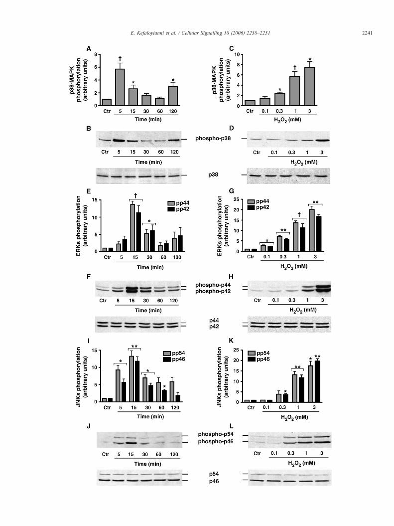
Fig. 1. Effect of H2O2 onMAPKs activation in skeletal myoblasts. C2 myoblasts were lefand J) or with various H2O2 concentrations for 15 min (G, H, K and L) or for 5 min (C andphospho-p38-MAPK (B andD top panels), p38-MAPK (B andD bottom panels), phosphoand L top panels) and JNK1/2 (J and L bottom panels). Phospho-MAPK bands were quancontrol values (A, C, E, G, I and K). *P<0.05, **P<0.01, †P<0.001 vs. control value.
10 min) and washed with PBS. The cell pellet was re-suspended in suspensionbuffer (100mMTris–HCl, pH 8, 200mMNaCl, 10 mMEDTA) and lysed by theaddition of an equal volume of digestion buffer (100 mM Tris–HCl, pH 8,200 mM NaCl, 10 mM EDTA, 0.4% SDS and 200 μg proteinase K). After anovernight incubation at 55 °C, DNA was extracted twice with a phenol–chloroform solution [1:1 (v/v)] and the aqueous phase was treated with RNase(20 μg/ml) for 30 min. DNAwas precipitated with ice-cold ethanol and resolvedin water. 5 μg DNA of each sample was subjected to electrophoresis on a 1%(w/v) agarose gel and visualised under ultraviolet light after 15 min incubation inethidium bromide.
2.9. Statistical evaluations
Western blots and EMSAs shown are representative of at least three inde-pendent experiments. All data are presented as means±S.E.M. Comparisonsbetween control and treatments were performed using Student's unpaired t-test.A value of at least P<0.05 was considered to be statistically significant.
3. Results
3.1. ERKs, JNKs and p38-MAPKs subfamilies are activated byH2O2 treatment
As the first step in the present study, the activation of MAPKsubfamilies by H2O2 using specific antibodies against the duallyphosphorylated (hence, activated) forms of the kinases wasexamined. The results revealed that all three MAPK subfamiliesexamined are rapidly and strongly activated by H2O2 treatment,following a time- and dose-dependent pattern. In particular, p38-MAPK activation displayed a rapid onset within 5 min of treat-ment (5.70±0.94 fold relative to controls, P<0.001), followedby a progressive decline, returning to basal levels within 30 min,while a second peak was observed (3.01±0.63-fold, relative tocontrol values, P<0.05) at 120 min of treatment (Fig. 1A and Btop panel). The minimal concentration of H2O2 inducing asignificant p38-MAPK activation was 0.3 mM (2.42±0.22-foldrelative to controls, P<0.05). Increasing H2O2 concentration ledto increased p38-MAPK phosphorylation (7.47±1.1-fold rela-tive to controls at 3 mM of H2O2, P<0.05) (Fig. 1C and D toppanel).
Activation of ERKs by H2O2 was intense, reaching maximumvalues within 15 min of treatment (13.71±0.94-fold relative tocontrol values for p44 and 11.30±2.03-fold for p42, P<0.001),decreasing thereafter and reaching control levels at 60 min(Fig. 1E and F top panel). ERKs activation was induced by lowerH2O2 concentration (0.1 mM) than that of p38 (2.80±0.28-foldrelative to control for p44 and 2.24±0.23-fold for p42, P<0.05)and maximal activation of the kinases was induced by 3 mM ofthe oxidant (20.12±1.51-fold for p44 and 16.70±1.01-fold forp42, P<0.01, respectively, above basal values) (Fig. 1G and Htop panel).
Activation of JNKs was also observed at 5 min of H2O2
treatment (9.26±1.3-fold, relative to control values for p54 and5.66±0.99-fold for p46, P<0.05), reaching maximal levels at
t untreated (Ctr) or were treated with 1 mMH2O2 for the indicated times (A, B, E, F, ID). The kinases were detected by immunoblotting, using specific antibodies against-ERK1/2 (F andH top panels), ERK1/2 (F andH bottom panels), phospho-JNK1/2 (Jtified by laser scanning densitometry and the results are presented as fold-relative to
2241E. Kefaloyianni et al. / Cellular Signalling 18 (2006) 2238–2251
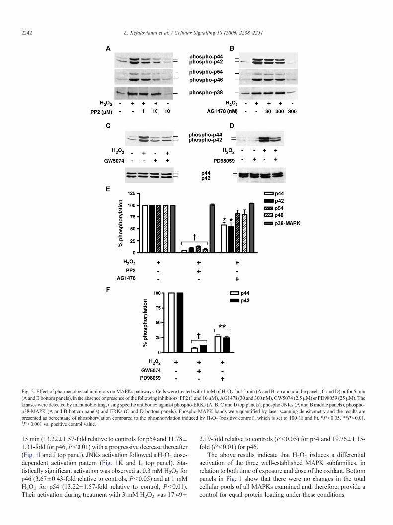
Fig. 2. Effect of pharmacological inhibitors onMAPKs pathways. Cells were treated with 1 mMof H2O2 for 15min (A and B top and middle panels; C and D) or for 5 min(AandBbottompanels), in the absence or presence of the following inhibitors: PP2 (1 and 10μM),AG1478 (30 and 300 nM),GW5074 (2.5μM) or PD98059 (25μM).Thekinases were detected by immunoblotting, using specific antibodies against phospho-ERKs (A, B, C and D top panels), phospho-JNKs (A and B middle panels), phospho-p38-MAPK (A and B bottom panels) and ERKs (C and D bottom panels). Phospho-MAPK bands were quantified by laser scanning densitometry and the results arepresented as percentage of phosphorylation compared to the phosphorylation induced by H2O2 (positive control), which is set to 100 (E and F). *P<0.05, **P<0.01,†P<0.001 vs. positive control value.
2242 E. Kefaloyianni et al. / Cellular Signalling 18 (2006) 2238–2251
15 min (13.22±1.57-fold relative to controls for p54 and 11.78±1.31-fold for p46, P<0.01) with a progressive decrease thereafter(Fig. 1I and J top panel). JNKs activation followed a H2O2 dose-dependent activation pattern (Fig. 1K and L top panel). Sta-tistically significant activation was observed at 0.3 mM H2O2 forp46 (3.67±0.43-fold relative to controls, P<0.05) and at 1 mMH2O2 for p54 (13.22±1.57-fold relative to control, P<0.01).Their activation during treatment with 3 mM H2O2 was 17.49±
2.19-fold relative to controls (P<0.05) for p54 and 19.76±1.15-fold (P<0.01) for p46.
The above results indicate that H2O2 induces a differentialactivation of the three well-established MAPK subfamilies, inrelation to both time of exposure and dose of the oxidant. Bottompanels in Fig. 1 show that there were no changes in the totalcellular pools of all MAPKs examined and, therefore, provide acontrol for equal protein loading under these conditions.
2243E. Kefaloyianni et al. / Cellular Signalling 18 (2006) 2238–2251
3.2. Src and, partially, EGFR mediate the phosphorylation ofERKs and JNKs, but not that of p38-MAPK
Although MAPK activation during oxidative stress has beenstudied in detail in diverse cell types, the signalling pathways
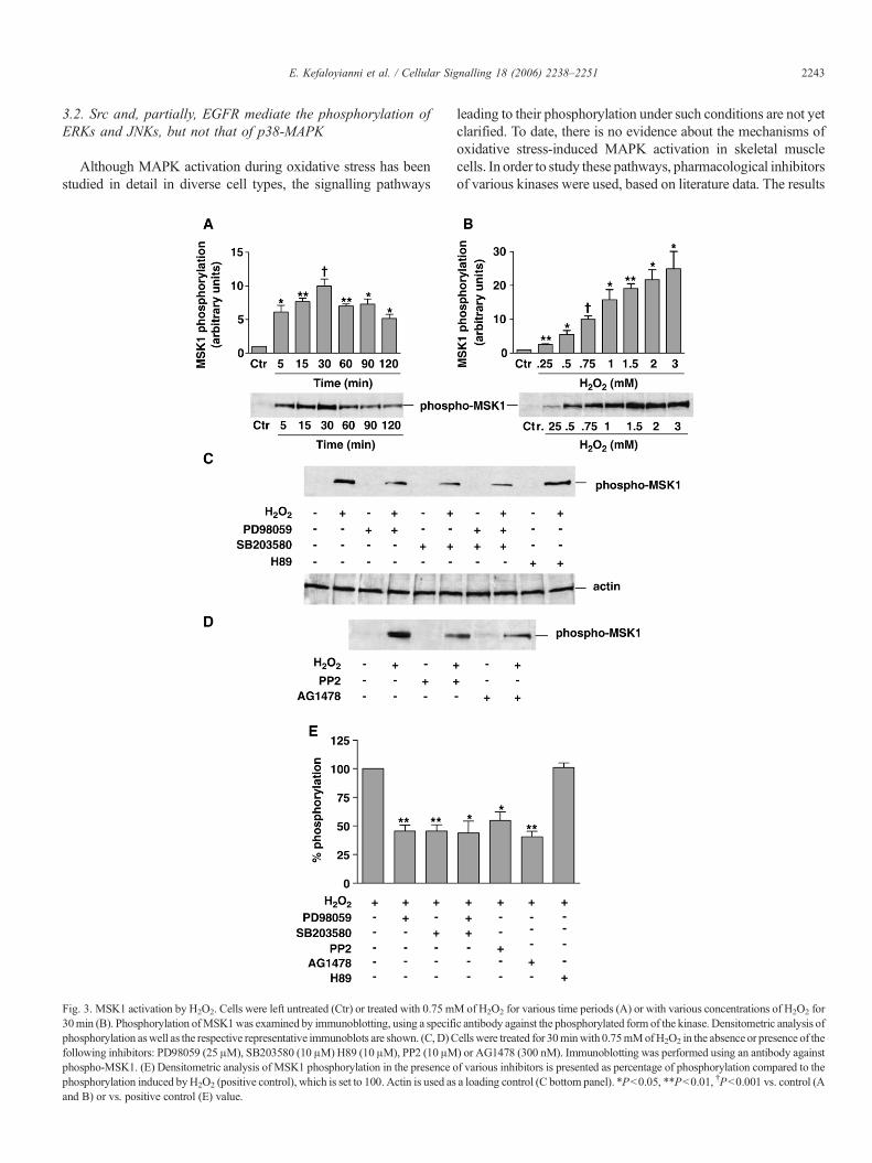
Fig. 3. MSK1 activation by H2O2. Cells were left untreated (Ctr) or treated with 0.75 m30min (B). Phosphorylation ofMSK1was examined by immunoblotting, using a specifphosphorylation aswell as the respective representative immunoblots are shown. (C,D)Cfollowing inhibitors: PD98059 (25 μM), SB203580 (10 μM)H89 (10 μM), PP2 (10 μMphospho-MSK1. (E) Densitometric analysis of MSK1 phosphorylation in the presence ophosphorylation induced byH2O2 (positive control), which is set to 100. Actin is used asand B) or vs. positive control (E) value.
leading to their phosphorylation under such conditions are not yetclarified. To date, there is no evidence about the mechanisms ofoxidative stress-induced MAPK activation in skeletal musclecells. In order to study these pathways, pharmacological inhibitorsof various kinases were used, based on literature data. The results
M of H2O2 for various time periods (A) or with various concentrations of H2O2 foric antibody against the phosphorylated form of the kinase. Densitometric analysis ofells were treated for 30minwith 0.75mMofH2O2 in the absence or presence of the) or AG1478 (300 nM). Immunoblotting was performed using an antibody againstf various inhibitors is presented as percentage of phosphorylation compared to thea loading control (C bottom panel). *P<0.05, **P<0.01, †P<0.001 vs. control (A
2244 E. Kefaloyianni et al. / Cellular Signalling 18 (2006) 2238–2251
of these experiments showed that 10 μMPP2 (a specific inhibitorof Src kinases) abolishes the activation of ERKs (Fig. 2A toppanel and E) and JNKs (Fig. 2A middle panel and E) induced by1 mM H2O2, whereas this inhibitor had no effect on p38-MAPKactivation (Fig. 2A bottom panel and E). Moreover, 300 nMAG1478 (a specific EGFR inhibitor) resulted in a partial inhi-bition of ERKs (41.99% reduction for p44 and 45.76% for p42,P<0.05) (Fig. 2B top panel and E), whereas it had not any sig-nificant inhibitory effect on JNKs activation by H2O2 (Fig. 2Bmiddle panel and E). Once more, p38-MAPK phosphorylationwas not affected by this inhibitor (Fig. 2B bottom panel and E). Toinvestigate whether the signalling pathway leading to ERK andJNK activation was Ras-Raf-mediated, GW5074 (a specificinhibitor of cRaf1) was used. 2.5 μM of GW5074 diminished theactivation of ERKs induced by H2O2 (Fig. 2C top panel and F),while it had no effect on the activation of JNKs induced by 1 mMH2O2 (data not shown). Finally, 25 μM of PD98059 (a selectiveMEK1/2 inhibitor) significantly reduced, as it was expected, theactivation of ERKs byH2O2 (Fig. 2D top panel and F). The aboveresults demonstrate that the activation mechanisms of ERKs andJNKs by H2O2 comprise common components, such as the Srckinase. On the contrary, the activation pathway of p38-MAPK byH2O2 seems to be Src kinase-independent.
3.3. ERKs and p38-MAPKmediate the phosphorylation of MSK1by H2O2
MSK1 is a substrate of ERKs and p38-MAPK and it was foundto phosphorylate the p65 subunit of NF-κB at Ser276 duringTNF-α stimulation [35]. Therefore, phosphorylation ofMSK1 (atThr581) was investigated during H2O2 treatment of C2 myo-blasts. As shown in Fig. 3A, the kinase was phosphorylated ra-pidly, within 5 min (6.11±0.99-fold relative to control values,P<0.05), reaching maximal phosphorylation levels after 30 minof treatment (9.98±1.03-fold relative to controls, P<0.001). This
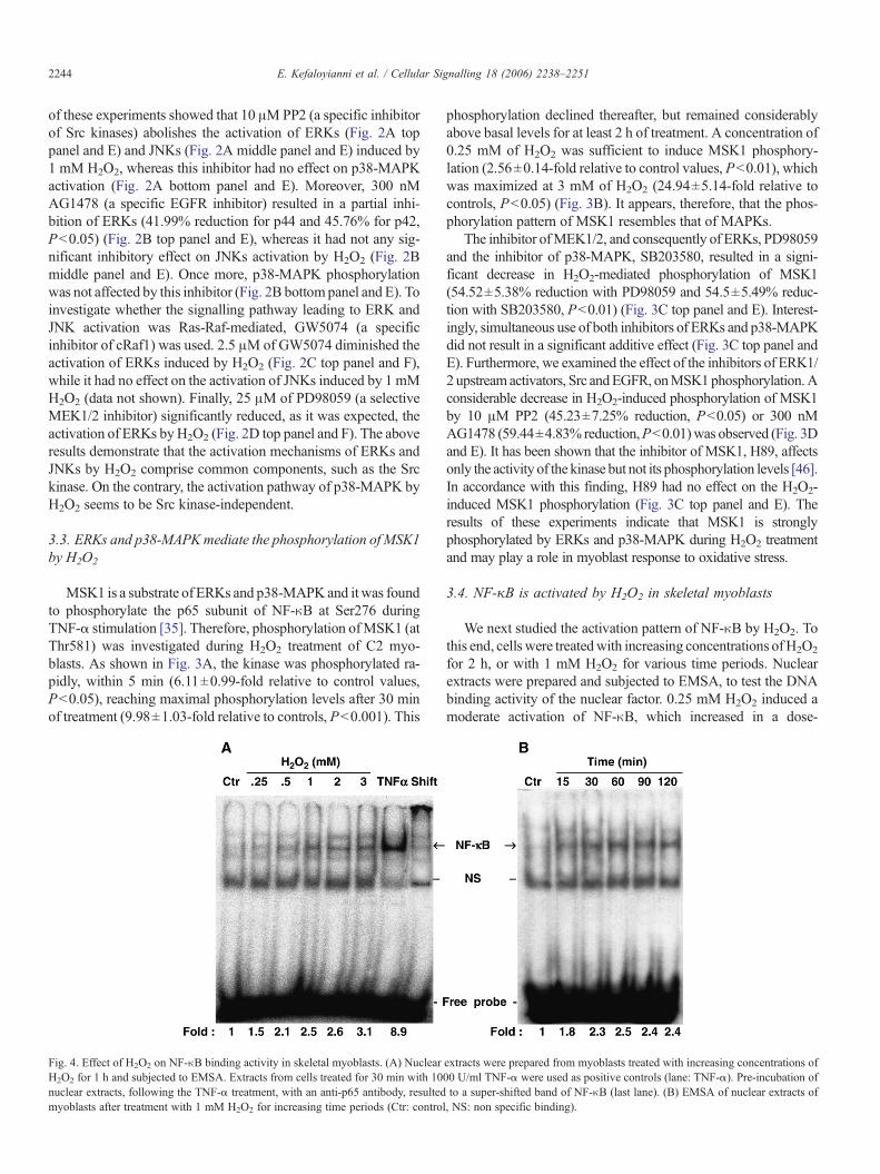
Fig. 4. Effect of H2O2 on NF-κB binding activity in skeletal myoblasts. (A) NuclearH2O2 for 1 h and subjected to EMSA. Extracts from cells treated for 30 min with 10nuclear extracts, following the TNF-α treatment, with an anti-p65 antibody, resultedmyoblasts after treatment with 1 mM H2O2 for increasing time periods (Ctr: contro
phosphorylation declined thereafter, but remained considerablyabove basal levels for at least 2 h of treatment. A concentration of0.25 mM of H2O2 was sufficient to induce MSK1 phosphory-lation (2.56±0.14-fold relative to control values, P<0.01), whichwas maximized at 3 mM of H2O2 (24.94±5.14-fold relative tocontrols, P<0.05) (Fig. 3B). It appears, therefore, that the phos-phorylation pattern of MSK1 resembles that of MAPKs.
The inhibitor ofMEK1/2, and consequently of ERKs, PD98059and the inhibitor of p38-MAPK, SB203580, resulted in a signi-ficant decrease in H2O2-mediated phosphorylation of MSK1(54.52±5.38% reduction with PD98059 and 54.5±5.49% reduc-tion with SB203580, P<0.01) (Fig. 3C top panel and E). Interest-ingly, simultaneous use of both inhibitors of ERKs and p38-MAPKdid not result in a significant additive effect (Fig. 3C top panel andE). Furthermore, we examined the effect of the inhibitors of ERK1/2 upstreamactivators, Src andEGFR, onMSK1phosphorylation.Aconsiderable decrease in H2O2-induced phosphorylation of MSK1by 10 μM PP2 (45.23±7.25% reduction, P<0.05) or 300 nMAG1478 (59.44±4.83% reduction,P<0.01)was observed (Fig. 3Dand E). It has been shown that the inhibitor of MSK1, H89, affectsonly the activity of the kinase but not its phosphorylation levels [46].In accordance with this finding, H89 had no effect on the H2O2-induced MSK1 phosphorylation (Fig. 3C top panel and E). Theresults of these experiments indicate that MSK1 is stronglyphosphorylated by ERKs and p38-MAPK during H2O2 treatmentand may play a role in myoblast response to oxidative stress.
3.4. NF-κB is activated by H2O2 in skeletal myoblasts
We next studied the activation pattern of NF-κB by H2O2. Tothis end, cells were treatedwith increasing concentrations ofH2O2
for 2 h, or with 1 mM H2O2 for various time periods. Nuclearextracts were prepared and subjected to EMSA, to test the DNAbinding activity of the nuclear factor. 0.25 mM H2O2 induced amoderate activation of NF-κB, which increased in a dose-
extracts were prepared from myoblasts treated with increasing concentrations of00 U/ml TNF-α were used as positive controls (lane: TNF-α). Pre-incubation ofto a super-shifted band of NF-κB (last lane). (B) EMSA of nuclear extracts of
l, NS: non specific binding).

Fig. 5. Effect of H2O2 on IκB phosphorylation and degradation. Cells were treated with 1 mM H2O2 for various time intervals (B) or with increasing concentrations ofH2O2 for 1 h (D) or with 1000 U/ml TNF-α for 30 min (E). IκB phosphorylation was assessed by immunoblotting using a specific anti-phospho (Ser32/36)-IκB-αantibody (B, D and E top panels) and total IκB levels were measured using an antibody against total IκB-α (B, D and E bottom panels). Densitometric analysis of thephosphorylated IκB bands during H2O2 treatments is also presented (A and C). *P<0.05, **P<0.01, †P<0.001 vs. control value.
2245E. Kefaloyianni et al. / Cellular Signalling 18 (2006) 2238–2251
dependent manner (Fig. 4A). H2O2-induced NF-κB activationwas also time-dependent, displaying an onset within 15 min,reaching maximal values within 1 h and remaining considerablyabove basal levels for at least 2 h of treatment (Fig. 4B). Nuclearextracts prepared from cells treated with TNF-α (1000 U/ml) for
Fig. 6. Effect of MAPK inhibition on NF-κB DNA binding activity and IκB phosphorthe following inhibitors: PD98059 (25 μM), SB203580 (10 μM), SP600125 (10 μMsubjected to EMSA. (B) Immunoblotting was performed using an antibody against
30 min were used as positive controls (Fig. 4A). To confirmbinding specificity, we used an antibody against the p65 subunitof NF-κB. Pre-incubation of nuclear extracts with the antibodyled to a shift of the band to high molecular mass (Fig. 4A). Theseresults reveal that NF-κB is activated by H2O2 and translocates
ylation. Cells were treated with 1 mM H2O2 for 1 h in the absence or presence of), PP2 (10 μM) or AG1478 (300 nM). (A) Nuclear extracts were prepared andthe phosphorylated form of IκB.
2246 E. Kefaloyianni et al. / Cellular Signalling 18 (2006) 2238–2251
into the nucleus. This activation, however, is weaker compared tothe one observed during treatment with its potent inducer, TNF-α.
3.5. H2O2 induces phosphorylation, but not degradation, of IκB-α
Phosphorylation and subsequent degradation of IκB is requiredfor NF-κB activation, according to the classical activation pathway.Therefore, IκB phosphorylation was examined, using a specificantibody against the phosphorylated (Ser 32/36) formof IκB-α.Wefound that IκB was phosphorylated during H2O2 treatment, in atime- and dose-dependent profile. This phosphorylation was rapid,within 5 min of treatment (1.91±0.05-fold relative to controls,P<0.01), reached maximal values within 30 min (3.88±0.19-foldrelative to controls, P<0.01) and remained at maximum levels forat least 2 h (Fig. 5A and B top panel). Phosphorylation of IκBcould be detected when concentration of H2O2 used was 0.5 mMor higher. Increasing H2O2 concentration induced increasing IκBphosphorylation (reaching 7.39±0.27-fold relative to controls,P<0.01, at 3 mM H2O2) (Fig. 5C and D top panel). Thus, we
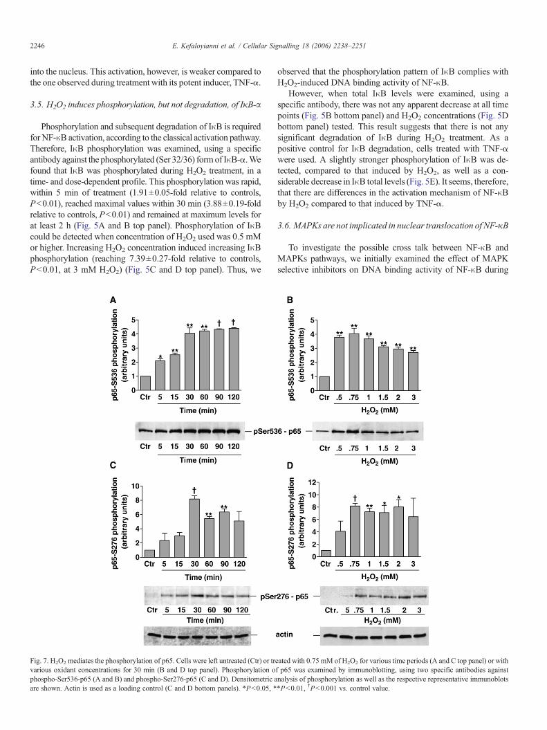
Fig. 7. H2O2 mediates the phosphorylation of p65. Cells were left untreated (Ctr) or trvarious oxidant concentrations for 30 min (B and D top panel). Phosphorylation ophospho-Ser536-p65 (A and B) and phospho-Ser276-p65 (C and D). Densitometric aare shown. Actin is used as a loading control (C and D bottom panels). *P<0.05, *
observed that the phosphorylation pattern of IκB complies withH2O2-induced DNA binding activity of NF-κB.
However, when total IκB levels were examined, using aspecific antibody, there was not any apparent decrease at all timepoints (Fig. 5B bottom panel) and H2O2 concentrations (Fig. 5Dbottom panel) tested. This result suggests that there is not anysignificant degradation of IκB during H2O2 treatment. As apositive control for IκB degradation, cells treated with TNF-αwere used. A slightly stronger phosphorylation of IκB was de-tected, compared to that induced by H2O2, as well as a con-siderable decrease in IκB total levels (Fig. 5E). It seems, therefore,that there are differences in the activation mechanism of NF-κBby H2O2 compared to that induced by TNF-α.
3.6. MAPKs are not implicated in nuclear translocation of NF-κB
To investigate the possible cross talk between NF-κB andMAPKs pathways, we initially examined the effect of MAPKselective inhibitors on DNA binding activity of NF-κB during
eated with 0.75 mM of H2O2 for various time periods (A and C top panel) or withf p65 was examined by immunoblotting, using two specific antibodies againstnalysis of phosphorylation as well as the respective representative immunoblots*P<0.01, †P<0.001 vs. control value.
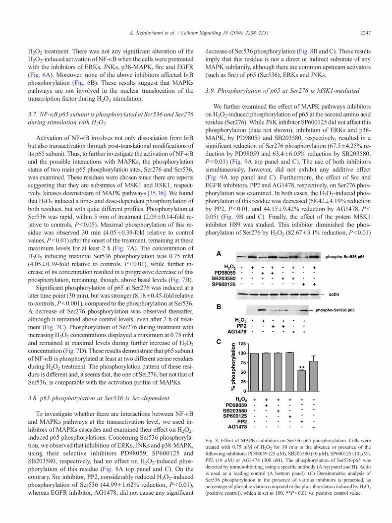
Fig. 8. Effect of MAPKs inhibitors on Ser536-p65 phosphorylation. Cells weretreated with 0.75 mM of H2O2 for 30 min in the absence or presence of thefollowing inhibitors: PD98059 (25 μM), SB203580 (10 μM), SP600125 (10 μM),PP2 (10 μM) or AG1478 (300 nM). The phosphorylation of Ser536-p65 wasdetected by immunoblotting, using a specific antibody (A top panel and B). Actinis used as a loading control (A bottom panel). (C) Densitometric analysis ofSer536 phosphorylation in the presence of various inhibitors is presented, aspercentage of phosphorylation compared to the phosphorylation induced by H2O2
(positive control), which is set to 100. **P<0.01 vs. positive control value.
2247E. Kefaloyianni et al. / Cellular Signalling 18 (2006) 2238–2251
H2O2 treatment. There was not any significant alteration of theH2O2-induced activation of NF-κBwhen the cells were pretreatedwith the inhibitors of ERKs, JNKs, p38-MAPK, Src and EGFR(Fig. 6A). Moreover, none of the above inhibitors affected IκBphosphorylation (Fig. 6B). These results suggest that MAPKspathways are not involved in the nuclear translocation of thetranscription factor during H2O2 stimulation.
3.7. NF-κB p65 subunit is phosphorylated at Ser536 and Ser276during stimulation with H2O2
Activation of NF-κB involves not only dissociation from IκBbut also transactivation through post-translational modifications ofits p65 subunit. Thus, to further investigate the activation of NF-κBand the possible interactions with MAPKs, the phosphorylationstatus of two main p65 phosphorylation sites, Ser276 and Ser536,was examined. These residues were chosen since there are reportssuggesting that they are substrates of MSK1 and RSK1, respect-ively, kinases downstream of MAPK pathways [35,36]. We foundthat H2O2 induced a time- and dose-dependent phosphorylation ofboth residues, but with quite different profiles. Phosphorylation atSer536 was rapid, within 5 min of treatment (2.09±0.14-fold re-lative to controls, P<0.05). Maximal phosphorylation of this re-sidue was observed 30 min (4.05±0.39-fold relative to controlvalues,P<0.01) after the onset of the treatment, remaining at thesemaximum levels for at least 2 h (Fig. 7A). The concentration ofH2O2 inducing maximal Ser536 phosphorylation was 0.75 mM(4.05±0.39-fold relative to controls, P<0.01), while further in-crease of its concentration resulted in a progressive decrease of thisphosphorylation, remaining, though, above basal levels (Fig. 7B).
Significant phosphorylation of p65 at Ser276 was induced at alater time point (30min), but was stronger (8.18±0.45-fold relativeto controls,P<0.001), compared to the phosphorylation at Ser536.A decrease of Ser276 phosphorylation was observed thereafter,although it remained above control levels, even after 2 h of treat-ment (Fig. 7C). Phosphorylation of Ser276 during treatment withincreasing H2O2 concentrations displayed a maximum at 0.75 mMand remained at maximal levels during further increase of H2O2
concentration (Fig. 7D). These results demonstrate that p65 subunitof NF-κB is phosphorylated at least at two different serine residuesduring H2O2 treatment. The phosphorylation pattern of these resi-dues is different and, it seems that, the one of Ser276, but not that ofSer536, is comparable with the activation profile of MAPKs.
3.8. p65 phosphorylation at Ser536 is Src-dependent
To investigate whether there are interactions between NF-κBand MAPKs pathways at the transactivation level, we used in-hibitors of MAPKs cascades and examined their effect on H2O2-induced p65 phosphorylations. Concerning Ser536 phosphoryla-tion, we observed that inhibition of ERKs, JNKs and p38-MAPK,using their selective inhibitors PD98059, SP600125 andSB203580, respectively, had no effect on H2O2-induced phos-phorylation of this residue (Fig. 8A top panel and C). On thecontrary, Src inhibitor, PP2, considerably reduced H2O2-inducedphosphorylation of Ser536 (44.99±1.62% reduction, P<0.01),whereas EGFR inhibitor, AG1478, did not cause any significant
decrease of Ser536 phosphorylation (Fig. 8B andC). These resultsimply that this residue is not a direct or indirect substrate of anyMAPK subfamily, although there are common upstream activators(such as Src) of p65 (Ser536), ERKs and JNKs.
3.9. Phosphorylation of p65 at Ser276 is MSK1-mediated
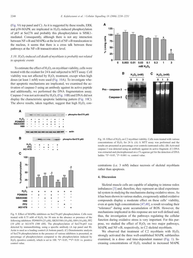
We further examined the effect of MAPK pathways inhibitorson H2O2-induced phosphorylation of p65 at the second amino acidresidue (Ser276).While JNK inhibitor SP600125 did not affect thisphosphorylation (data not shown), inhibition of ERKs and p38-MAPK, by PD98059 and SB203580, respectively, resulted in asignificant reduction of Ser276 phosphorylation (67.5±4.25% re-duction by PD98059 and 63.4±6.05% reduction by SB203580,P<0.01) (Fig. 9A top panel and C). The use of both inhibitorssimultaneously, however, did not exhibit any additive effect(Fig. 9A top panel and C). Furthermore, the effect of Src andEGFR inhibitors, PP2 and AG1478, respectively, on Ser276 phos-phorylation was examined. In both cases, the H2O2-induced phos-phorylation of this residue was decreased (68.42±4.19% reductionby PP2, P<0.01, and 44.15±9.42% reduction by AG1478, P<0.05) (Fig. 9B and C). Finally, the effect of the potent MSK1inhibitor H89 was studied. This inhibitor diminished the phos-phorylation of Ser276 by H2O2 (82.67±3.1% reduction, P<0.01)
2248 E. Kefaloyianni et al. / Cellular Signalling 18 (2006) 2238–2251
(Fig. 9A top panel and C). As it is suggested by these results, ERKand p38-MAPK are implicated in H2O2-induced phosphorylationof p65 at Ser276 and probably this phosphorylation is MSK1-mediated. Consequently, although there is not any interactionbetween NF-κB andMAPKs at the level of NF-κB translocation tothe nucleus, it seems that there is a cross talk between thesepathways at the NF-κB transactivation level.
3.10. H2O2-induced cell death of myoblasts is probably not relatedto apoptotic events
To estimate the effect of H2O2 onmyoblast viability, cells weretreated with the oxidant for 24 h and subjected toMTTassay. Cellviability was not affected by H2O2 treatment, except when highdoses (at least 1 mM) were used (Fig. 10A). To investigate whe-ther apoptotic mechanisms are implicated, we examined the ac-tivation of caspase-3 using an antibody against its active peptideand additionally, we performed the DNA fragmentation assay.Caspase-3was not activated byH2O2 (Fig. 10B) andDNAdid notdisplay the characteristic apoptotic laddering pattern (Fig. 10C).The above results, taken together, suggest that high H2O2 con-
Fig. 10. Effect of H2O2 on C2 myoblast viability. Cells were treated with variousconcentrations of H2O2 for 24 h. (A) A MTT assay was performed and theresults are presented as percentage over controls (untreated cells). (B) Activatedcaspase-3 was detected using an antibody against its active fragment. (C) DNAwas extracted and electrophoresed on a 1% agarose gel for the detection of DNAladder. *P<0.05, †P<0.001 vs. control value.
Fig. 9. Effect of MAPKs inhibitors on Ser276-p65 phosphorylation. Cells weretreated with 0.75 mM of H2O2 for 30 min in the absence or presence of thefollowing inhibitors: PD98059 (25 μM), SB203580 (10 μM), H89 (10 μM), PP2(10 μM) or AG1478 (300 nM). The phosphorylation of Ser276-p65 wasdetected by immunoblotting, using a specific antibody (A top panel and B).Actin is used as a loading control (A bottom panel). (C) Densitometric analysisof Ser276 phosphorylation in the presence of various inhibitors is presented, aspercentage of phosphorylation compared to the phosphorylation induced byH2O2 (positive control), which is set to 100. *P<0.05, **P<0.01 vs. positivecontrol value.
centrations (i.e. 3 mM) induce necrosis of skeletal myoblastsrather than apoptosis.
4. Discussion
Skeletal muscle cells are capable of adapting to intense redoximbalances [3] and, therefore, they represent an ideal experimen-tal system in studying the mechanisms during oxidative stress. Asit has been shown in various studies, exogenously added oxidativecompounds display a moderate effect on these cells' viability,even at quite high concentrations [47,48], a result revealing their“tolerance” during acute accumulation of ROS. However, themechanisms implicated in this response are not well defined and,thus, the investigation of the pathways regulating the cellularfunction during oxidative stress is very important. For this pur-pose, we studied the effect of H2O2 on two major pathways,MAPK and NF-κB, respectively, in C2 skeletal myoblasts.
We observed that treatment of C2 myoblasts with H2O2
strongly activated the three well-established MAPK subfamiliesexamined, in a dose- and time-dependent manner (Fig. 1). In-creasing concentrations of H2O2 resulted in increased MAPK
2249E. Kefaloyianni et al. / Cellular Signalling 18 (2006) 2238–2251
activation. However, the time-course of this activation differedamong the MAPK subfamilies. Activation of ERKs and JNKswas transient, maximized at 15 min, declining thereafter. On theother hand, p38-MAPK activation was more rapid, maximizingwithin 5min and displaying a biphasic pattern, with a second peakobtained at 2 h of treatment. This p38-MAPK re-activation couldbe attributed to a feedback mechanism, mediated by its eitherupstream activators or downstream targets, a phenomenon thathas been previously reported (p38 is also known as reactivatingkinase—RK) [14,49,50].
A question remaining unanswered is whether different celltypes share similar activation mechanisms of MAPKs pathways.EGFR and Src kinases have been shown tomediate ROS-inducedMAPK activation in vascular endothelial cells [20], renal epithe-lial cells [21], cardiomyocytes and heart fibroblasts [22]. Ourresults point as well to a Src-dependent activation of ERKs andJNKs by H2O2 in skeletal myoblasts (Fig. 2). Whether, in ourexperimental system, Src and EGFR lie in the same pathwayleading to MAPK activation is under investigation. In agreementwith Purdom and Chen [22], we observed that PP2 (a Src kinaseinhibitor) displayed much higher efficiency inhibiting MAPKactivation than AG1478 (an EGFR inhibitor) (Fig. 2). Src kinaseis a well-known downstream target of EGFR tyrosine kinase [51].Conversely, it has been shown that Src kinase can phosphorylateEGFR at Tyr845, stabilizing the activated state of the kinasedomain, leading, consequently, to the activation of the MAPKcascade [20,52]. Therefore, in the case of a ligand-independentactivation of EGFR, the sequential relationship between Src ki-nase and EGFR is not yet clarified.
H2O2-induced p38-MAPK phosphorylation was not affectedby the above inhibitors, implying the existence of a completelydifferent activation mechanism for this kinase. It is possible that
Fig. 11. A schematic model summarizing the resu
activation of p38-MAPK is regulated by Trx and ASK1 proteins[23], although this hypothesis remains to be elucidated.
Our results also indicate thatMSK1 is strongly phosphorylatedby H2O2 treatment and that this phosphorylation is mediated byboth, ERKs and p38-MAPK (Fig. 3). MSK1 phosphorylation byH2O2 was significantly reduced by ERK and p38-MAPK inhi-bitors as well as by inhibition of ERK upstream molecules, Srckinase and EGFR. The phosphorylation pattern of MSK1 fol-lowed closely the activation pattern of MAPKs. When both ERKand p38-MAPK were inhibited, we did not observe further in-hibition of MSK1 phosphorylation. This may seem inconsistent,but we should take into consideration that it is unclear how thekinetics of ERK and p38-MAPK phosphorylation interact andcoordinate MSK1 activation and that SB203580 can inhibit onlythe alpha and beta isoforms of p38-MAPK [53], while in skeletalmuscle p38 gamma is largely expressed and may contribute toMSK1 phosphorylation as well. MSK1 phosphorylation by oxi-dative stress has been also reported in other cell types [54,55],although in those cases MSK1 activation was mediated only byp38-MAPK, and not by ERK. The activation of the kinase in theabove studies was correlated with the subsequent activation oftranscription factors (CREB and AP1, respectively).
As the transcription factor NF-κB is subjected to acomplicated redox regulation in a cell-type specific manner[29], we examined its activation during oxidative stress inskeletal myoblasts. We found a mild time- and dose-dependentactivation of NF-κB, as it was demonstrated by the increase inits DNA-binding activity, assessed by EMSA (Fig. 4). We alsoobserved an increase in IκB phosphorylation (Ser32/36) byH2O2, exhibiting a similar profile with the DNA-bindingactivity of NF-κB (Fig. 5). However, we did not observe anydecrease in IκB total levels, a result indicating that there is not
lts of this study. → induction, ⊣ inhibition.
2250 E. Kefaloyianni et al. / Cellular Signalling 18 (2006) 2238–2251
any apparent degradation of IκB during oxidative stimulus. Thisresult implies that there is an alternative activation mechanismof the transcription factor during this treatment. It has beenreported that certain agents, including H2O2, activate NF-κB notthrough serine but through tyrosine phosphorylation of IκB andthis may not lead to IκB degradation [56]. Thus, IκB remainsbound to the dimer in the cytosol and only a small fraction of thetranscription factor translocates into the nucleus. In otherstudies, another mechanism of NF-κB activation has beenproposed, without the implication of IκB degradation, nor itstyrosine phosphorylation. This mechanism is based on reducedbinding of NF-κB to IκB due to increased phosphorylation ofp65 subunit of NF-κB [36]. These theories could explain themild activation of NF-κB by H2O2, compared to the strong oneobserved during treatment with its potent inducer, TNF-α.Activation of NF-κB by oxidative stress has been previouslyreported in skeletal myotubes [57,58], correlated with theirresistance to oxidative injury. The precise mechanism of NF-κBactivation, however, was not examined in these studies. Zhou etal. [57] used a mutant IκB-α to inhibit NF-κB activation. Thismutation involves the phosphorylation site of IκB. Thus, wemay assume that the phosphorylation of IκB is important forNF-κB activation, even if it does not lead to IκB degradation.
A growing body of evidence supports that post-translationalmodifications play particularly important roles in the activation ofNF-κB. Both phosphorylation and acetylation of p65 subunit ofthe transcription factor seem to be critical for the effective regu-lation of gene transcription by NF-κB. These modifications alterthe ability of NF-κB dimers to bind DNA, recruit co-activatorsand bind IκB [33,59]. Recently, the transactivation of NF-κB hasbeen linked with the MAPK substrates RSK1 and MSK1 thatwere shown to phosphorylate p65 subunit at serine 276 and serine536, respectively [35,36]. To further investigate oxidative stress-mediated activation of NF-κB, we examined the phosphorylationstatus of these two residues, duringH2O2 treatment. As it is shownin Fig. 7, these two sites are phosphorylated by H2O2, in a time-and dose-dependent manner. The phosphorylation pattern ofSer276 is similar with that of MAPK andMSK1. On the contrary,the phosphorylation pattern of Ser536 is completely differentcompared to that of MAPK. Overall, it seems that the translo-cation of NF-κB dimer to the nucleus (as shown by EMSA) isaccompanied with its phosphorylation at both aminoacid residuesexamined.
We further investigated whether MAPK pathways are im-plicated in any level of NF-κB activation. Using specific inhi-bitors of the kinases, we found that none of the MAPK pathwaysexamined had an effect onH2O2-inducedDNAbinding activity ofNF-κB or on Ser32/36 phosphorylation of IκB (Fig. 6). Theseresults suggest that MAPKs are not involved in the processesleading to dissociation of NF-κB dimer from IκB and to nucleartranslocation of the transcription factor.
Phosphorylation of p65 residue Ser536 was shown to bedependent on the Src kinase, while MAPKs were not implicatedin this phosphorylation (Fig. 8). A kinase that has often beenimplicated in this phosphorylation is IKK, as has been shownduring TNF-α treatment [59,60]. It is not known whether this isalso the case during oxidative stimulus. An interesting point,
according to our data, is that Src kinase seems to mediate theactivation of multiple, diverse signalling pathways during H2O2
treatment.On the other hand, our experiments showed that the phos-
horylation of the other residue (Ser276) examined was mediatedby MAPKs. In particular, we found that the inhibitors of p38-MAPK and ERKs, as well as the inhibitors of molecules upstreamof ERKs (Src kinase and EGFR) reduced the H2O2-mediatedphosphorylation of p65 at Ser276 (Fig. 9). Moreover, the potentinhibitor of MSK1, H89, diminished this phosphorylation. Theseresults suggest that Ser276 of p65 could be a target for MSK1during oxidative stimulus. This specific site is also phosphory-lated by protein kinase A (PKA), during LPS (lipopolysacharide)treatment [61] and H89 inhibits PKA as well. However, the factthat Ser276 phosphorylation is attenuated by inhibition of ERKand p38-MAPK, two kinases that have been shown to activateMSK1, further enhances the idea that this specific phosphoryla-tion is mediated by MSK1. Still, whether MSK1 is the onlySer276-p65 kinase is uncertain.
5. Conclusions
Taken together, our results (summarized in Fig. 11) demon-strate that both MAPK and NF-κB pathways are activated duringoxidative stress in skeletal myoblasts, providing evidence on theiractivationmechanisms.We show for the first time thatMAPKs, inparticular ERKs and p38-MAPK, are implicated in NF-κB trans-activation by oxidative stimulus, and that this action is probablymediated by MSK1.
Overall, the regulation mechanisms involved in NF-κB acti-vation are complicated; integrated signals arising from diversepathways are required for its translocation and/or transactiva-tion, reflecting the differential cell-type-dependent responsesobserved under oxidative stress.
Acknowledgments
This work was funded by a GSRT grant (PENED 01 ED5).
References
[1] K.J. Davies, A.T. Quintanilha, G.A. Brooks, L. Packer, Biochem. Biophys.Res. Commun. 107 (1982) 1198.
[2] M.J. Jackson, R.H. Edwards, M.C. Symons, Biochim. Biophys. Acta 847(1985) 185.
[3] F. McArdle, D.M. Pattwell, A. Vasilaki, A. McArdle, M.J. Jackson, FreeRadic. Biol. Med. 39 (2005) 651.
[4] M. Jackson, in: A.Z. Reznick, L. Packer, C.K. Sen, J.O. Holloszy, M.J.Jackson (Eds.), Oxidative Stress in Skeletal Muscle, Free Radical Me-chanisms in Exercise-related Muscle Damage, Birkhauser Verlag, Basel,1998, p. 75.
[5] T.A. Rando, Am. J. Phys. Med. Rehabil. 81 (2002) S175.[6] H. Kondo, I. Nakagaki, S. Sasaki, S. Hori, Y. Itokawa, Am. J. Physiol:
Endocrinol. Metab. 265 (1993) E839.[7] S.K. Powers, A.N. Kavazis, K.C. DeRuisseau, Am. J. Physiol., Regul.
Integr. Comp. Physiol. 288 (2005) R337.[8] A. McArdle, J.H. van der Meulen, M. Catapano, M.C.R. Symons, J.A.
Faulkner, M.J. Jackson, Free Radic. Biol. Med. 26 (1999) 1085.[9] V. Adler, Z. Yin, K.D. Tew, Z. Ronai, Oncogene 18 (1999) 6104.[10] J.J. Haddad, Cell. Signal. 14 (2002) 879.
2251E. Kefaloyianni et al. / Cellular Signalling 18 (2006) 2238–2251
[11] Y. Sun, L.W. Oberley, Free Radic. Biol. Med. 21 (1996) 335.[12] W. Droge, Physiol. Rev. 82 (2002) 47.[13] C. Widmann, S. Gibson, M.B. Jarpe, G.L. Johnson, Physiol. Rev. 79
(1999) 143.[14] J.M. Kyriakis, J. Avruch, Physiol. Rev. 81 (2001) 807.[15] S. Ghosh, M. Karin, Cell 109 (2002) S81.[16] G. Pearson, F. Robinson, T. Beers Gibson, B.E. Xu, M. Karandikar, K.
Berman, M.H. Cobb, Endocr. Rev. 22 (2001) 153.[17] P.P. Roux, J. Blenis, Microbiol. Mol. Biol. Rev. 68 (2004) 320.[18] J.I. Abe, B.C. Berk, J. Biol. Chem. 274 (1999) 21003.[19] M. Yoshizumi, J. Abe, J. Haendeler, Q. Huang, B.C. Berk, J. Biol. Chem.
275 (2000) 11706.[20] K.Chen, J.A.Vita, B.C.Berk, J.F.Keaney Jr., J. Biol. Chem. 276 (2001) 16045.[21] S. Zhougang, R.G. Schnellmann, Am. J. Physiol., Renal Fluid Electrolyte
Physiol. 286 (2004) F858.[22] S. Purdom, Q.M. Chen, J. Pharmacol. Exp. Ther. 312 (2005) 1179.[23] J. Matsukawa, A. Matsuzawa, K. Takeda, H. Ichijo, J. Biochem. (Tokyo)
136 (2004) 261.[24] W. Zhang, S. Zheng, P. Storz, W. Min, J. Biol. Chem. 280 (2005) 19036.[25] H. Ichijo, E. Nishida, K. Irie, P. ten Dijke, M. Saitoh, T. Moriguchi, M.
Takagi, K. Matsumoto, K. Miyazono, Y. Gotoh, Science 275 (1997) 90.[26] M. Saitoh, H. Nishitoh, M. Fujii, K. Takeda, K. Tobiume, Y. Sawada, M.
Kawabata, K. Miyazono, H. Ichijo, EMBO J. 17 (1998) 2596.[27] N.K. Tonks, Cell 121 (2005) 667.[28] R. Schreck, P.A. Baeuerle, Trends Cell Biol. 1 (1991) 39.[29] N. Li, M. Karin, FASEB J. 13 (1999) 1137.[30] S. Ghosh, M.J. May, E.B. Kopp, Annu. Rev. Immunol. 16 (1998) 225.[31] M. Karin, Y. Ben-Neriah, Annu. Rev. Immunol. 18 (2000) 621.[32] M.L. Schmitz, S. Bacher, M. Kracht, Trends Biochem. Sci. 26 (2001) 186.[33] L. Vermeulen, G. DeWilde, S. Notebaert, W. Vanden Berghe, G. Haegeman,
Biochem. Pharmacol. 64 (2002) 963.[34] L.F. Chen, W.C. Greene, Nat. Rev., Mol. Cell Biol. 5 (2004) 392.[35] L. Vermeulen, G. De Wilde, P. Van Damme, W. Vanden Berghe, G.
Haegeman, EMBO J. 22 (2003) 1313.[36] J. Bohuslav, L.F. Chen, H. Kwon, Y. Mu, W.C. Greene, J. Biol. Chem. 279
(2004) 26115.[37] W. Vanden Berghe, S. Plaisance, E. Boone, K. De Bosscher, M.L. Schmitz,
W. Fiers, G. Haegeman, J. Biol. Chem. 273 (1998) 3285.
[38] R. Craig, A. Larkin, A.M. Mingo, D.J. Thuerauf, C. Andrews, P.M.McDonough, C.C. Glembotski, J. Biol. Chem. 275 (2000) 23814.
[39] B. Baeza-Raja, P. Munoz-Canoves, Mol. Biol. Cell 15 (2004) 2013.[40] T.J. Hawke, D.J. Garry, J. Appl. Physiol. 91 (2001) 534.[41] D. Yaffe, O. Saxel, Nature 270 (1977) 725.[42] E. Schreiber, P. Matthias, M.M. Muller, W. Schaffner, Nucleic Acids Res.
17 (1989) 6419.[43] T. Markou, M. Hadzopoulou-Cladaras, A. Lazou, J. Mol. Cell. Cardiol. 37
(2004) 1001.[44] T. Mosmann, J. Immunol. Methods 65 (1983) 55.[45] H.N. Nguyen, C. Wang, D.C. Perry, Brain Res. 924 (2002) 159.[46] S. Thomson, A.L. Clayton, C.A. Hazzalin, S. Rose, M.J. Barratt, L.C.
Mahadevan, EMBO J. 18 (1999) 4779.[47] T.A. Rando, M.H. Disatnik, Y. Yu, A. Franco, Neuromuscul. Disord.
8 (1998) 14.[48] S.T.Winokur,K. Barrett, J.H.Martin, J.R. Forrester,M. Simon,R.Tawil, S.A.
Chung, P.S. Masny, D.A. Figlewicz, Neuromuscul. Disord. 13 (2003) 322.[49] J.C. Dai, P. He, X. Chen, E.M. Greenfield, Bone (2005), doi:10.1016/j.
bone.2005.10.007.[50] S.C. Lien, S. Usami, S. Chien, J.J. Chiu, Cell. Signal. (2005), doi:10.1016/
j.cellsig.2005.10.013.[51] P.A. Bromann, H. Korkaya, S.A. Courtneidge, Oncogene 23 (2004) 7957.[52] J.S. Biscardi, M.C. Maa, D.A. Tice, M.E. Cox, T.H. Leu, S.J. Parsons,
J. Biol. Chem. 274 (1999) 8335.[53] S. Kumar, J. Boehm, J.C. Lee, Nat. Rev. Drug Discov. 2 (2003) 717.[54] M. Deak, A.D. Clifton, L.M. Lucocq, D.R. Alessi, EMBO J. 17 (1998) 4426.[55] I.K. Aggeli, C. Gaitanaki, I. Beis, Cell. Signal. (2006), doi:10.1016/j.
cellsig.2006.02.001.[56] Y. Takada, A. Mukhopadhyay, G.C. Kundu, G.H. Mahabeleshwar, S.
Singh, B.B. Aggarwal, J. Biol. Chem. 278 (2003) 24233.[57] L.Z. Zhou,A.P. Johnson, T.A. Rando, FreeRadic. Biol.Med. 31 (2001) 1405.[58] M.V. Catani, I. Savini, G. Duranti, D. Caporossi, R. Ceci, S. Sabatini, L.
Avigliano, Free Radic. Biol. Med. 37 (2004) 1024.[59] M.S. Hayden, S. Ghosh, Genes Dev. 18 (2004) 2195.[60] H. Sakurai, S. Suzuki, N. Kawasaki, H. Nakano, T. Okazaki, A. Chino, T.
Doi, I. Saiki, J. Biol. Chem. 278 (2003) 36916.[61] H. Zhong, H. SuYang, H. Erdjument-Bromage, P. Tempst, S. Ghosh, Cell
89 (1997) 413.


![Mechanisms and functions of p38 MAPK signalling and functions of p38 MAPK signalling 405 Both MKK3 and MKK6 are highly specific for p38 MAPKs [14,23].Inaddition,p38αcanbealsophophorylatedbyMKK4,an](https://static.fdocument.org/doc/165x107/5ae2800d7f8b9a097a8d0b79/mechanisms-and-functions-of-p38-mapk-signalling-and-functions-of-p38-mapk-signalling.jpg)