
EPS Case presentation - Livemedia.gr · EPS Case presentation Looks like VT but it isn’t! ......
26
EPS Case presentation Looks like VT but it isn’t! E. Συμεωνίδου, MD, PhD Β’ Παν Καρδιολογική κλινική, Νοσ Aττικόν Σεμινάρια Ομάδων Εργασίας ΕΚΕ 2016 Ιωάννινα
Transcript of EPS Case presentation - Livemedia.gr · EPS Case presentation Looks like VT but it isn’t! ......
EPS Case presentation
Looks like VT but it isn’t!
E. Συμεωνίδου, MD, PhD
Β’ Παν Καρδιολογική κλινική, Νοσ Aττικόν
Σεμινάρια Ομάδων Εργασίας ΕΚΕ 2016
Ιωάννινα

Disclosures
• I have no conflict of interest to declare

Case presentation
• 18 yo high-school student presented at
emergencies with palpitations since 10
hours ago
• She neglected her condition due to intense
preparation for the national exams
• She denied any palpitations in the past
• Previous Cardiology history insignificant
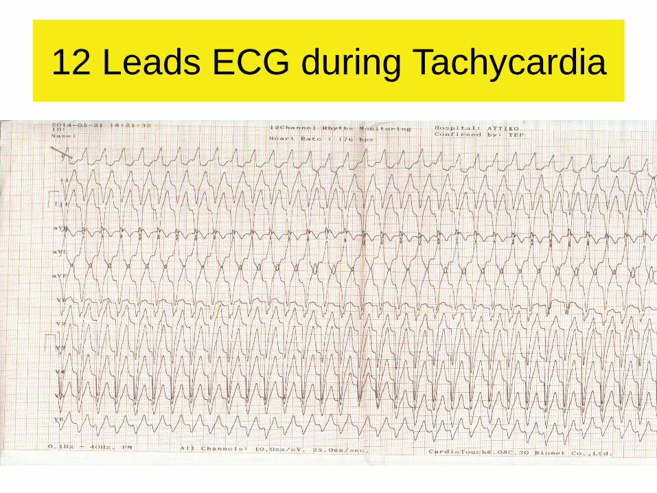
12 Leads ECG during Tachycardia
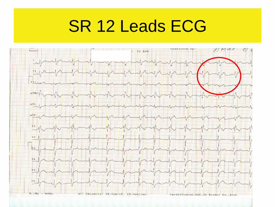
SR 12 Leads ECG
• She underwent cardiac echo, MRI.
Nothing significant
• Refused EPS
• 8 mo after the 1st episode she sustained a
new episode of tachycardia while
university student
• She was scheduled for EPS/RFA in our
hospital
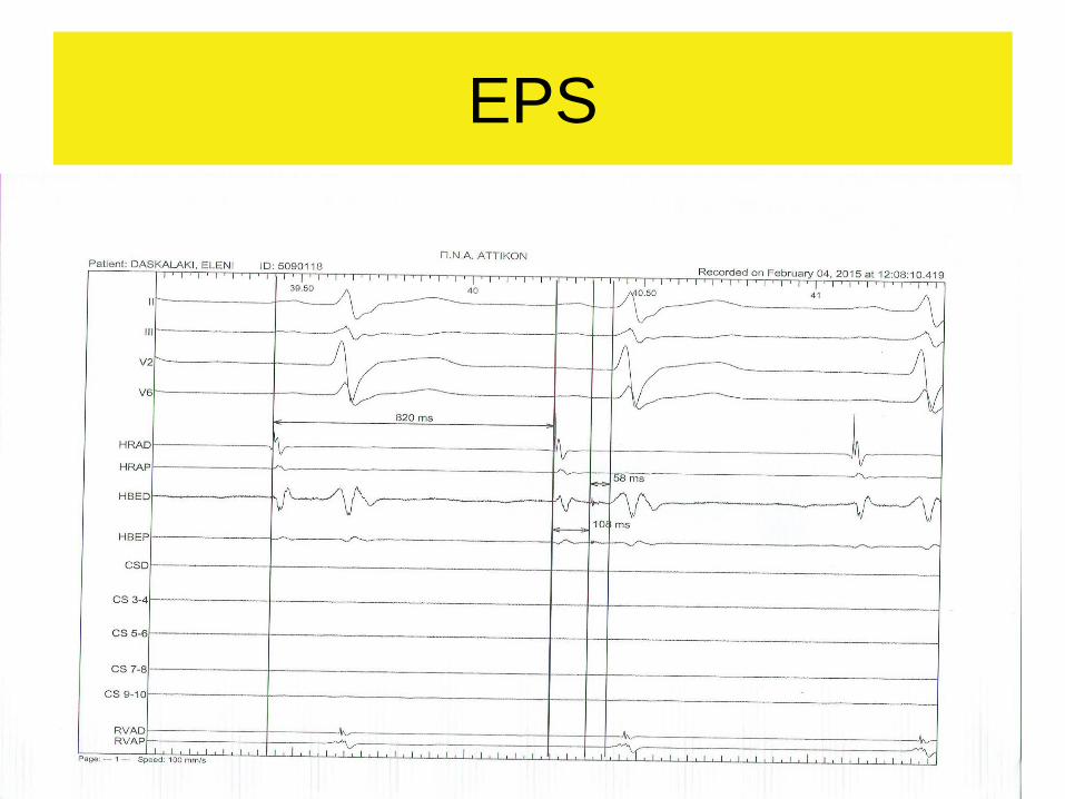
EPS
Atrial programmed stimulation
• Burst and Incremental atrial pacing gradual increase of QRS widening (preexcitation) associated with PR prolongation
• Wenckebach accessory pathway 250ms
• Wenckebach AVN 280ms
• AV interval progressive prolongation, AH interval lengthens, HV interval shortens and QRS widens until a steady value was achieved.
• During absence of pre-excitation, the HV interval was positive, whereas during preexcitation it was negative
Ventricular programmed stimulation
S1/S2 600, 500ms
VERP 240ms
• VA conduction 1:1 decremental concentric
• Ventricular pacing showed retrograde
decremental conduction via the AV node
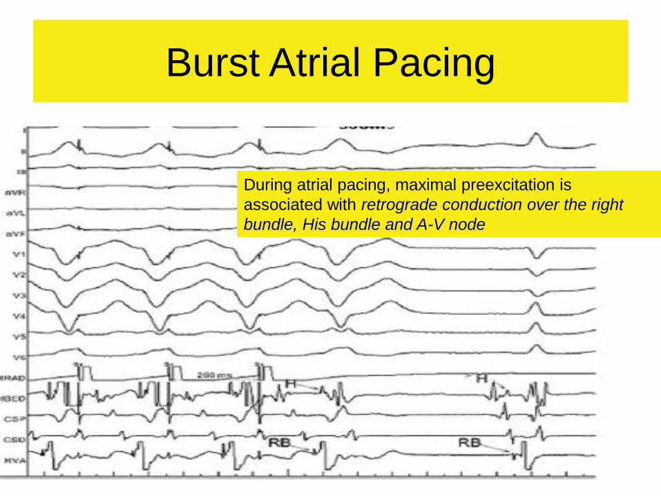
Burst Atrial Pacing
During atrial pacing, maximal preexcitation is
associated with retrograde conduction over the right
bundle, His bundle and A-V node
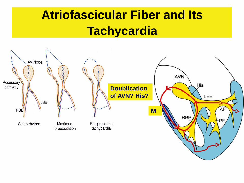
Atriofascicular Fiber and Its
Tachycardia
Doublication
of AVN? His?
M
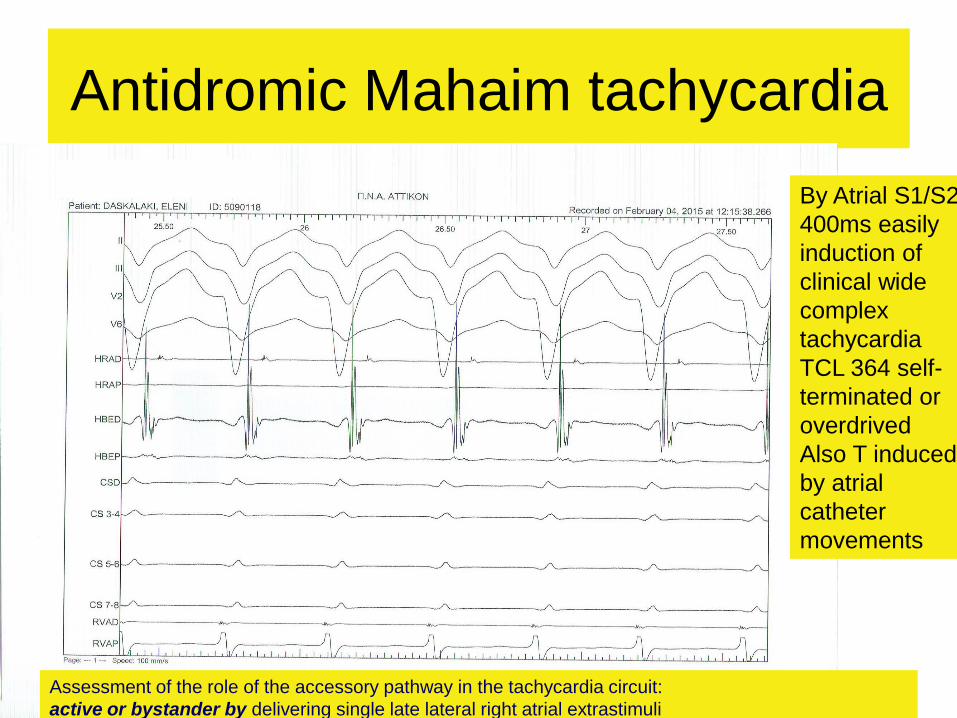
Antidromic Mahaim tachycardia
By Atrial S1/S2
400ms easily
induction of
clinical wide
complex
tachycardia
TCL 364 self-
terminated or
overdrived
Also T induced
by atrial
catheter
movements
Assessment of the role of the accessory pathway in the tachycardia circuit:
active or bystander by delivering single late lateral right atrial extrastimuli
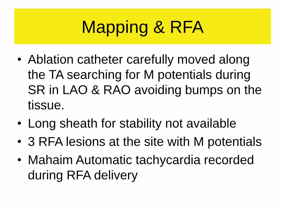
Mapping & RFA
• Ablation catheter carefully moved along
the TA searching for M potentials during
SR in LAO & RAO avoiding bumps on the
tissue.
• Long sheath for stability not available
• 3 RFA lesions at the site with M potentials
• Mahaim Automatic tachycardia recorded
during RFA delivery
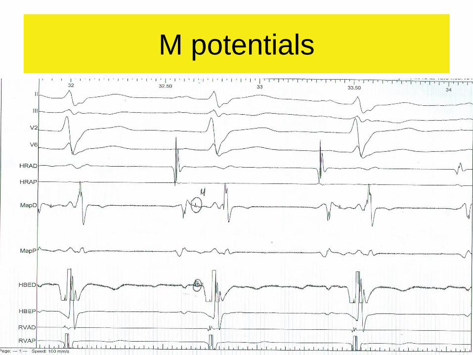
M potentials
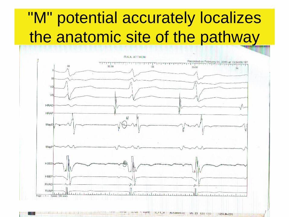
"M" potential accurately localizes
the anatomic site of the pathway

Three mapping strategies
• (i) M potential
• defined as a discrete deflection between atrial and ventricular signal with the interval between the M potential and ventricular electrogram remaining constant during the AV delay produced by atrial pacing,
• (ii) shortest stimulus-to-preexcitation (STP) intervaldefined by the shortest interval from a paced atrial site to the pre-excited QRS, and
• (iii) mechanical trauma induced loss of conductionover Mahaim fibre.
(located very close to the endocardium and thus catheter movement-related mechanical trauma resulting in transient loss of conduction is not uncommon).
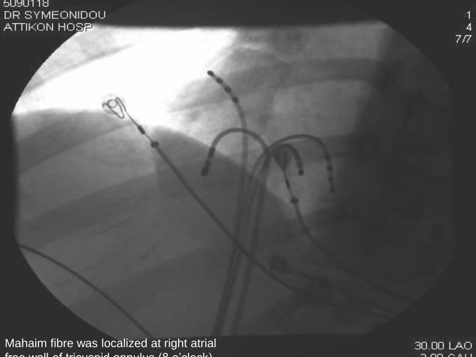
Mahaim fibre was localized at right atrial
free wall of tricuspid annulus (8 o’clock)
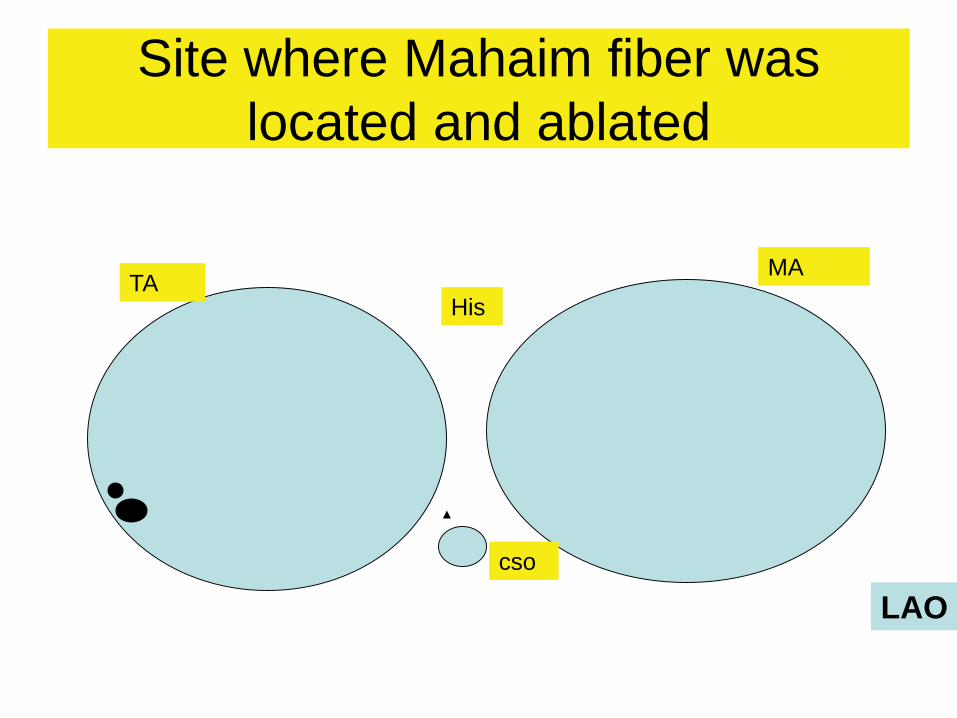
Site where Mahaim fiber was
located and ablated
TAHis
MA
LAO
cso
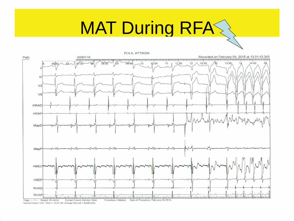
MAT During RFA
Mahaim automatic tachycardia
during RFA
• Probably due to heat-related
automaticity of nodal like tissue in a
similar fashion to junctional rhythm during
slow AVNRT ablation
• It seems to represent a hallmark for
successful RFA ablation of atriofascicular
pathways

SR post RFA
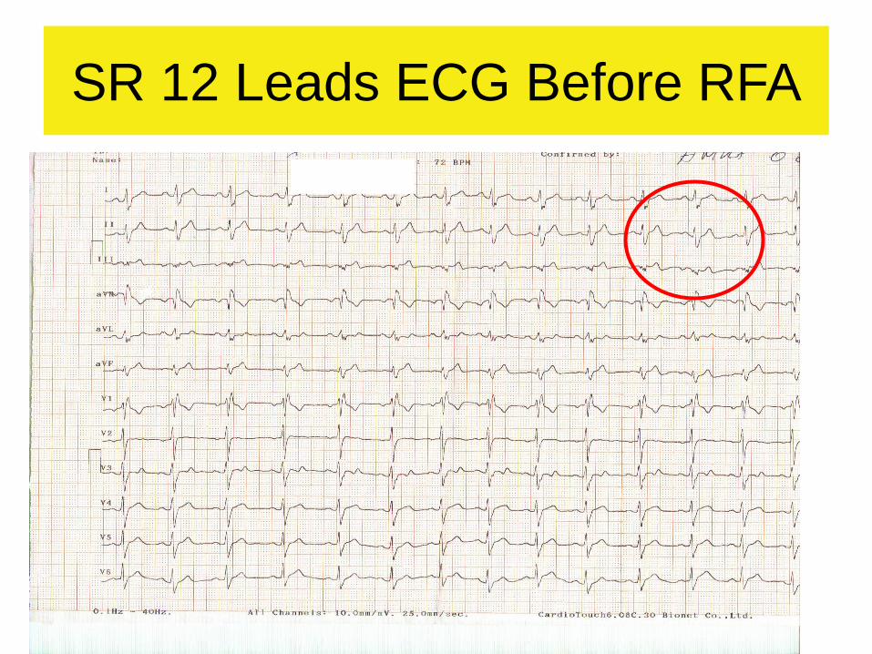
SR 12 Leads ECG Before RFA
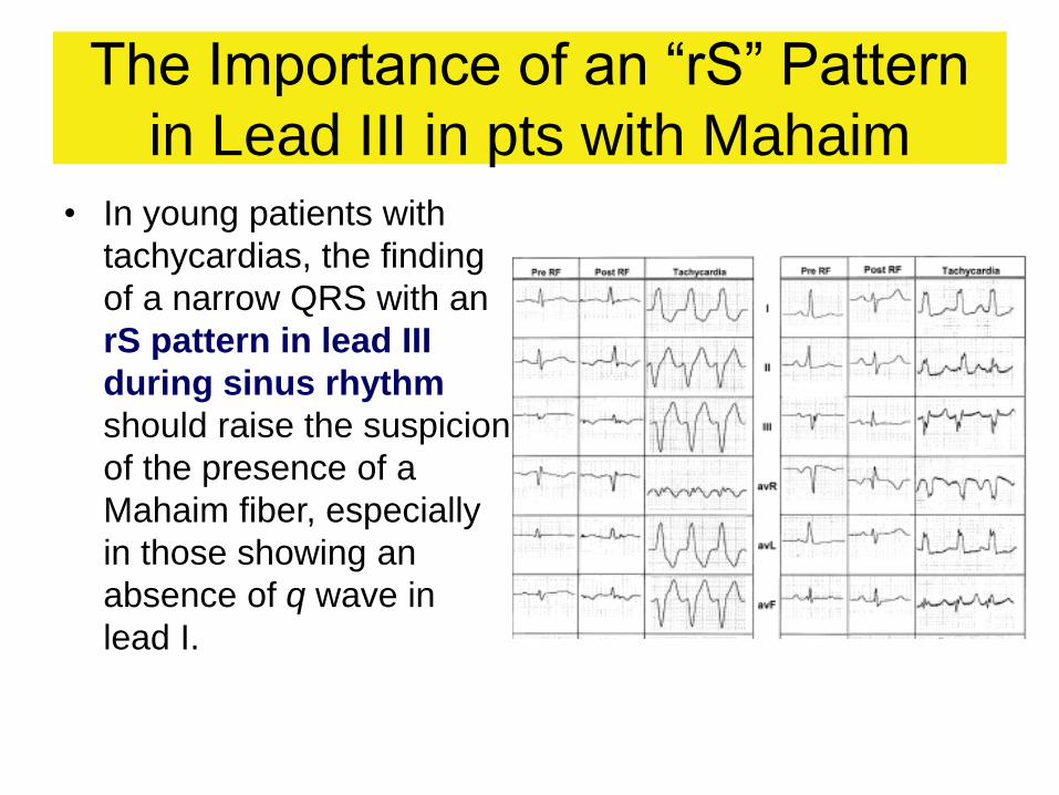
The Importance of an “rS” Pattern
in Lead III in pts with Mahaim
• In young patients with
tachycardias, the finding
of a narrow QRS with an
rS pattern in lead III
during sinus rhythm
should raise the suspicion
of the presence of a
Mahaim fiber, especially
in those showing an
absence of q wave in
lead I.
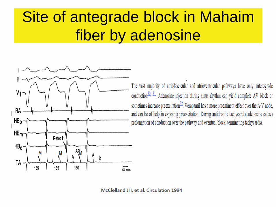
Site of antegrade block in Mahaim
fiber by adenosine
Conclusions
Atriofascicular (Mahaim) Pathway
• Atriofascicular (Mahaim) pathway is an uncommon but distinct form of preexcitation.
• Although initially the Mahaim fibre was thought to be nodofascicular, later studies showed it to be atriofascicular pathway.
• It exhibits certain unique properties such as
• unidirectional, anterograde conduction, and
• slow conduction with AV node-like decremental conduction properties.(Responds to adenosine (92%) and shows accessory pathway automaticity during ablation (91%).
• Most of Mahaim pathways originate from the RA free wall near the TA and terminate in or near the distal RBB (atriofascicular pathways) or in the RV near the TA (AV pathways).
• The usual target for ablation is at the site of a high-frequency potential (M-potential) along the TA.

Conclusions
Atriofascicular (Mahaim) Pathway
• Because of relatively low prevalence
(about 3% of all accessory pathways) of
Mahaim along with limited experience of
electrophysiologists, the diagnosis and
catheter ablation of Mahaim fibers often
presents a clinical and technical
challenge.