Engineered trivalent immunogen adjuvanted with a STING ... · x10 * * c α α Fig. 1...
13
Engineered trivalent immunogen adjuvanted with a STING agonist confers protection against Trypanosoma cruzi infection. Item type Article Authors Sanchez Alberti, Andrés; Bivona, Augusto E; Cerny, Natacha; Schulze, Kai; Weißmann, Sebastian; Ebensen, Thomas; Morales, Celina; Padilla, Angel M; Cazorla, Silvia I; Tarleton, Rick L; Guzmán, Carlos A; Malchiodi, Emilio L Citation Engineered trivalent immunogen adjuvanted with a STING agonist confers protection against Trypanosoma cruzi infection. 2017, 2:9 NPJ Vaccines DOI 10.1038/s41541-017-0010-z Journal NPJ vaccines Downloaded 14-May-2018 03:59:29 Item License http://creativecommons.org/licenses/by-nc-sa/4.0/ Link to item http://hdl.handle.net/10033/621359
Transcript of Engineered trivalent immunogen adjuvanted with a STING ... · x10 * * c α α Fig. 1...
Engineered trivalent immunogen adjuvanted with a STINGagonist confers protection against Trypanosoma cruzi infection.
Item type Article
Authors Sanchez Alberti, Andrés; Bivona, Augusto E; Cerny,Natacha; Schulze, Kai; Weißmann, Sebastian; Ebensen,Thomas; Morales, Celina; Padilla, Angel M; Cazorla,Silvia I; Tarleton, Rick L; Guzmán, Carlos A; Malchiodi,Emilio L
Citation Engineered trivalent immunogen adjuvanted with a STINGagonist confers protection against Trypanosoma cruziinfection. 2017, 2:9 NPJ Vaccines
DOI 10.1038/s41541-017-0010-z
Journal NPJ vaccines
Downloaded 14-May-2018 03:59:29
Item License http://creativecommons.org/licenses/by-nc-sa/4.0/
Link to item http://hdl.handle.net/10033/621359
ARTICLE OPEN
Engineered trivalent immunogen adjuvanted with a STINGagonist confers protection against Trypanosoma cruzi infectionAndrés Sanchez Alberti 1,2,3, Augusto E. Bivona1,2, Natacha Cerny1,2, Kai Schulze3, Sebastian Weißmann3, Thomas Ebensen3,Celina Morales4, Angel M. Padilla5, Silvia I. Cazorla1,2, Rick L. Tarleton5, Carlos A. Guzmán3 and Emilio L. Malchiodi 1,2
The parasite Trypanosoma cruzi is the causative agent of Chagas disease, a potentially life-threatening infection that represents amajor health problem in Latin America. Several characteristics of this protozoan contribute to the lack of an effective vaccine,among them: its silent invasion mechanism, T. cruzi antigen redundancy and immunodominance without protection. Taking intoaccount these issues, we engineered Traspain, a chimeric antigen tailored to present a multivalent display of domains from keyparasitic molecules, combined with stimulation of the STING pathway by c-di-AMP as a novel prophylactic strategy. This formulationproved to be effective for the priming of functional humoral responses and pathogen-specific CD8+ and CD4+ T cells, compatiblewith a Th1/Th17 bias. Interestingly, vaccine effectiveness assessed across the course of infection, showed a reduction in parasiteload and chronic inflammation in different proof of concept assays. In conclusion, this approach represents a promising tool againstparasitic chronic infections.
npj Vaccines (2017) 2:9 ; doi:10.1038/s41541-017-0010-z
INTRODUCTIONTrypanosoma cruzi is the etiological agent of Chagas disease, theworld’s leading cause of infectious myocarditis. It is recognized byWHO as a neglected tropical disease in Latin America, where one-sixth of the population is at risk of contracting the infection.1
The infection is a complex zoonosis transmitted by severalhematophagous Triatomine species. Despite the potent immuneresponse the parasite triggers in the mammalian host, T. cruzi isable to persist, establishing a chronic infection. One hundred yearsfollowing its discovery, there are still no effective vaccines or drugsto prevent or treat the chronic phase of the infection.As T. cruzi spends most of its time in mammals as amastigote
form replicating in the cytosol of host cells, cell-mediatedimmunity is essential for controlling the parasite.2 However, ithas been shown that the cytotoxic T lymphocyte (CTL) responsedeveloped is restricted to a few epitopes of the Transialidase (TS)superfamily,3 giving rise to the question of immunodominance asan immune evasion mechanism.4 The redundancy of some T. cruziantigens and the parasite silent invasion mechanism contribute tothe lack of detection of infected cells during the first moment ofentrance and represent a challenge in the design of anti-T. cruziprophylactic vaccines. This scenario allows speculation aboutwhether a chimeric immunogen could broaden the immuneresponse triggered by vaccination and achieve better protectionlevels. To test this hypothesis, we engineered Traspain, a structure-based chimeric antigen, relying on several properties of threeregions of T. cruzi proteins: (1) immunogenicity, (2) presence ofreported and predicted major histocompatibility complex class Ibinding peptides, (3) protection capacity, (4) expression profile,
and (5) structural signature. Thus, we selected the N-terminaldomain of Cruzipain (Cz)—the major cistein protease—, the centralregion of amastigote surface protein 2 (ASP2)—an antigenexpressed exclusively during the intracellular stage—, and aninactive transialidase (iTS)—a major antigen and virulence factor—,to generate a chimeric antigen presenting a multivalent display ofkey parasitic molecules.Subunit vaccines need not only a good immunogen but also
the right adjuvant. In search of novel components that canenhance cell-mediated immunity, we employed 3′5′-c-di-AMP (c-di-AMP), a cyclic di nucleotide (CDN), in vaccination protocols bythe intranasal route. CDNs are STING agonists that activate IRF3,NF-kB, and STAT6, inducing type I IFN and pro-inflammatorycytokines;5, 6 originally described as bacterial second messengersassociated with different metabolic process. Mammalian cells havealso an eukaryotic counterpart, 2′5 ′-c-GAMP, as part of their DNAsensing machinery.7 These small molecules have been recentlyintroduced as adjuvants.8 Here, we show how tailored antigencombined with this novel adjuvant represents a promisingstrategy for vaccines against parasitic infections.
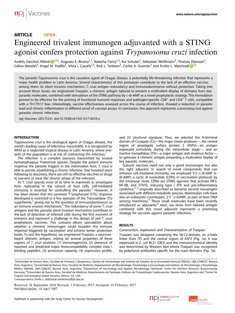
RESULTSConstruction, expression and characterization of TraspainTraspain was designed containing the Nt-Cz-domain, an α-helixlinker from iTS and the central region of ASP2 (Fig. 1a). It wasexpressed in E. coli BL21 (DE3) and the immunochemical identitywas determined by Western blot where Traspain was recognizedby polyclonal antibodies specific for the main domains (Fig. 1b).
Received: 28 September 2016 Revised: 1 February 2017 Accepted: 10 February 2017
1Universidad de Buenos Aires, Facultad de Farmacia y Bioquímica, Cátedra de Inmunología and Instituto de Estudios de la Inmunidad Humoral (IDEHU), UBA-CONICET, BuenosAires, Argentina; 2Universidad de Buenos Aires, Facultad de Medicina, Departamento de Microbiología, Parasitología e Inmunología and Instituto de Microbiología y ParasitologíaMédica (IMPaM), UBA-CONICET, Buenos Aires, Argentina; 3Department of Vaccinology and Applied Microbiology, Helmholtz Center for Infection Research, Braunschweig,Germany; 4Universidad de Buenos Aires, Facultad de Medicina, Departamento de Patología, Instituto de Fisiopatología Cardiovascular, Buenos Aires, Argentina and 5Center forTropical and Emerging Global Diseases, Athens, GA, USACorrespondence: Emilio L. Malchiodi ([email protected])
www.nature.com/npjvaccines
Published in partnership with the Sealy Center for Vaccine Development
Immunization with Traspain+c-di-AMP achieved an equal level ofpriming at both humoral and cellular levelsAs Traspain antigen contains domains from different proteinfamilies of the parasite, we decided to test whether anyinterference was developed between the main regions of theconstruction when the protein was administered to C3H mice inthe presence of a STING agonist as adjuvant.Antibodies raised against Traspain were able to recognize Nt-Cz,
ASP2 and GELRIIKSV peptide from the α-helix linker in an indirectELISA-assay (Fig. 1c). Remarkably, a similar level of Abs wasdetected against both Nt-Cz and ASP2, indicating absence ofinterference and inmunodominance at the humoral level.GELRIIKSV-specific IgG were detected in mice vaccinated withthe chimeric molecule but not in controls (c-di-AMP) nor in Nt-Cz+ASP2 vaccinated mice that lack this motive. The lower titer of this
specificity was expected considering the 9-mer peptidic nature ofthe coating molecule.Considering the report of ASP2 immunodominant
T cell epitopes in natural infection,9 we analyzed the performanceof Traspain+c-di-AMP to prime cell-mediated immune responsesagainst each domain. No differences were detected in theproliferation levels of the Nt-Cz and ASP2-specific subpopulations,PI: 7.17 ± 2.44 and 8.02 ± 1.03, respectively; representing nearlyhalf of the whole response (PI: 18.2 ± 5.78) upon restimulationwith Traspain (Fig. 1d).
Antibodies elicited by Traspain+c-di-AMP show neutralizationcapacityT. cruzi-specific antibodies have been shown to be beneficial forthe development of functional parasite-specific CD8+ T cells upon
a b
95
72
55
43
34
26
-helix linker
TSGHTVGATI KEEASSAV GELRIIKSV TEWETGQI VNHRFTLV
H-2k H-2b
ASP2 DomainNt-Cz Domain
c-di-
AMP
anti-
Nt-Cz+
ASP2
anti-
Trasp
ain0
20
40
60
80
Inhi
bitio
nof
inva
sion
( %)
**
Serum
anti-Nt-Cz anti-ASP2
kDa 1 2 3 kDa 1 2 3
95
72
55
43
34
26
d eSpleen
Traspain+c-di-AMP c-di-AMP0
5
10
15
20
25
30P
I
**TraspainNt-Cz
ASP2
Trasp
ainNt-C
z
ASP2
anti-
Trasp
ain
anti-
Nt-Cz+
ASP2
c-di-
AMP
0
100
200
300
400
500
0
100
200
300
400
500
-Lin
ker
Sp
e cifi
c-Ig
Gt it
re
Spe
cific
-IgG
t itre
x10
**
c
α
α
Fig. 1 Characterization of Traspain as a chimeric immunogen. Schematic representation of Traspain. Arrows point at CD8+ T cell epitopesincluded in the design with its respective mouse MHC-I haplotype (a). Immunochemical identity by Western blot. Domain-specific polyclonalantibodies (pAb) were used as primary antibody. SDS-PAGE gels were loaded as follows, lines: 1-MWM, 2-Traspain, 3-Nt-Cz (left) or ASP2 (right),arrows point at Traspain band (b). Specific antibodies response. Titers were determined by ELISA in serum samples from mice vaccinated witheither Traspain or antigen combination plus c-di-AMP at 15 days post vaccination. Plates were coated with Traspain, Nt-Cz, ASP2 (left) orGELRIIKSV α-linker-peptide (right). * p< 0.05, one-way ANOVA Kruskal-Wallis test + Dunn’s multiple comparisons test (c). Proliferative responsein vaccinated mice 30 days after the last dose. Spleen cells were re-stimulated with 10 µg/ml of the indicated protein. Results are expressed asproliferation index (PI), n= 6 mice per group, **p< 0.01, 2-way-ANOVA + Tukey’s multiple comparisons test (d). Neutralization assay of pAbgenerated upon vaccination. n= 5–6 per group, **p< 0.01, one-way-ANOVA Kruskal-Wallis test + Dunn’s multiple comparisons test (e). Resultsare expressed as mean± SEM and are representative of at least three independent experiments
Chimera and c-di-AMP protect against T. cruziA Sanchez Alberti et al
2
npj Vaccines (2017) 9 Published in partnership with the Sealy Center for Vaccine Development
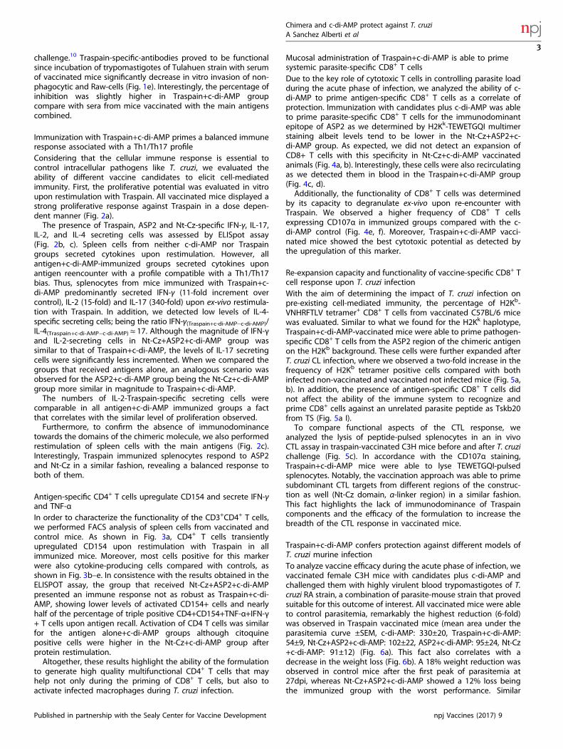
challenge.10 Traspain-specific-antibodies proved to be functionalsince incubation of trypomastigotes of Tulahuen strain with serumof vaccinated mice significantly decrease in vitro invasion of non-phagocytic and Raw-cells (Fig. 1e). Interestingly, the percentage ofinhibition was slightly higher in Traspain+c-di-AMP groupcompare with sera from mice vaccinated with the main antigenscombined.
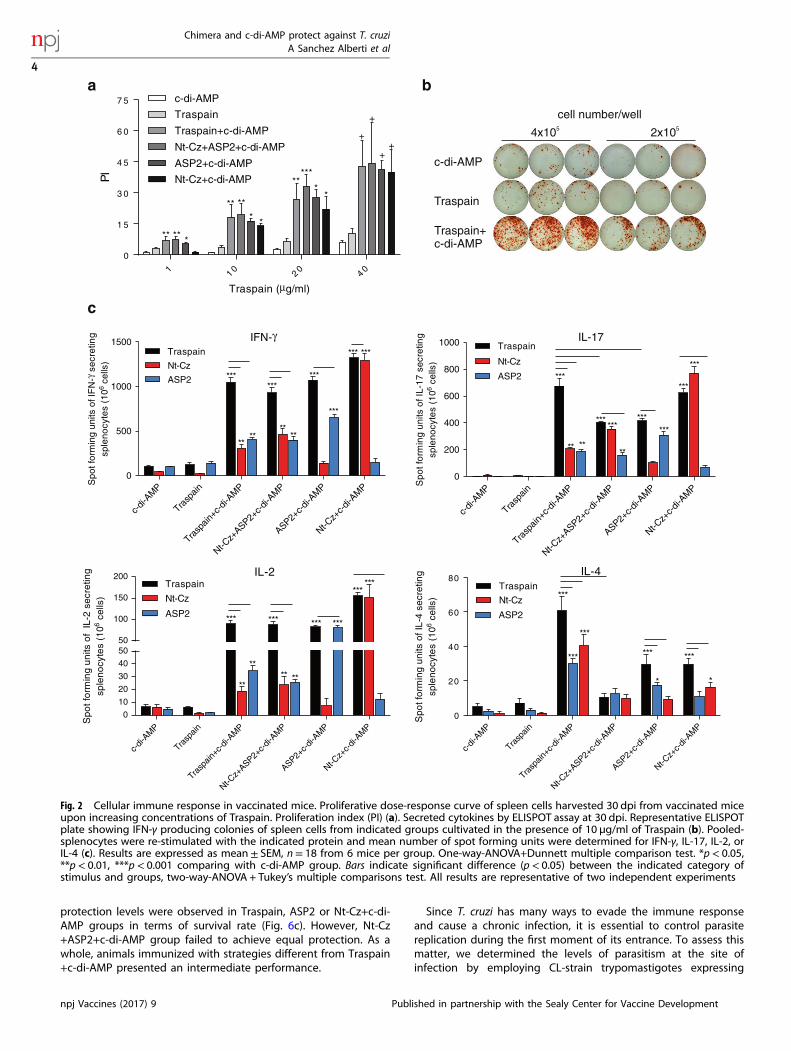
Immunization with Traspain+c-di-AMP primes a balanced immuneresponse associated with a Th1/Th17 profileConsidering that the cellular immune response is essential tocontrol intracellular pathogens like T. cruzi, we evaluated theability of different vaccine candidates to elicit cell-mediatedimmunity. First, the proliferative potential was evaluated in vitroupon restimulation with Traspain. All vaccinated mice displayed astrong proliferative response against Traspain in a dose depen-dent manner (Fig. 2a).The presence of Traspain, ASP2 and Nt-Cz-specific IFN-γ, IL-17,
IL-2, and IL-4 secreting cells was assessed by ELISpot assay(Fig. 2b, c). Spleen cells from neither c-di-AMP nor Traspaingroups secreted cytokines upon restimulation. However, allantigen+c-di-AMP-immunized groups secreted cytokines uponantigen reencounter with a profile compatible with a Th1/Th17bias. Thus, splenocytes from mice immunized with Traspain+c-di-AMP predominantly secreted IFN-γ (11-fold increment overcontrol), IL-2 (15-fold) and IL-17 (340-fold) upon ex-vivo restimula-tion with Traspain. In addition, we detected low levels of IL-4-specific secreting cells; being the ratio IFN-γ(Traspain+c-di-AMP−c-di-AMP)/IL-4(Traspain+c-di-AMP−c-di-AMP) ≈ 17. Although the magnitude of IFN-γand IL-2-secreting cells in Nt-Cz+ASP2+c-di-AMP group wassimilar to that of Traspain+c-di-AMP, the levels of IL-17 secretingcells were significantly less incremented. When we compared thegroups that received antigens alone, an analogous scenario wasobserved for the ASP2+c-di-AMP group being the Nt-Cz+c-di-AMPgroup more similar in magnitude to Traspain+c-di-AMP.The numbers of IL-2-Traspain-specific secreting cells were
comparable in all antigen+c-di-AMP immunized groups a factthat correlates with the similar level of proliferation observed.Furthermore, to confirm the absence of immunodominance
towards the domains of the chimeric molecule, we also performedrestimulation of spleen cells with the main antigens (Fig. 2c).Interestingly, Traspain immunized splenocytes respond to ASP2and Nt-Cz in a similar fashion, revealing a balanced response toboth of them.
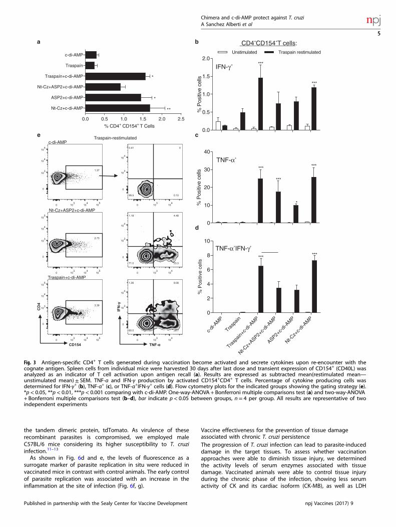
Antigen-specific CD4+ T cells upregulate CD154 and secrete IFN-γand TNF-αIn order to characterize the functionality of the CD3+CD4+ T cells,we performed FACS analysis of spleen cells from vaccinated andcontrol mice. As shown in Fig. 3a, CD4+ T cells transientlyupregulated CD154 upon restimulation with Traspain in allimmunized mice. Moreover, most cells positive for this markerwere also cytokine-producing cells compared with controls, asshown in Fig. 3b–e. In consistence with the results obtained in theELISPOT assay, the group that received Nt-Cz+ASP2+c-di-AMPpresented an immune response not as robust as Traspain+c-di-AMP, showing lower levels of activated CD154+ cells and nearlyhalf of the percentage of triple positive CD4+CD154+TNF-α+IFN-γ+ T cells upon antigen recall. Activation of CD4 T cells was similarfor the antigen alone+c-di-AMP groups although citoquinepositive cells were higher in the Nt-Cz+c-di-AMP group afterprotein restimulation.Altogether, these results highlight the ability of the formulation
to generate high quality multifunctional CD4+ T cells that mayhelp not only during the priming of CD8+ T cells, but also toactivate infected macrophages during T. cruzi infection.
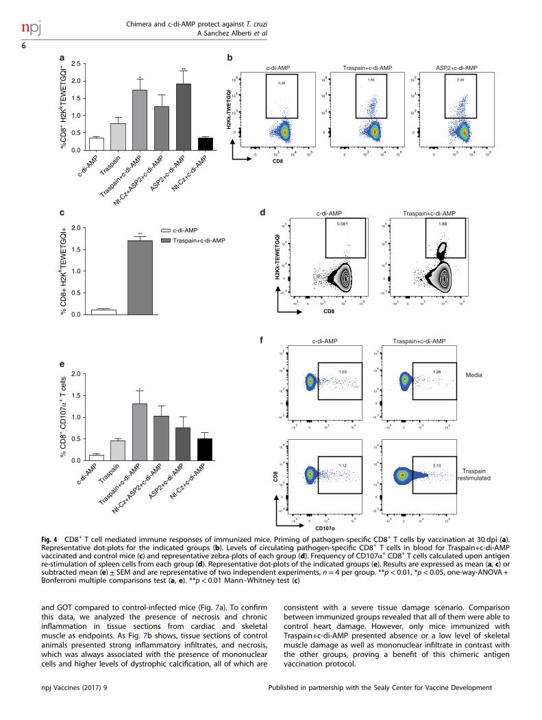
Mucosal administration of Traspain+c-di-AMP is able to primesystemic parasite-specific CD8+ T cellsDue to the key role of cytotoxic T cells in controlling parasite loadduring the acute phase of infection, we analyzed the ability of c-di-AMP to prime antigen-specific CD8+ T cells as a correlate ofprotection. Immunization with candidates plus c-di-AMP was ableto prime parasite-specific CD8+ T cells for the immunodominantepitope of ASP2 as we determined by H2Kk-TEWETGQI multimerstaining albeit levels tend to be lower in the Nt-Cz+ASP2+c-di-AMP group. As expected, we did not detect an expansion ofCD8+ T cells with this specificity in Nt-Cz+c-di-AMP vaccinatedanimals (Fig. 4a, b). Interestingly, these cells were also recirculatingas we detected them in blood in the Traspain+c-di-AMP group(Fig. 4c, d).Additionally, the functionality of CD8+ T cells was determined
by its capacity to degranulate ex-vivo upon re-encounter withTraspain. We observed a higher frequency of CD8+ T cellsexpressing CD107α in immunized groups compared with the c-di-AMP control (Fig. 4e, f). Moreover, Traspain+c-di-AMP vacci-nated mice showed the best cytotoxic potential as detected bythe upregulation of this marker.
Re-expansion capacity and functionality of vaccine-specific CD8+ Tcell response upon T. cruzi infectionWith the aim of determining the impact of T. cruzi infection onpre-existing cell-mediated immunity, the percentage of H2Kb-VNHRFTLV tetramer+ CD8+ T cells from vaccinated C57BL/6 micewas evaluated. Similar to what we found for the H2Kk haplotype,Traspain+c-di-AMP-vaccinated mice were able to prime pathogen-specific CD8+ T cells from the ASP2 region of the chimeric antigenon the H2Kb background. These cells were further expanded afterT. cruzi CL infection, where we observed a two-fold increase in thefrequency of H2Kb tetramer positive cells compared with bothinfected non-vaccinated and vaccinated not infected mice (Fig. 5a,b). In addition, the presence of antigen-specific CD8+ T cells didnot affect the ability of the immune system to recognize andprime CD8+ cells against an unrelated parasite peptide as Tskb20from TS (Fig. 5a I).To compare functional aspects of the CTL response, we
analyzed the lysis of peptide-pulsed splenocytes in an in vivoCTL assay in traspain-vaccinated C3H mice before and after T. cruzichallenge (Fig. 5c). In accordance with the CD107α staining,Traspain+c-di-AMP mice were able to lyse TEWETGQI-pulsedsplenocytes. Notably, the vaccination approach was able to primesubdominant CTL targets from different regions of the construc-tion as well (Nt-Cz domain, α-linker region) in a similar fashion.This fact highlights the lack of immunodominance of Traspaincomponents and the efficacy of the formulation to increase thebreadth of the CTL response in vaccinated mice.
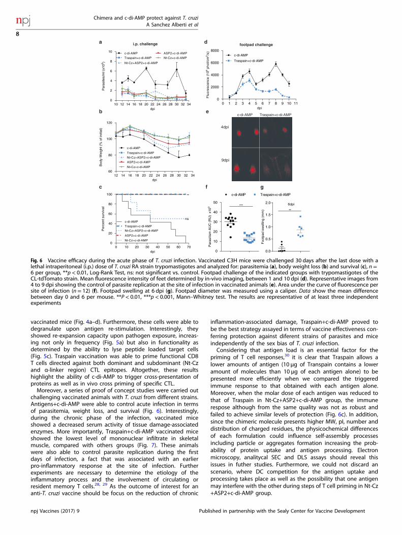
Traspain+c-di-AMP confers protection against different models ofT. cruzi murine infectionTo analyze vaccine efficacy during the acute phase of infection, wevaccinated female C3H mice with candidates plus c-di-AMP andchallenged them with highly virulent blood trypomastigotes of T.cruzi RA strain, a combination of parasite-mouse strain that provedsuitable for this outcome of interest. All vaccinated mice were ableto control parasitemia, remarkably the highest reduction (6-fold)was observed in Traspain vaccinated mice (mean area under theparasitemia curve ±SEM, c-di-AMP: 330±20, Traspain+c-di-AMP:54±9, Nt-Cz+ASP2+c-di-AMP: 102±22, ASP2+c-di-AMP: 95±24, Nt-Cz+c-di-AMP: 91±12) (Fig. 6a). This fact also correlates with adecrease in the weight loss (Fig. 6b). A 18% weight reduction wasobserved in control mice after the first peak of parasitemia at27dpi, whereas Nt-Cz+ASP2+c-di-AMP showed a 12% loss beingthe immunized group with the worst performance. Similar
Chimera and c-di-AMP protect against T. cruziA Sanchez Alberti et al
3
Published in partnership with the Sealy Center for Vaccine Development npj Vaccines (2017) 9
protection levels were observed in Traspain, ASP2 or Nt-Cz+c-di-AMP groups in terms of survival rate (Fig. 6c). However, Nt-Cz+ASP2+c-di-AMP group failed to achieve equal protection. As awhole, animals immunized with strategies different from Traspain+c-di-AMP presented an intermediate performance.
Since T. cruzi has many ways to evade the immune responseand cause a chronic infection, it is essential to control parasitereplication during the first moment of its entrance. To assess thismatter, we determined the levels of parasitism at the site ofinfection by employing CL-strain trypomastigotes expressing
1 1 0 2 0 4 0
0
1 5
3 0
4 5
6 0
7 5 c-di-AMP
Traspain
Traspain+c-di-AMP
Nt-Cz+ASP2+c-di-AMP
ASP2+c-di-AMP
Nt-Cz+c-di-AMP
Traspain ( g/ml)
PI
**
**
*
*** *
**
*****
**
+
+
++
IFN-
c-di-
AMP
Trasp
ain
Trasp
ain+c
-di-A
MP
Nt-Cz+
ASP2+c-
di-AM
P
ASP2+c-d
i-AM
P
Nt-Cz+
c-di-
AMP
0
500
1000
1500
ASP2
Nt-Cz
Traspain
Spo
tfor
min
gun
itsof
IFN
-se
cret
ing
sple
nocy
tes
(106
cells
)
***
****
***
****
*** ***
***
***
IL-17
c-di-
AMP
Trasp
ain
Trasp
ain+c
-di-A
MP
Nt-Cz+
ASP2+c-
di-AM
P
ASP2+c-
di-AM
P
Nt-Cz+
c-di-
AMP
0
200
400
600
800
1000
ASP2
Nt-Cz
Traspain
Spo
tfo
rmin
gu
nits
ofI
L-1
7s e
cre
ting
sple
nocy
tes
(106
cells
)
***
** **
***
***
***
***
**
*** ***
IL-2
c-di-
AMP
Trasp
ain
Trasp
ain+c
-di-A
MP
Nt-Cz+
ASP2+c-
di-AM
P
ASP2+c-
di-AM
P
Nt-Cz+
c-di-
AMP
0
10
20
30
40
5050
100
150
200
Spo
tfo
rmin
gu
nits
ofIL
-2se
cret
ing
sple
noc y
tes
(106
cells
)
Traspain
ASP2
Nt-Cz
*** ***
******
***
**
**
****
***
c-di-
AMP
Trasp
ain
Trasp
ain+c
-di-A
MP
Nt-Cz+
ASP2+c-
di-AM
P
ASP2+c-
di-AM
P
Nt-Cz+
c-di-
AMP
0
20
40
60
80IL-4
Spo
tfo
rmin
gun
itsof
IL-4
secr
etin
gsp
l eno
cyte
s(1
06ce
l ls)
Traspain
ASP2
Nt-Cz ***
******
***
***
**
c-di-AMP
Traspain
5 5 4x10 2x10
a b
cell number/well
c
Traspain+c-di-AMP
γ
μ
γ
Fig. 2 Cellular immune response in vaccinated mice. Proliferative dose-response curve of spleen cells harvested 30 dpi from vaccinated miceupon increasing concentrations of Traspain. Proliferation index (PI) (a). Secreted cytokines by ELISPOT assay at 30 dpi. Representative ELISPOTplate showing IFN-γ producing colonies of spleen cells from indicated groups cultivated in the presence of 10 µg/ml of Traspain (b). Pooled-splenocytes were re-stimulated with the indicated protein and mean number of spot forming units were determined for IFN-γ, IL-17, IL-2, orIL-4 (c). Results are expressed as mean± SEM, n= 18 from 6 mice per group. One-way-ANOVA+Dunnett multiple comparison test. *p< 0.05,**p< 0.01, ***p< 0.001 comparing with c-di-AMP group. Bars indicate significant difference (p< 0.05) between the indicated category ofstimulus and groups, two-way-ANOVA + Tukey’s multiple comparisons test. All results are representative of two independent experiments
Chimera and c-di-AMP protect against T. cruziA Sanchez Alberti et al
4
npj Vaccines (2017) 9 Published in partnership with the Sealy Center for Vaccine Development
the tandem dimeric protein, tdTomato. As virulence of theserecombinant parasites is compromised, we employed maleC57BL/6 mice considering its higher susceptibility to T. cruziinfection.11–13
As shown in Fig. 6d and e, the levels of fluorescence as asurrogate marker of parasite replication in situ were reduced invaccinated mice in contrast with control animals. The early controlof parasite replication was associated with an increase in theinflammation at the site of infection (Fig. 6f, g).
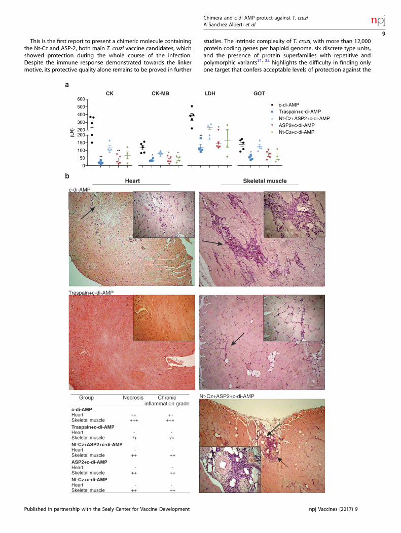
Vaccine effectiveness for the prevention of tissue damageassociated with chronic T. cruzi persistenceThe progression of T. cruzi infection can lead to parasite-induceddamage in the target tissues. To assess whether vaccinationapproaches were able to diminish tissue injury, we determinedthe activity levels of serum enzymes associated with tissuedamage. Vaccinated animals were able to control tissue injuryduring the chronic phase of the infection, showing less serumactivity of CK and its cardiac isoform (CK-MB), as well as LDH
0
10
20
30
40
%P
ositi
vece
lls
***
*
***
***
0.0
0.5
1.0
1.5
2.0
%P
ositi
vece
ll s
***
***
c-di-
AMP
Trasp
ain
Trasp
ain+c
-di-A
MP
Nt-Cz+
ASP2+c-
di-AM
P
ASP2+c-d
i-AM
P
Nt-Cz+
c-di-
AMP
0
2
4
6
8
10
%P
ositi
vece
lls
*** ***
a b
e c c-di-AMP
0.0 0.5 1.0 1.5 2.0 2.5
Nt-Cz+c-di-AMP
ASP2+c-di-AMP
Nt-Cz+ASP2+c-di-AMP
Traspaín+c-di-AMP
Traspaín
c-di-AMP
*
**
*
% CD4+ CD154+ T Cells
Nt-Cz+ASP2+c-di-AMP
Traspain+c-di-AMP
Traspain-restimulated
d
+ + CD4 CD154 T cells:
+TNF-α
+ + TNF-α IFN-γ
+ IFN-γ
Unstimulated Traspain restimulated
Fig. 3 Antigen-specific CD4+ T cells generated during vaccination become activated and secrete cytokines upon re-encounter with thecognate antigen. Spleen cells from individual mice were harvested 30 days after last dose and transient expression of CD154+ (CD40L) wasanalyzed as an indicator of T cell activation upon antigen recall (a). Results are expressed as subtracted mean(restimulated mean—unstimulated mean)± SEM. TNF-α and IFN-γ production by activated CD154+CD4+ T cells. Percentage of cytokine producing cells wasdetermined for IFN-γ+ (b), TNF-α+ (c), or TNF-α+IFN-γ+ cells (d). Flow cytometry plots for the indicated groups showing the gating strategy (e).*p< 0.05, **p< 0.01, ***p< 0.001 comparing with c-di-AMP. One-way-ANOVA + Bonferroni multiple comparisons test (a) and two-way-ANOVA+ Bonferroni multiple comparisons test (b–d), bar indicate p< 0.05 between groups, n= 4 per group. All results are representative of twoindependent experiments
Chimera and c-di-AMP protect against T. cruziA Sanchez Alberti et al
5
Published in partnership with the Sealy Center for Vaccine Development npj Vaccines (2017) 9
and GOT compared to control-infected mice (Fig. 7a). To confirmthis data, we analyzed the presence of necrosis and chronicinflammation in tissue sections from cardiac and skeletalmuscle as endpoints. As Fig. 7b shows, tissue sections of controlanimals presented strong inflammatory infiltrates, and necrosis,which was always associated with the presence of mononuclearcells and higher levels of dystrophic calcification, all of which are
consistent with a severe tissue damage scenario. Comparisonbetween immunized groups revealed that all of them were able tocontrol heart damage. However, only mice immunized withTraspain+c-di-AMP presented absence or a low level of skeletalmuscle damage as well as mononuclear infiltrate in contrast withthe other groups, proving a benefit of this chimeric antigenvaccination protocol.
c-di-AMP Traspain+c-di-AMP
Media
Traspainrestimulated
**c-di-AMP
Traspain+c-di-AMP
e
c c-di-AMP Traspain+c-di-AMP
c-di-AMP Traspain+c-di-AMP ASP2+c-di-AMP
c-di-
AMP
Trasp
ain
Trasp
ain+c-
di-AM
P
Nt-Cz+
ASP2+c-
di-AM
P
ASP2+c-
di-AM
P
Nt-Cz+
c-di-
AMP
***
%C
D8+
H2K
k TE
WE
TG
QI+
%C
D8+
H2K
k TE
WE
TG
QI+
%C
D8+
CD
107 α
+T
cells
c-di-
AMP
Trasp
ain
Trasp
ain+c-
di-AM
P
Nt-Cz+
ASP2+c-
di-AM
P
ASP2+c-
di-AM
P
Nt-Cz+
c-di-
AMP
0.0
0.5
1.0
1.5
2.0
0.0
0.5
1.0
1.5
2.0
2.5
0.0
0.5
1.0
1.5
2.0
*
a b
f
d
Fig. 4 CD8+ T cell mediated immune responses of immunized mice. Priming of pathogen-specific CD8+ T cells by vaccination at 30 dpi (a).Representative dot-plots for the indicated groups (b). Levels of circulating pathogen-specific CD8+ T cells in blood for Traspain+c-di-AMPvaccinated and control mice (c) and representative zebra-plots of each group (d). Frequency of CD107α+ CD8+ T cells calculated upon antigenre-stimulation of spleen cells from each group (d). Representative dot-plots of the indicated groups (e). Results are expressed as mean (a, c) orsubtracted mean (e)± SEM and are representative of two independent experiments, n= 4 per group. **p< 0.01, *p< 0.05, one-way-ANOVA +Bonferroni multiple comparisons test (a, e). **p< 0.01 Mann–Whitney test (c)
Chimera and c-di-AMP protect against T. cruziA Sanchez Alberti et al
6
npj Vaccines (2017) 9 Published in partnership with the Sealy Center for Vaccine Development
DISCUSSIONThe rational design of Traspain as a trivalent inmunogen wasbased on a series of criteria that guided us to select each of thecandidate to be incorporated in the chimeric gene. Thus, the Nt-Czwas chosen based on its protective capacity and excluding the C-terminal domain that distracts the immune response.14 It has a keyrole as a source of B and T cell epitopes. Secondly, the sequencefrom iTS was selected based on the α-helix structure that it adoptsin the native conformation. In Traspain, it works as a linkerbetween the N- and C-terminal domains acting as a molecularruler, a fact that may contribute to the independent folding ofeach region.15 As we have shown it is also a source ofsubdominant CD8+ T epitopes. Finally, the ASP2 central regionwas incorporated based on its protective properties, its locationwithin the parasite membrane and expression pattern.16, 17 Itrepresents a potent target for the CTL response.18, 19 Noteworthy,the B cell immune response triggered by the chimeric antigenTraspain proved to be directed against both main domains in asimilar fashion and in lesser extend, against a 9-mer peptide of thelinker, as expected (Fig. 1).Similar to previous reports,20 the profile of the immune
response triggered by c-di-AMP vaccination was associated witha Th1/Th17 bias and was detected, although not in the samemagnitude, in all animals that received antigens+c-di-AMP. Thus,we observed a 300-fold induction of IL-17 secreting cells in theTraspain+c-di-AMP group compared with c-di-AMP alone and 200-fold for the group that received Nt-Cz+ASP2 combined. The strong
production of this cytokine is associated on one hand to theadjuvant per se and on the other to the mucosal route ofadministration.21 IL-17 has been associated with protection duringthe acute phase of infection, contributing to parasite control andincreasing survival of infected animals.22 Moreover, it has beendescribed as a mechanism by which regulatory neutrophils arerecruited to avoid the damage of an otherwise exaggerated Th1response.23 Interestingly, it has been recently found that Th17cells confer stronger protection than Th1 cells.24 This fact maypartially explain the better control of T. cruzi infection of Traspainimmunized mice compared to Nt-Cz+ASP2 vaccinated animals.Flow cytometry data indicate that CD4+ T cells primed during
vaccination were able to secrete cytokines and transiently expressCD154 upon antigen recall ex-vivo. Interestingly, CD4+ T cells havean essential role in the efficient priming of CD8+ responses. TheCD8+ T cells primed in the absence of T-cell-help are impaired interms of their ability to respond to a secondary re-encounter withantigen, being compromised not only in the magnitude, but alsoin the quality of the CTL response. Upregulation of CD154 by CD4+
T cells plays a pivotal role during DC-licensing, a key step for theefficient priming of CD8+ T cells and optimal secondary CTLresponses.25, 26 This fact is also important in the context of T. cruziinfection considering that helpless CD8+ T cells are of lowerintensity and unable to control T. cruzi acute infection.27 In thisscenario, it is reasonable to assume that vaccination with c-di-AMPcould be able to prime pathogen-specific CD8+ T cells. Indeed, wedetected CD8+ CTL in spleen and blood of Traspain+c-di-AMP
%C
D8+
H2K
b VN
HR
FTLV
+
0
1
2
3
4
*
***
*
c-di-AMP
c-di-AMP + T. cruzi
Traspain+c-di-AMP
Vaccinated + T. cruzi
***
%S
peci
ficly
sis
KEEASSAV
GELRIIK
SV
TEWETGQI
0
20
40
60 Traspain + c-di-AMP
Vaccinated + T. cruzi
T. cruzi 10dpi
%C
D8+
Tsk
b20+
0
5
10
15
20c-di-AMP
Traspain+c-di-AMP
a b
c d
c-di-AMP Traspain+c-di-AMP Vaccinated+T.cruzi
c-di-AMP Traspain+c-di-AMP Vaccinated+T.cruzi
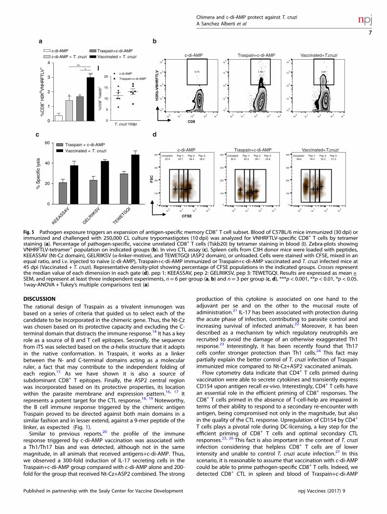
I
Fig. 5 Pathogen exposure triggers an expansion of antigen-specific memory CD8+ T cell subset. Blood of C57BL/6 mice immunized (30 dpi) orimmunized and challenged with 250,000 CL culture trypomastigotes (10 dpi) was analyzed for VNHRFTLV-specific CD8+ T cells by tetramerstaining (a). Percentage of pathogen-specific, vaccine unrelated CD8+ T cells (Tskb20) by tetramer staining in blood (I). Zebra-plots showingVNHRFTLV-tetramer+ population on indicated groups (b). In vivo CTL assay (c). Spleen cells from C3H donor mice were loaded with peptides,KEEASSAV (Nt-Cz domain), GELRIIKSV (α-linker-motive), and TEWETGQI (ASP2 domain), or unloaded. Cells were stained with CFSE, mixed in anequal ratio, and i.v. injected to naïve (c-di-AMP), Traspain+c-di-AMP immunized or Traspain+c-di-AMP vaccinated and T. cruzi infected mice at45 dpi (Vaccinated + T. cruzi). Representative density-plot showing percentage of CFSE populations in the indicated groups. Crosses representthe median value of each dimension in each gate (d). pep 1: KEEASSAV, pep 2: GELRIIKSV, pep 3: TEWETGQI. Results are expressed as mean ±SEM, and represent at least three independent experiments, n= 6 per group (a, b) and n= 3 per group (c, d), ***p< 0.001, **p< 0.01, *p< 0.05.1way-ANOVA + Tukey’s multiple comparisons test (a)
Chimera and c-di-AMP protect against T. cruziA Sanchez Alberti et al
7
Published in partnership with the Sealy Center for Vaccine Development npj Vaccines (2017) 9
vaccinated mice (Fig. 4a–d). Furthermore, these cells were able todegranulate upon antigen re-stimulation. Interestingly, theyshowed re-expansion capacity upon pathogen exposure, increas-ing not only in frequency (Fig. 5a) but also in functionality asdetermined by the ability to lyse peptide loaded target cells(Fig. 5c). Traspain vaccination was able to prime functional CD8T cells directed against both dominant and subdominant (Nt-Czand α-linker region) CTL epitopes. Altogether, these resultshighlight the ability of c-di-AMP to trigger cross-presentation ofproteins as well as in vivo cross priming of specific CTL.Moreover, a series of proof of concept studies were carried out
challenging vaccinated animals with T. cruzi from different strains.Antigens+c-di-AMP were able to control acute infection in termsof parasitemia, weight loss, and survival (Fig. 6). Interestingly,during the chronic phase of the infection, vaccinated miceshowed a decreased serum activity of tissue damage-associatedenzymes. More importantly, Traspain+c-di-AMP vaccinated miceshowed the lowest level of mononuclear infiltrate in skeletalmuscle, compared with others groups (Fig. 7). These animalswere also able to control parasite replication during the firstdays of infection, a fact that was associated with an earlierpro-inflammatory response at the site of infection. Furtherexperiments are necessary to determine the etiology of theinflammatory process and the involvement of circulating orresident memory T cells.28, 29 As the outcome of interest for ananti-T. cruzi vaccine should be focus on the reduction of chronic
inflammation-associated damage, Traspain+c-di-AMP proved tobe the best strategy assayed in terms of vaccine effectiveness con-ferring protection against diferent strains of parasites and miceindependently of the sex bias of T. cruzi infection.Considering that antigen load is an essential factor for the
priming of T cell responses,30 it is clear that Traspain allows alower amounts of antigen (10 µg of Transpain contains a loweramount of molecules than 10 µg of each antigen alone) to bepresented more efficiently when we compared the triggeredimmune response to that obtained with each antigen alone.Moreover, when the molar dose of each antigen was reduced tothat of Traspain in Nt-Cz+ASP2+c-di-AMP group, the immunerespose although from the same quality was not as robust andfailed to achieve similar levels of protection (Fig. 6c). In addition,since the chimeric molecule presents higher MW, pI, number anddistribution of charged residues, the physicochemical differencesof each formulation could influence self-assembly processesincluding particle or aggregates formation increasing the prob-ability of protein uptake and antigen processing. Electronmicroscopy, analitycal SEC and DLS assays should reveal thisissues in futher studies. Furthermore, we could not discard anscenario, where DC competition for the antigen uptake andprocessing takes place as well as the possibility that one antigenmay interfere with the other during steps of T cell priming in Nt-Cz+ASP2+c-di-AMP group.
c-di-AMP Traspain+c-di-AMP
4dpi
9dpi
a d
b e
c f
6dpi
Foo
tpad
swel
ling
(mm
)
0.0
0.5
1.0
1.5
2.0
**
c-di-AMP Traspain+c-di-AMP
Par
asiti
smA
UC
(RU
)x1
03
0
10
20
30
40
50***
dpi
Flu
ore
sce
nce
( 106
pho
t/cm
2/ s
)
0 1 2 3 4 5 6 7 8 9 10 110
2000
4000
6000
8000c-di-AMP
Traspain+c-di-AMP
10 12 14 16 18 20 22 24 26 28 30 32 340
2
4
6
8
10 c-di-AMP
Traspain+c-di-AMP
Nt-Cz+ASP2+c-di-AMP
ASP2+c-di-AMP
Nt-Cz+c-di-AMP
dpi
Par
asite
s/m
l(x1
05)
12 14 16 18 20 22 24 26 28 30 32 3460
80
100
120
Nt-Cz+c-di-AMP
ASP2+c-di-AMP
Nt-Cz+ASP2+c-di-AMP
Traspain+c-di-AMP
c-di-AMP
dpi
Bod
yW
eigh
t(%
ofin
i tial
)
0 10 20 30 40 50 60 700
20
40
60
80
100
dpi
Per
cent
surv
iva
l
Nt-Cz+c-di-AMP
ASP2+c-di-AMP
Nt-Cz+ASP2+c-di-AMP
Traspain+c-di-AMP
c-di-AMPns
**
footpad challengei.p. challenge
g
Fig. 6 Vaccine efficacy during the acute phase of T. cruzi infection. Vaccinated C3H mice were challenged 30 days after the last dose with alethal intraperitoneal (i.p.) dose of T. cruzi RA strain trypomastigotes and analyzed for: parasitemia (a), body weight loss (b) and survival (c), n=6 per group, **p< 0.01, Log-Rank Test, ns: not significant vs. control. Footpad challenge of the indicated groups with trypomastigotes of theCL-tdTomato strain. Mean fluorescence intensity of feet determined by in-vivo imaging, between 1 and 10 dpi (d). Representative images from4 to 9 dpi showing the control of parasite replication at the site of infection in vaccinated animals (e). Area under the curve of fluorescence persite of infection (n= 12) (f). Footpad swelling at 6 dpi (g). Footpad diameter was measured using a caliper. Data show the mean differencebetween day 0 and 6 per mouse. **P< 0.01, ***p< 0.001, Mann–Whitney test. The results are representative of at least three independentexperiments
Chimera and c-di-AMP protect against T. cruziA Sanchez Alberti et al
8
npj Vaccines (2017) 9 Published in partnership with the Sealy Center for Vaccine Development
This is the first report to present a chimeric molecule containingthe Nt-Cz and ASP-2, both main T. cruzi vaccine candidates, whichshowed protection during the whole course of the infection.Despite the immune response demonstrated towards the linkermotive, its protective quality alone remains to be proved in further
studies. The intrinsic complexity of T. cruzi, with more than 12,000protein coding genes per haploid genome, six discrete type units,and the presence of protein superfamilies with repetitive andpolymorphic variants31, 32 highlights the difficulty in finding onlyone target that confers acceptable levels of protection against the
Group
c-di-AMPHeart ++ ++Skeletal muscle +++ +++
Traspain+c-di-AMPHeart - -Skeletal muscle -/+ -/+
Nt-Cz+ASP2+c-di-AMPHeart - -Skeletal muscle ++ ++ ASP2+c-di-AMPHeart - -Skeletal muscle ++ ++ Nt-Cz+c-di-AMPHeart - -Skeletal muscle ++ ++
Necrosis Chronic inflammation grade
Heart
c-di-AMP
Traspain+c-di-AMP
Skeletal muscle
Nt-Cz+ASP2+c-di-AMP
(U/l)
0
50
100
150
200200
300
400
500
600
****
* * *
**
*
*
CK CK-MB LDH GOT
a
b
c-di-AMPTraspain+c-di-AMPNt-Cz+ASP2+c-di-AMPASP2+c-di-AMPNt-Cz+c-di-AMP
Chimera and c-di-AMP protect against T. cruziA Sanchez Alberti et al
9
Published in partnership with the Sealy Center for Vaccine Development npj Vaccines (2017) 9
infection. Designing an anti-T. cruzi vaccine is challenging not onlybecause of these facts but also due to the kind of immuneresponse required in order to achieve protection. Thus, cell-mediated immunity, particularly CD8+ T cells, plays a crucial role incontrolling parasite infection.2 Considering this scenario, webelieve that a multicomponent vaccine is appropriate in orderto increase the breadth of the immune response triggered byvaccination. Our results highlight the importance of designingchimeric molecules in multicomponent vaccines a fact that mustbe considered not only to reduce production costs but also toseek for an improvement in the magnitude and quality of theimmune response elicited by each component.Prompt control of T. cruzi infection might require the
identification of early antigens such as flagellar proteins33 andthe incorporation to new chimeric molecules as Traspainrepresent an attractive area of research for the development ofnovel anti-T. cruzi vaccines.
MATERIALS AND METHODSMice and parasitesFemale C3H/HeN (H-2k) mice 6–8-weeks-old were kept at theanimal facility of the Helmholtz Center for Infection Research underspecific pathogen-free (spf) conditions. C57BL/6 (H-2b) mice weremaintained in the University of Georgia animal facility under spfconditions. Animal experiments were approved by the ethical boardand conducted in accordance to the regulations of Lower Saxony No.09.4250204105/07, Germany, UBA-CONICET, Argentina and IACUC,UGA, USA. T. cruzi bloodstream trypomastigotes of the RA and therecombinant Tulahuen strain expressing β-galactosidase34 were isolatedfrom infected mice. Tissue culture trypomastigotes of the CL strain of T.cruzi wild-type or expressing tdTomato were obtained from passagethrough Vero cells.35
Recombinant proteinsThe Nt-terminal domain of Cz (Nt-Cz) was produced as previouslydescribed.14 The ASP2 transcript was obtained from T. cruzi amastigotecDNA from the RA strain as reported.36 The central region of ASP2protein (residues 261–570), herein referred to as ASP2, was amplifiedemploying gene-specific primers and cloned in a pET23a vector. ForTraspain construction, sequential PCR was done to build the alpha helixlinker incorporating nucleotides 1205–1281 from CL-Brener iTS(XM_811430.1) to Nt-Cz. Splicing by overlap extension PCR37, 38 wasperformed in order to obtain the hybrid gene with ASP2. Cloning wasdone in a pET23a vector. Gene sequencing was performed to confirm thechimeric gene. Traspain and ASP2 were expressed in E. coli BL21 (DE3) asinclusion bodies, purified under denaturing conditions by IMAC andin vitro refolded by dialysis method. Purity levels were determined by SDS-PAGE and presence of endotoxin was assessed by LPS-Detection Kit(invivogen).
Immunizations and challengeInbred female C3H/HeN mice 6–8-weeks-old were immunized by theintranasal route with three doses every 15 days as follows: (I) c-di-AMP, (II)Traspain, (III) Traspain+c-di-AMP, (IV) Nt-Cz+ASP2+c-di-AMP, (V) ASP2+c-di-AMP, and (VI) Nt-Cz+c-di-AMP. Each group received 10 µg of eachcomponent, with the exception of group IV that received equal molaramounts of each antigen. For analysis of the acute phase, 15–30 daysafter the last dose, mice were challenged with 103 T. cruzi RA strainblood trypomastigotes by the intraperitoneal route. For sublethal
assays 102 parasites were administered. Alternatively, male C57BL/6were immunized following the same schedule and challenged in the hindfootpads with 2.5 × 10⁵ T. cruzi tdTomato trypomastigotes.
ELISA titration of antigen-specific IgG AbsProtein-specific antibody titers were determined as previouslydescribed39 by ELISA using plates coated with 0.2 μg of Traspain, Nt-Cz,or ASP2 per well. Peroxidase conjugated goat immunoglobulins tomouse immunoglobulin (Ig) G were used as the secondary antibody.Plates were developed by adding o-phenylenediamine/H2O2. For peptide-ELISA, plates were coated with 10 µM of GELRIIKSV in carbonate buffer pH9.6 ON, washed and blocked, sera was incubated ON. Biotin conjugatedgoat immunoglobulins to mouse IgG plus streptavidin-peroxidasewere used as the secondary antibody. Plates were developed by addingTMB/H2O2.
Neutralization assayRaw cells (5 × 10³ cells/well) were infected with blood trypomastigotesexpressing β-galactosidase at a MOI of 10:1 for 24 h at 37 °C. Trypomas-tigotes were pre-incubated in triplicate with diluted serum (1/10) frommice belonging to each immunization group. After overnight incubationcells were washed and incubated for 5 days. CPRG was added to determinethe levels of parasites as previously described.40
Proliferation assaySpleen cells (5 × 10⁵ cells/well) of each vaccination group were incubatedin quadruplicates for 96 h in the presence of different concentrations ofTraspain (1, 10, 20, and 40 µg/ml) and proceeded as previously reported.20
Results were expressed as the ratio of mean values from stimulated andnon-stimulated samples, proliferation index (PI).
ELISPOT assaysSpleen cells (4 × 10⁵/2 × 105 cells/well) were incubated for 24 h (IFN-γ) or48 h (IL-2, IL-17, and IL-4), in the absence or presence of 10 µg/ml of eitherTraspain or ASP2 or Nt-Cz for ELISPOT assays (BD Pharmingen, USA). Afterincubation, cells were removed and the plates were processed accordingto the manufacturer’s instructions. Colored spots were counted with anELISPOT reader (CTL S5 Micro Analyzer) and analyzed using ImmunoSpotimage analyzer software v3.2 (CTL Europe GmbH, Germany).
Intracellular cytokine stainingIsolated splenocytes were stimulated overnight with 10 µg/ml of Traspainin the presence of anti-CD154-PE and anti-CD107 PE-Cy7. Brefeldin A plusmonensin was added to cultures during the last 6 h of incubation. Surfacestaining was performed with anti-CD3e-V500, anti-CD4-APC-H7 (BD), andanti-CD8α-Brilliant-Violet-650 (BioLegend). Cells were fixed with PFA 2%,permeabilized in 0.5% saponin, and stained using anti-IFN-γ-Brilliant-Violet-711 (BioLegend) and anti-TNF-α-eFluor450 (eBioscience) in accordancewith the manufacturer’s instructions.
MHC class I multimer stainingTo detect antigen-specific T cells, spleen or blood cells were first labeledwith H2Kk-TEWETGQI dextramer-APC (Immudex) and then with anti-CD3eV500, anti-CD4-APC-H7 (BD), and anti-CD8α-Brilliant Violet V650 (BioLe-gend) according to the manufacturer’s instructions. Alternatively, eitherH2Kb-VNHRFTLV-APC or H2Kb-ANYKFTLV-APC (Tskb20) tetramer (NIHTetramer Core Facility, Emory University, USA) were employed for C57BL/6 mice.
Fig. 7 Vaccine effectiveness for the prevention of tissue damage. Serum activity of cardiomyopathy-associated enzymes from immunized andinfected mice. Creatinine kinase (CK), creatinine kinase MB isoform (CK-MB), lactate dehydrogenase (LDH, U/I ×10), and glutamateoxaloacetate transaminase (GOT) (a). Results are expressed as mean± SEM. *p< 0.05, **p< 0.01, one-way-ANOVA Kruskal-Wallis test + Dunn’smultiple comparisons test. Histopathological analysis of T. cruzi-target organs during the chronic phase of infection. Representative tissuesections (H&E stained) for the indicated groups (b). Magnification level: 100×. Insets: 400×. Table shows Inflammation score semi-quantitativelyevaluated for each group (-, absence of inflammation; number of crosses indicate degree of inflammation). Solid arrows point to mononuclearcell infiltrates. Dashed arrow points to a nerve associated with mononuclear cell infiltrates. Results are representative of two independentexperiments
Chimera and c-di-AMP protect against T. cruziA Sanchez Alberti et al
10
npj Vaccines (2017) 9 Published in partnership with the Sealy Center for Vaccine Development
In vivo cytotoxicity assaySplenocytes collected from naïve C3H/HeN mice were incubated with 5 μMof peptides, pep1: TEWETGQI (ASP2), pep2: KEEASSAV (Nt-Cz), and pep3:GELRIIKSV (α-linker-iTS) or no peptide for 30min at 37 °C and 30min at 4 °C, washed, and then labeled with 2.5, 5, 10, and 0.5 μM of CFSE(CellTrace™ CFSE Cell Proliferation Kit), respectively. Cells were washed,combined, and transferred (4 × 10⁷ total cells) intravenously to syngenicnaïve (c-di-AMP), traspain+c-di-AMP immunized, and T. cruzi infected miceat 45 dpi. Sixteen hours after transfer, spleens were harvested andanalyzed by flow-cytometry. The percentage of specific lysis was calculatedas follows: % Specific lysis = 1−[(%CFSEpep/%CFSEunloaded) Immunized/(%CFSEpep /%CFSEunloaded)naïve] * 100
Assessment of vaccine efficacyParasitemia was monitored by counting peripheral parasites every 2 daysas previously described.40 In tdTomato T. cruzi infection the fluorescentintensity was measured using a whole animal imaging system (Maestro2 InVivo Imaging System CRi, USA) as a surrogate of parasite load.35 Muscleinjury was evaluated through the determination of a panel of myopathy-linked enzyme markers as previously described.39 The histological featuresof heart and skeletal muscles were also investigated. A blind histologicaltest was performed as previously described.41
Statistical analysisStatistical analysis was carried out with Graphpad Prism 6.0 software (SanDiego, CA, USA) using one-way ANOVA, n = 6 animals/group unlessotherwise specified in figure legends, p-values <0.05 were consideredsignificant.
ACKNOWLEDGEMENTFinancial support was received from: Consejo Nacional de Investigaciones Científicasy Técnicas (CONICET), University of Buenos Aires and Agencia Nacional deInvestigaciones Científicas y Técnicas (PICT-2013-2328 and PICT-2014-0854 to ELM),Argentina; the German Federal Ministry of Education and Research (BMBF) under theproject ARG 10/005 (01DN12004); and the National Institutes of Health of the UnitedStates Public Health Service grant to R01-AI089952 RLT. ELM is also supported by theFogarty International Center (TW007972) and International Center for GeneticEngineering and Biotechnology (CRP/ARG09-02). We are grateful to Blair Prochnowfor critical reading of the manuscript.
COMPETING INTERESTSThe authors have no financial conflicts of interest.
REFERENCES1. WHO. Sustaining the drive to overcome the global impact of neglected tropical
diseases. Second WHO Rep. Negl. Trop. Dis. 3.9, 67–71 (2013).2. Tarleton, R. L. CD8+ T cells in Trypanosoma cruzi infection. Semin. Immunopathol.
37, 233–238 (2015).3. Martin, D. L. et al. CD8+ T-cell responses to Trypanosoma cruzi are highly focused
on strain-variant trans-sialidase epitopes. PLoS Pathog. 2, 0731–0740 (2006).4. Junqueira, C. et al. The endless race between Trypanosoma cruzi and host
immunity: lessons for and beyond Chagas disease. Expert. Rev. Mol. Med. 12, e29(2010).
5. Burdette, D. L. & Vance, R. E. STING and the innate immune response to nucleicacids in the cytosol. Nat. Immunol. 14, 19–26 (2013).
6. Danilchanka, O. & Mekalanos, J. J. Cyclic dinucleotides and the innate immuneresponse. Cell 154, 962–970 (2013).
7. Sun, L., Wu, J., Du, F., Chen, X. & Chen, Z. J. Cyclic GMP-AMP synthase is a cytosolicDNA sensor that activates the type I interferon pathway. Science 339, 786–791(2013).
8. Libanova, R., Becker, P. D. & Guzmán, C. A. Cyclic di-nucleotides: new era for smallmolecules as adjuvants. Microb. Biotechnol. 5, 168–176 (2012).
9. Tzelepis, F. et al. Infection with Trypanosoma cruzi restricts the repertoire ofparasite-specific CD8+ T cells leading to immunodominance. J. Immunol. 180,1737–1748 (2008).
10. Sullivan, N. L., Eickhoff, C. S., Sagartz, J. & Hoft, D. F. Deficiency of antigen-specificB cells results in decreased Trypanosoma cruzi systemic but not mucosalimmunity due to CD8 T cell exhaustion. J. Immunol. 194, 1806–1818 (2015).
11. Hauschka, T. S. Sex of host as a factor in Chagas’ disease. J. Parasitol. 33, 399–404(1947).
12. Micucci, L. R. et al. [Importance of host sex in the development of trypanosomacruzi infection]. Rev. Fac. Cien. Med. Univ. Nac. Cordoba. 67, 73–76 (2010).
13. Vorraro, F. et al. Trypanosoma cruzi Infection in Genetically Selected Mouse Lines:Genetic Linkage with Quantitative Trait Locus Controlling Antibody Response.Mediators Inflamm. 2014, 1–15 (2014).
14. Cazorla, S. I. et al. Redirection of the immune response to the functional catalyticdomain of the cystein proteinase cruzipain improves protective immunity againstTrypanosoma cruzi infection. J. Infect. Dis. 202, 136–144 (2010).
15. Arai, R., Ueda, H., Kitayama, A., Kamiya, N. & Nagamune, T. Design of the linkerswhich effectively separate domains of a bifunctional fusion protein. Protein Eng.14, 529–532 (2001).
16. Vasconcelos, J. R. C. et al. A DNA-priming protein-boosting regimen significantlyimproves type 1 immune response but not protective immunity to Trypanosomacruzi infection in a highly susceptible mouse strain. Immunol. Cell Biol. 81,121–129 (2003).
17. Vasconcelos, J. R. et al. Pathogen-induced proapoptotic phenotype and highCD95 (Fas) expression accompany a suboptimal CD8+ T-cell response: reversal byadenoviral vaccine. PLoS Pathog. 8, e1002699 (2012).
18. Low, H. P., Santos, M. A., Wizel, B. & Tarleton, R. L. Amastigote surfaceproteins of Trypanosoma cruzi are targets for CD8+ CTL. J. Immunol. 160,1817–1823 (1998).
19. Dominguez, M. R. et al. Subdominant/cryptic CD8 T cell epitopes contribute toresistance against experimental infection with a human protozoan parasite. PLoSOne 6, e22011 (2011).
20. Sanchez, M. V. et al. Intranasal delivery of influenza rNP adjuvanted with c-di-AMPinduces strong humoral and cellular immune responses and provides protectionagainst virus challenge. PLoS One 9, e104824 (2014).
21. Fukuyama, Y. et al. Nanogel-based pneumococcal surface protein A nasal vaccineinduces microRNA-associated Th17 cell responses with neutralizing antibodiesagainst Streptococcus pneumoniae in macaques. Mucosal Immunol. 8, 1144–1153(2015).
22. Bermejo, D. A. et al. Trypanosoma cruzi trans-sialidase initiates a program inde-pendent of the transcription factors RORγt and Ahr that leads to IL-17 productionby activated B cells. Nat. Immunol. 14, 514–522 (2013).
23. Boari, J. T. et al. IL-17RA signaling reduces inflammation and mortality duringTrypanosoma cruzi infection by recruiting suppressive IL-10-producing neu-trophils. PLoS Pathog. 8, e1002658 (2012).
24. Cai, C. W., Blase, J. R., Zhang, X., Eickhoff, C. S. & Hoft, D. F. Th17 Cells Are MoreProtective Than Th1 Cells Against the Intracellular Parasite Trypanosoma cruzi.PLOS Pathog. 12, e1005902 (2016).
25. Laidlaw, B. J., Craft, J. E. & Kaech, S. M. The multifaceted role of CD4+ T cells inCD8+ T cell memory. Nat. Rev. Immunol. doi:10.1038/nri.2015.10 (2016).
26. Sokke Umeshappa, C. et al. CD154 and IL-2 signaling of CD4+ T cells play a criticalrole in multiple phases of CD8+ CTL responses following adenovirus vaccination.PLoS One 7, e47004 (2012).
27. Padilla, A., Xu, D., Martin, D. & Tarleton, R. Limited role for CD4+ T-cell help in theinitial priming of Trypanosoma cruzi-specific CD8+ T cells. Infect. Immun.75, 231–235 (2007).
28. Schenkel, J. M. & Masopust, D. Tissue-resident memory T cells. Immunity41, 886–897 (2014).
29. Mueller, S. N. & Mackay, L. K. Tissue-resident memory T cells: local specialists inimmune defence. Nat. Rev. Immunol. 1–11 (2015). doi:10.1038/nri.2015.3
30. Pennock, N. D., Kedl, J. D. & Kedl, R. M. T cell vaccinology: beyond the reflection ofinfectious responses. Trends Immunol. doi:10.1016/j.it.2016.01.001 (2016).
31. Ferella, M. et al. Proteomics in Trypanosoma cruzi—localization of novel proteinsto various organelles. Proteomics 8, 2735–2749 (2008).
32. Bustamante, J. & Tarleton, R. Reaching for the holy grail: insights from infection/cure models on the prospects for vaccines for Trypanosoma cruzi infection. Mem.Inst. Oswaldo Cruz 110, 445–451 (2015).
33. Kurup, S. P. & Tarleton, R. L. The Trypanosoma cruzi flagellum is discarded viaasymmetric cell division following invasion and provides early targets for pro-tective CD8+ T cells. Cell Host Microbe 16, 439–449 (2014).
34. Buckner, F. S., Verlinde, C. L. M. J., La Flamme, A. C. & Van Voorhis, W. C. Efficienttechnique for screening drugs for activity against Trypanosoma cruzi usingparasites expressing beta-galactosidase. Antimicrob. Agents Chemother. 40,2592–2597 (1996).
35. Canavaci, A. M. C. et al. In vitro and in vivo high-throughput assays forthe testing of anti-Trypanosoma cruzi compounds. PLoS Negl. Trop. Dis. 4, e740(2010).
36. Boscardin, S. B., Kinoshita, S. S., Fujimura, A. E. & Rodrigues, M. M. Immunizationwith cDNA expressed by amastigotes of Trypanosoma cruzi elicits protectiveimmune response against experimental infection. Infect. Immun. 71, 2744–2757(2003).
Chimera and c-di-AMP protect against T. cruziA Sanchez Alberti et al
11
Published in partnership with the Sealy Center for Vaccine Development npj Vaccines (2017) 9
37. Warrens, A. N., Jones, M. D. & Lechler, R. I. Splicing by overlap extensionby PCR using asymmetric amplification: an improved technique for thegeneration of hybrid proteins of immunological interest. Gene. 186, 29–35(1997).
38. Wurch, T., Lestienne, F. & Pauwels, P. J. A modified overlap extension PCR methodto create chimeric genes in the absence of restriction enzymes. Biotechnol. Tech.12, 653–657 (1998).
39. Matos, M. N. et al. Tc52 amino-terminal-domain DNA carried byattenuated Salmonella enterica serovar typhimurium induces protectionagainst a Trypanosoma cruzi lethal challenge. Infect. Immun. 82, 4265–4275(2014).
40. Matos, M. N., Sánchez Alberti, A., Morales, C. & Cazorla, S. I. A prime-boostimmunization with Tc52 N-terminal domain DNA and the recombinant proteinexpressed in Pichia pastoris protects against Trypanosoma cruzi infection. Vaccine34, 3243–3251 (2016).
41. Cazorla, S. I. et al. Oral multicomponent DNA vaccine delivered by attenuatedSalmonella elicited immunoprotection against American trypanosomiasis. J.Infect. Dis. 211, 698–707 (2015).
This work is licensed under a Creative Commons Attribution 4.0International License. The images or other third party material in this
article are included in the article’s Creative Commons license, unless indicatedotherwise in the credit line; if the material is not included under the Creative Commonslicense, users will need to obtain permission from the license holder to reproduce thematerial. To view a copy of this license, visit http://creativecommons.org/licenses/by/4.0/
© The Author(s) 2017
Chimera and c-di-AMP protect against T. cruziA Sanchez Alberti et al
12
npj Vaccines (2017) 9 Published in partnership with the Sealy Center for Vaccine Development
![Probing into Dopant Concentration Dependent Luminescence ... · trivalent rare earth ions such as Eu 3+, Pr Sm3+, Tb3+ as the luminous centers in sulfides [6], tungstates [7], titanates](https://static.fdocument.org/doc/165x107/604827c8f14a1c31824aab70/probing-into-dopant-concentration-dependent-luminescence-trivalent-rare-earth.jpg)


![Forming Alumina Parts Using Acrylamide Gels · 2018-12-26 · other ceramic processes especially slip-casting, ( 2)[12]. Firstly, by using magnetic or mechanical stirrer (with turbine](https://static.fdocument.org/doc/165x107/5f2af1608533fe5dad251b05/forming-alumina-parts-using-acrylamide-gels-2018-12-26-other-ceramic-processes.jpg)