Efficient Logistic Regression with Stochastic Gradient Descent – part 2
Research ArticleEfficient β-Carboline Alkaloid-Based Probe for Highly SensitiveImaging of Endogenous Glutathione in Wheat Germ Tissues
Xiaohui Ji,1 Dan Zhang ,1 Le Li,2 Lingxia Jin,1 and Rui Wu1
1Shaanxi Province Key Laboratory of Catalytic Foundation and Application, School of Chemistry and Environment Science,Shaanxi University of Technology, Hanzhong 723001, China2Shaanxi Key Laboratory of Industrial Automation, School of Mechanical Engineering, Shaanxi University of Technology,Hanzhong 723001, China
Correspondence should be addressed to Dan Zhang; [email protected]
Received 28 November 2019; Revised 3 July 2020; Accepted 6 September 2020; Published 15 September 2020
Academic Editor: Neil D. Danielson
Copyright © 2020 Xiaohui Ji et al. +is is an open access article distributed under the Creative Commons AttributionLicense, which permits unrestricted use, distribution, and reproduction in any medium, provided the original work isproperly cited.
Discriminative detection of GSH is achieved by employing a highly sensitive and selective fluorescent probe (KL-DN) that bearsβ-carboline alkaloid as a potential fluorophore and an azide group as the recognition unit. A rapid fluorescence off-on change iscaused by special redox reaction;KL-DN has the capability of monitoring endogenous GSH in wheat germ tissues, indicating thatthis probe holds great potential for biological applications in plant tissues.
1. Introduction
Wheat germ containing abundant GSH, a nutrition with highprotein, high vitamin E, and low cholesterol, is beneficial tohuman health [1]. Interestingly, research indicates that GSHdirectly or indirectly involves in many functional activities inwheat seedling leaves under osmotic stress, including themetabolism of drugs and hormone in the plant cells and thebiosynthesis of DNA, RNA, and protein [2–4]. As an essentialendogenic antioxidant, GSH can protect plant cells fromoxidative damage [5]. Accordingly, the development ofeconomical and effective methods for detection of GSH inwheat germ system is of considerable importance for betterunderstanding of its physiological functions and has becomethe significant subject of current chemical research.
Among the multiple analytical methods includingelectrochemical voltammetry, optical sensing, high-perfor-mance liquid chromatography, and mass spectrometry,fluorescence analysis is more popular because of its highsafety, low cost, simplicity of operation, and noninvasiveness[6–8]. In consideration of the sensitivity and great bio-imaging potential of fluorescence detection [9], considerableeffort had been paid in the development of fluorescent
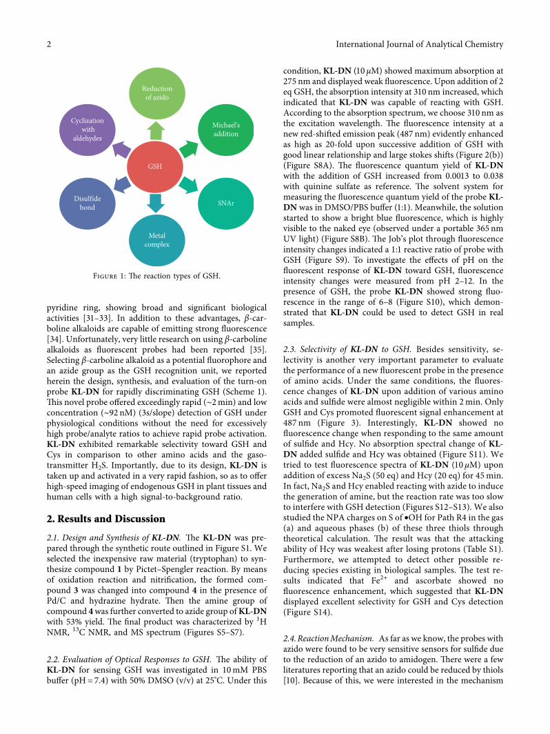
probes for GSH detection based on different reactivemechanisms (Figure 1). For instance, Xie et al. introducedthat the SH residue of GSH, and the aldehyde group of thefluorescent probe underwent the addition reaction resultingin the open-ring form in acid conditions [10]. Disulfidebonds have been abundantly exploited to measure GSH formediating the extracellular release of drugs [11–14]. +edisadvantages of long reaction time and high limit of de-tection were the ubiquitous challenge. +e fluorescentprobes with better SNAr leaving groups were susceptible toSNAr substitution by sulfhydryl thiols with poor selectivity[15–20]. Qu and coworkers reported a fluorescent turn-onmethod of Ag-S-GF taking advantage of specific interactionsbetween sulfur and silver cation [21]. +e mechanisms ofrecognizing GSH also included the selective cleavage of theselenium-nitrogen bond, Michael’s addition, the reductionof azido, and others [22–30]. Although these researchesgreatly accelerated the development of fluorescent detection,the design of a highly sensitive chemosensor on the basis ofnatural product structure for recognizing GSH in situ inplant organisms is still a significant challenge.
As an active natural product, β-carboline alkaloids withgood biocompatibility are composed of indole ring and
HindawiInternational Journal of Analytical ChemistryVolume 2020, Article ID 8675784, 7 pageshttps://doi.org/10.1155/2020/8675784
pyridine ring, showing broad and significant biologicalactivities [31–33]. In addition to these advantages, β-car-boline alkaloids are capable of emitting strong fluorescence[34]. Unfortunately, very little research on using β-carbolinealkaloids as fluorescent probes had been reported [35].Selecting β-carboline alkaloid as a potential fluorophore andan azide group as the GSH recognition unit, we reportedherein the design, synthesis, and evaluation of the turn-onprobe KL-DN for rapidly discriminating GSH (Scheme 1).+is novel probe offered exceedingly rapid (∼2min) and lowconcentration (∼92 nM) (3s/slope) detection of GSH underphysiological conditions without the need for excessivelyhigh probe/analyte ratios to achieve rapid probe activation.KL-DN exhibited remarkable selectivity toward GSH andCys in comparison to other amino acids and the gaso-transmitter H2S. Importantly, due to its design, KL-DN istaken up and activated in a very rapid fashion, so as to offerhigh-speed imaging of endogenous GSH in plant tissues andhuman cells with a high signal-to-background ratio.
2. Results and Discussion
2.1. Design and Synthesis of KL-DN. +e KL-DN was pre-pared through the synthetic route outlined in Figure S1. Weselected the inexpensive raw material (tryptophan) to syn-thesize compound 1 by Pictet–Spengler reaction. By meansof oxidation reaction and nitrification, the formed com-pound 3 was changed into compound 4 in the presence ofPd/C and hydrazine hydrate. +en the amine group ofcompound 4was further converted to azide group ofKL-DNwith 53% yield. +e final product was characterized by 1HNMR, 13C NMR, and MS spectrum (Figures S5–S7).
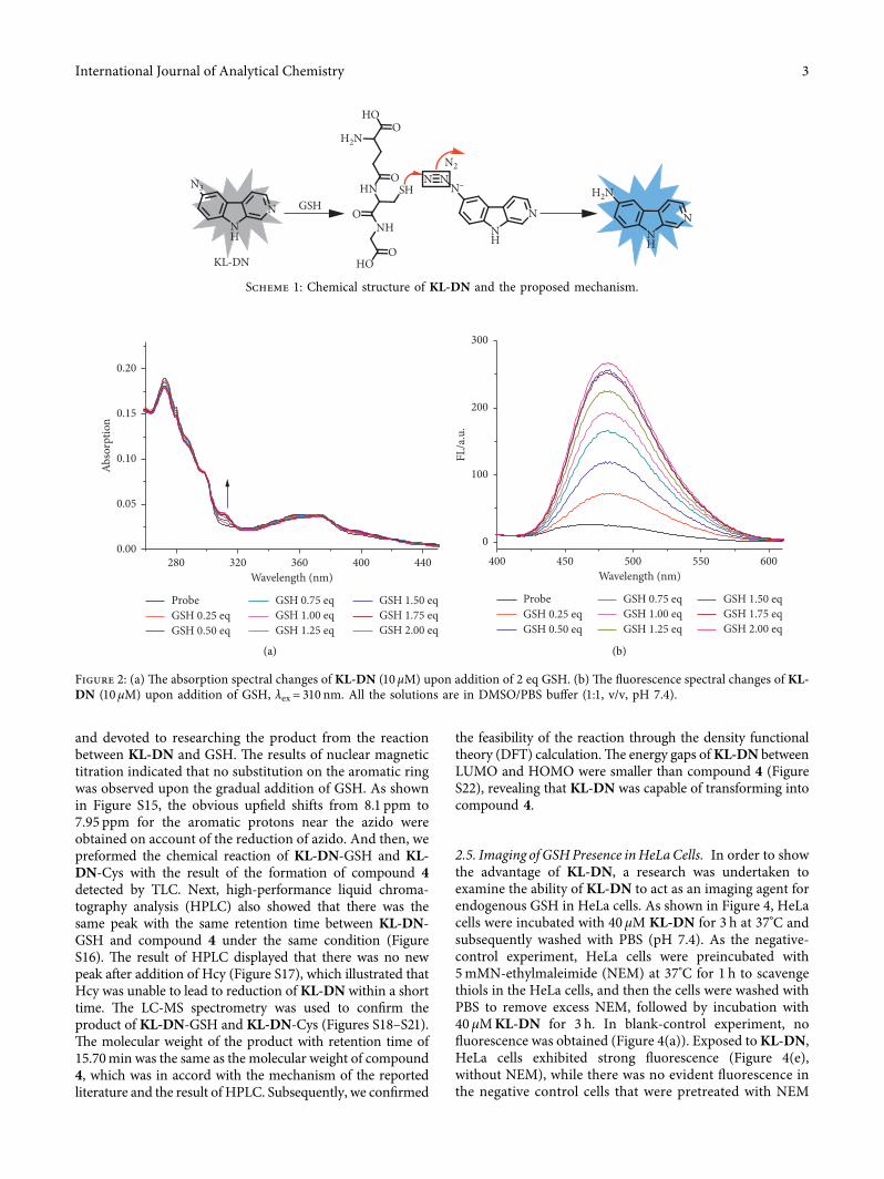
2.2. Evaluation of Optical Responses to GSH. +e ability ofKL-DN for sensing GSH was investigated in 10mM PBSbuffer (pH� 7.4) with 50% DMSO (v/v) at 25°C. Under this
condition, KL-DN (10 μM) showed maximum absorption at275 nm and displayed weak fluorescence. Upon addition of 2eq GSH, the absorption intensity at 310 nm increased, whichindicated that KL-DN was capable of reacting with GSH.According to the absorption spectrum, we choose 310 nm asthe excitation wavelength. +e fluorescence intensity at anew red-shifted emission peak (487 nm) evidently enhancedas high as 20-fold upon successive addition of GSH withgood linear relationship and large stokes shifts (Figure 2(b))(Figure S8A). +e fluorescence quantum yield of KL-DNwith the addition of GSH increased from 0.0013 to 0.038with quinine sulfate as reference. +e solvent system formeasuring the fluorescence quantum yield of the probe KL-DN was in DMSO/PBS buffer (1:1). Meanwhile, the solutionstarted to show a bright blue fluorescence, which is highlyvisible to the naked eye (observed under a portable 365 nmUV light) (Figure S8B). +e Job’s plot through fluorescenceintensity changes indicated a 1:1 reactive ratio of probe withGSH (Figure S9). To investigate the effects of pH on thefluorescent response of KL-DN toward GSH, fluorescenceintensity changes were measured from pH 2–12. In thepresence of GSH, the probe KL-DN showed strong fluo-rescence in the range of 6–8 (Figure S10), which demon-strated that KL-DN could be used to detect GSH in realsamples.
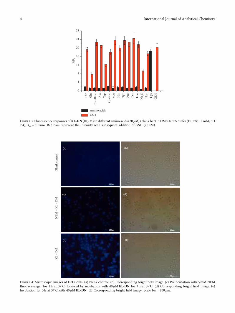
2.3. Selectivity of KL-DN to GSH. Besides sensitivity, se-lectivity is another very important parameter to evaluatethe performance of a new fluorescent probe in the presenceof amino acids. Under the same conditions, the fluores-cence changes of KL-DN upon addition of various aminoacids and sulfide were almost negligible within 2min. OnlyGSH and Cys promoted fluorescent signal enhancement at487 nm (Figure 3). Interestingly, KL-DN showed nofluorescence change when responding to the same amountof sulfide and Hcy. No absorption spectral change of KL-DN added sulfide and Hcy was obtained (Figure S11). Wetried to test fluorescence spectra of KL-DN (10 μM) uponaddition of excess Na2S (50 eq) and Hcy (20 eq) for 45min.In fact, Na2S and Hcy enabled reacting with azide to inducethe generation of amine, but the reaction rate was too slowto interfere with GSH detection (Figures S12–S13). We alsostudied the NPA charges on S of •OH for Path R4 in the gas(a) and aqueous phases (b) of these three thiols throughtheoretical calculation. +e result was that the attackingability of Hcy was weakest after losing protons (Table S1).Furthermore, we attempted to detect other possible re-ducing species existing in biological samples. +e test re-sults indicated that Fe2+ and ascorbate showed nofluorescence enhancement, which suggested that KL-DNdisplayed excellent selectivity for GSH and Cys detection(Figure S14).
2.4.ReactionMechanism. As far as we know, the probes withazido were found to be very sensitive sensors for sulfide dueto the reduction of an azido to amidogen. +ere were a fewliteratures reporting that an azido could be reduced by thiols[10]. Because of this, we were interested in the mechanism
Metalcomplex
SNAr
Michael'saddition
GSH
Reductionof azido
Cyclization with
aldehydes
Disulfidebond
Figure 1: +e reaction types of GSH.
2 International Journal of Analytical Chemistry
and devoted to researching the product from the reactionbetween KL-DN and GSH. +e results of nuclear magnetictitration indicated that no substitution on the aromatic ringwas observed upon the gradual addition of GSH. As shownin Figure S15, the obvious upfield shifts from 8.1 ppm to7.95 ppm for the aromatic protons near the azido wereobtained on account of the reduction of azido. And then, wepreformed the chemical reaction of KL-DN-GSH and KL-DN-Cys with the result of the formation of compound 4detected by TLC. Next, high-performance liquid chroma-tography analysis (HPLC) also showed that there was thesame peak with the same retention time between KL-DN-GSH and compound 4 under the same condition (FigureS16). +e result of HPLC displayed that there was no newpeak after addition of Hcy (Figure S17), which illustrated thatHcy was unable to lead to reduction of KL-DN within a shorttime. +e LC-MS spectrometry was used to confirm theproduct of KL-DN-GSH and KL-DN-Cys (Figures S18–S21).+e molecular weight of the product with retention time of15.70min was the same as the molecular weight of compound4, which was in accord with the mechanism of the reportedliterature and the result of HPLC. Subsequently, we confirmed
the feasibility of the reaction through the density functionaltheory (DFT) calculation.+e energy gaps ofKL-DN betweenLUMO and HOMO were smaller than compound 4 (FigureS22), revealing that KL-DN was capable of transforming intocompound 4.
2.5. Imaging of GSHPresence inHeLaCells. In order to showthe advantage of KL-DN, a research was undertaken toexamine the ability of KL-DN to act as an imaging agent forendogenous GSH in HeLa cells. As shown in Figure 4, HeLacells were incubated with 40 μM KL-DN for 3 h at 37°C andsubsequently washed with PBS (pH 7.4). As the negative-control experiment, HeLa cells were preincubated with5mMN-ethylmaleimide (NEM) at 37°C for 1 h to scavengethiols in the HeLa cells, and then the cells were washed withPBS to remove excess NEM, followed by incubation with40 μMKL-DN for 3 h. In blank-control experiment, nofluorescence was obtained (Figure 4(a)). Exposed toKL-DN,HeLa cells exhibited strong fluorescence (Figure 4(e),without NEM), while there was no evident fluorescence inthe negative control cells that were pretreated with NEM
280 320 360 400 4400.00
0.05
0.10
0.15
0.20
Abso
rptio
n
Wavelength (nm)
ProbeGSH 0.25 eqGSH 0.50 eq
GSH 0.75 eqGSH 1.00 eqGSH 1.25 eq
GSH 1.50 eqGSH 1.75 eqGSH 2.00 eq
(a)
ProbeGSH 0.25 eqGSH 0.50 eq
GSH 0.75 eqGSH 1.00 eqGSH 1.25 eq
GSH 1.50 eqGSH 1.75 eqGSH 2.00 eq
400 450 500 550 6000
100
200
300
FL/a
.u.
Wavelength (nm)
(b)
Figure 2: (a) +e absorption spectral changes of KL-DN (10 μM) upon addition of 2 eq GSH. (b) +e fluorescence spectral changes of KL-DN (10 μM) upon addition of GSH, λex � 310 nm. All the solutions are in DMSO/PBS buffer (1:1, v/v, pH 7.4).
HO
HO
O
ONH
HN SHO
O
NNN
N–NN2
N
HNH
GSHNNH
KL-DN
N3
H2N
H2N
Scheme 1: Chemical structure of KL-DN and the proposed mechanism.
International Journal of Analytical Chemistry 3
(a) (b)
Blan
k co
ntro
l
(c) (d)
NEM
+ K
L –
DN
(e) (f)
KL –
DN
Figure 4: Microscopic images of HeLa cells. (a) Blank control. (b) Corresponding bright field image. (c) Preincubation with 5mM NEMthiol scavenger for 1 h at 37°C, followed by incubation with 40 μMKL-DN for 3 h at 37°C. (d) Corresponding bright field image. (e)Incubation for 3 h at 37°C with 40 μMKL-DN. (f ) Corresponding bright field image. Scale bar� 200 μm.
0
4
8
12
16
20
24
28
F/F 0
Amino acidsGSH
Citr
ullin
e
GSHCy
sH
cyN
a 2S
Thr
Glu Ala
Trp
His
Tyr
Pro
Lys
Leu
Met
Cysti
ne
Figure 3: Fluorescence responses ofKL-DN (10 μM) to different amino acids (20 μM) (blank bar) in DMSO/PBS buffer (1:1, v/v, 10mM, pH7.4), λex � 310 nm. Red bars represent the intensity with subsequent addition of GSH (20 μM).
4 International Journal of Analytical Chemistry
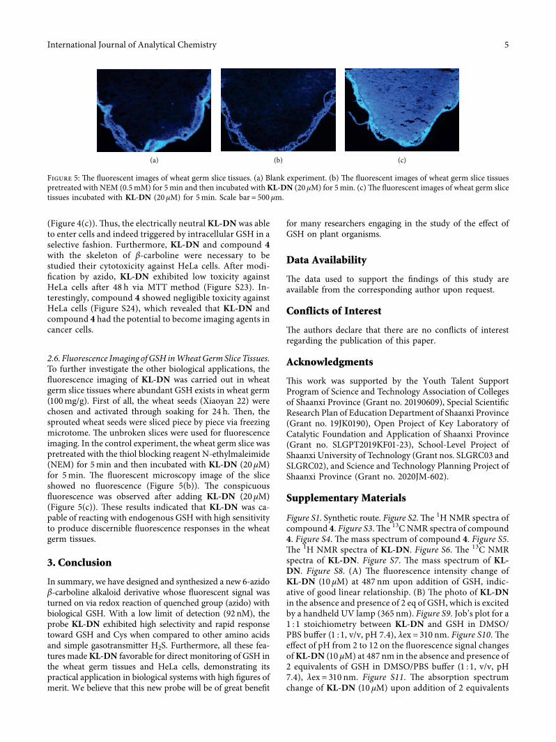
(Figure 4(c)). +us, the electrically neutral KL-DN was ableto enter cells and indeed triggered by intracellular GSH in aselective fashion. Furthermore, KL-DN and compound 4with the skeleton of β-carboline were necessary to bestudied their cytotoxicity against HeLa cells. After modi-fication by azido, KL-DN exhibited low toxicity againstHeLa cells after 48 h via MTT method (Figure S23). In-terestingly, compound 4 showed negligible toxicity againstHeLa cells (Figure S24), which revealed that KL-DN andcompound 4 had the potential to become imaging agents incancer cells.
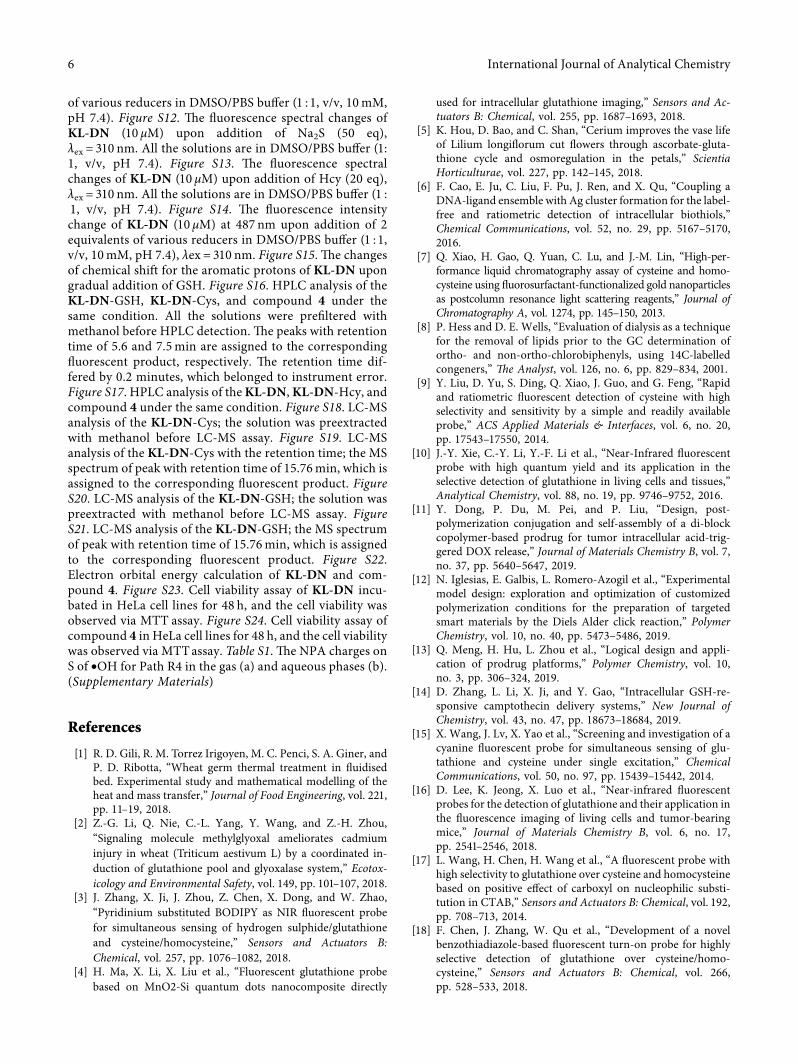
2.6. Fluorescence Imaging ofGSH inWheatGermSlice Tissues.To further investigate the other biological applications, thefluorescence imaging of KL-DN was carried out in wheatgerm slice tissues where abundant GSH exists in wheat germ(100mg/g). First of all, the wheat seeds (Xiaoyan 22) werechosen and activated through soaking for 24 h. +en, thesprouted wheat seeds were sliced piece by piece via freezingmicrotome. +e unbroken slices were used for fluorescenceimaging. In the control experiment, the wheat germ slice waspretreated with the thiol blocking reagent N-ethylmaleimide(NEM) for 5min and then incubated with KL-DN (20 μM)for 5min. +e fluorescent microscopy image of the sliceshowed no fluorescence (Figure 5(b)). +e conspicuousfluorescence was observed after adding KL-DN (20 μM)(Figure 5(c)). +ese results indicated that KL-DN was ca-pable of reacting with endogenous GSH with high sensitivityto produce discernible fluorescence responses in the wheatgerm tissues.
3. Conclusion
In summary, we have designed and synthesized a new 6-azidoβ-carboline alkaloid derivative whose fluorescent signal wasturned on via redox reaction of quenched group (azido) withbiological GSH. With a low limit of detection (92 nM), theprobe KL-DN exhibited high selectivity and rapid responsetoward GSH and Cys when compared to other amino acidsand simple gasotransmitter H2S. Furthermore, all these fea-tures madeKL-DN favorable for direct monitoring of GSH inthe wheat germ tissues and HeLa cells, demonstrating itspractical application in biological systems with high figures ofmerit. We believe that this new probe will be of great benefit
for many researchers engaging in the study of the effect ofGSH on plant organisms.
Data Availability
+e data used to support the findings of this study areavailable from the corresponding author upon request.
Conflicts of Interest
+e authors declare that there are no conflicts of interestregarding the publication of this paper.
Acknowledgments
+is work was supported by the Youth Talent SupportProgram of Science and Technology Association of Collegesof Shaanxi Province (Grant no. 20190609), Special ScientificResearch Plan of Education Department of Shaanxi Province(Grant no. 19JK0190), Open Project of Key Laboratory ofCatalytic Foundation and Application of Shaanxi Province(Grant no. SLGPT2019KF01-23), School-Level Project ofShaanxi University of Technology (Grant nos. SLGRC03 andSLGRC02), and Science and Technology Planning Project ofShaanxi Province (Grant no. 2020JM-602).
Supplementary Materials
Figure S1. Synthetic route. Figure S2. +e 1H NMR spectra ofcompound 4. Figure S3. +e 13C NMR spectra of compound4. Figure S4. +e mass spectrum of compound 4. Figure S5.+e 1H NMR spectra of KL-DN. Figure S6. +e 13C NMRspectra of KL-DN. Figure S7. +e mass spectrum of KL-DN. Figure S8. (A) +e fluorescence intensity change ofKL-DN (10 μM) at 487 nm upon addition of GSH, indic-ative of good linear relationship. (B) +e photo of KL-DNin the absence and presence of 2 eq of GSH, which is excitedby a handheld UV lamp (365 nm). Figure S9. Job’s plot for a1 : 1 stoichiometry between KL-DN and GSH in DMSO/PBS buffer (1 : 1, v/v, pH 7.4), λex � 310 nm. Figure S10. +eeffect of pH from 2 to 12 on the fluorescence signal changesofKL-DN (10 µM) at 487 nm in the absence and presence of2 equivalents of GSH in DMSO/PBS buffer (1 : 1, v/v, pH7.4), λex � 310 nm. Figure S11. +e absorption spectrumchange of KL-DN (10 μM) upon addition of 2 equivalents
(a) (b) (c)
Figure 5: +e fluorescent images of wheat germ slice tissues. (a) Blank experiment. (b) +e fluorescent images of wheat germ slice tissuespretreated with NEM (0.5mM) for 5min and then incubated withKL-DN (20 μM) for 5min. (c)+e fluorescent images of wheat germ slicetissues incubated with KL-DN (20 μM) for 5min. Scale bar� 500 μm.
International Journal of Analytical Chemistry 5
of various reducers in DMSO/PBS buffer (1 : 1, v/v, 10mM,pH 7.4). Figure S12. +e fluorescence spectral changes ofKL-DN (10 μM) upon addition of Na2S (50 eq),λex � 310 nm. All the solutions are in DMSO/PBS buffer (1:1, v/v, pH 7.4). Figure S13. +e fluorescence spectralchanges of KL-DN (10 μM) upon addition of Hcy (20 eq),λex � 310 nm. All the solutions are in DMSO/PBS buffer (1 :1, v/v, pH 7.4). Figure S14. +e fluorescence intensitychange of KL-DN (10 μM) at 487 nm upon addition of 2equivalents of various reducers in DMSO/PBS buffer (1 : 1,v/v, 10mM, pH 7.4), λex � 310 nm. Figure S15. +e changesof chemical shift for the aromatic protons of KL-DN upongradual addition of GSH. Figure S16. HPLC analysis of theKL-DN-GSH, KL-DN-Cys, and compound 4 under thesame condition. All the solutions were prefiltered withmethanol before HPLC detection. +e peaks with retentiontime of 5.6 and 7.5min are assigned to the correspondingfluorescent product, respectively. +e retention time dif-fered by 0.2 minutes, which belonged to instrument error.Figure S17. HPLC analysis of theKL-DN,KL-DN-Hcy, andcompound 4 under the same condition. Figure S18. LC-MSanalysis of the KL-DN-Cys; the solution was preextractedwith methanol before LC-MS assay. Figure S19. LC-MSanalysis of the KL-DN-Cys with the retention time; the MSspectrum of peak with retention time of 15.76min, which isassigned to the corresponding fluorescent product. FigureS20. LC-MS analysis of the KL-DN-GSH; the solution waspreextracted with methanol before LC-MS assay. FigureS21. LC-MS analysis of the KL-DN-GSH; the MS spectrumof peak with retention time of 15.76min, which is assignedto the corresponding fluorescent product. Figure S22.Electron orbital energy calculation of KL-DN and com-pound 4. Figure S23. Cell viability assay of KL-DN incu-bated in HeLa cell lines for 48 h, and the cell viability wasobserved via MTT assay. Figure S24. Cell viability assay ofcompound 4 in HeLa cell lines for 48 h, and the cell viabilitywas observed via MTTassay. Table S1. +e NPA charges onS of •OH for Path R4 in the gas (a) and aqueous phases (b).(Supplementary Materials)
References
[1] R. D. Gili, R. M. Torrez Irigoyen, M. C. Penci, S. A. Giner, andP. D. Ribotta, “Wheat germ thermal treatment in fluidisedbed. Experimental study and mathematical modelling of theheat and mass transfer,” Journal of Food Engineering, vol. 221,pp. 11–19, 2018.
[2] Z.-G. Li, Q. Nie, C.-L. Yang, Y. Wang, and Z.-H. Zhou,“Signaling molecule methylglyoxal ameliorates cadmiuminjury in wheat (Triticum aestivum L) by a coordinated in-duction of glutathione pool and glyoxalase system,” Ecotox-icology and Environmental Safety, vol. 149, pp. 101–107, 2018.
[3] J. Zhang, X. Ji, J. Zhou, Z. Chen, X. Dong, and W. Zhao,“Pyridinium substituted BODIPY as NIR fluorescent probefor simultaneous sensing of hydrogen sulphide/glutathioneand cysteine/homocysteine,” Sensors and Actuators B:Chemical, vol. 257, pp. 1076–1082, 2018.
[4] H. Ma, X. Li, X. Liu et al., “Fluorescent glutathione probebased on MnO2-Si quantum dots nanocomposite directly
used for intracellular glutathione imaging,” Sensors and Ac-tuators B: Chemical, vol. 255, pp. 1687–1693, 2018.
[5] K. Hou, D. Bao, and C. Shan, “Cerium improves the vase lifeof Lilium longiflorum cut flowers through ascorbate-gluta-thione cycle and osmoregulation in the petals,” ScientiaHorticulturae, vol. 227, pp. 142–145, 2018.
[6] F. Cao, E. Ju, C. Liu, F. Pu, J. Ren, and X. Qu, “Coupling aDNA-ligand ensemble with Ag cluster formation for the label-free and ratiometric detection of intracellular biothiols,”Chemical Communications, vol. 52, no. 29, pp. 5167–5170,2016.
[7] Q. Xiao, H. Gao, Q. Yuan, C. Lu, and J.-M. Lin, “High-per-formance liquid chromatography assay of cysteine and homo-cysteine using fluorosurfactant-functionalized gold nanoparticlesas postcolumn resonance light scattering reagents,” Journal ofChromatography A, vol. 1274, pp. 145–150, 2013.
[8] P. Hess and D. E. Wells, “Evaluation of dialysis as a techniquefor the removal of lipids prior to the GC determination ofortho- and non-ortho-chlorobiphenyls, using 14C-labelledcongeners,” =e Analyst, vol. 126, no. 6, pp. 829–834, 2001.
[9] Y. Liu, D. Yu, S. Ding, Q. Xiao, J. Guo, and G. Feng, “Rapidand ratiometric fluorescent detection of cysteine with highselectivity and sensitivity by a simple and readily availableprobe,” ACS Applied Materials & Interfaces, vol. 6, no. 20,pp. 17543–17550, 2014.
[10] J.-Y. Xie, C.-Y. Li, Y.-F. Li et al., “Near-Infrared fluorescentprobe with high quantum yield and its application in theselective detection of glutathione in living cells and tissues,”Analytical Chemistry, vol. 88, no. 19, pp. 9746–9752, 2016.
[11] Y. Dong, P. Du, M. Pei, and P. Liu, “Design, post-polymerization conjugation and self-assembly of a di-blockcopolymer-based prodrug for tumor intracellular acid-trig-gered DOX release,” Journal of Materials Chemistry B, vol. 7,no. 37, pp. 5640–5647, 2019.
[12] N. Iglesias, E. Galbis, L. Romero-Azogil et al., “Experimentalmodel design: exploration and optimization of customizedpolymerization conditions for the preparation of targetedsmart materials by the Diels Alder click reaction,” PolymerChemistry, vol. 10, no. 40, pp. 5473–5486, 2019.
[13] Q. Meng, H. Hu, L. Zhou et al., “Logical design and appli-cation of prodrug platforms,” Polymer Chemistry, vol. 10,no. 3, pp. 306–324, 2019.
[14] D. Zhang, L. Li, X. Ji, and Y. Gao, “Intracellular GSH-re-sponsive camptothecin delivery systems,” New Journal ofChemistry, vol. 43, no. 47, pp. 18673–18684, 2019.
[15] X. Wang, J. Lv, X. Yao et al., “Screening and investigation of acyanine fluorescent probe for simultaneous sensing of glu-tathione and cysteine under single excitation,” ChemicalCommunications, vol. 50, no. 97, pp. 15439–15442, 2014.
[16] D. Lee, K. Jeong, X. Luo et al., “Near-infrared fluorescentprobes for the detection of glutathione and their application inthe fluorescence imaging of living cells and tumor-bearingmice,” Journal of Materials Chemistry B, vol. 6, no. 17,pp. 2541–2546, 2018.
[17] L. Wang, H. Chen, H. Wang et al., “A fluorescent probe withhigh selectivity to glutathione over cysteine and homocysteinebased on positive effect of carboxyl on nucleophilic substi-tution in CTAB,” Sensors and Actuators B: Chemical, vol. 192,pp. 708–713, 2014.
[18] F. Chen, J. Zhang, W. Qu et al., “Development of a novelbenzothiadiazole-based fluorescent turn-on probe for highlyselective detection of glutathione over cysteine/homo-cysteine,” Sensors and Actuators B: Chemical, vol. 266,pp. 528–533, 2018.
6 International Journal of Analytical Chemistry
[19] S. Lee, J. Li, X. Zhou, J. Yin, and J. Yoon, “Recent progress onthe development of glutathione (GSH) selective fluorescentand colorimetric probes,” Coordination Chemistry Reviews,vol. 366, pp. 29–68, 2018.
[20] J. Huang, Y. Chen, J. Qi et al., “A dual-selective fluorescentprobe for discriminating glutathione and homocysteine si-multaneously,” Spectrochimica Acta PART A: Molecular andBiomolecular Spectroscopy, vol. 201, pp. 105–111, 2018.
[21] Y. Lin, Y. Tao, F. Pu, J. Ren, and X. Qu, “Combination ofgraphene oxide and thiol-activated DNA metallization forsensitive fluorescence turn-on detection of cysteine and theiruse for logic gate operations,” Advanced Functional Materials,vol. 21, no. 23, pp. 4565–4572, 2011.
[22] D. Zhang, Z. Yang, H. Li, Z. Pei, S. Sun, and Y. Xu, “A simpleexcited-state intramolecular proton transfer probe based on anew strategy of thiol-azide reaction for the selective sensing ofcysteine and glutathione,” Chemical Communications, vol. 52,no. 4, pp. 749–752, 2016.
[23] Q. Zhang, D. Yu, S. Ding, and G. Feng, “A low dose, highlyselective and sensitive colorimetric and fluorescent probe forbiothiols and its application in bioimaging,” ChemicalCommunications, vol. 50, no. 90, pp. 14002–14005, 2014.
[24] B. Zhu, X. Zhang, H. Jia, Y. Li, S. Chen, and S. Zhang, “+edetermination of thiols based using a probe that utilizes bothan absorption red-shift and fluorescence enhancement,” Dyesand Pigments, vol. 86, no. 1, pp. 87–92, 2010.
[25] J. Li, J. Ge, Z. Zhang et al., “A cyanine dye-based fluorescentprobe as indicator of copper clock reaction for tracing Cu2+-catalyzed oxidation of cysteine,” Sensors and Actuators B:Chemical, vol. 296, pp. 126578–126584, 2019.
[26] W. Chen, X. Yue, H. Zhang et al., “Simultaneous detection ofglutathione and hydrogen polysulfides from different emis-sion channels,” Analytical Chemistry, vol. 89, no. 23,pp. 12984–12991, 2017.
[27] H. Zhang, L. Xu, W. Chen et al., “A lysosome-targetablefluorescent probe for simultaneously sensing cys/hcy, GSH,and H2S from different signal patterns,” ACS Sensors, vol. 3,no. 12, pp. 2513–2517, 2018.
[28] J. Chen, X. Jiang, C. Zhang et al., “Reversible reaction-basedfluorescent probe for real-time imaging of glutathione dy-namics in mitochondria,” ACS Sensors, vol. 2, no. 9,pp. 1257–1261, 2017.
[29] J. Chen, X. Jiang, S. L. Carroll, J. Huang, and J. Wang,“+eoretical and experimental investigation of thermody-namics and kinetics of thiol-michael addition reactions: a casestudy of reversible fluorescent probes for glutathione imagingin single cells,” Organic Letters, vol. 17, no. 24, pp. 5978–5981,2015.
[30] W. Shen, J. Ge, S. He et al., “A self-quenching system based onbis-naphthalimide: a dual two-photon-channel GSH fluo-rescent probe,” Chemistry—An Asian Journal, vol. 12, no. 13,pp. 1532–1537, 2017.
[31] W. Horton, A. Sood, S. Peerannawar et al., “Synthesis andapplication of β-carbolines as novel multi-functional anti-Alzheimer’s disease agents,” Bioorganic & Medicinal Chem-istry Letters, vol. 27, no. 2, pp. 232–236, 2017.
[32] G. M. Olmedo, L. Cerioni, M. M. Gonzalez, F. M. Cabrerizo,V. A. Rapisarda, and S. I. Volentini, “Antifungal activity ofβ-carbolines on Penicillium digitatum and Botrytis cinerea,”Food Microbiology, vol. 62, pp. 9–14, 2017.
[33] N. A. Lunagariya, V. M. Gohil, V. Kushwah et al., “Design,synthesis and biological evaluation of 1,3,6-trisubstitutedβ-carboline derivatives for cytotoxic and anti-leishmanial
potential,” Bioorganic & Medicinal Chemistry Letters, vol. 26,no. 3, pp. 789–794, 2016.
[34] W. Chen, J. Shao, Y. Huang et al., “New β-carboline fluo-rophores with superior sensitivity and endoplasmic reticulumspecificity for tracking ER changes,” Chemical Communica-tions, vol. 55, no. 51, pp. 7327–7341, 2019.
[35] N. Li, J.-K. Dai, H.-T. Du, M.-S. Yuan, J.-W. Zhang, andJ.-R. Wang, “β-Carboline-functionalized dithioacetal asHg2+-selective fluorescence probe in water,” SpectrochimicaActa Part A: Molecular and Biomolecular Spectroscopy,vol. 136, pp. 900–905, 2015.
International Journal of Analytical Chemistry 7