Effects of impaired membrane interactions on α-synuclein ... · Received 21 January 2015 Revised...
14
Effects of impaired membrane interactions on α-synuclein aggregation and neurotoxicity Daniel Ysselstein a , Mehul Joshi a , Vartika Mishra a , Amy M. Griggs a,1 , Josephat M. Asiago a , George P. McCabe b , Lia A. Stanciu c , Carol Beth Post a , Jean-Christophe Rochet a, ⁎ a Department of Medicinal Chemistry and Molecular Pharmacology, Purdue University, West Lafayette, IN, USA b Department of Statistics, Purdue University, West Lafayette, IN, USA c Schools of Materials Engineering and Biomedical Engineering, Purdue University, West Lafayette, IN, USA abstract article info Article history: Received 21 January 2015 Revised 20 April 2015 Accepted 21 April 2015 Available online 27 April 2015 Keywords: Aggregation α-Helix α-Synuclein Familial mutation Membrane Neurodegeneration Oligomer Parkinson's disease Phospholipid Vesicle The post-mortem brains of individuals with Parkinson's disease (PD) and other synucleinopathy disorders are characterized by the presence of aggregated forms of the presynaptic protein α-synuclein (aSyn). Understanding the molecular mechanism of aSyn aggregation is essential for the development of neuroprotective strategies to treat these diseases. In this study, we examined how interactions between aSyn and phospholipid vesicles influence the protein's aggregation and toxicity to dopaminergic neurons. Two-dimensional NMR data revealed that two familial aSyn mutants, A30P and G51D, populated an exposed, membrane-bound conformer in which the central hydrophobic region was dissociated from the bilayer to a greater extent than in the case of wild-type aSyn. A30P and G51D had a greater propensity to undergo membrane-induced aggregation and elicited greater toxicity to primary dopaminergic neurons compared to the wild-type protein. In contrast, the non-familial aSyn mutant A29E exhibited a weak propensity to aggregate in the presence of phospholipid vesicles or to elicit neuro- toxicity, despite adopting a relatively exposed membrane-bound conformation. Our findings suggest that the aggregation of exposed, membrane-bound aSyn conformers plays a key role in the protein's neurotoxicity in PD and other synucleinopathy disorders. © 2015 Elsevier Inc. All rights reserved. Introduction Parkinson's disease (PD) is an age-related neurodegenerative disorder defined by the presence of cytoplasmic inclusions named Lewy bodies, which contain aggregated forms of the protein α-synuclein (aSyn) (Rochet et al., 2012; Shulman et al., 2011; Spillantini et al., 1997). aSyn is a highly expressed protein that localizes to presynaptic nerve terminals. Several autosomal dominant mutations in the SNCA gene encoding aSyn have been linked to familial forms of PD, including substitutions (A30P, E46K, H50Q, G51D, A53E, and A53T) (Kiely et al., 2013; Kruger et al., 1998; Lesage et al., 2013; Pasanen et al., 2014; Polymeropoulos et al., 1997; Proukakis et al., 2013; Shulman et al., 2011; Zarranz et al., 2004) and gene multiplications (Chartier-Harlin et al., 2004; Singleton et al., 2003). Some of the substitution mutants have been shown to have an increased propensity to form high molecular weight oligomers (Conway et al., 2000; Greenbaum et al., 2005; Li et al., 2001), and gene multi- plications are predicted to promote aSyn aggregation via mass action (Rochet et al., 2012). These observations suggest that aSyn oligo- merization is a key event in PD pathogenesis. Additionally, aSyn accumu- lation has been observed in other neurodegenerative diseases referred to as ‘synucleinopathies’, including dementia with Lewy bodies and multiple system atrophy (Spillantini et al., 1997; Wakabayashi et al., 1998). A better understanding of the events that initiate aSyn aggregation is critical for designing neuroprotective strategies to treat PD and other synucleinopathy disorders. aSyn is most commonly expressed as a 14 kDa (140-residue) protein. Biophysical studies have revealed that the protein is natively unfolded in solution (Weinreb et al., 1996). The protein consists of 3 regions: an N-terminal region, a central hydrophobic region, and a C-terminal region (Fig. 1A). The N-terminal region spans the first 67 amino acid residues and contains 5 conserved lysine-rich repeats. The Neurobiology of Disease 79 (2015) 150–163 Abbreviations: aSyn, α-synuclein; CD, circular dichroism; DOPC, 1,2-dioleoyl-sn- glycero-3-phosphocholine; DOPE, 1,2-dioleoyl-sn-glycero-3-phosphoethanolamine; DOPS, 1,2-dioleoyl-sn-glycero-3-phospho-L-serine; EGFP, enhanced green fluorescent protein; HSQC, heteronuclear single quantum coherence; MAP2, microtubule associated protein 2; MOI, multiplicity of infection; PBS-T, PBS + Tween 20; PC, L-α- phosphatidylcholine; PD, Parkinson's disease; PG, L-α-phosphatidylglycerol; POPG, 1- palmitoyl-2-oleoyl-sn-glycero-3-phosphoglycerol; PUFA, polyunsaturated fatty acid; SUV, small unilamellar vesicle; TH, tyrosine hydroxylase. ⁎ Corresponding author at: Department of Medicinal Chemistry and Molecular Pharmacology, Purdue University, 575 Stadium Mall Drive, West Lafayette, IN, USA. Fax: +1 765 494 1414. E-mail address: [email protected] (J.-C. Rochet). 1 Present address: Cook Research Incorporated, West Lafayette, IN, USA. Available online on ScienceDirect (www.sciencedirect.com). http://dx.doi.org/10.1016/j.nbd.2015.04.007 0969-9961/© 2015 Elsevier Inc. All rights reserved. Contents lists available at ScienceDirect Neurobiology of Disease journal homepage: www.elsevier.com/locate/ynbdi
Transcript of Effects of impaired membrane interactions on α-synuclein ... · Received 21 January 2015 Revised...
Neurobiology of Disease 79 (2015) 150–163
Contents lists available at ScienceDirect
Neurobiology of Disease
j ourna l homepage: www.e lsev ie r .com/ locate /ynbd i
Effects of impaired membrane interactions on α-synuclein aggregationand neurotoxicity
Daniel Ysselstein a, Mehul Joshi a, Vartika Mishra a, AmyM. Griggs a,1, Josephat M. Asiago a, George P. McCabe b,Lia A. Stanciu c, Carol Beth Post a, Jean-Christophe Rochet a,⁎a Department of Medicinal Chemistry and Molecular Pharmacology, Purdue University, West Lafayette, IN, USAb Department of Statistics, Purdue University, West Lafayette, IN, USAc Schools of Materials Engineering and Biomedical Engineering, Purdue University, West Lafayette, IN, USA
Abbreviations: aSyn, α-synuclein; CD, circular dichglycero-3-phosphocholine; DOPE, 1,2-dioleoyl-sn-glycDOPS, 1,2-dioleoyl-sn-glycero-3-phospho-L-serine; EGFprotein; HSQC, heteronuclear single quantum coherenceprotein 2; MOI, multiplicity of infection; PBS-T, Pphosphatidylcholine; PD, Parkinson's disease; PG, L-α-ppalmitoyl-2-oleoyl-sn-glycero-3-phosphoglycerol; PUFA, psmall unilamellar vesicle; TH, tyrosinehydroxylase.⁎ Corresponding author at: Department of Medici
Pharmacology, Purdue University, 575 Stadium Mall DFax: +1 765 494 1414.
E-mail address: [email protected] (J.-C. Rochet).1 Present address: Cook Research Incorporated, West L
Available online on ScienceDirect (www.sciencedir
http://dx.doi.org/10.1016/j.nbd.2015.04.0070969-9961/© 2015 Elsevier Inc. All rights reserved.
a b s t r a c t
a r t i c l e i n f oArticle history:Received 21 January 2015Revised 20 April 2015Accepted 21 April 2015Available online 27 April 2015
Keywords:Aggregationα-Helixα-SynucleinFamilial mutationMembraneNeurodegenerationOligomerParkinson's diseasePhospholipidVesicle
The post-mortem brains of individuals with Parkinson's disease (PD) and other synucleinopathy disorders arecharacterized by the presence of aggregated forms of the presynaptic proteinα-synuclein (aSyn). Understandingthe molecular mechanism of aSyn aggregation is essential for the development of neuroprotective strategies totreat these diseases. In this study, we examined how interactions between aSyn and phospholipid vesiclesinfluence the protein's aggregation and toxicity to dopaminergic neurons. Two-dimensional NMR data revealedthat two familial aSyn mutants, A30P and G51D, populated an exposed, membrane-bound conformer in whichthe central hydrophobic region was dissociated from the bilayer to a greater extent than in the case of wild-typeaSyn. A30P and G51D had a greater propensity to undergo membrane-induced aggregation and elicited greatertoxicity to primary dopaminergic neurons compared to the wild-type protein. In contrast, the non-familial aSynmutant A29E exhibited a weak propensity to aggregate in the presence of phospholipid vesicles or to elicit neuro-toxicity, despite adopting a relatively exposed membrane-bound conformation. Our findings suggest that theaggregation of exposed, membrane-bound aSyn conformers plays a key role in the protein's neurotoxicity in PDand other synucleinopathy disorders.
© 2015 Elsevier Inc. All rights reserved.
Introduction
Parkinson's disease (PD) is an age-related neurodegenerative disorderdefined by the presence of cytoplasmic inclusions named Lewy bodies,which contain aggregated forms of the protein α-synuclein (aSyn)(Rochet et al., 2012; Shulman et al., 2011; Spillantini et al., 1997). aSynis a highly expressed protein that localizes to presynaptic nerve terminals.Several autosomal dominant mutations in the SNCA gene encoding aSynhave been linked to familial forms of PD, including substitutions (A30P,
roism; DOPC, 1,2-dioleoyl-sn-ero-3-phosphoethanolamine;P, enhanced green fluorescent; MAP2,microtubule associatedBS + Tween 20; PC, L-α-hosphatidylglycerol; POPG, 1-olyunsaturated fatty acid; SUV,
nal Chemistry and Molecularrive, West Lafayette, IN, USA.
afayette, IN, USA.ect.com).
E46K, H50Q, G51D, A53E, and A53T) (Kiely et al., 2013; Kruger et al.,1998; Lesage et al., 2013; Pasanen et al., 2014; Polymeropoulos et al.,1997; Proukakis et al., 2013; Shulman et al., 2011; Zarranz et al., 2004)and gene multiplications (Chartier-Harlin et al., 2004; Singleton et al.,2003). Some of the substitution mutants have been shown to have anincreased propensity to form high molecular weight oligomers (Conwayet al., 2000; Greenbaum et al., 2005; Li et al., 2001), and gene multi-plications are predicted to promote aSyn aggregation via mass action(Rochet et al., 2012). These observations suggest that aSyn oligo-merization is a key event in PD pathogenesis. Additionally, aSyn accumu-lation has been observed in other neurodegenerative diseases referredto as ‘synucleinopathies’, including dementia with Lewy bodies andmultiple system atrophy (Spillantini et al., 1997; Wakabayashi et al.,1998). A better understanding of the events that initiate aSyn aggregationis critical for designing neuroprotective strategies to treat PD andother synucleinopathy disorders.
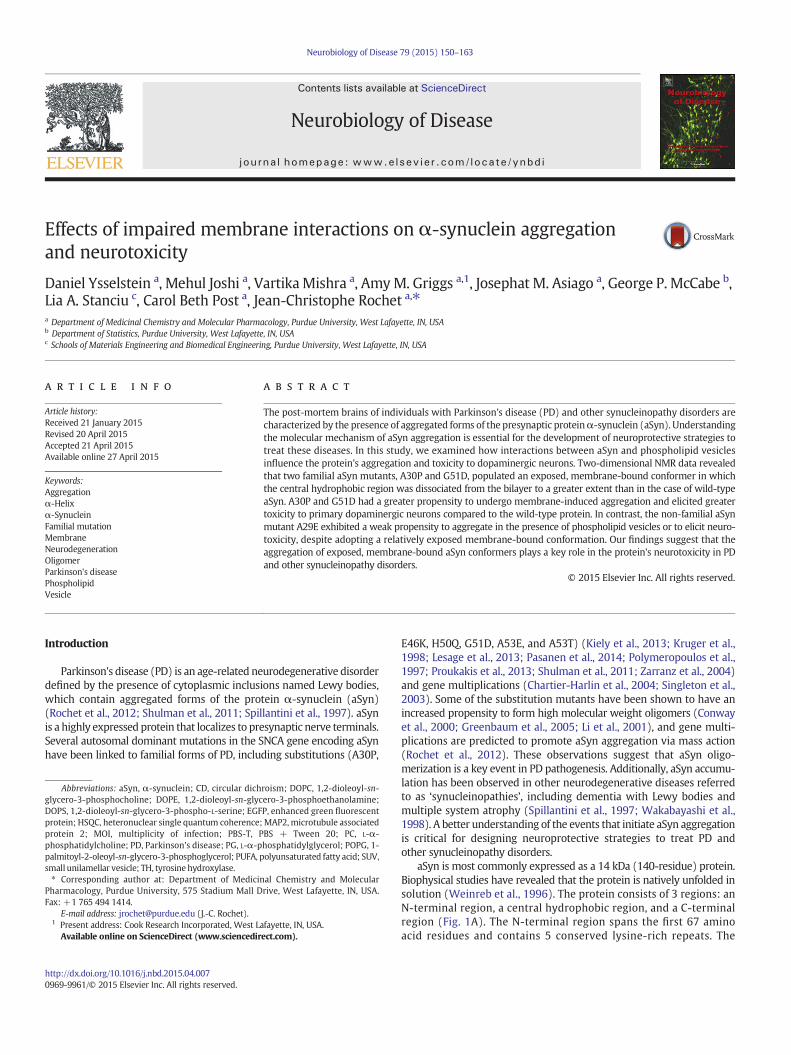
aSyn is most commonly expressed as a 14 kDa (140-residue) protein.Biophysical studies have revealed that the protein is natively unfolded insolution (Weinreb et al., 1996). The protein consists of 3 regions: anN-terminal region, a central hydrophobic region, and a C-terminalregion (Fig. 1A). The N-terminal region spans the first 67 aminoacid residues and contains 5 conserved lysine-rich repeats. The
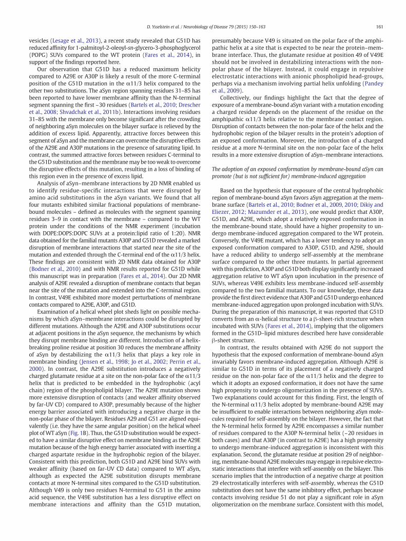
Fig. 1. aSyn adopts an α-helical structure upon binding phospholipid membranes. (A) Schematic representation of the secondary structure of membrane-bound aSyn. The amphipathicα-helix, shown here spanning the N-terminal region (blue) and the central hydrophobic region (red), and the unfolded C-terminal region (green) are depicted above a map of thepolypeptide chain with identical color coding. (B) Helical wheel plot portraying residues 23–66 in the membrane-binding region of aSyn as an amphipathic α11/3 helix. Because A29and G51 are located on the hydrophobic face of the helix embedded in the membrane, replacement of either residue by an acidic residue is predicted to disfavor membrane bindingand helix formation. In contrast, V49 is located on the hydrophilic face of the helix exposed to solvent. Accordingly, the replacement of this residue with an acidic residue should havea less pronounced effect on membrane binding and helix formation, though the proximity of residue 49 to the predicted membrane interface suggests that a negative charge at thisposition could be repelled by anionic lipid head groups. Residues examined in this study are outlined in red. Basic and acidic residues are highlighted with blue and orange shading,respectively.
151D. Ysselstein et al. / Neurobiology of Disease 79 (2015) 150–163
central region (spanning residues 61–95) is highly hydrophobicand contains a 6th lysine-rich repeat. Within the central region,residues 71–82 are required for aggregation of the protein (Giassonet al., 2001). The C-terminal region spanning residues 96–140contains many proline residues and acidic residues and is thoughtto be involved in long-range interactions that stabilize the proteinand prevent aggregation (Bertoncini et al., 2005; Dedmon et al.,2005).
The ability of aSyn to bind phospholipid membranes has beenwell documented and is presumably necessary for the protein's functionin regulating synaptic vesicle trafficking (Davidson et al., 1998; Jensenet al., 1998; Venda et al., 2010). A number of groups have shown thataSyn interacts with anionic phospholipid vesicles or detergent micellesby forming an amphipathic α-helix (most likely an α11/3 helix) withvarious lengths, ranging from a short helix spanning residues ~1–25to a long helix spanning residues ~1–97 and including the centralhydrophobic region (Bartels et al., 2010; Bodner et al., 2009, 2010;Bussell and Eliezer, 2003; Davidson et al., 1998; Jao et al., 2004;Jo et al., 2000) (Fig. 1A). The protein adopts a predominantly brokenor continuous α-helical structure upon binding high- or low-curvaturemembranes, respectively (Chandra et al., 2003; Ferreon et al., 2009;Georgieva et al., 2010; Trexler and Rhoades, 2009; Ulmer et al., 2005).Interactions involving an N-terminal segment spanning residues1–25 are critical for membrane binding and for the adoption of anα-helical structure (Bartels et al., 2010).
aSyn has been shown to undergo accelerated aggregation at themembrane surface when incubated with isolated rat membranehomogenates (Lee et al., 2002), synaptosomal membranes (Jo et al.,2004), exosomes (Grey et al., 2015), detergent micelles (Giehm et al.,2010; Necula et al., 2003), phospholipid vesicles (Galvagnion et al.,2015), and supported phospholipid bilayers (Haque et al., 2010;Pandey et al., 2009) at relatively high protein:lipid ratios. The increased
propensity of aSyn to undergo aggregation in the presence ofmembranesversus in solution could be because the two dimensional surface of thelipid bilayer increases the probability of molecular interactions neededfor oligomerization (Abedini and Raleigh, 2009; Pandey et al., 2009).It has also been observed that aSyn exhibits an enhanced propensity toform high molecular weight aggregates in the presence of polyunsatu-rated fatty acids (PUFAs) (Perrin et al., 2001; Sharon et al., 2003). PUFAsare thought to stimulate aSyn self-assembly because they are susceptibleto oxidation to 4-hydroxy-2-nonenal and 4-oxo-2-nonenal, and the incu-bation of aSyn with these oxidation products results in the formation oflarge, covalently crosslinked β-sheet-rich oligomers (Nasstrom et al.,2011; Qin et al., 2007). Membrane-induced aSyn aggregation mayplay a role in neurodegeneration by triggering membrane thinning, aprocess that could result in increased ion permeability (Comellas et al.,2012; Lee et al., 2012; Ouberai et al., 2013; Pfefferkorn et al., 2012;Reynolds et al., 2011).
Although evidence suggests that phospholipid membranes can sti-mulate aSyn aggregation, conformational properties of membrane-bound aSyn favoring its self-assembly are poorly understood. Wehypothesize that structural perturbations which disrupt interactionsbetween the central hydrophobic region and the phospholipid bi-layer promote the formation of neurotoxic aSyn aggregates at themem-brane surface. To address this hypothesis, we characterized a set of aSynmutants (A29E, A30P, V49E, G51D) in terms of their membrane affinity,membrane-bound conformation, propensity to undergo membrane-induced aggregation, and neurotoxicity as compared to WT aSyn.The rationale for examining these mutants was that their amino acidsubstitutions were predicted to disrupt aSyn-phospholipid inter-actions at various sites on the amphipathic α11/3 helix formed by themembrane-bound protein (Fig. 1B). Our findings suggest a molecularbasis for the enhanced neurotoxicity of the familial mutants A30P andG51D, and they yield insight into the molecular features of aSyn that
152 D. Ysselstein et al. / Neurobiology of Disease 79 (2015) 150–163
modulate membrane binding, membrane-induced aggregation, andneurotoxicity.
Materials and methods
Materials
Unless otherwise stated all chemicals were purchased fromSigma Chemical Co. (St. Louis, MO). 15N-labeled NH4Cl was obtainedfrom Cambridge Isotope Laboratories (Tewksbury, MA). L-α-Phos-phatidylcholine (PC), L-α-phosphatidylglycerol (PG), and a mixtureof 1,2-dioleoyl-sn-glycero-3-phosphoethanolamine (DOPE):1,2-dioleoyl-sn-glycero-3-phospho-L-serine (DOPS):1,2-dioleoyl-sn-glycero-3-phosphocholine (DOPC) (5:3:2 w/w/w) were purchasedfrom Avanti Polar Lipids (Alabaster, AL). Iodixanol, phospholipidextrusion membranes, and ECF substrate were obtained from GEHealthcare Life Sciences (Pittsburgh, PA). 100 kDa spin filters werepurchased fromMillipore (Billerica, MA). DMEM, penicillin–strepto-mycin, the ViraPower Adenoviral Expression System, and LR clonasewere obtained from Invitrogen (Carlsbad, CA). The Adeno-X qPCR ti-tration kit was purchased from Clontech (Mountain View, CA). FBSwas obtained from Sigma Chemical Co. or Atlanta Biologicals (Flow-ery Branch, GA). Nuserum and BCA assay reagents were purchasedfrom Thermo Fisher Scientific (Waltham, MA).
Antibodies
The following antibodieswere used in these studies: mouse anti-aSyn(Syn-1, catalog number 610787, BD Biosciences, San Jose, CA); AP-linkedanti-mouse (Cell Signaling Technology, Danvers,MA); chicken anti-MAP2(catalog number CPCA-MAP2, EnCor Biotechnology, Gainesville, FL);rabbit anti-TH (catalog number AB152, Millipore, Billerica, MA); andanti-rabbit IgG-Alexa Fluor 488, anti-rabbit IgG-Alexa Fluor 594, andanti-chicken IgG-Alexa Fluor 594 (Invitrogen, Carlsbad, CA).
Protein preparation
The pT7-7 constructs encoding A29E and V49E were a gift fromDr. Peter Lansbury and Dr. Michael Volles (Harvard Medical School)(Volles and Lansbury, 2007). A cDNA encoding G51D was generatedvia overlap-extension PCR and subcloned as an NdeI–BamHI fragmentinto the pT7-7 expression vector. Recombinant aSyn was purifiedfrom BL21 (DE3) cells transformed with pT7-7 constructs encodingWT or mutant aSyn as described (Zhang et al., 2013). To generate15N-labeled proteins for NMR analysis, E. coli cells transformed withpT7-7 constructs were grown overnight in LB media and then dilutedinto minimal media containing 15N-NH4Cl for protein expression asdescribed (Marley et al., 2001). Prior to each experiment, protein solu-tions prepared by resuspending purified, lyophilized aSyn variantswere filtered by successive centrifugation steps through a 0.22 μmspin filter and a 100 kDa centrifugal filter to remove aggregates andoligomers.
Lipid vesicle preparation
Egg PG and egg PC suspended in chloroform were mixed at equi-molar ratios in a round bottom flask. The PG:PC lipid mixture or a mix-ture of DOPE:DOPS:DOPC (5:3:2 w/w/w) was dried under a nitrogenstreamand placed in a vacuumdrier for 2 h to ensure complete removalof the chloroform. The dried lipids were suspended in PBS (10 mMphosphate buffer, 2.7 mM KCl, and 137 mM NaCl, pH 7.4), and smallunilamellar vesicles (SUVs) were prepared by extruding the suspensionthrough a 50 nmWhatman membrane 20 times using a mini-extrudersystem. The diameter of the SUVs was found to be between 50 and70 nm by dynamic light scattering on a Zetasizer Nano ZS instrument
(Malvern Instruments, Worcestershire, UK). SUVs were stored at 4 °Cprior to use.
Circular dichroism
Far-UV circular dichroism (CD) measurements were performedusing a Chirascan SC spectrometer (Applied Photophysics, Leatherhead,UK). Solutions of recombinant aSyn (5 μM in 20 mM KPi, pH 7.4) in theabsence or presence of SUVs (lipid:protein ratio, 50:1 to 1600:1mol/mol)were analyzed in a 1mmquartz cuvette at 22 °C. The ellipticity at 222 nmwas recorded, and the data were background corrected (eliminatingsignals associated with the buffer and SUVs) by subtracting ellipticityvalues obtained for equivalent samples minus protein. The mean residuemolar ellipticity at 222 nm ([θ]MR, 222) was calculated using the followingequation:
θ½ �MR; 222 ¼ θ222=10Cnl ð1Þ
where θ222 is the observed (background corrected) ellipticity at 222nm inmillidegrees, C is theprotein concentration inM, n is the number of aminoacid residues in the protein (=140), and l is the path length of the cuvettein cm. Binding curves generated by plotting [θ]MR, 222 versus the lipidconcentration were analyzed by fitting to the following equation:
R ¼ R0− R0−Rf� �Kd þ C þ L=N−
ffiffiffiffiffiffiffiffiffiffiffiffiffiffiffiffiffiffiffiffiffiffiffiffiffiffiffiffiffiffiffiffiffiffiffiffiffiffiffiffiffiffiffiffiffiffiffiffiffiffiffiffiffiKd þ C þ L=Nð Þ2−4CL=N
q
2Cð2Þ
where R is the measured [θ]MR, 222 at a given lipid concentration, R0 is the[θ]MR, 222 in the absence of lipid, Rf is the [θ]MR, 222 in the presence ofsaturating lipid, L is the total lipid concentration, C is the total proteinconcentration, Kd is the apparent macroscopic dissociation equilibriumconstant, and N is the binding stoichiometry (lipids/protein)(Shvadchak et al., 2011a). The data obtained for WT aSyn were fit toEq. (2) via non-linear regression with R0, Rf, Kd, and N as unconstrainedparameters. The data obtained for the aSyn mutants were fit to Eq. (2)with N set to a value determined from the fit of the WT aSyn data(N = 280 or 300 for WT aSyn incubated with PG:PC vesicles orPE:PS:PC vesicles, respectively). This approach of constraining N, neces-sary to obtain a satisfactory fit of the binding data for the lower-affinityaSyn mutants, is reasonable based on the assumption that the stoichio-metry of binding to SUVs with a particular phospholipid compositionshould be approximately equal for WT aSyn and variants with the samenumber of residues (Shvadchak et al., 2011a).
To determine themaximumhelical content of each aSyn variant, weused the following equations:
H ¼ 100% � θ−θcoilð Þθα−θcoilð Þ ð3Þ
θα ¼ −40000 1−2:5=nð Þ þ 100t ð4Þ
θcoil ¼ 640−45t ð5Þ
where H is the maximum % helicity, θ is the [θ]MR, 222 in the presence ofsaturating lipid (Rf in Eq. 2), θα is the [θ]MR, 222 value of an idealizedα-helical peptide, θcoil is the [θ]MR, 222 value of an idealized randomcoil peptide, n is the number of amino acid residues in the protein(=140), and t is the temperature (22 °C) (Scholtz et al., 1991;Shvadchak et al., 2011b).
2D NMR analysis
NMR measurements were carried out using a Bruker Advance800 MHz spectrometer equipped with a room temperature probe(Billerica, MA). Solutions of 15N-labeled aSyn (150 μM in 20 mM KPi,pH 7.4, 90% H2O/10% D2O (v/v)) in the absence or presence of SUVs
153D. Ysselstein et al. / Neurobiology of Disease 79 (2015) 150–163
(lipid:protein ratio, 20:1 mol/mol) were analyzed at 10 °C. 2D NMRanalyses were carried out by recording 1H–15N heteronuclear singlequantum coherence (HSQC) spectra with a data matrix of 2048(F2, 1H) × 256 (F1, 15N) increments in the direct and indirect dimen-sions, respectively. The spectra were processed with NMRPipe (Delaglioet al., 1995). Assignments listed in the BioMagResBank database (acces-sion number, 18857) matched assignments shown in a previous report(Eliezer et al., 2001) and were used in this study. Only small chemicalshift changes were observed for ≤3 residues immediately bracketingthe substitution site, consistent with previous findings (Bodner et al.,2010). Peak intensities (heights) were determined using Sparky (T.D.Goddard and D.G. Kneller, University of California San Francisco). Tomonitor residue-specific interactions of aSyn with the membrane, thefractional signal attenuation of each residue peak in the presence versusthe absence of SUVs was determined as described (Bodner et al., 2009;Bodner et al., 2010).
Lipid flotation assay
Solutions of recombinant aSyn in PBS with 0.02% (w/v) NaN3 wereincubated with PG:PC SUVs (lipid:protein ratio, 20:1 mol/mol) at37 °C for 72 or 96 h in a total volume of 60 μL. The final concentrationof aSyn in the protein–lipid mixture was 100 μM (determined with aBCA Protein Assay Kit). After incubation, each sample was mixed with4 mL of 30% (v/v) iodixanol solution and overlaid with 7.0 mL of 25%(v/v) iodixanol and 350 μL of 5% (v/v) iodixanol in a polyalomar tube(Beckman, Miami, FL). All of the above iodixanol solutions wereprepared in lipid flotation buffer (10 mM HEPES, pH 7.4, 150 mMNaCl). The samples were spun at 200,000 ×g in a Beckman SW 41 Tirotor for 4 h at 4 °C. The membrane fraction was carefully collectedfrom the 5% iodixanol layer at the top of the gradient and diluted in11mL of lipidflotation buffer.Membrane-bound proteinswere pelletedvia centrifugation at 200,000 ×g for 2 h at 4 °C, resuspended in 250 μL ofPBS, and stored at−20 °C prior to Western blot analysis. In general wefound that the rate of aSyn aggregation increased in the presence ofSUVs that had been stored for ~3–4 weeks at 4 °C, apparently becauseoxidized phospholipids that accumulated during vesicle storagepromoted aSyn–membrane binding and self-assembly of the proteinat the membrane surface (Griggs, A.M. and Rochet, J.-C., unpublisheddata).
Western blot analysis
Membrane-bound aSyn isolated by lipid flotation was analyzed byWestern blotting. Protein samples were combined with Laemmli buffer(final concentration, 60 mM Tris–HCl, pH 6.8, 2% (w/v) SDS, 10% (v/v)glycerol, 5% (v/v) β-mercaptoethanol, 0.01% (w/v) bromophenol blue)and boiled for 5 min. Denatured proteins were separated via SDS-PAGE on a 4–20% (w/v) polyacrylamide gradient gel (BioRad, Hercules,CA) and transferred to a PVDF membrane (pore size, 0.4 μm). Afterblocking in 10% (w/v) non-fat milk, the membrane was washed withPBS + 0.5% (w/v) Tween 20 (PBS-T) and probed for aSyn with theSyn-1 primary antibody, diluted 1/1500 in PBS-T with 1% (w/v) BSA.After washing in PBS-T, the membrane was probed with a secondaryanti-mouse antibody conjugated to alkaline phosphatase (diluted1/5000). To visualize the bands, the membrane was exposed to ECFsubstrate for 30 s, and images were acquired using a Typhoon imag-ing system (GE Life Sciences, Pittsburgh, PA). Band densities werequantified using ImageJ software (NIH, Bethesda, MD).
Preparation of adenoviral constructs
Adenoviruses expressing aSyn variants were prepared using theViraPower Adenoviral Expression System (Liu et al., 2008). cDNAsencoding WT or mutant aSyn were subcloned as KpnI–XhoI fragmentsinto the entry vector pENTR1A, and the inserts from the pENTR1A
constructs were then transferred into the pAd/CMV/V5 adenoviralexpression vector via recombination using Gateway LR Clonase.
A cDNA encoding enhanced green fluorescent protein (EGFP) wassubcloned as a KpnI–XhoI fragment into pENTR1A, yielding theconstruct pENTR–EGFP. A DNA fragment encoding the herpes simplexvirus thymidine kinase polyadenylation signal (TKpolyA)was amplifiedfrom pAd/CMV/V5 by PCR and subcloned as an XhoI–EcoRV fragmentinto pENTR–EGFP, yielding the construct pENTR–EGFP–TKpolyA.A DNA fragment encoding the human synapsin promoter sequencewas excised from the vector pAAV2 by digestion with SmaI and KpnIand subcloned into the vector pENTR–EGFP–TKpolyA cut with XmnIand KpnI, yielding the construct pENTR–synapsin–EGFP–TKpolyA. Thesynapsin–EGFP–TKpolyA insert from this construct was transferredinto the pAd/PL-DEST adenoviral vector via recombination usingGateway LR Clonase. pAd/PL-DEST is a ‘promoter-less’ version ofpAd/CMV/V5 (both have the same sequences required for packagingand production of human adenovirus type 5).
The sequence of the DNA insert in each adenoviral construct wasverified using an Applied Biosystems (ABI3700) DNA sequencer(Purdue University). Adenoviral constructs were packaged into virusvia lipid-mediated transient transfection of the HEK 293A packagingcell line. Adenovirus packaged with the plasmid pAd/V5-DEST/LacZ(Invitrogen), encoding the control protein β-galactosidase fused to theV5 epitope, was also prepared to control for non-specific effects ofviral transduction. Adenoviral titers were determined using theAdeno-X qPCR titration kit.
Analysis of relative aSyn expression levels
Relative expression levels of the aSyn variants encoded by differentadenoviral constructs were determined via Western blot analysis.Human SH-SY5Y neuroblastoma cells were plated on 6-well plates ata density of 300,000 cells per well in media consisting of RPMI, 10%(v/v) Nuserum, penicillin (100 U/mL), and streptomycin (100 μg/mL).Two days after plating, the cells were transduced with aSyn adenovirusat a multiplicity of infection (MOI) of 37.5–75. After 72 h, the cells weredislodged from the plate by trypsinization, collected by centrifugation,washed with PBS, and lysed in RIPA buffer (50 mM Tris HCl, pH 7.4,150 mM NaCl, 0.1% (w/v) SDS, 0.5% (w/v) sodium deoxycholate, 1%(v/v) Triton X-100) with protease inhibitor cocktail (Sigma P8340)and 1 mM PMSF. After centrifugation at 13,000 ×g, the detergent-soluble (supernatant) fraction was recovered and assayed for proteinconcentration using a BCA Protein Assay Kit. Equal amounts of proteinwere separated via SDS-PAGE on a 12% (w/v) polyacrylamide gel andanalyzed via Western blotting as described above for the analysis ofmembrane-bound aSyn isolated by lipid flotation.
Preparation and treatment of primary mesencephalic cultures
Primary midbrain cultures were prepared via dissection of day 17embryos obtained from pregnant Sprague–Dawley rats (Harlan,Indianapolis, IN) using the methods approved by the Purdue AnimalCare and Use Committee, as described (Cooper et al., 2006; Liu et al.,2008; Strathearn et al., 2014). The mesencephalic region containingthe substantia nigra and ventral tegmental areawas isolated stereoscop-ically, and the tissue was treated with trypsin (final concentration,26 μg/mL in 0.9% [w/v] NaCl). Dissociated cells were plated on 48-wellplates (pretreated with poly-L-lysine, 5 μg/mL) at a density of 163,500cells per well in media consisting of DMEM, 10% (v/v) FBS, 10% (v/v)horse serum, penicillin (100 U/mL), and streptomycin (100 μg/mL).Five days after plating, the cells were treated for 48 h with cytosinearabinofuranoside (20 μM, 48 h) to inhibit the growth of glial cells. Atthis stage (7 days in vitro), the cultures were transduced with adeno-virus encoding aSyn, β-galactosidase, or synapsin/EGFP for 72 h at anMOI of 7.5 to 15. The MOIs of adenoviruses encoding aSyn variantswere adjusted to ensure equal expression levels based on data from
154 D. Ysselstein et al. / Neurobiology of Disease 79 (2015) 150–163
Western blot analysis of lysates from transduced SH-SY5Y cells asoutlined above (for each virus, theMOI used to transduce primary mid-brain cultures was 5-fold less than that used for the transduction of SH-SY5Y cells).
After incubation in freshmedia for an additional 24 h, the cells werefixed in 4% (w/v) paraformaldehyde in PBS and subsequently per-meabilized for 1 h with PBS, 0.3% (v/v) Triton X-100, 1% (w/v) BSA,and 10% (v/v) FBS. For experiments aimed at monitoring relative dopa-minergic cell viability or neurite lengths, the cells were treated for 48 hat 4 °C with chicken anti-MAP2 (1:2000) and rabbit anti-TH (1:500)primary antibodies. The cells were then washed with PBS and treatedwith Alexa Fluor 594 goat anti-chicken and Alexa Fluor 488 goat anti-rabbit secondary antibodies (both at 1:1000) for 1 h at 22 °C. For expe-riments aimed at determining adenoviral transduction efficiencies, cellstransduced with virus encoding EGFP downstream of the synapsinpromoter were treated for 48 h at 4 °C with chicken anti-MAP2(1:2000) or rabbit anti-TH (1:500) primary antibody. The cells werethen washed with PBS and treated with Alexa Fluor 594 goat anti-chicken or Alexa Fluor 594 goat anti-rabbit secondary antibody (bothat 1:1000) for 1 h at 22 °C. After a final wash with PBS, prolong goldantifade reagent with DAPI was applied to each culture well beforesealing with a coverslip.
Measurements of primary dopaminergic cell viability
Relative dopaminergic cell viability was assessed by countingMAP2- and TH-immunoreactive primary neurons in a blinded man-ner using a Nikon TE2000-U inverted fluorescence microscope(Nikon Instruments, Melville, NY) equipped with a 20× objective(Cooper et al., 2006; Liu et al., 2008; Strathearn et al., 2014). A min-imum of 10 random fields of view were chosen to provide represen-tation of the whole well. Approximately 300–500 MAP2+ neuronswere counted per experiment for each treatment. Each experimentwas repeated using cultures isolated from at least 3 different preg-nant rats. The data are expressed as the percentage of MAP2+ neu-rons that were also TH+ (this ratiometric approach was used tocorrect for variations in cell plating density).
Neurite length measurements
Neurite length measurements were carried out on the identicalprimary midbrain cultures used to determine dopamine neuron via-bility. Images were taken with the automated Cytation 3 Cell ImagingReader (BioTek, Winooski, VT) using a 4× objective. Lengths ofMAP2+ processes extending from TH+/MAP2+ neurons with an intactcell body (∼90 neurons per sample) were assessed in a blindedmannerusing Nikon NIS Elements analysis software (Nikon Instruments,Melville, NY) (Strathearn et al., 2014).
Statistical analysis
Densitometry data from Western blots and primary neuron via-bility data were analyzed via ANOVA followed by Tukey's multiplecomparisons post hoc test using GraphPad Prism 6.0 (La Jolla, CA).In analyzing percentage cell viability data by ANOVA, square roottransformations were carried out to conform to ANOVA assumptions.Neurite length data were analyzed using an approach that accountsfor (i) the possibility of multiple neurites arising from a single celland (ii) the comparison across experiments conducted on differentdays. Neurite lengths for multiple treatment groups were comparedusing a general linear model implemented in the GLM procedure ofSAS Version 9.3 followed by Tukey's multiple comparisons post hoctest (Cary, NC).
Results
Membrane affinity and membrane-bound conformation of A30P and G51D
The goal of this study was to understand how site-specific perturba-tions of the amphipathic helical structure ofmembrane-bound aSyn im-pact aSyn aggregation at themembrane surface and aSyn neurotoxicity.As a first step, we characterized the familial mutants A30P and G51D interms of their membrane affinity and membrane-bound conformation,with the aim of then assessing how these properties relate to the vari-ants' relative propensities to undergo membrane-induced aggregationand elicit dopaminergic cell death (see next sections). We focused ini-tially on A30P and G51D because of their importance as familial PDmu-tants. A30P has been shown by several groups to have a reduced affinityfor phospholipids (Jensen et al., 1998; Jo et al., 2002; Perrin et al., 2000),and the G51D substitution was predicted to interfere with aSyn–mem-brane interactions by placing a negative charge on the non-polar face ofthe amphipathic α11/3 helix (Fig. 1B).
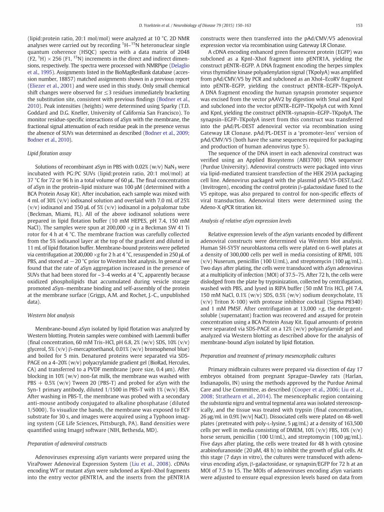
Recombinant, monomeric WT aSyn, A30P, and G51D were incu-bated with 50 nm SUVs at lipid:protein ratios of 50:1 to 1600:1(mol/mol). The SUVs had the following lipid compositions: (i) eggPG:egg PC (1:1 mol/mol) and (ii) DOPE:DOPS:DOPC (5:3:2 w/w/w). These lipid compositions were chosen as each contains anioniclipids necessary for aSyn membrane interactions (Davidson et al.,1998; Jo et al., 2000). In addition, PG:PC vesicles were used in ourprevious studies (Haque et al., 2010; Pandey et al., 2009; Zakharovet al., 2007), and SUVs consisting of DOPE:DOPS:DOPC have a com-position similar to that of synaptic vesicles (Takamori et al., 2006).Changes in aSyn conformation associated with membrane bindingwere monitored by far-UV CD, a technique that reports on the in-crease in aSyn α-helicity upon interaction of the protein with phos-pholipids (Davidson et al., 1998; Jo et al., 2000). Consistent with ourprevious data (Zakharov et al., 2007), the far-UV CD spectrum of WTaSynwas characterized by (i) aminimum at 198 nm in the absence ofSUVs and (ii) a maximum at 192 nm and minima at 208 nm and222 nm in the presence of SUVs (lipid:protein ratio, 1600:1,mol/mol) (Supplementary Fig. S1).
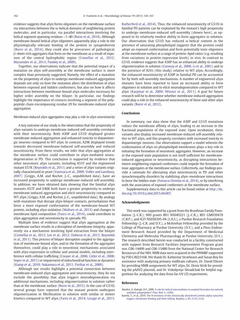
To compare different aSyn variants in terms of their propensities toadopt an α-helical structure upon binding to SUVs, we used the pre-viously described approach ofmonitoring themean residuemolar ellip-ticity at 222 nm ([θ]MR, 222) as a function of lipid:protein ratio (Scholtzet al., 1991). The data obtained for WT aSyn in the presence of PG:PCvesicles revealed a progressive decrease in [θ]MR, 222 with increasinglipid:protein ratios (Fig. 2A). The data were fit to Eq. (2) (see‘Materials and methods’), yielding a Kd value of 1.1 ± 0.1 μM, a bindingstoichiometry (N value) of 280 lipids per protein, and a maximumα-helical content of 49 ± 1% (Table 1). The lipid titration curves ob-tained for A30P andG51D in the presence of PG:PC vesicles were shiftedto the right compared to the WT aSyn curve, and the Kd values weremarkedly greater (Kd = 3.2 ± 0.4 μM for A30P and 10 ± 2 μM forG51D) (Fig. 2A, Table 1). Although A30P andWT aSyn exhibited similarα-helical contents in the presence of saturating PG:PC vesicles, themaximum helicity observed for G51D was considerably lower (~43 ±4%) (Table 1). Analysis of the far-UV CD data obtained for WT aSyn inthe presence of DOPE:DOPS:DOPC vesicles yielded a Kd value of0.21 ± 0.07 μM, a binding stoichiometry of 300 lipids per protein, anda maximum α-helical content of 50 ± 2% (Fig. 2B, Table 1). Hereagain, A30P and G51D bound to the lipids with higher Kd values(Kd =2.1 ± 0.5 μM for A30P and 3.1 ± 0.2 μM for G51D), and themax-imum helicity observed for G51D (40 ± 2%) was lower than that deter-mined for WT aSyn or A30P. Collectively, these data suggest that(i) G51D and A30P have a reduced affinity for PG:PC andDOPE:DOPS:DOPC vesicles compared to the WT protein; and (ii) theG51D substitution disrupts the α-helical structure of membrane-bound aSyn in the presence of saturating lipid to a greater extent thanthe A30P substitution.
Fig. 2. A30P and G51D display reduced membrane affinity relative to WT aSyn. Solutions of recombinant aSyn variants were analyzed by far-UV CD to determine [θ]MR, 222, a measure ofproteinα-helicity. Each aSyn variant was incubated with increasing concentrations of SUVs composed of egg PG:PC (1:1 mol:mol) (A) or DOPE:DOPS:DOPC (5:3:2 w/w/w) (B). The datawere fit to Eq. (2), and values for Kd, minimum [θ]MR, 222, andmaximumα-helical contentwere determined from the values of the fit parameters (see Table 1). The data are representativeof≥3 experiments (in total, including both lipid compositions).
155D. Ysselstein et al. / Neurobiology of Disease 79 (2015) 150–163
To compare themembrane-bound conformations ofWT aSyn, A30P,and G51D, 15N-labeled aSyn variants were analyzed via 2D NMRspectroscopy in the absence or presence of DOPE:DOPS:DOPC vesicles,and 1H–15NHSQC spectrawere recorded (Figs. 3A and B). This approachis ideal formonitoring residue-specific bindingbecause the signal atten-uation of the resonance from a given residue in the presence versus theabsence of SUVs is a measure of vesicle binding of that residue. This res-idue-level monitoring of membrane binding is possible because thepartitioning of the residue between a disordered, highlymobile solutionstate and a membrane-bound state is in the slow-exchange limit on theNMR frequency scale, and the slow rotational motion in themembrane-bound state results in a resonance linewidth too broad for detection. Foreach residue of aSyn, I/I0 is the proportion of molecules in an aSyn/SUVmixture in which the residue remains mobile and in solution, and thefractional attenuation (1 − I/I0) is the proportion of molecules in
Table 1Membrane affinities and maximum helix contents of aSyn variants titrated with SUVs.a
PG:PC
Variant Kdb
(μM)Ellipticity minimum(×103 deg·cm2/dmol)
Maximum helicity(%)
WT 1.1 ± 0.1 −18 ± 0.2 49 ± 1G51D 10 ± 2c −16 ± 1 43 ± 4A30P 3.2 ± 0.4 −18.6 ± 0.4 50 ± 2A29E 7.5 ± 0.7 −19.7 ± 0.6 53 ± 2V49E 2.5 ± 0.4 −17.7 ± 0.6 47 ± 2
PE:PS:PC
Variant Kdd
(μM)Ellipticity minimum(×103 deg·cm2/dmol)
Maximum helicity(%)
WT 0.21 ± 0.07e −18.6 ± 0.4 50 ± 2G51D 3.1 ± 0.2c −15.0 ± 0.2 40 ± 2A30P 2.1 ± 0.5 −18.7 ± 0.8 50 ± 3A29E 5 ± 1 −21 ± 1 55 ± 4V49E 1.8 ± 0.4 −18.4 ± 0.8 49 ± 3
a Values (± standard error) were determined from the far-UV CD data in Figs. 2 and 6using Eqs. (2)–(5); the concentration of aSyn was 5 μM.
b Fitting of theWT aSyn data to Eq. (2) yielded a binding stoichiometry (N value) of 280.The data obtained for the other variants were fit to Eq. (2) with N set to a value of 280.
c The reduced maximum helicity determined for G51D implies that this variant's N valuecould be lower than that ofWTaSyn. Because afit of the bindingdata to Eq. (2)withN set to alower value yields an even higher Kd value, the membrane affinity of G51D may in fact beover-estimated.
d Fitting of theWTaSyndata to Eq. (2) yielded a binding stoichiometry (N value) of 300.The data obtained for the other variants were fit to Eq. (2) with N set to a value of 300.
e This Kd value is considered approximate because it is 25-fold less than the concentrationof protein in the CD experiment.
which the residue is tightly associated with the membrane (Bodneret al., 2009, 2010). Thus, this approach is useful to analyze whichresidues on the protein are in greatest contact with the lipid vesicle. Weused this method to characterize the aSyn–membrane interaction, andin particular to determine whether the central hydrophobic(aggregation-prone) region was either dissociated or associated in thecase of conformers referred to here as ‘exposed’ or ‘hidden’, respectively.The fractional population of molecules bound to the membrane in eithermode (‘total bound population’) was determined from the mean attenu-ation of residues 3–9. Our rationale for this approach was that the N-terminal segment of aSyn has a higher membrane affinity compared tomore C-terminal regions (Bartels et al., 2010; Drescher et al., 2008;Shvadchak et al., 2011b) and is lipid-bound in the case of both exposedand hidden conformers (Bodner et al., 2010). To calculate the fractionalpopulation of protein in the hidden state we determined themean atten-uation of residues 66–80, based on the fact that this segment of the centralhydrophobic region contains residues that play a key role in aSyn self-assembly (Giasson et al., 2001), and consistent with a previous report(Bodner et al., 2010). The fractional population of protein with residues66–80 in the exposed state was determined by subtracting the hiddenpopulation from the total bound population. We found that ~84% of WTaSyn molecules were bound to the membrane (Figs. 3C and D; Table 2).Of the totalWT protein, ~36%was in the exposedmembrane-bound con-formation, whereas ~48% was in the hidden state. NMR analysis revealedthat ~85% of A30P or G51D molecules were bound to the membrane inthe N-terminal region, similar toWT aSyn (Figs. 3C and D; Table 2). How-ever, the percentage of total protein molecules in the exposed state wasincreased by a factor of 1.9 and 2.0 in the case of A30P and G51D, respec-tively. For each variant, examination of the sequence dependence of theNMR signal attenuation associated with membrane binding (from N- toC-terminus) revealed that the decrease in attenuation exhibited byA30P or G51D relative to WT aSyn begins in the region of the aminoacid substitution, suggesting that the mutation causes a lifting off of theprotein from the membrane (Figs. 3E and F).
Membrane-induced self-assembly of A30P and G51D
The CD and NMR data outlined above revealed that introduction ofthe A30P or G51D mutation led to a significant reduction in lipidinteractions involving the central hydrophobic region. As this regionplays a key role in aSyn aggregation in solution (Giasson et al., 2001),we postulated that exposure of this region by neighboring aSynmolecules at a high local concentration on the bilayer surface couldlead to self-assembly of the protein into membrane-bound aggregates.To address this hypothesis, we incubated each aSyn variant with
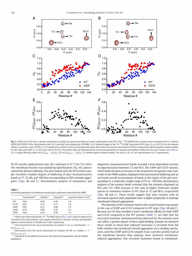
Fig. 3. A30P and G51D have a greater propensity to adopt an exposed membrane-bound conformation than WT aSyn. 15N-labeled aSyn variants were incubated with or withoutDOPE:DOPS:DOPC SUVs (lipid:protein ratio, 20:1 mol/mol) and analyzed by 2D NMR. (A, B) Selected region of the 1H–15N HSQC spectrum of WT aSyn (A) or G51D (B) in the absence(black) or presence (red) of SUVs. (C, D) Graphs of I/I0 (where I and I0 are the peak intensities observed in the presence and absence of SUVs, respectively) plotted against residue numberfor WT aSyn and A30P (C) or WT aSyn and G51D (D). From these data we determined the fractional populations of exposed and hidden conformers for each variant (see Table 2).(E, F) Graphs showing values of {I/Io}A30P − {I/Io}WT (E) or {I/Io}G51D − {I/Io}WT (F) plotted against residue number. The data in (A)–(F) are representative of ≥2 experiments.
156 D. Ysselstein et al. / Neurobiology of Disease 79 (2015) 150–163
PG:PC vesicles (lipid:protein ratio, 20:1mol/mol) at 37 °C for 72 or 96 h.The membrane fraction was isolated by lipid flotation (Fig. 4A) and ex-amined byWestern blotting. The lane loaded with theWT protein sam-ple revealed a modest degree of laddering of aSyn immunoreactivebands at 37, 55, 80, and 100 kDa corresponding to SDS-resistant aggre-gates (Figs. 4B and C). Densitometry analysis of monomeric and
Table 2Fractional populations of membrane-bound aSyn conformers determined by NMR.
Total bounda,b Hiddena,c Exposeda,d Exposed relative to WT
WT 0.84 0.48 0.36 1.0G51D 0.85 0.12 0.73 2.0A30P 0.85 0.17 0.68 1.9A29E 0.78 0.00 0.78 2.2V49E 0.84 0.34 0.49 1.4
a Valueswere determined from 1H–15N-HSQC data in Figs. 3 and 7 and are expressed asa fraction of the total protein; each sample consisted of a mixture of aSyn and phospho-lipids at concentrations of 150 μM and 3 mM, respectively.
b Determined from the mean attenuation of residues 3–9 (i.e. total bound = 1 −{I/Io}mean,3–9).
c Determined from the mean attenuation of residues 66–80 (i.e. hidden = 1 −{I/Io}mean,66–80).
d Calculated as the difference between the total bound population and the hiddenpopulation.
oligomeric immunoreactive bands revealed a time-dependent increasein oligomerization between 72 and 96 h. The A30P and G51D variants,which both showed an increase in the proportion of exposed-statemol-ecules in theNMR analysis, displayedmore pronounced laddering and anincreased overall accumulation of bands in the region of the gel corre-sponding to a molecular weight range of 55 to ~250 kDa. Densitometryanalysis of the protein bands revealed that the mutants displayed an85% and 115–140% increase in the ratio of higher molecular weightspecies to monomer relative to WT aSyn at 72 and 96 h, respectively(Figs. 4B and C). These results suggest that aSyn variants with anincreased exposed-state population have a higher propensity to undergomembrane-induced aggregation.
The intensity of themonomer band in the vesicle fractionwas greaterin the case of A30P and G51D compared to WT aSyn (Figs. 4B and C).Because our CD data revealed a lower membrane affinity for both A30Pand G51D compared to the WT protein (Table 1), we infer that theincreased monomer immunoreactivity observed for the mutants mustnot reflect a greater degree of direct lipid binding. Instead, we interpretthese results to mean that unbound monomer is recruited from thebulk solution into membrane-bound aggregates via a seeding mecha-nism, and thus A30P and G51D samples have a greater protein load atthe membrane because they undergo more extensive membrane-induced aggregation. Any recruited monomer bound to membrane-
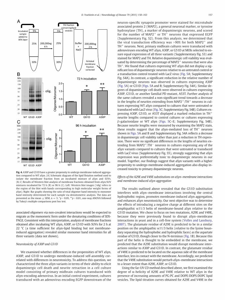
Fig. 4.A30P and G51D have a greater propensity to undergomembrane-induced aggrega-tion compared to WT aSyn. (A) Schematic diagram of the lipid flotation method used toisolate the membrane fraction from an incubated mixture of aSyn and SUVs.(B, C) Results of Western blot analysis of membrane fractions obtained from aSyn/SUVmixtures incubated for 72 h (B) or 96 h (C). Left: Western blot images (‘olig’ refers tothe region of the blot with bands corresponding to high molecular weight forms ofaSyn). Right: Bar graphs showing the ratio of total oligomer band intensity to monomerband intensity determined for each sample via densitometric analysis. The data arepresented as the mean ± SEM, n = 3; *p b 0.05, **p b 0.01, one-way ANOVA followedby Tukey's multiple comparisons post hoc test.
157D. Ysselstein et al. / Neurobiology of Disease 79 (2015) 150–163
associated oligomers via non-covalent interactions would be expected tomigrate as the monomeric form under the denaturing conditions of SDS-PAGE. Consistent with this interpretation, analysis of membrane fractionsobtained after incubating WT aSyn, A30P, or G51D with SUVs for 2 h at22 °C (a time sufficient for aSyn-lipid binding but not membrane-induced aggregation) revealed similar monomer band intensities for allthree variants (data not shown).
Neurotoxicity of A30P and G51D
We examined whether differences in the propensities of WT aSyn,A30P, and G51D to undergo membrane-induced self-assembly cor-related with differences in neurotoxicity. To address this question, wecharacterized the three aSyn variants in terms of their ability to elicitdopaminergic cell death and neurite retraction in a cell culturemodel consisting of primary midbrain cultures transduced withaSyn-encoding adenovirus. In an initial control experiment, culturestransduced with an adenovirus encoding EGFP downstream of the
neuron-specific synapsin promoter were stained for microtubuleassociated protein 2 (MAP2), a general neuronal marker, or tyrosinehydroxylase (TH), a marker of dopaminergic neurons, and scoredfor the number of MAP2+ or TH+ neurons that expressed EGFP(Supplementary Fig. S2). From this analysis, we determined thatthe viral transduction efficiency was N90% for both MAP2+ andTH+ neurons. Next, primary midbrain cultures were transduced withadenoviruses encodingWT aSyn, A30P, or G51D atMOIs selected to en-sure equal expression of all three variants (Supplementary Fig. S3) andstained for MAP2 and TH. Relative dopaminergic cell viability was eval-uated by determining the percentage of MAP2+ neurons that were alsoTH+. We found that cultures expressing WT aSyn did not display a sig-nificant loss of dopaminergic neurons relative to an untreated control ora transduction control treated with LacZ virus (Fig. 5A; SupplementaryFig. S4A). In contrast, a significant reduction in the relative number ofdopaminergic neurons was observed in cultures expressing A30P(Fig. 5A) or G51D (Figs. 5A and B; Supplementary Fig. S4A). Similar de-grees of dopaminergic cell death were observed in cultures expressingA30P, G51D, or another familial PD mutant, A53T. Further analysis ofthe same cultures revealed a non-significant trend towards a decreasein the lengths of neurites extending from MAP2+/TH+ neurons in cul-tures expressing WT aSyn compared to cultures that were untreated ortransducedwith LacZ virus (Fig. 5C; Supplementary Fig. S4B). Cultures ex-pressing A30P, G51D, or A53T displayed a marked reduction in TH+
neurite lengths compared to control cultures or cultures expressingβ-galactosidase or WT aSyn (Figs. 5C–E; Supplementary Fig. S4B).Because neurite lengths were measured by examining the MAP2 stain,these results suggest that the aSyn-mediated loss of TH+ neuronsshown in Figs. 5A and B and Supplementary Fig. S4A reflects a decreasein dopaminergic cell viability rather than just a reduction in TH expres-sion. There were no significant differences in the lengths of neurites ex-tending from MAP2+/TH− neurons in cultures expressing any of theaSyn variants compared to cultures that were untreated or transducedwith LacZ virus (Supplementary Fig. S5), strongly suggesting that aSynexpression was preferentially toxic to dopaminergic neurons in ourmodel. Together, our findings suggest that aSyn variants with a higherpropensity to undergo membrane-induced aggregation also display in-creased toxicity to primary dopaminergic neurons.
Effects of the A29E and V49E substitutions on aSyn–membrane interactionsand membrane-induced aSyn aggregation
The results outlined above revealed that the G51D substitutioninterferes with aSyn–membrane interactions involving the centralhydrophobic region, promotes membrane-induced aSyn aggregation,and enhances aSyn neurotoxicity. Our next objective was to determinethe effects of introducing a negative charge at different sites on theamphipathic α11/3 helix of membrane-bound aSyn relative to theG51D mutation. We chose to focus on two mutations, A29E and V49E,because they were previously found to disrupt aSyn–membraneinteractions in yeast and in a cell-free system (Volles and Lansbury,2007). The glutamate residue of A29E is predicted to be in a similarposition on the amphipathic α11/3 helix (relative to the lysine boun-dary separating the hydrophobic and hydrophilic faces) as the aspartateresidue of G51D, though closer to the N-terminus (Fig. 1B). Because thispart of the helix is thought to be embedded in the membrane, wepredicted that the A29E substitution would disrupt membrane inter-actions similar to A30P and G51D. In contrast, the glutamate residueof V49E is predicted to be located on the aqueous side of themembraneinterface, less in contact with themembrane. Accordingly, we predictedthat the V49E substitution would perturb aSyn–membrane interactionsto a lesser extent than A29E, A30P, or G51D.
Using the far-UV CDmethod described above, we first examined thedegree of α-helicity of A29E and V49E relative to WT aSyn in thepresence of increasing amounts of PG:PC and DOPE:DOPS:DOPC lipidvesicles. The lipid titration curves obtained for A29E and V49E in the
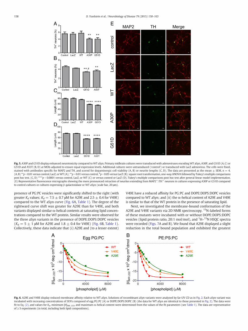
Fig. 5.A30P andG51D display enhanced neurotoxicity compared toWT aSyn. Primarymidbrain cultureswere transducedwith adenoviruses encodingWT aSyn, A30P, and G51D (A, C) orG51D and A53T (B, D) at MOIs adjusted to ensure equal expression levels. Additional cultures were untransduced (‘control’) or transduced with LacZ adenovirus. The cells were fixed,stained with antibodies specific for MAP2 and TH, and scored for dopaminergic cell viability (A, B) or neurite lengths (C, D). The data are presented as the mean ± SEM, n = 4.(A, B) **p b 0.01 versus control, LacZ, orWT (A); **p b 0.01 versus control, #p b 0.05 versus LacZ (B); square root transformation, one-wayANOVA followedby Tukey'smultiple comparisonspost hoc test. (C, D) ****p b 0.0001 versus control, LacZ, or WT (C) or versus control or LacZ (D), Tukey's multiple comparisons post hoc test after general linear model implementation.(E) Representative fluorescence micrographs showing the more pronounced retraction of neurites extending fromMAP2+/TH+ neurons in cultures expressing A30P or G51D comparedto control cultures or cultures expressing β-galactosidase or WT aSyn (scale bar, 20 μm).
158 D. Ysselstein et al. / Neurobiology of Disease 79 (2015) 150–163
presence of PG:PC vesicles were significantly shifted to the right (withgreater Kd values; Kd = 7.5 ± 0.7 μM for A29E and 2.5 ± 0.4 for V49E)compared to the WT aSyn curve (Fig. 6A, Table 1). The degree of therightward curve shift was greater for A29E than for V49E, and bothvariants displayed similarα-helical contents at saturating lipid concen-trations compared to the WT protein. Similar results were observed forthe three aSyn variants in the presence of DOPE:DOPS:DOPC vesicles(Kd = 5 ± 1 μM for A29E and 1.8 ± 0.4 for V49E) (Fig. 6B, Table 1).Collectively, these data indicate that (i) A29E and (to a lesser extent)
Fig. 6. A29E and V49E display reduced membrane affinity relative to WT aSyn. Solutions of reincubated with increasing concentrations of SUVs composed of egg PG:PC (A) or DOPE:DOPS:fit to Eq. (2), and values for Kd, minimum [θ]MR, 222, and maximum α-helical content were deof≥3 experiments (in total, including both lipid compositions).
V49E have a reduced affinity for PG:PC and DOPE:DOPS:DOPC vesiclescompared to WT aSyn; and (ii) the α-helical content of A29E and V49Eis similar to that of the WT protein in the presence of saturating lipid.
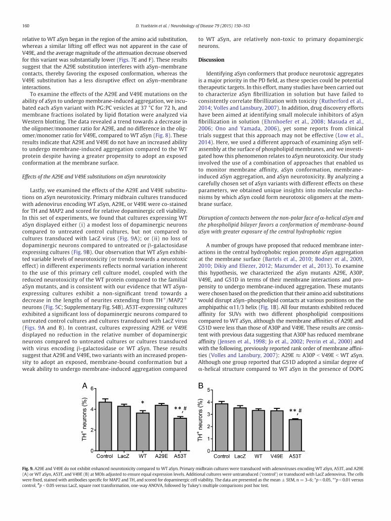
Next, we investigated the membrane-bound conformation of theA29E and V49E variants via 2D NMR spectroscopy. 15N-labeled formsof these mutants were incubated with or without DOPE:DOPS:DOPCvesicles (lipid:protein ratio, 20:1 mol/mol), and 1H–15N HSQC spectrawere recorded (Figs. 7A and B). We found that A29E displayed a slightreduction in the total bound population and exhibited the greatest
combinant aSyn variants were analyzed by far-UV CD as in Fig. 2. Each aSyn variant wasDOPC (B) (the data for WT aSyn are identical to those presented in Fig. 2). The data weretermined from the values of the fit parameters (see Table 1). The data are representative
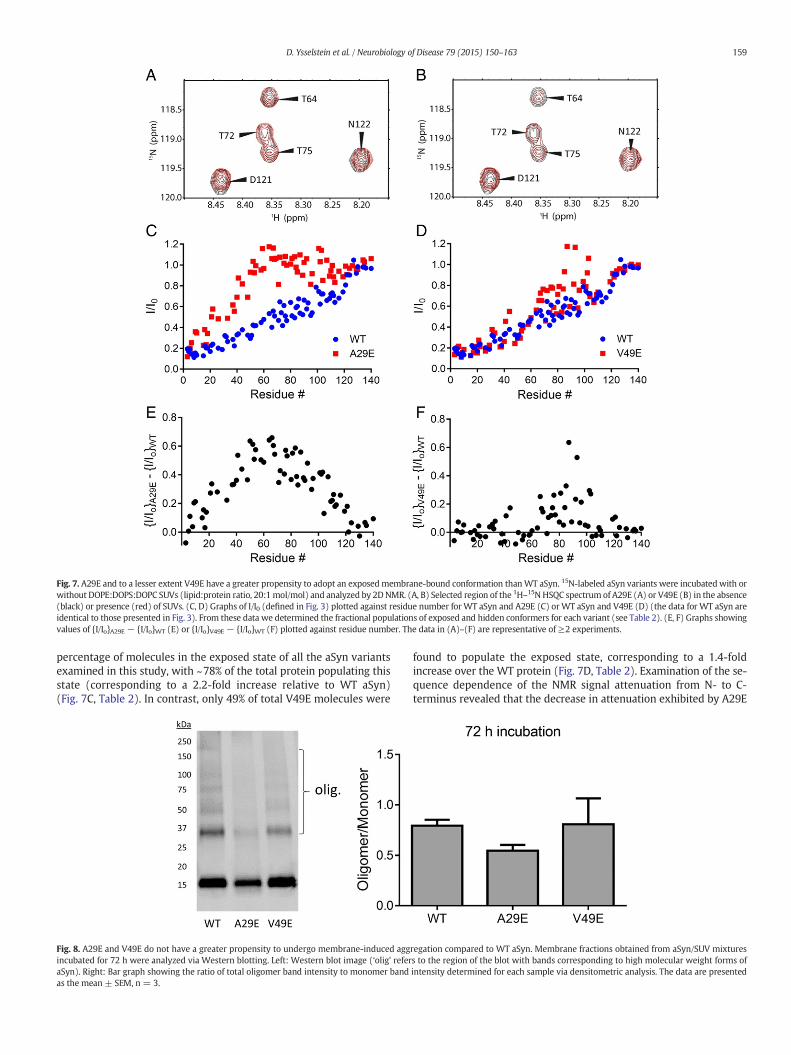
Fig. 7. A29E and to a lesser extent V49E have a greater propensity to adopt an exposedmembrane-bound conformation thanWT aSyn. 15N-labeled aSyn variants were incubated with orwithout DOPE:DOPS:DOPC SUVs (lipid:protein ratio, 20:1mol/mol) and analyzed by 2DNMR. (A, B) Selected region of the 1H–15N HSQC spectrum of A29E (A) or V49E (B) in the absence(black) or presence (red) of SUVs. (C, D) Graphs of I/I0 (defined in Fig. 3) plotted against residue number for WT aSyn and A29E (C) orWT aSyn and V49E (D) (the data for WT aSyn areidentical to those presented in Fig. 3). From these data we determined the fractional populations of exposed and hidden conformers for each variant (see Table 2). (E, F) Graphs showingvalues of {I/Io}A29E − {I/Io}WT (E) or {I/Io}V49E − {I/Io}WT (F) plotted against residue number. The data in (A)–(F) are representative of ≥2 experiments.
159D. Ysselstein et al. / Neurobiology of Disease 79 (2015) 150–163
percentage of molecules in the exposed state of all the aSyn variantsexamined in this study, with ~78% of the total protein populating thisstate (corresponding to a 2.2-fold increase relative to WT aSyn)(Fig. 7C, Table 2). In contrast, only 49% of total V49E molecules were
Fig. 8. A29E and V49E do not have a greater propensity to undergo membrane-induced aggincubated for 72 h were analyzed via Western blotting. Left: Western blot image (‘olig’ referaSyn). Right: Bar graph showing the ratio of total oligomer band intensity to monomer bandas the mean ± SEM, n = 3.
found to populate the exposed state, corresponding to a 1.4-foldincrease over the WT protein (Fig. 7D, Table 2). Examination of the se-quence dependence of the NMR signal attenuation from N- to C-terminus revealed that the decrease in attenuation exhibited by A29E
regation compared to WT aSyn. Membrane fractions obtained from aSyn/SUV mixturess to the region of the blot with bands corresponding to high molecular weight forms ofintensity determined for each sample via densitometric analysis. The data are presented
160 D. Ysselstein et al. / Neurobiology of Disease 79 (2015) 150–163
relative to WT aSyn began in the region of the amino acid substitution,whereas a similar lifting off effect was not apparent in the case ofV49E, and the average magnitude of the attenuation decrease observedfor this variant was substantially lower (Figs. 7E and F). These resultssuggest that the A29E substitution interferes with aSyn–membranecontacts, thereby favoring the exposed conformation, whereas theV49E substitution has a less disruptive effect on aSyn–membraneinteractions.
To examine the effects of the A29E and V49E mutations on theability of aSyn to undergo membrane-induced aggregation, we incu-bated each aSyn variant with PG:PC vesicles at 37 °C for 72 h, andmembrane fractions isolated by lipid flotation were analyzed viaWestern blotting. The data revealed a trend towards a decrease inthe oligomer/monomer ratio for A29E, and no difference in the olig-omer/monomer ratio for V49E, compared to WT aSyn (Fig. 8). Theseresults indicate that A29E and V49E do not have an increased abilityto undergo membrane-induced aggregation compared to the WTprotein despite having a greater propensity to adopt an exposedconformation at the membrane surface.
Effects of the A29E and V49E substitutions on aSyn neurotoxicity
Lastly, we examined the effects of the A29E and V49E substitu-tions on aSyn neurotoxicity. Primary midbrain cultures transducedwith adenovirus encoding WT aSyn, A29E, or V49E were co-stainedfor TH and MAP2 and scored for relative dopaminergic cell viability.In this set of experiments, we found that cultures expressing WTaSyn displayed either (i) a modest loss of dopaminergic neuronscompared to untreated control cultures, but not compared tocultures transduced with LacZ virus (Fig. 9A); or (ii) no loss ofdopaminergic neurons compared to untreated or β-galactosidaseexpressing cultures (Fig. 9B). Our observation that WT aSyn exhibi-ted variable levels of neurotoxicity (or trends towards a neurotoxiceffect) in different experiments reflects normal variation inherentto the use of this primary cell culture model, coupled with thereduced neurotoxicity of the WT protein compared to the familialaSyn mutants, and is consistent with our evidence that WT aSyn-expressing cultures exhibit a non-significant trend towards adecrease in the lengths of neurites extending from TH+/MAP2+
neurons (Fig. 5C; Supplementary Fig. S4B). A53T-expressing culturesexhibited a significant loss of dopaminergic neurons compared tountreated control cultures and cultures transduced with LacZ virus(Figs. 9A and B). In contrast, cultures expressing A29E or V49Edisplayed no reduction in the relative number of dopaminergicneurons compared to untreated cultures or cultures transducedwith virus encoding β-galactosidase or WT aSyn. These resultssuggest that A29E and V49E, two variants with an increased propen-sity to adopt an exposed, membrane-bound conformation but aweak ability to undergo membrane-induced aggregation compared
Fig. 9. A29E and V49E do not exhibit enhanced neurotoxicity compared to WT aSyn. Primary m(A) or WT aSyn, A53T, and V49E (B) at MOIs adjusted to ensure equal expression levels. Additiwere fixed, stained with antibodies specific for MAP2 and TH, and scored for dopaminergic cellcontrol, #p b 0.05 versus LacZ, square root transformation, one-way ANOVA, followed by Tukey
to WT aSyn, are relatively non-toxic to primary dopaminergicneurons.
Discussion
Identifying aSyn conformers that produce neurotoxic aggregatesis a major priority in the PD field, as these species could be potentialtherapeutic targets. In this effort, many studies have been carried outto characterize aSyn fibrillization in solution but have failed toconsistently correlate fibrillization with toxicity (Rutherford et al.,2014; Volles and Lansbury, 2007). In addition, drug discovery effortshave been aimed at identifying small molecule inhibitors of aSynfibrillization in solution (Ehrnhoefer et al., 2008; Masuda et al.,2006; Ono and Yamada, 2006), yet some reports from clinicaltrials suggest that this approach may not be effective (Low et al.,2014). Here, we used a different approach of examining aSyn self-assembly at the surface of phospholipid membranes, and we investi-gated how this phenomenon relates to aSyn neurotoxicity. Our studyinvolved the use of a combination of approaches that enabled usto monitor membrane affinity, aSyn conformation, membrane-induced aSyn aggregation, and aSyn neurotoxicity. By analyzing acarefully chosen set of aSyn variants with different effects on theseparameters, we obtained unique insights into molecular mecha-nisms by which aSyn could form neurotoxic oligomers at the mem-brane surface.
Disruption of contacts between the non-polar face of α-helical aSyn andthe phospholipid bilayer favors a conformation of membrane-boundaSyn with greater exposure of the central hydrophobic region
A number of groups have proposed that reduced membrane inter-actions in the central hydrophobic region promote aSyn aggregationat the membrane surface (Bartels et al., 2010; Bodner et al., 2009,2010; Dikiy and Eliezer, 2012; Mazumder et al., 2013). To examinethis hypothesis, we characterized the aSyn mutants A29E, A30P,V49E, and G51D in terms of their membrane interactions and pro-pensity to undergo membrane-induced aggregation. These mutantswere chosen based on the prediction that their amino acid substitutionswould disrupt aSyn–phospholipid contacts at various positions on theamphipathic α11/3 helix (Fig. 1B). All four mutants exhibited reducedaffinity for SUVs with two different phospholipid compositionscompared to WT aSyn, although the membrane affinities of A29E andG51D were less than those of A30P and V49E. These results are consis-tent with previous data suggesting that A30P has reduced membraneaffinity (Jensen et al., 1998; Jo et al., 2002; Perrin et al., 2000) andwith the following, previously reported rank order of membrane affini-ties (Volles and Lansbury, 2007): A29E ≈ A30P b V49E b WT aSyn.Although one group reported that G51D adopted a similar degree ofα-helical structure compared to WT aSyn in the presence of DOPG
idbrain cultures were transduced with adenoviruses encodingWT aSyn, A53T, and A29Eonal cultures were untransduced (‘control’) or transduced with LacZ adenovirus. The cellsviability. The data are presented as themean ± SEM, n= 3–6; *p b 0.05, **p b 0.01 versus's multiple comparisons post hoc test.
161D. Ysselstein et al. / Neurobiology of Disease 79 (2015) 150–163
vesicles (Lesage et al., 2013), a recent study revealed that G51D hasreduced affinity for 1-palmitoyl-2-oleoyl-sn-glycero-3-phosphoglycerol(POPG) SUVs compared to the WT protein (Fares et al., 2014), insupport of the findings reported here.
Our observation that G51D has a reduced maximum helicitycompared to A29E or A30P is likely a result of the more C-terminalposition of the G51D mutation in the α11/3 helix compared to theother two substitutions. The aSyn region spanning residues 31–85 hasbeen reported to have lower membrane affinity than the N-terminalsegment spanning the first ~30 residues (Bartels et al., 2010; Drescheret al., 2008; Shvadchak et al., 2011b). Interactions involving residues31–85 with the membrane only become significant after the crowdingof neighboring aSyn molecules on the bilayer surface is relieved by theaddition of excess lipid. Apparently, attractive forces between thissegment of aSyn and themembrane can overcome the disruptive effectsof the A29E and A30P mutations in the presence of saturating lipid. Incontrast, the summed attractive forces between residues C-terminal totheG51D substitution and themembranemaybe tooweak to overcomethe disruptive effects of this mutation, resulting in a loss of binding ofthis region even in the presence of excess lipid.
Analysis of aSyn–membrane interactions by 2D NMR enabled usto identify residue-specific interactions that were disrupted byamino acid substitutions in the aSyn variants. We found that allfour mutants exhibited similar fractional populations of membrane-bound molecules – defined as molecules with the segment spanningresidues 3–9 in contact with the membrane – compared to the WTprotein under the conditions of the NMR experiment (incubationwith DOPE:DOPS:DOPC SUVs at a protein:lipid ratio of 1:20). NMRdata obtained for the familial mutants A30P and G51D revealed amarkeddisruption of membrane interactions that started near the site of themutation and extended through the C-terminal end of the α11/3 helix.These findings are consistent with 2D NMR data obtained for A30P(Bodner et al., 2010) and with NMR results reported for G51D whilethis manuscript was in preparation (Fares et al., 2014). Our 2D NMRanalysis of A29E revealed a disruption of membrane contacts that begannear the site of the mutation and extended into the C-terminal region.In contrast, V49E exhibited more modest perturbations of membranecontacts compared to A29E, A30P, and G51D.
Examination of a helical wheel plot sheds light on possible mecha-nisms by which aSyn–membrane interactions could be disrupted bydifferent mutations. Although the A29E and A30P substitutions occurat adjacent positions in the aSyn sequence, the mechanisms by whichthey disrupt membrane binding are different. Introduction of a helix-breaking proline residue at position 30 reduces the membrane affinityof aSyn by destabilizing the α11/3 helix that plays a key role inmembrane binding (Jensen et al., 1998; Jo et al., 2002; Perrin et al.,2000). In contrast, the A29E substitution introduces a negativelycharged glutamate residue at a site on the non-polar face of the α11/3helix that is predicted to be embedded in the hydrophobic (acylchain) region of the phospholipid bilayer. The A29E mutation showsmore extensive disruption of contacts (and weaker affinity observedby far-UV CD) compared to A30P, presumably because of the higherenergy barrier associated with introducing a negative charge in thenon-polar phase of the bilayer. Residues A29 and G51 are aligned equi-valently (i.e. they have the same angular position) on the helical wheelplot ofWT aSyn (Fig. 1B). Thus, the G51D substitutionwould be expect-ed to have a similar disruptive effect on membrane binding as the A29Emutation because of the high energy barrier associated with inserting acharged aspartate residue in the hydrophobic region of the bilayer.Consistent with this prediction, both G51D and A29E bind SUVs withweaker affinity (based on far-UV CD data) compared to WT aSyn,although as expected the A29E substitution disrupts membranecontacts at more N-terminal sites compared to the G51D substitution.Although V49 is only two residues N-terminal to G51 in the aminoacid sequence, the V49E substitution has a less disruptive effect onmembrane interactions and affinity than the G51D mutation,
presumably because V49 is situated on the polar face of the amphi-pathic helix at a site that is expected to be near the protein–mem-brane interface. Thus, the glutamate residue at position 49 of V49Eshould not be involved in destabilizing interactions with the non-polar phase of the bilayer. Instead, it could engage in repulsiveelectrostatic interactions with anionic phospholipid head-groups,perhaps via a mechanism involving partial helix unfolding (Pandeyet al., 2009).
Collectively, our findings highlight the fact that the degree ofexposure of a membrane-bound aSyn variant with amutation encodinga charged residue depends on the placement of the residue on theamphipathic α11/3 helix relative to the membrane contact region.Disruption of contacts between the non-polar face of the helix and thehydrophobic region of the bilayer results in the protein's adoption ofan exposed conformation. Moreover, the introduction of a chargedresidue at a more N-terminal site on the non-polar face of the helixresults in a more extensive disruption of aSyn–membrane interactions.
The adoption of an exposed conformation by membrane-bound aSyn canpromote (but is not sufficient for) membrane-induced aggregation
Based on the hypothesis that exposure of the central hydrophobicregion of membrane-bound aSyn favors aSyn aggregation at the mem-brane surface (Bartels et al., 2010; Bodner et al., 2009, 2010; Dikiy andEliezer, 2012; Mazumder et al., 2013), one would predict that A30P,G51D, and A29E, which adopt a relatively exposed conformation inthe membrane-bound state, should have a higher propensity to un-dergo membrane-induced aggregation compared to the WT protein.Conversely, the V49E mutant, which has a lower tendency to adopt anexposed conformation compared to A30P, G51D, and A29E, shouldhave a reduced ability to undergo self-assembly at the membranesurface compared to the other three mutants. In partial agreementwith this prediction, A30P andG51Dbothdisplay significantly increasedaggregation relative to WT aSyn upon incubation in the presence ofSUVs, whereas V49E exhibits less membrane-induced self-assemblycompared to the two familial mutants. To our knowledge, these dataprovide the first direct evidence that A30P andG51Dundergo enhancedmembrane-induced aggregation upon prolonged incubationwith SUVs.During the preparation of this manuscript, it was reported that G51Dconverts from an α-helical structure to a β-sheet-rich structure whenincubated with SUVs (Fares et al., 2014), implying that the oligomersformed in the G51D–lipid mixtures described here have considerableβ-sheet structure.
In contrast, the results obtained with A29E do not support thehypothesis that the exposed conformation of membrane-bound aSyninvariably favors membrane-induced aggregation. Although A29E issimilar to G51D in terms of its placement of a negatively chargedresidue on the non-polar face of the α11/3 helix and the degree towhich it adopts an exposed conformation, it does not have the samehigh propensity to undergo oligomerization in the presence of SUVs.Two explanations could account for this finding. First, the length ofthe N-terminal α11/3 helix adopted by membrane-bound A29E maybe insufficient to enable interactions between neighboring aSyn mole-cules required for self-assembly on the bilayer. However, the fact thatthe N-terminal helix formed by A29E encompasses a similar numberof residues compared to the A30P N-terminal helix (~20 residues inboth cases) and that A30P (in contrast to A29E) has a high propensityto undergo membrane-induced aggregation is inconsistent with thisexplanation. Second, the glutamate residue at position 29 of neighbor-ing,membrane-boundA29Emoleculesmay engage in repulsive electro-static interactions that interfere with self-assembly on the bilayer. Thisscenario implies that the introduction of a negative charge at position29 electrostatically interferes with self-assembly, whereas the G51Dsubstitution does not have the same inhibitory effect, perhaps becausecontacts involving residue 51 do not play a significant role in aSynoligomerization on the membrane surface. Consistent with this model,
162 D. Ysselstein et al. / Neurobiology of Disease 79 (2015) 150–163
evidence suggests that aSyn forms oligomers on the membrane surfacevia interactions between the α-helical domains of neighboring proteinmolecules, and in particular, via parallel interactions involving thehelical segment spanning residues ~1–40 (Burre et al., 2014). Althoughmembrane-bound helical aSyn oligomers apparently play a role in thephysiologically relevant binding of the protein to synaptobrevin(Burre et al., 2014), they could also be precursors of pathological,β-sheet-rich aggregates that form on themembrane as a result of expo-sure of the central hydrophobic region (Comellas et al., 2012;Mazumder et al., 2013; Pandey et al., 2009).
Together, our observations indicate that the potential impact of amutation on aSyn self-assembly at the membrane surface is morecomplex than previously suggested. Namely, the effect of a mutationon the propensity of aSyn to undergo membrane-induced aggregationdepends not only on how the mutation alters the distribution of aSynbetween exposed and hidden conformers, but also on how it affectsinteractions between membrane-bound aSyn molecules necessary forhigher order assembly on the bilayer. In particular, our findingshighlight the importance of contacts involving a segment of the poly-peptide chain encompassing residue 29 for membrane-induced aSynaggregation.
Membrane-induced aSyn aggregation may play a role in aSyn neurotoxicity
A key outcome of our study is the observation that the propensity ofaSyn variants to undergo membrane-induced self-assembly correlateswith their neurotoxicity. Both A30P and G51D displayed greatermembrane-induced aggregation and enhanced toxicity to dopaminer-gic neurons compared to WT aSyn. In contrast, A29E displayed trendstowards decreased membrane-induced self-assembly and reducedneurotoxicity. From these results, we infer that aSyn aggregation atthe membrane surface could contribute to aSyn-mediated neuro-degeneration in PD. This conclusion is supported by evidence thatother neurotoxic aSyn variants, including A53T and the engineeredmutant E57K (Reynolds et al., 2011) and a series of aSyn variants origi-nally characterized in yeast (Vamvaca et al., 2009; Volles and Lansbury,2007) (Griggs, A.M. and Rochet, J.-C., unpublished data), have anincreased propensity to undergo membrane-induced self-assembly.In addition, we have obtained data showing that the familial aSynmutants A53T and E46K both have a greater propensity to undergomembrane-induced aggregation and elicit neurotoxicity compared toWT aSyn (Griggs, A.M. and Rochet, J.-C., unpublished data). By analogywith mutations that disrupt aSyn-bilayer contacts, perturbations thatfavor a more exposed conformation of the membrane-bound WTprotein, including aSyn oxidation (Maltsev et al., 2013) and changes inmembrane lipid composition (Fusco et al., 2014), could contribute toaSyn aggregation and neurotoxicity in sporadic PD.
Multiple lines of evidence suggest that aSyn aggregation at themembrane surface results in a disruption of membrane integrity, appa-rently via a mechanism involving lipid extraction from the bilayer(Comellas et al., 2012; Lee et al., 2012; Ouberai et al., 2013; Reynoldset al., 2011). This process of bilayer disruption coupled to the aggrega-tion of membrane-bound aSyn, and/or the formation of the aggregatesthemselves, could play a role in neurotoxic mechanisms associatedwith aSyn expression in cellular and animal models, including inter-ference with cellular trafficking (Cooper et al., 2006; Gitler et al., 2008;Soper et al., 2011) or impairment ofmitochondrial function or dynamics(Kamp et al., 2010; Nakamura et al., 2011; Parihar et al., 2009).
Although our results highlight a potential connection betweenmembrane-induced aSyn aggregation and neurotoxicity, they do notexclude the possibility that aSyn triggers neurodegeneration viaadditional mechanisms, including oligomerization in solution ratherthan at the membrane surface (Burre et al., 2015). In the case of G51D,several groups have reported that the mutant protein undergoesoligomerization or fibrillization in solution with similar or slowerkinetics compared to WT aSyn (Fares et al., 2014; Lesage et al., 2013;
Rutherford et al., 2014). Thus, the enhanced neurotoxicity of G51D infamilial PD patients can be explained by the mutant's high propensityto undergo membrane-induced self-assembly (shown here), as op-posed to its relatively modest ability to form aggregates in solution.Our observation that G51D has reduced α-helical content in thepresence of saturating phospholipid suggests that the protein couldadopt an exposed conformation and form potentially toxic oligomersat themembrane surface at a range of protein–lipid ratios (e.g. resultingfrom variations in protein expression levels) in vivo. In contrast toG51D, evidence suggests that A30P has an enhanced ability to undergooligomerization in solution (Conway et al., 2000; Li et al., 2001) and inthe presence of SUVs (this study) relative to WT aSyn. Accordingly,the enhanced neurotoxicity of A30P in familial PD can be accountedfor by both self-assembly mechanisms. A number of engineered aSynmutants have been reported to have an increased ability to formoligomers in solution and to elicit neurodegeneration compared to WTaSyn (Karpinar et al., 2009; Winner et al., 2011). A goal for futureresearch will be to determinewhether membrane-induced aggregationcould play a role in the enhanced neurotoxicity of these and other aSynvariants (Burre et al., 2012).
Conclusions
In summary, our data show that the A30P and G51D mutationsreduce the membrane affinity of aSyn, leading to an increase in thefractional population of the exposed state. Upon incubation, thesevariants also display increased membrane-induced self-assembly rela-tive to WT aSyn, and this property correlates with increased toxicity todopaminergic neurons. Our observations support a model wherein theconformation of aSyn on phospholipid membranes plays a key role ininitiating the formation of neurotoxic aggregates. However, an increasein the exposed state population is not itself sufficient for membrane-induced aggregation or neurotoxicity, as disrupting interactions be-tween neighboring exposed conformers could impede the formation oftoxic aggregates at themembrane surface. Collectively, our findings pro-vide a rationale for alleviating aSyn neurotoxicity in PD and othersynucleinopathy disorders by stabilizing aSyn–membrane interactionsto favor the hidden state (Fonseca-Ornelas et al., 2014), or by interferingwith the association of exposed conformers at the membrane surface.
Supplementary data to this article can be found online at http://dx.doi.org/10.1016/j.nbd.2015.04.007.
Acknowledgments
Thisworkwas supported by a grant from theBranfman Family Foun-dation (J.-C.R.), NIH grants R01 NS049221 (J.-C.R.), R01 GM039478(C.B.P.), and K25 NS058395-04 (L.A.S.), a Purdue Research FoundationFellowship (J.-C.R. and D.Y.), a McKeehan Fellowship provided by theCollege of Pharmacy at Purdue University (D.Y.), and a Floss Endow-ment Research Award provided by the Department of MedicinalChemistry and Molecular Pharmacology at Purdue University (D.Y.).The research described herein was conducted in a facility constructedwith support from Research Facilities Improvement Program grantnos. C06-14499 and C06-15480 from the National Center for ResearchResources of theNIH. NMRdatawere acquired in the PINMRF supportedby P30 CA023168.We thankDr. Katherine Strathearn and Susan Roy forassistance with analyzing primary midbrain cultures, Dr. David Eliezerfor providing NMR assignments forWT aSyn, Dr. Deniz Kirik for provid-ing the pAAV2 plasmid, and Dr. Volodymyr Shvadchak for helpful sug-gestions for analyzing the data from far-UV CD experiments.
References
Abedini, A., Raleigh, D.P., 2009. A role for helical intermediates in amyloid formation by nativelyunfolded polypeptides? Phys. Biol. 6, 15005.
Bartels, T., et al., 2010. The N-terminus of the intrinsically disordered protein alpha-synucleintriggers membrane binding and helix folding. Biophys. J. 99, 2116–2124.
163D. Ysselstein et al. / Neurobiology of Disease 79 (2015) 150–163
Bertoncini, C.W., et al., 2005. Release of long-range tertiary interactions potentiates aggregationof natively unstructured alpha-synuclein. Proc. Natl. Acad. Sci. U. S. A. 102, 1430–1435.
Bodner, C.R., et al., 2009. Multiple tight phospholipid-binding modes of alpha-synucleinrevealed by solution NMR spectroscopy. J. Mol. Biol. 390, 775–790.
Bodner, C.R., et al., 2010. Differential phospholipid binding of alpha-synuclein variants implicatedin Parkinson's disease revealed by solution NMR spectroscopy. Biochemistry 49, 862–871.
Burre, J., et al., 2012. Systematic mutagenesis of alpha-synuclein reveals distinct sequencerequirements for physiological and pathological activities. J. Neurosci. 32, 15227–15242.
Burre, J., et al., 2014. alpha-Synuclein assembles into higher-order multimers upon membranebinding to promote SNARE complex formation. Proc. Natl. Acad. Sci. U. S. A. 111, E4274–E4283.
Burre, J., et al., 2015. Definition of a molecular pathway mediating alpha-synuclein neuro-toxicity. J. Neurosci. 35, 5221–5232.
Bussell Jr., R., Eliezer, D., 2003. A structural and functional role for 11-mer repeats inα-synucleinand other exchangeable lipid binding proteins. J. Mol. Biol. 329, 763–778.
Chandra, S., et al., 2003. A broken α-helix in folded α-synuclein. J. Biol. Chem. 278, 15313–15318.Chartier-Harlin, M.C., et al., 2004. Alpha-synuclein locus duplication as a cause of familial
Parkinson's disease. Lancet 364, 1167–1169.Comellas, G., et al., 2012. Structural intermediates during alpha-synuclein fibrillogenesis on
phospholipid vesicles. J. Am. Chem. Soc. 134, 5090–5099.Conway, K.A., et al., 2000. Acceleration of oligomerization, not fibrillization, is a shared property
of both α-synuclein mutations linked to early-onset Parkinson's disease: implications forpathogenesis and therapy. Proc. Natl. Acad. Sci. U. S. A. 97, 571–576.
Cooper, A.A., et al., 2006. Alpha-synuclein blocks ER–Golgi traffic and Rab1 rescues neuron lossin Parkinson's models. Science 313, 324–328.
Davidson, W.S., et al., 1998. Stabilization of α-synuclein secondary structure upon binding tosynthetic membranes. J. Biol. Chem. 273, 9443–9449.
Dedmon, M.M., et al., 2005.Mapping long-range interactions in alpha-synuclein using spin-labelNMR and ensemble molecular dynamics simulations. J. Am. Chem. Soc. 127, 476–477.
Delaglio, F., et al., 1995. NMRPipe: a multidimensional spectral processing system based onUNIX pipes. J. Biomol. NMR 6, 277–293.
Dikiy, I., Eliezer, D., 2012. Folding and misfolding of alpha-synuclein on membranes. Biochim.Biophys. Acta 1818, 1013–1018.
Drescher, M., et al., 2008. Spin-label EPR on alpha-synuclein reveals differences in themembrane binding affinity of the two antiparallel helices. Chembiochem 9, 2411–2416.
Ehrnhoefer, D.E., et al., 2008. EGCG redirects amyloidogenic polypeptides into unstructured,off-pathway oligomers. Nat. Struct. Mol. Biol. 15, 558–566.
Eliezer, D., et al., 2001. Conformational properties of α-synuclein in its free and lipid-associatedstates. J. Mol. Biol. 307, 1061–1073.
Fares, M.B., et al., 2014. The novel Parkinson's disease linked mutation G51D attenuates in vitroaggregation and membrane binding of alpha-synuclein, and enhances its secretion andnuclear localization in cells. Hum. Mol. Genet. 23, 4491–4509.
Ferreon, A.C., et al., 2009. Interplay of alpha-synuclein binding and conformational switchingprobed by single-molecule fluorescence. Proc. Natl. Acad. Sci. U. S. A. 106, 5645–5650.
Fonseca-Ornelas, L., et al., 2014. Small molecule-mediated stabilization of vesicle-associated helicalalpha-synuclein inhibits pathogenic misfolding and aggregation. Nat. Commun. 5, 5857.
Fusco, G., et al., 2014. Direct observation of the three regions in alpha-synuclein that determineits membrane-bound behaviour. Nat. Commun. 5, 3827.
Galvagnion, C., et al., 2015. Lipid vesicles trigger alpha-synuclein aggregation by stimulatingprimary nucleation. Nat. Chem. Biol. 11, 229–234.
Georgieva, E.R., et al., 2010. The lipid-binding domain of wild type and mutant alpha-synuclein:compactness and interconversion between the broken and extended helix forms. J. Biol.Chem. 285, 28261–28274.
Giasson, B.I., et al., 2001. A hydrophobic stretch of 12 amino acid residues in the middle of α-synuclein is essential for filament assembly. J. Biol. Chem. 276, 2380–2386.
Giehm, L., et al., 2010. SDS-induced fibrillation of alpha-synuclein: an alternative fibrillationpathway. J. Mol. Biol. 401, 115–133.
Gitler, A.D., et al., 2008. The Parkinson's disease protein alpha-synuclein disrupts cellular Rabhomeostasis. Proc. Natl. Acad. Sci. U. S. A. 105, 145–150.
Greenbaum, E.A., et al., 2005. The E46K mutation in alpha-synuclein increases amyloid fibrilformation. J. Biol. Chem. 280, 7800–7807.
Grey, M., et al., 2015. Acceleration of alpha-synuclein aggregation by exosomes. J. Biol. Chem.290, 2969–2982.
Haque, F., et al., 2010. Adsorption of alpha-synuclein on lipid bilayers: modulating the structureand stability of protein assemblies. J. Phys. Chem. B 114, 4070–4081.
Jao, C.C., et al., 2004. Structure of membrane-bound alpha-synuclein studied by site-directedspin labeling. Proc. Natl. Acad. Sci. U. S. A. 101, 8331–8336.
Jensen, P.H., et al., 1998. Binding of α-synuclein to brain vesicles is abolished by familialParkinson's disease mutation. J. Biol. Chem. 273, 26292–26294.
Jo, E., et al., 2000. α-Synuclein membrane interactions and lipid specificity. J. Biol. Chem. 275,34328–34334.
Jo, E., et al., 2002. Defective membrane interactions of familial Parkinson's disease mutant A30Pα-synuclein. J. Mol. Biol. 315, 799–807.
Jo, E., et al., 2004. alpha-Synuclein-synaptosomal membrane interactions: implications forfibrillogenesis. Eur. J. Biochem. 271, 3180–3189.
Kamp, F., et al., 2010. Inhibition of mitochondrial fusion by alpha-synuclein is rescued by PINK1,Parkin and DJ-1. Embo J. 29, 3571–3589.
Karpinar, D.P., et al., 2009. Pre-fibrillar alpha-synuclein variants with impaired beta-structure in-crease neurotoxicity in Parkinson's disease models. Embo J. 28, 3256–3268.
Kiely, A.P., et al., 2013. alpha-Synucleinopathy associated with G51D SNCA mutation: a linkbetween Parkinson's disease andmultiple system atrophy? Acta Neuropathol. 125, 753–769.
Kruger, R., et al., 1998. Ala30Pro mutation in the gene encoding α-synuclein in Parkinson'sdisease. Nat. Genet. 18, 106–108.
Lee, H.-J., et al., 2002. Membrane-bound α-synuclein has a high aggregation propensity and theability to seed the aggregation of the cytosolic form. J. Biol. Chem. 277, 671–678.
Lee, J.H., et al., 2012. Radiating amyloid fibril formation on the surface of lipid membranesthrough unit-assembly of oligomeric species of alpha-synuclein. PLoS ONE 7, e47580.
Lesage, S., et al., 2013. G51D alpha-synuclein mutation causes a novel parkinsonian-pyramidalsyndrome. Ann. Neurol. 73, 459–471.
Li, J., et al., 2001. Effect of familial Parkinson's disease point mutations A30P and A53T on thestructural properties, aggregation, and fibrillation of human α-synuclein. Biochemistry40, 11604–11613.
Liu, F., et al., 2008. Methionine sulfoxide reductase A protects dopaminergic cells fromParkinson's disease-related insults. Free Radic. Biol. Med. 45, 242–255.
Low, P.A., et al., 2014. Efficacy and safety of rifampicin for multiple system atrophy: arandomised, double-blind, placebo-controlled trial. Lancet Neurol. 13, 268–275.
Maltsev, A.S., et al., 2013. Site-specific interaction between alpha-synuclein and membranesprobed by NMR-observed methionine oxidation rates. J. Am. Chem. Soc. 135, 2943–2946.
Marley, J., et al., 2001. A method for efficient isotopic labeling of recombinant proteins. J. Biomol.NMR 20, 71–75.
Masuda, M., et al., 2006. Small molecule inhibitors of alpha-synuclein filament assembly.Biochemistry 45, 6085–6094.
Mazumder, P., et al., 2013. Insight into alpha-synuclein plasticity and misfolding from differen-tial micelle binding. J. Phys. Chem. B 117, 11448–11459.
Nakamura, K., et al., 2011. Direct membrane association drives mitochondrial fission by theParkinson disease-associated protein α-synuclein. J. Biol. Chem. 286, 20710–20726.
Nasstrom, T., et al., 2011. The lipid peroxidation products 4-oxo-2-nonenal and 4-hydroxy-2-nonenal promote the formation of alpha-synuclein oligomers with distinct biochemical,morphological, and functional properties. Free Radic. Biol. Med. 50, 428–437.
Necula, M., et al., 2003. Rapid anionic micelle-mediated alpha-synuclein fibrillization in vitro.J. Biol. Chem. 278, 46674–46680.
Ono, K., Yamada, M., 2006. Antioxidant compounds have potent anti-fibrillogenic and fibril-destabilizing effects for alpha-synuclein fibrils in vitro. J. Neurochem. 97, 105–115.
Ouberai, M.M., et al., 2013. alpha-Synuclein senses lipid packing defects and induces lateralexpansion of lipids leading to membrane remodeling. J. Biol. Chem. 288, 20883–20895.
Pandey, A.P., et al., 2009. Clustering of alpha-synuclein on supported lipid bilayers: role ofanionic lipid, protein, and divalent ion concentration. Biophys. J. 96, 540–551.
Parihar, M.S., et al., 2009. Alpha-synuclein overexpression and aggregation exacerbatesimpairment of mitochondrial functions by augmenting oxidative stress in humanneuroblastoma cells. Int. J. Biochem. Cell Biol. 41, 2015–2024.
Pasanen, P., et al., 2014. A novel alpha-synuclein mutation A53E associated with atypical multiplesystem atrophy and Parkinson's disease-type pathology. Neurobiol. Aging 35 (2180), e1–e5.
Perrin, R.J., et al., 2000. Interaction of human α-synuclein and Parkinson's disease variants withphospholipids. Structural analysis using site-directed mutagenesis. J. Biol. Chem. 275,34393–34398.
Perrin, R.J., et al., 2001. Exposure to long chain polyunsaturated fatty acids triggers rapidmultimerization of synucleins. J. Biol. Chem. 276, 41958–41962.
Pfefferkorn, C.M., et al., 2012. Depth of alpha-synuclein in a bilayer determined by fluorescence,neutron reflectometry, and computation. Biophys. J. 102, 613–621.
Polymeropoulos, M.H., et al., 1997. Mutation in the α-synuclein gene identified in families withParkinson's disease. Science 276, 2045–2047.
Proukakis, C., et al., 2013. A novel alpha-synuclein missense mutation in Parkinson disease.Neurology 80, 1062–1064.
Qin, Z., et al., 2007. Effect of 4-hydroxy-2-nonenal modification on alpha-synuclein aggregation.J. Biol. Chem. 282, 5862–5870.
Reynolds, N.P., et al., 2011. Mechanism of membrane interaction and disruption by alpha-synuclein. J. Am. Chem. Soc. 133, 19366–19375.
Rochet, J.C., et al., 2012. Molecular insights into Parkinson's disease. Prog. Mol. Biol. Transl. Sci.107, 125–188.
Rutherford, N.J., et al., 2014. Divergent effects of the H50Q and G51D SNCA mutations on theaggregation of alpha-synuclein. J. Neurochem. 131, 859–867.
Scholtz, J.M., et al., 1991. Parameters of helix-coil transition theory for alanine-based peptides ofvarying chain lengths in water. Biopolymers 31, 1463–1470.
Sharon, R., et al., 2003. The formation of highly soluble oligomers of α-synuclein is regulated byfatty acids and enhanced in Parkinson's disease. Neuron 37, 583–595.
Shulman, J.M., et al., 2011. Parkinson's disease: genetics and pathogenesis. Annu. Rev. Pathol. 6,193–222.
Shvadchak, V.V., et al., 2011a. Specificity and kinetics of alpha-synuclein binding to modelmembranes determined with fluorescent excited state intramolecular proton transfer(ESIPT) probe. J. Biol. Chem. 286, 13023–13032.
Shvadchak, V.V., et al., 2011b. The mode of alpha-synuclein binding to membranes depends onlipid composition and lipid to protein ratio. FEBS Lett. 585, 3513–3519.
Singleton, A.B., et al., 2003. alpha-Synuclein locus triplication causes Parkinson's disease. Science302, 841.
Soper, J.H., et al., 2011. Aggregation of alpha-synuclein in S. cerevisiae is associated with defectsin endosomal trafficking and phospholipid biosynthesis. J. Mol. Neurosci. 43, 391–405.
Spillantini, M.G., et al., 1997. α-Synuclein in Lewy bodies. Nature 388, 839–840.Strathearn, K.E., et al., 2014. Neuroprotective effects of anthocyanin- and proanthocyanidin-rich
extracts in cellular models of Parkinson's disease. Brain Res. 1555, 60–77.Takamori, S., et al., 2006. Molecular anatomy of a trafficking organelle. Cell 127, 831–846.Trexler, A., Rhoades, E., 2009. Alpha-synuclein binds large unilamellar vesicles as an extended
helix. Biochemistry 48, 2304–2306.Ulmer, T.S., et al., 2005. Structure and dynamics of micelle-bound human alpha-synuclein. J. Biol.
Chem. 280, 9595–9603.Vamvaca, K., et al., 2009. The first N-terminal amino acids of alpha-synuclein are essential for
alpha-helical structure formation in vitro and membrane binding in yeast. J. Mol. Biol.389, 413–424.
Venda, L.L., et al., 2010. alpha-Synuclein and dopamine at the crossroads of Parkinson's disease.Trends Neurosci. 33, 559–568.
Volles, M.J., Lansbury Jr., P.T., 2007. Relationships between the sequence of alpha-synuclein and itsmembrane affinity, fibrillization propensity, and yeast toxicity. J. Mol. Biol. 366, 1510–1522.
Wakabayashi, K., et al., 1998. α-Synuclein immunoreactivity in glial cytoplasmic inclusions inmultiple system atrophy. Neurosci. Lett. 249, 180–182.
Weinreb, P.H., et al., 1996. NACP, a protein implicated in Alzheimer's disease and learning, isnatively unfolded. Biochemistry 35, 13709–13715.
Winner, B., et al., 2011. In vivo demonstration that alpha-synuclein oligomers are toxic. Proc.Natl. Acad. Sci. U. S. A. 108, 4194–4199.
Zakharov, S.D., et al., 2007. Helical alpha-synuclein forms highly conductive ion channels.Biochemistry 46, 14369–14379.
Zarranz, J.J., et al., 2004. The newmutation, E46K, of alpha-synuclein causes Parkinson and Lewybody dementia. Ann. Neurol. 55, 164–173.
Zhang, H., et al., 2013. In vitro study of alpha-synuclein protofibrils by cryo-EM suggests aCu(2+)-dependent aggregation pathway. Biophys. J. 104, 2706–2713.