Effects of Disulfide Bridges in Domain I of Bacillus thuringiensis ...
9
Effects of Disulfide Bridges in Domain I of Bacillus thuringiensis Cry1Aa δ-Endotoxin on Ion-Channel Formation in Biological Membranes O. Alzate,* ,‡,§,| T. You, ‡,⊥ M. Claybon, ‡ C. Osorio, ‡ A. Curtiss, ‡ and D. H. Dean ‡ Biochemistry Department, The Ohio State UniVersity, 484 West 12th AVenue, Columbus, Ohio 43210, and Parque Tecnolo ´ gico de Antioquia, Medellı ´n, Colombia ReceiVed July 20, 2006; ReVised Manuscript ReceiVed September 13, 2006 ABSTRACT: The δ-endotoxin family of toxic proteins represents the major component of the insecticidal capability of the bacterium Bacillus thuringiensis. Domain I of the toxins, which is largely R-helical, has been proposed to unfold at protein entry into the membrane of a target insect, following models known as the penknife and umbrella models. We extended the analysis of a previous work in which four disulfide bridges were constructed in domain I of the Cry1Aa δ-endotoxin that putatively prevented unfolding during membrane partitioning. Using bioassays and voltage clamping of whole insect midgut instead of artificial lipid bilayers, it was found that, while toxicity and inhibition of the short-circuit current were reduced, only one of the disulfide bridges eliminated the activity of the toxins in the insect midgut membrane, and in that case, the loss of toxicity was due to the single amino acid substitution, R99C. It is proposed that at least R helices 4, 5, 6, and 7 and domain II partition in the midgut membranes of target insects, in support of an insertion model in which the whole protein translocates into the midgut membrane. The partition of water-soluble proteins into biological membranes is an important step in the mode of action of some bacterial pathogens. One of these pathogens, Bacillus thuringiensis (Bt), 1 produces protein toxins with insecticidal capabilities that intoxicate by penetrating the midgut mem- brane of susceptible insects, thus inducing ion pores that disrupt the membrane potential. The Cry1A family of Bt toxins (also known as Cry1A δ-endotoxins) are released as 135 kDa protein molecules that become activated in the insect midgut by the proteolytic action of trypsin-like proteases. The 65 kDa activated toxins bind to specific receptors on the insect midgut lining columnar cells and possibly undergo conformational changes followed by membrane permeation and ion-channel formation, eventually resulting in the death of the insect (1). The activated Cry1Aa toxin (2) [Protein Data Bank (PDB) code 1CIY] is a cysteine-free protein composed of three distinct structural domains (Figure 1). Domain I contains seven R helices that account for 85-90% of the secondary structure of this domain. These R helices are numbered 1-7, with the highly hydrophobic R helix 5 located in the center and the other six surrounding it (Figure 1). As result of * To whom correspondence should be addressed: Neuroproteomics Center, Department of Neurobiology, Duke University Medical Center, 258 Bryan Research Building, DUMC Box 3209, Durham, NC 27710. Telephone: (919) 681-5855. Fax: (919) 668-0631. E-mail: alzate@ neuro.duke.edu. ‡ The Ohio State University. § Parque Tecnolo ´gico de Antioquia. | Current address: Neuroproteomics Center, Department of Neuro- biology, Duke University Medical Center, DUMC Box 3209, Durham, NC 27710. ⊥ Current address: Department of Biological Sciences, Campbell University, Buies Creek, NC 27506. 1 Abbreviations: BBMV, brush border membrane vesicles; Bt, Bacil- lus thuringiensis; -ME, -mercaptoethanol; CD, circular dichroism. FIGURE 1: Three-dimensional structure of Cry1Aa displaying three structural domains. Domain I is shown on the left in a ribbon-like representation. The highly hydrophobic R helix 5 is shown in dark gray. Disulfide bridge SS1 (C99-C144) producing a link between R helices 3 and 4 is shown in red. SS2, in green, is designed such that the external R helix 6 is locked to the inner R helix 5 (162C- 207C). SS3, in yellow, holds together R helices 5 and 7 through a linkage between C176 and C252. SS4, the only non-interhelical link, forms a bridge between R helix 7 (C224) and domain II (C279), shown in blue. Domains II and III are shown in a wire- frame representation on the right. The inset shows a diagram highlighting the disulfide linkages. 13597 Biochemistry 2006, 45, 13597-13605 10.1021/bi061474z CCC: $33.50 © 2006 American Chemical Society Published on Web 10/21/2006
Transcript of Effects of Disulfide Bridges in Domain I of Bacillus thuringiensis ...
Effects of Disulfide Bridges in Domain I ofBacillus thuringiensisCry1Aaδ-Endotoxin on Ion-Channel Formation in Biological Membranes
O. Alzate,*,‡,§,| T. You,‡,⊥ M. Claybon,‡ C. Osorio,‡ A. Curtiss,‡ and D. H. Dean‡
Biochemistry Department, The Ohio State UniVersity, 484 West 12th AVenue, Columbus, Ohio 43210, and Parque Tecnolo´gicode Antioquia, Medellı´n, Colombia
ReceiVed July 20, 2006; ReVised Manuscript ReceiVed September 13, 2006
ABSTRACT: The δ-endotoxin family of toxic proteins represents the major component of the insecticidalcapability of the bacteriumBacillus thuringiensis. Domain I of the toxins, which is largelyR-helical, hasbeen proposed to unfold at protein entry into the membrane of a target insect, following models knownas the penknife and umbrella models. We extended the analysis of a previous work in which four disulfidebridges were constructed in domain I of the Cry1Aaδ-endotoxin that putatively prevented unfoldingduring membrane partitioning. Using bioassays and voltage clamping of whole insect midgut instead ofartificial lipid bilayers, it was found that, while toxicity and inhibition of the short-circuit current werereduced, only one of the disulfide bridges eliminated the activity of the toxins in the insect midgutmembrane, and in that case, the loss of toxicity was due to the single amino acid substitution, R99C. Itis proposed that at leastR helices 4, 5, 6, and 7 and domain II partition in the midgut membranes of targetinsects, in support of an insertion model in which the whole protein translocates into the midgut membrane.
The partition of water-soluble proteins into biologicalmembranes is an important step in the mode of action ofsome bacterial pathogens. One of these pathogens,Bacillusthuringiensis(Bt),1 produces protein toxins with insecticidalcapabilities that intoxicate by penetrating the midgut mem-brane of susceptible insects, thus inducing ion pores thatdisrupt the membrane potential. The Cry1A family ofBttoxins (also known as Cry1Aδ-endotoxins) are released as135 kDa protein molecules that become activated in the insectmidgut by the proteolytic action of trypsin-like proteases.The 65 kDa activated toxins bind to specific receptors onthe insect midgut lining columnar cells and possibly undergoconformational changes followed by membrane permeationand ion-channel formation, eventually resulting in the deathof the insect (1).
The activated Cry1Aa toxin (2) [Protein Data Bank (PDB)code 1CIY] is a cysteine-free protein composed of threedistinct structural domains (Figure 1). Domain I containssevenR helices that account for 85-90% of the secondarystructure of this domain. TheseR helices are numbered 1-7,with the highly hydrophobicR helix 5 located in the centerand the other six surrounding it (Figure 1). As result of
* To whom correspondence should be addressed: NeuroproteomicsCenter, Department of Neurobiology, Duke University Medical Center,258 Bryan Research Building, DUMC Box 3209, Durham, NC 27710.Telephone: (919) 681-5855. Fax: (919) 668-0631. E-mail: [email protected].
‡ The Ohio State University.§ Parque Tecnolo´gico de Antioquia.| Current address: Neuroproteomics Center, Department of Neuro-
biology, Duke University Medical Center, DUMC Box 3209, Durham,NC 27710.
⊥ Current address: Department of Biological Sciences, CampbellUniversity, Buies Creek, NC 27506.
1 Abbreviations: BBMV, brush border membrane vesicles;Bt, Bacil-lus thuringiensis; â-ME, â-mercaptoethanol; CD, circular dichroism.
FIGURE 1: Three-dimensional structure of Cry1Aa displaying threestructural domains. Domain I is shown on the left in a ribbon-likerepresentation. The highly hydrophobicR helix 5 is shown in darkgray. Disulfide bridge SS1 (C99-C144) producing a link betweenR helices 3 and 4 is shown in red. SS2, in green, is designed suchthat the externalR helix 6 is locked to the innerR helix 5 (162C-207C). SS3, in yellow, holds togetherR helices 5 and 7 through alinkage between C176 and C252. SS4, the only non-interhelicallink, forms a bridge betweenR helix 7 (C224) and domain II(C279), shown in blue. Domains II and III are shown in a wire-frame representation on the right. The inset shows a diagramhighlighting the disulfide linkages.
13597Biochemistry2006,45, 13597-13605
10.1021/bi061474z CCC: $33.50 © 2006 American Chemical SocietyPublished on Web 10/21/2006
extensive research for the past 10 years, it has been foundthat the major functions of domain I are insertion into themidgut membrane and ion-channel formation (2-4) and thatthe major functions of domains II and III are involved inreceptor binding (5-9).
The mechanism ofδ-endotoxin partitioning into cellmembranes has been proposed as resulting from conforma-tional changes induced by the binding process, either thepresence of the receptor, the cell membrane, or both; aprocess not yet elucidated. Two main ideas have been usedto model the membrane-bound state of theδ-endotoxins inthe target membrane. These theories are known as the“penknife” model (10) and the “umbrella” model (3). The“penknife” model proposes thatR helices 5 and 6 flip outof the domain and insert into the plasma membrane as ahelical hairpin. This model is derived from the distributionof the hydrophobic faces of the helices and does not requiresubsequent rearrangement of the other helices of domain I.The second idea, the “umbrella” model, proposes thatRhelices 4 and 5 insert into the membrane as a helical hairpinand the other helices flatten out on the membrane surfacewith their hydrophobic faces facing the membrane. Bothmodels propose that domain II binds to the receptors andtriggers conformational changes needed for protein translo-cation. Both theories were proposed mainly on the basis ofanalogous properties with other membrane-translocatingproteins of similar structure. One such protein, colicin A,was used in support for both the “penknife” (11) and“umbrella” (12) models.
Two studies using artificial phospholipid vesicles con-cluded thatR helices 4 and 5 are important in drivinginsertion, thus giving support to the “umbrella” model. Thefirst of these discusses the introduction of disulfide bondsinto Cry1Aa to immobilize variousR helices of domain I(4). The double-cysteine mutants under study were eachexpected to form one disulfide bridge with the followingRhelices bound: 3-4, 5-6, 5-7, and 7-loop in domain II.These mutations produced trypsin-resistant activated toxins,whose formation of ion channels in artificial phospholipidvesicles was prevented. After the addition ofâ-mercapto-ethanol (â-ME), ion transport was recovered. The results ofthis work are very interesting forBt studies on artificialphospholipid vesicles. It follows from this work that studieson real insect midgut membranes under physiological condi-tions, including bioassays and ion-channel formation, mightbe useful to gain a better understanding of the mode of actionof these important biopesticides. The second study usedartificial peptides emulating theR helices of domain I (13).This study explored the ability of these individualR-helicalpeptides to insert into artificial phospholipid vesicles bythemselves. The results support the insertion ofR helices 4and 5 into the artificial membrane. Again, these results areattractive and invite us to perform protein translocationstudies under physiological conditions on insect midguts.
In addition to the umbrella and penknife models, analternative model has gained support in recent years. Thismodel proposes that other regions of the protein and probablythe whole toxin insert into the target membrane. Adding toprevious studies on the insertion ofR helices 4 and 5, it wasestablished conclusively thatR helix 7 is buried in themembrane and is involved in ion transport (14, 15). Onereview introduced a revised model, stating that domain III,
as well as domain I, inserts into the target membrane (16).Other studies suggest that the whole toxin inserts into thecell membrane (17-20). This new hypothesis supports amechanism for protein-membrane interaction in which theentire protein, with the possible exception ofR helix 1, insertsinto the membrane. This mechanism probably involvesmultimeric protein conformations (18, 21). Even with theextensive research that has been performed to uncover theinsertion mechanism of Cry1Aa, current models do notaccount for all of the experimental observations.
For the present study, eight cysteine mutations identicalto those mutants produced previously by Schwartz et al. (4)were constructed (see Table 1 and Figure 1). In total, sixdifferent Cry1Aa mutant proteins were developed, fourcontaining a different double-cysteine mutation expected toform a disulfide bridge and two single-mutant proteins. Thedisulfide bridge mutant proteins were named SS1, SS2, SS3,and SS4. SS1 (Arg99Cys-Ala144Cys) is expected to forma disulfide bridge betweenR helices 3 and 4; SS2(Val162Cys-Ala207Cys) is expected to form a disulfidebridge betweenR helices 5 and 6; SS3 (Ser176Cys-Ser252Cys) is expected to form a disulfide bridge betweenR helices 5 and 7; and SS4 (Arg224Cys-Ser279Cys) isexpected to form a disulfide bridge betweenR helix 7 anda loop at the beginning of domain II, respectively (Figure 1and Table 1).
MATERIALS AND METHODS
Site-Directed Mutagenesis.Amino acid residues for site-directed mutagenesis were selected as published by Schwartzet al. (4) (Table 1). To construct Cry1Aa mutants, theBtcry1Aa1gene from pOS4101 (5), which originally came frompES1 (22), was subcloned into the pBluescript KS- plasmid,to generate pBN-1Aa. To accommodate theNdeI fragmentcontaining thecry1Aacoding region, theEcoRV recognitionsequence in the multiple cloning site of the vector wasmodified to anNdeI recognition sequence by site-directedmutagenesis. The Cry1Aa mutant proteins were constructedusing single-stranded DNA of the wild-type Cry1Aa as atemplate. Mutagenic oligonucleotide DNA primers were fromGeneMed Synthesis, Inc. (South San Francisco, CA). Site-directed mutagenesis was performed according to the in-structions of the manufacturer (Bio-Rad), with the single-stranded DNA template prepared using the Bio-Rad muta-genesis kit.
Bacterial cultures were grown overnight and screened byplasmid extraction followed by double-stranded DNA se-quencing. Sequencing was performed in the PTC-150 Mini-cycler (MJ Research, Inc., MA) using the Perkin-Elmer
Table 1: Double-Cysteine Mutations Introduced in the Cry1Aaδ-Endotoxina
mutantprotein
originalresidues
expectedR-helices
boundbond length,
CR (Å)bond length,
Câ (Å)
SS1 Arg99-Ala144 3-4 5.777 4.057SS2 Val162-Ala207 5-6 7.056 5.466SS3 Ser176-Ser252 5-7 6.999 4.594SS4 Arg224-Ser279 7-domain II 5.033 3.549a The bond lengths given were determined with RasMol from the
crystal structure of the wild-type toxin (PDB code 1CIY).
13598 Biochemistry, Vol. 45, No. 45, 2006 Alzate et al.
Applied Biosystems DNA sequencing kit following theinstructions of the manufacturer. The sequencing results wereanalyzed using the ABI 373A DNA sequencer. Stock cultureswere established from cultures grown overnight in 5 mL ofLB liquid medium with 100µg/mL ampicillin.
Toxin Preparation.A single colony obtained from LB/ampicillin agar plates ofEscherichia coliMV1190 contain-ing the mutantcry1Aa1gene was grown in 500 mL of terrificbroth (TB) medium (23) (containing 100µg/mL ampi-cillin) for 72 h at 37°C, while shaking at 250 rpm. Cultureswere centrifuged for 10 min at 8000 rpm. Pelleted cells wereresuspended in lysis buffer [50 mM Tris at pH 8.0, 50 mMethylenediaminetetraacetic acid (EDTA), 15% sucrose, and10µg/mL lysozyme] and then incubated for 2 h withshakingat 37 °C. Purification of inclusion bodies was performedas described (17). The final pellet was solubilized in 50 mMsodium carbonate buffer at pH 10.5 for 4 h, whileshaking at 37°C. Digestion of the protoxin was carriedout by the addition of trypsin in a trypsin/protoxin ratio of1:20 (wt/wt) for 1 h at 37°C. An equivalent amount oftrypsin was added, and the digestion procedure was repeated.Both protoxin and toxin were analyzed by 10% sodiumdodecyl sulfate-polyacrylamide gel electrophoresis (SDS-PAGE).
Toxicity Bioassays.High-performance liquid chromatog-raphy (HPLC)-purified, trypsin-activated toxins were dilutedin 50 mM carbonate buffer at pH 9.5.Manduca sextabioassays were performed using the surface contaminationmethod of intoxication. Each concentration of toxin wasapplied with a volume of 50µL to a 2 cm2 well containingartificial diet in a 24-well tissue culture plate (Falcon). Twoneonate (2-4 days old) larvae were placed on each well,and analysis of insect mortality was recorded after 5 days.At least five concentrations for each toxin were used, andthe lethal concentration at which 50% insect mortality (LC50)occurred was obtained.Bombyx moribioassays were carriedout using the force-feeding method of intoxication. A singleconcentration of toxin was fed to fourth instar larvae invarying dosages. The larvae were then isolated and fed, andinsect mortality was recorded after 24 h. At least fivedifferent dosages were used, and the lethal dosage at which50% insect mortality (LD50) occurred was obtained. The LC50
and LD50 were determined with the PROBIT methodincluded in the computer program SoftTox (WindowChemSoftware, Inc.).
Secondary-Structure Analysis.Toxin was purified by size-exclusion chromatography with an A¨ kta Explorer workstation(Pharmacia, Sweden) with a HiLoad 16/60 Superdex200column (Pharmacia). A 2 mL volume of toxin was elutedwith 1 mM KH2PO4/K2HPO4 buffer at pH 7.4, previouslyfiltered through 20µm filter paper (Millipore). Conforma-tional changes in the secondary structure were determinedusing circular dichroism (CD) spectroscopy. The spectra werecollected with an AVIV CD2 spectropolarimeter at 25°Cin a 1 cmpath-length quartz cuvette (Hellma). Each samplecontained 30µg of purified toxin diluted up to 3 mL withMilli-Q water. Ellipticity was measured as a function of thewavelength from 250 to 200 nm in 1 nm increments. EachCD spectra is the average of 10 scans. These data were usedto calculate the contents of secondary structure (R helix, âsheet, and random coils) with the computer program K2D(24).
Thermal Denaturation Analysis.Toxin purification wasperformed as described under secondary-structure analysis.The stability of each toxin was determined by measuringthe percentage of native protein as a function of temperature.CD spectral analysis was obtained at the single wavelengthof 223 nm from 30 to 90°C. Each sample contained 30µgof toxin diluted up to 3 mL with Milli-Q water/1.5 Mguanidine hydrochloride. Data were normalized by regressionanalysis to sigmoidal equations using SigmaPlot-2000 (JandelScientific Co.).
Binding Experiments.Brush border membrane vesicles(BBMV) were prepared from fifth instarM. sextamidgutsby the magnesium precipitation method (25). Vesicles wereresuspended in binding buffer (8 mM NaHPO4/2 mM KH2-PO4/150 mM NaCl at pH 7.4) to a final protein concentrationof 1 mg/mL and stored in liquid nitrogen until further use.Iodination of toxins for competition and dissociation assayswas carried out as described (14). The homologous competi-tion assay was performed by competing 1 nM125I-labeledtoxin with an increasing concentration of the same unlabeledtoxin as described (26). For the dissociation experiments,50µg of M. sextaBBMV was incubated with 2 nM of either125I-labeled Cry1Aa or125I-labeled mutant proteins in 100µL of binding buffer at room temperature. After 1.0 h ofincubation, 500-fold excess of unlabeled toxin was addedto the 125I-labeled toxin-BBMV suspension. The reactionwas stopped at various time intervals (0-60 min) by spinningdown the mixture. The pellets were washed twice with 300µL of binding buffer to remove any unbound toxin. Theiodine content of the final pellets was determined in a gammacounter (Beckman instruments). Nonspecific binding wasdetermined by adding together labeled toxin and 500-foldexcess of the corresponding unlabeled toxin to the BBMV.Nonspecific binding was subtracted in the final data analysis.Binding data were analyzed with Sigma Plot (JandelScientific Co.).
Electrophysiology.Ion-channel formation was analyzed byvoltage clamping using the technique established by Harveyet al. (27). Experimental conditions and procedures were asdescribed (17). Each experiment was carried out in the pres-ence and absence of 500 mMâ-ME (Sigma). For each exper-iment, a fourth instar larva was chilled in ice for 15 min.Then, the peritrophic membrane and the gut contents wereremoved, and the isolated membrane was attached to a holderwith an effective area of 0.1 cm2. The holder was placedinto the voltage-clamp chamber that was previously filled withbuffer (17) under continuous oxygen. When the signal wasstable for about 20 min, the voltage was clamped to 0 V.After the current was stable for 10 min, 10µg of toxin wasapplied into the lumen side and the inhibition of the short-circuit current (Isc) was recorded. The inhibition ofIsc wasmeasured with a DVC-1000 voltage/current clamp (WorldPrecision Instruments, Sarasota, FL) connected to a Ma-cLab-4 (AD Instruments, Mountain View, CA). Data analysiswas performed with SigmaPlot 2000 (Jandel Scientific Co.).Recorded data was normalized to the percentage ofIsc remain-ing. Each experiment was repeated at least 3 times. Aftertoxin application, the time required for theIsc to drop 10%from its initial value was defined as the lag time,T0 (17,28). The rate of ion transport was determined by thenormalized slope of the linear portion of theIsc inhibitioncurve (inµA/min) as described by Liebig et al. (28).
Cry1Aa δ-Endotoxin Membrane Insertion Biochemistry, Vol. 45, No. 45, 200613599
RESULTS
Production of Stable Toxins.The techniques describedpreviously were successful in the production of 130-kDaprotoxins with the desired mutations as determined by DNAsequencing. The mutant protoxins were subsequently sub-jected to trypsin digestion. Each of the four mutant protoxinsproduced a stable trypsin-activated 66 kDa protein. The sizeand presence of each toxin and corresponding protoxin weredetermined by 10% SDS-PAGE (data not shown). Asdescribed below, the SS1 double-cysteine mutant showedthe most dramatic changes in biological activity; thus, twosingle-cysteine mutant proteins were created, Arg99Cys(R99C) and Ala144Cys (A144C), that correspond to theindividual mutations introduced to create the SS1 mutantprotein. These two mutations also produced protoxins withthe same size as the wild-type protein. The correspondingtrypsin-activated toxins displayed the same size and stabilityas the Cry1Aa wild type.
Toxicity Bioassays.The biological activity of the mutantproteins was assessed using toxicity bioassays (Table 2). Twospecies of insect larvae were used for the bioassays: thesilkworm and the tobacco hornworm.B. mori (silkworm)larvae were intoxicated by the force-feeding method to obtainthe LD50. AgainstB. mori mutant toxins, SS2 (LD50 ≈ 45.65ng/cm2), SS3 (LD50 ≈ 93.36 ng/cm2), and SS4 (LD50 ≈ 43.28ng/cm2) have decreased their toxicity 4-, 9-, and 4-fold,respectively, compared to the wild-type toxin (LD50 ≈ 10.45ng/cm2). The biological activity of the SS1 mutant proteinwas particularly affected by the mutations because thetoxicity was completely lost (LD50 > 4000 ng/cm2, where4000 ng/cm2 was the maximum amount of toxin tested).Bioassays were repeated in the presence of 20 mMâ-ME(Table 2). It was expected that some of the disulfide bridgeswould be broken; therefore, some toxicity would be recov-ered. The overlapping confidence limits indicate that therewere no changes in toxicity levels in the presence ofâ-ME.The toxicity of the SS1 mutant remained unchanged, becausethere was no detectable recovery in toxicity levels up to themaximum concentration tested.
M. sexta(tobacco hornworm) larvae were intoxicated bythe food-contamination method to obtain the LC50. The
toxicity effects are very similar in this insect as in thesilkworm. Mutant toxins SS2 (LC50 ≈ 13.57 ng/cm2), SS3(LC50 ≈ 45.37 ng/cm2), and SS4 (LC50 ≈ 33.23 ng/cm2) havedecreased their toxicity with respect to the Cry1Aa wild-type toxin (LC50 ≈ 3.23 ng/cm2) 4-, 14-, and 10-fold,respectively. The biological activity of the SS1 mutantprotein was again drastically affected by the mutationsbecause the toxicity was completely lost (LC50 > 4000 ng/cm2, maximum amount of toxin tested). Bioassays wereperformed in the presence of 20 mMâ-ME (Table 2). Theconfidence limits indicate that significant differences are notdetected. The SS1 mutant protein did not show any increasein toxicity levels. Toxicity assays onM. sextawith the single-cysteine mutant proteins making up the SS1 mutant proteinrevealed that the first site, R99C, completely eliminates thetoxicity of the protein. The second site, A144C, showed an11-fold decrease in toxicity compared to the Cry1Aa wildtype (Table 2).
Secondary-Structure Analysis.Conformational changes inthe secondary structure of the mutant proteins, relative tothe wild-type protein, determined by CD spectroscopy areshown in Figure 2. The ratios of the n-π* transition, that isresponsive to theR-helical content atλ ) 223 nm, [θ]223,and theπ-π* excitation band atλ ) 208 nm, [θ]208, foreach protein are shown in the first column of Table 3. Twomutant proteins, SS1 ([θ]223/[θ]208 ) 1.10) and SS2 ([θ]223/[θ]208 ) 1.09) are virtually identical to the wild-type Cry1Aa([θ]223/[θ]208 ) 1.11). The other two mutant proteins SS3([θ]223/[θ]208 ) 1.05) and SS4 ([θ]223/[θ]208 ) 1.02) displaya decrease in the ellipticity ratio. The calculation of second-ary-structural contents with K2D (24) (Table 3) indicatesthat SS1 is virtually identical to the wild-type toxin, with aspectrum that almost overlaps with the spectrum of the wild-type toxin (Figure 2). TheR-helical contents of SS2, SS3,and SS4 increased by 23.3, 30.0, and 73.3%, respectively,compared to theR-helical contents of the wild-type toxin.In contrast, theâ-sheet contents of SS2, SS3, and SS4decreased by 31.8, 68.20, and 27.3%, respectively (Table
Table 2: Toxicity Bioassaysa
B. mori LD50 (ng/cm2) M. sextaLC50 (ng/cm2)
protein (-) â-ME (+) â-ME (-) â-ME (+) â-ME
Cry1Aa 10.45(7.10-14.10)
no change 3.23(2.43-4.29)
no change
SS1 >4000 >4000 >4000 >4000SS2 45.65
(25.92-61.24)66.32(32.08-98.53)
13.57(7.00-28.03)
10.34(5.42-21.11)
SS3 93.36(44.39-169.69)
62.24(35.58-291.45)
45.37(24.87-110.80)
24.62(8.30-55.96)
SS4 43.28(31.94-56.26)
25.34(18.48-42.57)
33.23(16.31-215.22)
12.41(2.34-23.88)
R99C >4000A144C 37.31
(22.64-143.40)
a The bioassays were performed usingB. moriandM. sextawith orwithout â-ME. B. mori insects were force-feed, andM. sextainsectswere intoxicated by the food-contamination method. Confidence limitsat the 95% confidence level appear in parentheses. Control bioassayswith â-ME in 50 mM carbonate buffer at pH 9.5 (no toxin) showed notoxicity.
FIGURE 2: Structural analysis by CD. CD spectra for each toxinwere collected at room temperature between 190-260 nm as theaverage of 10 scans. The secondary-structural contents for eachprotein were calculated with K2D (24) using data between 200 and250 nm, as shown in the figure. The symbols used are (O) forCry1Aa, (0) for SS1, (4) for SS2, (3) for SS3, and (]) for SS4.Slight conformational changes at the secondary-structural level areobserved for mutant toxins SS3 and SS4 compared to the wild-type toxin.
13600 Biochemistry, Vol. 45, No. 45, 2006 Alzate et al.
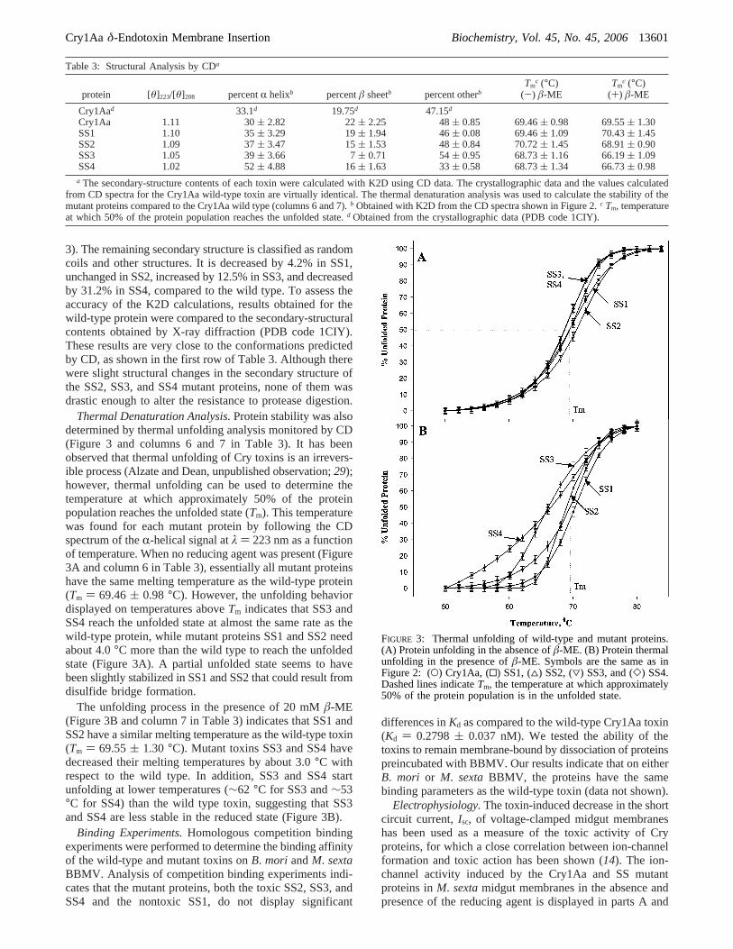
3). The remaining secondary structure is classified as randomcoils and other structures. It is decreased by 4.2% in SS1,unchanged in SS2, increased by 12.5% in SS3, and decreasedby 31.2% in SS4, compared to the wild type. To assess theaccuracy of the K2D calculations, results obtained for thewild-type protein were compared to the secondary-structuralcontents obtained by X-ray diffraction (PDB code 1CIY).These results are very close to the conformations predictedby CD, as shown in the first row of Table 3. Although therewere slight structural changes in the secondary structure ofthe SS2, SS3, and SS4 mutant proteins, none of them wasdrastic enough to alter the resistance to protease digestion.
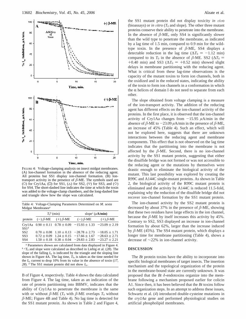
Thermal Denaturation Analysis.Protein stability was alsodetermined by thermal unfolding analysis monitored by CD(Figure 3 and columns 6 and 7 in Table 3). It has beenobserved that thermal unfolding of Cry toxins is an irrevers-ible process (Alzate and Dean, unpublished observation;29);however, thermal unfolding can be used to determine thetemperature at which approximately 50% of the proteinpopulation reaches the unfolded state (Tm). This temperaturewas found for each mutant protein by following the CDspectrum of theR-helical signal atλ ) 223 nm as a functionof temperature. When no reducing agent was present (Figure3A and column 6 in Table 3), essentially all mutant proteinshave the same melting temperature as the wild-type protein(Tm ) 69.46( 0.98 °C). However, the unfolding behaviordisplayed on temperatures aboveTm indicates that SS3 andSS4 reach the unfolded state at almost the same rate as thewild-type protein, while mutant proteins SS1 and SS2 needabout 4.0°C more than the wild type to reach the unfoldedstate (Figure 3A). A partial unfolded state seems to havebeen slightly stabilized in SS1 and SS2 that could result fromdisulfide bridge formation.
The unfolding process in the presence of 20 mMâ-ME(Figure 3B and column 7 in Table 3) indicates that SS1 andSS2 have a similar melting temperature as the wild-type toxin(Tm ) 69.55( 1.30 °C). Mutant toxins SS3 and SS4 havedecreased their melting temperatures by about 3.0°C withrespect to the wild type. In addition, SS3 and SS4 startunfolding at lower temperatures (∼62 °C for SS3 and∼53°C for SS4) than the wild type toxin, suggesting that SS3and SS4 are less stable in the reduced state (Figure 3B).
Binding Experiments.Homologous competition bindingexperiments were performed to determine the binding affinityof the wild-type and mutant toxins onB. mori andM. sextaBBMV. Analysis of competition binding experiments indi-cates that the mutant proteins, both the toxic SS2, SS3, andSS4 and the nontoxic SS1, do not display significant
differences inKd as compared to the wild-type Cry1Aa toxin(Kd ) 0.2798( 0.037 nM). We tested the ability of thetoxins to remain membrane-bound by dissociation of proteinspreincubated with BBMV. Our results indicate that on eitherB. mori or M. sextaBBMV, the proteins have the samebinding parameters as the wild-type toxin (data not shown).
Electrophysiology.The toxin-induced decrease in the shortcircuit current,Isc, of voltage-clamped midgut membraneshas been used as a measure of the toxic activity of Cryproteins, for which a close correlation between ion-channelformation and toxic action has been shown (14). The ion-channel activity induced by the Cry1Aa and SS mutantproteins inM. sextamidgut membranes in the absence andpresence of the reducing agent is displayed in parts A and
Table 3: Structural Analysis by CDa
protein [θ]223/[θ]208 percentR helixb percentâ sheetb percent otherbTm
c (°C)(-) â-ME
Tmc (°C)
(+) â-ME
Cry1Aad 33.1d 19.75d 47.15d
Cry1Aa 1.11 30( 2.82 22( 2.25 48( 0.85 69.46( 0.98 69.55( 1.30SS1 1.10 35( 3.29 19( 1.94 46( 0.08 69.46( 1.09 70.43( 1.45SS2 1.09 37( 3.47 15( 1.53 48( 0.84 70.72( 1.45 68.91( 0.90SS3 1.05 39( 3.66 7( 0.71 54( 0.95 68.73( 1.16 66.19( 1.09SS4 1.02 52( 4.88 16( 1.63 33( 0.58 68.73( 1.34 66.73( 0.98a The secondary-structure contents of each toxin were calculated with K2D using CD data. The crystallographic data and the values calculated
from CD spectra for the Cry1Aa wild-type toxin are virtually identical. The thermal denaturation analysis was used to calculate the stability of themutant proteins compared to the Cry1Aa wild type (columns 6 and 7).b Obtained with K2D from the CD spectra shown in Figure 2.c Tm, temperatureat which 50% of the protein population reaches the unfolded state.d Obtained from the crystallographic data (PDB code 1CIY).
FIGURE 3: Thermal unfolding of wild-type and mutant proteins.(A) Protein unfolding in the absence ofâ-ME. (B) Protein thermalunfolding in the presence ofâ-ME. Symbols are the same as inFigure 2: (O) Cry1Aa, (0) SS1, (4) SS2, (3) SS3, and (]) SS4.Dashed lines indicateTm, the temperature at which approximately50% of the protein population is in the unfolded state.
Cry1Aa δ-Endotoxin Membrane Insertion Biochemistry, Vol. 45, No. 45, 200613601
B of Figure 4, respectively. Table 4 shows the data calculatedfrom Figure 4. The lag time, taken as an indication of therate of protein partitioning into BBMV, indicates that theability of Cry1Aa to penetrate the membrane is the samewith or without â-ME (T0 with â-ME overlapsT0 withoutâ-ME; Figure 4B and Table 4). No lag time is detected forthe SS1 mutant protein. As shown in Table 2 and Figure 4,
the SS1 mutant protein did not display toxicityin ViVo(bioassays) orin Vitro (T0 and slope). The other three mutantproteins conserve their ability to penetrate into the membrane.In the absence ofâ-ME, only SS4 is significantly slowerthan the wild type to penetrate the membrane, as indicatedby a lag time of 1.5 min, compared to 0.9 min for the wild-type toxin. In the presence ofâ-ME, SS4 displays adetectable reduction in the lag time (∆T0 ) -1.12 min)compared to itsT0 in the absence ofâ-ME. SS2 (∆T0 )+0.40 min) and SS3 (∆T0 ) +0.52 min) showed slightdelays in membrane partitioning with the reducing agent.What is critical from these lag-time observations is thecapacity of the mutant toxins to form ion channels, both inthe oxidized and in the reduced states, indicating the abilityof the toxin to form ion channels in a conformation in whichtheR helices of domain I do not need to separate from eachother.
The slope obtained from voltage clamping is a measureof the ion-transport activity. The addition of the reducingagent has different effects on the ion-channel activity of theproteins. In the first place, it is observed that the ion-channelactivity of Cry1Aa changes from-15.95 µA/min in theabsence ofâ-ME to -23.09µA/min in the presence ofâ-ME,an increase of 45% (Table 4). Such an effect, which willnot be explored here, suggests that there are unknowninteractions between the reducing agent and membranecomponents. This effect that is not observed on the lag timeindicates that the partitioning into the membrane is notaffected by theâ-ME. Second, there is no ion-channelactivity by the SS1 mutant protein, suggesting that eitherthe disulfide bridge was not formed or was not accessible tothe reducing agent or the mutations by themselves weredrastic enough to eliminate the biological activity of themutant. This last possibility was explored by creating theR99C and A144C single-mutant proteins. As shown in Table2, the biological activity of the R99C mutant protein iseliminated and the activity by A144C is reduced 11.5-fold,explaining why the reduction of the disulfide bridge did notrecover ion-channel formation by the SS1 mutant protein.
The ion-channel activity by the SS2 mutant protein isdecreased by about 37% in the presence ofâ-ME, showingthat these two residues have large effects in the ion channel,because theâ-ME by itself increases this activity by 45%.Contrary to SS2, SS3 displayed an increase in ion-channelformation by about 62%, larger than the increase inducedby â-ME (45%). The SS4 mutant protein, which displays alonger time for membrane partitioning (Table 4), shows adecrease of∼22% in ion-channel activity.
DISCUSSION
The Bt protein toxins have the ability to incorporate intospecific biological membranes of target insects. The insertionmechanism and the topological organization of the proteinin the membrane-bound state are currently unknown. It wasproposed that theBt δ-endotoxins organize into the mem-brane following a mechanism proposed earlier for colicinA1. Since then, it has been believed that theBt toxins followsuch organization steps. In an attempt to address these issues,Schwartz et al. (4) introduced double-cysteine mutations inthe cry1Aa gene and performed physiological studies onartificial phospholipid membranes.
FIGURE 4: Voltage-clamping analysis on insect midgut membranes.(A) Ion-channel formation in the absence of the reducing agent.All proteins but SS1 display ion-channel formation. (B) Ion-transport activity in the presence ofâ-ME. The symbols used are(O) for Cry1Aa, (0) for SS1, (4) for SS2, (3) for SS3, and (])for SS4. The short-dashed line indicates the time at which the toxinwas added to the voltage-clamp chambers, and the long-dashed lineand triangle show how the slope was calculated.
Table 4: Voltage-Clamping Parameters Determined onM. sextaMidgut Membranesa
T0b (min) slopec (µA/min)
protein (-) â-ME (+) â-ME (-) â-ME (+) â-ME
Cry1Aa 0.90( 0.11 0.78( 0.09 -15.93( 1.33 -23.09( 2.19SS1d
SS2 0.70( 0.08 1.10( 0.13 -28.78( 2.73 -18.05( 1.71SS3 0.72( 0.09 1.24( 0.15 -17.66( 1.67 -28.63( 2.71SS4 1.50( 0.18 0.38( 0.04 -29.83( 2.83 -23.27( 2.21
a Parameters shown are calculated from data displayed in Figure 4.b ,cT0 and slope were calculated as described in Liebig et al. (28). Theslope of the fallingIsc is indicated by the triangle and the sloping lineshown in Figure 4A. The lag time,T0, is taken as the time needed forthe Isc current to drop 10% from its value in the absence of toxin (17,28). d The SS1 mutant protein did not showIsc.
13602 Biochemistry, Vol. 45, No. 45, 2006 Alzate et al.
Following similar approaches, we introduced the samedouble-cysteine mutations described by Schwartz et al. intothe Cry1Aa toxin to form disulfide bridges (Figure 1 andTable 1). The formation of such bridges would hold someconformations but not permit the partition of the toxin intothe membrane as proposed by the umbrella model. Theeffects of these mutations on the toxicity of the resultingproteins, on their ability to form ion channels, and on thestructure of these proteins were assessed as a mechanisticapproach to test the validity of the models in biologicalmembranes of susceptible insects.
Toxicity assays reveal that the SS1 mutant protein (R99C-A144C) did not show any toxic effect up to the maximumconcentration tested (4000 ng/cm2). The other three mutanttoxins have reduced toxicity compared to the wild-type toxin.AgainstM. sexta, SS2, SS3, and SS4 are 4, 14, and 10 timesless toxic than the wild-type protein, respectively. Somereasons could be argued to explain the drop in toxicity ofthe mutant proteins, including (i) structural alterationsresulting from the mutations, (ii) the formation of thedisulfide bridges, (iii) the direct involvement of the mutatedresidues in the protein-receptor binding interaction, and (iv)the direct involvement of the altered residues in ion-channelactivity.
The possibility of the toxicity loss as a result of confor-mational changes was examined by analyzing the secondarystructure of the proteins via CD (Figure 2 and Table 3). Fromthe information contained in the Protein Data Bank, it isknown that the Cry1Aa toxin has 33.10%R-helix, 19.75%â-sheet, and 47.15% remaining secondary structure andrandom-coil conformations. Our calculated secondary-structural contents for the wild-type protein derived fromCD using K2D indicate that the contents ofR helix is 30(2.82%, ofâ sheet is 22( 2.25%, and of other secondary-structural contents is 48( 0.85%, in agreement with thecrystallographic data. On the basis of these results, it is clearthat there are no CD-detectable changes in the structure ofthe SS1 mutant protein. On the other hand, SS2, SS3, andSS4 have increased theirR-helical contents while decreasingtheir amount ofâ-sheet conformations. These results suggestthat in SS2, SS3, and SS4 the disulfide bridges were formed,because more rigidR-helical conformations will lead to ahigherR-helical CD signal. SS4 shows the highest increasein R-helical contents, suggesting that a larger portion ofdomain I has reduced mobility, as expected from domain Ibeing strongly linked to domain II. This is further supportedby the small change in the amount ofâ sheet and theconsiderable decrease in the random-coil conformationdisplayed by SS4. SS3 has a considerable increase in randomcoil, suggesting a tendency toward more unfolded domainsII and/or III. The packing of domain I was also affected inmutant toxins SS3 and SS4 as evidenced by the [θ]223/[θ]208
ratio (Table 3). Nonetheless, none of the mutations alteredthe proteins to a level that would make them susceptible toprotease digestion as determined by 10% SDS-PAGE. It isreasonable to conclude, on the basis of the CD data, that thelack of toxicity in SS1 is not the result of changes in thesecondary structure of the protein and that disulfide bridgeswere formed in SS2, SS3, and SS4.
We then tested whether the disulfide bridges had effectson insecticidal activity. Toxic levels were determined in thepresence ofâ-ME to reduce the disulfide bridges. The
proteins were either mixed with 200 times molar excess ofâ-ME or preincubated over a 12 h period previous to thetest. There were no significant changes in toxicity levelscompared to the results withoutâ-ME (Table 2). Theseresults could be explained by several possibilities. It couldbe that the disulfide bridges were reduced in the insectmidgut; therefore, bioassays in the absence and presence ofâ-ME will not make any difference. Another possibilitywould be a protein inserting in the midgut membrane as awhole molecule or at least domains I and II together;therefore, the presence of disulfide bridges limiting theseparations of theR helices in domain I will not havesignificant contributions to toxicity levels. It also could bethat the disulfide bridges are not affected by the reducingagent and that the midgut environment is not strong enoughto reduce those disulfide bridges.
We tested whether the disulfide bridges were reduced byâ-ME using thermal denaturation analysis followed by CD.On the basis of our experience with disulfide introductioninto Cry3Aa (30), we expected the melting curve of the SSmutants in Cry1Aa to show an increase in theTm temperature.As shown in Figure 3A, in the absence ofâ-ME, changes inTm were small and not easily attributable to disulfide bridgeformation. However, in the presence ofâ-ME, SS2, SS3,and SS4 have decreased theirTm, suggesting that the disulfidebridges were broken by the reducing agent (Figure 3B). Thedecrease inTm is particularly large for SS3 and SS4,indicating that these mutant proteins were readily affectedby the absence of the wild-type amino acid residues. SS3shows a clear deviation from the wild-type stability bystarting to unfold at about 62°C, while the SS4 mutantprotein starts unfolding at about 53°C. These changes instructural stability explain the decrease in toxicity levelsobserved for SS3 and SS4 in the reduced state. It is alsoseen that a partially folded conformation is stabilized in SS1and SS2 that likely results from the presence of a disulfidebridge, which is reduced byâ-ME, allowing the proteins torecover their wild-type-like thermal stability. The datapresented up to this point are sufficient to conclude that SS1,SS2, SS3, and SS4 have formed disulfide bridges, that thesedisulfide bridges are reduced byâ-ME, and that the reductionof toxicity displayed by SS3 and SS4 is likely to result fromstructural alterations detected in the reduced state.
The possibility that the disulfide bridges are reduced bythe insect midgut and, therefore, that the addition ofâ-MEwill not have an effect on toxicity assays was further studiedby analyzing the ion-transport activity of these mutantproteins in insect midgut membranes. As observed in Figure4A, the SS2, SS3, and SS4 mutant proteins conserved theirion-channel function in the absence of the reducing agent.A detailed observation of the ion-transport parameters,T0
and slope, shed some light to the membrane-proteininteraction. The lag time for Cry1Aa in the absence andpresence ofâ-ME is the same, indicating that the reducingagent has no effect on protein translocation into the midgutmembrane. The lag times for the intrahelical disulfidebridges, SS2 and SS3, under reducing conditions are longerthan the lag times under nonreducing conditions. Theseresults suggest that a more rigid, more organizedR-helicaldomain I has a better ability to translocate into the mem-branes. These results are in agreement with previous studies,proposing that someR helices were important for the
Cry1Aa δ-Endotoxin Membrane Insertion Biochemistry, Vol. 45, No. 45, 200613603
initiation of the protein translocation process (13). The lagtime for SS4 shows that under nonreducing conditions thisprotein is slower inserting into the membrane, a propertythat is recovered by the reduction of the disulfide bridge.This demonstrates that the disulfide bridges are not reducedby the midgut alone and that a separation of domain I fromdomain II is important for the initiation of the translocationprocess. Analysis of the ion-transport activities, the slope,is a little more complicated because individual amino acidsubstitutions have an effect on toxicity and the presence ofâ-ME has a substantial effect on this process as shown bychanges in the slope for Cry1Aa under reducing andnonreducing conditions. However, it is important to realizethat, with the exception of SS1, the remaining mutant toxinshave ion-transport ability, suggesting that the disulfidebridges did not impede protein translocation into themembrane.
A particular situation is observed with the SS1 mutantprotein. There was not detectable toxicity against any of theinsects, under either oxidizing or reducing conditions.Structural changes were not observed, and ion-transportactivity was undetected for this mutant protein. Beforeconcluding thatR helices 3 and 4 need to be free from eachother for the protein to form ion channels, we tested theeffects of the individual mutations. Both residues, Arg99 andAla144, have affected the toxicity levels of the protein (Table2). The fact that Arg99Cys eliminated the toxic effect ofthe protein suggests that Arg at position 99 is required fortoxicity. The lack of toxicity was the result of removing thearginine residue from such a position and not the result ofthe disulfide bridge formation; therefore, this mutant proteindoes not offer functional information concerning the insertionmechanism of Cry1A toxins in biological membranes.
The main goal for this research was to obtain informationabout the insertion process of Cry1Aa into the midgutmembrane of two lepidopteran insects. The double-cysteinemutations in the SS1, SS2, SS3, and SS4 Cry1Aa mutantproteins were introduced to form disulfide bridges. It isobserved that SS2, SS3, and SS4 have reduced their toxicitylevels, but it has not been eliminated completely. The SS1mutant toxin, designed to form a disulfide bridge betweenR helices 3 and 4, lost all toxic activity that was neither theresult of conformational nor structural alterations. Thelocking of R helices 5 and 6 by SS2,R helices 5 and 7 bySS3, or domain I and domain II by SS4 was a factor thatneither prevented the toxins to partition into the membranenor eliminated ion-transport activity. However, eliminatingthe disulfide bridge in SS4 reduced the partition time,suggesting that some degree of freedom helps the penetrationof the toxin into the membrane.
We and others have demonstrated thatR helix 7 of Cry1Aalso participates in ion transport (14, 15). We demonstratedthat the preincubation of Cry1Ab with BBMV protects thewhole toxin from proteolytic digestion by proteinase K (17).In a more recent work, Tomimoto et al. (20) have proposeda “buried dragon model” in which the whole proteinassociates with the midgut membrane and is protected fromdigestion by pronase. The data presented here indicate thatthere is no need for theR helices to separate from each otherfor the protein to partition in the midgut membrane andtherefore to be toxic. These results do not support proteinpartition models in which individualR-helical hairpins are
required to penetrate the membrane by themselves. Rather,an alternative model, in which the protein partitions as awhole molecule in the lipid environment, will be in betteragreement with these data.
REFERENCES
1. Schnepf, E., Crickmore, N., van Rie, J., Lereclus, D., Baum, J.,Feitelson, J., Zeigler, D. R., and Dean, D. H. (1998)Bacillusthuringiensisand its pesticidal crystal proteins,Microbiol. Mol.Biol. ReV. 62, 775-806.
2. Grochulski, P., Masson, L., Borisova, S., Pusztai-Carey, M.,Schwartz, J. L., Brousseau, R., and Cygler, M. (1995)BacillusthuringiensisCryIA(a) insecticidal toxin: Crystal structure andchannel formation,J. Mol. Biol. 254, 447-464.
3. Li, J. D., Carroll, J., and Ellar, D. J. (1991) Crystal structure ofinsecticidal δ-endotoxin fromBacillus thuringiensisat 2.5 Åresolution,Nature 353, 815-821.
4. Schwartz, J. L., Juteau, M., Grochulski, P., Cygler, M., Prefontaine,G., Brousseau, R., and Masson, L. (1997) Restriction of intramo-lecular movements within the Cry1Aa toxin molecule ofBacillusthuringiensisthrough disulfide bond engineering,FEBS Lett. 410,397-402.
5. Ge, A. Z., Shivarova, N. I., and Dean, D. H. (1989) Location ofthe Bombyx morispecificity domain on aBacillus thuringiensisδ-endotoxin protein,Proc. Natl. Acad. Sci. U.S.A. 86, 4037-4041.
6. Lu, H., Rajamohan, F., and Dean, D. H. (1994) Identification ofamino acid residues ofBacillus thuringiensisδ-endotoxin CryIAaassociated with membrane binding and toxicity toBombyx mori,J. Bacteriol. 176, 5554-5559.
7. Rajamohan, F., Hussain, S. R., Cotrill, J. A., Gould, F., and Dean,D. H. (1996) Mutations at domain II, loop 3, ofBacillusthuringiensisCryIAa and CryIAbδ-endotoxins suggest loop 3 isinvolved in initial binding to lepidopteran midguts,J. Biol. Chem.271, 25220-25226.
8. Lee, M. K., Young, B. A., and Dean, D. H. (1995) Domain IIIexchanges ofBacillus thuringiensisCryIA toxins affect bindingto different gypsy moth midgut receptors,Biochem. Biophys. Res.Commun. 216, 306-312.
9. de Maagd, R. A., Kwa, M. S., van der Klei, H., Yamamoto, T.,Schipper, B., Vlak, J. M., Stiekema, W. J., and Bosch, D. (1996)Domain III substitution inBacillus thuringiensisδ-endotoxinCryIA(b) results in superior toxicity forSpodoptera exiguaandaltered membrane protein recognition,Appl. EnViron. Microbiol.62, 1537-1543.
10. Hodgman, T. C., and Ellar, D. J. (1990) Models for the structureand function of theBacillus thuringiensisδ-endotoxins determinedby compilational analysis,DNA Sequence 1, 97-106.
11. Lakey, J. H., Gonzalez-Manas, J. M., van der Goot, F. G., andPattus, F. (1992) The membrane insertion of colicins,FEBS Lett.307, 26-29.
12. Parker, M. W., Tucker, A. D., Tsernoglou, D., and Pattus, F. (1990)Insights into membrane insertion based on studies of colicins,Trends Biochem. Sci. 15, 126-129.
13. Gazit, E., La Rocca, P., Sansom, M. S., and Shai, Y. (1998) Thestructure and organization within the membrane of the helicescomposing the pore-forming domain ofBacillus thuringiensisδ-endotoxin are consistent with an “umbrella-like” structure ofthe pore,Proc. Natl. Acad. Sci. U.S.A. 95, 12289-12294.
14. Alcantara, E. P., Alzate, O., Lee, M. K., Curtiss, A., and Dean,D. H. (2001) Role ofR-helix seven ofBacillus thuringiensisCry1Ab δ-endotoxin in membrane insertion, structural stability,and ion channel activity,Biochemistry 40, 2540-2547.
15. Chandra, A., Ghosh, P., Mandaokar, A. D., Bera, A. K., Sharma,R. P., Das, S., and Kumar, P. A. (1999) Amino acid substitutionin R-helix 7 of Cry1Acδ-endotoxin ofBacillus thuringiensisleadsto enhanced toxicity toHelicoVerpa armigeraHubner,FEBS Lett.458, 175-179.
16. Dean, D. H., Rajamohan, F., Lee, M. K., Wu, S. J., Chen, X. J.,Alcantara, E., and Hussain, S. R. (1996) Probing the mechanismof action of Bacillus thuringiensisinsecticidal proteins by site-directed mutagenesissA minireview, Gene 179, 111-117.
17. Arnold, S., Curtiss, A., Dean, D. H., and Alzate, O. (2001) Therole of a proline-induced broken-helix motif inR-helix 2 ofBacillus thuringiensisδ-endotoxins,FEBS Lett. 490, 70-74.
18. Aronson, A. I., and Shai, Y. (2001) WhyBacillus thuringiensisinsecticidal toxins are so effective: Unique features of their modeof action,FEMS Microbiol. Lett. 195, 1-8.
13604 Biochemistry, Vol. 45, No. 45, 2006 Alzate et al.
19. Loseva, O. I., Tiktopulo, E. I., Vasiliev, V. D., Nikulin, A. D.,Dobritsa, A. P., and Potekhin, S. A. (2001) Structure of Cry3Aδ-endotoxin within phospholipid membranes,Biochemistry 40,14143-14151.
20. Tomimoto, K., Hayakawa, T., and Hori, H. (2006) Pronasedigestion of brush border membrane-bound Cry1Aa shows thatalmost the whole activated Cry1Aa molecule penetrates into themembrane,Comp. Biochem. Physiol., B: Biochem. Mol. Biol. 144,413-422.
21. Bravo, A., Gomez, I., Conde, J., Munoz-Garay, C., Sanchez, J.,Miranda, R., Zhuang, M., Gill, S. S., and Soberon, M. (2004)Oligomerization triggers binding of aBacillus thuringiensisCry1Ab pore-forming toxin to aminopeptidase N receptor leadingto insertion into membrane microdomains,Biochim. Biophys. Acta.1667, 38-46.
22. Schnepf, H. E., and Whiteley, H. R. (1981) Cloning and expressionof the Bacillus thuringiensiscrystal protein gene inEscherichiacoli, Proc. Natl. Acad. Sci. U.S.A. 78, 2893-2897.
23. Sambrook, J., Fritsch, E. F., Maniatis, T. (1989)MolecularCloning: A Laboratory Manual, 2 ed., Cold Spring HarborLaboratory Press, Plainview, NY.
24. Andrade, M. A., Chacon, P., Merelo, J. J., and Moran, F. (1993)Evaluation of secondary structure of proteins from UV circular
dichroism spectra using an unsupervised learning neural network,Protein Eng. 6, 383-390.
25. Wolfersberger, M. G. (1993) Preparation and partial characteriza-tion of amino acid transporting brush border membrane vesiclesfrom the larval midgut of the gypsy moth (Lymantria dispar),Arch. Insect Biochem. Physiol. 24, 139-147.
26. Liang, Y., Patel, S. S., and Dean, D. H. (1995) Irreversible bindingkinetics of Bacillus thuringiensisCryIA δ-endotoxins to gypsymoth brush border membrane vesicles is directly correlated totoxicity, J. Biol. Chem. 270, 24719-24724.
27. Harvey, W. R., Crawford, D. N., and Spaeth, D. D. (1990)Isolation, voltage clamping, and flux measurements in lepidopteranmidgut,Methods Enzymol. 192, 599-608.
28. Liebig, B., Stetson, D. L., and Dean, D. H. (1995) Quantificationof the effect ofBacillus thuringiensistoxins on short-circuit currentin the midgut ofBombyx mori, J. Insect Physiol. 41, 17-22.
29. Potekhin, S. A., Loseva, O. I., Tiktopulo, E. I., and Dobritsa, A.P. (1999) Transition state of the rate-limiting step of heatdenaturation of Cry3Aδ-endotoxin,Biochemistry 38, 4121-4127.
30. Wu, S. J. (1996) inBiochemistry, The Ohio State University,Columbus, OH.
BI061474Z
Cry1Aa δ-Endotoxin Membrane Insertion Biochemistry, Vol. 45, No. 45, 200613605


![Fibronectin Fibronectin exists as a dimer, consisting of two nearly identical polypeptide chains linked by a pair of C-terminal disulfide bonds. [3] Each.](https://static.fdocument.org/doc/165x107/56649d4e5503460f94a2e7cf/fibronectin-fibronectin-exists-as-a-dimer-consisting-of-two-nearly-identical.jpg)