Effect of Depolarization on Binding Kinetics of Scorpion α-Toxin Highlights Conformational Changes...
9
Effect of Depolarization on Binding Kinetics of Scorpion R-Toxin Highlights Conformational Changes of Rat Brain Sodium Channels ² Nicolas Gilles, ‡ Enrico Leipold, § Haijun Chen, § Stefan H. Heinemann, § and Dalia Gordon* ,| Commissaariat a ` l’Energie Atomique, De ´ partement d’Inge ´ nierie et d’Etudes des Prote ´ ines, Centre d’Etudes de Saclay, 91191 Gif-sur-YVette, France, Molecular and Cellular Biophysics, Medical Faculty of the Friedrich Schiller UniVersity Jena, Drackendorfer Strasse 1, D-07747 Jena, Germany, and Department of Plant Sciences, Tel-AViV UniVersity, Ramat-AViV, Tel AViV, 69978 Israel, and Sigyn Pharmaceuticals Ltd., Post Office Box 45010 Jerusalem, 91450 Israel ReceiVed May 11, 2001; ReVised Manuscript ReceiVed August 24, 2001 ABSTRACT: Binding of scorpion R-toxins to receptor site 3 on voltage-gated sodium channels inhibits sodium current inactivation and is voltage-dependent. To reveal the direct effect of depolarization, we analyzed binding kinetics of the R-toxin Lqh-II (from Leiurus quinquestriatus hebraeus) to rat brain synaptosomes and effects on rat brain II (rBII) channels expressed in mammalian cells. Our results indicated that the 33-fold decrease in toxin affinity for depolarized (0 mV, 90 mM [K + ] out , K d ) 5.85 ( 0.5 nM) versus polarized (-55 mV, 5 mM [K + ] out , K d ) 0.18 ( 0.04 nM) synaptosomes at steady state results from a 48-fold reduction in the association rate (k on at 5 mM [K + ] ) (12.0 ( 4) × 10 6 M -1 s -1 and (0.25 ( 0.03) × 10 6 M -1 s -1 at 90 mM [K + ] out ) with nearly no change in the dissociation rate. Electrophysiological analyses of rBII channels expressed in mammalian cells revealed that approximately 75% and 40% of rBII occupied fast- and slow-inactivated states, respectively, at resting membrane potential of synaptosomes (-55 mV), and Lqh-II markedly increased the steady-state fast and slow inactivation. To mimic electrophysiological conditions we induced fast depolarization of toxin-bound synaptosomes, which generated a biphasic unbinding of Lqh-II from toxin-receptor complexes. The first fast off rate closely resembled values determined electrophysiologically for rBII in mammalian cells. The second off rate was similar to the voltage-independent steady-state value, attributed to binding to the slow-inactivated channel states. Thus, the Lqh-II voltage-dependent affinity highlights two independent mechanisms representing conformational changes of sodium channels associated with transitions among electrically visible and invisible inactivated states. Voltage-gated sodium channels (Na + channels) are re- sponsible for the generation and propagation of action potentials in most excitable cells. These channels are composed of a pore-forming R-subunit, which contains four domains consisting each of six transmembrane segments (1). The fourth transmembrane segment (S4) in each domain contains several positive charges and serves as the voltage sensor of the channel (2, 3). The outward movements of S4 segments initiates the voltage-dependent activation of the Na + channel under membrane depolarization (2, 4, 5), with S4 of domain IV preceding the movement of the others (6). Domain IV prevails in the fast inactivation of Na + channels (6-12), a process that involves also the intracellular linker between domains III and IV (2, 13, 14). Many neurotoxins bind to Na + channels at several receptor sites and affect the Na + current in various fashions (15- 17). Toxins that target Na + channel receptor site 3 bind in a voltage-dependent manner and inhibit the fast inactivation process (15, 16, 18). Receptor site 3 involves external regions of channel domains I and IV (19, 20). A negatively charged amino acid residue at the external loop connecting segments S3 and S4 in domain IV has been shown to be very important for binding of site 3 toxins (21-23). In turn, the bioactive surface of site 3 toxins contains positively charged residues (22, 24, 25). Binding of scorpion R-toxins has been useful in probing subtle conformational changes at the channel surface that occur during voltage-dependent gating as well as during modulation provoked by other groups of neuro- toxins (15, 16, 26-29). However, the structural basis of the voltage dependency in binding of site 3 toxins and the mechanism by which binding to channel external regions affects fast inactivation, which is presumably mediated by intracellular protein components, is still elusive. Depolarization of the membrane induces a dramatic decrease of the scorpion R-toxin affinity to Na + channels, as has been demonstrated in both electrophysiological and binding studies (21, 26, 30-36). The voltage-dependent drop in binding affinity has been attributed to an increase in the dissociation rate constant of R-toxins under depolarized conditions (21, 30-32, 35-37). The voltage dependence of the apparent binding correlates well with the voltage dependence of channel activation and the coupled fast ² This work was supported in part by the Israeli Science Foundation (508/00, to D.G.). * Corresponding author: Department of Plant Sciences, Tel-Aviv University, Ramat-Aviv, Tel Aviv 69978, Israel. E-mail dgordon@ post.tau.ac.il; fax +972-3-640-6100; phone +972-3-640-9844. ‡ CEA, De ´partement d'Inge ´nierie et d'Etudes des Prote ´ines, C.E. Saclay. § Medical Faculty of the Friedrich Schiller University Jena. | Tel-Aviv University and Sigyn Pharmaceuticals Ltd. 14576 Biochemistry 2001, 40, 14576-14584 10.1021/bi010973r CCC: $20.00 © 2001 American Chemical Society Published on Web 10/30/2001
Transcript of Effect of Depolarization on Binding Kinetics of Scorpion α-Toxin Highlights Conformational Changes...

Effect of Depolarization on Binding Kinetics of ScorpionR-Toxin HighlightsConformational Changes of Rat Brain Sodium Channels†
Nicolas Gilles,‡ Enrico Leipold,§ Haijun Chen,§ Stefan H. Heinemann,§ and Dalia Gordon*,|
Commissaariat a` l’Energie Atomique, De´partement d’Inge´nierie et d’Etudes des Prote´ines, Centre d’Etudes de Saclay,91191 Gif-sur-YVette, France, Molecular and Cellular Biophysics, Medical Faculty of the Friedrich Schiller UniVersity Jena,
Drackendorfer Strasse 1, D-07747 Jena, Germany, and Department of Plant Sciences, Tel-AViV UniVersity, Ramat-AViV,Tel AViV, 69978 Israel, and Sigyn Pharmaceuticals Ltd., Post Office Box 45010 Jerusalem, 91450 Israel
ReceiVed May 11, 2001; ReVised Manuscript ReceiVed August 24, 2001
ABSTRACT: Binding of scorpionR-toxins to receptor site 3 on voltage-gated sodium channels inhibitssodium current inactivation and is voltage-dependent. To reveal the direct effect of depolarization, weanalyzed binding kinetics of theR-toxin Lqh-II (from Leiurus quinquestriatus hebraeus) to rat brainsynaptosomes and effects on rat brain II (rBII) channels expressed in mammalian cells. Our results indicatedthat the 33-fold decrease in toxin affinity for depolarized (0 mV, 90 mM [K+]out, Kd ) 5.85( 0.5 nM)versus polarized (-55 mV, 5 mM [K+]out, Kd ) 0.18 ( 0.04 nM) synaptosomes at steady state resultsfrom a 48-fold reduction in the association rate (kon at 5 mM [K+] ) (12.0( 4) × 106 M-1 s-1 and (0.25( 0.03) × 106 M-1 s-1 at 90 mM [K+]out) with nearly no change in the dissociation rate.Electrophysiological analyses of rBII channels expressed in mammalian cells revealed that approximately75% and 40% of rBII occupied fast- and slow-inactivated states, respectively, at resting membrane potentialof synaptosomes (-55 mV), and Lqh-II markedly increased the steady-state fast and slow inactivation.To mimic electrophysiological conditions we induced fast depolarization of toxin-bound synaptosomes,which generated a biphasic unbinding of Lqh-II from toxin-receptor complexes. The first fast off rateclosely resembled values determined electrophysiologically for rBII in mammalian cells. The second offrate was similar to the voltage-independent steady-state value, attributed to binding to the slow-inactivatedchannel states. Thus, the Lqh-II voltage-dependent affinity highlights two independent mechanismsrepresenting conformational changes of sodium channels associated with transitions among electricallyvisible and invisible inactivated states.
Voltage-gated sodium channels (Na+ channels) are re-sponsible for the generation and propagation of actionpotentials in most excitable cells. These channels arecomposed of a pore-formingR-subunit, which contains fourdomains consisting each of six transmembrane segments (1).The fourth transmembrane segment (S4) in each domaincontains several positive charges and serves as the voltagesensor of the channel (2, 3). The outward movements of S4segments initiates the voltage-dependent activation of theNa+ channel under membrane depolarization (2, 4, 5), withS4 of domain IV preceding the movement of the others (6).Domain IV prevails in the fast inactivation of Na+ channels(6-12), a process that involves also the intracellular linkerbetween domains III and IV (2, 13, 14).
Many neurotoxins bind to Na+ channels at several receptorsites and affect the Na+ current in various fashions (15-17). Toxins that target Na+ channel receptor site 3 bind in a
voltage-dependent manner and inhibit the fast inactivationprocess (15, 16, 18). Receptor site 3 involves external regionsof channel domains I and IV (19, 20). A negatively chargedamino acid residue at the external loop connecting segmentsS3 and S4 in domain IV has been shown to be very importantfor binding of site 3 toxins (21-23). In turn, the bioactivesurface of site 3 toxins contains positively charged residues(22, 24, 25). Binding of scorpionR-toxins has been usefulin probing subtle conformational changes at the channelsurface that occur during voltage-dependent gating as wellas during modulation provoked by other groups of neuro-toxins (15, 16, 26-29). However, the structural basis of thevoltage dependency in binding of site 3 toxins and themechanism by which binding to channel external regionsaffects fast inactivation, which is presumably mediated byintracellular protein components, is still elusive.
Depolarization of the membrane induces a dramaticdecrease of the scorpionR-toxin affinity to Na+ channels,as has been demonstrated in both electrophysiological andbinding studies (21, 26, 30-36). The voltage-dependent dropin binding affinity has been attributed to an increase in thedissociation rate constant ofR-toxins under depolarizedconditions (21, 30-32, 35-37). The voltage dependence ofthe apparent binding correlates well with the voltagedependence of channel activation and the coupled fast
† This work was supported in part by the Israeli Science Foundation(508/00, to D.G.).
* Corresponding author: Department of Plant Sciences, Tel-AvivUniversity, Ramat-Aviv, Tel Aviv 69978, Israel. E-mail [email protected]; fax+972-3-640-6100; phone+972-3-640-9844.
‡ CEA, Departement d'Inge´nierie et d'Etudes des Prote´ines, C.E.Saclay.
§ Medical Faculty of the Friedrich Schiller University Jena.| Tel-Aviv University and Sigyn Pharmaceuticals Ltd.
14576 Biochemistry2001,40, 14576-14584
10.1021/bi010973r CCC: $20.00 © 2001 American Chemical SocietyPublished on Web 10/30/2001

inactivation (21, 26, 31), suggesting that these conformationaltransitions weaken the toxin-channel interaction. However,the interactions ofR-toxins with the electrically silent fast-and slow-inactivated channel states have not been described.Only experiments in which the physical binding of toxinsto Na+ channels are measured can infer about the bindingproperties of such states, but thus far, no detailed analysisof the binding kinetics to the different channel states atpolarized and depolarized membrane potentials have beenreported.
To study the direct effect of membrane depolarization onthe interaction of a scorpionR-toxin with receptor site 3,we have undertaken a detailed kinetic analysis of the bindingof Lqh-II,1 a classical scorpionR-toxin highly active onmammals, fromLeiurus quinquestriatus hebraeus(38), torat brain synaptosomes. Lqh-II is highly similar in effectsand binding properties to previously studied scorpionR-tox-ins (e.g., LqTx or Lqq-V fromLeiurus qinquestriatusqinquestriatus, 15, 21, 26, 30-32) and is almost identicalin sequence to Aah-II, fromAndroctonus australis hector(18, 29, 33, 38). Synaptosomes are capable of retaining aresting membrane potential due to a passive K+ efflux, whichcan be modulated in the range of-55 to 0 mV by changingthe external K+ concentration (32, 33, 39-41). In our bindingstudies we found, unexpectedly, that the dramatic reductionin Lqh-II binding affinity between-55 and 0 mV did notcorrespond with an increase in the dissociation rate constant(as previously concluded;21, 26, 30, 31, 35) but with adecrease in the association rate. The expected markedincrease in the dissociation rate was obtained, however, underfast depolarization of synaptosomes. The physical bindingexperiments with synaptosomes were compared with elec-trophysiological data obtained for rBII Na+ channels ex-pressed in mammalian cells.
EXPERIMENTAL PROCEDURES
Materials. Scorpion toxin II fromLeiurus quinquestriatushebraeuswas from Latoxan (20, Rue Leon Blum, 2600Valance, France) and, in part, was a generous gift of Dr.Pierre Sautie`re (Institut Pasteur, Lille). Reverse-phase C18
(250 × 4.6 mm; 300 Å, 5µm particle size) HPLC columnwas from Vydac. Iodogen was from Pierce Chemical Co.(Rockford, IL). Carrier-free Na125I was from Amersham(Buckinghamshire, U.K.). All other chemicals were ofanalytical grade. Filters for binding assays were GF/C glassfiber (Whatman, Maidstone, U.K.) preincubated in 0.3%polyethylenimine (Sigma, Steinheim, Germany).
Rat Brain Synaptosome Preparation. Rat brain synapto-somes were prepared from adult albino Sprague-Dawleyrats (about 300 g, laboratory bred), according to the methoddescribed by Kanner (42). All buffers contained a mixtureof proteinase inhibitors composed of phenylmethanesulfonylfluoride (50µg/mL), pepstatin A (1µM), iodoacetamide (1mM), and 1,10-phenanthroline (1 mM). All processes wereperformed on ice. The enriched synaptosomal fraction wasfrozen in aliquots at-80 °C. Membrane protein concentra-
tion was determined by Bio-Rad protein assay, with bovineserum albumin (BSA) as standard.
Radioiodination of Lqh-II. Lqh-II was radioiodinated byIodogen (Pierce Chemical Co., Rockford, IL) with 5µg oftoxin and 0.5 mCi of carrier-free Na125I, and the monoiodo-toxin was purified on an analytical Vydac reverse-phase C18
column, as was previously described (43). The concentrationof the radiolabeled toxin was determined according to thespecific activity of the125I, corresponding to 2500-3000dpm/fmol of monoiodotoxin, depending on the age of theradiotoxin and by estimation of its biological activity (usually60-70%; 44).
Binding Assay. Rat brain synaptosomes were thawed at37 °C (for 30 s) and placed on ice. The synaptosomes weresuspended in 0.2 or 1 mL of binding buffer (13.6-78.8µgof protein/mL), containing 33-268 pM125I-Lqh-II, depend-ing on the type of experiment (see figure captions). Afterincubation for the designated time periods, the reactionmixture was diluted with 2 mL of ice-cold wash buffer andfiltered through GF/C filters under vacuum. Filters wererapidly washed twice with 2 mL of wash buffer. Terminationof reaction and washing lasted 10 s. Nonspecific binding ofthe toxin was determined in the presence of a highconcentration of Lqh-II, as specified in the figure legends,and typically accounted for 5-30% of total binding of125I-Lqh-II. Standard binding medium composition (millimolar)was as follows: choline chloride 130, CaCl2 1.8, KCl 5,MgSO4 0.8, HEPES 50, glucose 10, and BSA 2 mg/mL.Wash buffer composition (millimolar) was as follows:choline chloride 140, CaCl2 1.8, KCl 5.4, MgSO4 0.8,HEPES 50 (pH 7.2), and BSA 5 mg/mL. When membraneswere depolarized by high external K+ concentration, cholinechloride was substituted by KCl so that the total concentra-tion of choline and K+ was equal to 135 mM.
Equilibrium and Kinetic Analysis of Binding. Cold satura-tion assays were performed with increasing concentrationsof unlabeled Lqh-II in the presence of a constant lowconcentration of125I-Lqh-II. The data were subjected toanalysis by the iterative program LIGAND (Elsevier Biosoft,Cambridge, U.K.) by cold saturation analysis. The kineticdata for ligand association and dissociation rates weresubjected to analysis by LIGAND, by kinetic analysis. Eachcurve was subjected to multislope analysis to detect thepresence of one or two slopes. Toxin dissociation wasinduced by an excess of cold toxin and the dissociation rateconstant (koff) was determined directly from a first-order plotof toxin dissociation versus time. In some experiments(Figure 4), unbinding of toxin was determined by a timeconstant (τ ) ln 2/koff), estimated directly from the bindingcurves. The rate of toxin association (kon) was determinedfrom the equationkon ) kobs([RL]e/([L][RL] max)), where [L]is the concentration of ligand, [RL]e is the concentration ofthe complex at equilibrium, [RL]max is the maximum numberof receptors present (determined in a parallel saturationexperiment), andkobs is the slope of the pseudo-first-orderplot ln [RL]e/([RL]e - [RL] t) versus time (45). Eachexperiment was performed at least three times and each datapoint represents the mean of 2-3 samples (with up to 12%deviation between samples). Data are presented as mean(SEM of number (n) of independent experiments.
Expression of rBII Sodium Channels in Mammalian Cells.HEK293 or CHO cells were cultured according to standard
1 Abbreviations: Lqh-II, R-toxin II from the scorpionLeiurusquinquestriatus hebraeus; HEPES, 4-(2-hydroxyethyl)-1-piperazineethane-sulfonic acid; BSA, bovine serum albumin;Kd, dissociation constant;kon, kinetic constant of association;koff, kinetic constant of dissociation.
ScorpionR-Toxin Binding to Na+ Channels Biochemistry, Vol. 40, No. 48, 200114577

methods in a humid atmosphere incubator at 5% CO2.Transient transfection of plasmid DNA was performed withthe SuperFect transfection kit according to the manufacturer’sdescription (Qiagen, Hilden, Germany). Cells grown to 30-50% confluence in 35-mm Petri dishes were cotransfectedwith a 5:1 ratio of Na+ channel expression plasmid and avector encoding the CD8 antigen (46). Plasmid DNAencoding Na+ channels and the CD8 antigen was purifiedby Qiagen plasmid maxi kit (Qiagen). The coding segmentsof the rat brain II Na+ channel, rBII (1), was cloned into theexpression vector pCI-neo (Promega), kindly provided byDr. K. Imoto (Okazaki, Japan). No difference in function ofrBII channels was found between channels expressed inHEK293 or CHO cells.
Patch Clamp Recording. For patch clamp experiments, thecells were used 2-4 days after transfection. Before recording,transfected cells were incubated with Dynalbeads (DynalGmbH, Hamburg, Germany) coated with antibodies directedagainst CD8 antigen for at least 30 min at 37°C, in 5%CO2, and washed a few times with the bath solution. Patchclamp experiments were performed with an EPC9 amplifier(HEKA Elektronik, Lambrecht, Germany). Data acquisitionwas controlled with Pulse+PulseFit software (HEKA Ele-ktronik). The patch pipets were fabricated from borosilicateglass and had resistances of 0.8-2.0 MΩ when filled withinternal solution (see below). They were coated with RTV510 (General Electric) in order to reduce noise and electricalcapacitance. Series resistances below 5 MΩ were acceptedand a series resistance compensation of at least 80% wasapplied. Leak and capacitative transient currents weredigitally subtracted online with procedures supported by thePulse+PulseFit software. The holding voltage for all experi-ments was-120 mV.
Patch clamp pipets were filled with a solution containing(millimolar) NaCl 35, CsF 105, EGTA 10, and HEPES 10,pH 7.4. The bath solution contained (millimolar) NaCl 150,KCl 2, CaCl2 1.5, MgCl2 1, and HEPES 10, pH 7.4. Thetemperature was 20( 1 °C.
Lqh-II was dissolved in the bath solution, supplementedwith 1 mg/mL BSA in order to prevent adherence of thetoxin to the vials and the perfusion apparatus. The applicationof toxin was performed with an application pipet of about10 µM opening diameter. The collection of the control datawas initiated 15 min after the whole-cell voltage clampconfiguration was established.
Further data analysis was performed with IgorPro software(WaveMetrics, Lake Oswego, OR). Steady-state inactivationwas described with Boltzmann functions:
with the voltageV, the voltage of half-maximal inactivationVh, the slope factorkh, and the fraction of noninactivatingchannelsani.
Toxin dissociation was assayed by measuring the degreeof inactivation at 0 mV 4.5-5 ms after the start of thedepolarization. For this purpose, the mean current between4.5 and 5 ms was divided by the peak current:I5ms/Ipeak. Allexperiments were performed with twin pulses. In this wayit was assured by the first depolarization that full recoveryfrom slow inactivation and full toxin binding has happened
before conditioning pulses were applied (for further protocoldetails see ref47).
RESULTS
Monitoring Spontaneous Depolarization of Synaptosomes.At resting membrane potential of rat brain synaptosomes(∼-55 mV with 5 mM external K+; 30, 32, 37, 39), classicalR-toxins, such as Aah-II and Lqh-II, bind with high affinity(Kd of 0.2-0.3 nM; 27, 43). The saturable binding of theseR-toxins is reduced by∼90% upon membrane depolarizationwith 90-135 mM K+ (membrane potential of∼0 mV; 29,32, 33, 39, 48). It has been demonstrated that the decreasein binding affinity in high external [K+] was due to its effecton membrane depolarization and not a direct effect onscorpionR-toxin binding, as equivalent changes inR-toxinaffinity occurred when membrane potential was depolarizedby various methods (21, 30, 32, 33). Depolarization of themembrane potential occurs spontaneously during prolongedincubation of synaptosomes in 5 mM [K+] (∼-55 mV)buffer and is monitored by the change in125I-Lqh-II bindingwith time (Figure 1). Specific binding of125I-Lqh-II at 5 mM[K+] reached a maximum level and then decreased at variousrates, depending on the temperature. The decrease in125I-
I/Imax ) ani + (1 - ani)/(1 + exp[-(V - Vh)/kh]) (1)
FIGURE 1: Time course of Lqh-II binding to rat brain synaptosomesat different temperatures. Rat brain synaptosomes were incubatedin 200 µL of binding buffer (see Experimental Procedures) at 37°C (panel A; 78.8µg of protein/mL), 22°C (panel B; 20µg/mL),or 8 °C (panel C; 20µg/mL). Association kinetics were measuredin the presence of125I-Lqh-II at 126 pM (37°C) or 60 pM (8 and22 °C). Typical experiments are presented. Nonspecific bindingdetermined in parallel experiments in the presence of 1µM Lqh-IIwas time-invariant and was subtracted from the experimental points.The binding of125I-Lqh-II reached its maximum level after 5, 10,and 20 min respectively at 37, 22, and 8°C and was stable for 5,30, and more than 240 min before starting to decrease. Maximalbinding (100%) was 0.21, 0.017, and 0.007 fmol/mg at 37, 22, and8 °C, respectively.
14578 Biochemistry, Vol. 40, No. 48, 2001 Gilles et al.

Lqh-II binding was attributed to the passive dissipation ofthe K+ ion gradient with time, which caused progressivedepolarization of the membrane potential. The potential wasstable for 10 min at 37°C (Figure 1A), for 40 min at 22°C(Figure 1B), and for more than 4 h at 8°C (Figure 1C). Theshort-lived steady-state polarized membrane potential at 37°C as well as the low specific binding at 8°C (approximately40% of that at 22°C; see Figure 1 caption) posed difficultiesin performing a detailed kinetic analysis of Lqh-II binding.Thus, all subsequent studies were performed at 22°C, whichallowed the appropriate setting for precise binding analysesunder both kinetic and equilibrium conditions at polarizedmembrane potential at steady state. In addition, this tem-perature allowed a more direct comparison of the bindingdata with results obtained from electrophysiological experi-ments.
Affinity of 125I-Lqh-II for Polarized and Depolarized RatBrain Synaptosomes at Steady State.The change in equi-librium dissociation constant (Kd) of 125I-Lqh-II binding underpolarized (5 mM K+) and depolarized (90 mM K+) condi-tions was determined by Scatchard analysis of saturationbinding curves of Lqh-II (Figure 2). The concentration of125I-Lqh-II and receptor sites (membrane protein) at equi-librium was adjusted to the different apparent affinity valuesto keep the change in free125I-Lqh-II under 10% (see Figure2 caption;45). Under optimal conditions, the specific binding
was 95% and 70% of the total binding in 5 and 90 mM K+,respectively, which enabled an accurate estimation of thebinding parameters under steady-state conditions. The Scat-chard analyses indicated a single receptor binding site, aswas previously described (32, 33). The apparent bindingaffinity of Lqh-II dropped 33-fold from polarized (5 mMK+; Kd ) 0.18( 0.04 nM,n ) 3; mean( SE,n ) numberof experiments) to depolarized (90 mM K+; Kd ) 5.85 (0.5, n ) 4) conditions and was accompanied by a 1.6-foldincrease in receptor site capacity (Bmax ) 0.86( 0.3 pmol/mg of protein andn ) 3 for polarized membranes;Bmax )1.4 ( 0.2 pmol/mg of protein andn ) 4 for depolarizedmembranes) (Figure 2).
Association and Dissociation Kinetics of125I-Lqh-II Bind-ing. Analysis of the binding curves in Figures 1 and 2indicated the significance of measurements at steady-statepolarized conditions for the precise determination of bindingparameters. Hence,125I-Lqh-II dissociation at polarizedmembrane potential was induced by addition of excess Lqh-II (200 nM, Figure 3A, arrow) after 3 min of association.This ensured stable membrane potential throughout thedissociation measurements as indicated by the increase in125I-Lqh-II binding in the control (Figure 3A). Approximately80% of the bound125I-Lqh-II dissociated before spontaneousdepolarization of the membrane could be detected, as wasshown by the gradual decrease in binding after 30 min ofincubation in the control (Figure 3A). The calculatedassociation rate constant,kon, of Lqh-II at polarized mem-brane conditions was (12.0( 4.0) × 106 M-1 s-1 (n ) 6),and that of the dissociation rate,koff, was (0.82( 0.06)×10-3 s-1 (n ) 3). The correspondingKd, calculated from thekinetic constants, was 68.3 pM, which was in good ac-cordance with the value obtained at equilibrium (Figure 2).Kinetics of125I-Lqh-II binding in depolarized synaptosomes(90 mM K+) was easier to measure. Dissociation was inducedby 1 µM Lqh-II (arrow, Figure 3B), and the calculatedbinding parameters werekon ) (0.25 ( 0.03) × 106 M-1
s-1 (n ) 4) andkoff ) (1.12 ( 0.08) × 10-3 s-1 (n ) 3).The corresponding calculatedKd was 4.48 nM, which wasin good agreement with the equilibrium measurements(Figure 2).
FIGURE 2: Scatchard plots of the binding of125I-Lqh-II to rat brainsynaptosomes under polarized (5 mM K+) and depolarized (90 mMK+) conditions. Synaptosomes were incubated with125I-Lqh-II at22 °C for 20 min in binding buffer containing 5 mM K+ (13.6µgof protein in 1 mL reaction volume; 33 pM125I-Lqh-II) and for 60min in 90 mM K+ (11.6µg of protein in 0.2 mL reaction volume;268 pM 125I-Lqh-II), in the presence of increasing concentrationsof nonlabeled Lqh-II (cold saturation), so that [choline] plus [K+]was 135.5 mM. Nonspecific binding, determined in the presenceof 0.2 and 1 µM Lqh-II, respectively, was subtracted. Theequilibrium binding parameters were calculated by the programLIGAND (see Experimental Procedures) and were as follows (mean( SE; n ) number of experiments) under polarized conditions:Kd ) 0.18( 0.04 nM;Bmax ) 0.86( 0.3 pmol/mg of protein (n) 3). Under depolarized conditions:Kd ) 5.85( 0.5 nM;Bmax )1.4 ( 0.2 pmol/mg (n ) 4).
FIGURE 3: Kinetics of 125I-Lqh-II binding to polarized anddepolarized membranes. Rat brain synaptosomes were incubatedat 22 °C (in 200 µL) in the presence of (A) 5 mM K+ and 84pM125I-Lqh-II (35 µg/mL protein) and (B) 90 mM K+ and 220 pM125I-Lqh-II (73 µg/mL protein), for various periods of time.Nonspecific binding, determined in parallel in the presence of 200nM or 1µM Lqh-II, respectively, was time-invariant and subtractedfrom the experimental points. Dissociation was initiated by additionof (arrows) 200 nM or 1µM nonlabeled toxin after 3 or 60 min ofassociation at polarized and depolarized conditions, respectively.At polarized condition (A), 80% of the dissociation of the boundtoxin was achieved before the occurrence of spontaneous depolar-ization of the membranes (see text).
ScorpionR-Toxin Binding to Na+ Channels Biochemistry, Vol. 40, No. 48, 200114579

Unexpectedly, the difference between the dissociation ratesunder polarized and depolarized conditions could not explainthe drop in affinity. This result implied that the membranepotential per se had nearly no effect on the dissociation rateof the complex under our binding conditions. More unex-pected was the finding that the association rate constantdecreased 48-fold at depolarized membrane potential, whichcould account for the change inKd values at polarized anddepolarized synaptosomes.
Effect of Instantaneous Depolarization on the Binding of125I-Lqh-II. The increase in dissociation rate of scorpionR-toxin binding due to membrane depolarization has beeninferred from electrophysiological experiments in which itwas shown that the toxin effect on fast inactivation can bediminished by depolarizing prepulses (e.g.,21 and22). Tomimic a fast membrane depolarization in our binding studies,we quickly elevated [K+] to 90 mM with no change in thefree 125I-Lqh-II toxin concentration. Synaptosomes wereallowed to bind125I-Lqh-II at polarized conditions (5 mMK+) for 20 min; subsequently, the membranes were instan-taneously depolarized by 1:1 (v/v) addition of 180 mM K+
(90 mM K+ final) containing identical125I-Lqh-II concentra-tion (120 pM; see Figure 4). The small change in osmolarityof the binding buffer (from 300 to 330 mOsm) had no effecton 125I-Lqh-II binding (data not shown). In parallel, identicaladdition of choline or a lower [K+] (135 mM choline or 15mM K+ final concentrations) were performed in control
experiments (Figure 4, inset). The binding of125I-Lqh-IIremained practically constant for at least 40 min after additionof choline, except for a∼10% initial transient decreaseduring the first 2-4 min, indicating that the 2-fold dilutionof receptor sites per se had no significant effect on the toxin-receptor complex (Figure 4, inset). In contrast, dilution byhigh [K+] (90 mM final concentration) induced an immediate50% drop in bound toxin during the first minute (Figure 4,inset). Statistical analysis of the data and a semilogarithmicpresentation of the drop in binding yielded two slopes inthe binding curve (p < 0.01, by LIGAND kinetic analysis,Figure 4 main panel). The first slope corresponded to thefirst minute postdepolarization, during which the saturablebinding decreased by 35-40%, with a time constant of 15.8s (n ) 3). Assuming no significant association of the toxinduring the first 60 s, the calculated dissociation rate constant(koff) was (44( 14) × 10-3 s-1 (Table 1). This calculationis acceptable since the time constant of association atdepolarized membranes is 18 min, and less than 10% of thetoxin is able to rebind during the first minute, which is withinthe experimental error of thekoff calculation. The secondslope (starting after 60 s), with a time constant of 533(280 s (n ) 3), revealed a 34.5-fold slower decrease in bindingthan that of the first slope. The calculated off rate,koff )(1.3 ( 0.7) × 10-3 s-1, was very similar to that obtainedwith depolarized synaptosomes at steady state (Figure 3,Table 1). An intermediate depolarization by 15 mM K+
caused a decrease in125I-Lqh-II binding also typified by twoslopes. The first slope had a time constant of 33 s andrepresented only 15% decrease in binding, whereas thesecond slope displayed a 10-fold slower decrease in boundtoxin, with a time constant of 330 s (off rate of 2.1× 10⁻³
s-1), and reached 70% of the initial binding at steady state.The second slope was in concert with the dissociation rateconstant measured under steady-state depolarization (Figure3). The slower phase of toxin unbinding (Figure 4) reachesa new equilibrium (10% and 70% of the initial value ofbound toxin, at 90 or 15 mM K+, respectively) that can becalculated from the concentration of the bound toxin complex(RL) at each membrane potential, and theKd, by the equationRL) [R][L]/ Kd. The initialRLvalue was obtained from Lqh-II binding to polarized synaptosomes (lowKd and highRL;Table 1). The profound increase ofKd upon fast depolar-ization by 90 mM K+ explains the lowRL value obtained(10% of the initial level; Figure 4). The time required toachieve a new equilibrium is a function of thekoff, which isnearly independent of membrane potential, and of the slowkon under depolarized conditions (Figure 3, Table 1). Indeed,the close resemblance between the second apparent off rate(after more than 1 min from fast depolarization, Figure 4)and thekoff value obtained at steady state (Table 1) suggestthat the new equilibrium represents binding to equivalentchannel states, presumably the slow-inactivated states. Thisis supported by the off rate value calculated under partiallydepolarized conditions (15 mM K+, koff ) 2.1 × 10⁻³ s-1),which resembles that measured under steady-state conditions.
Effect of Lqh-II on Fast and Slow InactiVation of rBIISodium Channels.Upon heterologous expression in HEK293or CHO cells, the function of rBII channels was assayed withpatch clamp methods. As shown previously, application ofLqh-II to the extracellular side of the cell substantially slowsdown the fast inactivation of rBII sodium channels (Figure
FIGURE 4: Effects of fast depolarization of synaptosomes on thebinding of 125I-Lqh-II. Synaptosomes (90µg/mL protein) wereincubated at 22°C for 20 min in a 5 mM K+ binding buffer (-55mV membrane potential) with 120 pM125I-Lqh-II. Then, onevolume of 180 mM K+ (90 mM K+ final concentration; main paneland inset) or 1 volume of 180 mM choline (control; inset) bindingbuffer, each containing 120 pM125I-Lqh-II, were added and theevolution of the binding was monitored at different time periods.In some experiments, partial depolarization was obtained by additionof 0.058 volume of 180 mM K+ binding buffer (15 mM K+ finalconcentration) containing 120 pM of125I-Lqh-II (inset). Nonspecificbinding, determined in parallel experiments in the presence of 1µM Lqh-II, was time-invariant and was subtracted from theexperimental points. The additions induced a complete depolar-ization (0 mV, 90 mM K+), no change in membrane potential(control, choline buffer), or a partial depolarization (-30 mV; 15mM K+) and resulted in a 2-fold dilution of receptor sites. Duringthe first 60 s after each addition the binding was measured intriplicate at 10 s intervals (see Experimental Procedures). Main panelpresents enlargement of the binding curve of the first 8 min afterdepolarization to 0 mV (90 mM K+), drawn on a semilog scale,which revealed the two-slopes fit of the binding results. See textfor details.
14580 Biochemistry, Vol. 40, No. 48, 2001 Gilles et al.
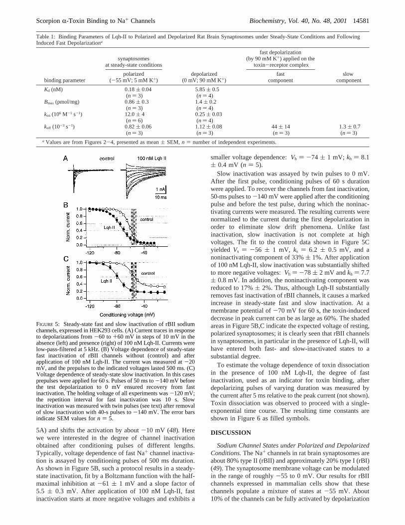
5A) and shifts the activation by about-10 mV (48). Herewe were interested in the degree of channel inactivationobtained after conditioning pulses of different lengths.Typically, voltage dependence of fast Na+ channel inactiva-tion is assayed by conditioning pulses of 500 ms duration.As shown in Figure 5B, such a protocol results in a steady-state inactivation, fit by a Boltzmann function with the half-maximal inhibition at-61 ( 1 mV and a slope factor of5.5 ( 0.3 mV. After application of 100 nM Lqh-II, fastinactivation starts at more negative voltages and exhibits a
smaller voltage dependence:Vh ) -74 ( 1 mV; kh ) 8.1( 0.4 mV (n ) 5).
Slow inactivation was assayed by twin pulses to 0 mV.After the first pulse, conditioning pulses of 60 s durationwere applied. To recover the channels from fast inactivation,50-ms pulses to-140 mV were applied after the conditioningpulse and before the test pulse, during which the noninac-tivating currents were measured. The resulting currents werenormalized to the current during the first depolarization inorder to eliminate slow drift phenomena. Unlike fastinactivation, slow inactivation is not complete at highvoltages. The fit to the control data shown in Figure 5Cyielded Vs ) -56 ( 1 mV, ks ) 6.2 ( 0.5 mV, and anoninactivating component of 33%( 1%. After applicationof 100 nM Lqh-II, slow inactivation was substantially shiftedto more negative voltages:Vh ) -78 ( 2 mV andkh ) 7.7( 0.8 mV. In addition, the noninactivating component wasreduced to 17%( 2%. Thus, although Lqh-II substantiallyremoves fast inactivation of rBII channels, it causes a markedincrease in steady-state fast and slow inactivation. At amembrane potential of-70 mV for 60 s, the toxin-induceddecrease in peak current can be as large as 60%. The shadedareas in Figure 5B,C indicate the expected voltage of resting,polarized synaptosomes; it is clearly seen that rBII channelsin synaptosomes, in particular in the presence of Lqh-II, willhave entered both fast- and slow-inactivated states to asubstantial degree.
To estimate the voltage dependence of toxin dissociationin the presence of 100 nM Lqh-II, the degree of fastinactivation, used as an indicator for toxin binding, afterdepolarizing pulses of varying duration was measured bythe current after 5 ms relative to the peak current (not shown).Toxin dissociation was observed to proceed with a single-exponential time course. The resulting time constants areshown in Figure 6 as filled symbols.
DISCUSSION
Sodium Channel States under Polarized and DepolarizedConditions.The Na+ channels in rat brain synaptosomes areabout 80% type II (rBII) and approximately 20% type I (rBI)(49). The synaptosome membrane voltage can be modulatedin the range of roughly-55 to 0 mV. Our results for rBIIchannels expressed in mammalian cells show that thesechannels populate a mixture of states at-55 mV. About10% of the channels can be fully activated by depolarization
Table 1: Binding Parameters of Lqh-II to Polarized and Depolarized Rat Brain Synaptosomes under Steady-State Conditions and FollowingInduced Fast Depolarizationa
synaptosomesat steady-state conditions
fast depolarization(by 90 mM K+) applied on the
toxin-receptor complex
binding parameterpolarized
(-55 mV; 5 mM K+)depolarized
(0 mV; 90 mM K+)fast
componentslow
component
Kd (nM) 0.18( 0.04(n ) 3)
5.85( 0.5(n ) 4)
Bmax (pmol/mg) 0.86( 0.3(n ) 3)
1.4( 0.2(n ) 4)
kon (106 M-1 s-1) 12.0( 4(n ) 6)
0.25( 0.03(n ) 4)
koff (10-3 s-1) 0.82( 0.06(n ) 3)
1.12( 0.08(n ) 3)
44 ( 14(n ) 3)
1.3( 0.7(n ) 3)
a Values are from Figures 2-4, presented as mean( SEM, n ) number of independent experiments.
FIGURE 5: Steady-state fast and slow inactivation of rBII sodiumchannels, expressed in HEK293 cells. (A) Current traces in responseto depolarizations from-60 to+60 mV in steps of 10 mV in theabsence (left) and presence (right) of 100 nM Lqh-II. Currents werelow-pass-filtered at 5 kHz. (B) Voltage dependence of steady-statefast inactivation of rBII channels without (control) and afterapplication of 100 nM Lqh-II. The current was measured at-20mV, and the prepulses to the indicated voltages lasted 500 ms. (C)Voltage dependence of steady-state slow inactivation. In this casesprepulses were applied for 60 s. Pulses of 50 ms to-140 mV beforethe test depolarization to 0 mV ensured recovery from fastinactivation. The holding voltage of all experiments was-120 mV;the repetition interval for fast inactivation was 10 s. Slowinactivation was measured with twin pulses (see text) after removalof slow inactivation with 40-s pulses to-140 mV. The error barsindicate SEM values forn ) 5.
ScorpionR-Toxin Binding to Na+ Channels Biochemistry, Vol. 40, No. 48, 200114581

to -55 mV (48), but the probability of the individualactivating units, i.e., the S4 segments, being in the activatedposition at-55 mV is substantially greater. However, onlya small fraction will actually be open at a holding voltageof -55 mV, because most channels will be inactivated.Indeed, the data in Figure 5 show that about 75% of thechannels are fast-inactivated and about 40% are slow-inactivated. In the presence of a saturating concentration (100nM) of Lqh-II, inactivation is even more prevalent: about80% of the channels are fast- and slow-inactivated. Therefore,at -55 mV, rBII Na+ channels in rat synaptosomes areexpected to populate a complex mixture of states: the S4segments are about half-activated and the channels havesubstantially entered fast- and slow-inactivated states.
Rapid depolarization, as induced in the experiments shownin Figure 4, will result in an alteration of the state population.Activation of the S4 segments will reach almost saturationand the extent of fast and slow inactivation will only increaseslightly in absolute terms (about an additional 10%). Inrelative terms, however, fast and slow inactivation increasesubstantially, i.e., from the noninactivated channels at-55mV, almost 100% will undergo fast and 50% will undergoslow inactivation.
These data show, at least for the fraction of rBII channelsin synaptosomes, that toxin-binding studies to Na+ channelswill mostly report on electrically silent conformationalchanges because most of the channels are not accessible byelectrophysiological methods at the voltages under consid-eration. Rat brain synaptosomes, however, contain about 20%rBI channels, which according to Smith and Goldin (50) areavailable for opening at-55 mV, when assayed inXenopusoocytes with coexpression ofâ1 andâ2 subunits. Thus, the
fraction of activable channels may indeed be greater thanestimated from the electrophysiological assessment of rBIIchannels alone.
Capacity of Lqh-II Binding Sites on Polarized and De-polarized Synaptosomes.We found that the capacity of Lqh-II to bind polarized synaptosomes (0.86 pmol/mg) represents60% of the capacity on depolarized synaptosomes (1.4 pmol/mg). This result suggests that 60% of the channels at-55mV are in states presenting a high-affinity binding site forLqh-II, and the rest are in other states, which correspond tothe low-affinity site. The reason, on polarized synaptosomes,we did not detect the low-affinity site is due to the lowconcentrations of125I-Lqh-II (compared to itsKd) used inthe binding studies (Figures 1 and 2). Such low toxinconcentration prevented detection of low-affinity bindingsites in polarized synaptosomes, because only 20% and 0.3%of the high- and low-affinity sites, respectively, wereoccupied. In contrast, in depolarized synaptosomes, close to100% of the Na+ channels occupy states presenting a moreuniform, low-affinity site to the toxin, and maximal bindingcapacity was obtained (Figure 2). This conclusion is sup-ported by the data obtained with the binding of a spider toxin,δ-atracotoxin, and allosteric modulation of site 3 toxinbinding by alkaloid sodium channel activators (unpublishedresults). The direct designation, however, of channel statesfollowed in the binding studies is not straightforward.Considering the state distribution obtained for rBII channelsin HEK293 cells, this likely reflects a transition fromdeactivated to activated S4 segments. However, it cannotbe excluded that it represents a transition of the slowinactivation gate, which is also about half-activated at-55mV. More insight may be gained by considering the kineticsand voltage dependence underlying such a transition.
Channel State Transition between High- and Low-AffinitySites.Electrophysiological studies suggested that binding ofR-toxins slow the transition from open to fast-inactivatedchannel states and that the decline in toxin binding affinityat depolarized conditions resulted from a strong increase inthe off rate (21, 30, 31, 35, 36, 51). However, transitionsthat occur between different inactivated states are electricallysilent and therefore are difficult to study.
To analyze the direct effect of fast depolarization onbinding of R-toxins, we have pursued the change in Lqh-IIbinding after instantaneous depolarization of membranes,which were preequilibrated with toxin at resting membranepotential. Induction of fast depolarization was achieved byincreasing the K+ concentration while maintaining a constantconcentration of free125I-Lqh-II in order to prevent toxindissociation by dilution (Figure 4), which mimicked theelectrophysiological experiments in the presence ofR-toxins(21). The fast toxin unbinding rate in the electrophysiologicalapproach was inferred from the loss of toxin effect on currentinactivation and was attributed to the transition from closedto fast-inactivated channel states through the open state (21,36). Judicious analysis of125I-Lqh-II unbinding after fastdepolarization provided an off-rate value of 44× 10-3 s-1
for the fast component (Figure 4 and Table 1), which isstrikingly similar to that obtained for Lqh-II in electrophysi-ological studies on rBII Na+ channels, expressed in mam-malian cells. This similarity can be demonstrated by plottingthe time constants of Lqh-II unbinding at different membranepotentials (filled symbols in Figure 6) together with those
FIGURE 6: Time constants of Lqh-II unbinding as a function ofthe membrane potential. Data from electrophysiological measure-ments are presented (on a semilog scale) together with results fromthe binding studies. The closed symbols represent the time constantsof Lqh-II unbinding at different membrane potentials measuredelectrophysiologically on rBII sodium channels expressed inmammalian cells. The open symbols represent time constants ofLqh-II unbinding of rat brain synaptosomes. Changes in membranepotential of synaptosomes were obtained by increasing external K+
concentrations (5, 15, and 90 mM) to obtain-55,-30, and 0 mV,respectively. The time constants of Lqh-II unbinding at-30 and 0mV are from Figure 4. The time constant at-55 mV was calculatedfrom the koff obtained in Figure 3. The error bars for theelectrophysiological data represent the errors obtained from fittingsingle-exponential functions to dissociation data. Notably, the timeconstants of Lqh-II unbinding at the various membrane potentialsof synaptosomes fit remarkably well to a line, which could beextrapolated from the electrophysiological measurements. Thestraight line indicates an exponential fit yielding a slope of (17mV/e)-fold change in the time constant.
14582 Biochemistry, Vol. 40, No. 48, 2001 Gilles et al.

obtained in the Lqh-II binding experiments (open symbolsin Figure 6). The data fit in Figure 6 according to a single-exponential function to the combined data shows that thetime constant of Lqh-II dissociation from rBII/rat synapto-some channels has a voltage dependence equivalent to (17mV/e)-fold change.
These equivalent off rates suggest a transition into identicalchannel states during the first phase after fast depolarizationof synaptosomes (Figure 4) and depolarization under voltage-clamp conditions. The high off rate is likely to represent afast conformational change at the toxin receptor site, whichdestabilizes the toxin-receptor complex and leads to toxinexpulsion. This change could occur during the rapid transitionfrom the closed to the activated and finally the fast-inactivated state, as was suggested in the electrophysiologicalstudies (21, 36). Owing to the similarity in rate and voltagedependence of this component determined in electrophysi-ological and binding assays, it can be concluded that it isnot associated with slow-inactivated channel states, becausethey would not be detected with the electrophysiologicalprotocols employed.
The second, slow unbinding component (Figure 4) pro-vides an equivalent off rate to that measured in depolarizedsynaptosomes at steady state (Figure 3 and Table 1). Thissimilarity suggests that the unbinding during this second slowphase occur from channels in slow-inactivated states. Thisinterpretation is supported by the kinetics (seconds tominutes) of entering into the slow-inactivated state of Na+
channels in various depolarized neural preparations (12, 52-56). Moreover, no marked slow component of toxin dis-sociation is observed in electrophysiological experiments,suggesting that this component, measured in binding experi-ments, represents electrically silent channel states. Such statescould become more populated after depolarization, leadingto slow unbinding of the toxin. Slow inactivation has beendescribed as an absorbing state, which is effectively sequen-tial to channel activation (55-57). The observation that onlyabout 50% of the bound Lqh-II at resting membrane potentialis expelled by fast depolarization (Figure 4) may indicatethat about half the channels with bound toxin undergo a directtransition to the slow-inactivated state. In addition, thestrength of toxin binding to such states may have a smallintrinsic voltage dependence, which may contribute to thesmall change in toxinkoff upon depolarization (Table 1).
The rapid and extended depolarization of synaptosomesmay induce slow inactivation of the Na+ channels, leadingto conformational changes of these proteins with timeconstants in the order of several seconds to minutes (seeFigure 4). In addition, application of the toxins will enhanceslow inactivation (see Figure 5) and therefore will alter theequilibrium of normal and slow-inactivated channel states,which may have different affinities for the toxins. Thevoltage-dependent decrease inkon suggests a creation of asteric hindrance and/or charge alteration at the vicinity ofreceptor site 3, which reduce toxin association to the slow-inactivated state. The smaller difference in association ratesof the structurally differentδ-atracotoxin to receptor site 3under polarized and depolarized conditions further supportsthis notion (unpublished results).
CONCLUDING REMARKS
Our results reveal that it is the association and not thedissociation rate, observed inR-toxin binding, which isvoltage-dependent. The association rate is substantiallyreduced in depolarized membranes, linked to the slow-inactivated state. A second distinct mechanism exists thatcauses a voltage-dependent expulsion of the bound toxin. Itis suggested that rapid membrane depolarization induces afast transition to channel states that are responsible for thedestabilization of the boundR-toxin and its subsequentexpulsion from the receptor site. The voltage dependenceof this process is similar to that observed in electrophysi-ological experiments (Figure 6 and, e.g., refs21 and 36),suggesting that it reports on structural changes of electricalvisible channel states, i.e., transitions of non-slow-inactivatedchannels. The altered association rate and the slow dissocia-tion may report on channel states that are not accessed byelectrophysiological methods, i.e., transitions to or amongslow-inactivated channel states, which are prevalent at therelatively low membrane voltages of rat brain synaptosomes.
This study paves the way to further analysis of the putativeeffects ofR-toxins on channel entrance to and recovery fromthe slow-inactivated state, which may shed light on structuralelements involved in modulation of receptor site 3.
REFERENCES
1. Noda, M., Ikeda, T., Suzuki, H., Takeshima, H., Takahashi,T., Kuno, M., and Numa, S. (1986)Nature 320, 188-192.
2. Stuhmer, W., Conti, F., Suzuki, H., Wang, X., Noda, M.,Yahagi, N., Kubo, H., and Numa, S. (1989)Nature 339, 597-603.
3. Aggarwal, S. K., and MacKinnon, R. (1996)Neuron 16,1169-1177.
4. Armstrong, C. M. (1981)Physiol. ReV. 61, 644-682.5. Yang, N., George, A. L., Jr, and Horn, R. (1996)Neuron 16,
113-122.6. Cha, A., Ruben, P. C., George, A. L., Jr., Fujimoto, E., and
Bezanilla, F. (1999)Neuron 22, 73-87.7. Chahine, M., George, A., Jr., Zhou, M., Ji, S., Sun, W., Barchi,
R. L., and Horn, R. (1994)Neuron 12, 281-294.8. Chen, L. Q., Santarelli, V., Horn, R., and Kallen. R. G. (1996)
J. Gen. Physiol. 108, 549-556.9. McPhee, J. C., Ragsdale, D. S., Scheuer, T., and Catterall,
W. A. (1994)Proc. Natl. Acad. Sci. U.S.A. 91, 12346-12350.10. McPhee, J. C., Ragsdale, D. S., Scheuer, T., and Catterall,
W. A. (1995)J. Biol. Chem. 270, 12025-12034.11. McPhee, J. C., Ragsdale, D. S., Scheuer, T., and Catterall,
W. A. (1998)J. Biol. Chem. 273, 1121-1129.12. Kontis, K. J., and Goldin, A. L. (1997)J. Gen. Physiol. 110,
403-413.13. Vassilev, P., Scheuer, T., and Catterall, W. A. (1988)Science
241, 1658-1661.14. West, J. W., Patton, D. E., Scheuer, T., Wang, Y., Goldin, A.
L., and Catterall, W. A. (1992)Proc. Natl. Acad. Sci. U.S.A.89, 10910-10914.
15. Catterall, W. A. (1992)Physiol. ReV. 72 (Suppl.), S15-S48.16. Gordon, D. (1997) inToxins and Signal Transduction(Laz-
arovici, P., and Gutman, Y., Eds.) pp 119-149, HarwoodPress, Amsterdam.
17. Gordon, D. (1997)InVertebr. Neurosci. 3, 103-116.18. Martin-Eauclaire, M. F., and Couraud, F. (1995) inHandbook
Neurotoxicology(Chang, L. W., and Dyer, R. S., Eds.) pp683-716, Marcel Dekker, New York.
19. Thomsen, W. J., and Catterall, W. A. (1989)Proc. Natl. Acad.Sci. U.S.A. 86, 10161-10165.
20. Tejedor, F., and Catterall, W. A. (1990)Cell. Mol. Neurobiol.10, 257-265.
ScorpionR-Toxin Binding to Na+ Channels Biochemistry, Vol. 40, No. 48, 200114583

21. Rogers, J. C., Qu, Y., Tanada, T. N., Scheuer, T., and Catterall,W. A. (1996)J. Biol. Chem. 271, 15950-15962.
22. Benzinger, G. R., Kyle, J. W., Blumenthal, K. M., and Hanck,D. A. (1998)J. Biol. Chem. 273, 80-84.
23. Ma, Z., Tang, L., Lu, S., Kong, J., Gordon, D., and Kallen,R. G. (2000)Biophys. J. 78, A1011.
24. Zilberberg, N., Froy, O., Loret, E., Ceste`le, S., Arad, D.,Gordon, D., and Gurevitz, M. (1997)J. Biol. Chem. 272,14810-14816.
25. Gurevitz, M., Gordon, D., Ben-Natan, S., Turkov, M. and Froy,O. (2001)FASEB J. 15, 1201-1205.
26. Catterall, W. A. (1977)J. Biol. Chem. 252, 8669-8676.27. Ceste`le, S., Ben Khalifa, R., Pelhate, M., Rochat, H., and
Gordon, D. (1995)J. Biol. Chem. 270, 15153-15161.28. Ceste`le, S., Sampieri, F., Rochat, H., and Gordon, D. (1996)
J. Biol. Chem. 271, 18329-18332.29. Ceste`le, S., and Gordon, D. (1998)J. Neurochem. 70, 1217-
1226.30. Catterall, W. A., Ray, R., and Morrow, C. S. (1976)Proc.
Natl. Acad. Sci. U.S.A. 73, 2682-2686.31. Catterall, W. A. (1977)J. Biol. Chem. 252, 8660-8668.32. Ray, R., Morrow, C. S., and Catterall, W. A. (1978)J. Biol.
Chem. 253, 7307-7313.33. Jover, E., Martin-Moutot, N., Couraud, F., and Rochat, H.
(1980)Biochemistry 19, 463-467.34. Mozhayeva, G. N., Naumov, A. P., Nosyreva, E. D., and
Grishin, E. V. (1980)Biochim. Biophys. Acta. 597, 587-602.35. Strichartz, G. R., and Wang, G. K. (1986)J. Gen. Physiol.
88, 413-435.36. Chen, H., Gordon, D., and Heinemann, S. H. (2000)Pflugers
Arch. 439, 423-432.37. Catterall, W. A., Morrow, C. S., and Hartshorne, R. P. (1979)
J. Biol. Chem. 254, 11379-11387.38. Sautie`re, P., Ceste`le, S., Kopeyan, C., Martinage, A., Drobecq,
H., Doljansky, Y., and Gordon, D. (1998)Toxicon 36, 1141-1154.
39. Blaustein, M. P., and Goldring, J. M. (1975)J. Physiol. 247,589-615.
40. Tamkun, M. M., and Catterall, W. A. (1981)Mol. Pharmcol.19, 78-86.
41. Sharkey, R. G., Jover, E., Couraud, F., Baden, D. G., andCatterall, W. A. (1987)Mol. Pharmacol. 31, 273-278.
42. Kanner, B. J. (1978)Biochemistry 17, 1207-1211.43. Gilles, N., Blanchet, C., Shichor, I., Zaninetti, M., Lotan, I.,
Bertrand, D., and Gordon, D. (1999)J. Neurosci. 19, 8730-8739.
44. Gilles, N., Krimm, I., Bouet, F., Froy, O., Gurevitz, M.,Lancelin, J.-M., and Gordon, D. (2000)J. Neurochem. 75,1735-1745.
45. Weiland, G. A., and Molinoff, P. B. (1981)Life Sci. 29, 313-330.
46. Jurman, M. E., Boland, L. M., Liu, Y., and Yellen, G. (1994)Biotechniques 17, 876-881.
47. Chen, H., and Heinemann S. H. (2001)J. Gen. Physiol. 117,505-518.
48. Gilles, N., Chen, H., Wilson, H., Le Gall, F., Montoya, G.,Molgo, J., Scho¨nherr, R., Nicholson, G., Heinemann, S. H.,and Gordon, D. (2000)Eur. J. Neurosci. 12, 2823-2832.
49. Gordon, D., Merrick, D., Auld, V., Dunn, R., Goldin, A. L.,Davidson, N., and Catterall, W. A. (1987)Proc. Natl. Acad.Sci. U.S.A. 84, 8682-8686.
50. Smith, R. D., and Goldin, A. L. (1998)J. Neurosci. 18, 811-820.
51. Sheets, M. F., Kyle, J. W., Kallen, R. G., and Hanck, D. A.(1999)Biophys. J. 77, 747-757.
52. Ruben, P. C., Starkus, J. G., and Rayner, M. D. (1990)Biophys.J. 58, 1169-1181.
53. Ruben, P. C., Starkus, J. G., and Rayner, M. D. (1992)Biophys.J. 61, 941-955.
54. Rudy, B. (1978)J. Physiol. (London) 283, 1-21.55. Adelman, W. J., Jr., and Palti, Y. (1969)J. Gen. Physiol. 54,
589-606.56. Rudy, B. (1981)J. Physiol. (London) 312, 531-549.57. Vedantham, V., and Cannon, S. C. (1998)J. Gen. Physiol.
111, 83-93.58. Featherstone, D. E., Richmond, J. E., and Ruben, P. C. (1996)
Biophys. J. 71, 3098-3109.59. Richmond, J. E., Featherstone, D. E., Hartmann, H. A., and
Ruben, P. C. (1998)Biophys. J. 74, 2945-2952.
BI010973R
14584 Biochemistry, Vol. 40, No. 48, 2001 Gilles et al.