Dual role of pericyte α6β1-integrin in tumour blood vessels · of α6-integrin in vivowas...
13
RESEARCH ARTICLE Dual role of pericyte α6β1-integrin in tumour blood vessels Louise E. Reynolds 1, ‡ , Gabriela D’Amico 1, *, Tanguy Lechertier 1, *, Alexandros Papachristodoulou 2 , Jose ́ M. Mun ̃ oz-Fe ́ lix 1 , Adè le De Arcangelis 3 , Marianne Baker 1 , Bryan Serrels 4 and Kairbaan M. Hodivala-Dilke 1 ABSTRACT The α6β1-integrin is a major laminin receptor, and formation of a laminin-rich basement membrane is a key feature in tumour blood vessel stabilisation and pericyte recruitment, processes that are important in the growth and maturation of tumour blood vessels. However, the role of pericyte α6β1-integrin in angiogenesis is largely unknown. We developed mice where the α6-integrin subunit is deleted in pericytes and examined tumour angiogenesis and growth. These mice had: (1) reduced pericyte coverage of tumour blood vessels; (2) reduced tumour blood vessel stability; (3) increased blood vessel diameter; (4) enhanced blood vessel leakiness, and (5) abnormal blood vessel basement membrane architecture. Surprisingly, tumour growth, blood vessel density and metastasis were not altered. Analysis of retinas revealed that deletion of pericyte α6-integrin did not affect physiological angiogenesis. At the molecular level, we provide evidence that pericyte α6-integrin controls PDGFRβ expression and AKT–mTOR signalling. Taken together, we show that pericyte α6β1-integrin regulates tumour blood vessels by both controlling PDGFRβ and basement membrane architecture. These data establish a novel dual role for pericyte α6-integrin as modulating the blood vessel phenotype during pathological angiogenesis. KEY WORDS: Integrin, Pericyte, Tumour growth, Angiogenesis INTRODUCTION Blood vessels comprise endothelial cells supported by mural cells, also known as pericytes. Although the role of integrins in endothelial biology has been studied extensively (Avraamides et al., 2008; Germain et al., 2010), almost nothing is known about the role of pericyte integrins, including α6-integrin (Garmy-Susini et al., 2005; Liu and Leask, 2012). Integrins are transmembrane cell surface receptors that mediate cell–cell and cell–extracellular matrix (ECM) interactions. One of the major components of the vascular basement membrane is laminin whose predominant adhesive receptors include the integrins α6β1 and α6β4 (Durbeej, 2010). Targeting endothelial integrins has proved to be relatively beneficial in treating cancer in preclinical studies (Yamada et al., 2006; Tabatabai et al., 2010) but has had limited success in clinical trials (Patel et al., 2001), therefore new targets, including pericytes are currently being examined. Tumour blood vessels have many structural abnormalities including decreased endothelial barrier function, reduced pericyte recruitment and poor basement membrane organisation when compared with normal quiescent vessels (Armulik et al., 2005). Studies have shown that pericyte recruitment and investment to blood vessels stimulates endothelial cell basement membrane (BM) deposition and organisation in vitro (Stratman et al., 2009). This is mediated mainly by secretion of endothelial platelet-derived growth factor (PDGF)-BB, attracting PDGF receptor β (PDGFRβ)-positive pericytes, which adhere to the BM surrounding endothelial cells (Stratman et al., 2010; Abramsson et al., 2003). In vivo, mice lacking PDGF-BB–PDGFRβ signalling fail to adequately recruit pericytes to newly formed blood vessels, resulting in severe perturbation of blood vessel stabilisation and maturation (Hellberg et al., 2010). Furthermore, interference with PDGF-BB–PDGFRβ signalling results in disruption of already established endothelial–pericyte associations and vessel destabilisation during retinal development (Benjamin et al., 1998). Whether pericyte α6-integrin might regulate tumour vessel stability was hitherto unknown. In the present study, we examined the role of pericyte α6-integrin on tumour blood vessel function using a genetic ablation approach. Surprisingly, loss of pericyte α6-integrin did not affect tumour growth, angiogenesis or metastasis but did cause a decrease in pericyte association with tumour blood vessels and poor basement membrane organisation, with an associated increase in vessel leakage and instability. At the molecular level, we demonstrate a novel mechanism by which pericyte α6-integrin reduces PDGFRβ expression on pericytes and therefore diminishes responses to PDGF-BB. This, in turn, is the likely mechanism for aberrant pericyte investment of tumour blood vessels. Taken together, our data suggest that pericyte α6β1-integrin plays a dual role in regulating PDGFRβ expression and BM organisation that likely increases vessel leakage and instability. RESULTS Generation and characterisation of pdgfrβcre+;α6fl/fl mice We have generated a new mouse model that enables us to study deletion of α6-integrin in pericytes. We bred α6-integrin floxed mice (denoted α6fl/fl ) (Bouvard et al., 2012; Germain et al., 2010) with mice expressing Cre-recombinase under the control of the PDGFRβ promoter (denoted pdgfrβcre+) (Foo et al., 2006), to generate pdgfrβcre-;α6fl/fl and pdgfrβcre+;α6fl/fl mice. Mice were born to pdgfrβcre+;α6fl/fl×pdgfrβcre-;α6fl/fl crosses at normal Mendelian ratios and male:female ratios with no obvious adverse phenotype (Fig. S1A–C). Mice were genotyped by PCR analysis (Fig. S1D). Histological analysis of H&E-stained sections of lung, heart, liver and spleen from pdgfrβcre-;α6fl/fl and pdgfrβcre+;α6fl/fl adult mice showed no apparent tissue defects (Fig. S1E). Furthermore, no apparent vascular abnormalities were observed in these tissues Received 23 September 2016; Accepted 8 March 2017 1 Adhesion and Angiogenesis Laboratory, Centre for Tumour Biology, Barts Cancer Institute - A CRUK Centre of Excellence, Queen Mary University of London, Charterhouse Square, London EC1M 6BQ, UK. 2 Laboratory for Molecular Neuro- Oncology, Dept. of Neurology, University Hospital Zurich, Frauenklinikstrasse 26, Zurich CH-8091, Switzerland. 3 IGBMC, UMR 7104, INSERM U964, Université de Strasbourg, BP. 10142, 1, Rue Laurent Fries, Illkirch Cedex 67404, France. 4 Cancer Research UK Edinburgh Centre, University of Edinburgh, Crewe Road South, Edinburgh EH4 2XR, UK. *These authors contributed equally to this work ‡ Author for correspondence ([email protected]) L.E.R., 0000-0001-6075-1808 This is an Open Access article distributed under the terms of the Creative Commons Attribution License (http://creativecommons.org/licenses/by/3.0), which permits unrestricted use, distribution and reproduction in any medium provided that the original work is properly attributed. 1583 © 2017. Published by The Company of Biologists Ltd | Journal of Cell Science (2017) 130, 1583-1595 doi:10.1242/jcs.197848 Journal of Cell Science
Transcript of Dual role of pericyte α6β1-integrin in tumour blood vessels · of α6-integrin in vivowas...

RESEARCH ARTICLE
Dual role of pericyte α6β1-integrin in tumour blood vesselsLouise E Reynolds1Dagger Gabriela DrsquoAmico1 Tanguy Lechertier1 Alexandros Papachristodoulou2Jose M Mun oz-Felix
1 Adele De Arcangelis3 Marianne Baker1 Bryan Serrels4 and Kairbaan M Hodivala-Dilke1
ABSTRACTThe α6β1-integrin is a major laminin receptor and formation of alaminin-rich basement membrane is a key feature in tumour bloodvessel stabilisation and pericyte recruitment processes that areimportant in the growth and maturation of tumour blood vesselsHowever the role of pericyte α6β1-integrin in angiogenesis is largelyunknown We developed mice where the α6-integrin subunit isdeleted in pericytes and examined tumour angiogenesis and growthThese mice had (1) reduced pericyte coverage of tumour bloodvessels (2) reduced tumour blood vessel stability (3) increasedblood vessel diameter (4) enhanced blood vessel leakiness and(5) abnormal blood vessel basement membrane architectureSurprisingly tumour growth blood vessel density and metastasiswere not altered Analysis of retinas revealed that deletion of pericyteα6-integrin did not affect physiological angiogenesis At themolecularlevel we provide evidence that pericyte α6-integrin controls PDGFRβexpression and AKTndashmTOR signalling Taken together we show thatpericyte α6β1-integrin regulates tumour blood vessels by bothcontrolling PDGFRβ and basement membrane architecture Thesedata establish a novel dual role for pericyte α6-integrin as modulatingthe blood vessel phenotype during pathological angiogenesis
KEY WORDS Integrin Pericyte Tumour growth Angiogenesis
INTRODUCTIONBlood vessels comprise endothelial cells supported by mural cellsalso known as pericytes Although the role of integrins in endothelialbiology has been studied extensively (Avraamides et al 2008Germain et al 2010) almost nothing is known about the role ofpericyte integrins including α6-integrin (Garmy-Susini et al 2005Liu and Leask 2012) Integrins are transmembrane cell surfacereceptors that mediate cellndashcell and cellndashextracellular matrix (ECM)interactions One of the major components of the vascular basementmembrane is laminin whose predominant adhesive receptors includethe integrins α6β1 and α6β4 (Durbeej 2010) Targeting endothelialintegrins has proved to be relatively beneficial in treating cancer in
preclinical studies (Yamada et al 2006 Tabatabai et al 2010) buthas had limited success in clinical trials (Patel et al 2001) thereforenew targets including pericytes are currently being examined
Tumour blood vessels have many structural abnormalitiesincluding decreased endothelial barrier function reduced pericyterecruitment and poor basement membrane organisation whencompared with normal quiescent vessels (Armulik et al 2005)Studies have shown that pericyte recruitment and investment toblood vessels stimulates endothelial cell basement membrane (BM)deposition and organisation in vitro (Stratman et al 2009) This ismediated mainly by secretion of endothelial platelet-derived growthfactor (PDGF)-BB attracting PDGF receptor β (PDGFRβ)-positivepericytes which adhere to the BM surrounding endothelial cells(Stratman et al 2010 Abramsson et al 2003) In vivo mice lackingPDGF-BBndashPDGFRβ signalling fail to adequately recruit pericytesto newly formed blood vessels resulting in severe perturbation ofblood vessel stabilisation and maturation (Hellberg et al 2010)Furthermore interference with PDGF-BBndashPDGFRβ signallingresults in disruption of already established endothelialndashpericyteassociations and vessel destabilisation during retinal development(Benjamin et al 1998) Whether pericyte α6-integrin mightregulate tumour vessel stability was hitherto unknown
In the present study we examined the role of pericyte α6-integrinon tumour blood vessel function using a genetic ablation approachSurprisingly loss of pericyte α6-integrin did not affect tumourgrowth angiogenesis or metastasis but did cause a decrease inpericyte association with tumour blood vessels and poor basementmembrane organisation with an associated increase in vesselleakage and instability At the molecular level we demonstrate anovel mechanism by which pericyte α6-integrin reduces PDGFRβexpression on pericytes and therefore diminishes responses toPDGF-BB This in turn is the likely mechanism for aberrantpericyte investment of tumour blood vessels Taken together ourdata suggest that pericyte α6β1-integrin plays a dual role inregulating PDGFRβ expression and BM organisation that likelyincreases vessel leakage and instability
RESULTSGeneration and characterisation of pdgfrβcre+α6flfl miceWe have generated a new mouse model that enables us to studydeletion of α6-integrin in pericytes We bred α6-integrin floxed mice(denoted α6flfl) (Bouvard et al 2012 Germain et al 2010) withmice expressing Cre-recombinase under the control of the PDGFRβpromoter (denoted pdgfrβcre+) (Foo et al 2006) to generatepdgfrβcre-α6flfl and pdgfrβcre+α6flfl mice Mice were born topdgfrβcre+α6flfltimespdgfrβcre-α6flfl crosses at normal Mendelianratios and malefemale ratios with no obvious adverse phenotype(Fig S1AndashC) Mice were genotyped by PCR analysis (Fig S1D)Histological analysis of HampE-stained sections of lung heart liver andspleen from pdgfrβcre-α6flfl and pdgfrβcre+α6flfl adult miceshowed no apparent tissue defects (Fig S1E) Furthermore noapparent vascular abnormalities were observed in these tissuesReceived 23 September 2016 Accepted 8 March 2017
1Adhesion and Angiogenesis Laboratory Centre for Tumour Biology Barts CancerInstitute - A CRUK Centre of Excellence Queen Mary University of LondonCharterhouse Square London EC1M 6BQ UK 2Laboratory for Molecular Neuro-Oncology Dept of Neurology University Hospital Zurich Frauenklinikstrasse 26Zurich CH-8091 Switzerland 3IGBMC UMR 7104 INSERM U964 Universite deStrasbourg BP 10142 1 Rue Laurent Fries Illkirch Cedex 67404 France 4CancerResearch UK Edinburgh Centre University of Edinburgh Crewe Road SouthEdinburgh EH4 2XR UKThese authors contributed equally to this work
DaggerAuthor for correspondence (lreynoldsqmulacuk)
LER 0000-0001-6075-1808
This is an Open Access article distributed under the terms of the Creative Commons AttributionLicense (httpcreativecommonsorglicensesby30) which permits unrestricted usedistribution and reproduction in any medium provided that the original work is properly attributed
1583
copy 2017 Published by The Company of Biologists Ltd | Journal of Cell Science (2017) 130 1583-1595 doi101242jcs197848
Journal
ofCe
llScience
(Fig S1F) or in the developing retina (Fig S1G) suggesting that lossof PDGFRβ-driven α6-integrin had no apparent effect onphysiological angiogenesis Finally to confirm pericyte-specific Creexpression in our mouse model we crossed pdgfrβcre- andpdgfrβcre+ mice with the mTmG reporter mouse which expressesmembrane-targeted tandem dimer Tomato (mT red) prior to Cre-mediated excision and membrane-targeted green fluorescent protein(mG green) after excision (Muzumdar et al 2007) Analysis oftumour blood vessels frompdgfrβcre-mTmG andpdgfrβcre+mTmGmice showed that although Tomato (mT) expression was observed inbloodvessels in bothpdgfrβcre-mTmG andpdgfrβcre+mTmGmiceGFP (mG) expression was present in mouse tissue only after Creexcision and only observed in pericytes in pdgfrβcre+mTmG mice(Fig S2AB) As expected α6-integrin is expressed on tumourendothelial cells shown by co-expression of α6-integrin and theendothelial cell marker CD31 (Fig S2C) suggesting that the deletionof α6-integrin in vivo was restricted to PDGFRβ-positive pericytes
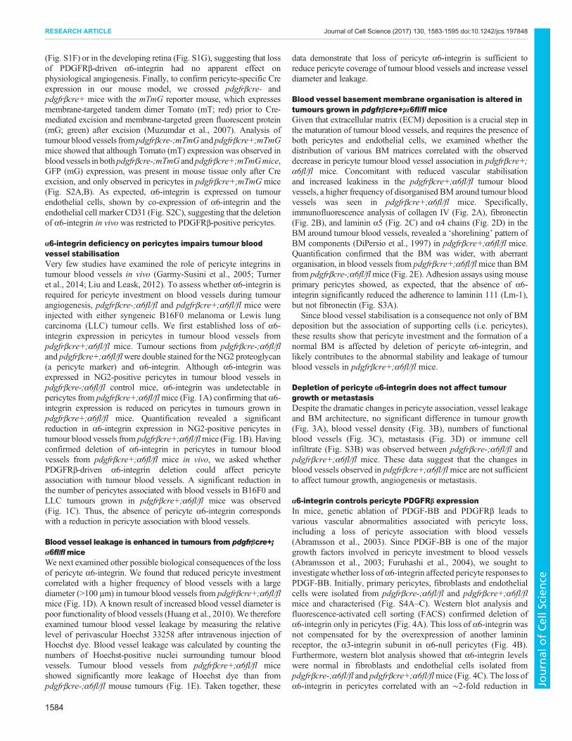
α6-integrin deficiency on pericytes impairs tumour bloodvessel stabilisationVery few studies have examined the role of pericyte integrins intumour blood vessels in vivo (Garmy-Susini et al 2005 Turneret al 2014 Liu and Leask 2012) To assess whether α6-integrin isrequired for pericyte investment on blood vessels during tumourangiogenesis pdgfrβcre-α6flfl and pdgfrβcre+α6flfl mice wereinjected with either syngeneic B16F0 melanoma or Lewis lungcarcinoma (LLC) tumour cells We first established loss of α6-integrin expression in pericytes in tumour blood vessels frompdgfrβcre+α6flfl mice Tumour sections from pdgfrβcre-α6flfland pdgfrβcre+α6flflwere double stained for the NG2 proteoglycan(a pericyte marker) and α6-integrin Although α6-integrin wasexpressed in NG2-positive pericytes in tumour blood vessels inpdgfrβcre-α6flfl control mice α6-integrin was undetectable inpericytes from pdgfrβcre+α6flflmice (Fig 1A) confirming that α6-integrin expression is reduced on pericytes in tumours grown inpdgfrβcre+α6flfl mice Quantification revealed a significantreduction in α6-integrin expression in NG2-positive pericytes intumour blood vessels from pdgfrβcre+α6flflmice (Fig 1B) Havingconfirmed deletion of α6-integrin in pericytes in tumour bloodvessels from pdgfrβcre+α6flfl mice in vivo we asked whetherPDGFRβ-driven α6-integrin deletion could affect pericyteassociation with tumour blood vessels A significant reduction inthe number of pericytes associated with blood vessels in B16F0 andLLC tumours grown in pdgfrβcre+α6flfl mice was observed(Fig 1C) Thus the absence of pericyte α6-integrin correspondswith a reduction in pericyte association with blood vessels
Blood vessel leakage is enhanced in tumours from pdgfrβcre+α6flflmiceWe next examined other possible biological consequences of the lossof pericyte α6-integrin We found that reduced pericyte investmentcorrelated with a higher frequency of blood vessels with a largediameter (gt100 microm) in tumour blood vessels from pdgfrβcre+α6flflmice (Fig 1D) A known result of increased blood vessel diameter ispoor functionality of blood vessels (Huang et al 2010)We thereforeexamined tumour blood vessel leakage by measuring the relativelevel of perivascular Hoechst 33258 after intravenous injection ofHoechst dye Blood vessel leakage was calculated by counting thenumbers of Hoechst-positive nuclei surrounding tumour bloodvessels Tumour blood vessels from pdgfrβcre+α6flfl miceshowed significantly more leakage of Hoechst dye than frompdgfrβcre-α6flfl mouse tumours (Fig 1E) Taken together these
data demonstrate that loss of pericyte α6-integrin is sufficient toreduce pericyte coverage of tumour blood vessels and increase vesseldiameter and leakage
Blood vessel basement membrane organisation is altered intumours grown in pdgfrβcre+α6flfl miceGiven that extracellular matrix (ECM) deposition is a crucial step inthe maturation of tumour blood vessels and requires the presence ofboth pericytes and endothelial cells we examined whether thedistribution of various BM matrices correlated with the observeddecrease in pericyte tumour blood vessel association in pdgfrβcre+α6flfl mice Concomitant with reduced vascular stabilisationand increased leakiness in the pdgfrβcre+α6flfl tumour bloodvessels a higher frequency of disorganised BM around tumour bloodvessels was seen in pdgfrβcre+α6flfl mice Specificallyimmunofluorescence analysis of collagen IV (Fig 2A) fibronectin(Fig 2B) and laminin α5 (Fig 2C) and α4 chains (Fig 2D) in theBM around tumour blood vessels revealed a lsquoshoreliningrsquo pattern ofBM components (DiPersio et al 1997) in pdgfrβcre+α6flfl miceQuantification confirmed that the BM was wider with aberrantorganisation in blood vessels from pdgfrβcre+α6flflmice than BMfrom pdgfrβcre-α6flflmice (Fig 2E) Adhesion assays using mouseprimary pericytes showed as expected that the absence of α6-integrin significantly reduced the adherence to laminin 111 (Lm-1)but not fibronectin (Fig S3A)
Since blood vessel stabilisation is a consequence not only of BMdeposition but the association of supporting cells (ie pericytes)these results show that pericyte investment and the formation of anormal BM is affected by deletion of pericyte α6-integrin andlikely contributes to the abnormal stability and leakage of tumourblood vessels in pdgfrβcre+α6flfl mice
Depletion of pericyte α6-integrin does not affect tumourgrowth or metastasisDespite the dramatic changes in pericyte association vessel leakageand BM architecture no significant difference in tumour growth(Fig 3A) blood vessel density (Fig 3B) numbers of functionalblood vessels (Fig 3C) metastasis (Fig 3D) or immune cellinfiltrate (Fig S3B) was observed between pdgfrβcre-α6flfl andpdgfrβcre+α6flfl mice These data suggest that the changes inblood vessels observed in pdgfrβcre+α6flflmice are not sufficientto affect tumour growth angiogenesis or metastasis
α6-integrin controls pericyte PDGFRβ expressionIn mice genetic ablation of PDGF-BB and PDGFRβ leads tovarious vascular abnormalities associated with pericyte lossincluding a loss of pericyte association with blood vessels(Abramsson et al 2003) Since PDGF-BB is one of the majorgrowth factors involved in pericyte investment to blood vessels(Abramsson et al 2003 Furuhashi et al 2004) we sought toinvestigatewhether loss of α6-integrin affected pericyte responses toPDGF-BB Initially primary pericytes fibroblasts and endothelialcells were isolated from pdgfrβcre-α6flfl and pdgfrβcre+α6flflmice and characterised (Fig S4AndashC) Western blot analysis andfluorescence-activated cell sorting (FACS) confirmed deletion ofα6-integrin only in pericytes (Fig 4A) This loss of α6-integrin wasnot compensated for by the overexpression of another lamininreceptor the α3-integrin subunit in α6-null pericytes (Fig 4B)Furthermore western blot analysis showed that α6-integrin levelswere normal in fibroblasts and endothelial cells isolated frompdgfrβcre-α6flfl and pdgfrβcre+α6flflmice (Fig 4C) The loss ofα6-integrin in pericytes correlated with an sim2-fold reduction in
1584
RESEARCH ARTICLE Journal of Cell Science (2017) 130 1583-1595 doi101242jcs197848
Journal
ofCe
llScience
levels of PDGFRβ in α6-null pericytes suggesting that α6-integrinwas indeed regulating the expression of PDGFRβ protein (Fig 4D)Additionally PDGFRβ levels were significantly reduced inPDGFRβ-immunostained α6-null pericytes compared with WTpericytes in vitro (Fig 4E)
PDGF-BB responses are diminished in α6-null pericytesSince changes in receptor expression levels do not always reflectchanges in downstream signalling of the corresponding tyrosinekinase receptor (Platta and Stenmark 2011) we next sought toconfirm whether the decreased expression of PDGFRβ correlated
Fig 1 See next page for legend
1585
RESEARCH ARTICLE Journal of Cell Science (2017) 130 1583-1595 doi101242jcs197848
Journal
ofCe
llScience
with diminished responses to PDGF-BB stimulation in α6-nullpericytes We showed that PDGF-BB-stimulated ex vivomicrovesselsprouting was reduced in aortic rings isolated from pdgfrβcre+α6flfl mice compared with PDGF-BB-stimulated sprouting frompdgfrβcre-α6flfl mouse aortic rings (Fig 5A) In this assay thevessel sprouts become surrounded by pericytes that proliferate andmigrate along the endothelium (Nicosia 2009) We were unable toanalyse pericyte coverage in pdgfrβcre+α6flfl aortic rings due tothe complete lack of sprouts that grew in response to PDGF-BB Inresponse to PDGF-BB α6-null pericyte migration and proliferationwas significantly reduced when compared with WT controls(Fig 5BC) We next examined the effect of α6-integrin deficiencyon PDGF-BB-stimulated downstream signalling in pericytesInteraction of PDGFRβ with PDGF-BB activates several signallingpathways including the MAPK pathway (through ERK12 alsoknown as MAPK3 and MAPK1) and phosphoinositide 3-kinase(PI3K) through the AKT pathway Western blot analysis showedthat in α6-null pericytes PDGF-BB-mediated stimulation ofERK12 and AKT (AKT123) pathways were both reducedsignificantly (Fig 5DE) Since integrins and PDGFR can activatemany downstream signalling pathways we performed a non-candidate proteomic study using Reverse Phase Protein Array(RPPA) analysis to identify other possible pathways that may beaffected by the absence of α6-integrin on pericytes We found thatcomponents of the AKTndashmTOR signalling pathway [AKT P70S6kinase (RPS6KB1) mTOR pS6 ribosomal protein (RPS6) and4E-BP1] were significantly downregulated in α6-null pericytes(Fig S4D) This pathway is activated downstream of integrins and isknown to be regulated by PDGFR expression and function (Zhanget al 2007) These results suggest that absence of α6-integrin resultsin a reduction of PDGFRβ levels significantly reducing pericyteresponses to PDGF-BB
Collectively these results demonstrate a novel role for pericyteα6-integrin in both the regulation of PDGFRβ levels which waspreviously unknown and in BM organisation This dual mechanismconfers a blood vessel phenotype that affects only the primarytumour
DISCUSSIONThe role of pericyte α6-integrin expression has not been addressedpreviously We have now shown that the combined effects of thedownregulation of PDGFRβ and the changes in pericyte adhesionupon deletion of α6β1-integrin induces destabilisation of tumourblood vessels Genetic ablation of pericyte α6β1-integrin correlateswith reduced pericyte investment to tumour blood vessels changesin BM architecture and reduced PDGFRβ expression levels andPDGF-BB-mediated downstream signalling These correspond withincreased blood vessel leakage without affecting tumour growth ormetastasis Taken together our results suggest a dual function ofα6-integrin on pericytes and here we will discuss both the effect ofreduced PDGFRβ levels and decreased pericyte adhesion in turn
Very few studies have linked pericyte integrins with growthfactor receptor regulation Currently α5β1-integrin has been shownto regulate signalling through PDGFRβ in vascular smooth musclecells (Veevers-Lowe et al 2011) and inactivation of PDGF-BBsignalling can decrease α1β1 integrin levels (Hosaka et al 2013) Inanother study NG2 depletion in pericytes was shown to reduce β1-integrin-mediated signalling (You et al 2014) Although limitedthese studies suggest possible cross-talk between pericyte integrinsand growth factor receptors a mechanism that has been shownpreviously between endothelial cell integrins and growth factorreceptors (da Silva et al 2010 Germain et al 2010 Reynoldset al 2002) In parallel in vitro studies using fibroblasts havehighlighted the ability of integrins to enhance PDGF-dependentresponses (DeMali et al 1999 Sundberg and Rubin 1996) Herewe show for the first time that α6β1-integrin acts as a regulator ofPDGFRβ ndash controlling its expression and signalling uponstimulation with PDGF-BB ndash in pericytes Our data indicate thatdepletion of α6-integrin on pericytes leads to a significant reductionin the levels of PDGFRβ In turn this leads to a downregulation inthe activation of the MAPK and AKT signalling pathways whichare both known to be critical for cell migration and proliferation(Lemmon and Schlessinger 2010) Our observation of reducedpericyte PDGFRβ levels signalling and resulting inhibitedresponses to PDGF-BB in α6-integrin deficient pericytes mayexplain the tumour blood vessel phenotypes we observe in thepdgfrβcre+α6flfl mice including reduced pericyte blood vesselinvestment and increased detachment of pericytes to endothelialcells since these functions have been reported to be mediated byPDGF-BB (Abramsson et al 2003 Hellberg et al 2010) It hasbeen well documented that tumours transplanted into PDGF-Bretention motif-deficient ( pdgf-bretret) mice have an sim50reduction in numbers of pericytes that associate poorly with theblood vessel wall and result in leaky vessels (Abramsson et al2003) It is noteworthy that although we observe a similar leakyblood vessel phenotype to that reported in tumour growth in pdgf-bretret mice our results indicate that α6β1-integrin is a regulator ofpericyte function rather than numbers as opposed to the reductionin numbers seen in the pdgf-bretret mice (Lindblom et al 2003)
One surprising observation was that despite such striking bloodvessel defects in the pdgfrβcre+α6flfl mouse tumours includingincreased vessel leakage we did not observe any changes in tumourgrowth angiogenesis or lung metastasis (Cooke et al 2012 Zanget al 2015) Our work is in line with previous studies showing that
Fig 1 Reduced pericyte investment and increased vessel leakage inpdgfrβcre+α6flfl mice (A) B16F0 tumour sections from pdgfrβcre-α6flfland pdgfrβcre+α6flflmice were double-immunostained for NG2 (red) and α6-integrin (green) expression to determine in vivo expression of α6-integrin inpericytes α6-integrin was observed in NG2-positive pericytes in tumour bloodvessels from pdgfrβcre-α6flfl mice In contrast α6-integrin expression wassignificantly reduced in pericytes in tumour blood vessels from pdgfrβcre+α6flflmice Magnified regions show α6-integrin-positive pericytes (to give a yellowsignal) on pdgfrβcre-α6flfl blood vessels but α6-integrin-negative pericytes onpdgfrβcre+α6flfl blood vessels Arrows α6 and NG2 co-expressionarrowheads NG2 expression alone (B) Quantification of α6-integrinexpression in NG2-positive pericytes from tumour blood vessels in pdgfrβcre-α6flfl in pdgfrβcre+α6flfl mice Bar chart represents mean+sem pixelintensity of α6-integrin expression in NG2-positive cells n=9ndash10 tumours pergenotype (C) Pericyte association with blood vessels The percentage ofblood vessels with associated NG2-positive pericytes in B16F0 and LLCtumours grown in pdgfrβcre+α6flfl mice was reduced significantly comparedwith pdgfrβcre-α6flfl mice Scatter graphs represent the mean+sempercentage blood vessels that are NG2-positive in B16F0 and LLC tumoursn=4ndash6 micegroup Representative images of tumour sections stained with thepericyte marker NG2 (green) and for endomucin (red) Arrows endomucin-positive staining arrowheads NG2-positive staining (D) Blood vesseldiameter The frequency of vessels with a diameter ge100 microm was greater inboth B16F0 and LLC tumours grown in pdgfrβcre+α6flflmice when comparedwith pdgfrβcre-α6flfl mice Bar charts represent the mean+sem percentageof blood vessels ge100 microm diameter (E) Blood vessel leakage Mice wereinjected via the tail vein with Hoechst 33258 dye and tumour sections wereanalysed for blood vessel leakage by measuring the numbers of tumour cellsthat had taken up Hoechst Blood vessels in the tumours grown in pdgfrβcre+α6flfl mice showed significantly more leakage than blood vessels in tumoursgrown in pdgfrβcre-α6flfl mice Bar chart shows relative mean+semleakage n=6 tumoursgenotype Dotted lines Hoechst leakage Plt005Plt0005 Plt00009 Scale bars 50 microm (A) 100 microm (C) 200 microm (E)
1586
RESEARCH ARTICLE Journal of Cell Science (2017) 130 1583-1595 doi101242jcs197848
Journal
ofCe
llScience
early ablation of NG2+ cells or depletion of PDGFβR+ pericytesdoes not necessarily affect tumour growth and metastasis (Keskinet al 2015) suggesting that increased leakage alone is not sufficientto enhance metastasis Indeed increased vascular leakage has beenshown to be insufficient for metastasis per se (Thurston et al 1999)
We hypothesise that despite the observed decrease in blood vesselpericyte coverage in pdgfrβcre+α6flfl mice the remainingpericytes attached to the endothelial cells provide enough survivalfactors for example VEGF and Ang-1 (angiopoietin-1 also knownas Angpt1) to allow endothelial cells to survive allowing for
Fig 2 Tumours grown in pdgfrβcre+α6flfl mice have aberrant BM organisation around blood vessels LLC tumours were stained with antibodies to theECM proteins (A) collagen IV (B) fibronectin (C) laminin α5 and (D) laminin α4 chains For all matrices disorganisation of BM with a lsquoshorelinersquo pattern wasobserved more frequently around blood vessels in tumours grown in pdgfrβcre+α6flfl mice when compared with pdgfrβcre-α6flfl mice Boxes show magnifiedregions of BM Brackets identify representative BM widths Scale bars 50 microm (E) The width of the BM surrounding blood vessels was analysed (mean+sem)n=8ndash10 sectionsgenotype Plt0005
1587
RESEARCH ARTICLE Journal of Cell Science (2017) 130 1583-1595 doi101242jcs197848
Journal
ofCe
llScience
Fig 3 See next page for legend
1588
RESEARCH ARTICLE Journal of Cell Science (2017) 130 1583-1595 doi101242jcs197848
Journal
ofCe
llScience
normal tumour angiogenesis and tumour growth Studies haveshown that decreased pericyte coverage can lead to regression ofblood vessels but the magnitude is tumour specific and does notnecessarily retard tumour growth (Sennino et al 2007) Indeedtreatment of RipTag2 tumours with anti-PDGFRβ antibody reducespericyte numbers and enlarges blood vessels but does not reducetumour vascular density (Song et al 2005) Although not within thescope of this study we believe that the phenotype observed in ourpdgfrβcre+α6flfl mice may help to improve chemotherapyefficacy for primary tumours owing to the leaky vessel defectAs well as pericyte α6-integrin deficiency affecting PDGFRβ
levels and upstream signalling we also show that deletion of α6-integrin affects the ECMadhesion properties of pericytes in vitro andin vivo The BM that surrounds blood vessels is necessary for vesselintegrity stability and maturation Recent studies have highlightedthe importance of normal endothelial cellndashpericyte interactions forproper BM organisation (Davis et al 2013 Stratman et al 2009Baluk et al 2003) When this interaction is de-stabilised BMorganisation is affected resulting in decreased vessel integrity(Stratman et al 2009 Davis et al 2013) Therefore it is possiblethat the phenotypewe observe in themicrovessel BMof pdgfrβcre+α6flflmice may be due at least in part to a loss of pericyte adhesionto the endothelial cell basement membrane Similar phenotypeshave been shown in other studies for example inactivation of theβ1-integrin subunit in mural cells leads to failure of these cells toassociate with the subendothelial BM (Abraham et al 2008) andgenetic ablation of a related laminin receptor α3-integrinresults in an epidermal BM defect in which components of theBM show a disorganised expression pattern (DiPersio et al 1997
Georges-Labouesse et al 1996 Has et al 2012) very similar to thatobserved in tumour blood vessels of mice deficient in α6-integrinpericyte Foxf2 (a forkhead transcription factor specificallyexpressed by pericytes) deficiency in brain pericytes leads tosignificantly reduced PDGFRβ and αvβ8-integrin levels withthinning of the vascular basal lamina resulting in a leaky bloodndashbrain barrier (Reyahi et al 2015) inactivation of PDGF-BBsignalling decreases α1β1-integrin levels and impairs pericyteadhesion to ECMcomponents of blood vessels (Hosaka et al 2013)
Taken together these studies all support a common notion thatcrosstalk between pericyte integrins with PDGFRβ signalling canaffect vascular BM organisation and vessel function It is alsoconceivable that microvessel BM disorganisation may be acontributing cause to decreased supporting cell coverage in thetumour blood vessels in pdgfrβcre+α6flfl mice For example ithas been shown that deletion of laminin-α4 chain in mice causesimpaired vessel growth due to reduced pericyte recruitment to bloodvessels (Abrass et al 2010) In vitro pericyte recruitment duringtube formation is necessary to stimulate endothelial BM formation(Stratman et al 2009) Overall our data suggest that the absence ofpericyte α6-integrin leads to (1) a reduced investment of pericytes totumour microvessels possibly due to reduced PDGFRβ levels and(2) that this is associated with poor vessel BM architecture leadingto vascular leakage
Our study provides new insights into the regulation of tumourblood vessels by pericyte α6-integrin which points towards animportant role in the regulation of tumour vessel leakage
MATERIALS AND METHODSGeneration of miceα6-integrin floxed mice (Bouvard et al 2012 Germain et al 2010) werebred with mice expressing Cre-recombinase under the control of thePDGFRβ promoter PDGFRβCre (Foo et al 2006) to generate pdgfrβcre-α6flfl and pdgfrβcre+α6flfl mice
Immunostaining of tumour sectionsUnless otherwise stated frozen sections were fixed in ice-cold acetone for10 min followed by permeabilisation with 05 NP-40 for 10 min Sectionswere blocked for 45 min with 10 bovine serum albumin (BSA) and 01Tween 20 in PBS Primary antibodies were incubated overnight at 4degCfollowed by incubation with a fluorescently conjugated secondary antibodyfor 1 h at room temperature (11000 Invitrogen) Primary antibodies againstthe following were used α6-integrin (GoH3 Chemicon) NG2 (AB5320Millipore) endomucin (sc-65495 Santa Cruz Biotechnology) (all 1100)
Quantification of α6-integrin on pericytesQuantification of α6-integrin expression on NG2-positive pericytes ontumour blood vessels from pdgfrβcre-α6flfl and pdgfrβcre+α6flfl micewas performed using ImageJ software The mean pixel intensity of α6-integrin expression on NG2-positive pericytes was quantified
Immunostaining and quantification of BMImmunostaining for laminin α4 chain (antibody was a kind gift from TakakoSasaki Dept Matrix Medicine Oita University Japan) was performed asdescribed previously (Sasaki et al 2001) For laminin α5 chain sectionswere fixed in 4 paraformaldehyde (PFA) washed twice with PBS thenblocked with 1 BSA for 30 min Sections were incubated overnight at 4degCwith primary antibody against laminin α5 (1400 dilution in blocking bufferkind gift from Jeffrey H Miner Division of Biology amp BiomedicalSciences Washington University in St Louis USA Pierce et al 2000)followed by several washes incubation with Alexa-Fluor-488-conjugatedanti-rabbit-IgG secondary antibody (11000 Invitrogen) and mounted
For collagen IV (ab19808 Abcam) and fibronectin (ab23750 Abcam)staining sections were fixed in 4 PFA blocked with 3 normal goatserum (NGS) 01 Triton X-100 (TX-100) in PBS for 30 min at room
Fig 3 Tumour growth angiogenesis and metastasis are not affected inpdgfrβcre+α6flfl mice (A) Subcutaneous B16F0 and LLC tumour growthwas similar in both pdgfrβcre+α6flfl mice and pdgfrβcre-α6flfl mice The barcharts represent meanplusmnsem tumour volumes n=20ndash30 micegenotype(B) Endomucin staining of midline sections of age- and size-matched B16F0and LLC tumours showed no significant differences in blood vessel densitybetween pdgfrβcre+α6flfl mice and pdgfrβcre-α6flfl mice Blood vesseldensity is given as the number of blood vesselsmm2 for the midline tumoursection Representative images of endomucin-stained tumour blood vesselsare shown Bar chart represents mean+sem tumour blood vessel density ofsize-matched tumours n=6 tumour sectionsgenotype ns no significantdifference (C) The number of perfused tumour blood vessels was assessedafter tail vein injection of PE-conjugated anti-CD31 antibody before tumourexcision in pdgfrβcre+α6flfl mice and pdgfrβcre-α6flfl control mice andcomparing the numbers of CD31-positive vessels with numbers of endomucin-stained vessels B16F0 and LLC tumours grown in pdgfrβcre+α6flflmice hada similar number of functional PEndashCD31 endomucin-expressing tumour bloodvessels compared to pdgfrβcre-α6flfl littermate control mice Bar chartsrepresent the percentage of PEndashCD31-perfused vessels over total number ofendomucin-positive blood vessels for the midline tumour section frompdgfrβcre+α6flfl mice and pdgfrβcre-α6flfl mice+sem n=10 tumoursections per genotype ns no significant difference Representative images ofendomucin-stained PEndashCD31-positive perfused blood vessels from B16F0tumours are shown (D) Metastasis is not affected in pdgfrβcre+α6flfl miceSubcutaneous LLC tumours were resected when they reached sim100 mm3 At3 weeks post-resection mice were killed and lungs removed to assessmetastasis Lungs were fixed and the number of surface metastases wascounted There were no significant differences in the total number of surfacemetastases between pdgfrβcre+α6flfl mice and pdgfrβcre-α6flfl mice (barchart mean+sem) Lungs were then sectioned and stained with HampEMeasurement of individual metastatic nodule areas showed no differencebetween genotypes (scatter graph) Representative high-power images ofHampE-stained lung sections show areas of metastasis (dotted line upperimages) as well as low-power images of metastatic lung tissue (arrows lowerimages) Scale bars 100 microm (B) 50 microm (C) 50 microm (D upper panel) 5000 microm(D lower panel)
1589
RESEARCH ARTICLE Journal of Cell Science (2017) 130 1583-1595 doi101242jcs197848
Journal
ofCe
llScience
Fig 4 See next page for legend
1590
RESEARCH ARTICLE Journal of Cell Science (2017) 130 1583-1595 doi101242jcs197848
Journal
ofCe
llScience
temperature Primary antibody was diluted 1200 in 1 NGS 01 TX-100in PBS and incubated overnight at 4degC Sections were washed three timeswith PBS incubated with Alexa-Fluor-488-conjugated anti-rabbitsecondary antibody (Invitrogen) diluted 1100 in 1 NGS TX-100washed three times and mounted with Prolong Gold anti-fade with DAPI
Fluorescence staining was visualised using the Axioplan microscope(Zeiss) Images were captured using Axiovision Rel 40 software TheAxiovision software linear measuring tool was used to analyse the spread (inmicrom) of BM surrounding blood vessels
Pericyte associationFor analysis of pericyte coverage tumour sections were doubleimmunostained for endomucin and NG2 (for details see lsquoImmunostainingof tumour sectionsrsquo) Pericyte coverage was quantified by counting the totalnumber of endomucin-positive blood vessels across whole tumour sectionsfollowed by the numbers of blood vessels positive for both endomucin andNG2 The percentage of blood vessels with associated pericytes wascalculated
Tumour blood vessel leakageTo analyse tumour blood vessel leakage Hoechst 33258 dye (4 microgmlSigma H33258) was used Hoechst dyes diffuse quickly from vessels andbind to the DNA of cells surrounding blood vessels allowing forquantification of areas of uptake by perivascular tumour cells and henceblood vessel leakage (Janssen et al 2005) Briefly mice were injectedsequentially with 100 μl phycoerythrin (PE)-conjugated CD31 (Biolegendto stain functional blood vessels) followed 9 min later with 100 μl Hoechst33258 (4 microgml Sigma H33258) via the tail vein and were killed 1 minlater Tumours were excised and snap-frozen A total of 8ndash10 fields at times20magnification were analysed by ImageJ For quantification blood vesselleakage was calculated by counting the numbers of Hoechst 33258-positivenuclei surrounding PEndashCD31-positive tumour areas Results are shown inarbitrary units (AU)
Tumour growth and angiogenesisThe syngeneic mouse tumour cell lines B16F0 (melanoma derived fromC57BL6) and Lewis Lung Carcinoma (LLC) (both from the ATCC) wereused in subcutaneous tumour growth experiments 1times106 B16F0 cells or05times106 LLC cells resuspended in 100 microl of phosphate-buffered saline(PBS) were injected subcutaneously into the flank of 12ndash14-week-oldpdgfrβcre+α6flfl and littermate control mice ( pdgfrβcre-α6flfl mice)Tumour growth was measured every 2 days using calipers After 14 daysanimals were culled tumours excised and either fixed in 4 formaldehydein PBS overnight or snap-frozen in liquid nitrogen for subsequentimmunohistochemical analysis
Blood vessel densitySize-matched tumours from pdgfrβcre-α6flfl and pdgfrβcre+α6flfl micewere snap-frozen and bisected and cryosections made Frozen sections werefixed in 100 acetone at minus20degC rehydrated in PBS for 10 min and thenblocked (PBS 1 BSA 01 Tween-20) for 45 min at room temperatureAfter a 5 min wash in PBS sections were incubated with 1100 anti-endomucin (as above) in blocking buffer for 45 min at room temperatureAfter three 5 min washes in PBS sections were incubated with Alexa-Fluor-488-conjugated anti-rat-IgG secondary antibody (Invitrogen) diluted inblocking buffer After three 5 min washes in PBS sections were washedbriefly with distilled water before being mounted The number ofendomucin-positive blood vessels present across the entire area of eachmid-line tumour section from size and age-matched tumours was countedand divided by the area of the section to determine tumour blood vesseldensity
Tumour blood vessel diameterThe diameter of endomucin-positive blood vessels were quantified by usingthe Axiovision software linear measuring tool
Perfused blood vessel analysisFor analysis of the percentage of perfused functional tumour vessels 100 μlPEndashCD31 antibody (Biolegend London UK) was injected via the tail vein10 min (mins) prior to killing of mice Tumours were dissected immediatelysnap-frozen and sectioned Frozen sections were then immunostained forendomucin as described above To calculate the percentage of functionalvessels the number of PEndashCD31-positive blood vessels was divided by thetotal number of endomucin-positive blood vessels
Tumour metastasis05times106 LLC tumour cells were injected subcutaneously and tumours wereallowed to reach a size of 100 mm3 before surgical resection The mice weremonitored for up to 3 weeks after which the metastatic burden wasquantified by counting numbers of surface lungmetastases Lungs were thenfixed and sections stained with HampE for further analysis The numbers ofsurface metastases was counted immediately after fixation to give the totalnumber of lung metastases per mouse To examine internal metastasesHampE-stained sections were analysed and the area of individual metastaseswas measured using Axiovision software to give the internal metastaticnodule area
Primary endothelial cell fibroblast and pericyte isolationPrimary mouse endothelial cells were isolated from lungs and maintained asdescribed previously (Reynolds and Hodivala-Dilke 2006) Brieflypdgfrβcre-α6flfl and pdgfrβcre+α6flfl mouse lungs were mincedcollagenase digested (Type I Gibco) strained through a 70 microm cellstrainer (BD Falcon) and the resulting cell suspension plated on flaskscoated with a mixture of 01 gelatin (Sigma) 10 microgml fibronectin(Millipore) and 30 microgml rat tail collagen (Sigma) Endothelial cells werepurified by a single negative (FCγ sort-RIIIII Pharmingen) and twopositive cell sorts (ICAM-2 Pharmingen) using anti-rat IgG-conjugatedmagnetic beads (Dynal) During preparation of primary endothelial cellslung fibroblasts were isolated from the non-endothelial cell population thatwas generated during the first positive sort For all cell types passagingoccurred when cells reached 70 confluency Cells were trypsinisedcentrifuged washed with PBS and replated on pre-coated flasks forendothelial cells and pericytes and non-coated flasks for fibroblastsFibroblasts were cultured in Dulbeccorsquos modified Eaglersquos medium(DMEM) with 10 fetal calf serum (FCS) to passage 4 endothelial cellsin MLEC [Hamrsquos F-12 DMEM (low glucose) 10 FCS heparin andendothelial mitogen (Generon)] to passage 4ndash5 Pericytes were isolated frommouse brains as described previously (Tigges et al 2012) and cultured inPericyte medium (ScienCell) to passage 9
FACS analysisPrimary mouse brain pericytes isolated from pdgfrβcre-α6flfl andpdgfrβcre+α6flfl mice were incubated with an anti-α6-integrin antibody
Fig 4 Pericyte α6-integrin regulates PDGFRβ expression (A) Pericyteswere isolated from pdgfrβcre-α6flfl and pdgfrβcre+α6flflmice andwestern blotanalysis performed to assess α6-integrin deletion α6 deletion was onlyobserved in pericytes from pdgfrβcre+α6flfl mice FACS analysis alsoconfirmed loss ofα6-integrin surface levels in pericytes isolated from pdgfrβcre+α6flfl mice compared with pericytes isolated from pdgfrβcre-α6flfl mice (linegraph and equivalent data dot plot) Bar charts represent mean+semdensitometric readings of western blots corrected for loading n=3 separatelysatesgenotype (B) Western blot analysis confirmed that wild-type (WT) andα6-null pericytes have similar levels of α3-integrin a second laminin receptorsuggesting that there was no compensation of α3-integrin in the absence ofα6-integrin Results are mean+sem n=3 separate lysatesgenotype(C) Fibroblasts and endothelial cells were isolated from pdgfrβcre-α6flfl andpdgfrβcre+α6flfl mice and western blot analysis performed to assessα6-integrin levels α6-integrin levels were not affected in either cell type isolatedfrom either pdgfrβcre-α6flfl and pdgfrβcre+α6flfl mice Results are mean+sem n=3 separate lysatesgenotype (D) Western blot analysis revealedPDGFRβ protein levels were significantly reduced in primary α6-null pericytesThe bar chart shows mean+sem densitometric values of PDGFRβ levelscorrected for loading HSC70 was used as a loading control n=3 separatelysatesgenotype (E) Immunostaining of PDGFRβ in α6-null pericytes in culturewas significantly reduced compared with WT pericytes Plt005 ns nosignificant difference Scale bar 50 microm
1591
RESEARCH ARTICLE Journal of Cell Science (2017) 130 1583-1595 doi101242jcs197848
Journal
ofCe
llScience
Fig 5 See next page for legend
1592
RESEARCH ARTICLE Journal of Cell Science (2017) 130 1583-1595 doi101242jcs197848
Journal
ofCe
llScience
(1100 GoH3 Abcam) to determine expression levels for 30 min at 4degCThis was followed by incubation for 30 min at 4degC with an appropriateFITC-conjugated secondary antibody Unstained cells were used as acontrol For characterisation of primary mouse brain pericytes cells werewashed with PBS and trypsinised at 37degC The cell suspensions werewashed in medium containing serum and centrifuged at 214 g for 3 minCells were washed with cold FACS buffer (1 BSA in PBS) and fixed with4 formaldehyde for 10 min at room temperature Cells were washed withFACS buffer and the cell suspensions were incubated with the followingprimary antibodies (all 1100) for 30 min PE-conjugated anti-CD31(102507 Biolegend) PE-conjugated anti-Mac1 (CD11b 101207Biolegend) PE-conjugated anti-GFAP (561483 BD Biosciences) APC-conjugated anti-PDGFRβ (136007 Biolegend) PEndashCy7-conjugated anti-CD146 (134713 Biolegend) Cells were then washed three times in samplebuffer and resuspended in a final volume of 400 ml As a control unstainedcells were sorted by FACS Primary mouse lung endothelial cells wereincubated with PE-conjugated anti-CD31 (as above)
Western blot analysisPrimary lung endothelial cells lung fibroblasts and brain pericytes isolatedfrom pdgfrβcre-α6flfl and pdgfrβcre+α6flflmice were grown to 70ndash80confluency then lysed in RIPA buffer 15ndash30 microg protein was run on 8polyacrylamide gels then transferred to nitrocellulose membranesMembranes were probed with primary antibody overnight at 4degC Allantibodies (against ERK12 cat no 9102 phosphoERK12 cat no 9101AKT cat no 9272 phosphoAKT cat no 4058α6-integrin cat no 3750) forthe signalling studies were purchased from Cell Signaling and used at a11000 dilution α3-integrin antibody was purchased from Millipore(AB1920 11000) The anti-HSC70 antibody used as a loading controlwas fromSantaCruzBiotechnology (cat no sc-7298) andwas used at 15000dilution For PDGF-BB stimulation pericytes were serum starved for 6 h inOptimem with 05 FCS then stimulated with PDGF-BB (30 ngmlPeprotech UK) for 0 30 s 60 s 5 min 15 min and 30 min beforelysis Densitometric readings of band intensities were obtained using theImageJ software
PDGFRβ immunostaining of cellsImmunostaining of brain pericytes isolated from pdgfrβcre-α6flfl andpdgfrβcre+α6flfl mice for PDGFRβ (11000 28E1 Cell Signaling) wasperformed according to the manufacturerrsquos protocol
Aortic ring assayThoracic aortas were isolated from pdgfrβcre-α6flfl and pdgfrβcre+α6flfl8ndash10-week-old mice and prepared for culture as described previously(Baker et al 2012) Where indicated culture medium was supplementedwith PDGF-BB at 30 ngml (Peprotech London UK) PBS was used as acontrol Aortic rings were fed every 3 days with fresh medium with orwithout PDFG-BB (30 ngml) Sprouting microvessels were counted after9 days in culture fixed and stained to identify endothelial cells andpericytes as described previously (Baker et al 2012)
Scratch wound assaysPrimary mouse brain pericytes from pdgfrβcre-α6flfl and pdgfrβcre+α6flfl mice were grown to confluency in Pericyte medium (ScienCell cat no0010) in six-well plates coated with 01 gelatin and fibronectin Cells wereserum starved overnight in Optimem containing 05 FCS The followingday the monolayers were scratched horizontally and vertically throughthe centre of each well The cells were either stimulated with PDGF-BB(30 ngml) or not and cell migration monitored over a 72 h period At eachtime point 12 photographs were taken of each scratch and the wound widthwas measured using ImageJ software Results were normalised to the woundwidth at time 0
Proliferation assayPrimary WT and α6-null brain pericyte proliferation was assessed using theCellTiter 96reg Aqueous One Solution Reagent (Promega) according to themanufacturerrsquos instructions Plates were read at a wavelength of 490 nm withabsorbance measured relative to blank wells containing reagent only Plateswere coated with 01 gelatin and fibronectin prior to seeding the pericytes
Characterising transgenic miceThe primers for the Cre PCR were forward primer 5prime-GCCGCATTACC-GGTCGATGCAAGA-3prime and reverse primer 5prime-GTGGCAGATGGCGC-GGCAACACCATT-3prime The reaction generates a fragment ofsim1000 bp Theprimers for α6-floxed PCR were forward primer 5prime-AGAAGGTGATGT-TACCCT-3prime and reverse primer 5prime-AATGTAACTAGCATTCAAGT-3primeThe PCR generates a 154 bp fragment for the α6-floxed allele and a 120 bpfragment for the wild-type allele (described in Germain et al 2010)
Whole-mount immunofluorescence on retinasPostnatal day (P)9 eyes were fixed in 4 PFA overnight at 4degC Retinas weredissected in PBS and after five washes in PBS incubated in blockingsolution [1 FBS (Sigma) 3 BSA (Sigma) 05 Triton X-100 (Sigma)001 Na deoxycholate (Sigma) 002 Na Azide (Sigma) in PBS pH 74]for 2 h at room temperature Retinas were incubated overnight at4degC with rabbit polyclonal anti-NG2 (Millipore AB5320 dilution1500) in blocking solutionPBS (11) at 4degC Retinas were then washedseveral times in PBS and incubated with secondary antibody conjugated toAlexa Fluor 488 and 548 (Life Technologies) all diluted 1300 overnight at4degC After several washes in PBS retinas were post-fixed in 1 PFA and re-blocked in 1 BSA and 05 Triton X-100 in PBS for 1 h at roomtemperature After two rinses in PBlec (01 nM CaCl2 01 mMMgCl2 01mM MnCl2 and 1 Triton X-100 in PBS pH 68) retinas were incubatedwith biotinylated Isolectin B4 (B1205VectorLabs dilution 1125) in PBlecovernight at 4degC After several washes in PBS retinas were incubated withAlexa Fluor 647-conjugated Streptavidin diluted 1100 in 05 BSA with03 Triton X-100 in PBS overnight at 4degC After several washes in PBSretinas were incubated with Hoechst 33342 dye as a nuclear counterstain(H3570 Life Technologies) washed and mounted with ProLong Gold(Molecular Probes) Fluorescently labelled samples were imaged with aconfocal microscope (Carl Zeiss LSM 710 Carl Zeiss) in multichannelmode Three-dimensional projections were digitally reconstructed fromconfocal z-stacks using the LSMZen 2009 software and Image J open sourceimage processing software (version200-rc-43151d)
Adhesion assayAdhesion assays were performed as previously described (Germain et al2010) using brain pericytes isolated from pdgfrβcre-α6flfl and pdgfrβcre+
Fig 5 Responses to PDGF-BB are reduced in α6-null pericytes (A) Aorticrings were isolated from pdgfrβcre-α6flfl and pdgfrβcre+α6flfl mice andtreated with either PBS or PDGF-BB (30 ngml) for up to 10 days PDGF-BBstimulation increased vessel sprouting in pdgfrβcre-α6flfl but not pdgfrβcre+α6flfl aortic rings Bar charts show the mean+sem number of aortic sproutsgenotype n=40ndash50 ringsgenotype from triplicate experiments (B) Confluentmonolayers of primary wild-type (WT) and α6-null pericytes were woundedmanually after 24 h serum starvation then stimulated or not with PDGF-BB(30 ngml) Wound width was quantified for up to 48 h Wound closure wassignificantly reduced in α6-null pericytes in response to PDGF-BB Bar chartsshow mean+sem wound width in the absence (left panel) or presence (rightpanel) of PDGF-BB normalised to time 0 n=6genotypetime point fromtriplicate experiments (C) Proliferation of primary WT and α6-null pericytes inthe presence of Optimem or PDGF-BB was measured α6-null pericytesproliferated significantly less in the presence of PDGF-BB compared with WTpericytes Graphs represent meanplusmnsem relative proliferation n=3 biologicalrepeats (D) Western blot analysis of phosphorylated-ERK12 (pErk) totalERK12 (tErk) phosphorylated AKT (pAkt) and total AKT (tAkt) from WT andα6-null pericyte lysates after serum starvation and stimulation with PDGF-BBfor 0 30 s 60 s 5 min 15 min 30 min Downstream signalling responses toPDGF-BB in α6-null pericytes were reduced significantly Individual croppedblots are representative of samples run on the same gel under identicalexperimental conditions HSC70 acted as the loading control (E) Graphs showmeanplusmnsem densitometric readings of fold change for p-AKT to total AKTnormalised to time 0 and fold change of ratios p-ERK12 to total ERK12normalised to time 0 n=3 independent experiments Plt005 Plt0005 nsno significant difference
1593
RESEARCH ARTICLE Journal of Cell Science (2017) 130 1583-1595 doi101242jcs197848
Journal
ofCe
llScience
α6flfl mice Adhesion of pericytes is presented relative to adhesion ofpericytes to fibronectin for the same genotype
Immunostaining of immune cells fibroblasts and endothelialcellsImmunostaining of immune cells from B16F0 tumour sections wasperformed as described previously (Reynolds et al 2010)
Primary mouse lung fibroblasts and endothelial cells were fixed with 4PFA for 10 min washed twice and blocked with 5NGS in PBS for 30 minat room temperature Primary antibodies to vimentin (1100 5741 CellSignaling) NG2 (1100 MAB 5385 Millipore) endomucin (1100 V7C7Santa Cruz Biotechnology) and α-Sma (clone 1A4 Sigma) were incubatedfor 1 h at room temperature washed three times with PBS and incubatedwith the relevant secondary antibody for 45 min at room temperature
RPPA nitrocellulose slidesCell lysates were prepared using ice-cold RIPA buffer After normalisationto adjust protein concentrations triplicate spots of each lysate weredeposited onto 16-pad Avid Nitrocellulose slides (Grace Bio) underconditions of constant 70 humidity using an Aushon 2470 Arrayplatform (Aushon BioSystems) After printing and washing steps thearrays were blocked by incubation in Superblock (Thermo Scientific37535) for 10 min The protein array chips were subsequently incubatedfor 1 h with primary antibody followed by repeat blocking with Superblockand 30 min incubation with anti-rabbit Dylight-800-conjugated secondaryantibody (Cell Signaling cat no 5151)
Following secondary antibody incubation and subsequent wash steps theimmune-stained arrays were imaged using an Innopsys 710IR scanner(Innopsys France) Microarray images were obtained at the highest gainwithout saturation of fluorescent signal detection Image analysis wasperformed using Mapix software (Innopsys France) to calculate the relativefluorescence intensity (RFI) value for each sample An estimate of totalprotein printed per feature on the array was determined by staining an arrayslide with fastgreen protein stain Readout values for all antibodies tested areexpressed as a ratio of the total protein loaded and are presented as the meanof technical replicates
Ethical regulationsAll animals were used in accord with United Kingdom Home Officeregulations (Home Office license number 707449) The in-house EthicsCommittee at Queen Mary University of London has approved allexperiments using mice under the project license
Statistical analysisStatistical significance was calculated by using a Studentrsquos t-test Plt005was considered statistically significant
AcknowledgementsWe thank Julie Holdsworth (Bart Cancer Institute QMUL UK) and Bruce Williams(CancerResearchUK) for their technicalhelpwithanimalexperimentsDrRalf Adams(Max Planck Institute for Molecular Biomedicine Munster Germany) generouslyprovided the pdgfrβcremice Dr Elisabeth Georges-Labouesse generously providedthe α6 floxedmice mTmGmicewere generously provided by TaijaMakinen UppsalaUniversity Sweden Rebecca Pike provided FACS technical support
Competing interestsThe authors declare no competing or financial interests
Author contributionsLER and KMH-D conceived and designed the experiments LER performedthe experiments GD performed confocal microscopy and retinal angiogenesisstudies TL performed image analysis and mTmG studies AP performed immunecell immunostaining and quantification JMM-F and LER performed the FACSanalysis ADA provided the a6-integrin floxed mice MB undertook the initialtumour experiments BS performed theRPPA analysis LER and KMH-D wrotethe paper KMH-D supervised the project
FundingThis work was supported by a grant from Cancer Research UK (C8218A18673)Deposited in PMC for immediate release
Supplementary informationSupplementary information available online athttpjcsbiologistsorglookupdoi101242jcs197848supplemental
ReferencesAbraham S Kogata N Fassler R and Adams R H (2008) Integrin beta1
subunit controls mural cell adhesion spreading and blood vessel wall stabilityCirc Res 102 562-570
Abramsson A Lindblom P and Betsholtz C (2003) Endothelial andnonendothelial sources of PDGF-B regulate pericyte recruitment and influencevascular pattern formation in tumors J Clin Invest 112 1142-1151
Abrass C K Hansen K M and Patton B L (2010) Laminin alpha4-null mutantmice develop chronic kidney disease with persistent overexpression of platelet-derived growth factor Am J Pathol 176 839-849
Armulik A Abramsson A and Betsholtz C (2005) Endothelialpericyteinteractions Circ Res 97 512-523
Avraamides C J Garmy-Susini B and Varner J A (2008) Integrins inangiogenesis and lymphangiogenesis Nat Rev Cancer 8 604-617
Baker M Robinson S D Lechertier T Barber P R Tavora B Drsquoamico GJones D T Vojnovic B and Hodivala-Dilke K (2012) Use of the mouseaortic ring assay to study angiogenesis Nat Protoc 7 89-104
Baluk P Morikawa S Haskell A Mancuso M and Mcdonald D M (2003)Abnormalities of basement membrane on blood vessels and endothelial sproutsin tumors Am J Pathol 163 1801-1815
Benjamin L E Hemo I and Keshet E (1998) A plasticity window for bloodvessel remodelling is defined by pericyte coverage of the preformed endothelialnetwork and is regulated by PDGF-B and VEGF Development 125 1591-1598
Bouvard C De Arcangelis A Dizier B Galy-Fauroux I Fischer A-MGeorges-Labouesse E and Helley D (2012) Tie2-dependent knockout ofalpha6 integrin subunit in mice reduces post-ischaemic angiogenesisCardiovasc Res 95 39-47
Cooke V G Lebleu V S Keskin D Khan Z OrsquoConnell J T Teng YDuncan M B Xie L Maeda G Vong S et al (2012) Pericyte depletionresults in hypoxia-associated epithelial-to-mesenchymal transition andmetastasis mediated by met signaling pathway Cancer Cell 21 66-81
Da Silva R G Tavora B Robinson S D Reynolds L E Szekeres CLamar J Batista S Kostourou V Germain M A Reynolds A R et al(2010) Endothelial alpha3beta1-integrin represses pathological angiogenesisand sustains endothelial-VEGF Am J Pathol 177 1534-1548
Davis G E Kim D J Meng C-X Norden P R Speichinger K R DavisM T Smith A O Bowers S L andStratman A N (2013) Control of vasculartube morphogenesis and maturation in 3D extracellular matrices by endothelialcells and pericytes Methods Mol Biol 1066 17-28
Demali K A Balciunaite E and Kazlauskas A (1999) Integrins enhanceplatelet-derived growth factor (PDGF)-dependent responses by altering the signalrelay enzymes that are recruited to the PDGF beta receptor J Biol Chem 27419551-19558
Dipersio C M Hodivala-Dilke K M Jaenisch R Kreidberg J A and HynesR O (1997) alpha3beta1 Integrin is required for normal development of theepidermal basement membrane J Cell Biol 137 729-742
Durbeej M (2010) Laminins Cell Tissue Res 339 259-268Foo S S Turner C J Adams S Compagni A Aubyn D Kogata N
Lindblom P Shani M Zicha D and Adams R H (2006) Ephrin-B2 controlscell motility and adhesion during blood-vessel-wall assembly Cell 124 161-173
Furuhashi M Sjoblom T Abramsson A Ellingsen J Micke P Li HBergsten-Folestad E Eriksson U Heuchel R Betsholtz C et al (2004)Platelet-derived growth factor production by B16 melanoma cells leads toincreased pericyte abundance in tumors and an associated increase in tumorgrowth rate Cancer Res 64 2725-2733
Garmy-Susini B Jin H Zhu Y Sung R J Hwang R and Varner J (2005)Integrin alpha4beta1-VCAM-1-mediated adhesion between endothelial andmuralcells is required for blood vessel maturation J Clin Invest 115 1542-1551
Georges-Labouesse E Messaddeq N Yehia G Cadalbert L Dierich Aand Le Meur M (1996) Absence of integrin alpha 6 leads to epidermolysisbullosa and neonatal death in mice Nat Genet 13 370-373
Germain M DeArcangelis A Robinson S D Baker M Tavora B DrsquoamicoG Silva R Kostourou V Reynolds L E Watson A et al (2010) Geneticablation of the alpha 6-integrin subunit in Tie1Cre mice enhances tumourangiogenesis J Pathol 220 370-381
Has C Sparta G Kiritsi D Weibel L Moeller A Vega-Warner V WatersA He Y Anikster Y Esser P et al (2012) Integrin alpha3 mutations withkidney lung and skin disease N Engl J Med 366 1508-1514
Hellberg C Ostman A and Heldin C-H (2010) PDGF and vessel maturationRecent Results Cancer Res 180 103-114
Hosaka K Yang Y Seki T Nakamura M Andersson P Rouhi P Yang XJensen L Lim S Feng N et al (2013) Tumour PDGF-BB expression levelsdetermine dual effects of anti-PDGF drugs on vascular remodelling andmetastasis Nat Commun 4 2129
1594
RESEARCH ARTICLE Journal of Cell Science (2017) 130 1583-1595 doi101242jcs197848
Journal
ofCe
llScience
Huang F-J You W-K Bonaldo P Seyfried T N Pasquale E B andStallcup W B (2010) Pericyte deficiencies lead to aberrant tumorvascularizaton in the brain of the NG2 null mouse Dev Biol 344 1035-1046
Janssen H L Ljungkvist A S Rijken P F Sprong D Bussink J Van DerKogel A J Haustermans K M and Begg A C (2005) Thymidine analoguesto assess microperfusion in human tumors Int J Radiat Oncol Biol Phys 621169-1175
Keskin D Kim J Cooke V G Wu C-C Sugimoto H Gu C De Palma MKalluri R and Lebleu V S (2015) Targeting vascular pericytes in hypoxictumors increases lung metastasis via angiopoietin-2 Cell Rep 10 1066-1081
Lemmon M A and Schlessinger J (2010) Cell signaling by receptor tyrosinekinases Cell 141 1117-1134
Lindblom P Gerhardt H Liebner S Abramsson A Enge M HellstromMBackstrom G Fredriksson S Landegren U Nystrom H C et al (2003)Endothelial PDGF-B retention is required for proper investment of pericytes in themicrovessel wall Genes Dev 17 1835-1840
Liu S and Leask A (2012) Integrin beta1 is required for maintenance of vasculartone in postnatal mice J Cell Commun Signal 6 175-180
Muzumdar M D Tasic B Miyamichi K Li L and Luo L (2007) A globaldouble-fluorescent Cre reporter mouse Genesis 45 593-605
Nicosia R F (2009) The aortic ring model of angiogenesis a quarter century ofsearch and discovery J Cell Mol Med 13 4113-4136
Patel S R Jenkins J Papadopolous N Burgess M A Plager CGutterman J and Benjamin R S (2001) Pilot study of Vitaxin - anangiogenesis inhibitor - in patients with advanced leiomyosarcomas Cancer92 1347-1348
Pierce R A Griffin G L Miner J H and Senior R M (2000) Expressionpatterns of laminin alpha1 and alpha5 in human lung during developmentAm J Respir Cell Mol Biol 23 742-747
Platta H W and Stenmark H (2011) Endocytosis and signaling Curr Opin CellBiol 23 393-403
Reyahi A Nik A M Ghiami M Gritli-Linde A Ponten F Johansson B Rand Carlsson P (2015) Foxf2 is required for brain pericyte differentiation anddevelopment and maintenance of the blood-brain barrier Dev Cell 34 19-32
Reynolds L E Wyder L Lively J C Taverna D Robinson S D Huang XSheppard D Hynes R O and Hodivala-Dilke K M (2002) Enhancedpathological angiogenesis in mice lacking beta3 integrin or beta3 and beta5integrins Nat Med 8 27-34
Reynolds L E and Hodivala-Dilke K M (2006) Primary mouse endothelial cellculture for assays of angiogenesis Methods Mol Med 120 503-509
Reynolds L E Watson A R Baker M Jones T A DrsquoAmico G RobinsonS D Joffre C Garrido-Urbani S Rodriguez-Manzaneque J C Martino-Echarri E et al (2010) Tumour angiogenesis is reduced in the Tc1 mousemodel of Downrsquos syndrome Nature 465 813-817
Sasaki T Mann K and Timpl R (2001) Modification of the laminin alpha 4 chainby chondroitin sulfate attachment to its N-terminal domain FEBS Lett 505173-178
Sennino B Falcon B L McCauley D Le T McCauley T Kurz J CHaskell A Epstein D M and McDonald D M (2007) Sequential loss oftumor vessel pericytes and endothelial cells after inhibition of platelet-derivedgrowth factor B by selective aptamer AX102 Cancer Res 67 7358-7367
Song S Ewald A J Stallcup W Werb Z and Bergers G (2005)PDGFRbeta+ perivascular progenitor cells in tumours regulate pericytedifferentiation and vascular survival Nat Cell Biol 7 870-879
Stratman A N Malotte K M Mahan R D Davis M J and Davis G E(2009) Pericyte recruitment during vasculogenic tube assembly stimulatesendothelial basement membrane matrix formation Blood 114 5091-5101
Stratman A N Schwindt A E Malotte K M and Davis G E (2010)Endothelial-derived PDGF-BB and HB-EGF coordinately regulate pericyterecruitment during vasculogenic tube assembly and stabilization Blood 1164720-4730
Sundberg C and Rubin K (1996) Stimulation of beta1 integrins on fibroblastsinduces PDGF independent tyrosine phosphorylation of PDGF beta-receptorsJ Cell Biol 132 741-752
Tabatabai G Weller M Nabors B Picard M Reardon D Mikkelsen TRuegg C and Stupp R (2010) Targeting integrins in malignant glioma TargetOncol 5 175-181
Thurston G Suri C Smith K McClain J Sato T N Yancopoulos G Dand McDonald D M (1999) Leakage-resistant blood vessels in micetransgenically overexpressing angiopoietin-1 Science 286 2511-2514
Tigges U Welser-Alves J V Boroujerdi A and Milner R (2012) A novel andsimple method for culturing pericytes from mouse brain Microvasc Res 8474-80
Turner C J Badu-Nkansah K Crowley D Van Der Flier A and Hynes R O(2014) Integrin-alpha5beta1 is not required for mural cell functions duringdevelopment of blood vessels but is required for lymphatic-blood vesselseparation and lymphovenous valve formation Dev Biol 392 381-392
Veevers-Lowe J Ball S G Shuttleworth A and Kielty C M (2011)Mesenchymal stem cell migration is regulated by fibronectin through alpha5beta1-integrin-mediated activation of PDGFR-beta and potentiation of growth factorsignals J Cell Sci 124 1288-1300
Yamada S Bu X-Y Khankaldyyan V Gonzales-Gomez I McComb J Gand Laug W E (2006) Effect of the angiogenesis inhibitor cilengitide (EMD121974) on glioblastoma growth in nude mice Neurosurgery 59 1304-1312
You W-K Yotsumoto F Sakimura K Adams R H and Stallcup W B(2014) NG2 proteoglycan promotes tumor vascularization via integrin-dependenteffects on pericyte function Angiogenesis 17 61-76
Zang G Gustafsson K Jamalpour M Hong JW Genove G andWelsh M(2015) Vascular dysfunction and increased metastasis of B16F10 melanomas inShb deficient mice as compared with their wild type counterparts BMC Cancer15 234
Zhang H B Bajraszewski N Wu E X Wang HW Moseman A P DaboraS L Griffin J D and Kwiatkowski D J (2007) PDGFRs are critical for PI3KAkt activation and negatively regulated by mTOR J Clin Investig 117 730-738
1595
RESEARCH ARTICLE Journal of Cell Science (2017) 130 1583-1595 doi101242jcs197848
Journal
ofCe
llScience
(Fig S1F) or in the developing retina (Fig S1G) suggesting that lossof PDGFRβ-driven α6-integrin had no apparent effect onphysiological angiogenesis Finally to confirm pericyte-specific Creexpression in our mouse model we crossed pdgfrβcre- andpdgfrβcre+ mice with the mTmG reporter mouse which expressesmembrane-targeted tandem dimer Tomato (mT red) prior to Cre-mediated excision and membrane-targeted green fluorescent protein(mG green) after excision (Muzumdar et al 2007) Analysis oftumour blood vessels frompdgfrβcre-mTmG andpdgfrβcre+mTmGmice showed that although Tomato (mT) expression was observed inbloodvessels in bothpdgfrβcre-mTmG andpdgfrβcre+mTmGmiceGFP (mG) expression was present in mouse tissue only after Creexcision and only observed in pericytes in pdgfrβcre+mTmG mice(Fig S2AB) As expected α6-integrin is expressed on tumourendothelial cells shown by co-expression of α6-integrin and theendothelial cell marker CD31 (Fig S2C) suggesting that the deletionof α6-integrin in vivo was restricted to PDGFRβ-positive pericytes
α6-integrin deficiency on pericytes impairs tumour bloodvessel stabilisationVery few studies have examined the role of pericyte integrins intumour blood vessels in vivo (Garmy-Susini et al 2005 Turneret al 2014 Liu and Leask 2012) To assess whether α6-integrin isrequired for pericyte investment on blood vessels during tumourangiogenesis pdgfrβcre-α6flfl and pdgfrβcre+α6flfl mice wereinjected with either syngeneic B16F0 melanoma or Lewis lungcarcinoma (LLC) tumour cells We first established loss of α6-integrin expression in pericytes in tumour blood vessels frompdgfrβcre+α6flfl mice Tumour sections from pdgfrβcre-α6flfland pdgfrβcre+α6flflwere double stained for the NG2 proteoglycan(a pericyte marker) and α6-integrin Although α6-integrin wasexpressed in NG2-positive pericytes in tumour blood vessels inpdgfrβcre-α6flfl control mice α6-integrin was undetectable inpericytes from pdgfrβcre+α6flflmice (Fig 1A) confirming that α6-integrin expression is reduced on pericytes in tumours grown inpdgfrβcre+α6flfl mice Quantification revealed a significantreduction in α6-integrin expression in NG2-positive pericytes intumour blood vessels from pdgfrβcre+α6flflmice (Fig 1B) Havingconfirmed deletion of α6-integrin in pericytes in tumour bloodvessels from pdgfrβcre+α6flfl mice in vivo we asked whetherPDGFRβ-driven α6-integrin deletion could affect pericyteassociation with tumour blood vessels A significant reduction inthe number of pericytes associated with blood vessels in B16F0 andLLC tumours grown in pdgfrβcre+α6flfl mice was observed(Fig 1C) Thus the absence of pericyte α6-integrin correspondswith a reduction in pericyte association with blood vessels
Blood vessel leakage is enhanced in tumours from pdgfrβcre+α6flflmiceWe next examined other possible biological consequences of the lossof pericyte α6-integrin We found that reduced pericyte investmentcorrelated with a higher frequency of blood vessels with a largediameter (gt100 microm) in tumour blood vessels from pdgfrβcre+α6flflmice (Fig 1D) A known result of increased blood vessel diameter ispoor functionality of blood vessels (Huang et al 2010)We thereforeexamined tumour blood vessel leakage by measuring the relativelevel of perivascular Hoechst 33258 after intravenous injection ofHoechst dye Blood vessel leakage was calculated by counting thenumbers of Hoechst-positive nuclei surrounding tumour bloodvessels Tumour blood vessels from pdgfrβcre+α6flfl miceshowed significantly more leakage of Hoechst dye than frompdgfrβcre-α6flfl mouse tumours (Fig 1E) Taken together these
data demonstrate that loss of pericyte α6-integrin is sufficient toreduce pericyte coverage of tumour blood vessels and increase vesseldiameter and leakage
Blood vessel basement membrane organisation is altered intumours grown in pdgfrβcre+α6flfl miceGiven that extracellular matrix (ECM) deposition is a crucial step inthe maturation of tumour blood vessels and requires the presence ofboth pericytes and endothelial cells we examined whether thedistribution of various BM matrices correlated with the observeddecrease in pericyte tumour blood vessel association in pdgfrβcre+α6flfl mice Concomitant with reduced vascular stabilisationand increased leakiness in the pdgfrβcre+α6flfl tumour bloodvessels a higher frequency of disorganised BM around tumour bloodvessels was seen in pdgfrβcre+α6flfl mice Specificallyimmunofluorescence analysis of collagen IV (Fig 2A) fibronectin(Fig 2B) and laminin α5 (Fig 2C) and α4 chains (Fig 2D) in theBM around tumour blood vessels revealed a lsquoshoreliningrsquo pattern ofBM components (DiPersio et al 1997) in pdgfrβcre+α6flfl miceQuantification confirmed that the BM was wider with aberrantorganisation in blood vessels from pdgfrβcre+α6flflmice than BMfrom pdgfrβcre-α6flflmice (Fig 2E) Adhesion assays using mouseprimary pericytes showed as expected that the absence of α6-integrin significantly reduced the adherence to laminin 111 (Lm-1)but not fibronectin (Fig S3A)
Since blood vessel stabilisation is a consequence not only of BMdeposition but the association of supporting cells (ie pericytes)these results show that pericyte investment and the formation of anormal BM is affected by deletion of pericyte α6-integrin andlikely contributes to the abnormal stability and leakage of tumourblood vessels in pdgfrβcre+α6flfl mice
Depletion of pericyte α6-integrin does not affect tumourgrowth or metastasisDespite the dramatic changes in pericyte association vessel leakageand BM architecture no significant difference in tumour growth(Fig 3A) blood vessel density (Fig 3B) numbers of functionalblood vessels (Fig 3C) metastasis (Fig 3D) or immune cellinfiltrate (Fig S3B) was observed between pdgfrβcre-α6flfl andpdgfrβcre+α6flfl mice These data suggest that the changes inblood vessels observed in pdgfrβcre+α6flflmice are not sufficientto affect tumour growth angiogenesis or metastasis
α6-integrin controls pericyte PDGFRβ expressionIn mice genetic ablation of PDGF-BB and PDGFRβ leads tovarious vascular abnormalities associated with pericyte lossincluding a loss of pericyte association with blood vessels(Abramsson et al 2003) Since PDGF-BB is one of the majorgrowth factors involved in pericyte investment to blood vessels(Abramsson et al 2003 Furuhashi et al 2004) we sought toinvestigatewhether loss of α6-integrin affected pericyte responses toPDGF-BB Initially primary pericytes fibroblasts and endothelialcells were isolated from pdgfrβcre-α6flfl and pdgfrβcre+α6flflmice and characterised (Fig S4AndashC) Western blot analysis andfluorescence-activated cell sorting (FACS) confirmed deletion ofα6-integrin only in pericytes (Fig 4A) This loss of α6-integrin wasnot compensated for by the overexpression of another lamininreceptor the α3-integrin subunit in α6-null pericytes (Fig 4B)Furthermore western blot analysis showed that α6-integrin levelswere normal in fibroblasts and endothelial cells isolated frompdgfrβcre-α6flfl and pdgfrβcre+α6flflmice (Fig 4C) The loss ofα6-integrin in pericytes correlated with an sim2-fold reduction in
1584
RESEARCH ARTICLE Journal of Cell Science (2017) 130 1583-1595 doi101242jcs197848
Journal
ofCe
llScience
levels of PDGFRβ in α6-null pericytes suggesting that α6-integrinwas indeed regulating the expression of PDGFRβ protein (Fig 4D)Additionally PDGFRβ levels were significantly reduced inPDGFRβ-immunostained α6-null pericytes compared with WTpericytes in vitro (Fig 4E)
PDGF-BB responses are diminished in α6-null pericytesSince changes in receptor expression levels do not always reflectchanges in downstream signalling of the corresponding tyrosinekinase receptor (Platta and Stenmark 2011) we next sought toconfirm whether the decreased expression of PDGFRβ correlated
Fig 1 See next page for legend
1585
RESEARCH ARTICLE Journal of Cell Science (2017) 130 1583-1595 doi101242jcs197848
Journal
ofCe
llScience
with diminished responses to PDGF-BB stimulation in α6-nullpericytes We showed that PDGF-BB-stimulated ex vivomicrovesselsprouting was reduced in aortic rings isolated from pdgfrβcre+α6flfl mice compared with PDGF-BB-stimulated sprouting frompdgfrβcre-α6flfl mouse aortic rings (Fig 5A) In this assay thevessel sprouts become surrounded by pericytes that proliferate andmigrate along the endothelium (Nicosia 2009) We were unable toanalyse pericyte coverage in pdgfrβcre+α6flfl aortic rings due tothe complete lack of sprouts that grew in response to PDGF-BB Inresponse to PDGF-BB α6-null pericyte migration and proliferationwas significantly reduced when compared with WT controls(Fig 5BC) We next examined the effect of α6-integrin deficiencyon PDGF-BB-stimulated downstream signalling in pericytesInteraction of PDGFRβ with PDGF-BB activates several signallingpathways including the MAPK pathway (through ERK12 alsoknown as MAPK3 and MAPK1) and phosphoinositide 3-kinase(PI3K) through the AKT pathway Western blot analysis showedthat in α6-null pericytes PDGF-BB-mediated stimulation ofERK12 and AKT (AKT123) pathways were both reducedsignificantly (Fig 5DE) Since integrins and PDGFR can activatemany downstream signalling pathways we performed a non-candidate proteomic study using Reverse Phase Protein Array(RPPA) analysis to identify other possible pathways that may beaffected by the absence of α6-integrin on pericytes We found thatcomponents of the AKTndashmTOR signalling pathway [AKT P70S6kinase (RPS6KB1) mTOR pS6 ribosomal protein (RPS6) and4E-BP1] were significantly downregulated in α6-null pericytes(Fig S4D) This pathway is activated downstream of integrins and isknown to be regulated by PDGFR expression and function (Zhanget al 2007) These results suggest that absence of α6-integrin resultsin a reduction of PDGFRβ levels significantly reducing pericyteresponses to PDGF-BB
Collectively these results demonstrate a novel role for pericyteα6-integrin in both the regulation of PDGFRβ levels which waspreviously unknown and in BM organisation This dual mechanismconfers a blood vessel phenotype that affects only the primarytumour
DISCUSSIONThe role of pericyte α6-integrin expression has not been addressedpreviously We have now shown that the combined effects of thedownregulation of PDGFRβ and the changes in pericyte adhesionupon deletion of α6β1-integrin induces destabilisation of tumourblood vessels Genetic ablation of pericyte α6β1-integrin correlateswith reduced pericyte investment to tumour blood vessels changesin BM architecture and reduced PDGFRβ expression levels andPDGF-BB-mediated downstream signalling These correspond withincreased blood vessel leakage without affecting tumour growth ormetastasis Taken together our results suggest a dual function ofα6-integrin on pericytes and here we will discuss both the effect ofreduced PDGFRβ levels and decreased pericyte adhesion in turn
Very few studies have linked pericyte integrins with growthfactor receptor regulation Currently α5β1-integrin has been shownto regulate signalling through PDGFRβ in vascular smooth musclecells (Veevers-Lowe et al 2011) and inactivation of PDGF-BBsignalling can decrease α1β1 integrin levels (Hosaka et al 2013) Inanother study NG2 depletion in pericytes was shown to reduce β1-integrin-mediated signalling (You et al 2014) Although limitedthese studies suggest possible cross-talk between pericyte integrinsand growth factor receptors a mechanism that has been shownpreviously between endothelial cell integrins and growth factorreceptors (da Silva et al 2010 Germain et al 2010 Reynoldset al 2002) In parallel in vitro studies using fibroblasts havehighlighted the ability of integrins to enhance PDGF-dependentresponses (DeMali et al 1999 Sundberg and Rubin 1996) Herewe show for the first time that α6β1-integrin acts as a regulator ofPDGFRβ ndash controlling its expression and signalling uponstimulation with PDGF-BB ndash in pericytes Our data indicate thatdepletion of α6-integrin on pericytes leads to a significant reductionin the levels of PDGFRβ In turn this leads to a downregulation inthe activation of the MAPK and AKT signalling pathways whichare both known to be critical for cell migration and proliferation(Lemmon and Schlessinger 2010) Our observation of reducedpericyte PDGFRβ levels signalling and resulting inhibitedresponses to PDGF-BB in α6-integrin deficient pericytes mayexplain the tumour blood vessel phenotypes we observe in thepdgfrβcre+α6flfl mice including reduced pericyte blood vesselinvestment and increased detachment of pericytes to endothelialcells since these functions have been reported to be mediated byPDGF-BB (Abramsson et al 2003 Hellberg et al 2010) It hasbeen well documented that tumours transplanted into PDGF-Bretention motif-deficient ( pdgf-bretret) mice have an sim50reduction in numbers of pericytes that associate poorly with theblood vessel wall and result in leaky vessels (Abramsson et al2003) It is noteworthy that although we observe a similar leakyblood vessel phenotype to that reported in tumour growth in pdgf-bretret mice our results indicate that α6β1-integrin is a regulator ofpericyte function rather than numbers as opposed to the reductionin numbers seen in the pdgf-bretret mice (Lindblom et al 2003)
One surprising observation was that despite such striking bloodvessel defects in the pdgfrβcre+α6flfl mouse tumours includingincreased vessel leakage we did not observe any changes in tumourgrowth angiogenesis or lung metastasis (Cooke et al 2012 Zanget al 2015) Our work is in line with previous studies showing that
Fig 1 Reduced pericyte investment and increased vessel leakage inpdgfrβcre+α6flfl mice (A) B16F0 tumour sections from pdgfrβcre-α6flfland pdgfrβcre+α6flflmice were double-immunostained for NG2 (red) and α6-integrin (green) expression to determine in vivo expression of α6-integrin inpericytes α6-integrin was observed in NG2-positive pericytes in tumour bloodvessels from pdgfrβcre-α6flfl mice In contrast α6-integrin expression wassignificantly reduced in pericytes in tumour blood vessels from pdgfrβcre+α6flflmice Magnified regions show α6-integrin-positive pericytes (to give a yellowsignal) on pdgfrβcre-α6flfl blood vessels but α6-integrin-negative pericytes onpdgfrβcre+α6flfl blood vessels Arrows α6 and NG2 co-expressionarrowheads NG2 expression alone (B) Quantification of α6-integrinexpression in NG2-positive pericytes from tumour blood vessels in pdgfrβcre-α6flfl in pdgfrβcre+α6flfl mice Bar chart represents mean+sem pixelintensity of α6-integrin expression in NG2-positive cells n=9ndash10 tumours pergenotype (C) Pericyte association with blood vessels The percentage ofblood vessels with associated NG2-positive pericytes in B16F0 and LLCtumours grown in pdgfrβcre+α6flfl mice was reduced significantly comparedwith pdgfrβcre-α6flfl mice Scatter graphs represent the mean+sempercentage blood vessels that are NG2-positive in B16F0 and LLC tumoursn=4ndash6 micegroup Representative images of tumour sections stained with thepericyte marker NG2 (green) and for endomucin (red) Arrows endomucin-positive staining arrowheads NG2-positive staining (D) Blood vesseldiameter The frequency of vessels with a diameter ge100 microm was greater inboth B16F0 and LLC tumours grown in pdgfrβcre+α6flflmice when comparedwith pdgfrβcre-α6flfl mice Bar charts represent the mean+sem percentageof blood vessels ge100 microm diameter (E) Blood vessel leakage Mice wereinjected via the tail vein with Hoechst 33258 dye and tumour sections wereanalysed for blood vessel leakage by measuring the numbers of tumour cellsthat had taken up Hoechst Blood vessels in the tumours grown in pdgfrβcre+α6flfl mice showed significantly more leakage than blood vessels in tumoursgrown in pdgfrβcre-α6flfl mice Bar chart shows relative mean+semleakage n=6 tumoursgenotype Dotted lines Hoechst leakage Plt005Plt0005 Plt00009 Scale bars 50 microm (A) 100 microm (C) 200 microm (E)
1586
RESEARCH ARTICLE Journal of Cell Science (2017) 130 1583-1595 doi101242jcs197848
Journal
ofCe
llScience
early ablation of NG2+ cells or depletion of PDGFβR+ pericytesdoes not necessarily affect tumour growth and metastasis (Keskinet al 2015) suggesting that increased leakage alone is not sufficientto enhance metastasis Indeed increased vascular leakage has beenshown to be insufficient for metastasis per se (Thurston et al 1999)
We hypothesise that despite the observed decrease in blood vesselpericyte coverage in pdgfrβcre+α6flfl mice the remainingpericytes attached to the endothelial cells provide enough survivalfactors for example VEGF and Ang-1 (angiopoietin-1 also knownas Angpt1) to allow endothelial cells to survive allowing for
Fig 2 Tumours grown in pdgfrβcre+α6flfl mice have aberrant BM organisation around blood vessels LLC tumours were stained with antibodies to theECM proteins (A) collagen IV (B) fibronectin (C) laminin α5 and (D) laminin α4 chains For all matrices disorganisation of BM with a lsquoshorelinersquo pattern wasobserved more frequently around blood vessels in tumours grown in pdgfrβcre+α6flfl mice when compared with pdgfrβcre-α6flfl mice Boxes show magnifiedregions of BM Brackets identify representative BM widths Scale bars 50 microm (E) The width of the BM surrounding blood vessels was analysed (mean+sem)n=8ndash10 sectionsgenotype Plt0005
1587
RESEARCH ARTICLE Journal of Cell Science (2017) 130 1583-1595 doi101242jcs197848
Journal
ofCe
llScience
Fig 3 See next page for legend
1588
RESEARCH ARTICLE Journal of Cell Science (2017) 130 1583-1595 doi101242jcs197848
Journal
ofCe
llScience
normal tumour angiogenesis and tumour growth Studies haveshown that decreased pericyte coverage can lead to regression ofblood vessels but the magnitude is tumour specific and does notnecessarily retard tumour growth (Sennino et al 2007) Indeedtreatment of RipTag2 tumours with anti-PDGFRβ antibody reducespericyte numbers and enlarges blood vessels but does not reducetumour vascular density (Song et al 2005) Although not within thescope of this study we believe that the phenotype observed in ourpdgfrβcre+α6flfl mice may help to improve chemotherapyefficacy for primary tumours owing to the leaky vessel defectAs well as pericyte α6-integrin deficiency affecting PDGFRβ
levels and upstream signalling we also show that deletion of α6-integrin affects the ECMadhesion properties of pericytes in vitro andin vivo The BM that surrounds blood vessels is necessary for vesselintegrity stability and maturation Recent studies have highlightedthe importance of normal endothelial cellndashpericyte interactions forproper BM organisation (Davis et al 2013 Stratman et al 2009Baluk et al 2003) When this interaction is de-stabilised BMorganisation is affected resulting in decreased vessel integrity(Stratman et al 2009 Davis et al 2013) Therefore it is possiblethat the phenotypewe observe in themicrovessel BMof pdgfrβcre+α6flflmice may be due at least in part to a loss of pericyte adhesionto the endothelial cell basement membrane Similar phenotypeshave been shown in other studies for example inactivation of theβ1-integrin subunit in mural cells leads to failure of these cells toassociate with the subendothelial BM (Abraham et al 2008) andgenetic ablation of a related laminin receptor α3-integrinresults in an epidermal BM defect in which components of theBM show a disorganised expression pattern (DiPersio et al 1997
Georges-Labouesse et al 1996 Has et al 2012) very similar to thatobserved in tumour blood vessels of mice deficient in α6-integrinpericyte Foxf2 (a forkhead transcription factor specificallyexpressed by pericytes) deficiency in brain pericytes leads tosignificantly reduced PDGFRβ and αvβ8-integrin levels withthinning of the vascular basal lamina resulting in a leaky bloodndashbrain barrier (Reyahi et al 2015) inactivation of PDGF-BBsignalling decreases α1β1-integrin levels and impairs pericyteadhesion to ECMcomponents of blood vessels (Hosaka et al 2013)
Taken together these studies all support a common notion thatcrosstalk between pericyte integrins with PDGFRβ signalling canaffect vascular BM organisation and vessel function It is alsoconceivable that microvessel BM disorganisation may be acontributing cause to decreased supporting cell coverage in thetumour blood vessels in pdgfrβcre+α6flfl mice For example ithas been shown that deletion of laminin-α4 chain in mice causesimpaired vessel growth due to reduced pericyte recruitment to bloodvessels (Abrass et al 2010) In vitro pericyte recruitment duringtube formation is necessary to stimulate endothelial BM formation(Stratman et al 2009) Overall our data suggest that the absence ofpericyte α6-integrin leads to (1) a reduced investment of pericytes totumour microvessels possibly due to reduced PDGFRβ levels and(2) that this is associated with poor vessel BM architecture leadingto vascular leakage
Our study provides new insights into the regulation of tumourblood vessels by pericyte α6-integrin which points towards animportant role in the regulation of tumour vessel leakage
MATERIALS AND METHODSGeneration of miceα6-integrin floxed mice (Bouvard et al 2012 Germain et al 2010) werebred with mice expressing Cre-recombinase under the control of thePDGFRβ promoter PDGFRβCre (Foo et al 2006) to generate pdgfrβcre-α6flfl and pdgfrβcre+α6flfl mice
Immunostaining of tumour sectionsUnless otherwise stated frozen sections were fixed in ice-cold acetone for10 min followed by permeabilisation with 05 NP-40 for 10 min Sectionswere blocked for 45 min with 10 bovine serum albumin (BSA) and 01Tween 20 in PBS Primary antibodies were incubated overnight at 4degCfollowed by incubation with a fluorescently conjugated secondary antibodyfor 1 h at room temperature (11000 Invitrogen) Primary antibodies againstthe following were used α6-integrin (GoH3 Chemicon) NG2 (AB5320Millipore) endomucin (sc-65495 Santa Cruz Biotechnology) (all 1100)
Quantification of α6-integrin on pericytesQuantification of α6-integrin expression on NG2-positive pericytes ontumour blood vessels from pdgfrβcre-α6flfl and pdgfrβcre+α6flfl micewas performed using ImageJ software The mean pixel intensity of α6-integrin expression on NG2-positive pericytes was quantified
Immunostaining and quantification of BMImmunostaining for laminin α4 chain (antibody was a kind gift from TakakoSasaki Dept Matrix Medicine Oita University Japan) was performed asdescribed previously (Sasaki et al 2001) For laminin α5 chain sectionswere fixed in 4 paraformaldehyde (PFA) washed twice with PBS thenblocked with 1 BSA for 30 min Sections were incubated overnight at 4degCwith primary antibody against laminin α5 (1400 dilution in blocking bufferkind gift from Jeffrey H Miner Division of Biology amp BiomedicalSciences Washington University in St Louis USA Pierce et al 2000)followed by several washes incubation with Alexa-Fluor-488-conjugatedanti-rabbit-IgG secondary antibody (11000 Invitrogen) and mounted
For collagen IV (ab19808 Abcam) and fibronectin (ab23750 Abcam)staining sections were fixed in 4 PFA blocked with 3 normal goatserum (NGS) 01 Triton X-100 (TX-100) in PBS for 30 min at room
Fig 3 Tumour growth angiogenesis and metastasis are not affected inpdgfrβcre+α6flfl mice (A) Subcutaneous B16F0 and LLC tumour growthwas similar in both pdgfrβcre+α6flfl mice and pdgfrβcre-α6flfl mice The barcharts represent meanplusmnsem tumour volumes n=20ndash30 micegenotype(B) Endomucin staining of midline sections of age- and size-matched B16F0and LLC tumours showed no significant differences in blood vessel densitybetween pdgfrβcre+α6flfl mice and pdgfrβcre-α6flfl mice Blood vesseldensity is given as the number of blood vesselsmm2 for the midline tumoursection Representative images of endomucin-stained tumour blood vesselsare shown Bar chart represents mean+sem tumour blood vessel density ofsize-matched tumours n=6 tumour sectionsgenotype ns no significantdifference (C) The number of perfused tumour blood vessels was assessedafter tail vein injection of PE-conjugated anti-CD31 antibody before tumourexcision in pdgfrβcre+α6flfl mice and pdgfrβcre-α6flfl control mice andcomparing the numbers of CD31-positive vessels with numbers of endomucin-stained vessels B16F0 and LLC tumours grown in pdgfrβcre+α6flflmice hada similar number of functional PEndashCD31 endomucin-expressing tumour bloodvessels compared to pdgfrβcre-α6flfl littermate control mice Bar chartsrepresent the percentage of PEndashCD31-perfused vessels over total number ofendomucin-positive blood vessels for the midline tumour section frompdgfrβcre+α6flfl mice and pdgfrβcre-α6flfl mice+sem n=10 tumoursections per genotype ns no significant difference Representative images ofendomucin-stained PEndashCD31-positive perfused blood vessels from B16F0tumours are shown (D) Metastasis is not affected in pdgfrβcre+α6flfl miceSubcutaneous LLC tumours were resected when they reached sim100 mm3 At3 weeks post-resection mice were killed and lungs removed to assessmetastasis Lungs were fixed and the number of surface metastases wascounted There were no significant differences in the total number of surfacemetastases between pdgfrβcre+α6flfl mice and pdgfrβcre-α6flfl mice (barchart mean+sem) Lungs were then sectioned and stained with HampEMeasurement of individual metastatic nodule areas showed no differencebetween genotypes (scatter graph) Representative high-power images ofHampE-stained lung sections show areas of metastasis (dotted line upperimages) as well as low-power images of metastatic lung tissue (arrows lowerimages) Scale bars 100 microm (B) 50 microm (C) 50 microm (D upper panel) 5000 microm(D lower panel)
1589
RESEARCH ARTICLE Journal of Cell Science (2017) 130 1583-1595 doi101242jcs197848
Journal
ofCe
llScience
Fig 4 See next page for legend
1590
RESEARCH ARTICLE Journal of Cell Science (2017) 130 1583-1595 doi101242jcs197848
Journal
ofCe
llScience
temperature Primary antibody was diluted 1200 in 1 NGS 01 TX-100in PBS and incubated overnight at 4degC Sections were washed three timeswith PBS incubated with Alexa-Fluor-488-conjugated anti-rabbitsecondary antibody (Invitrogen) diluted 1100 in 1 NGS TX-100washed three times and mounted with Prolong Gold anti-fade with DAPI
Fluorescence staining was visualised using the Axioplan microscope(Zeiss) Images were captured using Axiovision Rel 40 software TheAxiovision software linear measuring tool was used to analyse the spread (inmicrom) of BM surrounding blood vessels
Pericyte associationFor analysis of pericyte coverage tumour sections were doubleimmunostained for endomucin and NG2 (for details see lsquoImmunostainingof tumour sectionsrsquo) Pericyte coverage was quantified by counting the totalnumber of endomucin-positive blood vessels across whole tumour sectionsfollowed by the numbers of blood vessels positive for both endomucin andNG2 The percentage of blood vessels with associated pericytes wascalculated
Tumour blood vessel leakageTo analyse tumour blood vessel leakage Hoechst 33258 dye (4 microgmlSigma H33258) was used Hoechst dyes diffuse quickly from vessels andbind to the DNA of cells surrounding blood vessels allowing forquantification of areas of uptake by perivascular tumour cells and henceblood vessel leakage (Janssen et al 2005) Briefly mice were injectedsequentially with 100 μl phycoerythrin (PE)-conjugated CD31 (Biolegendto stain functional blood vessels) followed 9 min later with 100 μl Hoechst33258 (4 microgml Sigma H33258) via the tail vein and were killed 1 minlater Tumours were excised and snap-frozen A total of 8ndash10 fields at times20magnification were analysed by ImageJ For quantification blood vesselleakage was calculated by counting the numbers of Hoechst 33258-positivenuclei surrounding PEndashCD31-positive tumour areas Results are shown inarbitrary units (AU)
Tumour growth and angiogenesisThe syngeneic mouse tumour cell lines B16F0 (melanoma derived fromC57BL6) and Lewis Lung Carcinoma (LLC) (both from the ATCC) wereused in subcutaneous tumour growth experiments 1times106 B16F0 cells or05times106 LLC cells resuspended in 100 microl of phosphate-buffered saline(PBS) were injected subcutaneously into the flank of 12ndash14-week-oldpdgfrβcre+α6flfl and littermate control mice ( pdgfrβcre-α6flfl mice)Tumour growth was measured every 2 days using calipers After 14 daysanimals were culled tumours excised and either fixed in 4 formaldehydein PBS overnight or snap-frozen in liquid nitrogen for subsequentimmunohistochemical analysis
Blood vessel densitySize-matched tumours from pdgfrβcre-α6flfl and pdgfrβcre+α6flfl micewere snap-frozen and bisected and cryosections made Frozen sections werefixed in 100 acetone at minus20degC rehydrated in PBS for 10 min and thenblocked (PBS 1 BSA 01 Tween-20) for 45 min at room temperatureAfter a 5 min wash in PBS sections were incubated with 1100 anti-endomucin (as above) in blocking buffer for 45 min at room temperatureAfter three 5 min washes in PBS sections were incubated with Alexa-Fluor-488-conjugated anti-rat-IgG secondary antibody (Invitrogen) diluted inblocking buffer After three 5 min washes in PBS sections were washedbriefly with distilled water before being mounted The number ofendomucin-positive blood vessels present across the entire area of eachmid-line tumour section from size and age-matched tumours was countedand divided by the area of the section to determine tumour blood vesseldensity
Tumour blood vessel diameterThe diameter of endomucin-positive blood vessels were quantified by usingthe Axiovision software linear measuring tool
Perfused blood vessel analysisFor analysis of the percentage of perfused functional tumour vessels 100 μlPEndashCD31 antibody (Biolegend London UK) was injected via the tail vein10 min (mins) prior to killing of mice Tumours were dissected immediatelysnap-frozen and sectioned Frozen sections were then immunostained forendomucin as described above To calculate the percentage of functionalvessels the number of PEndashCD31-positive blood vessels was divided by thetotal number of endomucin-positive blood vessels
Tumour metastasis05times106 LLC tumour cells were injected subcutaneously and tumours wereallowed to reach a size of 100 mm3 before surgical resection The mice weremonitored for up to 3 weeks after which the metastatic burden wasquantified by counting numbers of surface lungmetastases Lungs were thenfixed and sections stained with HampE for further analysis The numbers ofsurface metastases was counted immediately after fixation to give the totalnumber of lung metastases per mouse To examine internal metastasesHampE-stained sections were analysed and the area of individual metastaseswas measured using Axiovision software to give the internal metastaticnodule area
Primary endothelial cell fibroblast and pericyte isolationPrimary mouse endothelial cells were isolated from lungs and maintained asdescribed previously (Reynolds and Hodivala-Dilke 2006) Brieflypdgfrβcre-α6flfl and pdgfrβcre+α6flfl mouse lungs were mincedcollagenase digested (Type I Gibco) strained through a 70 microm cellstrainer (BD Falcon) and the resulting cell suspension plated on flaskscoated with a mixture of 01 gelatin (Sigma) 10 microgml fibronectin(Millipore) and 30 microgml rat tail collagen (Sigma) Endothelial cells werepurified by a single negative (FCγ sort-RIIIII Pharmingen) and twopositive cell sorts (ICAM-2 Pharmingen) using anti-rat IgG-conjugatedmagnetic beads (Dynal) During preparation of primary endothelial cellslung fibroblasts were isolated from the non-endothelial cell population thatwas generated during the first positive sort For all cell types passagingoccurred when cells reached 70 confluency Cells were trypsinisedcentrifuged washed with PBS and replated on pre-coated flasks forendothelial cells and pericytes and non-coated flasks for fibroblastsFibroblasts were cultured in Dulbeccorsquos modified Eaglersquos medium(DMEM) with 10 fetal calf serum (FCS) to passage 4 endothelial cellsin MLEC [Hamrsquos F-12 DMEM (low glucose) 10 FCS heparin andendothelial mitogen (Generon)] to passage 4ndash5 Pericytes were isolated frommouse brains as described previously (Tigges et al 2012) and cultured inPericyte medium (ScienCell) to passage 9
FACS analysisPrimary mouse brain pericytes isolated from pdgfrβcre-α6flfl andpdgfrβcre+α6flfl mice were incubated with an anti-α6-integrin antibody
Fig 4 Pericyte α6-integrin regulates PDGFRβ expression (A) Pericyteswere isolated from pdgfrβcre-α6flfl and pdgfrβcre+α6flflmice andwestern blotanalysis performed to assess α6-integrin deletion α6 deletion was onlyobserved in pericytes from pdgfrβcre+α6flfl mice FACS analysis alsoconfirmed loss ofα6-integrin surface levels in pericytes isolated from pdgfrβcre+α6flfl mice compared with pericytes isolated from pdgfrβcre-α6flfl mice (linegraph and equivalent data dot plot) Bar charts represent mean+semdensitometric readings of western blots corrected for loading n=3 separatelysatesgenotype (B) Western blot analysis confirmed that wild-type (WT) andα6-null pericytes have similar levels of α3-integrin a second laminin receptorsuggesting that there was no compensation of α3-integrin in the absence ofα6-integrin Results are mean+sem n=3 separate lysatesgenotype(C) Fibroblasts and endothelial cells were isolated from pdgfrβcre-α6flfl andpdgfrβcre+α6flfl mice and western blot analysis performed to assessα6-integrin levels α6-integrin levels were not affected in either cell type isolatedfrom either pdgfrβcre-α6flfl and pdgfrβcre+α6flfl mice Results are mean+sem n=3 separate lysatesgenotype (D) Western blot analysis revealedPDGFRβ protein levels were significantly reduced in primary α6-null pericytesThe bar chart shows mean+sem densitometric values of PDGFRβ levelscorrected for loading HSC70 was used as a loading control n=3 separatelysatesgenotype (E) Immunostaining of PDGFRβ in α6-null pericytes in culturewas significantly reduced compared with WT pericytes Plt005 ns nosignificant difference Scale bar 50 microm
1591
RESEARCH ARTICLE Journal of Cell Science (2017) 130 1583-1595 doi101242jcs197848
Journal
ofCe
llScience
Fig 5 See next page for legend
1592
RESEARCH ARTICLE Journal of Cell Science (2017) 130 1583-1595 doi101242jcs197848
Journal
ofCe
llScience
(1100 GoH3 Abcam) to determine expression levels for 30 min at 4degCThis was followed by incubation for 30 min at 4degC with an appropriateFITC-conjugated secondary antibody Unstained cells were used as acontrol For characterisation of primary mouse brain pericytes cells werewashed with PBS and trypsinised at 37degC The cell suspensions werewashed in medium containing serum and centrifuged at 214 g for 3 minCells were washed with cold FACS buffer (1 BSA in PBS) and fixed with4 formaldehyde for 10 min at room temperature Cells were washed withFACS buffer and the cell suspensions were incubated with the followingprimary antibodies (all 1100) for 30 min PE-conjugated anti-CD31(102507 Biolegend) PE-conjugated anti-Mac1 (CD11b 101207Biolegend) PE-conjugated anti-GFAP (561483 BD Biosciences) APC-conjugated anti-PDGFRβ (136007 Biolegend) PEndashCy7-conjugated anti-CD146 (134713 Biolegend) Cells were then washed three times in samplebuffer and resuspended in a final volume of 400 ml As a control unstainedcells were sorted by FACS Primary mouse lung endothelial cells wereincubated with PE-conjugated anti-CD31 (as above)
Western blot analysisPrimary lung endothelial cells lung fibroblasts and brain pericytes isolatedfrom pdgfrβcre-α6flfl and pdgfrβcre+α6flflmice were grown to 70ndash80confluency then lysed in RIPA buffer 15ndash30 microg protein was run on 8polyacrylamide gels then transferred to nitrocellulose membranesMembranes were probed with primary antibody overnight at 4degC Allantibodies (against ERK12 cat no 9102 phosphoERK12 cat no 9101AKT cat no 9272 phosphoAKT cat no 4058α6-integrin cat no 3750) forthe signalling studies were purchased from Cell Signaling and used at a11000 dilution α3-integrin antibody was purchased from Millipore(AB1920 11000) The anti-HSC70 antibody used as a loading controlwas fromSantaCruzBiotechnology (cat no sc-7298) andwas used at 15000dilution For PDGF-BB stimulation pericytes were serum starved for 6 h inOptimem with 05 FCS then stimulated with PDGF-BB (30 ngmlPeprotech UK) for 0 30 s 60 s 5 min 15 min and 30 min beforelysis Densitometric readings of band intensities were obtained using theImageJ software
PDGFRβ immunostaining of cellsImmunostaining of brain pericytes isolated from pdgfrβcre-α6flfl andpdgfrβcre+α6flfl mice for PDGFRβ (11000 28E1 Cell Signaling) wasperformed according to the manufacturerrsquos protocol
Aortic ring assayThoracic aortas were isolated from pdgfrβcre-α6flfl and pdgfrβcre+α6flfl8ndash10-week-old mice and prepared for culture as described previously(Baker et al 2012) Where indicated culture medium was supplementedwith PDGF-BB at 30 ngml (Peprotech London UK) PBS was used as acontrol Aortic rings were fed every 3 days with fresh medium with orwithout PDFG-BB (30 ngml) Sprouting microvessels were counted after9 days in culture fixed and stained to identify endothelial cells andpericytes as described previously (Baker et al 2012)
Scratch wound assaysPrimary mouse brain pericytes from pdgfrβcre-α6flfl and pdgfrβcre+α6flfl mice were grown to confluency in Pericyte medium (ScienCell cat no0010) in six-well plates coated with 01 gelatin and fibronectin Cells wereserum starved overnight in Optimem containing 05 FCS The followingday the monolayers were scratched horizontally and vertically throughthe centre of each well The cells were either stimulated with PDGF-BB(30 ngml) or not and cell migration monitored over a 72 h period At eachtime point 12 photographs were taken of each scratch and the wound widthwas measured using ImageJ software Results were normalised to the woundwidth at time 0
Proliferation assayPrimary WT and α6-null brain pericyte proliferation was assessed using theCellTiter 96reg Aqueous One Solution Reagent (Promega) according to themanufacturerrsquos instructions Plates were read at a wavelength of 490 nm withabsorbance measured relative to blank wells containing reagent only Plateswere coated with 01 gelatin and fibronectin prior to seeding the pericytes
Characterising transgenic miceThe primers for the Cre PCR were forward primer 5prime-GCCGCATTACC-GGTCGATGCAAGA-3prime and reverse primer 5prime-GTGGCAGATGGCGC-GGCAACACCATT-3prime The reaction generates a fragment ofsim1000 bp Theprimers for α6-floxed PCR were forward primer 5prime-AGAAGGTGATGT-TACCCT-3prime and reverse primer 5prime-AATGTAACTAGCATTCAAGT-3primeThe PCR generates a 154 bp fragment for the α6-floxed allele and a 120 bpfragment for the wild-type allele (described in Germain et al 2010)
Whole-mount immunofluorescence on retinasPostnatal day (P)9 eyes were fixed in 4 PFA overnight at 4degC Retinas weredissected in PBS and after five washes in PBS incubated in blockingsolution [1 FBS (Sigma) 3 BSA (Sigma) 05 Triton X-100 (Sigma)001 Na deoxycholate (Sigma) 002 Na Azide (Sigma) in PBS pH 74]for 2 h at room temperature Retinas were incubated overnight at4degC with rabbit polyclonal anti-NG2 (Millipore AB5320 dilution1500) in blocking solutionPBS (11) at 4degC Retinas were then washedseveral times in PBS and incubated with secondary antibody conjugated toAlexa Fluor 488 and 548 (Life Technologies) all diluted 1300 overnight at4degC After several washes in PBS retinas were post-fixed in 1 PFA and re-blocked in 1 BSA and 05 Triton X-100 in PBS for 1 h at roomtemperature After two rinses in PBlec (01 nM CaCl2 01 mMMgCl2 01mM MnCl2 and 1 Triton X-100 in PBS pH 68) retinas were incubatedwith biotinylated Isolectin B4 (B1205VectorLabs dilution 1125) in PBlecovernight at 4degC After several washes in PBS retinas were incubated withAlexa Fluor 647-conjugated Streptavidin diluted 1100 in 05 BSA with03 Triton X-100 in PBS overnight at 4degC After several washes in PBSretinas were incubated with Hoechst 33342 dye as a nuclear counterstain(H3570 Life Technologies) washed and mounted with ProLong Gold(Molecular Probes) Fluorescently labelled samples were imaged with aconfocal microscope (Carl Zeiss LSM 710 Carl Zeiss) in multichannelmode Three-dimensional projections were digitally reconstructed fromconfocal z-stacks using the LSMZen 2009 software and Image J open sourceimage processing software (version200-rc-43151d)
Adhesion assayAdhesion assays were performed as previously described (Germain et al2010) using brain pericytes isolated from pdgfrβcre-α6flfl and pdgfrβcre+
Fig 5 Responses to PDGF-BB are reduced in α6-null pericytes (A) Aorticrings were isolated from pdgfrβcre-α6flfl and pdgfrβcre+α6flfl mice andtreated with either PBS or PDGF-BB (30 ngml) for up to 10 days PDGF-BBstimulation increased vessel sprouting in pdgfrβcre-α6flfl but not pdgfrβcre+α6flfl aortic rings Bar charts show the mean+sem number of aortic sproutsgenotype n=40ndash50 ringsgenotype from triplicate experiments (B) Confluentmonolayers of primary wild-type (WT) and α6-null pericytes were woundedmanually after 24 h serum starvation then stimulated or not with PDGF-BB(30 ngml) Wound width was quantified for up to 48 h Wound closure wassignificantly reduced in α6-null pericytes in response to PDGF-BB Bar chartsshow mean+sem wound width in the absence (left panel) or presence (rightpanel) of PDGF-BB normalised to time 0 n=6genotypetime point fromtriplicate experiments (C) Proliferation of primary WT and α6-null pericytes inthe presence of Optimem or PDGF-BB was measured α6-null pericytesproliferated significantly less in the presence of PDGF-BB compared with WTpericytes Graphs represent meanplusmnsem relative proliferation n=3 biologicalrepeats (D) Western blot analysis of phosphorylated-ERK12 (pErk) totalERK12 (tErk) phosphorylated AKT (pAkt) and total AKT (tAkt) from WT andα6-null pericyte lysates after serum starvation and stimulation with PDGF-BBfor 0 30 s 60 s 5 min 15 min 30 min Downstream signalling responses toPDGF-BB in α6-null pericytes were reduced significantly Individual croppedblots are representative of samples run on the same gel under identicalexperimental conditions HSC70 acted as the loading control (E) Graphs showmeanplusmnsem densitometric readings of fold change for p-AKT to total AKTnormalised to time 0 and fold change of ratios p-ERK12 to total ERK12normalised to time 0 n=3 independent experiments Plt005 Plt0005 nsno significant difference
1593
RESEARCH ARTICLE Journal of Cell Science (2017) 130 1583-1595 doi101242jcs197848
Journal
ofCe
llScience
α6flfl mice Adhesion of pericytes is presented relative to adhesion ofpericytes to fibronectin for the same genotype
Immunostaining of immune cells fibroblasts and endothelialcellsImmunostaining of immune cells from B16F0 tumour sections wasperformed as described previously (Reynolds et al 2010)
Primary mouse lung fibroblasts and endothelial cells were fixed with 4PFA for 10 min washed twice and blocked with 5NGS in PBS for 30 minat room temperature Primary antibodies to vimentin (1100 5741 CellSignaling) NG2 (1100 MAB 5385 Millipore) endomucin (1100 V7C7Santa Cruz Biotechnology) and α-Sma (clone 1A4 Sigma) were incubatedfor 1 h at room temperature washed three times with PBS and incubatedwith the relevant secondary antibody for 45 min at room temperature
RPPA nitrocellulose slidesCell lysates were prepared using ice-cold RIPA buffer After normalisationto adjust protein concentrations triplicate spots of each lysate weredeposited onto 16-pad Avid Nitrocellulose slides (Grace Bio) underconditions of constant 70 humidity using an Aushon 2470 Arrayplatform (Aushon BioSystems) After printing and washing steps thearrays were blocked by incubation in Superblock (Thermo Scientific37535) for 10 min The protein array chips were subsequently incubatedfor 1 h with primary antibody followed by repeat blocking with Superblockand 30 min incubation with anti-rabbit Dylight-800-conjugated secondaryantibody (Cell Signaling cat no 5151)
Following secondary antibody incubation and subsequent wash steps theimmune-stained arrays were imaged using an Innopsys 710IR scanner(Innopsys France) Microarray images were obtained at the highest gainwithout saturation of fluorescent signal detection Image analysis wasperformed using Mapix software (Innopsys France) to calculate the relativefluorescence intensity (RFI) value for each sample An estimate of totalprotein printed per feature on the array was determined by staining an arrayslide with fastgreen protein stain Readout values for all antibodies tested areexpressed as a ratio of the total protein loaded and are presented as the meanof technical replicates
Ethical regulationsAll animals were used in accord with United Kingdom Home Officeregulations (Home Office license number 707449) The in-house EthicsCommittee at Queen Mary University of London has approved allexperiments using mice under the project license
Statistical analysisStatistical significance was calculated by using a Studentrsquos t-test Plt005was considered statistically significant
AcknowledgementsWe thank Julie Holdsworth (Bart Cancer Institute QMUL UK) and Bruce Williams(CancerResearchUK) for their technicalhelpwithanimalexperimentsDrRalf Adams(Max Planck Institute for Molecular Biomedicine Munster Germany) generouslyprovided the pdgfrβcremice Dr Elisabeth Georges-Labouesse generously providedthe α6 floxedmice mTmGmicewere generously provided by TaijaMakinen UppsalaUniversity Sweden Rebecca Pike provided FACS technical support
Competing interestsThe authors declare no competing or financial interests
Author contributionsLER and KMH-D conceived and designed the experiments LER performedthe experiments GD performed confocal microscopy and retinal angiogenesisstudies TL performed image analysis and mTmG studies AP performed immunecell immunostaining and quantification JMM-F and LER performed the FACSanalysis ADA provided the a6-integrin floxed mice MB undertook the initialtumour experiments BS performed theRPPA analysis LER and KMH-D wrotethe paper KMH-D supervised the project
FundingThis work was supported by a grant from Cancer Research UK (C8218A18673)Deposited in PMC for immediate release
Supplementary informationSupplementary information available online athttpjcsbiologistsorglookupdoi101242jcs197848supplemental
ReferencesAbraham S Kogata N Fassler R and Adams R H (2008) Integrin beta1
subunit controls mural cell adhesion spreading and blood vessel wall stabilityCirc Res 102 562-570
Abramsson A Lindblom P and Betsholtz C (2003) Endothelial andnonendothelial sources of PDGF-B regulate pericyte recruitment and influencevascular pattern formation in tumors J Clin Invest 112 1142-1151
Abrass C K Hansen K M and Patton B L (2010) Laminin alpha4-null mutantmice develop chronic kidney disease with persistent overexpression of platelet-derived growth factor Am J Pathol 176 839-849
Armulik A Abramsson A and Betsholtz C (2005) Endothelialpericyteinteractions Circ Res 97 512-523
Avraamides C J Garmy-Susini B and Varner J A (2008) Integrins inangiogenesis and lymphangiogenesis Nat Rev Cancer 8 604-617
Baker M Robinson S D Lechertier T Barber P R Tavora B Drsquoamico GJones D T Vojnovic B and Hodivala-Dilke K (2012) Use of the mouseaortic ring assay to study angiogenesis Nat Protoc 7 89-104
Baluk P Morikawa S Haskell A Mancuso M and Mcdonald D M (2003)Abnormalities of basement membrane on blood vessels and endothelial sproutsin tumors Am J Pathol 163 1801-1815
Benjamin L E Hemo I and Keshet E (1998) A plasticity window for bloodvessel remodelling is defined by pericyte coverage of the preformed endothelialnetwork and is regulated by PDGF-B and VEGF Development 125 1591-1598
Bouvard C De Arcangelis A Dizier B Galy-Fauroux I Fischer A-MGeorges-Labouesse E and Helley D (2012) Tie2-dependent knockout ofalpha6 integrin subunit in mice reduces post-ischaemic angiogenesisCardiovasc Res 95 39-47
Cooke V G Lebleu V S Keskin D Khan Z OrsquoConnell J T Teng YDuncan M B Xie L Maeda G Vong S et al (2012) Pericyte depletionresults in hypoxia-associated epithelial-to-mesenchymal transition andmetastasis mediated by met signaling pathway Cancer Cell 21 66-81
Da Silva R G Tavora B Robinson S D Reynolds L E Szekeres CLamar J Batista S Kostourou V Germain M A Reynolds A R et al(2010) Endothelial alpha3beta1-integrin represses pathological angiogenesisand sustains endothelial-VEGF Am J Pathol 177 1534-1548
Davis G E Kim D J Meng C-X Norden P R Speichinger K R DavisM T Smith A O Bowers S L andStratman A N (2013) Control of vasculartube morphogenesis and maturation in 3D extracellular matrices by endothelialcells and pericytes Methods Mol Biol 1066 17-28
Demali K A Balciunaite E and Kazlauskas A (1999) Integrins enhanceplatelet-derived growth factor (PDGF)-dependent responses by altering the signalrelay enzymes that are recruited to the PDGF beta receptor J Biol Chem 27419551-19558
Dipersio C M Hodivala-Dilke K M Jaenisch R Kreidberg J A and HynesR O (1997) alpha3beta1 Integrin is required for normal development of theepidermal basement membrane J Cell Biol 137 729-742
Durbeej M (2010) Laminins Cell Tissue Res 339 259-268Foo S S Turner C J Adams S Compagni A Aubyn D Kogata N
Lindblom P Shani M Zicha D and Adams R H (2006) Ephrin-B2 controlscell motility and adhesion during blood-vessel-wall assembly Cell 124 161-173
Furuhashi M Sjoblom T Abramsson A Ellingsen J Micke P Li HBergsten-Folestad E Eriksson U Heuchel R Betsholtz C et al (2004)Platelet-derived growth factor production by B16 melanoma cells leads toincreased pericyte abundance in tumors and an associated increase in tumorgrowth rate Cancer Res 64 2725-2733
Garmy-Susini B Jin H Zhu Y Sung R J Hwang R and Varner J (2005)Integrin alpha4beta1-VCAM-1-mediated adhesion between endothelial andmuralcells is required for blood vessel maturation J Clin Invest 115 1542-1551
Georges-Labouesse E Messaddeq N Yehia G Cadalbert L Dierich Aand Le Meur M (1996) Absence of integrin alpha 6 leads to epidermolysisbullosa and neonatal death in mice Nat Genet 13 370-373
Germain M DeArcangelis A Robinson S D Baker M Tavora B DrsquoamicoG Silva R Kostourou V Reynolds L E Watson A et al (2010) Geneticablation of the alpha 6-integrin subunit in Tie1Cre mice enhances tumourangiogenesis J Pathol 220 370-381
Has C Sparta G Kiritsi D Weibel L Moeller A Vega-Warner V WatersA He Y Anikster Y Esser P et al (2012) Integrin alpha3 mutations withkidney lung and skin disease N Engl J Med 366 1508-1514
Hellberg C Ostman A and Heldin C-H (2010) PDGF and vessel maturationRecent Results Cancer Res 180 103-114
Hosaka K Yang Y Seki T Nakamura M Andersson P Rouhi P Yang XJensen L Lim S Feng N et al (2013) Tumour PDGF-BB expression levelsdetermine dual effects of anti-PDGF drugs on vascular remodelling andmetastasis Nat Commun 4 2129
1594
RESEARCH ARTICLE Journal of Cell Science (2017) 130 1583-1595 doi101242jcs197848
Journal
ofCe
llScience
Huang F-J You W-K Bonaldo P Seyfried T N Pasquale E B andStallcup W B (2010) Pericyte deficiencies lead to aberrant tumorvascularizaton in the brain of the NG2 null mouse Dev Biol 344 1035-1046
Janssen H L Ljungkvist A S Rijken P F Sprong D Bussink J Van DerKogel A J Haustermans K M and Begg A C (2005) Thymidine analoguesto assess microperfusion in human tumors Int J Radiat Oncol Biol Phys 621169-1175
Keskin D Kim J Cooke V G Wu C-C Sugimoto H Gu C De Palma MKalluri R and Lebleu V S (2015) Targeting vascular pericytes in hypoxictumors increases lung metastasis via angiopoietin-2 Cell Rep 10 1066-1081
Lemmon M A and Schlessinger J (2010) Cell signaling by receptor tyrosinekinases Cell 141 1117-1134
Lindblom P Gerhardt H Liebner S Abramsson A Enge M HellstromMBackstrom G Fredriksson S Landegren U Nystrom H C et al (2003)Endothelial PDGF-B retention is required for proper investment of pericytes in themicrovessel wall Genes Dev 17 1835-1840
Liu S and Leask A (2012) Integrin beta1 is required for maintenance of vasculartone in postnatal mice J Cell Commun Signal 6 175-180
Muzumdar M D Tasic B Miyamichi K Li L and Luo L (2007) A globaldouble-fluorescent Cre reporter mouse Genesis 45 593-605
Nicosia R F (2009) The aortic ring model of angiogenesis a quarter century ofsearch and discovery J Cell Mol Med 13 4113-4136
Patel S R Jenkins J Papadopolous N Burgess M A Plager CGutterman J and Benjamin R S (2001) Pilot study of Vitaxin - anangiogenesis inhibitor - in patients with advanced leiomyosarcomas Cancer92 1347-1348
Pierce R A Griffin G L Miner J H and Senior R M (2000) Expressionpatterns of laminin alpha1 and alpha5 in human lung during developmentAm J Respir Cell Mol Biol 23 742-747
Platta H W and Stenmark H (2011) Endocytosis and signaling Curr Opin CellBiol 23 393-403
Reyahi A Nik A M Ghiami M Gritli-Linde A Ponten F Johansson B Rand Carlsson P (2015) Foxf2 is required for brain pericyte differentiation anddevelopment and maintenance of the blood-brain barrier Dev Cell 34 19-32
Reynolds L E Wyder L Lively J C Taverna D Robinson S D Huang XSheppard D Hynes R O and Hodivala-Dilke K M (2002) Enhancedpathological angiogenesis in mice lacking beta3 integrin or beta3 and beta5integrins Nat Med 8 27-34
Reynolds L E and Hodivala-Dilke K M (2006) Primary mouse endothelial cellculture for assays of angiogenesis Methods Mol Med 120 503-509
Reynolds L E Watson A R Baker M Jones T A DrsquoAmico G RobinsonS D Joffre C Garrido-Urbani S Rodriguez-Manzaneque J C Martino-Echarri E et al (2010) Tumour angiogenesis is reduced in the Tc1 mousemodel of Downrsquos syndrome Nature 465 813-817
Sasaki T Mann K and Timpl R (2001) Modification of the laminin alpha 4 chainby chondroitin sulfate attachment to its N-terminal domain FEBS Lett 505173-178
Sennino B Falcon B L McCauley D Le T McCauley T Kurz J CHaskell A Epstein D M and McDonald D M (2007) Sequential loss oftumor vessel pericytes and endothelial cells after inhibition of platelet-derivedgrowth factor B by selective aptamer AX102 Cancer Res 67 7358-7367
Song S Ewald A J Stallcup W Werb Z and Bergers G (2005)PDGFRbeta+ perivascular progenitor cells in tumours regulate pericytedifferentiation and vascular survival Nat Cell Biol 7 870-879
Stratman A N Malotte K M Mahan R D Davis M J and Davis G E(2009) Pericyte recruitment during vasculogenic tube assembly stimulatesendothelial basement membrane matrix formation Blood 114 5091-5101
Stratman A N Schwindt A E Malotte K M and Davis G E (2010)Endothelial-derived PDGF-BB and HB-EGF coordinately regulate pericyterecruitment during vasculogenic tube assembly and stabilization Blood 1164720-4730
Sundberg C and Rubin K (1996) Stimulation of beta1 integrins on fibroblastsinduces PDGF independent tyrosine phosphorylation of PDGF beta-receptorsJ Cell Biol 132 741-752
Tabatabai G Weller M Nabors B Picard M Reardon D Mikkelsen TRuegg C and Stupp R (2010) Targeting integrins in malignant glioma TargetOncol 5 175-181
Thurston G Suri C Smith K McClain J Sato T N Yancopoulos G Dand McDonald D M (1999) Leakage-resistant blood vessels in micetransgenically overexpressing angiopoietin-1 Science 286 2511-2514
Tigges U Welser-Alves J V Boroujerdi A and Milner R (2012) A novel andsimple method for culturing pericytes from mouse brain Microvasc Res 8474-80
Turner C J Badu-Nkansah K Crowley D Van Der Flier A and Hynes R O(2014) Integrin-alpha5beta1 is not required for mural cell functions duringdevelopment of blood vessels but is required for lymphatic-blood vesselseparation and lymphovenous valve formation Dev Biol 392 381-392
Veevers-Lowe J Ball S G Shuttleworth A and Kielty C M (2011)Mesenchymal stem cell migration is regulated by fibronectin through alpha5beta1-integrin-mediated activation of PDGFR-beta and potentiation of growth factorsignals J Cell Sci 124 1288-1300
Yamada S Bu X-Y Khankaldyyan V Gonzales-Gomez I McComb J Gand Laug W E (2006) Effect of the angiogenesis inhibitor cilengitide (EMD121974) on glioblastoma growth in nude mice Neurosurgery 59 1304-1312
You W-K Yotsumoto F Sakimura K Adams R H and Stallcup W B(2014) NG2 proteoglycan promotes tumor vascularization via integrin-dependenteffects on pericyte function Angiogenesis 17 61-76
Zang G Gustafsson K Jamalpour M Hong JW Genove G andWelsh M(2015) Vascular dysfunction and increased metastasis of B16F10 melanomas inShb deficient mice as compared with their wild type counterparts BMC Cancer15 234
Zhang H B Bajraszewski N Wu E X Wang HW Moseman A P DaboraS L Griffin J D and Kwiatkowski D J (2007) PDGFRs are critical for PI3KAkt activation and negatively regulated by mTOR J Clin Investig 117 730-738
1595
RESEARCH ARTICLE Journal of Cell Science (2017) 130 1583-1595 doi101242jcs197848
Journal
ofCe
llScience
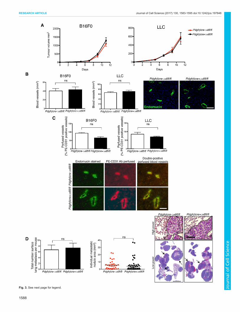