Integrin α high cell population functions as an initiator in...
38
Integrin α6 marks stem cell-like cells in human liposarcoma 1 Integrin α6 high cell population functions as an initiator in tumorigenesis and relapse of human liposarcoma Lu Wang 1 , Lingxian Wang 1 , Yanhong Gu 1,2 , Yongqian Shu 2 , Yan Shen 1 and Qiang Xu 1* 1 State Key Laboratory of Pharmaceutical Biotechnology, School of Life Sciences, Nanjing University, Nanjing 210093, China; 2 Department of Clinical Oncology, The First Affiliated Hospital of Nanjing Medical University, Nanjing 210029, China Running Title: Integrin α6 marks stem cell-like cells in human liposarcoma Key words: liposarcoma, cancer stem cells, integrin α6, tumorigenesis Abbreviations List: ADCs, human normal adipose derived cells; CSC, cancer stem cells; ECM, Extracellular matrix Footnotes: 1. This study was supported by national Natural Science Foundation of China (Nos. 30730107 and 90913023), Science Fund for Creative Research Groups (No. 30821006), and Natural Science Foundation of Jiangsu Province (BK2008022). on June 19, 2018. © 2011 American Association for Cancer Research. mct.aacrjournals.org Downloaded from Author manuscripts have been peer reviewed and accepted for publication but have not yet been edited. Author Manuscript Published OnlineFirst on October 6, 2011; DOI: 10.1158/1535-7163.MCT-11-0487
Transcript of Integrin α high cell population functions as an initiator in...
Integrin α6 marks stem cell-like cells in human liposarcoma
1
Integrin α6high cell population functions as an initiator in
tumorigenesis and relapse of human liposarcoma
Lu Wang1, Lingxian Wang1, Yanhong Gu1,2, Yongqian Shu2 , Yan Shen1 and
Qiang Xu1*
1 State Key Laboratory of Pharmaceutical Biotechnology, School of Life Sciences,
Nanjing University, Nanjing 210093, China; 2 Department of Clinical Oncology,
The First Affiliated Hospital of Nanjing Medical University, Nanjing 210029, China
Running Title: Integrin α6 marks stem cell-like cells in human liposarcoma
Key words: liposarcoma, cancer stem cells, integrin α6, tumorigenesis
Abbreviations List: ADCs, human normal adipose derived cells; CSC, cancer
stem cells; ECM, Extracellular matrix
Footnotes:
1. This study was supported by national Natural Science Foundation of China
(Nos. 30730107 and 90913023), Science Fund for Creative Research Groups
(No. 30821006), and Natural Science Foundation of Jiangsu Province
(BK2008022).
on June 19, 2018. © 2011 American Association for Cancer Research. mct.aacrjournals.org Downloaded from
Author manuscripts have been peer reviewed and accepted for publication but have not yet been edited. Author Manuscript Published OnlineFirst on October 6, 2011; DOI: 10.1158/1535-7163.MCT-11-0487
Integrin α6 marks stem cell-like cells in human liposarcoma
2
2.*Corresponding author. State Key Laboratory of Pharmaceutical Biotechnology,
School of Life Sciences, Nanjing University, 22 Han Kou Road, Nanjing 210093,
China Tel & Fax: 86-25-8359-7620 E-mail: [email protected] (Q. Xu)
The authors indicate no potential conflicts of interest.
on June 19, 2018. © 2011 American Association for Cancer Research. mct.aacrjournals.org Downloaded from
Author manuscripts have been peer reviewed and accepted for publication but have not yet been edited. Author Manuscript Published OnlineFirst on October 6, 2011; DOI: 10.1158/1535-7163.MCT-11-0487
Integrin α6 marks stem cell-like cells in human liposarcoma
3
Abstract
The relapse and resistance to chemo- and radiotherapy are main problems in the
treatment of human liposarcoma. It is important to find a functional marker
existing in the liposarcoma cells for targeting. In this report, we established a new
sub-cell line SW872-S cells with high tumorigenicity from human liposarcoma
SW-872 cells by repeated inoculation approach. The characteristic of the sub-cell
line is linked to the high levels of integrin α6 on the surface. The integrin α6high
cells show much higher tumor initiation and self-renewal potential in vivo than
integrin α6low cells do. Targeting integrin α6 with its specific siRNA and antibody
significantly inhibits the cell adhesion to laminin and the tumor growth in vitro and
in vivo, respectively. Interestingly, integrin α6 marks almost all of the surgical
biopsy specimens of patients with liposarcoma relapse. Moreover, integrin α6 is
found to co-express with CD13, which might contribute to anti-apoptosis ability of
integrin α6high cells. Consistently, integrin α6high cells are more sensitive to the
CD13 inhibitor bestatin, and 61% of 23 other human tumor cell lines also contain
integrin α6high CD13high subgroup. These results provide evidence, for the first
time, to our knowledge, that integrin α6 and CD13 can serve as functional
markers of the tumor-initiation sub-cell population in human liposarcoma as well
as other cancers for therapeutic targeting.
on June 19, 2018. © 2011 American Association for Cancer Research. mct.aacrjournals.org Downloaded from
Author manuscripts have been peer reviewed and accepted for publication but have not yet been edited. Author Manuscript Published OnlineFirst on October 6, 2011; DOI: 10.1158/1535-7163.MCT-11-0487
Integrin α6 marks stem cell-like cells in human liposarcoma
4
Introduction
Liposarcoma is the most common soft tissue sarcoma and accounts for at least
20% of all sarcomas in adults (1). The two major location of liposarcoma are the
extremities and the retroperitoneum (2). Surgery almost serves as the sole
therapy for localized liposarcoma and there is few effective treatment option (3).
For high grade liposarcoma such as lipoblastic sarcoma, 100% of local
recurrence will be present within 5 years, and half of them had a distant relapse.
Moreover, liposarcoma is highly resistant to chemotherapy and radiotherapy. All
these findings suggest the existence of a sub-group of cells with higher
tumorigenicity in human liposarcoma.
Extracellular matrix (ECM) proteins are key structural components of the
perivascular niche. The proteins are known to play a role in the proliferation and
migration of normal stem cells and tumor cells (4) and in liposarcoma
progression (5). One of the functions of ECM in regulating cell behavior is
through their receptors on the cell surface called heterodimer integrins, which
consist of α and β subunits (6). Integrin α6, the receptor for the ECM protein
laminin, forms heterodimers with integrin β1 or β4. Integrin α6 is highly
expressed in gastrointestinal stromal tumors (7), embryonic, hematopoeitic and
neural stem cells (8). Resent studies suggest that integrin α6 is important for the
tumourigenicity of stem cell-like subpopulation within breast cancer cells (9). It is
also known that integrin α6-positive cells are enriched in adult prostatic
on June 19, 2018. © 2011 American Association for Cancer Research. mct.aacrjournals.org Downloaded from
Author manuscripts have been peer reviewed and accepted for publication but have not yet been edited. Author Manuscript Published OnlineFirst on October 6, 2011; DOI: 10.1158/1535-7163.MCT-11-0487
Integrin α6 marks stem cell-like cells in human liposarcoma
5
progenitor/stem cells (10). Justin D. Lathia et al. demonstrated that integrin α6
may serve as a functional marker of human glioblastoma stem cells (11).
In the present study, for the first time, we established a sub-cell line (SW872-S)
derived from human liposarcoma cells SW872 by repeated inoculation approach
in nude mice, which showed high tumor initiation ability in vivo. The SW872-S
cells revealed higher adhesion ability to laminin but not to fibronectin than
SW872 cells did. This result was then linked to the up-regulated expression of
integrin α6 in SW872-S cells, which is rarely expressed in normal adipose
derived cells (12). Furthermore, integrin α6high cells showed much higher tumor
formation in mice than integrin α6low cells did. Consistently, integrin α6-positive
cells were confirmed in tumor tissues of patients who suffered from liposarcoma
relapse within 2 years after surgery. Moreover, the co-high expression of integrin
α6 with aminopeptidase N (CD13) was observed and the functions were
examined. The findings obtained here provide a novel characteristic of the new
sub-cell line of human liposarcoma.
Materials and Methods
Animals
Six-week-old NCR-nu/nu (nude) female mice were purchased from the Shanghai
Laboratory Animal Center (Shanghai, China). They were maintained in pathogen-
free condition at 22±2 °C and kept on a 12-h light–dark cycle. Animal welfare and
on June 19, 2018. © 2011 American Association for Cancer Research. mct.aacrjournals.org Downloaded from
Author manuscripts have been peer reviewed and accepted for publication but have not yet been edited. Author Manuscript Published OnlineFirst on October 6, 2011; DOI: 10.1158/1535-7163.MCT-11-0487
Integrin α6 marks stem cell-like cells in human liposarcoma
6
experimental procedures were carried out strictly in accordance with the Guide
for the Care and Use of Laboratory Animals (The Ministry of Science and
Technology of China, 2006) and the related ethical regulations of our university.
All efforts were made to minimize the animals’ suffering and to reduce the
number of animals used.
Isolation of high tumorigenicity sub-cell population using repeating
inoculation approach
SW872 cells at the logarithmic growth phase were suspended in ice-cold PBS to
a final density of 107/ml, and injected into the right flank of nude mice with 100 μl.
Six months later, neoplasm formed in 2 of 30 mice. The mice with tumor were
then sacrificed and the isolated cells from the tumor tissue were cultured as
method mentioned above. These cells were re-inoculated into nude mice after in
vitro culture for 5 generations. After 3 times of the repeated inoculation approach,
the sub-cell line with high tumorigenicity in vivo was designed as SW872-S cells.
Cell Culture
Human liposarcoma cell line SW872 cells were purchased from the American
Type Culture Collection (Rockville, MD). Cells were maintained in L-15 Medium
supplemented with 10% fetal bovine serum (FBS, Life Technologies), 100 U/ml
penicillin and 100 μg/ml streptomycin and incubated at 37 °C. Human melanoma
cell lines SKMEL-1, A875, A375; human liver cancer cell lines HepG2, BEL-7402;
human breast cancer cell lines MCF-7, MDA-MB-435S; human ovarian cancer
on June 19, 2018. © 2011 American Association for Cancer Research. mct.aacrjournals.org Downloaded from
Author manuscripts have been peer reviewed and accepted for publication but have not yet been edited. Author Manuscript Published OnlineFirst on October 6, 2011; DOI: 10.1158/1535-7163.MCT-11-0487
Integrin α6 marks stem cell-like cells in human liposarcoma
7
cell lines CAOV3, SKOV3; human renal cell carcinoma cell lines 786-O; human
colon cancer cell lines Colo 205, HT29, SW620, HUT-116; human fibrosarcoma
cell line HT1080; human pancreatic cancer cell line PANC-1; human lung cancer
cell line A549; human cervical cancer cell line Hela; human prostate cell line PC3
were obtained from American Type Culture Collection (ATCC). Human
melanoma cell line M14, human liver cancer cell line SMMC7221; human renal
cell carcinoma cell line OS-RC2; human gallbladder carcinoma cell line GBC-SD
were purchased from the Cell Institute Country Cell Bank, Institute of
Biochemistry and Cell Biology, Chinese Academy of Sciences, Shanghai, China.
These cells were maintained with DMEM containing 10% FBS, at 37 °C with 5%
CO2. No independent authentication of these cells was done by the authors.
Lipoaspirates from surgery (supplied by obtained from The First Affiliated
Hospital of Nanjing Medical University) were washed extensively with ice-cold
PBS to remove the containing debris and blood cells. Then the tissues were
treated with 0.15% collagenase (type I) containing 0.1% BSA in PBS for 30 min
at 37 °C with gentle agitation. The cellular pellet digested from tissues was then
resuspended in low-glucose DMEM/10% FBS. Human sample investigations
were preceded by local institutional review board approval.
Cell adhesion assay
The cell adhesion assay was performed as before (Wu et al., 2004). In brief, 96-
well, flat-bottom culture plates were coated with 50 μl of fibronectin (10 μg/ml),
laminin (10 μg/ml) (Calbiochem, San Diego, CA), or collagen type I (50 μg/ml)
on June 19, 2018. © 2011 American Association for Cancer Research. mct.aacrjournals.org Downloaded from
Author manuscripts have been peer reviewed and accepted for publication but have not yet been edited. Author Manuscript Published OnlineFirst on October 6, 2011; DOI: 10.1158/1535-7163.MCT-11-0487
Integrin α6 marks stem cell-like cells in human liposarcoma
8
(Sigma Chemical Co.) in PBS overnight at 4°C, respectively. Plates were then
blocked with 0.2% bovine serum albumin for 2 hours at room temperature
followed by washing three times with DMEM. The cells were harvested with
trypsin/EDTA, washed with ice cold PBS for twice, and re-suspended in DMEM
medium. Cells (2 × 10⁴/well) were added to each well in triplicate and incubated
for 30 minutes at 37 °C. Plates were then washed three times with DMEM
medium to remove unbounded cells. Cells remaining attached to the plates were
quantified with MTT assay.
Real-time PCR
Quantitative PCR was performed with the ABI Prism 7000 sequence detection
system (Applied Biosystems, Foster City, CA) using SYBR Green I dye (Biotium,
Inc., Hayward, CA), and threshold cycle numbers were obtained using ABI Prism
7000 SDS software version 1.0. Conditions for amplification were one cycle of
94°C for 5 minutes followed by 35 cycles of 94°C for 30 seconds, 59°C for 35
second, and 72°C for 45 seconds. The primer sequences used in this study were
as follows: integrin a1 forwad, 5’-GGTTCCTACTTTGGCAG-3’; reverse, 5’-
AACCTTGTCTGATTGAGAG-3’; integrin a2 forwad, 5’-
GGAACGGGACTTTCGCAT-3’; reverse, 5’-GGTACTTCGGCTTTCT-3’; integrin
a3 forwad, 5’-AAGGGACCTTCAGGTGCA-3’; reverse, 5’-
TGTAGCCGGTGATTTACCAT-3’; integrin a4 forwad, 5’-
GCTTCTCAGATCTGCTCGTG-3’; reverse, 5’-GTCACTTCCAACGAGGTT-3’;
integrin a5 forwad, 5’-TGCAGTGTGAGGCTGTGTACA-3’; reverse, 5’-
on June 19, 2018. © 2011 American Association for Cancer Research. mct.aacrjournals.org Downloaded from
Author manuscripts have been peer reviewed and accepted for publication but have not yet been edited. Author Manuscript Published OnlineFirst on October 6, 2011; DOI: 10.1158/1535-7163.MCT-11-0487
Integrin α6 marks stem cell-like cells in human liposarcoma
9
GTGGCCACCTGACGCTCT-3’; integrin a6 forwad, 5’-
TTGAATATACTGCTAACC-3’; reverse, 5’-TCGAAACTGAACTCTTGAGGA-3’;
integrin a7 forwad, 5’-CTGTTTCAGCTACATTGCA-3’; reverse, 5’-
GCCTGGTGCTTGGGTT-3’; integrin av forwad, 5’-AATCTTCCAATTGAGGATA-
3’; reverse, 5’-AAAACAGCCAGTAGCAA-3’; integrin a11 forwad, 5’-
GGAGGAAGACTTGCG-3’; reverse, 5’-CACAGGTTCCCCAGTA-3’; CD9 forwad,
5’-ACTGTTCTTCGGCTT-3’; reverse, 5’-AAAATCCCAAAAATCT-3’; CD10
forwad, 5’-TGTGGCCAGATTGATT-3’; reverse, 5’-TTGTAGGTTCGGCTGA-3’;
CD13 forwad, 5’-CCCAAGATGTCCACGTA-3’; reverse, 5’-
GGTGCTGATGGCATTAACCT-3’; CD44 forwad, 5’-
CAACTCCATCTGTGCAGCA-3’; reverse, 5’-GTAACCTCCTGAAGTGCT-3’;
CD55 forwad, 5’-AAGTAATCTTTGGCTGTAAGG-3’; reverse, 5’-
TTCACCAGCATGTTTTACCTTT-3’; CD59 forwad, 5’-
CGTAGTCCGCTTTCTCTTG-3’; reverse, 5’-CTGCCAGAAGTCCTCTA-3’; CD90
forwad, 5’-CTAGTGGACCAGAGC-3’; reverse, 5’-GCCCTCACACTTGACCAG-3’;
CD166 forwad, 5’-CCAAGAAGGAGGAGGAATA-3’; reverse, 5’-
CCATTCCTGTACCAT-3’; β-actin forwad, 5’-GCTGTGCTACGTCGC-3’; reverse,
5’-GGAGGAGCTGGAAGC-3’;
RNA interference
Chemically synthesized 21-nucleotide sense and antisense RNA
oligonucleotides were obtained from Invitrogen. Cells were plated on six-well
plates at 3×10⁵ cells per well and transfected with 100 pmol of siRNA duplex per
on June 19, 2018. © 2011 American Association for Cancer Research. mct.aacrjournals.org Downloaded from
Author manuscripts have been peer reviewed and accepted for publication but have not yet been edited. Author Manuscript Published OnlineFirst on October 6, 2011; DOI: 10.1158/1535-7163.MCT-11-0487
Integrin α6 marks stem cell-like cells in human liposarcoma
10
well using Oligofectamine (Invitrogen). CD13 siRNA sequences were as follows:
5’- AACGAUCUCUUCAGCACAUCA -3’, Luciferase siRNA was used as
described before (13). Integrin α6 specific SMARTpool® siRNA reagent
(Dharmacon, IL) was used to knockdown endogenous integrin α6 in cells.
Immunohistochemistry and H&E staining assay.
Immunostaining of Integrin α6 was done using Real Envision Detection kit (Gene
Tech Company, Nanjing, China) according to the manufacturer’s instructions.
H&E staining in tumor tissues were performed following manufacturer’s protocol.
Western blot
Western blot assay was performed as described (13). Antibodies to Src,
phosphorylated Src(Tyr416), Akt, phosphorylated Akt (Ser473), phosphorylated
ERK, Bcl-2, cleaved caspase 3, cleaved PARP (Cell Signaling Technology,
Danvers, MA); antibody to integrin α6, GAPDH, CD13 (Santa Cruz, CA) ;
antibody to FAK, phosphorylated FAK (Tyr397) (BD Biosciences, San Jose, CA)
were respectively used for blotting, and detection was performed by enhanced
chemiluminescence (Amersham Pharmacia Biotech, Piscataway, NJ).
Colony formation in soft agar
Two hundreds of SW872 or SW872-S cells were embedded in 0.3% Noble agar
in DMEM containing 10% FBS. Then the clotted agar was plated on a 0.6% agar
on June 19, 2018. © 2011 American Association for Cancer Research. mct.aacrjournals.org Downloaded from
Author manuscripts have been peer reviewed and accepted for publication but have not yet been edited. Author Manuscript Published OnlineFirst on October 6, 2011; DOI: 10.1158/1535-7163.MCT-11-0487
Integrin α6 marks stem cell-like cells in human liposarcoma
11
base in the same medium. Fourteen days later, the plates were photographed
using a Nikon inverted microscope and colonies (> 50 cells) were counted.
Flow cytometry and cell sorting
Laminin specific integrins (integrin α1, α2, α3, α6, β1, β4) were measured by
using Laminin-Specific Integrins Investigator Kit (ECM420, Millipore), isotype-
matched mouse immunoglobulins served as control. For flow cytometry, samples
were analyzed using a FACSCalibur flow cytometer and CellQuest software (BD
Biosciences). For cell sorting by flow cytometry, samples were analyzed and
sorted on a BD FACSVantage SE (BD Biosciences). For the integrin α6 high and
low populations, the stained cells with the upper 30% or lower 30% of
fluorescence intensity were selected, respectively.
Statistical analysis
Data are expressed as mean ± SD. Student’s t test was used to evaluate the
difference between two groups. Kaplan-Meier method was used to evaluate the
survival test. P < 0.05 was considered to be significant.
on June 19, 2018. © 2011 American Association for Cancer Research. mct.aacrjournals.org Downloaded from
Author manuscripts have been peer reviewed and accepted for publication but have not yet been edited. Author Manuscript Published OnlineFirst on October 6, 2011; DOI: 10.1158/1535-7163.MCT-11-0487
Integrin α6 marks stem cell-like cells in human liposarcoma
12
Results:
A new sub-cell line derived from human liposarcoma cells SW872 is
established and characterized with much stronger tumor-initiating potential
in mice.
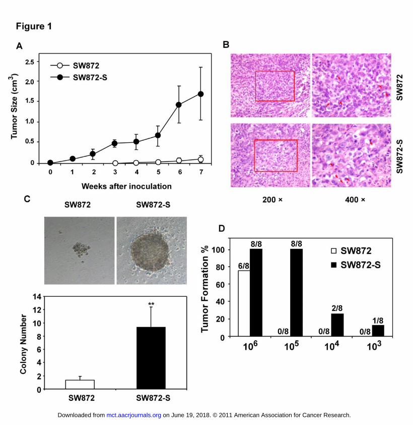
SW872 cells (3×10⁵) were injected into the right flank of nude mice (n=30). Six
months later, the mice with tumors (2 in 30) were sacrificed and the tumor cells
collected were re-inoculated into the naive nude mice. After 3 rounds of selection
as mentioned in Methods section, we obtained a sub-cell line of SW872 cells
(SW872-S) with high aggressive tumor formation potential in nude mice, while
SW872 cells only showed a slight tumor growth (Fig. 1A). H & E staining
revealed that the tumor tissues formed by both cell lines were composed by
lipoblastoma cells (Fig. 1B), indicating that both cell lines belong to the same
classification. In soft agar, SW872-S cells formed bigger and more clones, and
possessed 5-fold colony formation numbers (>50 cells) than SW872 cells (Fig.
1C). As compared with no visible tumor in SW872 cell-inoculated nude mice, we
observed tumor formation in all of the mice (n=8) injected with the same amount
of SW872-S cells (1×10⁵). The smaller amount of 1×10⁴ and even as few as
1×10³ SW872-S cells formed tumor in 2 of 8, and 1 of 8 nude mice, respectively
(Fig. 1D).
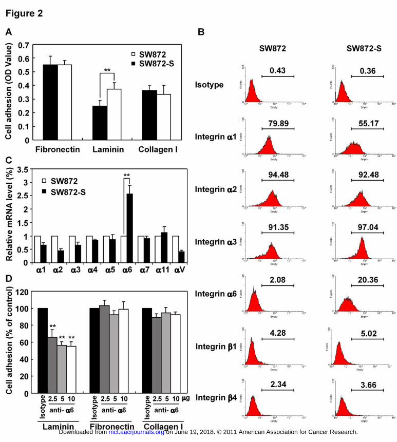
SW872-S cells show a specifically increased adhesion to laminin with an
elevated expression of integrin α6.
on June 19, 2018. © 2011 American Association for Cancer Research. mct.aacrjournals.org Downloaded from
Author manuscripts have been peer reviewed and accepted for publication but have not yet been edited. Author Manuscript Published OnlineFirst on October 6, 2011; DOI: 10.1158/1535-7163.MCT-11-0487
Integrin α6 marks stem cell-like cells in human liposarcoma
13
In order to detect the underlying mechanism of tumor-initiating potential between
the two cell lines, we compared the adhesion ability of SW872 and SW872-S
cells. SW872-S cells showed higher adherent ability to laminin but not to
fibronectin or collagen (Fig. 2A), suggesting the involvement of laminin-specific
integrins in the sub-line cells. FACS analyzing revealed that among all the
binding integrins to laminin, integrin α6 was the only high-expressing one on cell
surface of SW872-S cells, while there was a decrease of integrin α1(Fig. 2B).
Figure 2C showed that among integrin α family, integrin α6 the only higher
expression one in SW872-S cells, indicating integrin α6 may contribute largely to
the high adhesion ability on laminin of SW872-S cells. Consistent with the result,
blocking with integrin α6 specific antibody (Clone: CoH3) significantly inhibited
the adhesion of SW872-S cells to laminin in a dose-dependent manner (Fig. 2D).
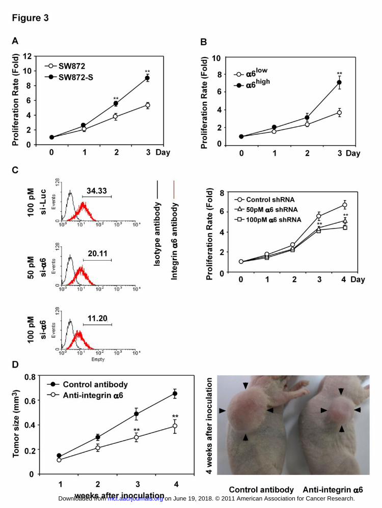
Integrin α6 contributes to the increased liposarcoma growth in vitro and in
vivo.
As shown in Fig. 3A, SW872-S cells grew faster than SW872 cells in vitro. To
examine the relation of such enhanced cell growth to the increased expression of
integrin α6, we divided SW872-S cells into two compartments according to
integrin α6 levels (low and high) by FACS sorting and used them for comparison.
After 72-hour culture, the α6high cells proliferated 2 times as α6low cells (Fig. 3B),
and knocking down of integrin α6 with its specific smartpool siRNA significantly
decreased the growth of SW872-S cells (Fig. 3C). We then inoculated into right
flank of nude mice with 1×10⁶ SW872-S cells that were pre-incubated with
on June 19, 2018. © 2011 American Association for Cancer Research. mct.aacrjournals.org Downloaded from
Author manuscripts have been peer reviewed and accepted for publication but have not yet been edited. Author Manuscript Published OnlineFirst on October 6, 2011; DOI: 10.1158/1535-7163.MCT-11-0487
Integrin α6 marks stem cell-like cells in human liposarcoma
14
control antibody or integrin α6 monoclonal antibody for 30 min, respectively. As
shown in Fig. 3D, treating with integrin α6 antibody significantly inhibited the
growth of SW872-S cells in mice. In this case, the tumor formation rate of
SW872-S cells was also reduced to 60%, as compared with the rate of 100% of
control.
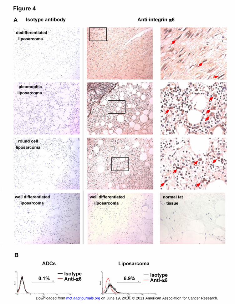
Integrin α6 expression associates with human liposarcoma relapse.
Patients with high grade liposarcoma always suffered from high relapse
frequency within 5 years. This fact leads us to presume that there exists a sub-
group of cells with high tumorigenicity in human liposarcoma. To test this
assumption, we collected liposarcoma specimens from 30 patients who suffered
from recurrence within two years, 6 samples of well-differentiated liposarcoma
with good prognosis and 3 samples of human normal fat tissues. We examined
the involvement of integrin α6 in human liposarcoma recurrence by means of
immunohistochemistry. As the result, 90% (27 in 30) of recurrent samples
contained integrin α6-positive cells and the representative pictures respectively
from dedifferentiated, pleomorphic and round cell liposarcoma, while we could
not detect remarkable integrin α6 positive staining cells in neither well-
differentiated liposarcoma samples, nor the normal fat tissue samples.
Accordingly, FACS analysis revealed that about 6.9% of integrin α6-positive cells
in human primary liposarcoma while human normal adipose derived cells (ADCs)
contained no integrin α6-positive cells (Fig. 4B).
on June 19, 2018. © 2011 American Association for Cancer Research. mct.aacrjournals.org Downloaded from
Author manuscripts have been peer reviewed and accepted for publication but have not yet been edited. Author Manuscript Published OnlineFirst on October 6, 2011; DOI: 10.1158/1535-7163.MCT-11-0487
Integrin α6 marks stem cell-like cells in human liposarcoma
15
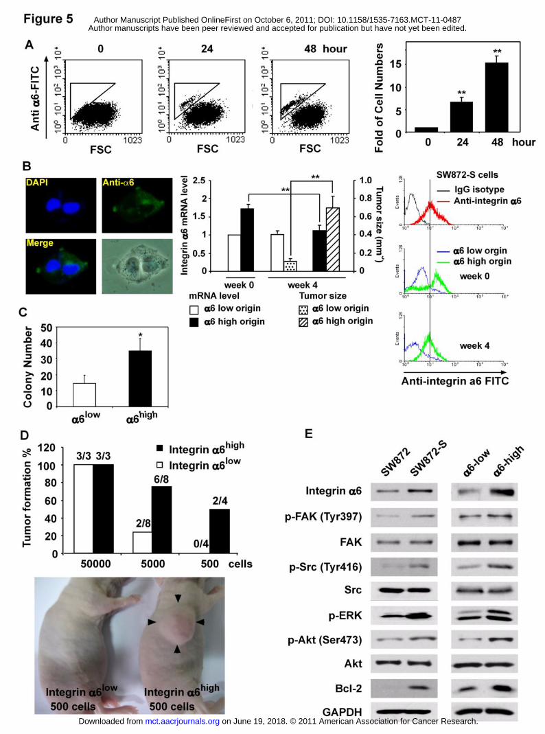
Integrin α6high liposarcoma cells show high self-renewal and tumorigenicity
with activated FAK/Src/ERK signaling.
When cultured in serum-free medium, SW872-S cells with integrin α6 high
expression still proliferate well and the cell numbers were increased by 6.8 and
15.1 times after 24 and 48 hours, respectively (Fig. 5A). The SW872-S cells
showed an asymmetrical distribution of the integrin α6 expression in a cell as
well as in divided daughter cells during cell division (Fig. 5B, left panel). Further
analysis was performed to test the self-renewal ability of integrin α6 cells. The
α6high cells and α6low cells at 106 were inoculated into the right flank of nude mice,
and allowed to grow for 4 weeks. Interestingly, a totally reduced integrin α6
expression was observed in the tumor tissues from integrin α6high cells inoculated
mice by RT-PCR (Fig. 5B, middle panel) or FACS (right panel) , and in this case,
there was a greatly enhanced tumor growth in the mice. In soft agar, integrin
α6high cells have stronger colony formation than integrin α6low cells (Fig. 5C).
When injected into nude mice, 5000 α6high cells initiated tumors in 6 of 8 animals,
while the same amount of α6low cells only did in 2 of 8 mice. Similarly, 500 α6high
cells and α6low cells formed tumors in 2 of 4 and 0 mice, respectively (Fig. 5D). In
SW872-S cells, we found increased phosphorylation levels of FAK (tyr925), Src
(Tyr416), ERK and Akt (ser473), as well as up-regulated expression of Bcl-2.
Consistently, the sub-population with integrin α6high isolated from SW872-S cells
also showed stronger activation of FAK/Src/ERK pathway, and expression of Bcl-
2 (Fig. 5E).
on June 19, 2018. © 2011 American Association for Cancer Research. mct.aacrjournals.org Downloaded from
Author manuscripts have been peer reviewed and accepted for publication but have not yet been edited. Author Manuscript Published OnlineFirst on October 6, 2011; DOI: 10.1158/1535-7163.MCT-11-0487
Integrin α6 marks stem cell-like cells in human liposarcoma
16
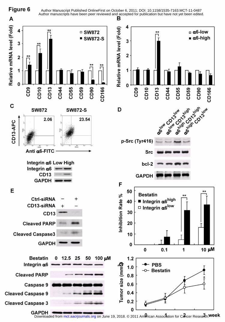
Integrin α6high liposarcoma cells express high levels of aminopeptidase N
(CD13).
For liposarcoma is a malignant tumor that arises in fat cells, we measured
several markers that have been reported to express on the surface of human
adipose derived cells (ADCs) (12) by real-time PCR. The results revealed that
there were no significant changes in the expressions of CD44, CD55, and CD59
between SW872 and SW872-S cells. However, the expressions of CD9, CD10
and CD13 were significantly elevated in SW872-S cells, while those of CD90 and
CD166 were decreased in the cells (Fig. 6A). We further identified that in
SW872-S cells, the α6high group displayed 3 times CD13 expression as α6low
group, while there were no significant or just slender changes in the other
markers (Fig. 6B). The co-high expression of integrin α6 and CD13 was found in
SW872-S cells about 12 times than in SW872 cells (Fig. 6C, upper level). The
high expression of CD13 could also been observed in integrin α6high cells (Fig.
6C, lower panel). In addition, we found that in 23 other human cancer cell lines,
61% (Supplementary Fig.1) of which contained integrin α6- and CD13- double
positive sub groups (Supplementary Fig. 2). The integrin α6high CD13 high cells
showed the highest level of phosphorylated Src as well as the expression of Bcl-
2, as compared with α6howCD13low, α6highCD13low, and α6lowCD13high cells (Fig.
6D). Knocking down of CD13 expression induced apoptosis in SW872-S cells
(Fig. 6E, upper panel) as well as in some other malignant cancer cells
(Supplementary Fig. 3). CD13 specific inhibitor bestatin also induced apoptosis in
SW872-S cells in dose-dependent manner (Fig. 6E, lower panel). The integrin
on June 19, 2018. © 2011 American Association for Cancer Research. mct.aacrjournals.org Downloaded from
Author manuscripts have been peer reviewed and accepted for publication but have not yet been edited. Author Manuscript Published OnlineFirst on October 6, 2011; DOI: 10.1158/1535-7163.MCT-11-0487
Integrin α6 marks stem cell-like cells in human liposarcoma
17
α6high cells were more sensitive to the bestatin treatment than integrin α6low cells,
in which the proliferation of both cells were dose-dependently inhibited (Fig. 6F).
Consistent with the in vitro experiment, 20 mg/kg/day treatment of bestatin could
also inhibite the SW872-S cells growth in nude mice (Fig. 6G).
Discussion
Patients with liposarcoma always suffered from high risk of relapse, and 100% of
local recurrence will be present within 5 years such as lipoblastic liposarcoma
despite of thorough surgery treatment (14). This fact lets us to think about if there
is a sub-group of cells with high tumorigenicity in human liposarcoma tissues.
In the present study, we tried to generate the possibly existing sub-population by
an in vivo repeated inoculation approach with a small amount of cells as
mentioned in Materials and Methods. After nine-month selection, a sub-cell line
(SW872-S) was obtained from human liposarcoma SW872 cells, which have
been previously demonstrated unable or difficult to initiate tumors in nude mice
(15). The SW872-S cells obtained revealed extremely high tumorigenicity in the
nude mice. The elevated tumorigenicity of SW872-S cells was also exhibited in
the assays of colony formation (Fig. 1C), and specific cell adhesion to laminin but
not to fibronectin or collagen type I (Fig. 2A). This result suggests an involvement
of laminin-specific integrins in the functions of the sub-line cells. In order to prove
this assumption, we compared the expressions of various integrins associated
with laminin adhesion on the surface of SW872-S cells with SW872 cells, and
found that integrin α6 but not all the other integrins was elevated. The decrease
on June 19, 2018. © 2011 American Association for Cancer Research. mct.aacrjournals.org Downloaded from
Author manuscripts have been peer reviewed and accepted for publication but have not yet been edited. Author Manuscript Published OnlineFirst on October 6, 2011; DOI: 10.1158/1535-7163.MCT-11-0487
Integrin α6 marks stem cell-like cells in human liposarcoma
18
of integrin α1 in SW872-S cells seemed to be in less relationship with cell
adhesion ability on laminin, at least in our system, for integrin a1 antibody could
not inhibit neither SW872 cells nor SW872-S cells adhesion on laminin (data not
shown). Furthermore, blocking with integrin α6 specific antibody (Clone CoH3)
significantly inhibited the adhesion of SW872-S cells to laminin in vitro (Fig. 2D),
and the tumor growth in vivo (40.77% of inhibition rate) (Fig. 3D). These findings
strongly suggest that the high tumorigenicity of the sub-cell population could be
linked to integrin α6 at least as one of the important factors.
In previous studies, integrin α6 has been reported as subunit form heterodimers
with either β1 or β4 integrins, which both function as receptors for the laminin
family of ECM. Activated α6β1 or α6β4 components then bring extra-cellular
signal to active PI3K/Akt pathway or hif dependent pathway, respectively (16).
High expression of the α6 subunit in women with breast cancer has been
demonstrated to be correlated significantly with reduced survival times (9).
Recently, integrin α6 has been suggested as a potential marker of cancer stem
cells in human glioblastoma (11). In addition, integrin α6 is critical to the self-
renewal and tumor formation of the glioblastoma stem cells, and knocking-down
or blocking with α6 specific antibody could significantly inhibit glioblastoma
growth in vitro and in vivo (11). This report is in consistence with our data shown
in Fig. 2, suggesting that integrin α6 may be crucial for initiating tumor growth in
some kinds of cancers.
on June 19, 2018. © 2011 American Association for Cancer Research. mct.aacrjournals.org Downloaded from
Author manuscripts have been peer reviewed and accepted for publication but have not yet been edited. Author Manuscript Published OnlineFirst on October 6, 2011; DOI: 10.1158/1535-7163.MCT-11-0487
Integrin α6 marks stem cell-like cells in human liposarcoma
19
As reported by D-S. Kim, et. al., rapid adhering cells of human epidermal
keratinocytes with high levels of integrin α6 show a smaller size, which have high
ability of colony formation and skin equivalents reconstruction (17). In our study,
we noticed that in SW872-S cells, integrin α6high sub-group could grow in serum-
free medium at least more than 72 hours (Fig. 5A), and the size of proliferated
integrin α6high cells were also smaller than the others according to FACS analysis
(Fig. 5A). The underlying mechanism, however, remains unknown. Nevertheless,
we have evidenced that integrin α6 was distributed asymmetrically on the surface
of a cell and the expression level was quite different in divided daughter cells
during cell division. This finding suggests that integrin α6high cells could
differentiate into both integrin α6high and α6low cells. In fact, the integrin α6high
cells showed much higher proliferation in mice than α6low cells. Perhaps due to
the dilution of increased α6low cells after 4-week growth in mice, the total content
of integrin α6 in the tumor tissues was reduced (Fig. 5B). Although cancer stem
cells are believed to have stem cell characteristics (18), such as tumor initiation,
proliferation (19) and self-renewal (20), which may lead to metastasis and
relapse after therapy (21), it is still lacks of gold standard definition of cancer
stem cells in all types of cancers. Our results highlight the involvement of integrin
α6 in the self-renewal capacity, colony formation ability, and tumor initiation in
vivo of liposarcoma cells. All these results revealed the tumor initiation function of
integrin α6high sub group cells, at least in human liposarcoma.
on June 19, 2018. © 2011 American Association for Cancer Research. mct.aacrjournals.org Downloaded from
Author manuscripts have been peer reviewed and accepted for publication but have not yet been edited. Author Manuscript Published OnlineFirst on October 6, 2011; DOI: 10.1158/1535-7163.MCT-11-0487
Integrin α6 marks stem cell-like cells in human liposarcoma
20
It should be noted that in some experiments mentioned above, we did not
observe the complete blockade of tumor proliferation or growth by silencing
integrin α6 or using its specific monoclonal antibody. This could be partially
explained by incomplete silence due to technique restriction. We can also
assume that it might be due to another factor involved in integrin α6-mediated
tumor initiation upon these data. As the evidence for this, Fig. 6B and C showed
that integrin α6 co-highly expressed with CD13, which shows a variety of
functions of one member of this family, aminopeptidase N (22).
CD13 has been proved to be a transcript target of Ras signaling pathways (23),
playing an important role in tumor progression in ovarian carcinoma cells (24). It
was also reported that CD13 could block the apoptosis induction by tumor
necrosis factor-alpha (25), and enhance sensitivity of tumor cells to cisplatin (26).
CD13 was also proved to be involved in cell adhesion (27) as well as in
endothelial invasion (28). Recent study made by Haraguchi et al. proved that
CD13 may be a therapeutic target in human liver cancer stem cells and inhibition
of CD13 by monoclonal antibody could induce cell apoptosis (29). In the present
study, knocking down of CD13 with its specific siRNA induced apoptosis in
SW872-S cells (Fig. 6E), and small molecule inhibitor of CD13 such as bestatin
showed higher inhibition rate on SW872-S-integrin α6high cells (Fig. 6F). All these
results suggest that CD13 may be an essential co-operator in integrin α6high sub-
group of cells, which defends cells from apoptosis induction. In order to confirm
the implication of these two proteins simultaneously appeared in the cells, we
on June 19, 2018. © 2011 American Association for Cancer Research. mct.aacrjournals.org Downloaded from
Author manuscripts have been peer reviewed and accepted for publication but have not yet been edited. Author Manuscript Published OnlineFirst on October 6, 2011; DOI: 10.1158/1535-7163.MCT-11-0487
Integrin α6 marks stem cell-like cells in human liposarcoma
21
measured the co-expression of both proteins in 23 other human cancer cell lines.
As the result, we found that 61% (14 in 23) of them contained CD13 and integrin
α6 double-positive sub-group cells (Supplementary Fig. 1). Knocking down of
CD13 induced apoptosis in A375, A549 and Bel-7402 cells with CD13 and
integrin α6 co-high expression (Supplementary Fig. 3). These results indicate a
possibility that both integrin α6 and CD13 may serve as functional markers of not
only liposarcoma but also various human cancers and sarcomas, which may be
new therapeutic targets for the clinical cancer treatment.
on June 19, 2018. © 2011 American Association for Cancer Research. mct.aacrjournals.org Downloaded from
Author manuscripts have been peer reviewed and accepted for publication but have not yet been edited. Author Manuscript Published OnlineFirst on October 6, 2011; DOI: 10.1158/1535-7163.MCT-11-0487
Integrin α6 marks stem cell-like cells in human liposarcoma
22
Acknowledgements:
We want to sincerely consecrate this paper to Prof. Ting Chen who had been our
colleague and had struggled with retroperitoneum liposarcoma for 15 years, and
who is also the donor of some of the samples used in this study.
on June 19, 2018. © 2011 American Association for Cancer Research. mct.aacrjournals.org Downloaded from
Author manuscripts have been peer reviewed and accepted for publication but have not yet been edited. Author Manuscript Published OnlineFirst on October 6, 2011; DOI: 10.1158/1535-7163.MCT-11-0487
Integrin α6 marks stem cell-like cells in human liposarcoma
23
References
1. Mack T. Sarcomas and Other Malignancies of Soft-Tissue,
Retroperitoneum, Peritoneum, Pleura, Heart, Mediastinum, and Spleen. Cancer.
1995;75:211-44.
2. Amato G, Martella A, Ferraraccio F, Di Martino N, Maffettone V, Landolfi V,
et al. Well differentiated "lipoma-like" liposarcoma of the sigmoid mesocolon and
multiple lipomatosis of the rectosigmoid colon. Report of a case. Hepato-
Gastroenterology. 1998;45:2151-6.
3. Singer S, Socci N, Ambrosini G, Sambol E, Decarolis P, Wu Y, et al. Gene
expression profiling of liposarcoma identifies distinct biological types/subtypes
and potential therapeutic targets in well-differentiated and dedifferentiated
liposarcoma. Cancer Research. 2007;67:6626-36.
4. Gilbertson RJ, Rich JN. Making a tumour's bed: glioblastoma stem cells
and the vascular niche. Nature Reviews Cancer. 2007;7:733-6.
5. Roomi M, Ivanov V, Kalinovsky T, Niedzwiecki A, Rath M. Inhibition of cell
invasion and MMP production by a nutrient mixture in malignant liposarcoma cell
line SW-872. Medical Oncology. 2007;24:394-401.
6. Hynes R. Integrins: Bidirectional, allosteric signaling machines. Cell.
2002;110:673-87.
7. Takeyama H, Funahashi H, Sawai H, Takahashi H, Yamamoto M, Akamo
Y, et al. Expression of alpha(6) integrin subunit is associated with malignancy in
gastric gastrointestinal stromal tumors. Med Sci Monitor. 2007;13:CR51-CR6.
on June 19, 2018. © 2011 American Association for Cancer Research. mct.aacrjournals.org Downloaded from
Author manuscripts have been peer reviewed and accepted for publication but have not yet been edited. Author Manuscript Published OnlineFirst on October 6, 2011; DOI: 10.1158/1535-7163.MCT-11-0487
Integrin α6 marks stem cell-like cells in human liposarcoma
24
8. Evsikov A, Solter D. Comment on " 'Stemness': Transcriptional profiling of
embryonic and adult stem cells" and "A stem cell molecular signature" (II).
Science. 2003;302.
9. Wewer U, Shaw L, Albrechtsen R, Mercurio A. The integrin alpha 6 beta 1
promotes the survival of metastatic human breast carcinoma cells in mice.
American Journal of Pathology. 1997;151:1191-8.
10. Barclay W, Axanova L, Chen W, Romero L, Maund SL, Soker S, et al.
Characterization of adult prostatic progenitor/stem cells exhibiting self-renewal
and multilineage differentiation. Stem Cells. 2008;26:600-10.
11. Lathia J, Gallagher J, Heddleston J, Wang J, Eyler C, MacSwords J, et al.
Integrin Alpha 6 Regulates Glioblastoma Stem Cells. Cell Stem Cell. 2010;6:421-
32.
12. Katz AJ, Tholpady A, Tholpady SS, Shang HL, Ogle RC. Cell surface and
transcriptional characterization of human adipose-derived adherent stromal
(hADAS) cells. Stem Cells. 2005;23:412-23.
13. Wang L, Shen Y, Song R, Sun Y, Xu J, Xu Q. An Anticancer Effect of
Curcumin Mediated by Down-Regulating Phosphatase of Regenerating Liver-3
Expression on Highly Metastatic Melanoma Cells. Molecular Pharmacology.
2009;76:1238-45.
14. Chang H, Hajdu SI, Collin C, Brennan M. The Prognostic Value of
Histologic Subtypes in Primary Extremity Liposarcoma. Cancer. 1989;64:1514-20.
on June 19, 2018. © 2011 American Association for Cancer Research. mct.aacrjournals.org Downloaded from
Author manuscripts have been peer reviewed and accepted for publication but have not yet been edited. Author Manuscript Published OnlineFirst on October 6, 2011; DOI: 10.1158/1535-7163.MCT-11-0487
Integrin α6 marks stem cell-like cells in human liposarcoma
25
15. Guo Y, Me J, Rubin E, Tang Y, Lin F, Zi X, et al. Frzb, a secreted wnt
antagonist, decreases growth and invasiveness of fibrosarcoma cells associated
with inhibition of met signalling. Cancer Research. 2008;68:3350-60.
16. Lipscomb E, Simpson K, Lyle S, Ring J, Dugan A, Mercurio A. The alpha
6 beta 4 integrin promotes tumor formation by regulated cell survival. Molecular
Biology of the Cell. 2004;15:342.
17. Kim D, Cho H, Choi H, Kwon S, Park K. Isolation of human epidermal
stem cells by adherence and the reconstruction of skin equivalents. Cellular and
Molecular Life Sciences. 2004;61:2774-81.
18. Alison, R M. Stem cells and cancer in the aerodigestive tract. Eur J
Cancer. 2009;45 Suppl 1:175-85.
19. Dean M, Fojo T, Bates S. Tumour stem cells and drug resistance. Nature
Reviews Cancer. 2005;5:275-84.
20. Schatton T, Murphy G, Frank NY, Yamaura K, Waaga-Gasser A, Gasser
M, et al. Identification of cells initiating human melanomas. Nature.
2008;451:345-U11.
21. Reya T, Morrison S, Clarke M, Weissman I. Stem cells, cancer, and
cancer stem cells. Nature. 2001;414:105-11.
22. Mina-Osorio P. The moonlighting enzyme CD13: old and new functions to
target. Trends in Molecular Medicine. 2008;14:361-71.
23. Bhagwat S, Petrovic N, Okamoto Y, Shapiro L. The angiogenic regulator
CD13/APN is a transcriptional target of Ras signaling pathways in endothelial
morphogenesis. Blood. 2003;101:1818-26.
on June 19, 2018. © 2011 American Association for Cancer Research. mct.aacrjournals.org Downloaded from
Author manuscripts have been peer reviewed and accepted for publication but have not yet been edited. Author Manuscript Published OnlineFirst on October 6, 2011; DOI: 10.1158/1535-7163.MCT-11-0487
Integrin α6 marks stem cell-like cells in human liposarcoma
26
24. Terauchi M, Kajiyama H, Shibata K, Ino K, Nawa A, Mizutani S, et al.
Inhibition of APN/CD13 leads to suppressed progressive potential in ovarian
carcinoma cells. Bmc Cancer. 2007;7.
25. Cowburn A, Sobolewski A, Reedo B, Deighton J, Murray J, Cadwallader K,
et al. Aminopeptidase N (CD13) regulates tumor necrosis factor-alpha-induced
apoptosis in human neutrophils. Journal of Biological Chemistry.
2006;281:12458-67.
26. van Hensbergen Y, Broxterman H, Rana S, van Diest P, Duyndam M,
Hoekman K, et al. Reduced growth, increased vascular area, and reduced
response to cisplatin in CD13-overexpressing human ovarian cancer xenografts.
Clinical Cancer Research. 2004;10:1180-91.
27. Mina-Osorio P, Shapiro L, Ortega E. CD13 in cell adhesion:
aminopeptidase N (CD13) mediates homotypic aggregation of monocytic cells.
Journal of Leukocyte Biology. 2006;79:719-30.
28. Petrovic N, Schacke W, Gahagan J, O'Conor C, Winnicka B, Conway R,
et al. CD13/APN regulates endothelial invasion and filopodia fonnation. Blood.
2007;110:142-50.
29. Haraguchi N, Ishii H, Mimori K, Tanaka F, Ohkuma M, Kim H, et al. CD13
is a therapeutic target in human liver cancer stem cells. Journal of Clinical
Investigation. 2010;120:3326-39.
on June 19, 2018. © 2011 American Association for Cancer Research. mct.aacrjournals.org Downloaded from
Author manuscripts have been peer reviewed and accepted for publication but have not yet been edited. Author Manuscript Published OnlineFirst on October 6, 2011; DOI: 10.1158/1535-7163.MCT-11-0487
Integrin α6 marks stem cell-like cells in human liposarcoma
27
Figure Legends
Figure 1
Establishment and characterization of a new sub-cell line derived from
human liposarcoma cells SW872 with much stronger tumor-initiating
potential in vivo.
A. 3×10⁶ of SW872 and SW872-S cells were injected into the right flank of nude
mice. Seven weeks later, the growth of tumor formed in mice. Data are mean ±
SD. of five nude mice in each group. B. Tumor sample sections from each group
were stained with H&E. C. 200 of SW872 and SW872-S cells were seeded in soft
agar as described in Materials and Methods. Two weeks later, the clones formed
were counted (lower panel), and the representative picture of clones were shown
(upper panel). Data are mean ± SD of three independent experiments. * p < 0.05,
** p < 0.01 vs the colony number of SW872 cells.
D. SW872 and SW872-S cells were diluted from 106 to 103, and injected into the
right flank of nude mice and allowed to grow for 2 months, the tumor formation
rate were presented.
Figure 2
Increased adhesion to laminin and elevated expression of integrin α6 in
SW872-S cells.
A. The adhesion of SW872 and SW872-S cells ability to fibronectin, laminin and
collagen type I were measured, respectively. B. The surface expressions of
on June 19, 2018. © 2011 American Association for Cancer Research. mct.aacrjournals.org Downloaded from
Author manuscripts have been peer reviewed and accepted for publication but have not yet been edited. Author Manuscript Published OnlineFirst on October 6, 2011; DOI: 10.1158/1535-7163.MCT-11-0487
Integrin α6 marks stem cell-like cells in human liposarcoma
28
laminin-specific integrins of SW872 and SW872-S cells were measured by FACS
more than 3 times, and representative result was presented. C. The mRNA levels
of the integrin α family members between SW872 and SW872-S cells were
qualified by real-time PCR. β-actin was used as invariant control. D. SW872-S
cells were incubated with various amounts of integrin α6 monoclonal antibody for
30 min, and the adhesion ability to laminin, fibronectin and collagen was
measured. Data are mean ± SD of three independent experiments. * p < 0.05, **
p < 0.01 vs control.
Figure 3
Identification of integrin α6high cells in liposarcoma cells’ growth in vitro
and in vivo.
A. The proliferation of SW872 and SW872-S cells was determined by MTT assay.
B. The proliferation of integrin α6high and integrin α6low cells of SW872-S was
determined by MTT assay. C. SW872-S cells were transfected with various
amounts of integrin α6 smartpool siRNA or luciferase siRNA for 72 hours,
respectively. The surface expression of integrin α6 was measured by FACS (left
panel). The proliferation rate of cells transfected by integrin a6 siRNA or
luciferase siRNA was determined by MTT assay (right panel). D. 106 of SW872-S
cells pre-incubated with control antibody or integrin α6 specific blocking antibody
(clone: CoH3), were inoculated into the right flank of nude mice, respectively.
Time course of tumor growth (left panel), representative pictures (right panel)
on June 19, 2018. © 2011 American Association for Cancer Research. mct.aacrjournals.org Downloaded from
Author manuscripts have been peer reviewed and accepted for publication but have not yet been edited. Author Manuscript Published OnlineFirst on October 6, 2011; DOI: 10.1158/1535-7163.MCT-11-0487
Integrin α6 marks stem cell-like cells in human liposarcoma
29
were presented. Data are mean ± SD of three independent experiments. * p <
0.05, ** p < 0.01 vs control.
Figure 4
Integrin α6-positive cells in human tumor tissues with liposarcoma relapse.
Immunohistochemical staining assays was carried out for integrin α6 by using
human liposarcoma specimens. Paraffin section of human liposarcoma tissues
were collected from 30 patients who had undergone liposarcoma relapse within 2
years; 6 samples from patients underwent well-differentiated liposarcoma with
good prognosis; 3 samples of normal fat tissue. A. The representative pictures
respectively from dedifferentiated, pleomorphic, round cell, well differentiated
liposarcoma and normal fat tissue were shown. B. FACS analyze between
human normal adipose derived cells and human primary liposarcoma cells
isolated from tumor tissues with relapse.
Figure 5
Multiple tumor formation potential in integrin α6high cells with activated
FAK/Src/ERK signaling.
A. SW872-S cells were cultured in serum free medium for 24 and 48 hours.
Integrin α6 level was measured by FACS, the fold induction of integrin α6high sub-
group was shown. B. Single cell suspension of SW872-S cells were seeded at
the concentration of 1000/ml, the surface expression of integrin α6 was analyzed
by immunofluorescence (left panel). Integrin α6high and integrin α6low cells (106
on June 19, 2018. © 2011 American Association for Cancer Research. mct.aacrjournals.org Downloaded from
Author manuscripts have been peer reviewed and accepted for publication but have not yet been edited. Author Manuscript Published OnlineFirst on October 6, 2011; DOI: 10.1158/1535-7163.MCT-11-0487
Integrin α6 marks stem cell-like cells in human liposarcoma
30
cells/mouse) were s.c. inoculated into the flank of nude mice, respectively. Four
weeks later, the tumor xenografts were collected and Integrin α6 expression was
detected by real-time PCR (middle panel) and FACS (right panel) respectively,
for comparison with the tumor size. C. The colony formation ability of Integrin
α6high and integrin α6low cells divided from SW872-S was measured. D. Integrin
α6high and integrin α6low cells divided from SW872-S cells were diluted from
50000 to 500, respectively, and injected into the right flank of nude mice and
allowed to grow for 2 months. The tumor formation rate (upper panel),
representative pictures of tumor formed (lower panel) were presented. E. The
protein levels of total integrin α6, FAK, Src, ERK, Akt, Bcl-2 as well as
phosphorylated FAK, Src, ERK, Akt of SW872, SW872-S, integrin α6high, and
integrin α6low of SW872-S cells were determined by western blot.
Figure 6
Co-expression of aminopeptidase N (CD13) in integrin α6high cells and
increased sensitivity to CD13 inhibitor bestatin.
A. The mRNA levels of CD9, CD10, CD13, CD44, CD55, CD59, CD90, and
CD166 between SW872 and SW872-S cells. B. The mRNA levels of the above
markers between SW872-S-integrin α6high and SW872-S-integrin α6low cells. C.
Flow cytometric analysis for expressions of CD13 and integrin α6 in SW872 and
SW872-S cells (upper panel). The protein level of CD13 was also determined in
α6low and α6high cells (lower panel). D. SW872-S cells were sorted into four
groups: α6low CD13low, α6low CD13high, α6high CD13high, α6high CD13low, the protein
on June 19, 2018. © 2011 American Association for Cancer Research. mct.aacrjournals.org Downloaded from
Author manuscripts have been peer reviewed and accepted for publication but have not yet been edited. Author Manuscript Published OnlineFirst on October 6, 2011; DOI: 10.1158/1535-7163.MCT-11-0487
Integrin α6 marks stem cell-like cells in human liposarcoma
31
levels of phosphorylated Src, Src, and Bcl-2 were determined by western blot. E.
SW872-S cells were transfected with control siRNA and CD13 specific siRNA for
72 hours. The protein levels of CD13, cleaved PARP, and cleaved caspase 3
were determined by western blot (upper panel). SW872-S cells were incubated
with various amount of bestatin for 24 hours, the protein level of integrin α6,
cleaved PARP, Caspase9, cleaved Caspase3 were determined by western blot
(lower panel). F. The 72-hour inhibition rates of Bestatin on SW872-S-integrin
α6high and SW872-S-integrin α6low cells were determined by MTT assay. G. The
mice with tumor formed of SW872-S cells were treated with PBS or 20 mg/kg
bestatin daily. The tumor size was measured every week. Data are mean ± SEM
of five mice in each group. * p < 0.05, ** p < 0.01 vs control.
on June 19, 2018. © 2011 American Association for Cancer Research. mct.aacrjournals.org Downloaded from
Author manuscripts have been peer reviewed and accepted for publication but have not yet been edited. Author Manuscript Published OnlineFirst on October 6, 2011; DOI: 10.1158/1535-7163.MCT-11-0487
Published OnlineFirst October 6, 2011.Mol Cancer Ther Lu Wang, Lingxian Wang, Yanhong Gu, et al. tumorigenesis and relapse of human liposarcomaIntegrin a6high cell population functions as an initiator in
Updated version
10.1158/1535-7163.MCT-11-0487doi:
Access the most recent version of this article at:
Material
Supplementary
http://mct.aacrjournals.org/content/suppl/2011/10/03/1535-7163.MCT-11-0487.DC1
Access the most recent supplemental material at:
Manuscript
Authoredited. Author manuscripts have been peer reviewed and accepted for publication but have not yet been
E-mail alerts related to this article or journal.Sign up to receive free email-alerts
Subscriptions
Reprints and
To order reprints of this article or to subscribe to the journal, contact the AACR Publications
Permissions
Rightslink site. Click on "Request Permissions" which will take you to the Copyright Clearance Center's (CCC)
.http://mct.aacrjournals.org/content/early/2011/10/06/1535-7163.MCT-11-0487To request permission to re-use all or part of this article, use this link
on June 19, 2018. © 2011 American Association for Cancer Research. mct.aacrjournals.org Downloaded from
Author manuscripts have been peer reviewed and accepted for publication but have not yet been edited. Author Manuscript Published OnlineFirst on October 6, 2011; DOI: 10.1158/1535-7163.MCT-11-0487