
DesigningaLongActingErythropoietinbyFusingThree...
8
Hindawi Publishing Corporation International Journal of Cell Biology Volume 2011, Article ID 275063, 7 pages doi:10.1155/2011/275063 Research Article Designing a Long Acting Erythropoietin by Fusing Three Carboxyl-Terminal Peptides of Human Chorionic Gonadotropin β Subunit to the N -Terminal and C -Terminal Coding Sequence Fuad Fares, 1 Avri Havron, 2, 3 and Eyal Fima 2, 3 1 Department of Human Biology, Faculty of Natural Sciences, University of Haifa and Department of Molecular Genetics, Carmel Medical Center, Mount Carmel, 31905 Haifa, Israel 2 ModigeneTech, Weizmann Science Park, 74140 Nes-Ziona, Israel 3 PROLOR Biotech, Weizmann Science Park, 74140 Nes-Ziona, Israel Correspondence should be addressed to Fuad Fares, ff[email protected] Received 26 February 2011; Accepted 26 June 2011 Academic Editor: Johannes Vogel Copyright © 2011 Fuad Fares et al. This is an open access article distributed under the Creative Commons Attribution License, which permits unrestricted use, distribution, and reproduction in any medium, provided the original work is properly cited. A new analog of EPO was designed by fusing one and two CTPs to the N-terminal and C-terminal ends of EPO (EPO-(CTP) 3 ), respectively. This analog was expressed and secreted efficiently in CHO cells. The in vitro test shows that the activity of EPO- (CTP) 3 in TFI-1 cell proliferation assay is similar to that of EPO-WT and commercial rHEPO. However, in vivo studies indicated that treatment once a week with EPO-(CTP) 3 (15 μg/kg) dramatically increased (∼8 folds) haematocrit as it was compared to rHuEPO. Moreover, it was found that EPO-(CTP) 3 is more effective than rHuEPO and Aranesp in increasing reticulocyte number in mice blood. The detected circulatory half-lives of rHuEPO, Aranesp, and EPO-(CTP) 3 following IV injection of 20 IU were 4.4, 10.8, and 13.1 h, respectively. These data established the rational for using this chimera as a long-acting EPO analog in clinics. The therapeutic efficacy of EPO-CTP analog needs to be established in higher animals and in human clinical trials. 1. Introduction Erythropoietin (EPO) is a 34-kDa glycoprotein hormone produced primarily by cells of the per tubular capillary endothelium of the kidney and regulates red blood cell pro- duction through stimulation of erythropoiesis [1, 2]. EPO synthesis in the kidney is increased following reduction in tissue oxygenation, it binds to specific receptors on red blood cell precursors in the bone marrow leading to proliferation, differentiation, and to an increase in haematocrit [3]. Bio- logical responses associated with EPO include activation of intracellular signaling molecules such as transcription factors like signal transducer and activator of transcription (STAT) proteins leading to cellular growth and differentiation. EPO receptor belongs to a family of homodimerization receptors where dimerization of the receptor is required to trigger the biological responses associated with EPO [4–7]. Anemia in patients with chronic kidney disease is due to a number of factors, the most common of which is abnormally low erythropoietin levels. Anemia of EPO deficiency is recog- nized in advanced renal failure but not in early renal disease. Deficiency in EPO production results in anemia in humans and in animal models. EPO is heavily glycosylated with one O-linked and three N-linked oligosaccharide chains. It was found that O-linked oligosaccharide chain has no effect on secretion, receptor binding affinity, and in vitro or in vivo bioactivity. On the other hand, N-linked oligosaccharide- chains have no role in in vitro activity, but it is critical for in vivo bioactivity [8]. The gene encoding human erythropoietin was cloned in 1985 leading to the production of recombinant human EPO (rHuEPO) [9, 10]. rHuEPO was used successfully in treating anemia associated with chronic kidney disease. It has also been approved for the treatment of anemia associated with cancer, HIV infection, and in the surgical setting in order to reduce blood transfusions [11–13]. One major issue regarding the clinical use of EPO is its relatively short half- life in vivo due to its rapid clearance (∼5 hours) from the
Transcript of DesigningaLongActingErythropoietinbyFusingThree...
Hindawi Publishing CorporationInternational Journal of Cell BiologyVolume 2011, Article ID 275063, 7 pagesdoi:10.1155/2011/275063
Research Article
Designing a Long Acting Erythropoietin by Fusing ThreeCarboxyl-Terminal Peptides of Human Chorionic Gonadotropinβ Subunit to the N -Terminal and C -Terminal Coding Sequence
Fuad Fares,1 Avri Havron,2, 3 and Eyal Fima2, 3
1 Department of Human Biology, Faculty of Natural Sciences, University of Haifa and Department of Molecular Genetics,Carmel Medical Center, Mount Carmel, 31905 Haifa, Israel
2 ModigeneTech, Weizmann Science Park, 74140 Nes-Ziona, Israel3 PROLOR Biotech, Weizmann Science Park, 74140 Nes-Ziona, Israel
Correspondence should be addressed to Fuad Fares, [email protected]
Received 26 February 2011; Accepted 26 June 2011
Academic Editor: Johannes Vogel
Copyright © 2011 Fuad Fares et al. This is an open access article distributed under the Creative Commons Attribution License,which permits unrestricted use, distribution, and reproduction in any medium, provided the original work is properly cited.
A new analog of EPO was designed by fusing one and two CTPs to the N-terminal and C-terminal ends of EPO (EPO-(CTP)3),respectively. This analog was expressed and secreted efficiently in CHO cells. The in vitro test shows that the activity of EPO-(CTP)3 in TFI-1 cell proliferation assay is similar to that of EPO-WT and commercial rHEPO. However, in vivo studies indicatedthat treatment once a week with EPO-(CTP)3 (15 μg/kg) dramatically increased (∼8 folds) haematocrit as it was compared torHuEPO. Moreover, it was found that EPO-(CTP)3 is more effective than rHuEPO and Aranesp in increasing reticulocyte numberin mice blood. The detected circulatory half-lives of rHuEPO, Aranesp, and EPO-(CTP)3 following IV injection of 20 IU were 4.4,10.8, and 13.1 h, respectively. These data established the rational for using this chimera as a long-acting EPO analog in clinics. Thetherapeutic efficacy of EPO-CTP analog needs to be established in higher animals and in human clinical trials.
1. Introduction
Erythropoietin (EPO) is a 34-kDa glycoprotein hormoneproduced primarily by cells of the per tubular capillaryendothelium of the kidney and regulates red blood cell pro-duction through stimulation of erythropoiesis [1, 2]. EPOsynthesis in the kidney is increased following reduction intissue oxygenation, it binds to specific receptors on red bloodcell precursors in the bone marrow leading to proliferation,differentiation, and to an increase in haematocrit [3]. Bio-logical responses associated with EPO include activation ofintracellular signaling molecules such as transcription factorslike signal transducer and activator of transcription (STAT)proteins leading to cellular growth and differentiation. EPOreceptor belongs to a family of homodimerization receptorswhere dimerization of the receptor is required to trigger thebiological responses associated with EPO [4–7]. Anemia inpatients with chronic kidney disease is due to a numberof factors, the most common of which is abnormally low
erythropoietin levels. Anemia of EPO deficiency is recog-nized in advanced renal failure but not in early renal disease.Deficiency in EPO production results in anemia in humansand in animal models. EPO is heavily glycosylated with oneO-linked and three N-linked oligosaccharide chains. It wasfound that O-linked oligosaccharide chain has no effect onsecretion, receptor binding affinity, and in vitro or in vivobioactivity. On the other hand, N-linked oligosaccharide-chains have no role in in vitro activity, but it is critical forin vivo bioactivity [8].
The gene encoding human erythropoietin was clonedin 1985 leading to the production of recombinant humanEPO (rHuEPO) [9, 10]. rHuEPO was used successfully intreating anemia associated with chronic kidney disease. It hasalso been approved for the treatment of anemia associatedwith cancer, HIV infection, and in the surgical setting inorder to reduce blood transfusions [11–13]. One major issueregarding the clinical use of EPO is its relatively short half-life in vivo due to its rapid clearance (∼5 hours) from the
2 International Journal of Cell Biology
circulation when it is injected intravenously [14]. Thus, theclinical therapeutic protocols of available stimulating agentsused in the treatment of patients require frequent injectionsof EPO. The recommended therapy with rHuEPO is 2-3times per week by subcutaneous or intravenous injections.Therefore, it can be anticipated that enhancing the in vivohalf-life of EPO would reduce the number of injectionsper week. Previous studies indicated that there is a directrelationship between the sialic acid-containing carbohydratecontent of the molecule and its serum half-life and in vivobioactivity [15–17]. It was shown that fusing the carboxyl-terminal peptide (CTP) of hCGβ subunit that associatedwith four sites of O-linked oligosaccharide chains to the C-terminal of FSH, TSH, GH, and EPO cDNA did not affectsecretion, receptor binding affinity, and in vitro bioactivity.On the other hand, the addition of O-linked oligosaccharidesto the backbone of the protein significantly increased thehalf-life and longevity in vivo [18–22]. We hypothesis thatthe addition of 12 O-linked oligosaccharide chains to thebackbone of EPO will dramatically increase the longevityof EPO. Therefore, in the present study, three carboxyl-terminal peptides (CTP) of hCGβ subunit that each containsfour O-linked oligosaccharide recognition sites was fused toN-terminal (one) and to the C-terminal (two) of humanEPO coding sequence, respectively. Our results indicate thatligation of three CTPs to the coding sequence of EPOdramatically increased both in vivo potency and half-life inthe circulation.
2. Materials and Methods
2.1. Materials. Enzymes used in the construction of DNAvectors and constructs were purchased from New EnglandBioLabs (Beverly, Mass, USA). Cell culture media andreagents were obtained from Biological Industries (BeitHaemek, Israel). Rabbit antisera against EPO were purchasedfrom Fitzgerald (Concord, Mass, USA). The eukaryoticexpression vector (pCI-DHFR, Dihydrofolate reductase) intowhich the cDNA encoding for the corresponding hEPOvariants were inserted was purchased from Promega, (SanLuis Obispo, Calif, USA). Commercial human recombinantEPO (Eprex) was purchased from Janssen-Cilag (NorthRyde, NSW, Australia).
2.2. Crystallography. The interaction between EPO and itsreceptor was crystallized as described previously [23] by theDepartment of Structural Biology, Weizmann Institute ofScience, Rehovot, Israel.
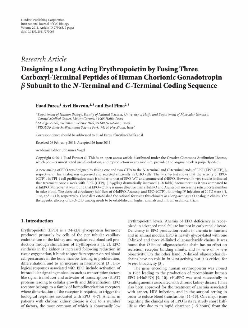
2.3. Construction of Chimeric Genes and Expression Vectors. Acassette gene containing the CTP of hCGβ was fused in tan-dem to the coding sequence of EPO at the N-terminal (oneCTP) and to the C-terminal end (two CTPs) (Figure 1). DNAfragment containing sequences of hEPO-cDNA and codingsequence of CTP were synthesized by GeneArt (Regensburg,Germany). The DNA fragments contain the recognition sitesof the restriction enzymes; Xba I (in the N-terminal) and NotI (in the C-terminal). Fragment containing hEPO and CTP
sequences was completely sequenced to ensure that no errorswere introduced during synthesis and ligated into the XbaI—Not I sites at the cloning site of the eukaryotic expressionvector, pCI-DHFR. Similarly, cDNA of human EPO (EPO-WT) was constructed into pCI-DHFR vector.
2.4. Cell Culture and DNA Transfection. Chinese hamsterovary (CHO)-DG44 cells, which are DHFR negative, wereused. Cells were cultured in MEM-α medium (Gibco BRL,USA) supplemented with penicillin (100 U/mL), strepto-mycin (100 mg/mL), L-glutamine (2 mM), and 10% heat-inactivated fetal bovine serum at 37◦C in humidified incu-bator containing 5% CO2. These cells were transfected with2 μg DNA of plasmid by using FuGENE6 (Roche, Mannheim,Germany) according to manufacturer protocol.
Cells were selected for insertion of the plasmid DNA bygrowth in culture medium of CD DG44 without hypoxan-thine and thymidine (HT) (Gibco BRL, USA) supplementedwith 8 mM L-Glutamine (Biological Industries, Beit Haimic,Israel) and 18 mL/L of 10% Pluronic F-68 solution (GibcoBRL, USA).
2.5. Western Blotting. Samples of condition medium whichwere collected from stable clones were electrophorisedon denaturing 15% SDS-polyacrylamide gels as describedbefore [24]. Gels were allowed to equilibrate for 10 minin 25 mM Tris and 192 mM glycine in 20% (vol/vol)methanol. Proteins were transferred to a 0.2 μm pore sizenitrocellulose membrane (Sigma, Saint Louis, Mo, USA)at 250 mA for 3 h using a Mini Trans-Blot electrophoresiscell (Biorad Laboratories, Richmond, CA) according to themethod described in the manual accompanying the unit.The nitrocellulose membrane was incubated in 5% nonfatdry milk for 2 h at room temperature. The membranewas incubated with EPO antiserum (1 : 1000 titers) forovernight at 4◦C followed by three consecutive washes in PBScontaining 0.1% Tween (10 min/wash). Then, the membranewas incubated with secondary antibody conjugated to HorseRadish Peroxidase (HRP) (Zymed, San Francisco, CA) for2 h at room temperature followed by three washes. Finally,the nitrocellulose paper was reacted with enhanced chemi-luminescent substrate (ECL) (Pierce, Rockford, Ill, USA) for5 min, dried with Whatman sheet and exposed to X-ray film.
2.6. In Vitro Bioactivity. Bioactivity of EPO variants wasassayed by testing the proliferation dependence of thehuman erythroleukemic cell line TF-1 (Kitamura) (DSMZ)in the presence of EPO and EPO variants [25]. Cultureswere routinely grown at 37◦C, 5% CO2 for 72 hrs inRPMI 1640 medium supplemented with 10% fetal bovineserum (FBS),10 mM Hepes, 1 mM sodium pyruvate, 2.5 g/Lglucose, 2 mM glutamine, and 2 ng/mL rhGM-CSF. Beforetransferring the cells to 96-well plates, the TF-1 cells werewashed three times with cold PBS and suspended in theassay medium (1640 medium supplemented with 10% fetalbovine serum (FBS) but without addition of rHGM-CSF) ata density of 200,000 cells/mL. The assay was performed in96-well plates containing 50 μL of cell suspension per well.
International Journal of Cell Biology 3
CTP CTP CTPEPO-cDNA
5- -3
TCCTCTTCC.. ...CTCCCACAATAASer Ser Ser .. .. Leu Pro Gln Term118 119 120 .. .. 143 144 145
hCGβ-CTP
ATGGGGGTG.. ..GGGGACAGAMet Gly Val .. .. Gly Asp Arg
1 2 3 .. .. 190 191 192
EPO-cDNA
Not IXba I
Figure 1: Construction of EPO-(CTP)3 chimeric gene. The chimeric gene contains the cDNA of human erythropoietin and three hCGβcarboxyl-terminal peptides that were ligated to the N-terminal (one) and to C-terminal (two) coding sequences.
50 μL of assay medium containing 3 IU of EPO variants werethen added to the wells of 96-well plates for 72 hrs. Cellviability was measured using MTT reagent kit (Cell Biolabs,San Diego, CA) according to manufacturer procedures.
2.7. Animals. Male ICR mice were obtained from CharlesRiver Laboratories, Jerusalem, Israel, and housed in air-conditioned quarters with a 12 h light/dark schedule. Stan-dard food and water were available ad libitum. Instituteethical committee approved the in vivo protocols. Animalswere treated with EPO variants as specified.
2.8. In Vivo Bioassay. Groups of 7 male ICR mice (7-week-old males) were used. EPO-WT, EPO-(CTP)3, or commercialrHuEPO were injected to anesthetized animals as describedin Table 1.
The animals were weighed, and each received an identicalamount (5 or 15 μg/kg) of EPO variants by IV injections(0.2 mL/animal). The frequency of treatment was eitherthrice weekly (days 1, 3, and 5) or once weekly. The levelof haematocrit was determined three times a week andthe experiment was stopped after three weeks. Haematocritwas determined using blood samples obtained by fillingtwo heparinized microhematocrit tubes from the inferiorcaval vein under anesthesia. In addition, reticulocyte countswere conducted since these cells are present in blood for∼48 hours before developing into mature red blood cells.Reticulocytes represent an appropriate evaluation for theacute experimental system employed. Blood was obtainedfrom each pup by cardiac puncture and placed in EDTAcoated tubes. This was then mixed with brilliant cresyl blueand incubated for 20 min at 37◦C. The blood and stainwere then smeared onto a glass slide and the number ofreticulocytes assessed using a × 100 oil objective lens.
2.9. Metabolic Clearance Rate. The metabolic clearance ofEPO-WT, Aranesp, and EPO-(CTP)3 was determined afterIV injection of 20 IU/animal into male ICR mice. At selectedintervals after injection, blood samples were collected andEPO immunoreactivity was determined by RIA.
2.10. Statistical Analysis. Data were expressed as the mean± SEM. Statistical analysis of the data were performedusing Student’s t-test and 1-way multivariate analysis of
N-terminal
C-terminal
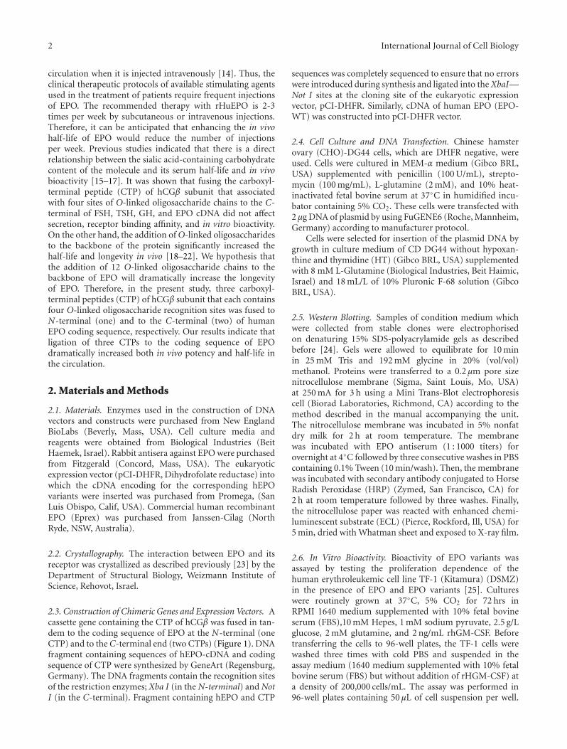
Figure 2: Model of erythropoietin binding to its receptor. Thediagram showing the complex of EPO bound to the receptor.Note that the N-terminal and C-terminal of EPO are free and notinvolved in the binding site.
variance (ANOVA1) to calculate P value. P values < 0.05 wereconsidered statistically significant.
3. Results and Discussion
Chrystallographic studies indicated that the N-terminal andC-terminal of EPO are not involved in the binding of thehormone to the receptor (Figure 2).
Therefore, we hypothesized that ligation of CTP to theN-terminal and to the C-terminal of EPO will not affectreceptor binding affinity and thus bioactivity. Therefore,three CTPs (one in the N-terminal and two in the C-terminal) were ligated to the coding sequence of EPO. ThecDNA of human EPO-WT and EPO-(CTP)3 was insertedinto the eukaryotic expression vector, pCI-DHFR, andtransfected into CHO cells. Stable clone expressing humanEPO-WT or EPO-(CTP)3 was selected. Secretion of EPOwas assessed by Western blot analysis under denaturingconditions using human EPO-specific antiserum. The EPO-WT migrated faster than EPO-(CTP)3 (Figure 3).
4 International Journal of Cell Biology
Table 1: Comparative 3 week induction of haematocrit by EPO-(CTP)3 and rHuEPO.
Group number Mice/groupTreatment
Regimen: IVCompound Dose μg/kg
1
n = 7
Vehicle (control) 0One dose per week2 rHuEPO 15 μg/kg
3 EPO-(CTP)3 15 μg/kg
4 Commercial rHuEPO 5 3 doses per week
EPO-WT EPO (CTP)3
57
37
28
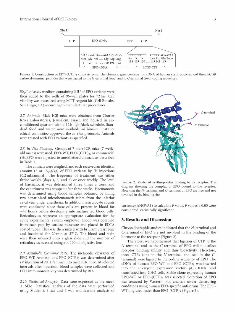
Figure 3: Expression of EPO-WT and EPO-(CTP)3 from trans-fected CHO cells. Conditioned media from transfected cells wereprepared for SDS/PAGE and proteins were detected by Western blotas described under “Materials and Methods.”
EPO-(CTP)3 exhibited high molecular weight (∼57 kDa)comparing to EPO-WT (∼36 kDa) due to the addition of84 amino acids and the O-linked oligosaccharides linked toCTP. These data may indicate that the O-linked glycosylationrecognition site of the C-terminal region is preserved eventhough the sequence is fused to different proteins. Levelsof EPO-WT and EPO-(CTP)3 were quantities in conditionmedium by using a monoclonal antibody-based RIA.
The in vitro biological activity of EPO analogs wasdemonstrated by measuring their ability to stimulate theproliferation of TF-1 cells as described under “Materialsand Methods.” The activity of EPO-(CTP)3 in TF-1 cellproliferation assay was similar to that of EPO wild-type(prepared by Modigene Tech) and commercial rHuEPO(Figure 4).
For further pharmacological evaluation of EPO-(CTP)3,comparative pharmacodynamic studies of EPO-(CTP)3 andcommercial rHuEPO were performed in male ICR mice(n = 7/group) using different frequencies and dose rangeas described in Table 1. The in vivo efficacy was obtained bymeasuring the mean values of haematocrit percentage in theblood. The results indicated that EPO-(CTP)3 is significantly(P < 0.001) more efficient than rHuEPO when administered
0
40
80
120
160
200
Cel
lnu
mbe
r(c
ontr
ol(%
))
EPO-WT rHuEPO EPO-(CTP)3
Figure 4: In vitro biological activity of recombinant hEPO deriva-tives. Bioactivity of EPO variants was tested by measuring theproliferation dependence of human erythroleukemicTF-1 cells inthe absence or presence of 3 IU of EPO variants. Cell proliferationwas measured using MTT reagent kit.
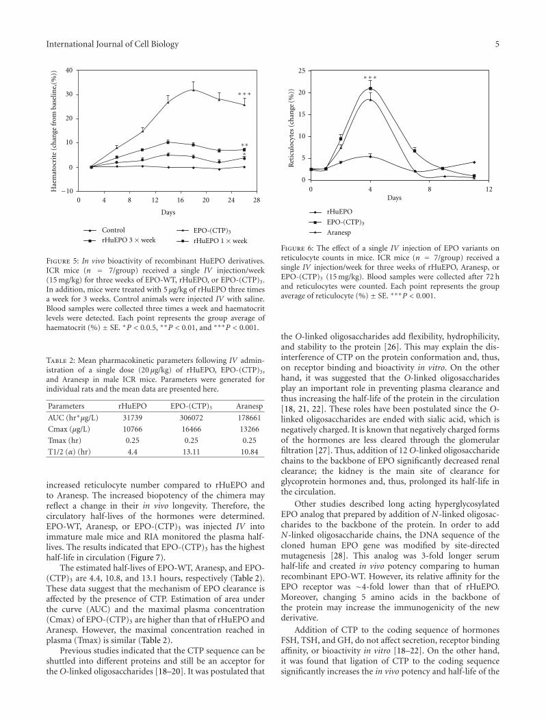
IV once a week with a dose of 15 μg/kg (Figure 5). EPO-(CTP)3 can successfully increase the haematocrit whenadministered once a week with a dose of 15 μg/kg (Figure 5).Once weekly dosing with the same concentration of com-mercial rHuEPO or EPO-WT was significantly (P < 0.001)less efficient than once weekly dosing of EPO-(CTP)3. Aninteresting observation from the present study was the abilityof a single injection once a week of EPO-CTP (15 μg/kg) toincrease dramatically (∼8 folds) the levels of haematocrit.Whereas administration of the same total dose of rHuEPOadministered three times a week as 5 μg/kg per injectionresulted in significantly (P < 0.001) lower effect (Figure 5).
Previously, we have shown that single injection oncea week of EPO-CTP, an EPO that contains one CTP atthe carboxyl-terminal end, (15 μg/kg) increased the level ofhaematocrit, whereas the same effect was achieved by admin-istration of the same total dose of rHuEPO administeredthree times a week as 5 μg/kg per injection [21]. These resultsindicated the importance of sustained blood levels, ratherthan total dose of EPO. These findings are consistent with thehypothesis that the ability of a single injection of EPO-CTPto increase haematocrit results from its increased stability inthe circulation.
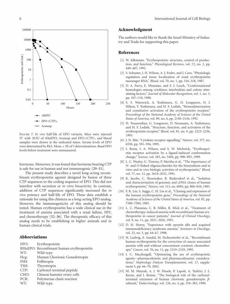
Effect of EPO-WT, (EPO-CTP)3, and Aranesp in reticu-locyte counts is shown in Figure 6. The results indicated thata single IV injection of 15 μg/kg (EPO-CTP)3 dramatically
International Journal of Cell Biology 5
Control
rHuEPO 3 × week rHuEPO 1 × week
EPO (CTP)3
0
10
20
30
40
0 4 8 12 16 20 24 28
Days
Hae
mat
ocri
te(c
han
gefr
omba
selin
e,(%
))
−10
∗∗∗
∗∗∗
-
Figure 5: In vivo bioactivity of recombinant HuEPO derivatives.ICR mice (n = 7/group) received a single IV injection/week(15 mg/kg) for three weeks of EPO-WT, rHuEPO, or EPO-(CTP)3.In addition, mice were treated with 5 μg/kg of rHuEPO three timesa week for 3 weeks. Control animals were injected IV with saline.Blood samples were collected three times a week and haematocritlevels were detected. Each point represents the group average ofhaematocrit (%) ± SE. ∗P < 0.0.5, ∗∗P < 0.01, and ∗∗∗P < 0.001.
Table 2: Mean pharmacokinetic parameters following IV admin-istration of a single dose (20 μg/kg) of rHuEPO, EPO-(CTP)3,and Aranesp in male ICR mice. Parameters were generated forindividual rats and the mean data are presented here.
Parameters rHuEPO EPO-(CTP)3 Aranesp
AUC (hr∗μg/L) 31739 306072 178661
Cmax (μg/L) 10766 16466 13266
Tmax (hr) 0.25 0.25 0.25
T1/2 (α) (hr) 4.4 13.11 10.84
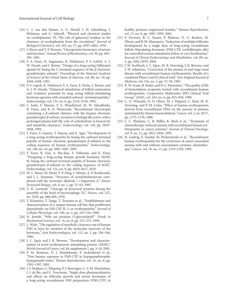
increased reticulocyte number compared to rHuEPO andto Aranesp. The increased biopotency of the chimera mayreflect a change in their in vivo longevity. Therefore, thecirculatory half-lives of the hormones were determined.EPO-WT, Aranesp, or EPO-(CTP)3 was injected IV intoimmature male mice and RIA monitored the plasma half-lives. The results indicated that EPO-(CTP)3 has the highesthalf-life in circulation (Figure 7).
The estimated half-lives of EPO-WT, Aranesp, and EPO-(CTP)3 are 4.4, 10.8, and 13.1 hours, respectively (Table 2).These data suggest that the mechanism of EPO clearance isaffected by the presence of CTP. Estimation of area underthe curve (AUC) and the maximal plasma concentration(Cmax) of EPO-(CTP)3 are higher than that of rHuEPO andAranesp. However, the maximal concentration reached inplasma (Tmax) is similar (Table 2).
Previous studies indicated that the CTP sequence can beshuttled into different proteins and still be an acceptor forthe O-linked oligosaccharides [18–20]. It was postulated that
Days
25
20
15
10
5
00 4 8 12
Aranesp
Ret
icu
locy
tes
(ch
ange
(%))
rHuEPO
∗∗∗
EPO (CTP)3-
Figure 6: The effect of a single IV injection of EPO variants onreticulocyte counts in mice. ICR mice (n = 7/group) received asingle IV injection/week for three weeks of rHuEPO, Aranesp, orEPO-(CTP)3 (15 mg/kg). Blood samples were collected after 72 hand reticulocytes were counted. Each point represents the groupaverage of reticulocyte (%) ± SE. ∗∗∗P < 0.001.
the O-linked oligosaccharides add flexibility, hydrophilicity,and stability to the protein [26]. This may explain the dis-interference of CTP on the protein conformation and, thus,on receptor binding and bioactivity in vitro. On the otherhand, it was suggested that the O-linked oligosaccharidesplay an important role in preventing plasma clearance andthus increasing the half-life of the protein in the circulation[18, 21, 22]. These roles have been postulated since the O-linked oligosaccharides are ended with sialic acid, which isnegatively charged. It is known that negatively charged formsof the hormones are less cleared through the glomerularfiltration [27]. Thus, addition of 12 O-linked oligosaccharidechains to the backbone of EPO significantly decreased renalclearance; the kidney is the main site of clearance forglycoprotein hormones and, thus, prolonged its half-life inthe circulation.
Other studies described long acting hyperglycosylatedEPO analog that prepared by addition of N-linked oligosac-charides to the backbone of the protein. In order to addN-linked oligosaccharide chains, the DNA sequence of thecloned human EPO gene was modified by site-directedmutagenesis [28]. This analog was 3-fold longer serumhalf-life and created in vivo potency comparing to humanrecombinant EPO-WT. However, its relative affinity for theEPO receptor was ∼4-fold lower than that of rHuEPO.Moreover, changing 5 amino acids in the backbone ofthe protein may increase the immunogenicity of the newderivative.
Addition of CTP to the coding sequence of hormonesFSH, TSH, and GH, do not affect secretion, receptor bindingaffinity, or bioactivity in vitro [18–22]. On the other hand,it was found that ligation of CTP to the coding sequencesignificantly increases the in vivo potency and half-life of the
6 International Journal of Cell Biology
r EPO
Aranesp
100000
10000
1000
100
10
1
0.1
0.010 50 100 150 200 250
Hours
Ert
h
h
ropo
ieti
n(m
U/m
L)
EPO (CTP)3-
Figure 7: In vivo half-life of EPO variants. Mice were injectedIV with 20 IU of rHuEPO, Aranesp and EPO-(CTP)3 and bloodsamples were drawn at the indicated times. Serum levels of EPOwere determined by RIA. Mean± SE of 5 determinations. Basal EPOlevels before treatment were unmeasured.
hormone. Moreover, it was found that hormone bearing CTPis safe for use in human and not immunogenic [29–31].
The present study describes a novel long-acting recom-binant erythropoietin agonist designed by fusion of threeCTP sequences to the coding sequence of EPO. This did notinterfere with secretion or in vitro bioactivity. In contrast,addition of CTP sequences significantly increased the invivo potency and half-life of EPO. These data establish arationale for using this chimera as a long-acting EPO analog.However, the immunogenicity of this analog should betested. Human erythropoietin has a wide clinical use in thetreatment of anemia associated with a renal failure, HIV,and chemotherapy [32–36]. The therapeutic efficacy of thisanalog needs to be establishing in higher animals and inhuman clinical trials.
Abbreviations
EPO: ErythropoietinRHuEPO: Recombinant human erythropoietinWT: Wild-typeHcg: Human Chorionic GonadotropinFSH: FollitropinTSH: ThyrotropinCTP: Carboxyl-terminal peptideCHO: Chinese hamster ovary cellsPCR: Polymerase chain reactionWT: Wild-type.
Acknowledgment
The authors would like to thank the Israel Ministry of Indus-try and Trade for supporting this paper.
References
[1] W. Jelkmann, “Erythropoietin: structure, control of produc-tion, and function,” Physiological Reviews, vol. 72, no. 2, pp.449–487, 1992.
[2] S. Schuster, J. H. Wilson, A. J. Erslev, and J. Caro, “Physiologicregulation and tissue localization of renal erythropoietinmessenger RNA,” Blood, vol. 70, no. 1, pp. 316–318, 1987.
[3] D. A. Parry, E. Minasian, and S. J. Leach, “Conformationalhomologies among cytokines: interleukins and colony stim-ulating factors,” Journal of Molecular Recognition, vol. 1, no. 3,pp. 107–110, 1988.
[4] S. S. Watowich, A. Yoshimura, G. D. Longmore, D. J.Hilton, Y. Yoshimura, and H. F. Lodish, “Homodimerizationand constitutive activation of the erythropoietin receptor,”Proceedings of the National Academy of Sciences of the UnitedStates of America, vol. 89, no. 6, pp. 2140–2144, 1992.
[5] H. Youssoufian, G. Longmore, D. Neumann, A. Yoshimura,and H. F. Lodish, “Structure, function, and activation of theerythropoietin receptor,” Blood, vol. 81, no. 9, pp. 2223–2236,1993.
[6] J. N. Ihle, “Cytokine receptor signalling,” Nature, vol. 377, no.6550, pp. 591–594, 1995.
[7] I. Remy, I. A. Wilson, and S. W. Michnick, “Erythropoi-etin receptor activation by a ligand-induced conformationchange,” Science, vol. 283, no. 5404, pp. 990–993, 1999.
[8] L. C. Wasley, G. Timony, P. Murtha et al., “The importance ofN- and O-linked oligosaccharides for the biosynthesis and invitro and in vivo biologic activities of erythropoietin,” Blood,vol. 77, no. 12, pp. 2624–2632, 1991.
[9] K. Jacobs, C. Shoemaker, R. Rudersdorf et al., “Isolationand characterization of genomic and cDNA clones of humanerythropoietin,” Nature, vol. 313, no. 6005, pp. 806–810, 1985.
[10] F. K. Lin, S. Suggs, C. H. Lin et al., “Cloning and expression ofthe human erythropoietin gene,” Proceedings of the NationalAcademy of Sciences of the United States of America, vol. 82, pp.7580–7584, 1985.
[11] L. C. Platanias, C. B. Miller, R. Mick et al., “Treatment ofchemotherapy-induced anemia with recombinant human ery-thropoietin in cancer patients,” Journal of Clinical Oncology,vol. 9, no. 11, pp. 2021–2026, 1991.
[12] D. H. Henry, “Experience with epoetin alfa and acquiredimmunodeficiency syndrome anemia,” Seminars in Oncology,vol. 25, no. 3, pp. 64–67, 1998.
[13] H. Ludwig, E. Sundal, M. Pecherstorfer et al., “Recombinanthuman erythropoietin for the correction of cancer associatedanemia with and without concomitant cytotoxic chemother-apy,” Cancer, vol. 76, no. 11, pp. 2319–2329, 1995.
[14] I. C. Macdougall, “Optimizing the use of erythropoieticagents—pharmacokinetic and pharmacodynamic considera-tions,” Nephrology Dialysis Transplantation, vol. 17, supple-ment 5, pp. 66–70, 2002.
[15] M. M. Matzuk, A. J. W. Hsueh, P. Lapolt, A. Tsafriri, J. L.Keene, and I. Boime, “The biological role of the carboxyl-terminal extension of human chorionic gonadotropin β-subunit,” Endocrinology, vol. 126, no. 4, pp. 376–383, 1990.
International Journal of Cell Biology 7
[16] C. J. van den Hamer, A. G. Morell, I. H. Scheinberg, J.Hickman, and G. Ashwell, “Physical and chemical studieson ceruloplasmin. IX. The role of galactosyl residues in theclearance of ceruloplasmin from the circulation,” Journal ofBiological Chemistry, vol. 245, no. 17, pp. 4397–4402, 1970.
[17] J. Pierce and T. F. Parsons, “Glycoprotein hormones: structureand function,” Annual Review of Biochemistry, vol. 50, pp. 465–495, 1981.
[18] F. A. Fares, N. Suganuma, K. Nishimori, P. S. LaPolt, A. J.W. Hsueh, and I. Boime, “Design of a long-acting follitropinagonist by fusing the C-terminal sequence of the β chorionicgonadotropin subunit,” Proceedings of the National Academyof Sciences of the United States of America, vol. 89, no. 10, pp.4304–4308, 1992.
[19] P. S. Lapolt, K. Nishimori, F. A. Fares, E. Perlas, I. Boime, andA. J. W. Hsueh, “Enhanced stimulation of follicle maturationand ovulatory potential by long acting follicle-stimulatinghormone agonists with extended carboxyl- terminal peptides,”Endocrinology, vol. 131, no. 6, pp. 2514–2520, 1992.
[20] L. Joshi, Y. Murata, F. E. Wondisford, M. W. Szkudlinski,R. Desai, and B. D. Weintraub, “Recombinant thyrotropincontaining a β-subunit chimera with the human chorionicgonadotropin-β carboxy-terminus is biologically active, with aprolonged plasma half-life: role of carbohydrate in bioactivityand metabolic clearance,” Endocrinology, vol. 136, pp. 3839–3848, 1994.
[21] F. Fares, S. Ganem, T. Hajouj, and E. Agai, “Development ofa long-acting erythropoietin by fusing the carboxyl-terminalpeptide of human chorionic gonadotropin β-subunit to thecoding sequence of human erythropoietin,” Endocrinology,vol. 148, no. 10, pp. 5081–5087, 2007.
[22] F. Fares, R. Guy, A. Bar-Ilan, Y. Felikman, and E. Fima,“Designing a long-acting human growth hormone (hGH)by fusing the carboxyl-terminal peptide of human chorionicgonadotropin β-subunit to the coding sequence of hGH,”Endocrinology, vol. 151, no. 9, pp. 4410–4417, 2010.
[23] M. L. Raves, M. Harel, Y. P. Pang, I. Silman, A. P. Kozikowski,and J. L. Sussman, “Structure of acetylcholinesterase com-plexed with the nootropic alkaloid, (-)-huperzine A,” NatureStructural Biology, vol. 4, no. 1, pp. 57–63, 1997.
[24] U. K. Laemmli, “Cleavage of structural proteins during theassembly of the head of bacteriophage T4,” Nature, vol. 227,no. 5259, pp. 680–685, 1970.
[25] T. Kitamura, T. Tange, T. Terasawa et al., “Establishment andcharacterization of a unique human cell line that proliferatesdependently on GM-CSF, IL-3, or erythropoietin,” Journal ofCellular Physiology, vol. 140, no. 2, pp. 323–334, 1989.
[26] N. Jentoft, “Why are proteins O-glycosylated?” Trends inBiochemical Sciences, vol. 14, no. 8, pp. 272–275, 1990.
[27] L. Wide, “The regulation of metabolic clearance rate of humanFSH in mice by variation of the molecular structure of thehormone,” Acta Endocrinologica, vol. 112, no. 3, pp. 336–344,1986.
[28] J. C. Egrie and J. K. Browne, “Development and character-ization of novel erythropoiesis stimulating protein (NESP),”British Journal of Cancer, vol. 84, supplement 1, pp. 3–10, 2001.
[29] P. M. Bouloux, D. J. Handelsman, F. Jockenhovel et al.,“First human exposure to FSH-CTP in hypogonadotrophichypogonadal males,” Human Reproduction, vol. 16, no. 8, pp.1592–1597, 2001.
[30] I. J. Duijkers, C. Klipping, P. J. Boerrigter, C. S. M. Machielsen,J. J. de Bie, and G. Voortman, “Single dose pharmacokineticsand effects on follicular growth and serum hormones ofa long-acting recombinant FSH preparation (FSH-CTP) in
healthy pituitary-suppressed females,” Human Reproduction,vol. 17, no. 8, pp. 1987–1993, 2002.
[31] P. Devroey, B. C. Fauser, P. Platteau, N. G. Beckers, M.Dhont, and B. M. Mannaerts, “Induction of multiple folliculardevelopment by a single dose of long-acting recombinantfollicle-Stimulating hormone (FSH-CTP, corifollitropin alfa)for controlled ovarian stimulation before in vitro fertilization,”Journal of Clinical Endocrinology and Metabolism, vol. 89, no.5, pp. 2062–2070, 2004.
[32] J. W. Eschbach, J. C. Egrie, M. R. Downing, J. K. Browne, andJ. W. Adamson, “Correction of the anemia of end-stage renaldisease with recombinant human erythropoietin. Results of acombined Phase I and II clinical trial,” New England Journal ofMedicine, vol. 316, no. 2, pp. 73–78, 1987.
[33] R. W. Evans, B. Rader, and D. L. Manninen, “The quality of lifeof hemodialysis recipients treated with recombinant humanerythropoietin. Cooperative Multicenter EPO Clinical TrialGroup,” JAMA, vol. 263, no. 6, pp. 825–830, 1990.
[34] C. G. Winearls, D. O. Oliver, M. J. Pippard, C. Reid, M. R.Downing, and P. M. Coles, “Effect of human erythropoietinderived from recombinant DNA on the anemia of patientsmaintained by chronic haemodialysis,” Lancet, vol. 2, no. 8517,pp. 1175–1178, 1986.
[35] L. C. Platanias, C. B. Miller, R. Mick et al., “Treatment ofchemotherapy-induced anemia with recombinant human ery-thropoietin in cancer patients,” Journal of Clinical Oncology,vol. 9, no. 11, pp. 2021–2026, 1991.
[36] H. Ludwig, E. Sundal, M. Pecherstorfer et al., “Recombinanthuman erythropoietin for the correction of cancer associatedanemia with and without concomitant cytotoxic chemother-apy,” Cancer, vol. 76, no. 11, pp. 2319–2329, 1995.

Submit your manuscripts athttp://www.hindawi.com
Hindawi Publishing Corporationhttp://www.hindawi.com Volume 2014
Anatomy Research International
PeptidesInternational Journal of
Hindawi Publishing Corporationhttp://www.hindawi.com Volume 2014
Hindawi Publishing Corporation http://www.hindawi.com
International Journal of
Volume 2014
Zoology
Hindawi Publishing Corporationhttp://www.hindawi.com Volume 2014
Molecular Biology International
GenomicsInternational Journal of
Hindawi Publishing Corporationhttp://www.hindawi.com Volume 2014
The Scientific World JournalHindawi Publishing Corporation http://www.hindawi.com Volume 2014
Hindawi Publishing Corporationhttp://www.hindawi.com Volume 2014
BioinformaticsAdvances in
Marine BiologyJournal of
Hindawi Publishing Corporationhttp://www.hindawi.com Volume 2014
Hindawi Publishing Corporationhttp://www.hindawi.com Volume 2014
Signal TransductionJournal of
Hindawi Publishing Corporationhttp://www.hindawi.com Volume 2014
BioMed Research International
Evolutionary BiologyInternational Journal of
Hindawi Publishing Corporationhttp://www.hindawi.com Volume 2014
Hindawi Publishing Corporationhttp://www.hindawi.com Volume 2014
Biochemistry Research International
ArchaeaHindawi Publishing Corporationhttp://www.hindawi.com Volume 2014
Hindawi Publishing Corporationhttp://www.hindawi.com Volume 2014
Genetics Research International
Hindawi Publishing Corporationhttp://www.hindawi.com Volume 2014
Advances in
Virolog y
Hindawi Publishing Corporationhttp://www.hindawi.com
Nucleic AcidsJournal of
Volume 2014
Stem CellsInternational
Hindawi Publishing Corporationhttp://www.hindawi.com Volume 2014
Hindawi Publishing Corporationhttp://www.hindawi.com Volume 2014
Enzyme Research
Hindawi Publishing Corporationhttp://www.hindawi.com Volume 2014
International Journal of
Microbiology


![Introduction Abstract - Neurology...medulla oblongata were dissected [27] and were stored in RNA later solution for RNA isolation. Whole brain (n = 5 per group) weighing 80-90mg) and](https://static.fdocument.org/doc/165x107/5f7aaac355c0bb44193d6438/introduction-abstract-neurology-medulla-oblongata-were-dissected-27-and.jpg)