Contents 23
42
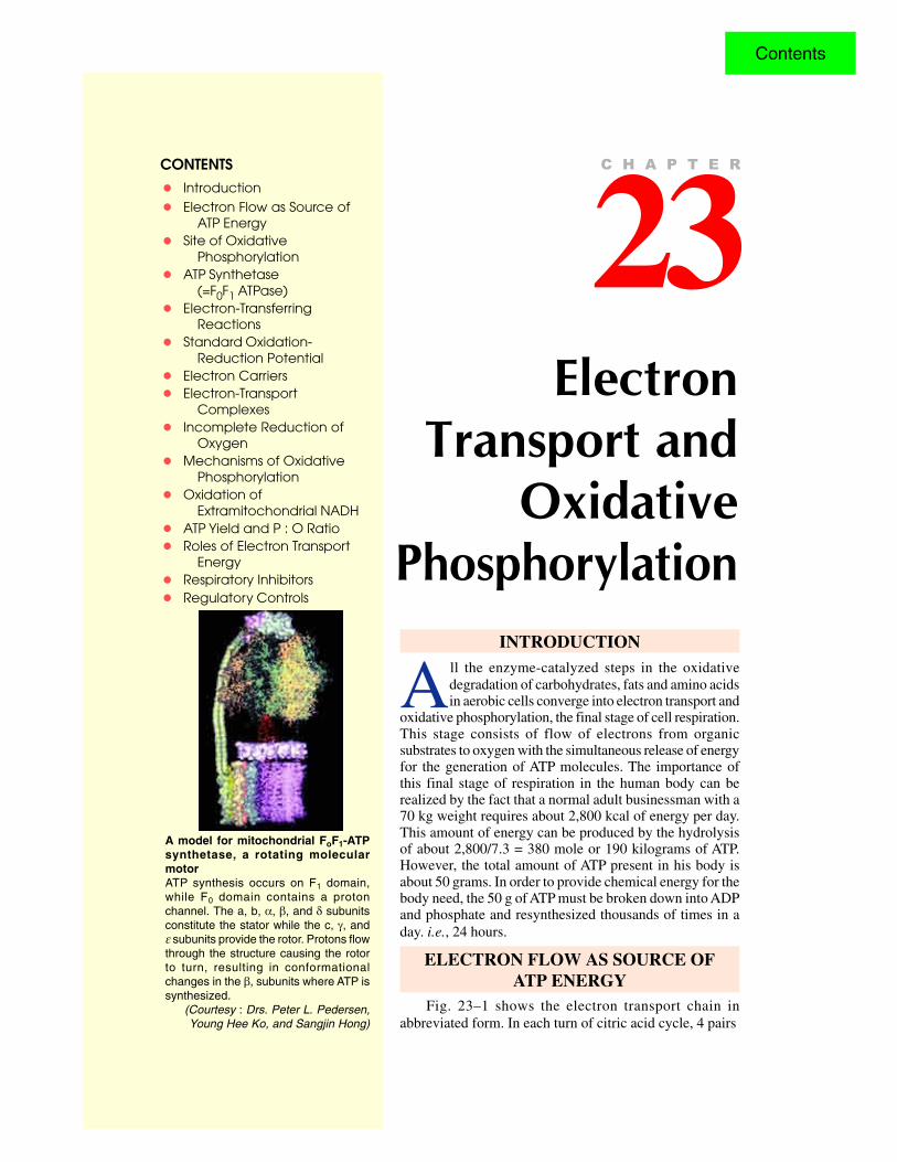
CONTENTS • Introduction • Electron Flow as Source of ATP Energy • Site of Oxidative Phosphorylation • ATP Synthetase (=F 0 F 1 ATPase) • Electron-Transferring Reactions • Standard Oxidation- Reduction Potential • Electron Carriers • Electron-Transport Complexes • Incomplete Reduction of Oxygen • Mechanisms of Oxidative Phosphorylation • Oxidation of Extramitochondrial NADH • ATP Yield and P : O Ratio • Roles of Electron Transport Energy • Respiratory Inhibitors • Regulatory Controls Electron Transport and Oxidative Phosphorylation 23 C H A P T E R A model for mitochondrial F o F 1 -ATP synthetase, a rotating molecular motor ATP synthesis occurs on F 1 domain, while F 0 domain contains a proton channel. The a, b, α, β, and δ subunits constitute the stator while the c, γ, and ε subunits provide the rotor. Protons flow through the structure causing the rotor to turn, resulting in conformational changes in the β, subunits where ATP is synthesized. (Courtesy : Drs. Peter L. Pedersen, Young Hee Ko, and Sangjin Hong) INTRODUCTION A ll the enzyme-catalyzed steps in the oxidative degradation of carbohydrates, fats and amino acids in aerobic cells converge into electron transport and oxidative phosphorylation, the final stage of cell respiration. This stage consists of flow of electrons from organic substrates to oxygen with the simultaneous release of energy for the generation of ATP molecules. The importance of this final stage of respiration in the human body can be realized by the fact that a normal adult businessman with a 70 kg weight requires about 2,800 kcal of energy per day. This amount of energy can be produced by the hydrolysis of about 2,800/7.3 = 380 mole or 190 kilograms of ATP. However, the total amount of ATP present in his body is about 50 grams. In order to provide chemical energy for the body need, the 50 g of ATP must be broken down into ADP and phosphate and resynthesized thousands of times in a day. i.e., 24 hours. ELECTRON FLOW AS SOURCE OF ATP ENERGY Fig. 23–1 shows the electron transport chain in abbreviated form. In each turn of citric acid cycle, 4 pairs Contents
Transcript of Contents 23
CONTENTS
• Introduction
• Electron Flow as Source ofATP Energy
• Site of OxidativePhosphorylation
• ATP Synthetase(=F0F1 ATPase)
• Electron-TransferringReactions
• Standard Oxidation-Reduction Potential
• Electron Carriers
• Electron-TransportComplexes
• Incomplete Reduction ofOxygen
• Mechanisms of OxidativePhosphorylation
• Oxidation ofExtramitochondrial NADH
• ATP Yield and P : O Ratio
• Roles of Electron TransportEnergy
• Respiratory Inhibitors
• Regulatory Controls
ElectronTransport and
OxidativePhosphorylation
23C H A P T E R
A model for mitochondrial FoF1-ATPsynthetase, a rotating molecularmotorATP synthesis occurs on F1 domain,while F0 domain contains a protonchannel. The a, b, α, β, and δ subunitsconstitute the stator while the c, γ, andε subunits provide the rotor. Protons flowthrough the structure causing the rotorto turn, resulting in conformationalchanges in the β, subunits where ATP issynthesized.
(Courtesy : Drs. Peter L. Pedersen,Young Hee Ko, and Sangjin Hong)
INTRODUCTION
All the enzyme-catalyzed steps in the oxidativedegradation of carbohydrates, fats and amino acidsin aerobic cells converge into electron transport and
oxidative phosphorylation, the final stage of cell respiration.This stage consists of flow of electrons from organicsubstrates to oxygen with the simultaneous release of energyfor the generation of ATP molecules. The importance ofthis final stage of respiration in the human body can berealized by the fact that a normal adult businessman with a70 kg weight requires about 2,800 kcal of energy per day.This amount of energy can be produced by the hydrolysisof about 2,800/7.3 = 380 mole or 190 kilograms of ATP.However, the total amount of ATP present in his body isabout 50 grams. In order to provide chemical energy for thebody need, the 50 g of ATP must be broken down into ADPand phosphate and resynthesized thousands of times in aday. i.e., 24 hours.
ELECTRON FLOW AS SOURCE OFATP ENERGY
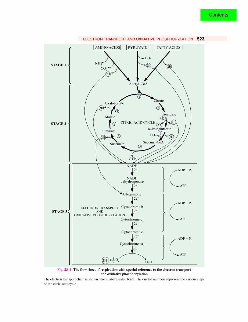
Fig. 23–1 shows the electron transport chain inabbreviated form. In each turn of citric acid cycle, 4 pairs
Contents
ELECTRON TRANSPORT AND OXIDATIVE PHOSPHORYLATION 523
Fig. 23–1. The flow sheet of respiration with special reference to the electron transportand oxidative phosphorylation
The electron transport chain is shown here in abbreviated form. The circled numbers represent the various stepsof the citric acid cycle.
Contents

524 FUNDAMENTALS OF BIOCHEMISTRY
of hydrogen atoms areeliminated, one eachfrom isocitrate, a-ketoglutarate, succinateand malate, by theaction of specificdehydrogenases. Thesehydrogen atoms donatetheir electrons to theelectron transport chainand become H+ ions,which escape into theaqueous medium. Theseelectrons are transportedalong a chain ofe l e c t r o n - c a r r y i n gmolecules to ultimatelyreach cytochrome aa3 orcytochrome oxidase,which promotes thetransfer of electrons tooxygen, the finalelectron acceptor inaerobic organisms. Aseach atom of oxygenaccepts two electronsfrom the chain, two H+
are taken up from theaqueous medium to form water.
Besides the citric acid cycle, other pairs of hydrogen atoms are also released from thedehydrogenases that act upon pyruvate, fatty acid and amino acids during their degradation to acetyl-CoA and other products. All these hydrogen atoms virtually donate their electrons ultimately to therespiratory chain with oxygen as the terminal electron acceptor.
The respiratory chain consists of a series of proteins with tightly bound prosthetic groups capableof accepting and donating electrons. Each member of the chain can accept electrons from the precedingmember and transfer them to the following one, in a specific sequence. The electrons entering theelectron-transport chain are energy-rich, but as they pass down the chain step-by-step to oxygen, theylose free energy. Much of this energy is conserved in the form of ATP in the inner mitochondrialmembrane. As each pair of electrons passes down the respiratory chain from NADH to oxygen, thesynthesis of 3 moles of ATP from ADP and phosphate takes place. The 3 segments of the respiratorychain that provide energy to generate ATP by oxidative phosphorylation are called their energy-conserving sites.
The role of electron flow and ATP production is highlighted in Fig- 23-2.
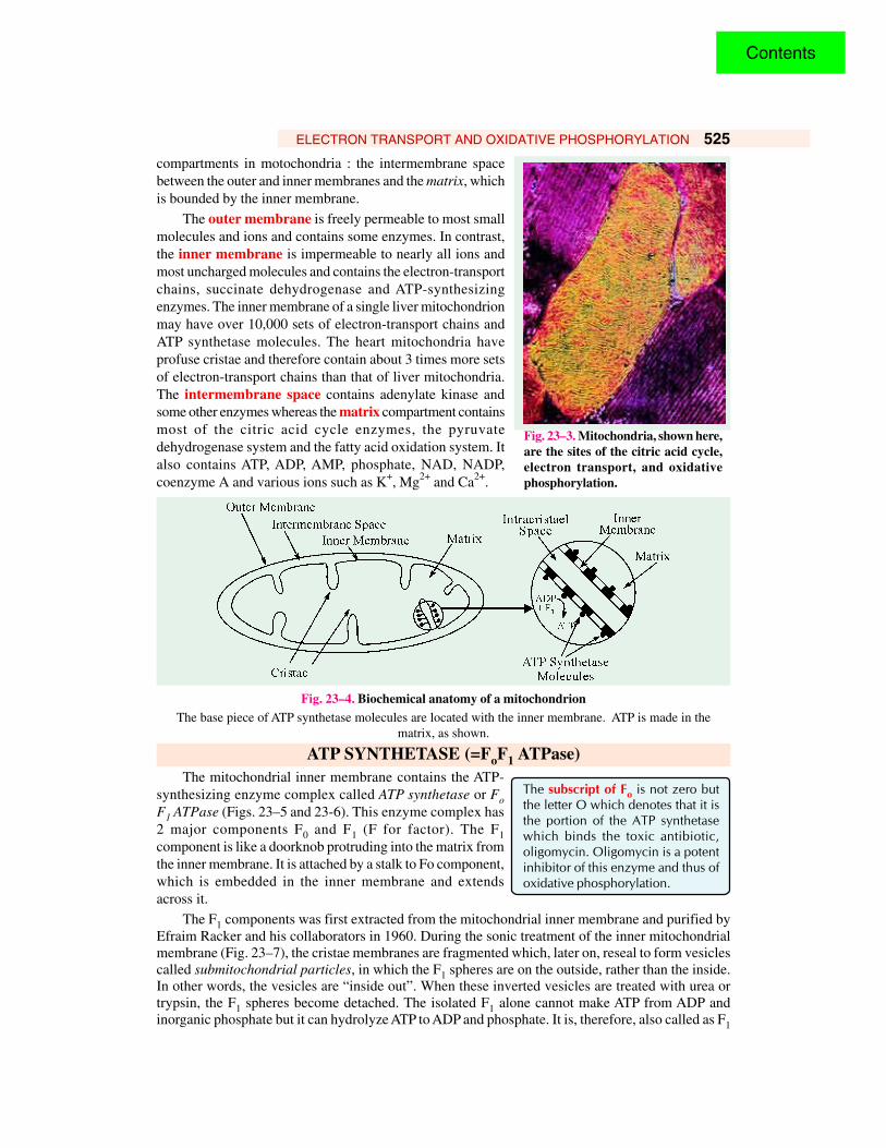
SITE OF OXIDATIVE PHOSPHORYLATIONMitochondria (Fig. 23–3) are ovate organelles, about 2 µm in length and 0.5 µm in the diameter.
Eugene P. Kennedy and Albert L. Lehninger discovered that mitochondria contain the respiratoryassembly, the enzymes of the citric acid cycle and the enzymes of fatty acid oxidation. Electronmicroscopic studies by George Palade and Fritjof Sjöstrand have revealed that each mitochondrion(Fig. 23–4) has two membrane systems : an outer membrane and an extensive inner membrane,which is highly-folded into a series of internal ridges called cristae. Obviously, there are two
Fig. 23–2. The role of electron transfer and ATP production in metabolismNAD+, FAD, and ATP are constantly recycled.
Nutrients(such as glucose)
Catabolism AnabolismExcretion
NADP+,NADP+, ADP
NADPH+,NADH, ATP
Nutrients
CO2
Citric acid cycle
NADP+,ADP
NADH,ATP
NADH,FADH2
NAD+,FAD
ADP ATP
O2 H
2O
Oxidative phosphorylation
Contents
ELECTRON TRANSPORT AND OXIDATIVE PHOSPHORYLATION 525
compartments in motochondria : the intermembrane spacebetween the outer and inner membranes and the matrix, whichis bounded by the inner membrane.
The outer membrane is freely permeable to most smallmolecules and ions and contains some enzymes. In contrast,the inner membrane is impermeable to nearly all ions andmost uncharged molecules and contains the electron-transportchains, succinate dehydrogenase and ATP-synthesizingenzymes. The inner membrane of a single liver mitochondrionmay have over 10,000 sets of electron-transport chains andATP synthetase molecules. The heart mitochondria haveprofuse cristae and therefore contain about 3 times more setsof electron-transport chains than that of liver mitochondria.The intermembrane space contains adenylate kinase andsome other enzymes whereas the matrix compartment containsmost of the citric acid cycle enzymes, the pyruvatedehydrogenase system and the fatty acid oxidation system. Italso contains ATP, ADP, AMP, phosphate, NAD, NADP,coenzyme A and various ions such as K+, Mg2+ and Ca2+.
Fig. 23–4. Biochemical anatomy of a mitochondrionThe base piece of ATP synthetase molecules are located with the inner membrane. ATP is made in the
matrix, as shown.
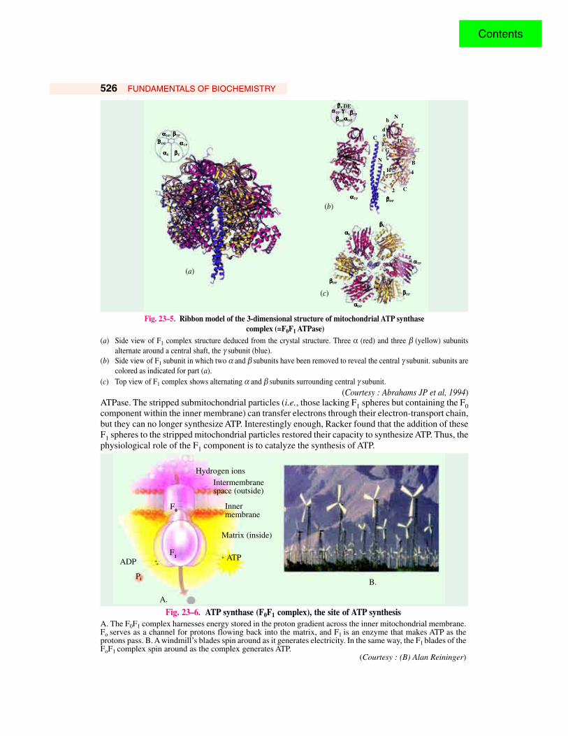
ATP SYNTHETASE (=FoF1 ATPase)The mitochondrial inner membrane contains the ATP-
synthesizing enzyme complex called ATP synthetase or FoF1 ATPase (Figs. 23–5 and 23-6). This enzyme complex has2 major components F0 and F1 (F for factor). The F1component is like a doorknob protruding into the matrix fromthe inner membrane. It is attached by a stalk to Fo component,which is embedded in the inner membrane and extendsacross it.
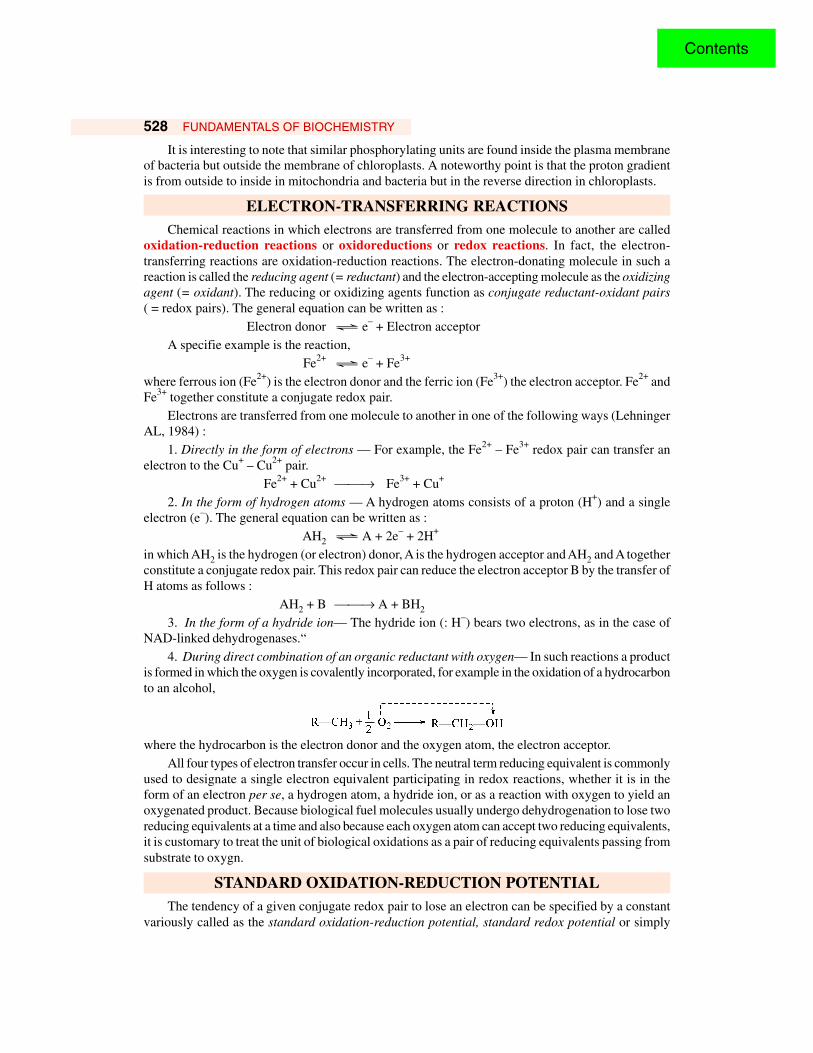
The F1 components was first extracted from the mitochondrial inner membrane and purified byEfraim Racker and his collaborators in 1960. During the sonic treatment of the inner mitochondrialmembrane (Fig. 23–7), the cristae membranes are fragmented which, later on, reseal to form vesiclescalled submitochondrial particles, in which the F1 spheres are on the outside, rather than the inside.In other words, the vesicles are “inside out”. When these inverted vesicles are treated with urea ortrypsin, the F1 spheres become detached. The isolated F1 alone cannot make ATP from ADP andinorganic phosphate but it can hydrolyze ATP to ADP and phosphate. It is, therefore, also called as F1
The subscript of Fo is not zero butthe letter O which denotes that it isthe portion of the ATP synthetasewhich binds the toxic antibiotic,oligomycin. Oligomycin is a potentinhibitor of this enzyme and thus ofoxidative phosphorylation.
Fig. 23–3. Mitochondria, shown here,are the sites of the citric acid cycle,electron transport, and oxidativephosphorylation.
Contents
526 FUNDAMENTALS OF BIOCHEMISTRY
Hydrogen ionsIntermembranespace (outside)
Innermembrane
Matrix (inside)
ATPADP
Pi
F1
F0
A.
B.
Fig. 23–6. ATP synthase (F0F1 complex), the site of ATP synthesisA. The F0F1 complex harnesses energy stored in the proton gradient across the inner mitochondrial membrane.Fo serves as a channel for protons flowing back into the matrix, and F1 is an enzyme that makes ATP as theprotons pass. B. A windmill’s blades spin around as it generates electricity. In the same way, the F1 blades of theFoF1 complex spin around as the complex generates ATP.
(Courtesy : (B) Alan Reininger)
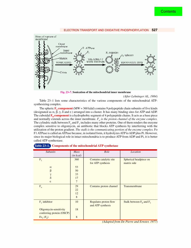
Fig. 23–5. Ribbon model of the 3-dimensional structure of mitochondrial ATP synthase complex (=F0F1 ATPase)
(a) Side view of F1 complex structure deduced from the crystal structure. Three α (red) and three β (yellow) subunitsalternate around a central shaft, the γ subunit (blue).
(b) Side view of F1 subunit in which two α and β subunits have been removed to reveal the central γ subunit. subunits arecolored as indicated for part (a).
(c) Top view of F1 complex shows alternating α and β subunits surrounding central γ subunit.(Courtesy : Abrahams JP et al, 1994)
αααααDP βββββTP
αααααE
ΒΒΒΒΒDE
βββββE
αααααTP
αααααTP γγγγγ
fda e
DEC
N
G
HI
B
4
C2αααααTP
βββββE
αααααE
αααααTP
βββββTP
αααααDP
βββββDP
(c)
βββββDP
(a)
(b)
βββββE DE
αααααOP
βββββTP
βββββDP hN
ATPase. The stripped submitochondrial particles (i.e., those lacking F1 spheres but containing the F0component within the inner membrane) can transfer electrons through their electron-transport chain,but they can no longer synthesize ATP. Interestingly enough, Racker found that the addition of theseF1 spheres to the stripped mitochondrial particles restored their capacity to synthesize ATP. Thus, thephysiological role of the F1 component is to catalyze the synthesis of ATP.
Contents
ELECTRON TRANSPORT AND OXIDATIVE PHOSPHORYLATION 527
Fig. 23–7. Sonication of the mitochondrial inner membrane
(After Lehninger AL, 1984)Table 23–1 lists some characteristics of the various components of the mitochondrial ATP-
synthesizing complex.The spheric F1 component (MW = 360 kdal) contains 9 polypeptide chain subunits of five kinds
(designated as α, β, γ, δ and ∈) arranged into a cluster. It has many binding sites for ATP and ADP.The cuboidal F0 component is a hydrophobic segment of 4 polypeptide chains. It acts as a base pieceand normally extends across the inner membrane. Fo is the proton channel of the enzyme complex.The cylindric stalk between Fo and F1 includes many other proteins. One of them renders the enzymecomplex sensitive to oligomycin, an antibiotic that blocks ATP synthesis by interferring with theutilization of the proton gradient. The stalk is the communicating portion of the enzyme complex. FoF1 ATPase is called an ATPase because, in isolated form, it hydrolyzes ATP to ADP plus Pi. However,since its major biological role in intact mitochondria is to produce ATP from ADP and Pi, it is bettercalled ATP synthestase.
Table 23–1. Components of the mitochondrial ATP synthetase
Subunits Mass Role Location(in kcal)
F1 360 Contains catalytic site Spherical headpiece onfor ATP synthesis matrix side
α 53β 50γ 33δ 17∈ 7
Fo 29 Contains proton channel Transmembrane22128
F1 inhibitor 10 Regulates proton flow Stalk between F0 and F1and ATP synthesis
Oligomycin-sensitivity 18conferring protein (OSCP)
Fc2 (F6) 8
(Adapted from De Pierre and Ernster, 1977)
Contents
528 FUNDAMENTALS OF BIOCHEMISTRY
It is interesting to note that similar phosphorylating units are found inside the plasma membraneof bacteria but outside the membrane of chloroplasts. A noteworthy point is that the proton gradientis from outside to inside in mitochondria and bacteria but in the reverse direction in chloroplasts.
ELECTRON-TRANSFERRING REACTIONSChemical reactions in which electrons are transferred from one molecule to another are called
oxidation-reduction reactions or oxidoreductions or redox reactions. In fact, the electron-transferring reactions are oxidation-reduction reactions. The electron-donating molecule in such areaction is called the reducing agent (= reductant) and the electron-accepting molecule as the oxidizingagent (= oxidant). The reducing or oxidizing agents function as conjugate reductant-oxidant pairs( = redox pairs). The general equation can be written as :
Electron donor Ö e– + Electron acceptorA specifie example is the reaction,
Fe2+ Ö e– + Fe3+
where ferrous ion (Fe2+) is the electron donor and the ferric ion (Fe3+) the electron acceptor. Fe2+ andFe3+ together constitute a conjugate redox pair.
Electrons are transferred from one molecule to another in one of the following ways (LehningerAL, 1984) :
1. Directly in the form of electrons — For example, the Fe2+ – Fe3+ redox pair can transfer anelectron to the Cu+ – Cu2+ pair.
Fe2+ + Cu2+ → Fe3+ + Cu+
2. In the form of hydrogen atoms — A hydrogen atoms consists of a proton (H+) and a singleelectron (e–). The general equation can be written as :
AH2 Ö A + 2e– + 2H+
in which AH2 is the hydrogen (or electron) donor, A is the hydrogen acceptor and AH2 and A togetherconstitute a conjugate redox pair. This redox pair can reduce the electron acceptor B by the transfer ofH atoms as follows :
AH2 + B → A + BH2
3. In the form of a hydride ion— The hydride ion (: H–) bears two electrons, as in the case ofNAD-linked dehydrogenases.“
4. During direct combination of an organic reductant with oxygen— In such reactions a productis formed in which the oxygen is covalently incorporated, for example in the oxidation of a hydrocarbonto an alcohol,
where the hydrocarbon is the electron donor and the oxygen atom, the electron acceptor.All four types of electron transfer occur in cells. The neutral term reducing equivalent is commonly
used to designate a single electron equivalent participating in redox reactions, whether it is in theform of an electron per se, a hydrogen atom, a hydride ion, or as a reaction with oxygen to yield anoxygenated product. Because biological fuel molecules usually undergo dehydrogenation to lose tworeducing equivalents at a time and also because each oxygen atom can accept two reducing equivalents,it is customary to treat the unit of biological oxidations as a pair of reducing equivalents passing fromsubstrate to oxygn.
STANDARD OXIDATION-REDUCTION POTENTIALThe tendency of a given conjugate redox pair to lose an electron can be specified by a constant
variously called as the standard oxidation-reduction potential, standard redox potential or simply
Contents
ELECTRON TRANSPORT AND OXIDATIVE PHOSPHORYLATION 529
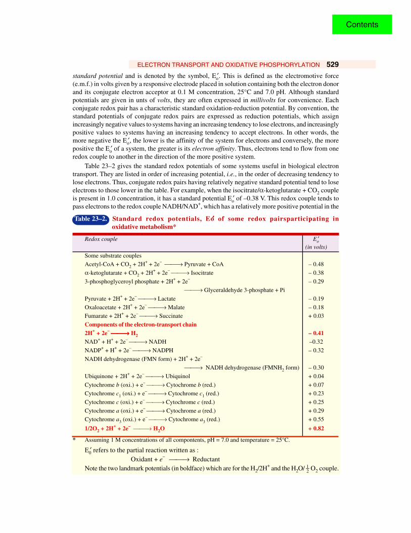
standard potential and is denoted by the symbol, Eo′. This is defined as the electromotive force(e.m.f.) in volts given by a responsive electrode placed in solution containing both the electron donorand its conjugate electron acceptor at 0.1 M concentration, 25°C and 7.0 pH. Although standardpotentials are given in unts of volts, they are often expressed in millivolts for convenience. Eachconjugate redox pair has a characteristic standard oxidation-reduction potential. By convention, thestandard potentials of conjugate redox pairs are expressed as reduction potentials, which assignincreasingly negative values to systems having an increasing tendency to lose electrons, and increasinglypositive values to systems having an increasing tendency to accept electrons. In other words, themore negative the Eo′, the lower is the affinity of the system for electrons and conversely, the morepositive the Eo′ of a system, the greater is its electron affinity. Thus, electrons tend to flow from oneredox couple to another in the direction of the more positive system.
Table 23–2 gives the standard redox potentials of some systems useful in biological electrontransport. They are listed in order of increasing potential, i.e., in the order of decreasing tendency tolose electrons. Thus, conjugate redox pairs having relatively negative standard potential tend to loseelectrons to those lower in the table. For example, when the isocitrate/α-ketoglutarate + CO2 coupleis present in 1.0 concentration, it has a standard potential Eo′ of –0.38 V. This redox couple tends topass electrons to the redox couple NADH/NAD+, which has a relatively more positive potential in the
Table 23–2. Standard redox potentials, Eo′′′′′ of some redox pairsparticipating in oxidative metabolism*
Redox couple Eo′(in volts)
Some substrate couples
Acetyl-CoA + CO2 + 2H+ + 2e– → Pyruvate + CoA – 0.48
α-ketoglutarate + CO2 + 2H+ + 2e– → Isocitrate – 0.38
3-phosphoglyceroyl phosphate + 2H+ + 2e– – 0.29
→ Glyceraldehyde 3-phosphate + Pi
Pyruvate + 2H+ + 2e– → Lactate – 0.19
Oxaloacetate + 2H+ + 2e– → Malate – 0.18
Fumarate + 2H+ + 2e– → Succinate + 0.03
Components of the electron-transport chain2H+ + 2e– →→→→→ H2 – 0.41NAD+ + H+ + 2e– → NADH –0.32
NADP+ + H+ + 2e– → NADPH – 0.32
NADH dehydrogenase (FMN form) + 2H+ + 2e–
→ NADH dehydrogenase (FMNH2 form) – 0.30
Ubiquinone + 2H+ + 2e– → Ubiquinol + 0.04
Cytochrome b (oxi.) + e– → Cytochrome b (red.) + 0.07
Cytochrome c1 (oxi.) + e– → Cytochrome c1 (red.) + 0.23
Cytochrome c (oxi.) + e– → Cytochrome c (red.) + 0.25
Cytochrome a (oxi.) + e– → Cytochrome a (red.) + 0.29
Cytochrome a3 (oxi.) + e– → Cytochrome a3 (red.) + 0.55
1/2O2 + 2H+ + 2e– → H2O + 0.82
* Assuming 1 M concentrations of all compontents, pH = 7.0 and temperature = 25°C.
E0′ refers to the partial reaction written as :Oxidant + e– → Reductant
Note the two landmark potentials (in boldface) which are for the H2/2H+ and the H2O/ 12 O2 couple.
Contents

530 FUNDAMENTALS OF BIOCHEMISTRY
presence of isocitrate dehydrogenase. Conversely, the strongly positive standard potential of H2O/O2couple, 0.82 V, indicates that water molecule has very little tendency to lose electrons to form molecularoxygen. In other words, molecular oxygen has a very high affinity for electrons or hydrogen atoms.
In oxidation systems, the electrons will tend to flow from a relatively electronegative conjugateredox pair, such as NADH/NAD+ (E0′ = – 0.32 V), to the more electropositive pair, such as reducedcytochrome c/oxidized cytochrome c (E0′ = + 0.25 V). Likewise, they will also tend to flow from thecytochrome c redox pair to the water/oxygen pair (E0′ = + 0.82 V). The greater the difference in thestandard potentials between two redox pairs, the greater is the free-energy loss as electrons pass fromthe electronegative to the electropositive pair. Therefore, when electrons flow down the completeelectron-transport chain from NADH to oxygen via several electron-carrying molecules, they lose alarge amount of free energy.
The amount of free-energy which becomes available as a pair of electrons passes from NADH toO2 can also be calculated. The standard-free-energy change of an electron-transferring reaction isgiven by the equation,
∆G°′ = –nF ∆E0′where, ∆G°′ is the standard-free-energy change in calories, n is the number of electrons transferred, Fis the caloric equivalent of the constant, the faraday (23.062 kcal/V. mol) and ∆E0′ is the differencebetween the standard potential of the electron-donor system and that of the electron-acceptor system.The standard-free-energy change as a pair of electron equivalents passes from the NADH/NAD+ pair(E0′ = – 0.32 V) to the H2O/O2 pair (E0′ = + 0.82 V) is
∆G°′ = – 2 (23.062) [0.82 – (– 0.32)]= – 2 × 23.062 × 1.14= –52.58 kcal/mol
This amount of free energy (i.e., –52.58 kcal) is more than sufficient to bring about the synthesisof 3 moles of ATP, which requires an input of 3 (7.3) = 21.9 kcal under standard conditions.
Fig. 23–8. The energy relationships in the respiratory chain of mitochondria[E-FMN = NADH dehydrogenase; Q = ubiquinone; b, c1, c and a = cytochromes]
Note that there are 3 steps (shown with boldface arrows) in the electron-transport chain in which relatively largedecreases in free-energy occur as electrons pass. These are, in fact, the steps that provide free energy for ATPsynthesis.
Contents

ELECTRON TRANSPORT AND OXIDATIVE PHOSPHORYLATION 531
Likewise using the expression ∆G°′ = – nF ∆E0′, the free-energy changes for individual segmentsof the electron-transport chains can be calculated from the differences in the standard potentials ofthe electron-donating redox pair and the electron-accepting pair. Fig. 23–8 is an energy diagramshowing (a) the standard potentials of the electron carriers of the respiratory chain, (b) the directionof electron flow, which is always “downhill” toward oxygen, and (c) the relative free-energy changeat each step.
ELECTRON CARRIERSElectrons are transferred from substrates to oxygen through a series of electron carriers— flavins,
iron-sulfur complexes, quinones and hemes (Fig. 23–9). The 3 noteworthy features of the electroncarriers are enumerated below :
1. Although the figure shows the respiratory chain to have 17 electron carriers, there are some15 or more chemical groups in the electron-transport chain that can accept or transfer reducingequivalents (hydrogen and electrons) in sequence.
2. All electron carriers, except for the quinones, are prosthetic groups of proteins. They includenicotinamide adenine dinucleotide (NAD), active with various dehydrogenases ; flavinmononucleotide (FMN), in NADH dehydrogenases; ubiquinone or coenzyme Q, whichfunctions in association with one or more proteins ; two different kinds of iron-containingproteins, the iron-sulfur centres (Fe-S)and the cytochromes ; and copper ofcytochrome aa3.
3. Almost all the electron-carrying proteinsare water insoluble and are embeded inthe inner mitochondrial membrane.
1. Pyridine NucleotidesMost of the electron pairs entering the
respiratory chains arise from the action ofdehydrogenases that utilize the coenzymes NAD+
or NADP+ (Fig. 23–6) as electron acceptors. Asa group, they are designated as NAD(P)-linkeddehydrogenases. They catalyze the reversiblereactions of the following general types :
Reduced substrate + NAD+ Ö Oxidizedsubstrate + NADH + H+
Reduced substrate + NADP+Ö Oxidizedsubstrate + NADPH + H+
Most of the pyridine-linked dehydrogenasesare specific for NAD+ (refer Table 23–3).However, certain others require NADP+ aselectron acceptor, such as glucose 6-phosphatedehydrogenase. Very few, such as glutamatedehydrogenase can react with either NAD+ orNADP+. Some pyridine-linked dehydrogenasesare located in the mitochondria, some in thecytosol and still others in both.
The pyridine-linked dehydrogenases remove2 hydrogen atoms from their substrates. One ofthese is transferred as a hydride ion ( : H–) to theNAD+ or NADP+ ; the other appears as H+ in themedium. Each hydride ion carries two reducing
Fig. 23–9. The complete set of electron carriersof the respiratory chain
The precise sequence and function of all the oxidation-reduction centres is not exactly known.
Contents
532 FUNDAMENTALS OF BIOCHEMISTRY
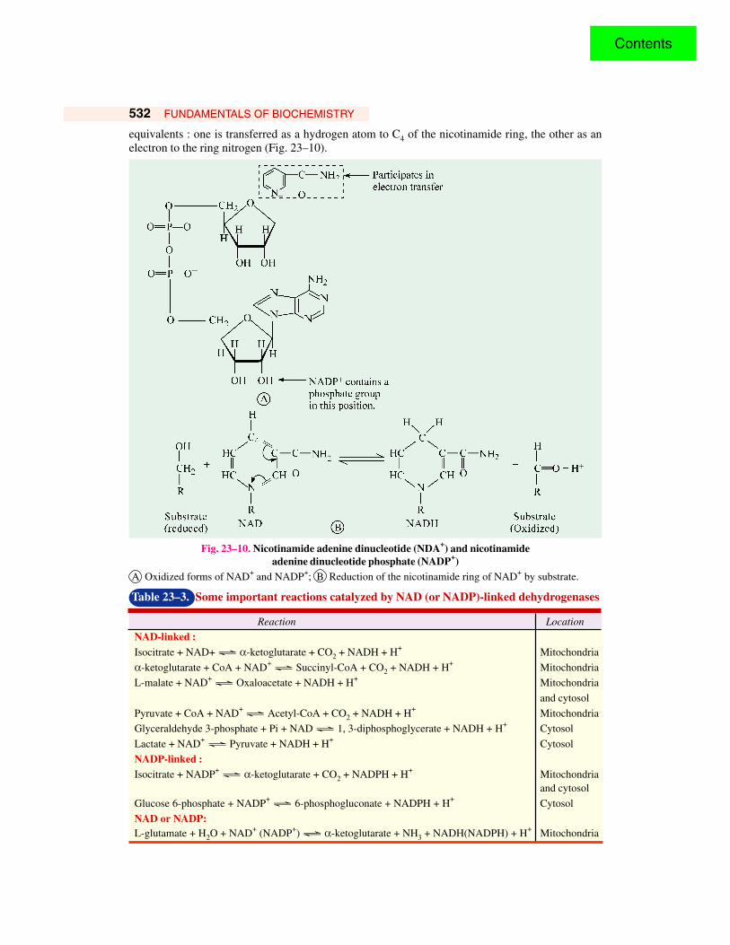
equivalents : one is transferred as a hydrogen atom to C4 of the nicotinamide ring, the other as anelectron to the ring nitrogen (Fig. 23–10).
Fig. 23–10. Nicotinamide adenine dinucleotide (NDA+) and nicotinamideadenine dinucleotide phosphate (NADP+)
A Oxidized forms of NAD+ and NADP+; B Reduction of the nicotinamide ring of NAD+ by substrate.
Table 23–3. Some important reactions catalyzed by NAD (or NADP)-linked dehydrogenases
Reaction Location
NAD-linked :Isocitrate + NAD+ Ö α-ketoglutarate + CO2 + NADH + H+ Mitochondria
α-ketoglutarate + CoA + NAD+ Ö Succinyl-CoA + CO2 + NADH + H+ Mitochondria
L-malate + NAD+ Ö Oxaloacetate + NADH + H+ Mitochondria
and cytosol
Pyruvate + CoA + NAD+ Ö Acetyl-CoA + CO2 + NADH + H+ Mitochondria
Glyceraldehyde 3-phosphate + Pi + NAD Ö 1, 3-diphosphoglycerate + NADH + H+ Cytosol
Lactate + NAD+ Ö Pyruvate + NADH + H+ Cytosol
NADP-linked :Isocitrate + NADP+ Ö α-ketoglutarate + CO2 + NADPH + H+ Mitochondria
and cytosol
Glucose 6-phosphate + NADP+ Ö 6-phosphogluconate + NADPH + H+ Cytosol
NAD or NADP:L-glutamate + H2O + NAD+ (NADP+) Ö α-ketoglutarate + NH3 + NADH(NADPH) + H+ Mitochondria
Contents
ELECTRON TRANSPORT AND OXIDATIVE PHOSPHORYLATION 533
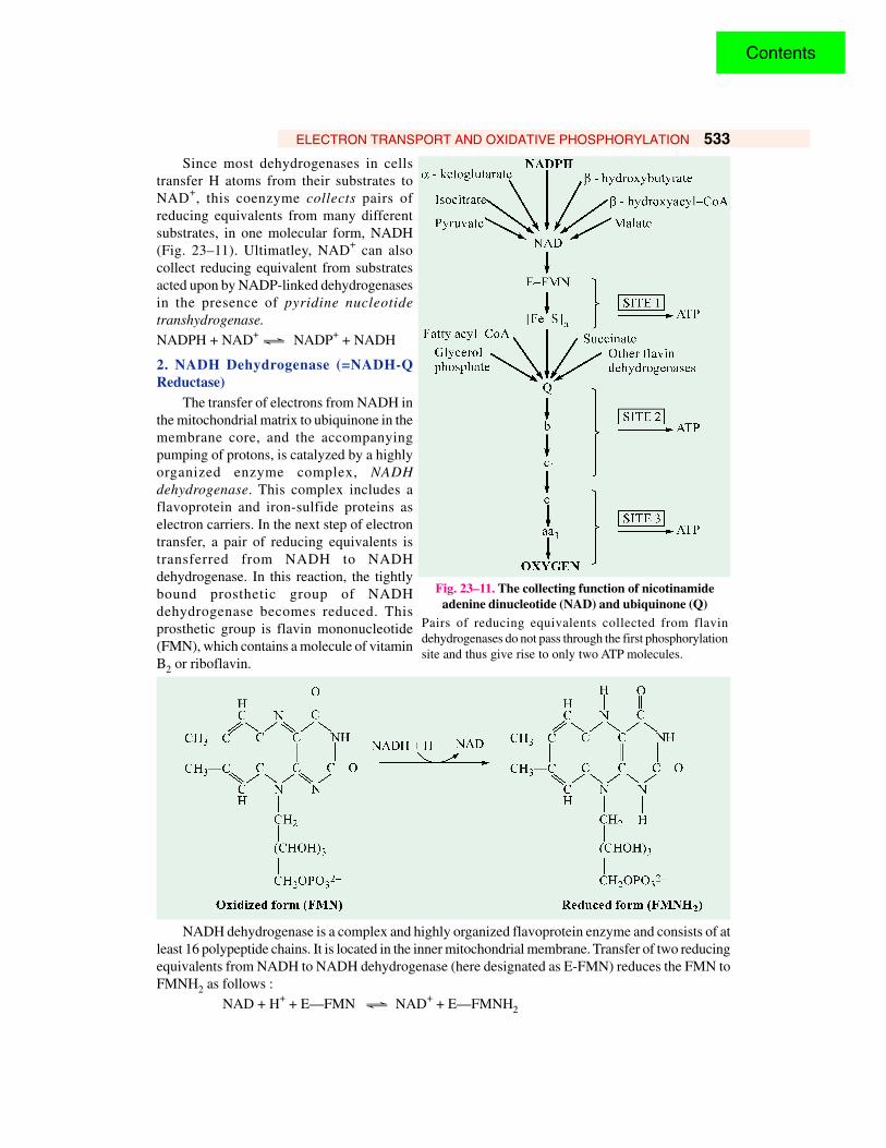
Since most dehydrogenases in cellstransfer H atoms from their substrates toNAD+, this coenzyme collects pairs ofreducing equivalents from many differentsubstrates, in one molecular form, NADH(Fig. 23–11). Ultimatley, NAD+ can alsocollect reducing equivalent from substratesacted upon by NADP-linked dehydrogenasesin the presence of pyridine nucleotidetranshydrogenase.
NADPH + NAD+ Ö NADP+ + NADH
2. NADH Dehydrogenase (=NADH-QReductase)
The transfer of electrons from NADH inthe mitochondrial matrix to ubiquinone in themembrane core, and the accompanyingpumping of protons, is catalyzed by a highlyorganized enzyme complex, NADHdehydrogenase. This complex includes aflavoprotein and iron-sulfide proteins aselectron carriers. In the next step of electrontransfer, a pair of reducing equivalents istransferred from NADH to NADHdehydrogenase. In this reaction, the tightlybound prosthetic group of NADHdehydrogenase becomes reduced. Thisprosthetic group is flavin mononucleotide(FMN), which contains a molecule of vitaminB2 or riboflavin.
NADH dehydrogenase is a complex and highly organized flavoprotein enzyme and consists of atleast 16 polypeptide chains. It is located in the inner mitochondrial membrane. Transfer of two reducingequivalents from NADH to NADH dehydrogenase (here designated as E-FMN) reduces the FMN toFMNH2 as follows :
NAD + H+ + E—FMN Ö NAD+ + E—FMNH2
Fig. 23–11. The collecting function of nicotinamideadenine dinucleotide (NAD) and ubiquinone (Q)
Pairs of reducing equivalents collected from flavindehydrogenases do not pass through the first phosphorylationsite and thus give rise to only two ATP molecules.
Contents

534 FUNDAMENTALS OF BIOCHEMISTRY
The electrons are then transferred from FMNH2 to a series of iron-sulfur complexes (abbreviatedas Fe—S), the second type of prosthetic group in NADH dehydrogenase. The iron is not a part of aheme group and so iron-sulfur proteins were referred to in the older literature as nonheme iron proteins(NHI proteins). Recall that Fe—S centres are also associated with succinate dehydrogenase (see page400). Three principal types of Fe—S complexes are known (Fig. 23-12). In all of these, the ironatoms are chelated with sulfur atoms, which are in part supplied by cysteine residues in the associatedprotein and in part as inorganic sulfide ions. The number of iron and acid-labile-sulfur atoms in thesecomplexes is always equal.
1. FeCys4 (or FeS) type—In this simplest kind,a single iron atom is tetrahedrally coordinated tothe sulfhydryl groups of 4 cysteine residues of theprotein ; there being no inorganic sulfides.
2. Fe2S2Cys4 (or Fe2S2) type— This contains2 iron atoms and 2 inorganic sulfides, in additionto 4 cysteine residues. The bonding occurs in sucha manner that a total of 4 sulfur atoms is linked toeach iron atom.
3. Fe4S4Cys4 (or Fe4S4) type — This contains4 iron atoms, 4 inorganic sulfides and 4 cysteineresidues. Here also, 4 sulfur atoms are linked toeach iron atom.
The iron atoms in these complexes can be inthe reduced (Fe2+) or oxidized (Fe3+) state. Animportant feature of the iron-sulfur proteins is thattheir relative affinity for electrons can be variedover a wide range by changing the nature of thepolypeptide chain. Some are relatively strongoxidizing agents ; others are powerful reducingagents — even stronger than NADH. NADHdehydrogenase contains both the Fe2S2Cys4 andFe4S4Cys4 types of complexes.
3. Ubiquinone (=Coenzyme Q)The next carrier of reducing equivalents in
the respiratory chain is ubiquinone (UQ), aname reflecting its ubiquitous nature, as it occursvirtually in all cells. It was formerly called ascoenzyme Q (Q for quinone) and abbreviated asCoQ or simply Q.
Ubiquinone is actually a group of compounds,all containing the same quinone structure butsubstituted with a long side chain composed ofvarying numbers (from 6 to 10) of isoprene units(=prenyl groups), linked head to tail. For example,certain microorganisms contain 6 isoprene units,and in which case the compound is referred to asQ6 or CoQ6 or also as UQ30, where the subscriptnumber 30 represents the total number of carbon atoms in the side chain. However, the most commonform in mammals contains 10 isoprene units, and when its designation is Q10 or CoQ10 or UQ50. The
Cys
Cys Cys
Cys
Fe
[Fe-S]
Cys
Cys
Cys
Cys
S2–
S2–
Fe Fe
[Fe2-S2]
Cys
[Fe4-S
4]
Cys
Cys
Cys
S2–
FeS2–
S2–
S2–
Fe
Fig. 23–12. Three principal forms of iron-sulfideproteins.
Contents
ELECTRON TRANSPORT AND OXIDATIVE PHOSPHORYLATION 535
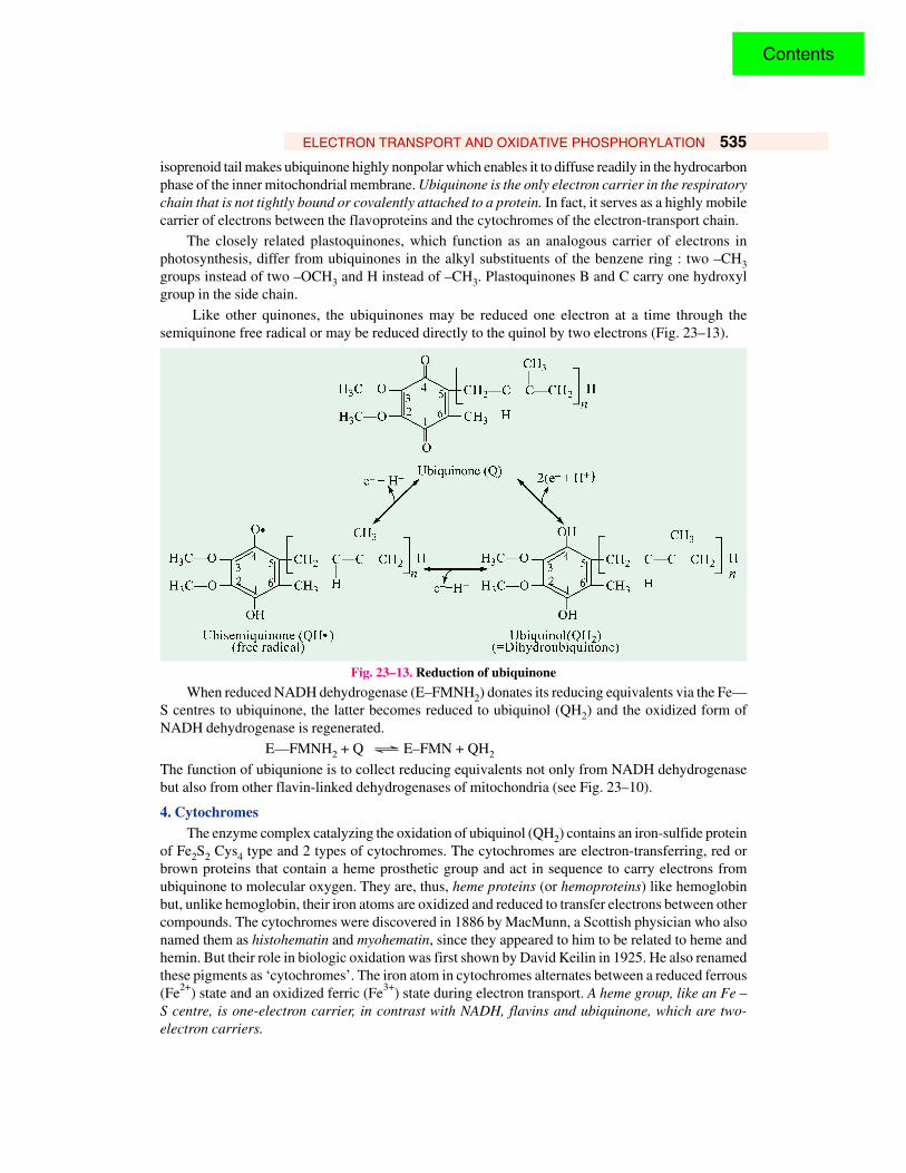
isoprenoid tail makes ubiquinone highly nonpolar which enables it to diffuse readily in the hydrocarbonphase of the inner mitochondrial membrane. Ubiquinone is the only electron carrier in the respiratorychain that is not tightly bound or covalently attached to a protein. In fact, it serves as a highly mobilecarrier of electrons between the flavoproteins and the cytochromes of the electron-transport chain.
The closely related plastoquinones, which function as an analogous carrier of electrons inphotosynthesis, differ from ubiquinones in the alkyl substituents of the benzene ring : two –CH3groups instead of two –OCH3 and H instead of –CH3. Plastoquinones B and C carry one hydroxylgroup in the side chain.
Like other quinones, the ubiquinones may be reduced one electron at a time through thesemiquinone free radical or may be reduced directly to the quinol by two electrons (Fig. 23–13).
Fig. 23–13. Reduction of ubiquinone
When reduced NADH dehydrogenase (E–FMNH2) donates its reducing equivalents via the Fe—S centres to ubiquinone, the latter becomes reduced to ubiquinol (QH2) and the oxidized form ofNADH dehydrogenase is regenerated.
E—FMNH2 + Q Ö E–FMN + QH2
The function of ubiqunione is to collect reducing equivalents not only from NADH dehydrogenasebut also from other flavin-linked dehydrogenases of mitochondria (see Fig. 23–10).
4. CytochromesThe enzyme complex catalyzing the oxidation of ubiquinol (QH2) contains an iron-sulfide protein
of Fe2S2 Cys4 type and 2 types of cytochromes. The cytochromes are electron-transferring, red orbrown proteins that contain a heme prosthetic group and act in sequence to carry electrons fromubiquinone to molecular oxygen. They are, thus, heme proteins (or hemoproteins) like hemoglobinbut, unlike hemoglobin, their iron atoms are oxidized and reduced to transfer electrons between othercompounds. The cytochromes were discovered in 1886 by MacMunn, a Scottish physician who alsonamed them as histohematin and myohematin, since they appeared to him to be related to heme andhemin. But their role in biologic oxidation was first shown by David Keilin in 1925. He also renamedthese pigments as ‘cytochromes’. The iron atom in cytochromes alternates between a reduced ferrous(Fe2+) state and an oxidized ferric (Fe3+) state during electron transport. A heme group, like an Fe –S centre, is one-electron carrier, in contrast with NADH, flavins and ubiquinone, which are two-electron carriers.
Contents

536 FUNDAMENTALS OF BIOCHEMISTRY
There are 5 types of cytochromes between ubiquinol (QH2) and oxygen in the electron-transportchain. Each type is given a letter designation— a, b, c and so on, based on the differences in theirlight-absorption spectra — the form absorbing at the longest wavelength called cytochrome a, thatabsorbing the next longest wavelength called cytochrome b, and so on. Unfortunately, the order ofwavelength does not correspond to the physiological sequence in which they function :
b → c1 → c → a → a3
The subscript numbers were added as new individual cytochromes within the same type werefound, i.e., those with similar prosthetic groups but with different apoproteins.
Fig. 23–14. The three principal types of cytochrome hemesHeme B is the prosthetic group of cytochrome b; heme C, that of cytochromes c and c1; and heme A, that ofcytochromes a and a3.
The various cytochromes differ from each other in the nature of the prosthetic group and itsmode of attachment to the apoprotein part (Fig. 23–14). The prosthetic group of cytochromes b, c andc1 is iron-protoporphyrin IX, commonly called heme or hemin. Heme is also the prosthetic group ofmyoglobin, hemoglobin, catalase and peroxidase. In cytochrome b, the heme is not covalently bondedto the protein, whereas in cytochromes c and c1, the heme is covalently attached to the protein bythioether linkages. These linkages are formed by the addition of the sulfhydryl groups of two cysteineresidues to the vinyl groups of the heme. The type of heme present in cytochrome b is called heme Band the one present in cytochromes c and c1 as heme C.
Contents
ELECTRON TRANSPORT AND OXIDATIVE PHOSPHORYLATION 537
The cytochromes a and a3 have a different iron-porphyrin prosthetic group, called heme A. Itdiffers from the heme in cytochromes c and c1 in that a formyl group replaces one of the methylgroups, and a hydrophobic polyprenyl chain replaces one of the vinyl groups. Cytochromes a and a3are the terminal members of the respiratory chain. They exist as complex, which is sometimes calledcytochrome oxidase. The cytochrome aa3 complex, thus, differs from other cytochromes as it contains2 moles of highly-bound heme A. Moreover, cytochrome aa3 also contains 2 essential copper atoms.It is the terminal member of the electron-transport chain and was first identified by Warburg asAtmungsferment.
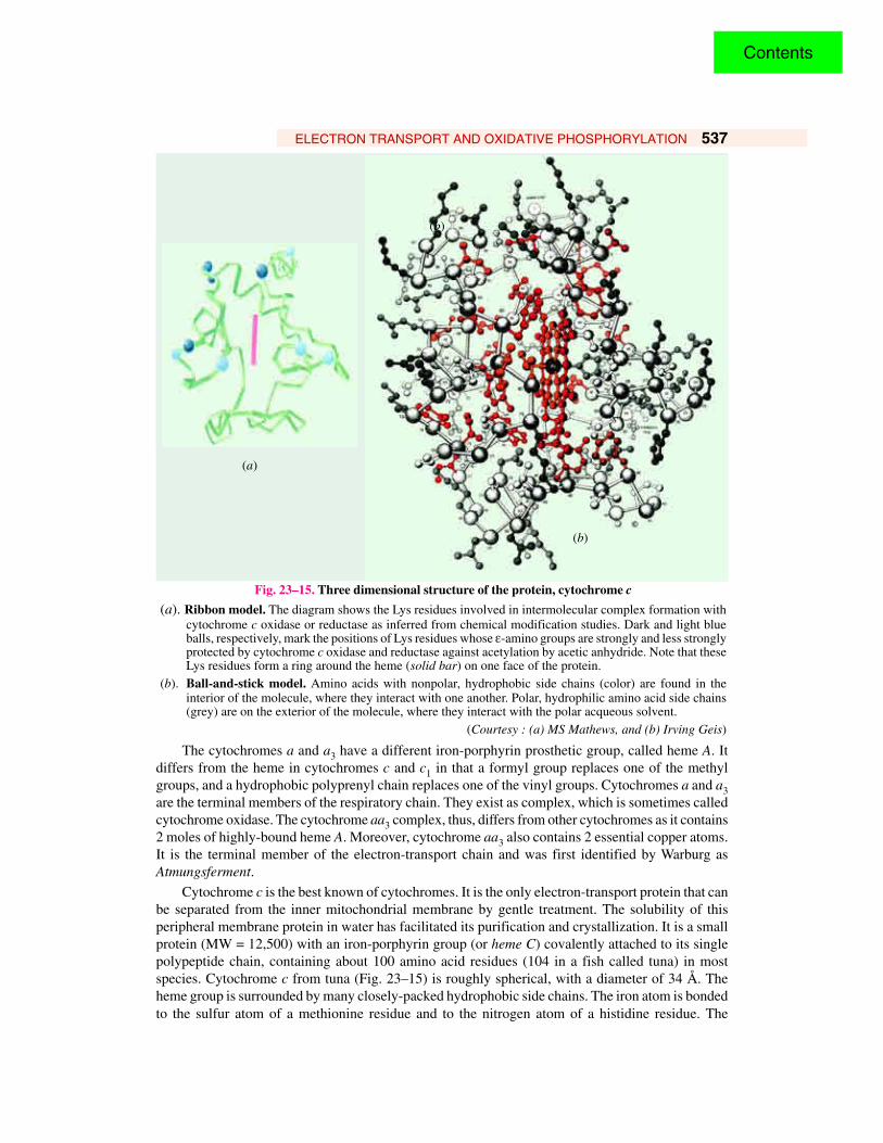
Cytochrome c is the best known of cytochromes. It is the only electron-transport protein that canbe separated from the inner mitochondrial membrane by gentle treatment. The solubility of thisperipheral membrane protein in water has facilitated its purification and crystallization. It is a smallprotein (MW = 12,500) with an iron-porphyrin group (or heme C) covalently attached to its singlepolypeptide chain, containing about 100 amino acid residues (104 in a fish called tuna) in mostspecies. Cytochrome c from tuna (Fig. 23–15) is roughly spherical, with a diameter of 34 Å. Theheme group is surrounded by many closely-packed hydrophobic side chains. The iron atom is bondedto the sulfur atom of a methionine residue and to the nitrogen atom of a histidine residue. The
(a)
(b)
Fig. 23–15. Three dimensional structure of the protein, cytochrome c
(a). Ribbon model. The diagram shows the Lys residues involved in intermolecular complex formation withcytochrome c oxidase or reductase as inferred from chemical modification studies. Dark and light blueballs, respectively, mark the positions of Lys residues whose ε-amino groups are strongly and less stronglyprotected by cytochrome c oxidase and reductase against acetylation by acetic anhydride. Note that theseLys residues form a ring around the heme (solid bar) on one face of the protein.
(b). Ball-and-stick model. Amino acids with nonpolar, hydrophobic side chains (color) are found in theinterior of the molecule, where they interact with one another. Polar, hydrophilic amino acid side chains(grey) are on the exterior of the molecule, where they interact with the polar acqueous solvent.
(Courtesy : (a) MS Mathews, and (b) Irving Geis)
(b)
Contents
538 FUNDAMENTALS OF BIOCHEMISTRY
hydrophobic nature of the heme environment makes the redox potential of cytochrome c more positive,corresponding to a higher electron affinity.
The overall structure of the cytochrome c molecule resembles that of a shell, one residue thicksurrounding the heme. The side chains make up the interior of the shell. The main polypeptide chaincomes next, followed by charged side chains on the surface. There is a very little a-helix and no β-pleated sheet. In essence, the polypeptide chain is coiled around the heme. Residues 1 to 47 are on thehistidine-18 side of the heme (called the right side), and residues 48 to 91 are on the methionine-80side (called the left side). The remaining 92 to 104 residues come back across the heme to the rightside. Cytochorme c is an ancient protein, since its amino acid sequence has many points of similarityin all eukaryotes— microbes, plants and animals.
ELECTRON-TRANSPORT COMPLEXES( = Complexes of the Respiratory Chain)
It is now a well-established fact that the electron carriers in the respiratory chain function in aspecific sequence. The following evidences support the statement :
1. Firstly, as expected their standard redox potentials (refer Table 23–2 and Fig. 23–6) aremore positive going toward oxygen, since electrons tend to flow from electronegative toelectropositive systems, causing a decrease in free energy.
2. Secondly, each member of the chain is specific for a given electron donor and acceptor. Forinstance, NADH can transfer electrons to NADH dehydrogenase but cannot transfer themdirectly to cytochrome b or to cytochrome c.
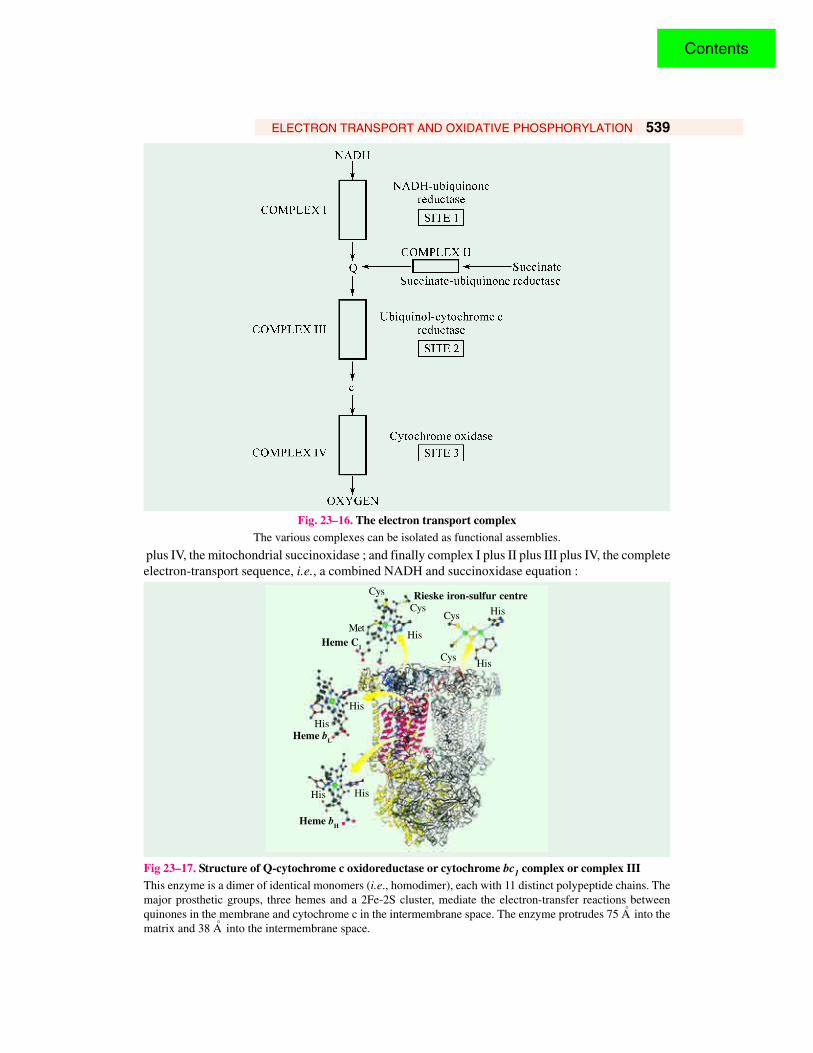
3. Lastly, four structured complexes of functionally related electron carriers have been isolatedfrom mitochondrial membrane (refer Fig. 23–16) :
Complex I consists of NADH dehydrogenase and its iron-sulfur centres, which are closely linkedin their function. Complex I carries out the following characteristic reaction :
NADH + H+ + CoQNADH : CoQ (oxido)
reductaseComplex I
→ NAD+ + CoQH2
Complex II consists of succinate dehydrogenase and its iron-sulfur centres. This complex carriesout the following characteristic reaction :
Succinate + CoQSuccinate : CoQ (oxido)
reductaseComplex II
→ Fumarate + CoQH2
Complex III consists of cytochromes b and c, and a specific iron-sulfur centre. This brings aboutthe following characteristic reaction :
CoQ H2 + 2 cyt c (Fe3+)Hydro-CoQ : Cytochrome
(oxido) reductaseComplex III
→ CoQ + 2 cyt c (Fe2+)
Complex IV consists only of cytochromes a and a3, which is sometimes called as cytochromeoxidase. This brings about the following characteristic reaction :
4 cyt c (Fe2+) + O22
Cytochrome oxidase(Cytochrome c : O oxido reductase)
Complex IV→ 4 cyt c (Fe3+) + H2O
Ubiquinone is the connecting link between complexes I , II and III, and cytochrome c connectsthe complexes. III and IV. It is evident that in the presence of 2 mobile carriers (CoQ and cytochrome c),this accounts for all the oxidoreductions of the mitochondrial electron transport system. Thus, complexI plus complex III reconstitute the mitochondrial NADH : cytochrome c (oxido) reductase ; complexII plus III, the mitochondrial succinate : cytochrome c (oxido) reductase ; complex I plus III
Contents
ELECTRON TRANSPORT AND OXIDATIVE PHOSPHORYLATION 539
Fig. 23–16. The electron transport complexThe various complexes can be isolated as functional assemblies.
plus IV, the mitochondrial succinoxidase ; and finally complex I plus II plus III plus IV, the completeelectron-transport sequence, i.e., a combined NADH and succinoxidase equation :
Fig 23–17. Structure of Q-cytochrome c oxidoreductase or cytochrome bc1 complex or complex IIIThis enzyme is a dimer of identical monomers (i.e., homodimer), each with 11 distinct polypeptide chains. Themajor prosthetic groups, three hemes and a 2Fe-2S cluster, mediate the electron-transfer reactions betweenquinones in the membrane and cytochrome c in the intermembrane space. The enzyme protrudes 75 A° into thematrix and 38 A° into the intermembrane space.
Cys
CysCys
Cys
MetHeme C1
His
His
His
His
His His
Heme bL
Heme bH
Rieske iron-sulfur centreHis
Contents
540 FUNDAMENTALS OF BIOCHEMISTRY
The ubiquinol-cytochrome c reductase complex (Fig 23-17) transfers electrons from ubiquinol(QH2) to cytochrome c.
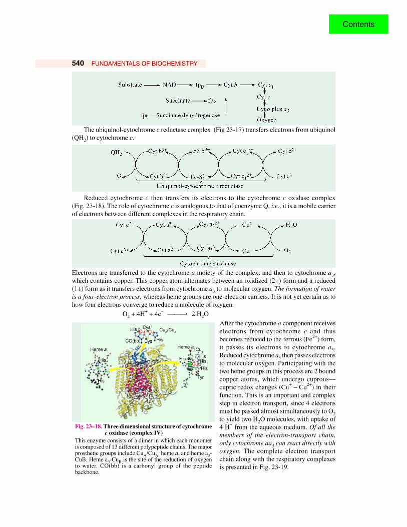
Reduced cytochrome c then transfers its electrons to the cytochrome c oxidase complex(Fig. 23-18). The role of cytochrome c is analogous to that of coenzyme Q, i.e., it is a mobile carrierof electrons between different complexes in the respiratory chain.
Electrons are transferred to the cytochrome a moiety of the complex, and then to cytochrome a3,which contains copper. This copper atom alternates between an oxidized (2+) form and a reduced(1+) form as it transfers electrons from cytochrome a3 to molecular oxygen. The formation of wateris a four-electron process, whereas heme groups are one-electron carriers. It is not yet certain as tohow four electrons converge to reduce a molecule of oxygen.
O2 + 4H+ + 4e– → 2 H2O
After the cytochrome a component receiveselectrons from cytochrome c and thusbecomes reduced to the ferrous (Fe2+) form,it passes its electrons to cytochrome a3.Reduced cytochrome a3 then passes electronsto molecular oxygen. Participating with thetwo heme groups in this process are 2 boundcopper atoms, which undergo cuprous—cupric redox changes (Cu+ – Cu2+) in theirfunction. This is an important and complexstep in electron transport, since 4 electronsmust be passed almost simultaneously to O2to yield two H2O molecules, with uptake of4 H+ from the aqueous medium. Of all themembers of the electron-transport chain,only cytochrome aa3 can react directly withoxygen. The complete electron transportchain along with the respiratory complexesis presented in Fig. 23-19.
Fig. 23–18. Three dimensional structure of cytochromec oxidase (complex IV)
This enzyme consists of a dimer in which each monomeris composed of 13 different polypeptide chains. The majorprosthetic groups include CuA/CuA’ heme a, and heme a3-CuB. Heme a3-CuB is the site of the reduction of oxygento water. CO(bb) is a carbonyl group of the peptidebackbone.
Cug
HisHis
His
Tyr
His
Heme a3
His Cys
His
CuA/Cu
A
CysCO(bb)
Cu Cu
His
Heme a
His
CuFe
Fe
Contents
ELECTRON TRANSPORT AND OXIDATIVE PHOSPHORYLATION 541
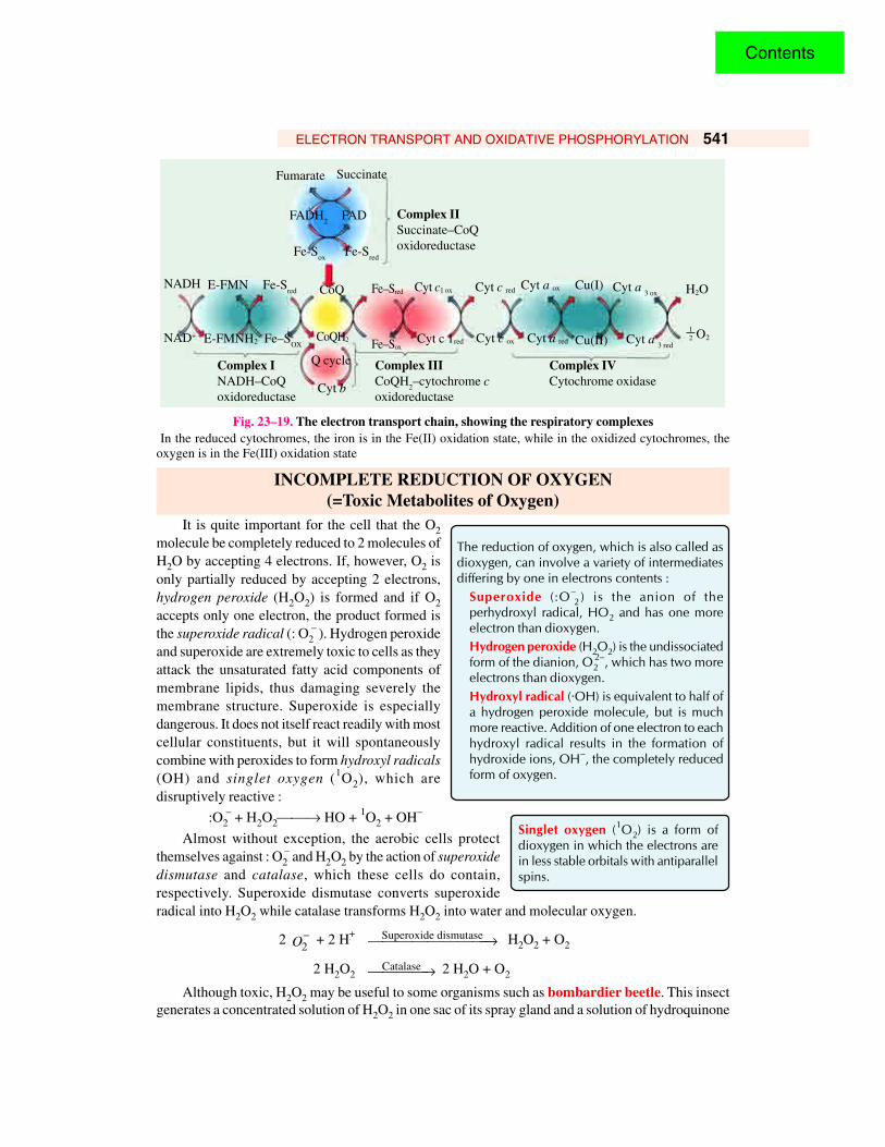
Fig. 23–19. The electron transport chain, showing the respiratory complexes In the reduced cytochromes, the iron is in the Fe(II) oxidation state, while in the oxidized cytochromes, theoxygen is in the Fe(III) oxidation state
INCOMPLETE REDUCTION OF OXYGEN(=Toxic Metabolites of Oxygen)
It is quite important for the cell that the O2molecule be completely reduced to 2 molecules ofH2O by accepting 4 electrons. If, however, O2 isonly partially reduced by accepting 2 electrons,hydrogen peroxide (H2O2) is formed and if O2accepts only one electron, the product formed isthe superoxide radical (: O2
– ). Hydrogen peroxideand superoxide are extremely toxic to cells as theyattack the unsaturated fatty acid components ofmembrane lipids, thus damaging severely themembrane structure. Superoxide is especiallydangerous. It does not itself react readily with mostcellular constituents, but it will spontaneouslycombine with peroxides to form hydroxyl radicals(OH) and singlet oxygen (1O2), which aredisruptively reactive :
:O2– + H2O2→ HO + 1O2 + OH–
Almost without exception, the aerobic cells protectthemselves against : O2
– and H2O2 by the action of superoxidedismutase and catalase, which these cells do contain,respectively. Superoxide dismutase converts superoxideradical into H2O2 while catalase transforms H2O2 into water and molecular oxygen.
2 2O− + 2 H+ Superoxide dismutase→ H2O2 + O2
2 H2O2Catalase→ 2 H2O + O2
Although toxic, H2O2 may be useful to some organisms such as bombardier beetle. This insectgenerates a concentrated solution of H2O2 in one sac of its spray gland and a solution of hydroquinone
The reduction of oxygen, which is also called asdioxygen, can involve a variety of intermediatesdiffering by one in electrons contents :
Superoxide (:O–2 ) is the anion of the
perhydroxyl radical, HO2 and has one moreelectron than dioxygen.Hydrogen peroxide (H2O2) is the undissociatedform of the dianion, O2
2–, which has two moreelectrons than dioxygen.Hydroxyl radical (·OH) is equivalent to half ofa hydrogen peroxide molecule, but is muchmore reactive. Addition of one electron to eachhydroxyl radical results in the formation ofhydroxide ions, OH–, the completely reducedform of oxygen.
Singlet oxygen (1O2) is a form ofdioxygen in which the electrons arein less stable orbitals with antiparallelspins.
Fumarate Succinate
FADH2
FAD
Fe-Sox
Fe-Sred
Complex IISuccinate–CoQoxidoreductase
NADH
NAD+
E-FMN
E-FMNH2 CoQH2
CoQ
Fe–Sox
Fe–Sred
Cyt c 1red
Cyt c1 ox Cyt c red
Cyt c ox
Cyt a ox
Cyt a red
Cu(I)
Cu(II)
Cyt a 3 ox
Cyt a 3 red
H2O
O212
Complex IIICoQH
2–cytochrome c
oxidoreductase
Complex INADH–CoQoxidoreductase
Complex IVCytochrome oxidase
Q cycle
Cyt b
Fe-Sred
Fe–SOX
Contents
542 FUNDAMENTALS OF BIOCHEMISTRY
in the other sac. When threatened, the insect frightens (and also poisons) its enemy by firing a hot(100°F) spray of toxic quinone, which is produced from the oxidation of hydroquinone by H2O2.
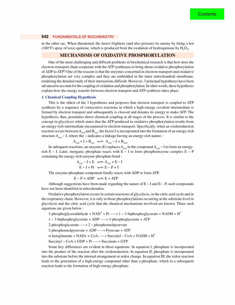
MECHANISMS OF OXIDATIVE PHOSPHORYLATIONOne of the most challenging and difficult problems in biochemical research is that how does the
electron-transport chain cooperate with the ATP synthetase to bring about oxidative phosphorylationof ADP to ATP? One of the reasons is that the enzymes concerned in electron-transport and oxidativephosphorylation are very complex and they are embedded in the inner mitochondrial membrane,rendering the detailed study of their interactions difficult. However, 3 principal hypotheses have beenadvanced to account for the coupling of oxidation and phosphorylation. In other words, these hypothesesexplain how the energy transfer between electron transport and ATP synthesis takes place.
1. Chemical Coupling HypothesisThis is the oldest of the 3 hypotheses and proposes that electron transport is coupled to ATP
synthesis by a sequence of consecutive reactions in which a high-energy covalent intermediate isformed by electron transport and subsequently is cleaved and donates its energy to make ATP. Thehypothesis, thus, postulates direct chemical coupling at all stages of the process. It is similar to theconcept in glycolysis which states that the ATP produced in oxidative phosphorylation results froman energy-rich intermediate encountered in electron transport. Specifically, when an oxidoreductionreaction occurs between Ared and Boxi, the factor I is incorporated into the formation of an energy-richstructure Aoxi ~ I, where the ~ indicates a linkage having an energy-rich nature :
Ared + I + Boxi Ö Aoxi ~ I + Bred
In subsquent reactions, an enzyme (E) replaces Aoxi in the compound Aoxi ~ I to form an energy-rich E ~ I. Later, inorganic phosphate reacts with E ~ I to form phosphoenzyme complex E ~ Pcontaining the energy-rich enzyme-phosphate bond :
Aoxi ~ I + E Ö Aoxi + E ~ I
E ~ I + Pi Ö E ~ P + I
The enzyme-phosphate component finally reacts with ADP to form ATP.
E ~ P + ADP Ö E + ATP
Although suggestions have been made regarding the nature of E ~ I and E ~ P, such compoundshave not been identified in mitochondria.
Oxidative phosphorylation occurs in certain reactions of glycolysis, in the citric acid cycle and inthe respiratory chain. However, it is only in those phosphorylations occurring at the substrate level inglycolysis and the citric acid cycle that the chemical mechanisms involved are known. Three suchequations are given below :
3-phosphoglyceraldehyde + NAD+ + Pi → 1 ~ 3-biphosphoglycerate + NADH + H+
1 ~ 3-biphosphoglycerate + ADP → 3-phosphoglycerate + ATP
2-phosphoglycerate → 2 ~ phosphoenolpyurvate
2-phosphoenolpyruvate + ADP → Pyruvate + ATP
α-ketoglutarate + NAD+ + CoA → Succinyl ~ CoA + NADH + H+
Succinyl ~ CoA + GDP + Pi → Succinate + GTP
Some key differences are evident in these equations. In equation I, phosphate is incorporatedinto the product of the reaction after the oxidoreduction. In equation II, phosphate is incorporatedinto the substrate before the internal arrangement or redox change. In equation III, the redox reactionleads to the generation of a high-energy compound other than a phosphate, which in a subsequentreaction leads to the formation of high-energy phosphate.
Contents
ELECTRON TRANSPORT AND OXIDATIVE PHOSPHORYLATION 543
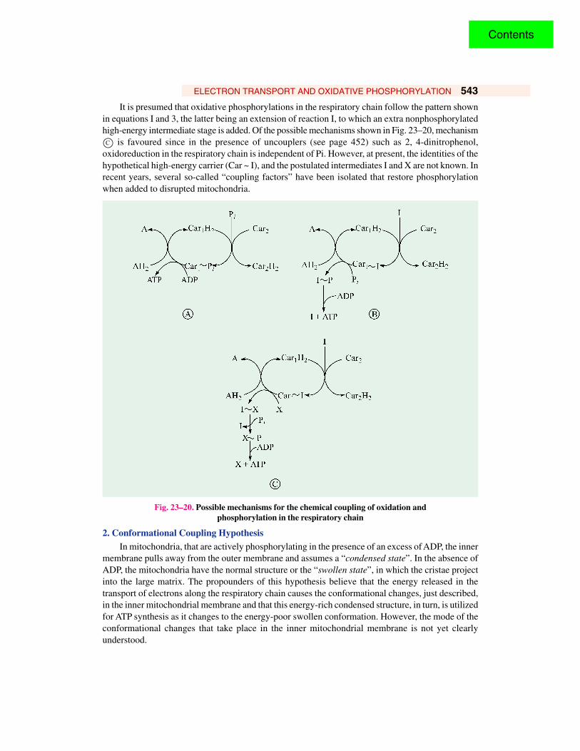
It is presumed that oxidative phosphorylations in the respiratory chain follow the pattern shownin equations I and 3, the latter being an extension of reaction I, to which an extra nonphosphorylatedhigh-energy intermediate stage is added. Of the possible mechanisms shown in Fig. 23–20, mechanism
is favoured since in the presence of uncouplers (see page 452) such as 2, 4-dinitrophenol,oxidoreduction in the respiratory chain is independent of Pi. However, at present, the identities of thehypothetical high-energy carrier (Car ~ I), and the postulated intermediates I and X are not known. Inrecent years, several so-called “coupling factors” have been isolated that restore phosphorylationwhen added to disrupted mitochondria.
Fig. 23–20. Possible mechanisms for the chemical coupling of oxidation andphosphorylation in the respiratory chain
2. Conformational Coupling HypothesisIn mitochondria, that are actively phosphorylating in the presence of an excess of ADP, the inner
membrane pulls away from the outer membrane and assumes a “condensed state”. In the absence ofADP, the mitochondria have the normal structure or the “swollen state”, in which the cristae projectinto the large matrix. The propounders of this hypothesis believe that the energy released in thetransport of electrons along the respiratory chain causes the conformational changes, just described,in the inner mitochondrial membrane and that this energy-rich condensed structure, in turn, is utilizedfor ATP synthesis as it changes to the energy-poor swollen conformation. However, the mode of theconformational changes that take place in the inner mitochondrial membrane is not yet clearlyunderstood.
Contents
544 FUNDAMENTALS OF BIOCHEMISTRY
Peter Mitchell (LT, 1920 – 1992)Mitchell, a British biochemist, is a rare example of the truly independentscientist since, in his native England, he was not affiliated with a university,industry or government. He received 1978 Nobel Prize in Chemistry forhis work on the coupling of oxidation and phosphorylation. He proposedthat electron transport and ATP synthesis are coupled by a proton gradient,rather than by a covalent high-energy intermediate or an activated protein.
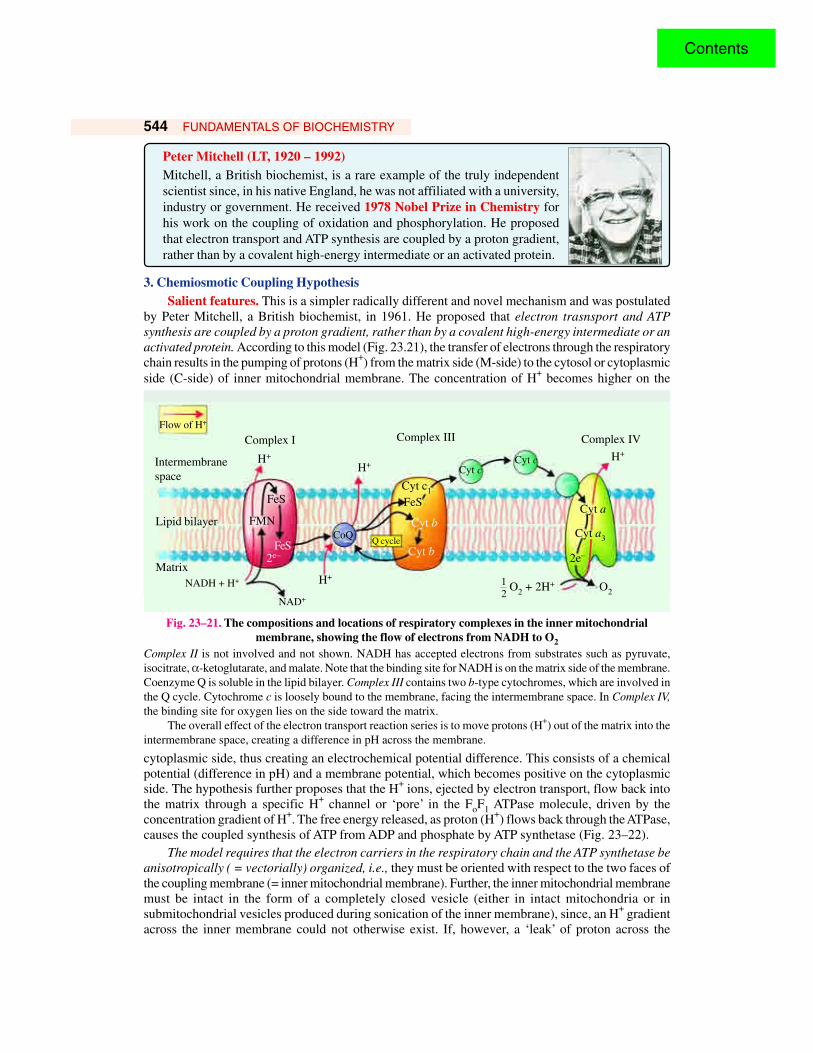
3. Chemiosmotic Coupling HypothesisSalient features. This is a simpler radically different and novel mechanism and was postulated
by Peter Mitchell, a British biochemist, in 1961. He proposed that electron trasnsport and ATPsynthesis are coupled by a proton gradient, rather than by a covalent high-energy intermediate or anactivated protein. According to this model (Fig. 23.21), the transfer of electrons through the respiratorychain results in the pumping of protons (H+) from the matrix side (M-side) to the cytosol or cytoplasmicside (C-side) of inner mitochondrial membrane. The concentration of H+ becomes higher on the
Fig. 23–21. The compositions and locations of respiratory complexes in the inner mitochondrialmembrane, showing the flow of electrons from NADH to O2
Complex II is not involved and not shown. NADH has accepted electrons from substrates such as pyruvate,isocitrate, α-ketoglutarate, and malate. Note that the binding site for NADH is on the matrix side of the membrane.Coenzyme Q is soluble in the lipid bilayer. Complex III contains two b-type cytochromes, which are involved inthe Q cycle. Cytochrome c is loosely bound to the membrane, facing the intermembrane space. In Complex IV,the binding site for oxygen lies on the side toward the matrix.
The overall effect of the electron transport reaction series is to move protons (H+) out of the matrix into theintermembrane space, creating a difference in pH across the membrane.
cytoplasmic side, thus creating an electrochemical potential difference. This consists of a chemicalpotential (difference in pH) and a membrane potential, which becomes positive on the cytoplasmicside. The hypothesis further proposes that the H+ ions, ejected by electron transport, flow back intothe matrix through a specific H+ channel or ‘pore’ in the FoF1 ATPase molecule, driven by theconcentration gradient of H+. The free energy released, as proton (H+) flows back through the ATPase,causes the coupled synthesis of ATP from ADP and phosphate by ATP synthetase (Fig. 23–22).
The model requires that the electron carriers in the respiratory chain and the ATP synthetase beanisotropically ( = vectorially) organized, i.e., they must be oriented with respect to the two faces ofthe coupling membrane (= inner mitochondrial membrane). Further, the inner mitochondrial membranemust be intact in the form of a completely closed vesicle (either in intact mitochondria or insubmitochondrial vesicles produced during sonication of the inner membrane), since, an H+ gradientacross the inner membrane could not otherwise exist. If, however, a ‘leak’ of proton across the
NADH + H+
Intermembranespace
Flow of H+
Complex I
FeS
H+
Complex III Complex IV
H+
Lipid bilayer
Matrix
NAD+
FeS
O2 + 2H+12
O2
FMN
2e–
H+
Q cycle
Cyt c1
FeS
Cyt b
Cyt b
H+
CoQ
Cyt cCyt c
Cyt a
Cyt a3
2e–
Contents
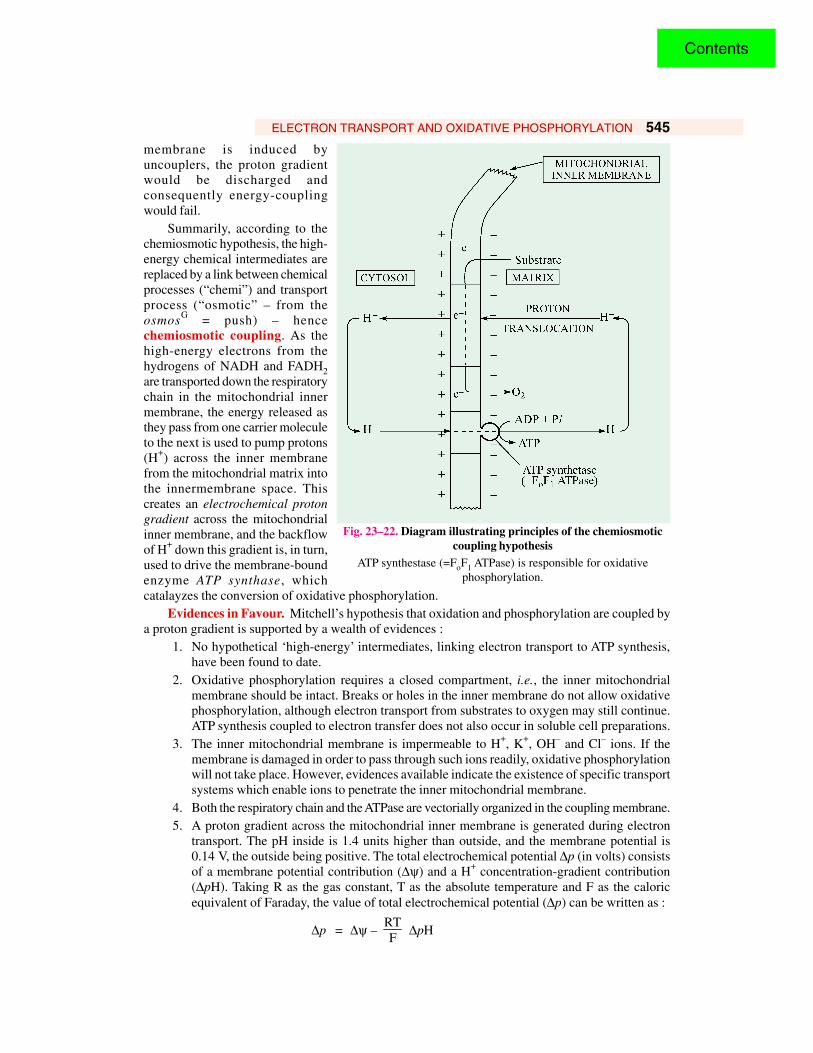
ELECTRON TRANSPORT AND OXIDATIVE PHOSPHORYLATION 545membrane is induced byuncouplers, the proton gradientwould be discharged andconsequently energy-couplingwould fail.
Summarily, according to thechemiosmotic hypothesis, the high-energy chemical intermediates arereplaced by a link between chemicalprocesses (“chemi”) and transportprocess (“osmotic” – from theosmosG = push) – hencechemiosmotic coupling. As thehigh-energy electrons from thehydrogens of NADH and FADH2are transported down the respiratorychain in the mitochondrial innermembrane, the energy released asthey pass from one carrier moleculeto the next is used to pump protons(H+) across the inner membranefrom the mitochondrial matrix intothe innermembrane space. Thiscreates an electrochemical protongradient across the mitochondrialinner membrane, and the backflowof H+ down this gradient is, in turn,used to drive the membrane-boundenzyme ATP synthase, whichcatalayzes the conversion of oxidative phosphorylation.
Evidences in Favour. Mitchell’s hypothesis that oxidation and phosphorylation are coupled bya proton gradient is supported by a wealth of evidences :
1. No hypothetical ‘high-energy’ intermediates, linking electron transport to ATP synthesis,have been found to date.
2. Oxidative phosphorylation requires a closed compartment, i.e., the inner mitochondrialmembrane should be intact. Breaks or holes in the inner membrane do not allow oxidativephosphorylation, although electron transport from substrates to oxygen may still continue.ATP synthesis coupled to electron transfer does not also occur in soluble cell preparations.
3. The inner mitochondrial membrane is impermeable to H+, K+, OH– and Cl– ions. If themembrane is damaged in order to pass through such ions readily, oxidative phosphorylationwill not take place. However, evidences available indicate the existence of specific transportsystems which enable ions to penetrate the inner mitochondrial membrane.
4. Both the respiratory chain and the ATPase are vectorially organized in the coupling membrane.5. A proton gradient across the mitochondrial inner membrane is generated during electron
transport. The pH inside is 1.4 units higher than outside, and the membrane potential is0.14 V, the outside being positive. The total electrochemical potential ∆p (in volts) consistsof a membrane potential contribution (∆ψ) and a H+ concentration-gradient contribution(∆pH). Taking R as the gas constant, T as the absolute temperature and F as the caloricequivalent of Faraday, the value of total electrochemical potential (∆p) can be written as :
∆p = ∆ψ – RTF
∆pH
Fig. 23–22. Diagram illustrating principles of the chemiosmoticcoupling hypothesis
ATP synthestase (=FoF1 ATPase) is responsible for oxidativephosphorylation.
Contents
546 FUNDAMENTALS OF BIOCHEMISTRY
= ∆ψ – 0.06 ∆pH= 0.14 – 0.06 (–1.4)
= 0.224 V
This total proton-motive force of 0.224 V corresponds to a free energy of 5.2 kcal per moleof protons.
6. ATP is synthesized when a pH gradient is imposed on mitochondria or chloroplasts in theabsence of electron transport.
7. Oxidative phosphorylation can be checked by uncouplers and certain ionophores (see page453). Uncouplers such as 2,4-dinitrophenol increase the permeability of mitochondria toprotons, thus reducing the electrochemical potential and short-circuiting the vectorial ATPsynthetase system for the production of ATP.
8. Addition of acid to the external medium, establishing a proton gradient, leads to the synthesisof ATP.
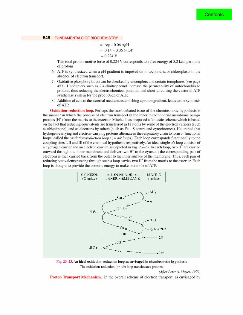
Oxidation-reduction loop. Perhaps the most debated issue of the chemiosmotic hypothesis isthe manner in which the process of electron transport in the inner mitochondrial membrane pumpsprotons (H+) from the matrix to the exterior. Mitchell has proposed a fantastic scheme which is basedon the fact that reducing equivalents are transferred as H atoms by some of the electron carriers (suchas ubiquinone), and as electrons by others (such as Fe—S centre and cytochromes). He opined thathydrogen-carrying and electron-carrying proteins alternate in the respiratory chain to form 3 ‘functionalloops’ called the oxidation-reduction loops ( = o/r loops). Each loop corresponds functionally to thecoupling sites I, II and III of the chemical hypothesis respectively. An ideal single o/r loop consists ofa hydrogen carrier and an electron carrier, as depicted in Fig. 23–23. In each loop, two H+ are carriedoutward through the inner membrane and deliver two H+ to the cytosol ; the corresponding pair ofelectrons is then carried back from the outer to the inner surface of the membrane. Thus, each pair ofreducing equivalents passing through such a loop carries two H+ from the matrix to the exterior. Eachloop is thought to provide the osmotic energy to make one mole of ATP.
Fig. 23–23. An ideal oxidation-reduction loop as envisaged in chemiosmotic hypothesisThe oxidation-reduction (or o/r) loop translocates protons.
(After Peter A. Mayes, 1979)
Proton Transport Mechanism. In the overall scheme of electron transport, as envisaged by
Contents
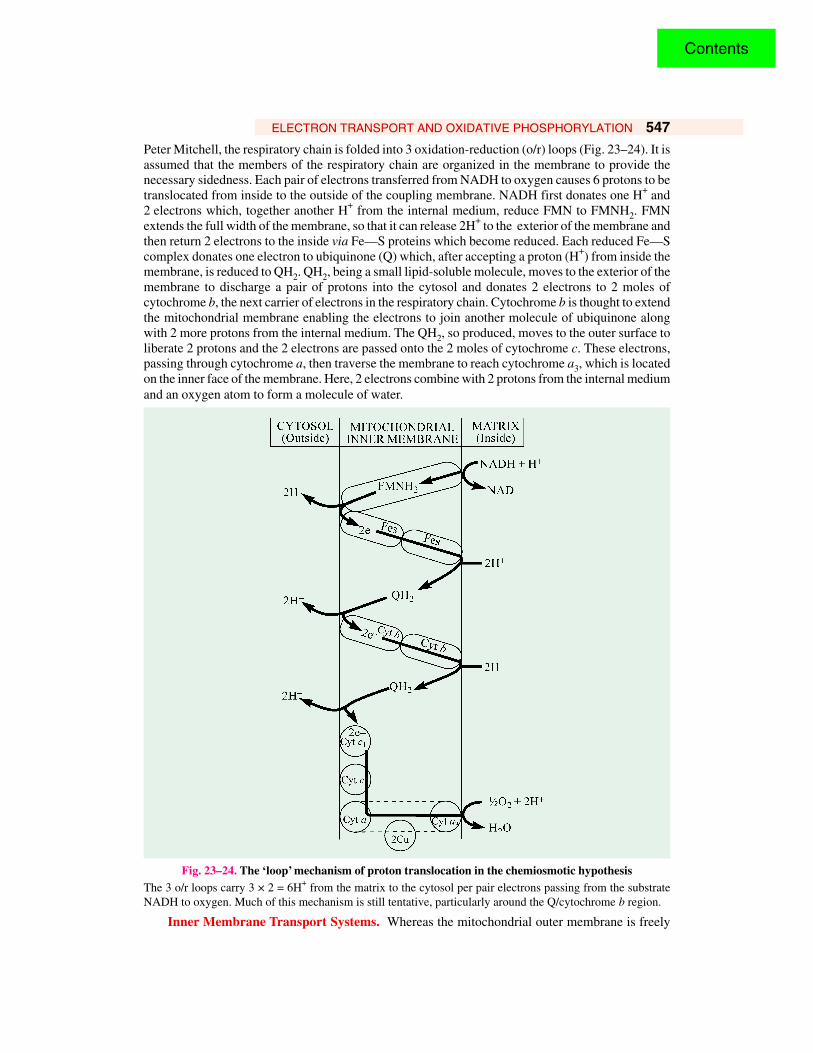
ELECTRON TRANSPORT AND OXIDATIVE PHOSPHORYLATION 547Peter Mitchell, the respiratory chain is folded into 3 oxidation-reduction (o/r) loops (Fig. 23–24). It isassumed that the members of the respiratory chain are organized in the membrane to provide thenecessary sidedness. Each pair of electrons transferred from NADH to oxygen causes 6 protons to betranslocated from inside to the outside of the coupling membrane. NADH first donates one H+ and2 electrons which, together another H+ from the internal medium, reduce FMN to FMNH2. FMNextends the full width of the membrane, so that it can release 2H+ to the exterior of the membrane andthen return 2 electrons to the inside via Fe—S proteins which become reduced. Each reduced Fe—Scomplex donates one electron to ubiquinone (Q) which, after accepting a proton (H+) from inside themembrane, is reduced to QH2. QH2, being a small lipid-soluble molecule, moves to the exterior of themembrane to discharge a pair of protons into the cytosol and donates 2 electrons to 2 moles ofcytochrome b, the next carrier of electrons in the respiratory chain. Cytochrome b is thought to extendthe mitochondrial membrane enabling the electrons to join another molecule of ubiquinone alongwith 2 more protons from the internal medium. The QH2, so produced, moves to the outer surface toliberate 2 protons and the 2 electrons are passed onto the 2 moles of cytochrome c. These electrons,passing through cytochrome a, then traverse the membrane to reach cytochrome a3, which is locatedon the inner face of the membrane. Here, 2 electrons combine with 2 protons from the internal mediumand an oxygen atom to form a molecule of water.
Fig. 23–24. The ‘loop’ mechanism of proton translocation in the chemiosmotic hypothesisThe 3 o/r loops carry 3 × 2 = 6H+ from the matrix to the cytosol per pair electrons passing from the substrateNADH to oxygen. Much of this mechanism is still tentative, particularly around the Q/cytochrome b region.
Inner Membrane Transport Systems. Whereas the mitochondrial outer membrane is freely
Contents
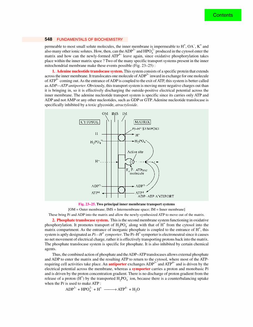
548 FUNDAMENTALS OF BIOCHEMISTRY
permeable to most small solute molecules, the inner membrane is impermeable to H+, OA–, K+ andalso many other ionic solutes. How, then, can the ADP3– and HPO4
2– produced in the cytosol enter thematrix and how can the newly-formed ATP4– leave again, since oxidative phosphorylation takesplace within the inner matrix space ? Two of the many specific transport systems present in the innermitochondrial membrane make these events possible (Fig. 23–25) :
1. Adenine nucleotide translocase system. This system consists of a specific protein that extendsacross the inner membrane. It translocates one molecule of ADP3– inward in exchange for one moleculeof ATP4– coming out. As the entrance of ADP is coupled to the exit of ATP, this system is better calledas ADP—ATP antiporter. Obviously, this transport system is moving more negative charges out thanit is bringing in, so it is effectively discharging the outside-positive electrical potential across theinner membrane. The adenine nucleotide transport system is specific since its carries only ATP andADP and not AMP or any other nucleotides, such as GDP or GTP. Adenine nucleotide translocase isspecifically inhibited by a toxic glycoside, atractyloside.
Fig. 23–25. Two principal inner membrane transport systems[OM = Outer membrane; IMS = Intermembrane space; IM = Inner membrane]
These bring Pi and ADP into the matrix and allow the newly-synthesized ATP to move out of the matrix.
2. Phosphate translocase system. This is the second membrane system functioning in oxidativephosphorylation. It promotes transport of H2PO4
– along with that of H+ from the cytosol into thematrix compartment. As the entrance of inorganic phosphate is coupled to the entrance of H+, thissystem is aptly designated as Pi—H+ symporter. The Pi–H+ symporter is electroneutral since it causesno net movement of electrical charge, rather it is effectively transporting protons back into the matrix.The phosphate translocase system is specific for phosphate. It is also inhibited by certain chemicalagents.
Thus, the combined action of phosphate and the ADP–ATP translocases allows external phosphateand ADP to enter the matrix and the resulting ATP to return to the cytosol, where most of the ATP-requiring cell activities take place. An antiporter exchanges ADP3– and ATP4– and is driven by theelectrical potential across the membrane, whereas a symporter carries a proton and monobasic Piand is driven by the proton concentration gradient. There is no discharge of proton gradient from therelease of a proton (H+) by the transported H2PO4
– ion, because there is a counterbalancing uptakewhen the Pi is used to make ATP :
ADP3– + HPO42– + H+ → ATP4– + H2O
Contents
ELECTRON TRANSPORT AND OXIDATIVE PHOSPHORYLATION 549
OXIDATION OF EXTRAMITOCHONDRIAL NADH(=NADH Shuttle Systems)
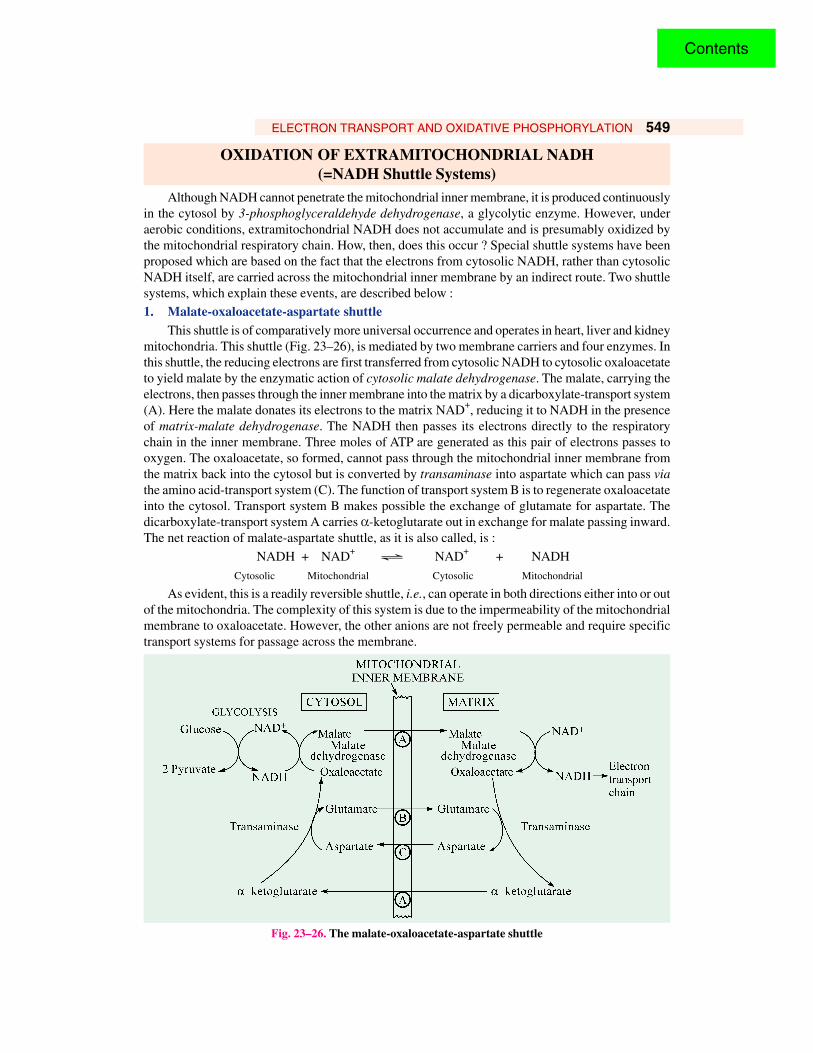
Although NADH cannot penetrate the mitochondrial inner membrane, it is produced continuouslyin the cytosol by 3-phosphoglyceraldehyde dehydrogenase, a glycolytic enzyme. However, underaerobic conditions, extramitochondrial NADH does not accumulate and is presumably oxidized bythe mitochondrial respiratory chain. How, then, does this occur ? Special shuttle systems have beenproposed which are based on the fact that the electrons from cytosolic NADH, rather than cytosolicNADH itself, are carried across the mitochondrial inner membrane by an indirect route. Two shuttlesystems, which explain these events, are described below :1. Malate-oxaloacetate-aspartate shuttle
This shuttle is of comparatively more universal occurrence and operates in heart, liver and kidneymitochondria. This shuttle (Fig. 23–26), is mediated by two membrane carriers and four enzymes. Inthis shuttle, the reducing electrons are first transferred from cytosolic NADH to cytosolic oxaloacetateto yield malate by the enzymatic action of cytosolic malate dehydrogenase. The malate, carrying theelectrons, then passes through the inner membrane into the matrix by a dicarboxylate-transport system(A). Here the malate donates its electrons to the matrix NAD+, reducing it to NADH in the presenceof matrix-malate dehydrogenase. The NADH then passes its electrons directly to the respiratorychain in the inner membrane. Three moles of ATP are generated as this pair of electrons passes tooxygen. The oxaloacetate, so formed, cannot pass through the mitochondrial inner membrane fromthe matrix back into the cytosol but is converted by transaminase into aspartate which can pass viathe amino acid-transport system (C). The function of transport system B is to regenerate oxaloacetateinto the cytosol. Transport system B makes possible the exchange of glutamate for aspartate. Thedicarboxylate-transport system A carries α-ketoglutarate out in exchange for malate passing inward.The net reaction of malate-aspartate shuttle, as it is also called, is :
NADH + NAD+ Ö NAD+ + NADHCytosolic Mitochondrial Cytosolic Mitochondrial
As evident, this is a readily reversible shuttle, i.e., can operate in both directions either into or outof the mitochondria. The complexity of this system is due to the impermeability of the mitochondrialmembrane to oxaloacetate. However, the other anions are not freely permeable and require specifictransport systems for passage across the membrane.
Fig. 23–26. The malate-oxaloacetate-aspartate shuttle
Contents
550 FUNDAMENTALS OF BIOCHEMISTRY
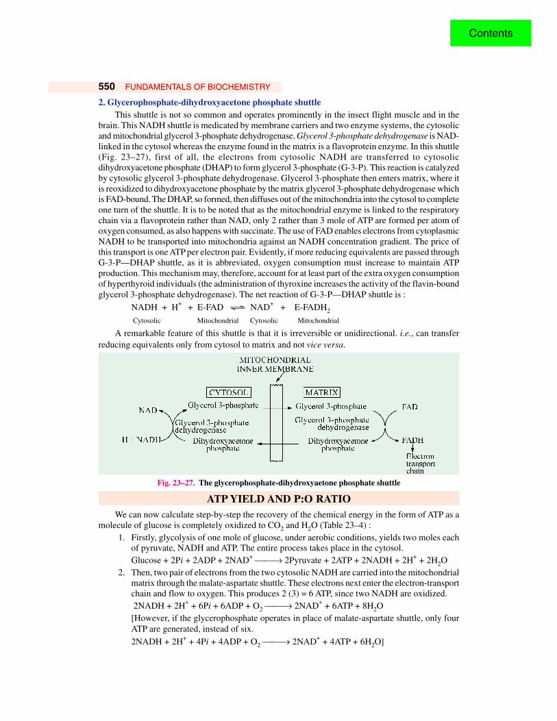
2. Glycerophosphate-dihydroxyacetone phosphate shuttleThis shuttle is not so common and operates prominently in the insect flight muscle and in the
brain. This NADH shuttle is medicated by membrane carriers and two enzyme systems, the cytosolicand mitochondrial glycerol 3-phosphate dehydrogenase. Glycerol 3-phosphate dehydrogenase is NAD-linked in the cytosol whereas the enzyme found in the matrix is a flavoprotein enzyme. In this shuttle(Fig. 23–27), first of all, the electrons from cytosolic NADH are transferred to cytosolicdihydroxyacetone phosphate (DHAP) to form glycerol 3-phosphate (G-3-P). This reaction is catalyzedby cytosolic glycerol 3-phosphate dehydrogenase. Glycerol 3-phosphate then enters matrix, where itis reoxidized to dihydroxyacetone phosphate by the matrix glycerol 3-phosphate dehydrogenase whichis FAD-bound. The DHAP, so formed, then diffuses out of the mitochondria into the cytosol to completeone turn of the shuttle. It is to be noted that as the mitochondrial enzyme is linked to the respiratorychain via a flavoprotein rather than NAD, only 2 rather than 3 mole of ATP are formed per atom ofoxygen consumed, as also happens with succinate. The use of FAD enables electrons from cytoplasmicNADH to be transported into mitochondria against an NADH concentration gradient. The price ofthis transport is one ATP per electron pair. Evidently, if more reducing equivalents are passed throughG-3-P—DHAP shuttle, as it is abbreviated, oxygen consumption must increase to maintain ATPproduction. This mechanism may, therefore, account for at least part of the extra oxygen consumptionof hyperthyroid individuals (the administration of thyroxine increases the activity of the flavin-boundglycerol 3-phosphate dehydrogenase). The net reaction of G-3-P—DHAP shuttle is :
NADH + H+ + E-FAD Ö NAD+ + E-FADH2
Cytosolic Mitochondrial Cytosolic Mitochondrial
A remarkable feature of this shuttle is that it is irreversible or unidirectional. i.e., can transferreducing equivalents only from cytosol to matrix and not vice versa.
Fig. 23–27. The glycerophosphate-dihydroxyaetone phosphate shuttle
ATP YIELD AND P:O RATIOWe can now calculate step-by-step the recovery of the chemical energy in the form of ATP as a
molecule of glucose is completely oxidized to CO2 and H2O (Table 23–4) :1. Firstly, glycolysis of one mole of glucose, under aerobic conditions, yields two moles each
of pyruvate, NADH and ATP. The entire process takes place in the cytosol.Glucose + 2Pi + 2ADP + 2NAD+ → 2Pyruvate + 2ATP + 2NADH + 2H+ + 2H2O
2. Then, two pair of electrons from the two cytosolic NADH are carried into the mitochondrialmatrix through the malate-aspartate shuttle. These electrons next enter the electron-transportchain and flow to oxygen. This produces 2 (3) = 6 ATP, since two NADH are oxidized. 2NADH + 2H+ + 6Pi + 6ADP + O2 → 2NAD+ + 6ATP + 8H2O[However, if the glycerophosphate operates in place of malate-aspartate shuttle, only fourATP are generated, instead of six.
2NADH + 2H+ + 4Pi + 4ADP + O2 → 2NAD+ + 4ATP + 6H2O]
Contents
ELECTRON TRANSPORT AND OXIDATIVE PHOSPHORYLATION 551
3. Then, two moles of pyruvate are dehydrogenated to yield two moles each of acetyl-CoA andCO2 in the mitochondria. This results in the formation of two NADH. The two electron pairsfrom two NADH are carried to O2 via the electron-transport chain, each mole providingthree moles of ATP.2 Pyruvate + 2CoA + 6Pi + 6ADP + O2 → 2Acetyl-CoA + 2CO2 + 6ATP + 8H2O
4. Ultimately, two moles of acetyl-CoA are oxidized to CO2 and H2O via the citric acid cycle,along with the oxidative phosphorylation coupled to electron transport from isocitrate, α-ketoglutarate and malate to O2, each of which yields 3 moles of ATP. The oxidation ofsuccinate, however, yields 2 ATP and another two ATPs are generated from succinyl-CoAvia GTP.
2 Acetyl-CoA + 24 Pi + 24 ADP + 4 O2 → 2 CoA —SH + 4 CO2 + 24 ATP + 26 H2O
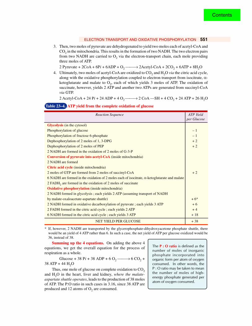
Table 23–4. ATP yield from the complete oxidation of glucose
Reaction Sequence ATP Yieldper Glucose
Glycolysis (in the cytosol)
Phosphorylation of glucose – 1
Phosphorylation of fructose 6-phosphate – 1
Dephosphorylation of 2 moles of 1, 3-DPG + 2
Dephosphorylation of 2 moles of PEP + 2
2 NADH are formed in the oxidation of 2 moles of G-3-P
Conversion of pyruvate into acetyl-CoA (inside mitochondria)
2 NADH are formed
Citric acid cycle (inside mitochondria)
2 moles of GTP are formed from 2 moles of succinyl-CoA + 2
6 NADH are formed in the oxidation of 2 moles each of isocitrate, α-ketoglutarate and malate
2 FADH2 are formed in the oxidation of 2 moles of succinate
Oxidative phosphorylation (inside mitochondria)
2 NADH formed in glycolysis ; each yields 2 ATP (assuming transport of NADH
by malate-oxaloacetate-aspartate shuttle) + 6*
2 NADH formed in oxidative decarboxylation of pyruvate ; each yields 3 ATP + 6
2 FADH formed in the citric acid cycle ; each yields 2 ATP + 4
6 NADH formed in the citric acid cycle ; each yields 3 ATP + 18
NET YIELD PER GLUCOSE + 38
* If, however, 2 NADH are transported by the glycerophosphate-dihydroxyacetone phosphate shuttle, therewould be an yield of 4 ATP rather than 6. In such a case, the net yield of ATP per glucose oxidized would be36, instead of 38.
Summing up the 4 equations. On adding the above 4equations, we get the overall equation for the process ofrespiration as a whole.
Glucose + 38 Pi + 38 ADP + 6 O2 → 6 CO2 +38 ATP + 44 H2O
Thus, one mole of glucose on complete oxidation to CO2and H2O in the heart, liver and kidney, where the malate-aspartate shuttle operates, leads to the production of 38 molesof ATP. The P:O ratio in such cases in 3.16, since 38 ATP areproduced and 12 atoms of O2 are consumed.
The P : O ratio is defined as thenumber of moles of inorganicphosphate incorporated intoorganic form per atom of oxygenconsumed. In other words, theP : O ratio may be taken to meanthe number of moles of high-energy phosphate generated peratom of oxygen consumed.
Contents
552 FUNDAMENTALS OF BIOCHEMISTRY
[However, in the skeletal muscles, where glycerophosphate shuttle operates, a total of 36 ATPmoles is generated per mole of glucose oxidized. In that case, the overall equation is modified to:
Glucose + 36 Pi + 36 ADP + 6O2 → 6 CO2 + 36 ATP + 42 H2OHere the P:O ratio is 36/12= 3.0.]Under standard conditions (1.0 M), the theoretical recovery of free energy in the complete oxidation
of glucose is :38 (7.3/686) (100) = 40%
[or 36 (7.3/686) (100) = 38%, when glycerophosphate shuttle operates]. But in the intact cell, theefficiency of this process is high (over 70%) because the cellular concentration of glucose, oxygen,Pi, ADP and ATP are unequal and much lower than the standard concentration of 1.0 M. The trappingof this amount of energy is a noteworthy achievement for the living cell.
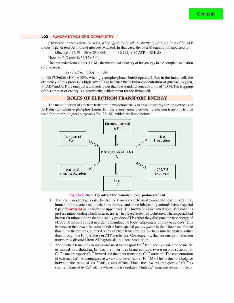
ROLES OF ELECTRON TRANSPORT ENERGYThe main function of electron transport in mitochondria is to provide energy for the synthesis of
ATP during oxidative phosphorylation. But the energy generated during electron transport is alsoused for other biological purposes (Fig. 23–28), which are listed below :
Fig. 23–28. Some key roles of the transmembrane proton gradient
1. The proton gradient generated by electron transport can be used to generate heat. For example,human infants, other mammals born hairless and some hibernating animals have a specialtype of brown fat in the neck and upper back. The brown fat is so named because it containsprofuse mitochondria which, in turn, are rich in the red-brown cytochromes. These specializedbrown-fat mitochondria do not usually produce ATP, rather they dissipate the free energy ofelectron transport as heat in order to maintain the body temperature of the young ones. Thisis because the brown-fat mitochondria have special proton pores in their inner membranethat allow the protons, pumped on by electron transport, to flow back into the matrix, ratherthan through the FoF1 ATPase or ATP synthetase. Consequently, the free energy of electrontransport is diverted from ATP synthesis into heat production.
2. The electron transport energy is also used to transport Ca2+ from the cytosol into the matrixof animal mitochondria. In fact, the inner membrane contains two transport systems forCa2+ : one transports Ca2+ inward and the other transports Ca2+ outward. The concentrationof external Ca2+ is maintained at a very low level (about 10–7 M). This is due to a balancebetween the rates of Ca2+ influx and efflux. Thus, the inward transport of Ca2+ iscounterbalanced by Ca2+ efflux whose rate is regulated. High Ca2+ concentrations initiate or
Contents
ELECTRON TRANSPORT AND OXIDATIVE PHOSPHORYLATION 553promote many cell functions such as muscle concentration, glycogen breakdown and theoxidation of pyruvate ; low Ca2+ concentrations have inhibitory effects on these functions.
3. Rotation of bacterial flagella is also controlled by the proton gradient generated across themembrane.
4. Transfer fo electrons from NADH to NADPH is also powered by the proton gradient.5. The entry of some amino acids and sugars is also governed by the energy generated during
electron transport.
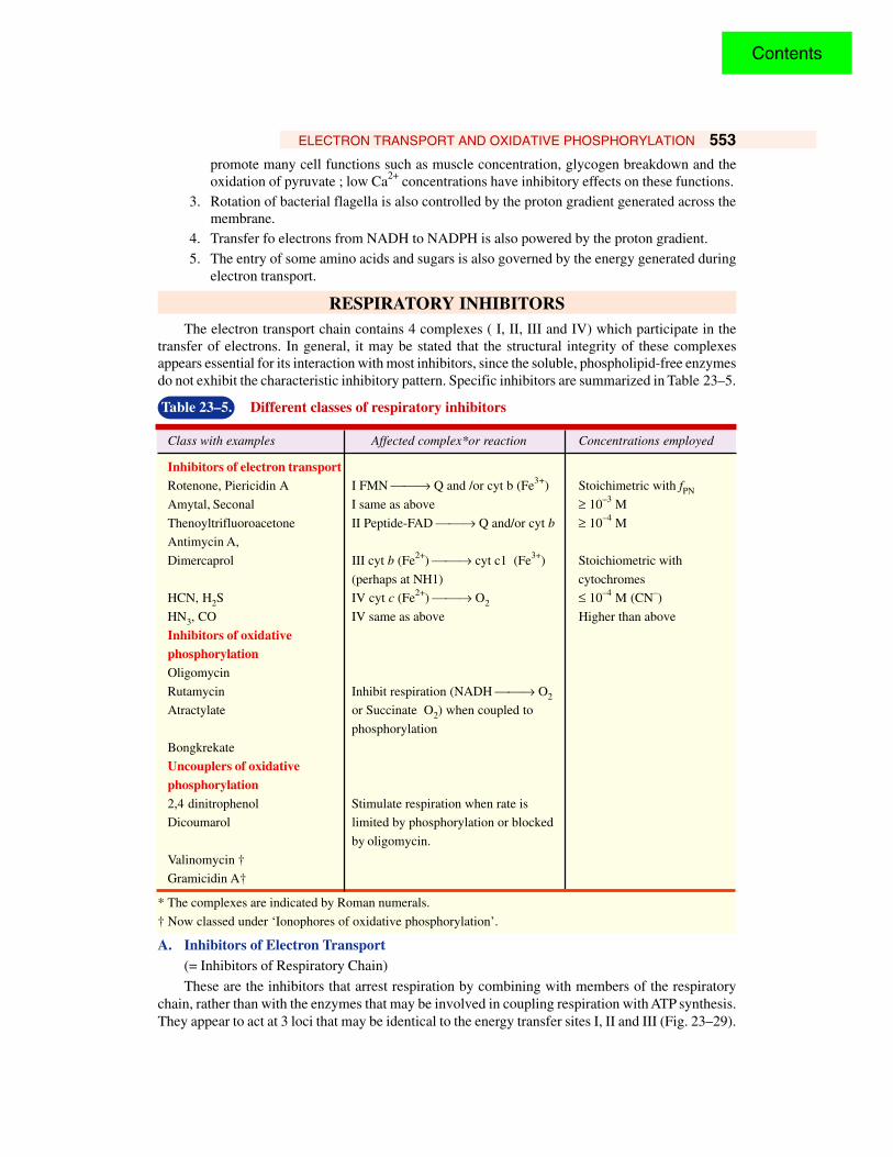
RESPIRATORY INHIBITORSThe electron transport chain contains 4 complexes ( I, II, III and IV) which participate in the
transfer of electrons. In general, it may be stated that the structural integrity of these complexesappears essential for its interaction with most inhibitors, since the soluble, phospholipid-free enzymesdo not exhibit the characteristic inhibitory pattern. Specific inhibitors are summarized in Table 23–5.
Table 23–5. Different classes of respiratory inhibitors
Class with examples Affected complex*or reaction Concentrations employed
Inhibitors of electron transportRotenone, Piericidin A I FMN → Q and /or cyt b (Fe3+) Stoichimetric with fPN
Amytal, Seconal I same as above ≥ 10–3 M
Thenoyltrifluoroacetone II Peptide-FAD → Q and/or cyt b ≥ 10–4 M
Antimycin A,
Dimercaprol III cyt b (Fe2+) → cyt c1 (Fe3+) Stoichiometric with
(perhaps at NH1) cytochromes
HCN, H2S IV cyt c (Fe2+) → O2 ≤ 10–4 M (CN–)
HN3, CO IV same as above Higher than above
Inhibitors of oxidativephosphorylationOligomycin
Rutamycin Inhibit respiration (NADH → O2
Atractylate or Succinate O2) when coupled to
phosphorylation
Bongkrekate
Uncouplers of oxidativephosphorylation2,4 dinitrophenol Stimulate respiration when rate is
Dicoumarol limited by phosphorylation or blocked
by oligomycin.
Valinomycin †
Gramicidin A†
* The complexes are indicated by Roman numerals.
† Now classed under ‘Ionophores of oxidative phosphorylation’.
A. Inhibitors of Electron Transport(= Inhibitors of Respiratory Chain)
These are the inhibitors that arrest respiration by combining with members of the respiratorychain, rather than with the enzymes that may be involved in coupling respiration with ATP synthesis.They appear to act at 3 loci that may be identical to the energy transfer sites I, II and III (Fig. 23–29).
Contents
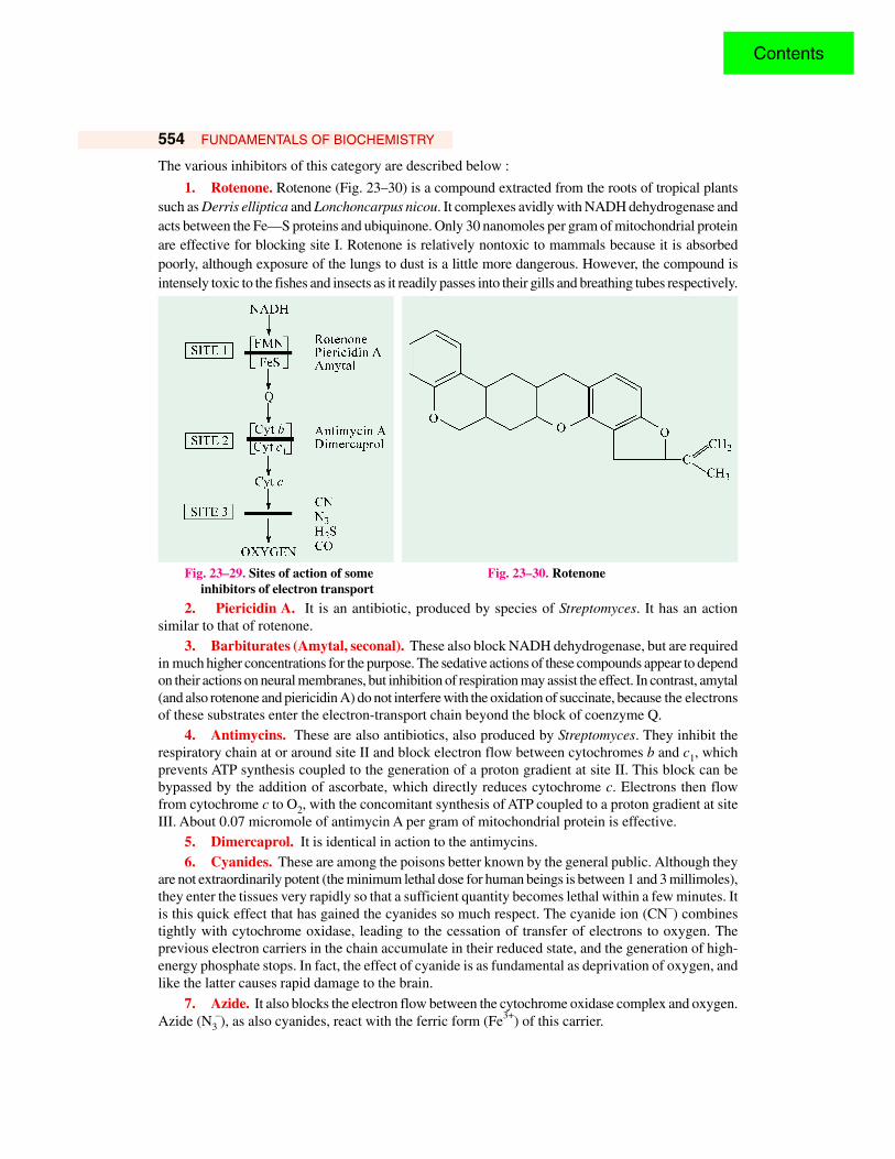
554 FUNDAMENTALS OF BIOCHEMISTRY
The various inhibitors of this category are described below :
1. Rotenone. Rotenone (Fig. 23–30) is a compound extracted from the roots of tropical plantssuch as Derris elliptica and Lonchoncarpus nicou. It complexes avidly with NADH dehydrogenase andacts between the Fe—S proteins and ubiquinone. Only 30 nanomoles per gram of mitochondrial proteinare effective for blocking site I. Rotenone is relatively nontoxic to mammals because it is absorbedpoorly, although exposure of the lungs to dust is a little more dangerous. However, the compound isintensely toxic to the fishes and insects as it readily passes into their gills and breathing tubes respectively.
Fig. 23–29. Sites of action of some Fig. 23–30. Rotenone inhibitors of electron transport
2. Piericidin A. It is an antibiotic, produced by species of Streptomyces. It has an actionsimilar to that of rotenone.
3. Barbiturates (Amytal, seconal). These also block NADH dehydrogenase, but are requiredin much higher concentrations for the purpose. The sedative actions of these compounds appear to dependon their actions on neural membranes, but inhibition of respiration may assist the effect. In contrast, amytal(and also rotenone and piericidin A) do not interfere with the oxidation of succinate, because the electronsof these substrates enter the electron-transport chain beyond the block of coenzyme Q.
4. Antimycins. These are also antibiotics, also produced by Streptomyces. They inhibit therespiratory chain at or around site II and block electron flow between cytochromes b and c1, whichprevents ATP synthesis coupled to the generation of a proton gradient at site II. This block can bebypassed by the addition of ascorbate, which directly reduces cytochrome c. Electrons then flowfrom cytochrome c to O2, with the concomitant synthesis of ATP coupled to a proton gradient at siteIII. About 0.07 micromole of antimycin A per gram of mitochondrial protein is effective.
5. Dimercaprol. It is identical in action to the antimycins.6. Cyanides. These are among the poisons better known by the general public. Although they
are not extraordinarily potent (the minimum lethal dose for human beings is between 1 and 3 millimoles),they enter the tissues very rapidly so that a sufficient quantity becomes lethal within a few minutes. Itis this quick effect that has gained the cyanides so much respect. The cyanide ion (CN–) combinestightly with cytochrome oxidase, leading to the cessation of transfer of electrons to oxygen. Theprevious electron carriers in the chain accumulate in their reduced state, and the generation of high-energy phosphate stops. In fact, the effect of cyanide is as fundamental as deprivation of oxygen, andlike the latter causes rapid damage to the brain.
7. Azide. It also blocks the electron flow between the cytochrome oxidase complex and oxygen.Azide (N3
–), as also cyanides, react with the ferric form (Fe3+) of this carrier.
Contents
ELECTRON TRANSPORT AND OXIDATIVE PHOSPHORYLATION 555
8. Hydrogen sulfide. Few realize that H2S is as toxic as HCN. However, its disagreeing odourgives more warning. It is a lethal menace in all drilling operations, especially so on oil-drilling platformsat sea. In vitro tests reveal that 0.1 mM sulfide inhibits cytochrome oxidase more than does 0.3 mMcyanide — 96% against 90%.
9. Carbon monoxide. It also attacks between cytochrome oxidase and O2 but, unlike cyanideand azide, CO inhibits the ferrous form (Fe2+) of the electron carrier.
Phosphorylation coupled to the generation of a proton gradient at site III does not occur in thepresence of these inhibitors (i.e., cyanides, azide, H2S and CO) because electron flow is blocked.
B. Inhibitors of Oxidative Phosphorylation
These compounds inhibit electron transport in cells in which such transport is coupled with ATPsynthesis, but they do not affect electron transport in cells in which such transport is not coupled withphosphorylation. Thus, these compounds inhibit both electron transport and oxidative phosphorylation.
1. Oligomycins. These polypeptide antibiotics are obtained from various species ofStreptomyces. They inhibit the transfer of high-energy phosphate to ADP and, therefore, also inhibitelectron transfers coupled to phosphorylation. However, they do not affect those redox reactions thatare not coupled. Henceforth, they are widely employed as experimental tools for differentiating betweenthe two kinds of reactions. The Fo component of ATP synthetase binds oligomycin, which is a potentinhibitor of this enzyme and, thus, of oxidative phosphorylation. Apparently, oligomycin appears toblock one of the primary phosphorylation steps.
2. Rutamycin. This antibiotic also inhibits both electron transport and oxidative phosphorylation.
3. Atractylate (= Atractyloside). This toxic glycoside is extracted from the rhizomes ofAtractylis gummifera, a plant native to Italy. It blocks oxidative phosphorylation by competing withATP and ADP for a site on the ADP—ATP antiport of the inner mitochondrial membrane. Hence, itchecks renewal of ATP supply in the cytosol. Evidently, atractylate inhibits at a step beyond that blockedby oligomycin, i.e., one specifically concerned with the entry and exit of ADP/ATP into a mitochondrialcompartment.
4. Bongkrekate. It is a toxin formed by a bacteria (Pseudomonas) in a coconut preparation(called ‘bongkrek’) from Java. It also blocks the ADP—ATP antiport. Only 2 micromoles per gramof mitochondrial protein are effective.
C. Uncouplers of Oxidative Phosphorylation
Uncoupling agents are compounds which dissociate (or ‘uncouple’) the synthesis of ATP fromthe transport of electrons through the cytochrome system. This means that the electron transportcontinues to function, leading to oxygen consumption but phosphorylation of ADP is inhibited. In theintact mitochondria, these two processes are intimately associated. When they are uncoupled, thetransport of electrons speeds up, thereby pointing out that the phosphorylation of ADP has been a ratelimiting process. In the presence of uncouplers, the free energy released by electron transport appearsas heat, rather than as newly-made ATP. Uncoupling agents greatly enhance the permeability of theinner membrane to H+. They are lipophilic and bind H+ from one side of the membrane and carry itthrough the membrane toward the side with the lower H+ concentration. In Mitchell’s hypothesis,uncouplers are agents that are capable of destroying the vectorial, anisotropic structure of the membrane,leading to elimination of the pH gradient. As the uncouplers bind and carry protons, they are alsocalled protonophores. Uncoupling can be distinguished from inhibition. Uncoupling causes anincreased oxygen consumption in the absence of increased utilization of ATP, whereas inhibition ofphosphorylation (or inhibition of ADP—ATP antiport) diminishes oxygen consumption in normalcoupled mitochondria. Some uncouplers commonly employed are :
Contents
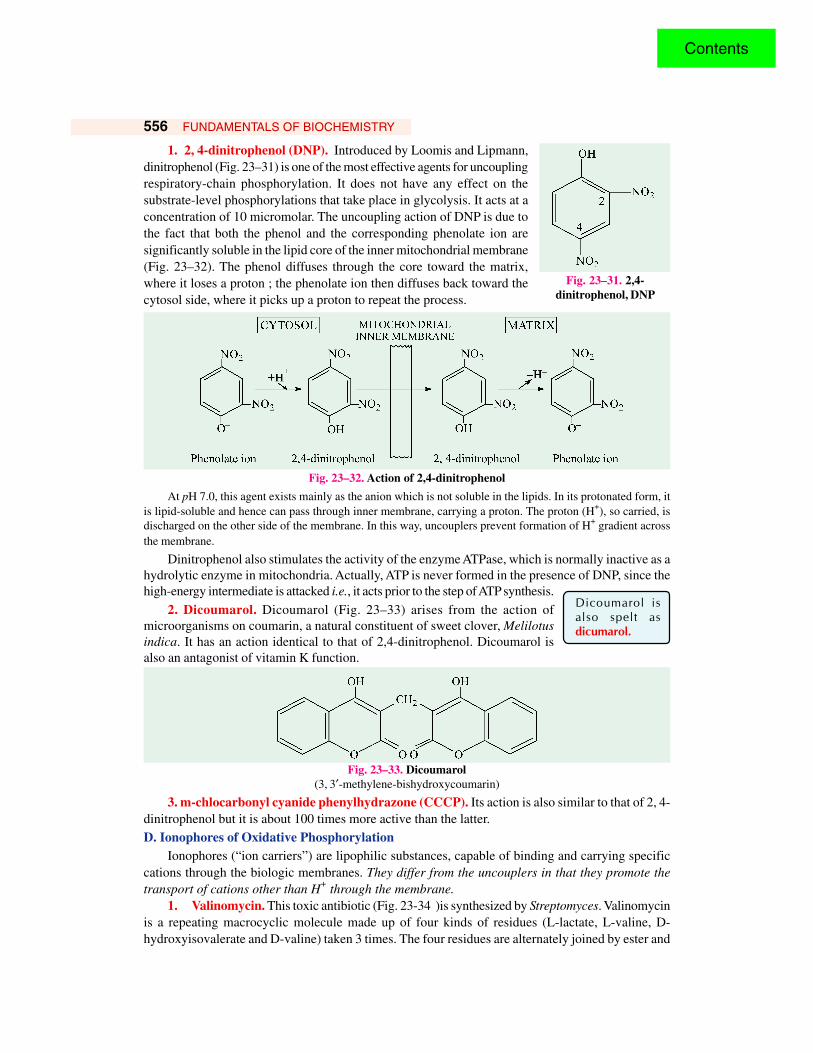
556 FUNDAMENTALS OF BIOCHEMISTRY
1. 2, 4-dinitrophenol (DNP). Introduced by Loomis and Lipmann,dinitrophenol (Fig. 23–31) is one of the most effective agents for uncouplingrespiratory-chain phosphorylation. It does not have any effect on thesubstrate-level phosphorylations that take place in glycolysis. It acts at aconcentration of 10 micromolar. The uncoupling action of DNP is due tothe fact that both the phenol and the corresponding phenolate ion aresignificantly soluble in the lipid core of the inner mitochondrial membrane(Fig. 23–32). The phenol diffuses through the core toward the matrix,where it loses a proton ; the phenolate ion then diffuses back toward thecytosol side, where it picks up a proton to repeat the process.
Fig. 23–32. Action of 2,4-dinitrophenol
At pH 7.0, this agent exists mainly as the anion which is not soluble in the lipids. In its protonated form, itis lipid-soluble and hence can pass through inner membrane, carrying a proton. The proton (H+), so carried, isdischarged on the other side of the membrane. In this way, uncouplers prevent formation of H+ gradient acrossthe membrane.
Dinitrophenol also stimulates the activity of the enzyme ATPase, which is normally inactive as ahydrolytic enzyme in mitochondria. Actually, ATP is never formed in the presence of DNP, since thehigh-energy intermediate is attacked i.e., it acts prior to the step of ATP synthesis.
2. Dicoumarol. Dicoumarol (Fig. 23–33) arises from the action ofmicroorganisms on coumarin, a natural constituent of sweet clover, Melilotusindica. It has an action identical to that of 2,4-dinitrophenol. Dicoumarol isalso an antagonist of vitamin K function.
Fig. 23–33. Dicoumarol(3, 3′-methylene-bishydroxycoumarin)
3. m-chlocarbonyl cyanide phenylhydrazone (CCCP). Its action is also similar to that of 2, 4-dinitrophenol but it is about 100 times more active than the latter.D. Ionophores of Oxidative Phosphorylation
Ionophores (“ion carriers”) are lipophilic substances, capable of binding and carrying specificcations through the biologic membranes. They differ from the uncouplers in that they promote thetransport of cations other than H+ through the membrane.
1. Valinomycin. This toxic antibiotic (Fig. 23-34 )is synthesized by Streptomyces. Valinomycinis a repeating macrocyclic molecule made up of four kinds of residues (L-lactate, L-valine, D-hydroxyisovalerate and D-valine) taken 3 times. The four residues are alternately joined by ester and
Fig. 23–31. 2,4-dinitrophenol, DNP
Dicoumarol isalso spelt asdicumarol.
Contents
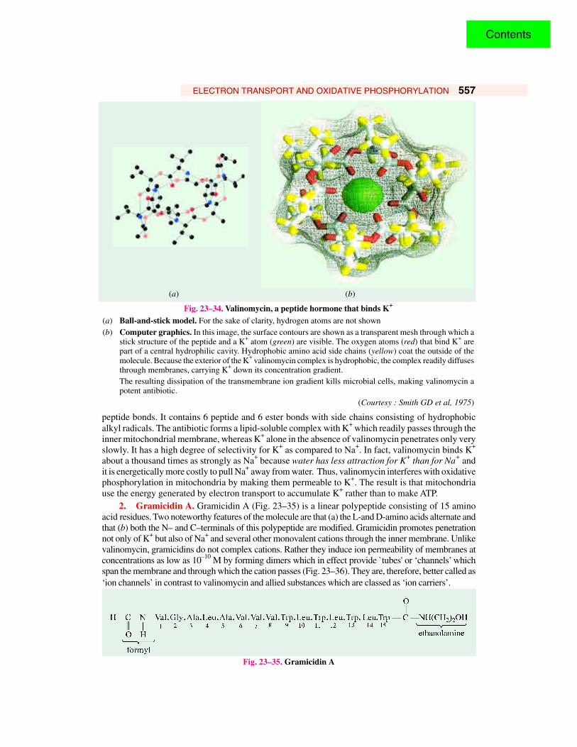
ELECTRON TRANSPORT AND OXIDATIVE PHOSPHORYLATION 557
peptide bonds. It contains 6 peptide and 6 ester bonds with side chains consisting of hydrophobicalkyl radicals. The antibiotic forms a lipid-soluble complex with K+ which readily passes through theinner mitochondrial membrane, whereas K+ alone in the absence of valinomycin penetrates only veryslowly. It has a high degree of selectivity for K+ as compared to Na+. In fact, valinomycin binds K+
about a thousand times as strongly as Na+ because water has less attraction for K+ than for Na+ andit is energetically more costly to pull Na+ away from water. Thus, valinomycin interferes with oxidativephosphorylation in mitochondria by making them permeable to K+. The result is that mitochondriause the energy generated by electron transport to accumulate K+ rather than to make ATP.
2. Gramicidin A. Gramicidin A (Fig. 23–35) is a linear polypeptide consisting of 15 aminoacid residues. Two noteworthy features of the molecule are that (a) the L-and D-amino acids alternate andthat (b) both the N– and C–terminals of this polypeptide are modified. Gramicidin promotes penetrationnot only of K+ but also of Na+ and several other monovalent cations through the inner membrane. Unlikevalinomycin, gramicidins do not complex cations. Rather they induce ion permeability of membranes atconcentrations as low as 10–10 M by forming dimers which in effect provide `tubes' or ‘channels’ whichspan the membrane and through which the cation passes (Fig. 23–36). They are, therefore, better called as‘ion channels’ in contrast to valinomycin and allied substances which are classed as ‘ion carriers’.
Fig. 23–35. Gramicidin A
(a) (b)
Fig. 23–34. Valinomycin, a peptide hormone that binds K+
(a) Ball-and-stick model. For the sake of clarity, hydrogen atoms are not shown(b) Computer graphics. In this image, the surface contours are shown as a transparent mesh through which a
stick structure of the peptide and a K+ atom (green) are visible. The oxygen atoms (red) that bind K+ arepart of a central hydrophilic cavity. Hydrophobic amino acid side chains (yellow) coat the outside of themolecule. Because the exterior of the K+ valinomycin complex is hydrophobic, the complex readily diffusesthrough membranes, carrying K+ down its concentration gradient.The resulting dissipation of the transmembrane ion gradient kills microbial cells, making valinomycin apotent antibiotic.
(Courtesy : Smith GD et al, 1975)
Contents
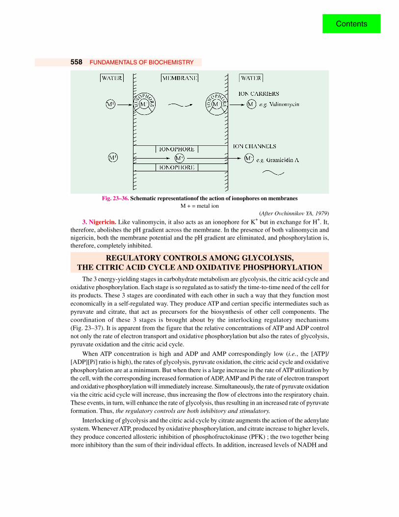
558 FUNDAMENTALS OF BIOCHEMISTRY
Fig. 23–36. Schematic representationof the action of ionophores on membranesM + = metal ion
(After Ovchinnikov YA, 1979)
3. Nigericin. Like valinomycin, it also acts as an ionophore for K+ but in exchange for H+. It,therefore, abolishes the pH gradient across the membrane. In the presence of both valinomycin andnigericin, both the membrane potential and the pH gradient are eliminated, and phosphorylation is,therefore, completely inhibited.
REGULATORY CONTROLS AMONG GLYCOLYSIS,THE CITRIC ACID CYCLE AND OXIDATIVE PHOSPHORYLATION
The 3 energy-yielding stages in carbohydrate metabolism are glycolysis, the citric acid cycle andoxidative phosphorylation. Each stage is so regulated as to satisfy the time-to-time need of the cell forits products. These 3 stages are coordinated with each other in such a way that they function mosteconomically in a self-regulated way. They produce ATP and certian specific intermediates such aspyruvate and citrate, that act as precursors for the biosynthesis of other cell components. Thecoordination of these 3 stages is brought about by the interlocking regulatory mechanisms(Fig. 23–37). It is apparent from the figure that the relative concentrations of ATP and ADP controlnot only the rate of electron transport and oxidative phosphorylation but also the rates of glycolysis,pyruvate oxidation and the citric acid cycle.
When ATP concentration is high and ADP and AMP correspondingly low (i.e., the [ATP]/[ADP][Pi] ratio is high), the rates of glycolysis, pyruvate oxidation, the citric acid cycle and oxidativephosphorylation are at a minimum. But when there is a large increase in the rate of ATP utilization bythe cell, with the corresponding increased formation of ADP, AMP and Pi the rate of electron transportand oxidative phosphorylation will immediately increase. Simultaneously, the rate of pyruvate oxidationvia the citric acid cycle will increase, thus increasing the flow of electrons into the respiratory chain.These events, in turn, will enhance the rate of glycolysis, thus resulting in an increased rate of pyruvateformation. Thus, the regulatory controls are both inhibitory and stimulatory.
Interlocking of glycolysis and the citric acid cycle by citrate augments the action of the adenylatesystem. Whenever ATP, produced by oxidative phosphorylation, and citrate increase to higher levels,they produce concerted allosteric inhibition of phosphofructokinase (PFK) ; the two together beingmore inhibitory than the sum of their individual effects. In addition, increased levels of NADH and
Contents
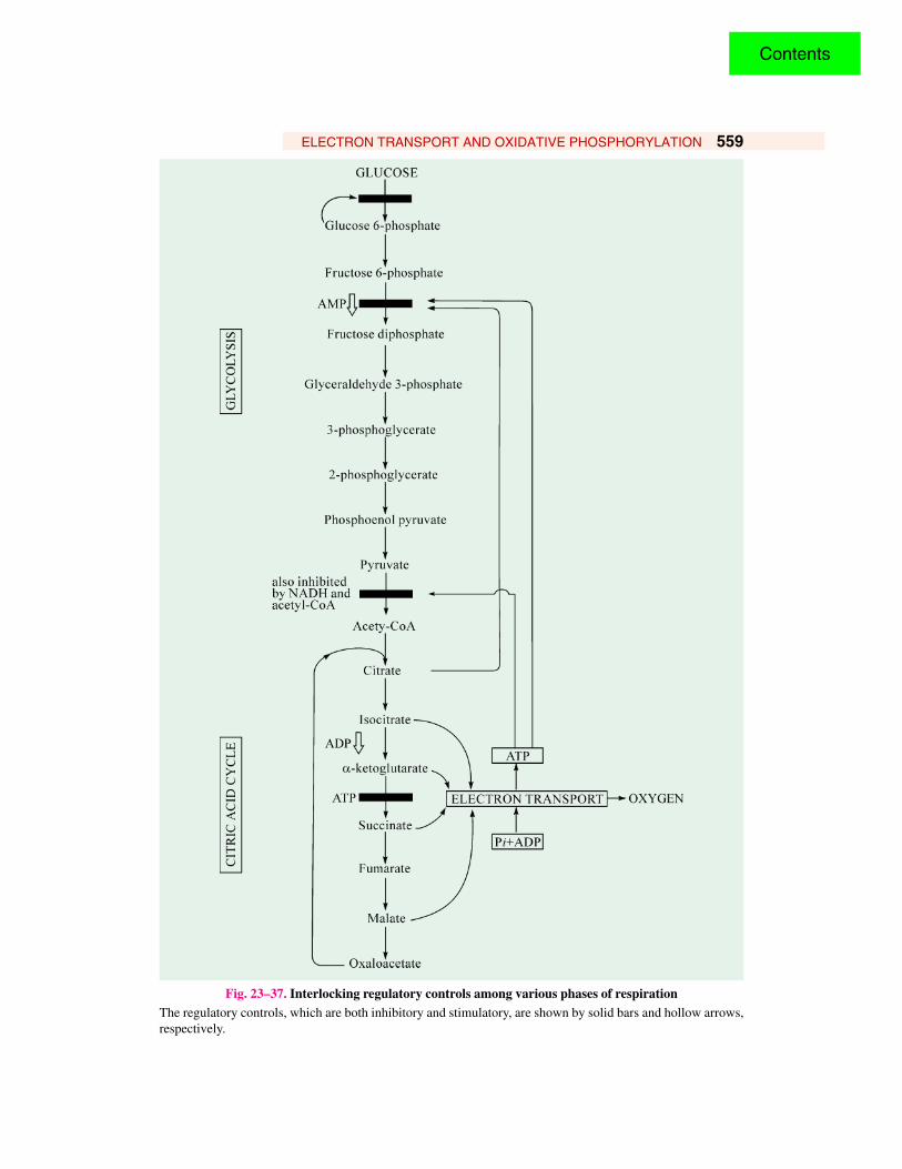
ELECTRON TRANSPORT AND OXIDATIVE PHOSPHORYLATION 559
Fig. 23–37. Interlocking regulatory controls among various phases of respirationThe regulatory controls, which are both inhibitory and stimulatory, are shown by solid bars and hollow arrows,respectively.
Contents

560 FUNDAMENTALS OF BIOCHEMISTRY
acetyl-CoA also inhibit the oxidation of pyruvate to acetyl-CoA. In nutshell, interlocking and regulatorymechanisms control glycolysis so that pyruvate is produced at a rate at which it is required by thecitric acid cycle, a process which donates electrons for the oxidative phosphorylation.
REFERENCES1. Abrahams JP, Leslie AGW, Lutter R, Walker JE : Structure at 2. 8 Å resolution of F1-
ATPase, from bovine heart mitochondria. Nature. 370 : 621-628, 1994.
2. Baltscheffsky H, Baltscheffsky M : Electron transport phosphorylation. Ann. Rev. Biochem.43 : 871, 1974.
3. Boyer PD : A perspective of the binding change mechanism for ATP synthesis. FASEB J.3 : 2164-2178, 1989.
4. Boyer PD, Chance B, Ernster L, Mitchell P, Racker E, Slater EC : Oxidativephosphorylation and photophosphorylation. Ann. Rev. Biochem. 46 : 955-1026, 1977.
5. Capaldi RA ; Structure and function of cytochrome C oxidase. Ann. Rev. Biochem. 59 :569-596, 1990.
6. Cross RL : The mechanism and regulation of ATP synthesis by F1-ATPases. Ann. Rev.Biochem. 50 : 681, 1981.
7. DePierre JW, Ernster L : Enzyme topology of intracellular membranes. Ann. Rev. Biochem.46 : 201-262, 1977.
8. Dickerson RE : Cytochrome c and the evolution of energy metabolism. Sci. Amer. 242(3) :137-153, 1980.
9. Erecinska M, Wilson DF : Cytochromeic oxidase. A synopsis. Arch. Biochem. Biophys.188 : 1, 1978.
10. Ernster L (editor) : Molecular Mechanisms in Bioenergetics. Elsevier. 1992.11. Ferguson SJ, Sorgato MC : Proton electrochemical gradients and energy-transduction
processes. Ann. Rev. Biochem. 51 : 185, 1982.12. Fillingame RH : The proton-translocating pumps of oxidation phosphorylation. Ann. Rev.
Biochem. 49 : 1079-1114, 1980.13. Fridovich I : Superoxide dismutase. Ann. Rev. Biochem. 44 : 147-159, 1975.14. Green DE, MacLennan DH : Structure and function of the mitochondrial cristael
membranes. Bioscience. 19 : 213 - 222, 1969.15. Hall DO, Rao KK, Cammack R : The iron-sulfur proteins : Structure, function and
evaluation of a ubiquitous group of proteins. Sci. Prog. 62 : 285-316, 1975.16. Harold FM : The Vital Force : A study of Bioenergetics. Academic Press, Inc., NewYork.
1986.17. Hinckle PC, McCarty RE : How cells make ATP. Sci. Amer. 238(3) : 104-123, 1978.18. Kalckar HM (editor) : Biological Phosphorylations : Development of Concepts. Prentice-
Hall, 1969.19. Kalckar HM : Fitfy years of biological research– From oxidative phosphorylation to energy
requiring transport and regulation. Ann. Rev. Biochem. 60 : 1-37. 1991.20. Keilin D : The History of Cell Respiration and Cytochromes. Cambridge Univ. Press, 1966.21. Klingenberg M : Structure-function of the ADP/ATP carrier. Biochem. Soc. Trans. 20 :
547-550, 1992.22. Krebs HA, Kornberg HL : Energy Transformation in Living Matter, A Survey. Springer,
Berlin. 1957.23. Lehninger AL : How cells transform energy. Sci. Amer. 205 (3) : 62, 1961.24. Lehninger AL : The Mitochondrion : Molecular Basis of Structure and Function. Benjamin,
New York. 1965.
Contents

ELECTRON TRANSPORT AND OXIDATIVE PHOSPHORYLATION 561
25. Lehninger AL, Wadkins CL : Oxidative phosphorylation. Ann. Rev. Biochem. 31 : 47-78,1962.
26. Lemberg R, Barrett J : Cytochromes. Academic Press Inc., New York. 1973.
27. Lovenberg W (editor) : Iron-sulfur Proteins. vol 3. Academic Press Inc., New York. 1977.
28. Malmstr´m BG : Enzymology of oxygen. Ann. Rev. Biochem. 51 : 21, 1982.
29. Martonosi AN : Membranes and Transport. Plenum, New York. 1982.
30. Massey V, Veeger C : Biological oxidations. Ann. Rev. Biochem. 32 : 579, 1963.
31. Mitchell P : Chemiosmotic Coupling and Energy Transduction. Glynn Research, Bodmin,England. 1968.
32. Mitchell P : Keilin’s respiratory chain concept and its chemiosmotic consequences. Science.206 : 1148-1159, 1979.
33. Munn EA : The Structure of Mitochondria. Academic Press Inc., New York. 1974.34. Nicholls DG, Ferguson SJ : Bioenergetics 2. Academic Press, Inc., New York. 1992.35. Parsons DS : Biological Membranes. Oxford, London. 1975.36. Pederson PL, Amzel LM : ATP synthases. Structure, reaction centre, mechanism, and
regulation of one of nature's most unique machines. J. Biol. Chem. 268 : 9937-9940, 1993.37. Racker E : Mechanisms in Bioenergetics. Academic Press Inc., New York. 1965.38. Racker E : The two faces of the inner mitochondrial membrane. Essays in Biochemistry. 6 :
1-22, 1970.39. Racker E : A New Look at Mechanisms in Bioenergetics. Academic Press Inc., New York.
1976.40. Racker E : From Pascher to Mitchell. A hundred years of Bioenergetics. Fed. Pro. 39 210-
215, 1980.41. Salemme SR : Structure and function of cytochromes c. Ann. Rev. Biochem. 46 : 299-329,
1977.42. Senior AE : ATP synthesis by oxidative phosphorylation. Physiol. Rev. 68 : 177-231, 1988.43. Siekevitz P : Powerhouse of the cell. Sci. Amer. 197(1) : 131, 1957.44. Singer TP (editor) : Flavins and Flavoproteins. Elsevier, Amsterdam. 1976.45. Singer TP (editor) : Biological Oxidations. Interscience, 1968.46. Skulachev VP : Energy transformations in the respiratory chain. Curr. Top. Bioenerg. 4 :
127-190, 1971.47. Skulachev VP : The laws of cell energetics. Eur. J. Biochem. 208 : 203-209, 1992.48. Spiro TG : Iron-Sulfur Proteins. Wiley-Interscience. 1982.49. Stayman CL (editor) : Electronic ion pumps. Curr. Top. Membr. Transp. vol. 16, 1982.50. Sund H (editor) : Pyridine Nucleotide Dependent Dehydrogenases. Springer-Verlag, New
York. 1970.51. Sweeney WV, Rabinowitz JC : Proteins containing 4Fe-4S clusters. An overview. Ann.
Rev. Biochem. 49 : 131-162, 1980.52. Tedeschi H : Mitochondria : Structure, Biogenesis and Transducing Functions. Springer-
Verlag, New York. 197653. Thomas PJ, Bianchet M, Garboczi DN, Hullihen J, Amzel LM, Pederson PL : ATP
synthase : Structure-function relationships. Biochem. Biophys. Acta. 1101 : 228-231, 1992.54. Tzagoloff A : Mitochondria. Plenum, New York. 1982.55. Von Jagow G, Sebald W : b-type cytochromes. Ann. Rev. Biochem. 49 : 281-314, 1980.56. Wainio WW : The Mammalian Mitochondrial Respiratory Chain. Academic Press Inc.,
New York. 1970.
Contents
562 FUNDAMENTALS OF BIOCHEMISTRY
57. Whittaker DA, Danks SM : Mitochondria ; Structure, Function and Assembly. Longman,London. 1978.
58. Wikström M, Krebs K, Suraste M : Proton-translocating cytochrome complexes. Rev.Biochem. 50 : 623-655, 1981.
PROBLEMS1. What is the yield of ATP when each of the following substrates is completely oxidized to
CO2 by a mammalian cell homogenate ? Assume that glycolysis, the citric acid cycle, andoxidative phosphorylation are fully active.
(a) Pyruvate (d) Phosphoenolpyruvate
(b) Lactate (e) Galactose(c) Fructose 1, 6-bisphosphate (f) Dihydroxyacetone phosphate
2. The standard oxidation–reduction potential for the reduction of O2 to H2O is given as0.82 V in Table 23-2. However, the value given in textbooks of chemistry is 1.23 V. Accountfor this difference.
3. What is the effect of each of the following inhibitors on electron transport and ATP formationby the respiratory chain ?
(a) Azide (d) DNP(b) Atractyloside (e) Carbon monoxide
(c) Rotenone (f) Antimycin A
4. The number of molecules of inorganic phosphate incorporated into organic form per atomof oxygen consumed, termed the P : O ratio, was frequently used as an index of oxidativephosphorylation.(a) What is the relation of the P : O ratio to the ratio of the number of protons translocated
per electron pair (H+/2e–) and the ratio of the number of protons needed to synthesizeATP and transport it to the cytosol (P/H+) ?
(b) What are the P : O ratios for electrons donated by matrix NADH and by succinate ?
5. The immediate administration of nitrite is a highly effective treatment for cyanide poisoning.What is the basis for the action of this antidote ? (Hint : Nitrite oxidizes ferrohemoglobin toferrihemoglobin.)
6. Suppose that the mitochondria of a patient oxidizes NADH irrespective of whether ADP ispresent. The P : O ratio for oxidative phosphorylation by these mitochondria is less thannormal. Predict the likely symptoms of this disorder.
7. Years ago, it was suggested that uncouplers would make wonderful diet drugs. Explain whythis idea was proposed and why it was rejected. Why might the producers of antiperspirantsbe supportive of the idea ?
8. You are asked to determine whether a chemical is an electron-transport-chain inhibitor or aninhibitor of ATP synthase. Design an experiment to determine this.
9. Years ago there was interest in using uncouplers such as dinitrophenol as weight controlagents. Presumably, fat could be oxidized without concomitant ATP synthesis for re-formationof fat or carbohydrate. Why was this a bad idea ?
10. The NADH dehydrogenase complex of the mitochondrial respiratory chain promotes thefollowing series of oxidation–reduction reactions, in which Fe3+ and Fe2+ represent the ironin iron–sulfur centres, UQ is ubiquinone, UQH2 is ubiquinol, and E is the enzyme :
(1) NADH + H+ + E–FMN → NAD+ + E–FMNH2
Contents
ELECTRON TRANSPORT AND OXIDATIVE PHOSPHORYLATION 563
(2) E–FMNH2 + 2Fe3+ → E–FMN + 2Fe2+ + 2H+
(3) 2Fe2+ + 2H+ + UQ → 2Fe3+ + UQH2
Sum : NADH + H+ + UQ → NAD+ + UQH2
For each of the three reactions catalyzed by the NADH dehydrogenase complex, identify(a) the electron donor, (b) the electron acceptor, (c) the conjugate redox pair, (d) the reducingagent, and (e) the oxidizing agent.
11. The standard reduction potential of any redox couple is defined for the half-cell reaction (orhalf-reaction) :
Oxidizing agent + n electrons → reducing agent
The standard reduction potentials of the NAD+/NADH and pyruvate/lactate redox pairs are– 0.320 and – 0.185 V, respectively.
(a) Which redox pair has the greater tendency to lose electrons ? Explain.(b) Which is the stronger oxidizing agent ? Explain.
(c) Beginning with 1 M concentrations of each reactant and product at pH 7, in whichdirection will the following reaction proceed ?
Pyruvate + NADH + H+ l lactate + NAD+
(d) What is the standard free-energy change, ∆G°′, at 25 °C for this reaction ?
(e) What is the equilibrium constant for this reaction at 25 °C ?
12. Electron transfer functions to translocate protons from the mitochondrial matrix to the externalmedium to establish a pH gradient across the inner membrane, the outside more acidic thanthe inside. The tendency of protons to diffuse from the outside into the matrix, where [H+]is lower, is the driving force for ATP synthesis via the ATP synthase. During oxidativephosphorylation by a suspension of mitochondria in a medium of pH 7.4, the internal pH ofthe matrix has been measured as 7.7.(a) Calculate [H+] in the external medium and in the matrix under these conditions.
(b) What is the outside : inside ratio of [H+] ? Comment on the energy inherent in thisconcentration.
(c) Calculate the number of protons in a respiring liver mitochondrion, assuming its innermatrix compartment is a sphere of diameter 15 µm.
(d) From these data would you think the pH gradient alone is sufficiently great to generateATP ?
(e) If not, can you suggest how the necessary energy for synthesis of ATP arises ?
13. ATP production in the flight muscles of the fly Lucilia sericata results almost exclusivelyfrom oxidative phosphorylation. During flight, 187 ml of O2/h • g of fly body weight isneeded to maintain an ATP concentration of 7 µmol/g of flight muscle. Assuming that theflight muscles represent 20% of the weight of the fly, calculate the rate at which the flight-muscle ATP pool turns over. How long would the reservoir of ATP last in the absence ofoxidative phosphorylation ? Assume that reducing equivalents are transferred by the glycerol-3-phosphate shuttle and that O2 is at 25 °C and 101.3 kPa (1 atm). (Note : Concentrationsare expressed in micromoles per gram of flight muscle.)
14. Iron-containing compounds that act as hydrogen acceptors in the respiratory chain are :
(a) flavoproteins
(b) dehydrogenases(c) cytochromes
(d) oxidases
Contents


![Contents€¦ · Contents 1 Molecular ... [10], and the bioartificial pancreas [11, 12]. In September of 2016, Medtronic announced FDA approval of its automated ... the endocrine](https://static.fdocument.org/doc/165x107/5f0a6dd17e708231d42b96c7/contents-contents-1-molecular-10-and-the-bioartificial-pancreas-11-12.jpg)