Conformational transitions in the γ subunit of the archaeal translation initiation factor 2
10
research papers 658 doi:10.1107/S1399004713032240 Acta Cryst. (2014). D70, 658–667 Acta Crystallographica Section D Biological Crystallography ISSN 1399-0047 Conformational transitions in the c subunit of the archaeal translation initiation factor 2 Oleg Nikonov,* Elena Stolboushkina, Valentina Arkhipova, Olesya Kravchenko, Stanislav Nikonov and Maria Garber Institute of Protein Research, Russian Academy of Sciences, Pushchino 142290, Moscow Region, Russian Federation Correspondence e-mail: [email protected] # 2014 International Union of Crystallography In eukaryotes and archaea, the heterotrimeric translation initiation factor 2 (e/aIF2) is pivotal for the delivery of methionylated initiator tRNA (Met-tRNA i ) to the ribosome. It acts as a molecular switch that cycles between inactive (GDP-bound) and active (GTP-bound) states. Recent studies show that eIF2 can also exist in a long-lived eIF2 –GDP–P i (inorganic phosphate) active state. Here, four high-resolution crystal structures of aIF2 from Sulfolobus solfataricus are reported: aIF2 –GDPCP (a nonhydrolyzable GTP analogue), aIF2 –GDP–formate (in which a formate ion possibly mimics P i ), aIF2 –GDP and nucleotide-free aIF2 . The structures describe the different states of aIF2 and demonstrate the conformational transitions that take place in the aIF2 ‘life cycle’. Received 31 August 2013 Accepted 26 November 2013 PDB references: aIF2–GDP, 4m0l; nucleotide-free aIF2, 4m2l; aIF2–GDP–formate, 4m4s; aIF2–GDPCP, 4m53 1. Introduction Heterotrimeric ( ) translation initiation factor 2 (e/aIF2) in its GTP-bound state interacts specifically with methionylated initiator tRNA (Met-tRNA i ) and contributes to its adjustment in the ribosomal P site. Both the eukaryotic translation initiation factor (eIF2) and the archaeal translation initiation factor (aIF2) have three subunits: , and . The subunit belongs to the superfamily of GTP-binding proteins, and in the case of aIF2 it is most closely related to the translation elongation factor eEF1A and its bacterial counterpart EF1A (EF-Tu), which are known to form a ternary complex with GTP and aminoacylated elongator tRNAs (Krab & Parmeg- giani, 1998). In the cell, e/aIF2 acts as a molecular switch that cycles between inactive (GDP-bound) and active (GTP-bound) states, passing through an intermediate nucleotide-free state. The crystal structures of aIF2 in nucleotide-free (Schmitt et al., 2002; Roll-Mecak et al., 2004; Sokabe et al. , 2006; Nikonov et al., 2007), GDP-bound (Schmitt et al., 2002; Sokabe et al. , 2006; Nikonov et al. , 2007; Yatime et al. , 2007) and GDPNP- bound (a nonhydrolyzable GTP analogue) states (Schmitt et al., 2002; Yatime et al. , 2006) demonstrate a conformation with a three-domain arrangement similar to that of EF-Tu in complex with GDPNP (Berchtold et al., 1993). In the case of EF-Tu, however, an active GTP-bound ON state and an inactive GDP-bound OFF state (Berchtold et al., 1993; Kjeldgaard et al., 1993; Song et al., 1999) with two different conformations, EF-Tu–GDPNP and EF-Tu–GDP, have been observed. The conformational changes involve drastic relative motions between domain I (the G domain) and domains II and III of the protein, as well as concerted motions of two flexible regions of the G domain called switch 1 and switch 2. The relative orientations of domains II and III are very similar
Transcript of Conformational transitions in the γ subunit of the archaeal translation initiation factor 2
research papers
658 doi:10.1107/S1399004713032240 Acta Cryst. (2014). D70, 658–667
Acta Crystallographica Section D
BiologicalCrystallography
ISSN 1399-0047
Conformational transitions in the c subunit of thearchaeal translation initiation factor 2
Oleg Nikonov,* Elena
Stolboushkina, Valentina
Arkhipova, Olesya Kravchenko,
Stanislav Nikonov and
Maria Garber
Institute of Protein Research, Russian Academy
of Sciences, Pushchino 142290, Moscow
Region, Russian Federation
Correspondence e-mail: [email protected]
# 2014 International Union of Crystallography
In eukaryotes and archaea, the heterotrimeric translation
initiation factor 2 (e/aIF2) is pivotal for the delivery of
methionylated initiator tRNA (Met-tRNAi) to the ribosome.
It acts as a molecular switch that cycles between inactive
(GDP-bound) and active (GTP-bound) states. Recent studies
show that eIF2 can also exist in a long-lived eIF2�–GDP–Pi
(inorganic phosphate) active state. Here, four high-resolution
crystal structures of aIF2� from Sulfolobus solfataricus are
reported: aIF2�–GDPCP (a nonhydrolyzable GTP analogue),
aIF2�–GDP–formate (in which a formate ion possibly mimics
Pi), aIF2�–GDP and nucleotide-free aIF2�. The structures
describe the different states of aIF2� and demonstrate the
conformational transitions that take place in the aIF2� ‘life
cycle’.
Received 31 August 2013
Accepted 26 November 2013
PDB references: aIF2�–GDP,
4m0l; nucleotide-free aIF2�,
4m2l; aIF2�–GDP–formate,
4m4s; aIF2�–GDPCP, 4m53
1. Introduction
Heterotrimeric (���) translation initiation factor 2 (e/aIF2) in
its GTP-bound state interacts specifically with methionylated
initiator tRNA (Met-tRNAi) and contributes to its adjustment
in the ribosomal P site. Both the eukaryotic translation
initiation factor (eIF2) and the archaeal translation initiation
factor (aIF2) have three subunits: �, � and �. The � subunit
belongs to the superfamily of GTP-binding proteins, and in
the case of aIF2� it is most closely related to the translation
elongation factor eEF1A and its bacterial counterpart EF1A
(EF-Tu), which are known to form a ternary complex with
GTP and aminoacylated elongator tRNAs (Krab & Parmeg-
giani, 1998).
In the cell, e/aIF2 acts as a molecular switch that cycles
between inactive (GDP-bound) and active (GTP-bound)
states, passing through an intermediate nucleotide-free state.
The crystal structures of aIF2� in nucleotide-free (Schmitt et
al., 2002; Roll-Mecak et al., 2004; Sokabe et al., 2006; Nikonov
et al., 2007), GDP-bound (Schmitt et al., 2002; Sokabe et al.,
2006; Nikonov et al., 2007; Yatime et al., 2007) and GDPNP-
bound (a nonhydrolyzable GTP analogue) states (Schmitt et
al., 2002; Yatime et al., 2006) demonstrate a conformation with
a three-domain arrangement similar to that of EF-Tu in
complex with GDPNP (Berchtold et al., 1993). In the case of
EF-Tu, however, an active GTP-bound ON state and an
inactive GDP-bound OFF state (Berchtold et al., 1993;
Kjeldgaard et al., 1993; Song et al., 1999) with two different
conformations, EF-Tu–GDPNP and EF-Tu–GDP, have been
observed. The conformational changes involve drastic relative
motions between domain I (the G domain) and domains II
and III of the protein, as well as concerted motions of two
flexible regions of the G domain called switch 1 and switch 2.
The relative orientations of domains II and III are very similar
in the two EF-Tu states, and these two domains can be
considered as a single structural unit. The structure of EF-Tu
in the nucleotide-free state is unknown. Domain rearrange-
ments and conformational changes of switch regions corre-
sponding to the nucleotide-free and nucleotide-bound states
of aIF2� have also been observed (Schmitt et al., 2002; Roll-
Mecak et al., 2004; Sokabe et al., 2006).
In eukaryotes, the nucleotide state of eIF2 is regulated with
the help of several initiation factors. In particular, hydrolysis
of eIF2-bound GTP is controlled by eIF5, a GTPase-activating
protein (GAP). Previously, it was thought that the action of
eIF5 was triggered by recognition of the start codon. However,
detailed biochemical experiments show that this protein is
able to promote GTP hydrolysis by eIF2 in the absence of
mRNA, producing the eIF2–GDP–Pi complex. The initiation
factor eIF1 blocks the release of inorganic phosphate (Pi) from
the ternary complex on the ribosome (Algire et al., 2005).
Upon start-codon recognition, eIF1 is displaced from the P
site of the ribosome, leading to release of Pi and dissociation
of eIF2–GDP. The tightly bound GDP must be released from
eIF2 so that eIF2 can be reactivated by the binding of GTP.
The dissociation of GDP from eIF2 is catalyzed by the
nucleotide exchange factor (GEF) eIF2B (Kyrpides & Woese,
1998). In archaea, eIF5 and eIF2B have no orthologues, and
GTP hydrolysis and the recycling of aIF2–GDP into aIF2–
GTP should be spontaneous (Schmitt et al., 2002; Pedulla et
al., 2005).
The conformational transitions that take place in aIF2 upon
switching between its active and inactive states have been
investigated using molecular-dynamics (MD) simulations
(Satpati & Simonson, 2012). The authors used the known
crystal structures of aIF2� from Sulfolobus solfataricus (Sso
aIF2�) to model the aIF2�–GTP ON state and the aIF2�–
GDP OFF state, whereas the aIF2�–GDP–Pi ON state was
modelled by breaking the bond between the � and � phos-
phates in the aIF2�–GTP structure and allowing the product
to gently relax. The MD structure of aIF2�–GDP–Pi demon-
strated some evident changes in the conformation of the
nucleotide-binding pocket and a new Mg2+-ion position close
to that of the � phosphate in the aIF2�� heterodimer (Yatime
et al., 2006). In contrast to eIF2–GDP–Pi in the eukaryotic
ternary complex (Algire et al., 2005), the archaeal post-
hydrolysis complex, aIF2�–GDP–Pi, appears to be very
unstable (Satpati & Simonson, 2012).
A structure that possibly indicates the state of Sso aIF2 one
step after GTP hydrolysis into GDP and Pi and one step
before pairing of tRNA with the start codon has been obtained
by Yatime et al. (2007). In this low-resolution (3.2 A) GDP-
bound form of Sso aIF2�3�� the conformation of the switch 2
region and the domain arrangement of aIF2� corresponded to
the typical ON conformation. No Mg2+ ion was found in the
nucleotide-binding pocket. Rather, the position expected for
an Mg2+ ion was occupied by the side chain of His37. The
conformation of switch 1 corresponded neither to the OFF
state nor to the ON state. The stability of this intermediate
state was maintained by a ligand, possibly a phosphate ion,
which interacted with both switch regions.
Here, we report four high-resolution crystal structures of
wild-type aIF2� and its mutants from S. solfataricus (Sso
aIF2�): aIF2�–GDPCP (a nonhydrolyzable GTP analogue),
aIF2�–GDP–formate (in which a formate ion possibly mimics
Pi), aIF2�–GDP and nucleotide-free aIF2�. The structures
demonstrate the conformational transitions that take place in
this protein upon nucleotide binding and switching between its
different states. In the case of aIF2�–GDPCP the mutant form
of aIF2� with deletion of the central part of switch 1 was used
for crystallization; the structure of this mutant did not differ
from that of the wild-type protein and the deletion did not
preclude aIF2�–GDPCP complex formation. It should be
emphasized that the aIF2�–GDP–formate and aIF2�–GDP
crystals were obtained by co-crystallization of aIF2� in the
presence of purified GTP, but GDP was found in the nucleo-
tide-binding pockets of both complexes. Our data demonstrate
that sodium formate at high concentrations is able to stabilize
the switch regions in positions characteristic of the ON state
of aIF2� and EF-Tu. Nucleotide-free Sso aIF2� shows a
conformation closer to the ON state, in contrast to other
known nucleotide-free aIF2� structures.
2. Materials and methods
2.1. Overproduction and purification of the mutant forms ofS. solfataricus aIF2c
In our investigations, three mutant structures were used. A
very flexible central part of switch 1 was deleted to evaluate its
role in the binding of nucleotides. Fortunately, these deletions
and substitutions in the noncanonical nucleotide-binding
pocket resulted in higher resolution diffraction data. Deletion
of the sequences corresponding to residues 41–45 and 37–47
(switch 1) and the replacement of residues Phe221/Lys225/
Arg280 of aIF2� by alanines in aIF2� were achieved by site-
directed mutagenesis of pET-11d-� using the QuikChange
method (Stratagene). The mutant proteins were overproduced
and purified as described previously (Nikonov et al., 2007).
2.2. GTP purification
5 mg GTP (Sigma) dissolved in 500 ml buffer (20 mM Tris–
HCl pH 7.5) was loaded onto a Mono Q column (1 ml)
equilibrated with the same buffer. The column was washed
with 10 ml of the same buffer, and a 30 ml linear gradient of
NaCl concentration (0–1 M) in the same buffer was used for
elution of the nucleotide. The optical density of fractions was
estimated at 220, 260 and 280 nm. Peak fractions were tested
by thin-layer ion-exchange chromatography on plates with
PEI-cellulose (Merck) in 0.1 M Tris–HCl pH 8.0 with 0.5 M
LiCl in a glass camera during 15 min. Pure GTP and GDP
were used as markers. Spots were detected using a UV lamp.
2.3. Crystallization of the mutant forms of Sso aIF2c with andwithout nucleotides
Crystallization trials were performed at 28�C by the
hanging-drop vapour-diffusion technique using mutant Sso
aIF2� protein at a concentration of 50 mg ml�1 in buffer
research papers
Acta Cryst. (2014). D70, 658–667 Nikonov et al. � Archaeal translation initiation factor 2 659
consisting of 50 mM Tris–HCl pH 7.5, 400 mM NaCl, 10 mM
�-mercaptoethanol. The protein was mixed with reservoir
solution in equal amounts. A reservoir solution consisting of
100 mM sodium cacodylate pH 6.5, 4 M sodium formate was
used for the crystallization of Sso aIF2� (�37–47) with
GDPCP and of Sso aIF2� (F221A/K225A/R280A) with GTP.
Commercial preparations of nucleotides (GDPCP from Jena
Bioscience and additionally purified GTP from Sigma) were
added to the drop to a final concentration about 5 mM.
Crystals of Sso aIF2�(�41–45) in a nucleotide-free form were
obtained using a reservoir solution consisting of 50 mM Na
HEPES pH 7.0, 50 mM MgSO4, 1.6 M Li2SO4. Cryoprotection
of the crystals was achieved by adding ethylene glycol to the
reservoir solution to a final concentration of 15%(v/v).
2.4. Crystallization of wild-type Sso aIF2c in complex withGDP
Crystallization trials were performed at 22�C by the
hanging-drop diffusion technique using Sso aIF2� protein at
a concentration of 25 mg ml�1 in a buffer consisting of 10 mM
Tris–HCl pH 7.5, 100 mM NaCl, 100 mM KCl, 10 mM MgCl2,
10 mM �-mercaptoethanol. The protein solution (3 ml) was
mixed with 0.5 ml 6 mM purified GTP pH 7.5 and 2 ml reservoir
solution consisting of 100 mM Tris–HCl pH 7.5, 180 mM KBr,
13% PEG 4000 (Clear Strategy Screen 1 solution No. 10).
Crystals of Sso aIF2�–GDP were obtained within 7 d. Cryo-
protection of the crystals was achieved by adding ethylene
glycol to the reservoir solution to a final concentration of
15%(v/v).
2.5. Data collection, structure determination and refinement
X-ray diffraction data were collected using synchrotron
radiation on the BL14.1 HZB beamline at BESSY, Berlin,
Germany with an MX-225 CCD detector (Rayonics, Evan-
ston, USA) and using a rotating-anode radiation source
(Bruker AXS MICROSTAR) with a CCD detector (Bruker
PLATINUM 135) at RAS, Pushchino, Russia.
The data were processed and merged with the XDS
program suite (Kabsch, 1993) and the PROTEUMplus software
package (Bruker AXS). A molecular-replacement solution
was obtained with Phaser (Storoni et al., 2004). To find unique
positions of the � subunits in the aIF2�–GDPCP and aIF2�–
GDP–formate structures, the previously determined structure
of aIF2�–GDP (PDB entry 2pmd; Nikonov et al., 2007) was
used as a search model. Nucleotides, ions and water were
excluded from the model. The crystal of the aIF2�–GDP
complex belonged to space group P21 and contained six
molecules in the asymmetric unit. This fact, together with the
high flexibility of the molecule, led to difficulties in finding a
search model. Only the G subunit of one of the four copies of
the aIF2 trimer (PDB entry 3cw2; Stolboushkina et al., 2008)
could be used. The structure of nucleotide-free aIF2� was
solved using the nucleotide-free form of aIF2 from Pyrococcus
research papers
660 Nikonov et al. � Archaeal translation initiation factor 2 Acta Cryst. (2014). D70, 658–667
Table 1Data-collection and refinement statistics.
Values in parentheses are for the highest resolution shell.
Structure aIF2�–GDPCP aIF2�–GDP–formate aIF2�–GDP aIF2�
Data collectionSource BESSY BL14.1 BESSY BL14.1 Bruker AXS MICROSTAR BESSY BL14.1Wavelength (A) 0.91841 0.91841 1.54179 0.91841Space group I23 R32 P21 P3121Unit-cell parameters
a (A) 186.89 142.86 86.05 95.81b (A) 186.89 142.86 106.55 95.81c (A) 186.89 218.41 156.30 165.24� (�) 90.00 90.00 90.00 90.00� (�) 90.00 90.00 90.63 90.00� (�) 90.00 120.00 90.00 120.00
Resolution (A) 19.70–2.00 (2.10–2.00) 45.73–2.25 (2.35–2.25) 19.81–2.60 (2.70–2.60) 47.91–2.15 (2.25–2.15)Observed reflections 365256 (48649) 452939 (54385) 284650 (25550) 284714 (35794)Unique reflections 72719 (9781) 40715 (4843) 84101 (8374) 48518 (6088)Completeness (%) 99.6 (98.6) 99.8 (99.0) 96.7 (90.5) 99.9 (99.7)Multiplicity 5.01 (4.90) 11.10 (11.12) 3.27 (2.76) 5.86 (5.87)hI/�(I)i 12.77 (2.73) 19.35 (4.47) 14.29 (1.93) 18.01 (3.83)Rmerge (%) 10.76 (49.07) 8.41 (42.12) 5.60 (44.40) 5.68 (37.40)
RefinementResolution range (A) 19.70–2.00 (2.03–2.00) 45.73–2.25 (2.31–2.25) 19.81–2.60 (2.63–2.60) 47.91–2.15 (2.19–2.15)Rwork/Rfree† (%) 16.00/18.45 (25.09/29.33) 15.20/18.20 (22.04/27.67) 23.40/28.60 (33.76/37.58) 18.80/22.80 (25.75/31.12)No. of atoms
Protein 3221 3250 18901 3080Ligands 147 121 180 148Water 654 437 363 255
R.m.s.d.Bond lengths (A) 0.006 0.006 0.005 0.007Bond angles (�) 1.143 1.075 0.965 1.127
B factor (A2) 19.97 36.94 54.60 42.54
† Rfree factors were calculated for randomly selected test sets of 5.0% of the reflections that were not used in the refinement.
abyssi (PDB entry 1kk0; Schmitt et al., 2002)
as a search model.
The models were subjected to several
rounds of computational refinement and
map calculation with PHENIX (Adams et
al., 2002) and manual model inspection and
modification with Coot (Emsley et al., 2010).
The data-collection and refinement statistics
are summarized in Table 1. The model bias
existing in the initial molecular-replacement
solutions was tackled using composite
OMIT cross-validated �A-weighted maps as
implemented in CNS (Brunger et al., 1998).
3. Results
3.1. Overall description of the structures
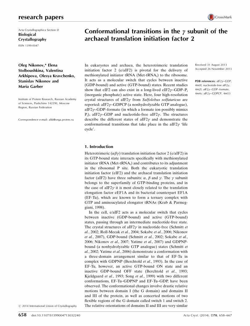
3.1.1. The aIF2c–GDPCP complex. In all
known structures of aIF2� the switch 2
region can adopt OFF or ON conformations
only. In contrast, the central part of the
switch 1 region displays irregular confor-
mations that are presumably influenced by
crystal packing and not by the nature of the
bound nucleotide (Nikonov et al., 2007;
Yatime et al., 2007; Stolboushkina et al.,
2008). Because switch 1 showed a confor-
mation in the structure of the aIF2��heterodimer from S. solfataricus bound to
GDPNP that was similar to that of EF-Tu–
GDPNP, it was suggested that this switch
participates in the binding of GTP (Yatime
et al., 2006). However, this function of switch
1 was not evident. Therefore, we deleted the
central part of switch 1 (amino-acid residues
37–47) in aIF2� and cocrystallized this
mutant with GDPCP. Additionally, these
crystals demonstrate a higher resolution
in comparison with the aIF2��–GDPNP
complex (Yatime et al., 2006).
The structure of aIF2�–GDPCP from
S. solfataricus (subsequently indicated as
structure S1) has been solved at 2.0 A
resolution. A stereoview of the structure is
shown in Fig. 1 and the amino-acid sequence
of the � subunit is shown in Supplementary
Fig. S11. The mutant structure clearly
demonstrates the presence of GDPCP–Mg2+
in the nucleotide-binding pocket and a lack
of significant interactions (apart from a
hydrogen bond that is accessible to the solvent formed by the
NZ atom of Lys48 and the O3G atom of GDPCP) betweentruncated switch 1 and GDPCP. This structure and the struc-
ture of aIF2��–GDPNP previously determined at 3.0 A
resolution (Yatime et al., 2006) are closely similar and can be
superimposed with an r.m.s.d. of 0.610 A for 94% compared
C� atoms. The positions of domains II and III relative to
domain G are identical in both structures.
research papers
Acta Cryst. (2014). D70, 658–667 Nikonov et al. � Archaeal translation initiation factor 2 661
Figure 1A stereoview of S. solfataricus aIF2�–GDPCP. The �L1 loop, P loop and the switch 1 andswitch 2 regions are shown in red, yellow, magenta and green, respectively. The Mg2+ ion isshown as an orange ball. The GDPCP molecules are shown in cyan. Domains are numberedwith roman numerals.
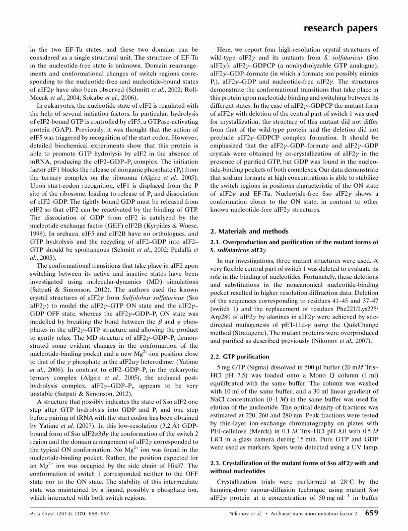
Figure 2The nucleotide-binding pocket of the aIF2�–GDPCP complex. The side chains of Asp152 andSer184 mimic the interactions characteristic of a canonical G–C pair. The P loop (residues 19–23) forms a bed for the �-phosphate and �-phosphate groups. The O3G �-phosphate O atomforms hydrogen bonds to the main-chain N atom of Gly96 and the NZ atom of Lys22. The C3B�–� bridge atom (O3B in GTP) forms a hydrogen bond to the main-chain N atom of Asp19.The Mg2+ ion is coordinated by the O2B and O2G atoms of GDPCP, the OG1 atom of Thr23,the NZ atom of Lys48 and two water molecules. Water molecule A618 occupies a position closeto that required for nucleophilic attack.
1 Supporting information has been deposited in the IUCr electronic archive(Reference: MN5043).
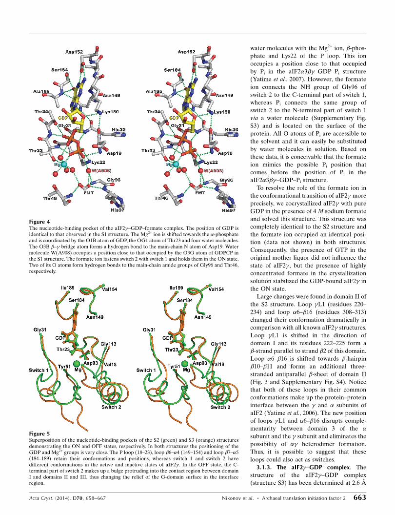
The nucleotide-binding pocket of aIF2� is built up from the
P loop (residues 18–23), the switch 2 region (residues 93–113),
loop �6–�4 (residues 149–152) and loop �7–�5 (residues 185–
187) (Fig. 2 and Supplementary Fig. S1). The two latter regions
surround the base of the nucleotide. The side chains of Asp152
and Ser184 mimic the interactions characteristic of a canonic
G–C pair. These interactions are conserved in all known
G-protein structures and ensure the specificity for guanine
nucleotides (Simonetti et al., 2013).
It is interesting to note that except for the switch 1 region,
superposition of C� atoms of the nucleotide-binding pockets
of aIF2� and Thermus thermophilus EF-Tu (Tth EF-Tu;
Berchtold et al., 1993), both in the ON state, produces an r.m.s.
deviation of 0.498 A, thus demonstrating the high stability
of this region in the superfamily of ribosomal GTP-binding
proteins. The positions of GDPCP, GDPNP and Mg2+ are very
similar in these aIF2� and EF-Tu structures. Moreover, a
hydrogen bond that is crucial for GTP hydrolysis by elonga-
tion factor EF-Tu between the main-chain amide N atom of
Gly96 (Sso aIF2 numbering) and the �-phosphate (Knudsen
et al., 2001) is found in the S1 structure (Fig. 2) and in the
structure of aIF2��–GDPNP (Yatime et al., 2006). The iden-
tity of interactions between the protein and nucleotide in the
aIF2� structure with a truncated switch 1 region, the wild-type
(Yatime et al., 2006) protein and EF-Tu (Berchtold et al., 1993)
shows that at least the central part of the switch 1 region of the
� subunit (amino-acid residues 37–47) does not take part in
the binding of the GTP analogues.
Similar to the previously determined aIF2�–GDP complex
(Nikonov et al., 2007), the second GDPCP molecule was also
found in the noncanonical nucleotide-binding pocket of the S1
structure. In both structures, the locations of the bases and
riboses of the extra G nucleotides were identical, whereas the
locations of the phosphate moieties were different. Super-
position of domain II of the S1 structure on that of the ternary
complex (Stolboushkina et al., 2013) showed that the non-
canonical nucleotide-binding pocket possibly corresponds to
the positioning of the G2 nucleotide base of the initiator
tRNA.
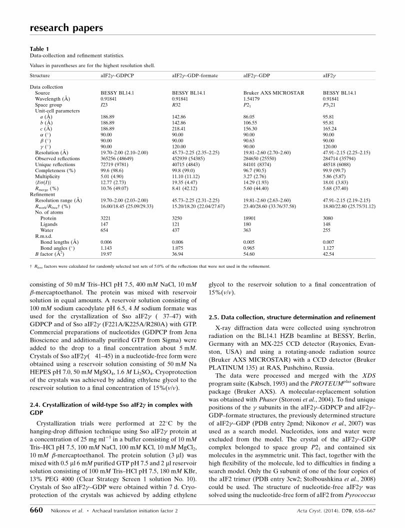
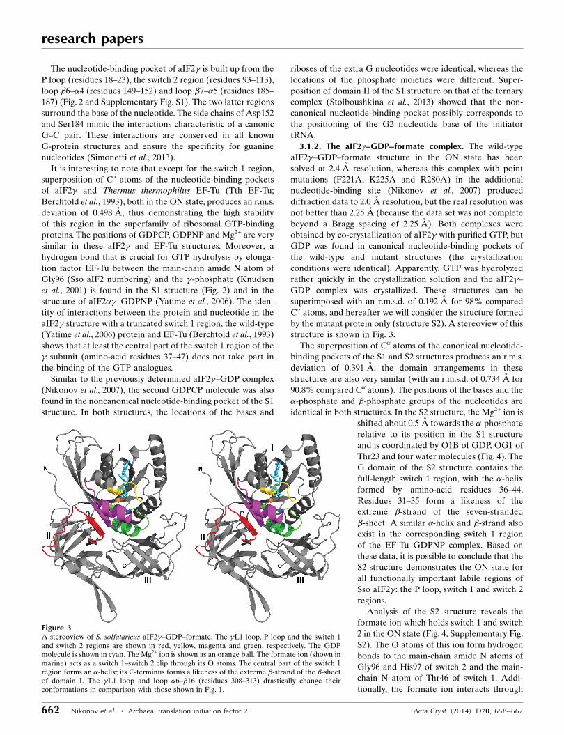
3.1.2. The aIF2c–GDP–formate complex. The wild-type
aIF2�–GDP–formate structure in the ON state has been
solved at 2.4 A resolution, whereas this complex with point
mutations (F221A, K225A and R280A) in the additional
nucleotide-binding site (Nikonov et al., 2007) produced
diffraction data to 2.0 A resolution, but the real resolution was
not better than 2.25 A (because the data set was not complete
beyond a Bragg spacing of 2.25 A). Both complexes were
obtained by co-crystallization of aIF2� with purified GTP, but
GDP was found in canonical nucleotide-binding pockets of
the wild-type and mutant structures (the crystallization
conditions were identical). Apparently, GTP was hydrolyzed
rather quickly in the crystallization solution and the aIF2�–
GDP complex was crystallized. These structures can be
superimposed with an r.m.s.d. of 0.192 A for 98% compared
C� atoms, and hereafter we will consider the structure formed
by the mutant protein only (structure S2). A stereoview of this
structure is shown in Fig. 3.
The superposition of C� atoms of the canonical nucleotide-
binding pockets of the S1 and S2 structures produces an r.m.s.
deviation of 0.391 A; the domain arrangements in these
structures are also very similar (with an r.m.s.d. of 0.734 A for
90.8% compared C� atoms). The positions of the bases and the
�-phosphate and �-phosphate groups of the nucleotides are
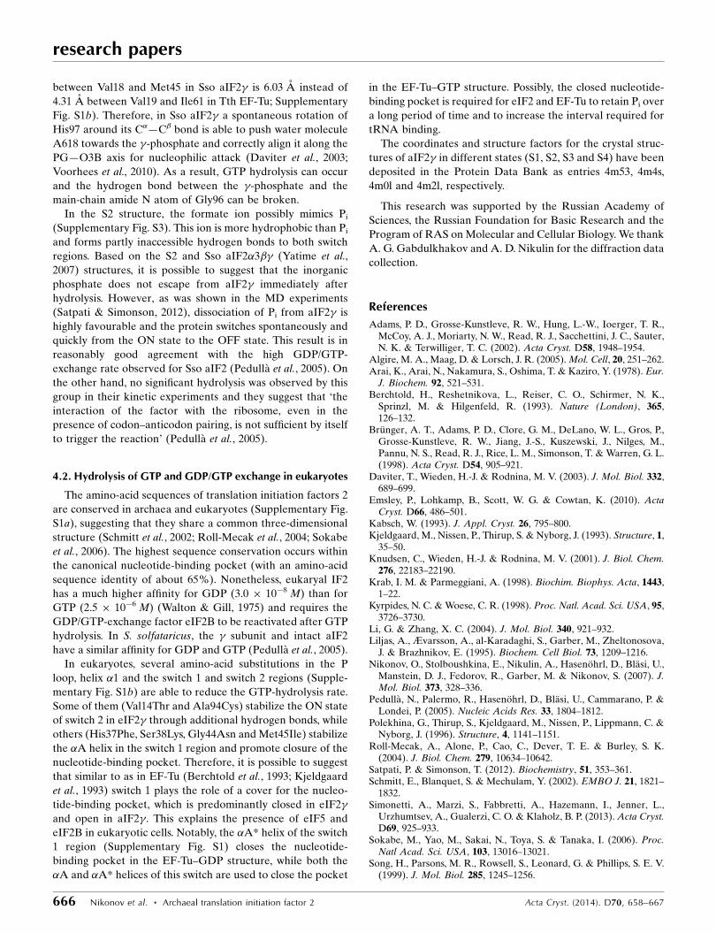
identical in both structures. In the S2 structure, the Mg2+ ion is
shifted about 0.5 A towards the �-phosphate
relative to its position in the S1 structure
and is coordinated by O1B of GDP, OG1 of
Thr23 and four water molecules (Fig. 4). The
G domain of the S2 structure contains the
full-length switch 1 region, with the �-helix
formed by amino-acid residues 36–44.
Residues 31–35 form a likeness of the
extreme �-strand of the seven-stranded
�-sheet. A similar �-helix and �-strand also
exist in the corresponding switch 1 region
of the EF-Tu–GDPNP complex. Based on
these data, it is possible to conclude that the
S2 structure demonstrates the ON state for
all functionally important labile regions of
Sso aIF2�: the P loop, switch 1 and switch 2
regions.
Analysis of the S2 structure reveals the
formate ion which holds switch 1 and switch
2 in the ON state (Fig. 4, Supplementary Fig.
S2). The O atoms of this ion form hydrogen
bonds to the main-chain amide N atoms of
Gly96 and His97 of switch 2 and the main-
chain N atom of Thr46 of switch 1. Addi-
tionally, the formate ion interacts through
research papers
662 Nikonov et al. � Archaeal translation initiation factor 2 Acta Cryst. (2014). D70, 658–667
Figure 3A stereoview of S. solfataricus aIF2�–GDP–formate. The �L1 loop, P loop and the switch 1and switch 2 regions are shown in red, yellow, magenta and green, respectively. The GDPmolecule is shown in cyan. The Mg2+ ion is shown as an orange ball. The formate ion (shown inmarine) acts as a switch 1–switch 2 clip through its O atoms. The central part of the switch 1region forms an �-helix; its C-terminus forms a likeness of the extreme �-strand of the �-sheetof domain I. The �L1 loop and loop �6–�16 (residues 308–313) drastically change theirconformations in comparison with those shown in Fig. 1.
water molecules with the Mg2+ ion, �-phos-
phate and Lys22 of the P loop. This ion
occupies a position close to that occupied
by Pi in the aIF2�3��–GDP–Pi structure
(Yatime et al., 2007). However, the formate
ion connects the NH group of Gly96 of
switch 2 to the C-terminal part of switch 1,
whereas Pi connects the same group of
switch 2 to the N-terminal part of switch 1
via a water molecule (Supplementary Fig.
S3) and is located on the surface of the
protein. All O atoms of Pi are accessible to
the solvent and it can easily be substituted
by water molecules in solution. Based on
these data, it is conceivable that the formate
ion mimics the possible Pi position that
comes before the position of Pi in the
aIF2�3��–GDP–Pi structure.
To resolve the role of the formate ion in
the conformational transition of aIF2� more
precisely, we cocrystallized aIF2� with pure
GDP in the presence of 4 M sodium formate
and solved this structure. This structure was
completely identical to the S2 structure and
the formate ion occupied an identical posi-
tion (data not shown) in both structures.
Consequently, the presence of GTP in the
original mother liquor did not influence the
state of aIF2�, but the presence of highly
concentrated formate in the crystallization
solution stabilized the GDP-bound aIF2� in
the ON state.
Large changes were found in domain II of
the S2 structure. Loop �L1 (residues 220–
234) and loop �6–�16 (residues 308–313)
changed their conformation dramatically in
comparison with all known aIF2� structures.
Loop �L1 is shifted in the direction of
domain I and its residues 222–225 form a
�-strand parallel to strand �2 of this domain.
Loop �6–�16 is shifted towards �-hairpin
�10–�11 and forms an additional three-
stranded antiparallel �-sheet of domain II
(Fig. 3 and Supplementary Fig. S4). Notice
that both of these loops in their common
conformations make up the protein–protein
interface between the � and � subunits of
aIF2 (Yatime et al., 2006). The new position
of loops �L1 and �6–�16 disrupts comple-
mentarity between domain 3 of the �subunit and the � subunit and eliminates the
possibility of �� heterodimer formation.
Thus, it is possible to suggest that these
loops could also act as switches.
3.1.3. The aIF2c–GDP complex. The
structure of the aIF2�–GDP complex
(structure S3) has been determined at 2.6 A
Acta Cryst. (2014). D70, 658–667 Nikonov et al. � Archaeal translation initiation factor 2 663
Figure 5Superposition of the nucleotide-binding pockets of the S2 (green) and S3 (orange) structuresdemonstrating the ON and OFF states, respectively. In both structures the positioning of theGDP and Mg2+ groups is very close. The P loop (18–23), loop �6–�4 (149–154) and loop �7–�5(184–189) retain their conformations and positions, whereas switch 1 and switch 2 havedifferent conformations in the active and inactive states of aIF2�. In the OFF state, the C-terminal part of switch 2 makes up a bulge protruding into the contact region between domainI and domains II and III, thus changing the relief of the G-domain surface in the interfaceregion.
Figure 4The nucleotide-binding pocket of the aIF2�–GDP–formate complex. The position of GDP isidentical to that observed in the S1 structure. The Mg2+ ion is shifted towards the �-phosphateand is coordinated by the O1B atom of GDP, the OG1 atom of Thr23 and four water molecules.The O3B �–� bridge atom forms a hydrogen bond to the main-chain N atom of Asp19. Watermolecule W(A908) occupies a position close to that occupied by the O3G atom of GDPCP inthe S1 structure. The formate ion fastens switch 2 with switch 1 and holds them in the ON state.Two of its O atoms form hydrogen bonds to the main-chain amide groups of Gly96 and Thr46,respectively.

resolution. The asymmetric unit of the crystal contains six
molecules with slightly different positions of small helices and
short �-strands in their long loops. The molecules in the
asymmetric unit of the crystal are packed into two trimers
which are related by a noncrystallographic twofold translation
axis parallel to the b axis of the cell. The switch 1 region and
the �L1 loop are both positioned within the inner hole of the
trimer and have different conformations in the different
monomers. The central parts of these loops (residues 37–44
and 228–230) show poor electron density in all six molecules.
After exclusion of the switch 1 region and the �L1 loop,
pairwise superposition of all of the monomers yielded an r.m.s.
deviation for the remaining 378 C� atoms of around 0.7 A.
This shows that the relative orientation of the domains in all of
the complexes is similar.
Similar to the S2 structure, the crystals of this complex were
obtained by co-crystallization of aIF2� with purified GTP,
but GDP–Mg2+ was found in the canonical nucleotide-binding
pockets of all six molecules. Thus, similar to the previous case,
GTP could be spontaneously hydrolyzed in solution during
crystallization. The N-terminal parts of the switch 2 regions
in all six aIF2�–GDP molecules in the asymmetric unit of the
crystal adopt a helical conformation (Fig. 5).
The C-terminal residues 107–109 of this
switch makes up a bulge that protrudes from
the core of the G domain into the space
between this domain and domain III,
forming an interdomain parallel �-sheet
with loop �17–�18 of domain III and initi-
ating the new domain arrangement.
Monomer A of the S3 structure and aIF2�of the previously determined structure of
aIF2�3�1�–GDP (Yatime et al., 2007) in the
OFF state are closely similar and can be
superimposed with an r.m.s.d. of 0.843 A for
86% compared C� atoms. A similar result
was obtained on superposition of monomer
A of the S3 structure and the aIF2�–GDP
complex from P. abyssi (Pab aIF2�) solved
at 1.9 A resolution (Schmitt et al., 2002). In
contrast, the superposition of this monomer
and aIF2� in the ON state from the aIF2��heterodimer (Yatime et al., 2006) produced
an r.m.s. deviation of 1.83 A for the same set
of C� atoms. These results unambiguously
demonstrate the OFF state of the S3 struc-
ture.
The comparison of the aIF2� structures in
the OFF and ON states shows a clearly
visible displacement of domains II and III
relative to domain G; these domains are
rotated by about 10–15� around an axis
approximately coincident with a line
connecting the C� atoms of residues 292–293
of domain II (Fig. 6). This confirms that Sso
aIF2 acts similarly to other GTPases
(Berchtold et al., 1993; Song et al., 1999;
Vetter & Wittinghofer, 2001; Liljas et al.,
1995); aIF2� also has a different arrange-
ment of domains II and III relative to
domain G in the ON and OFF states.
However, the domain reorganization is not
as dramatic as that observed in the EF-Tu–
GDPNP and EF-Tu–GDP structures.
At the same time, the switch 1 regions
of either of the two copies of aIF2�–GDP
can be superimposed on each other with an
r.m.s. deviation within the range 2.3–4.1 A.
research papers
664 Nikonov et al. � Archaeal translation initiation factor 2 Acta Cryst. (2014). D70, 658–667
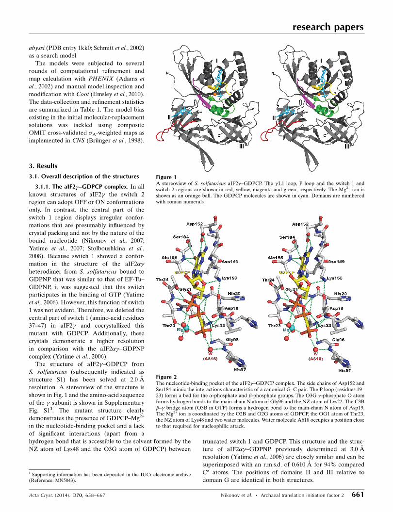
Figure 7Stereoview of S. solfataricus aIF2�. The �L1 loop, P loop and the switch 1 and switch 2 regionsare shown in red, yellow, magenta and green, respectively. The P loop and the N-terminal partof switch 2 change their conformations (residues 96–98 of switch 2 are not visible in thestructure). The conformation of switch 2 corresponds to the ON state.
Figure 6Domain displacement in aIF2� during transition from the active to the inactive state. The Gdomains of the S2 and S3 structures are superimposed. Domains are numbered with romannumerals and shown in dark and light grey for the OFF and ON states, respectively. In the OFFstate, switch 2 and loop 334–353 are shown in orange. The same elements in the ON state areshown in blue. Upon transition from the OFF state to the ON state, domains II and III rotateabout an axis approximately coincident with a line connecting the C� atoms of residues 292–293 (magenta spheres) approaching domain G.

Only small parts of switch 1, which include amino-acid resi-
dues 31–34 and 49–51, appear to retain their conformation in
the different monomers. Comparison with the conformation of
switch 1 regions in other known aIF2� structures indicates that
this region generally retains its mobility in all observed states
of aIF2� and may adopt any conformation dictated by crystal
packing or other factors, independently of the type of
nucleotide bound. Moreover, its central part plays no role in
either GTP or GDP binding.
3.1.4. Nucleotide-free aIF2c. Upon going from the GDP-
bound to the GTP-bound form, the GTPase passes through
the nucleotide-free form. This form is unknown for EF-Tu.
However, several structures of nucleotide-free aIF2� from
different organisms are available (Schmitt et al., 2002; Roll-
Mecak et al., 2004; Sokabe et al., 2006). Some years ago, the
structure of free aIF2� from S. solfataricus was solved by our
group at 2.9 A resolution (Nikonov et al., 2007). The confor-
mation and location of the switch 2 region in this structure
dramatically differed from that observed in all other nucleo-
tide-free aIF2� structures, which demonstrated the classical
OFF form. To prove that this was not an artifact, the structure
of a mutant Sso aIF2� (structure S4) was solved in a nucleo-
tide-free form (Fig. 7). In this structure, the deletion of amino-
acid residues 41–45 resulted in higher resolution (2.15 A)
diffraction data. The two nucleotide-free Sso aIF2� forms
were closely similar and could be superimposed with an
r.m.s.d. of 0.512 A for 87% compared C� atoms. The domain
arrangement and location of the N- and C-terminal parts of
switch 1 (residues 31–34 and 49–51, respectively) were iden-
tical in both structures. Differences were only found in the N-
terminus of the P loop (residues 18–20) and the switch 2 region
(residues 93–101). Residues 96–98 were not visible in the S4
structure. In the P loop, the conformational changes emerged
as a result of the disruption of two hydrogen bonds from the
main-chain O atoms of Asp19 and His20 to the NZ atom of
Lys150 and the ND atom of Asn149, respectively, which
stabilized the P loop in the nucleotide-bound forms. The OFF
conformation of switch 2 is stabilized by hydrogen bonds
formed by the main-chain O atoms of Ala94 and Pro95 to
water molecules coordinating the Mg2+ ion, whereas the ON
conformation of this switch is stabilized by the hydrogen bond
formed by the main-chain O atom of Gly96 to the O atom of
the �-phosphate. All of these hydrogen bonds are nucleotide-
dependent.
In contrast to the nucleotide-free forms of aIF2� from
P. abyssi (Schmitt et al., 2002), Methanocaldococcus jannaschii
(Roll-Mecak et al., 2004) and P. furiosus (Sokabe et al., 2006),
in the nucleotide-free forms of Sso aIF2� the position of
the switch 2 helix is shifted from spanning residues 97–107
corresponding to the GDP-bound form to residues 100–110
corresponding to the GTP-bound form. The superposition of
domain I of the S4 structure on those of the S1 and S3
structures shows that in the nucleotide-free form of aIF2�domains II and III also demonstrate a position relative to
domain I which is closer to the ON state (Supplementary Fig.
S5). In summary, we can say that the nucleotide-free form of
Sso aIF2� is probably preferred to the binding of GTP. This
possibly explains the similar affinity for GTP and GDP
obtained in Sso aIF2 activity assays (Pedulla et al., 2005).
4. Discussion
4.1. Hydrolysis of GTP and GDP/GTP exchange in archaea
A comparison of the canonical nucleotide-binding pockets
of the S1, S2 and S3 complexes shows that with the exception
of the switch regions they have very stable conformations
which are identical in both the active and inactive states of
aIF2�. Identical structures of the pocket are observed in the
EF-Tu–GDPNP (Berchtold et al., 1993) and EF-Tu–GDP
complexes (Polekhina et al., 1996; Song et al., 1999). A
comparison (Supplementary Fig. S6) shows that the base,
ribose and �- and �-phosphates of G nucleotides occupy the
same positions in aIF2� and EF-Tu in both the ON and OFF
states. The Mg2+ ion can move slightly, but its displacement is
not more than 1.0 A. Previously, there was reason to believe
that the nucleotide-binding pocket of e/aIF2� is distinct from
that in the G domain of monomeric bacterial IF2 (Kyrpides &
Woese, 1998). However, the crystal structure of the protein
core of translation initiation factor 2 from T. thermophilus
in the nucleotide-free, GTP-bound and GDP-bound forms
(Simonetti et al., 2013) showed that the nucleotide-binding
pocket of IF2 is identical to that of aIF2� and other ribosomal
GTPases.
At the same time, it is known that guanine nucleotides bind
with a dissociation constant of 10�8–10�9 M to EF-Tu from
T. thermophilus (Arai et al., 1978), while the � subunit and
intact aIF2 from S. solfataricus have a more limited affinity for
GDP and GTP (�4 � 10�7 M; Pedulla et al., 2005). Moreover,
in in vitro experiments at the optimal growth temperature for
S. solfataricus aIF2, radiolabelled GDP can be replaced
by either GDP or GTP at a comparable rate and nucleotide
exchange was complete within 4 min (Pedulla et al., 2005). To
account for this phenomenon, we compared the conforma-
tions of the switch 1 regions in the S2 and EF-Tu structures. It
turned out that the nucleotide-binding pocket of aIF2� is open
and accessible to the solvent, in contrast to the pocket of
EF-Tu, which is closed by Tyr47 and Ile61 (Trp33 and Met45 in
Sso aIF2�).
The catalysis of GTP hydrolysis in ribosomal GTPases
requires an invariant histidine (His97 in Sso aIF2� and His85
in EF-Tu from T. thermophilus) that acts as a general base,
abstracting a proton from a water molecule for inline attack on
the �-phosphate of GTP (Berchtold et al., 1993; Kjeldgaard et
al., 1993; Li & Zhang, 2004; Simonetti et al., 2013). In the S1
structure, the nucleophilic water molecule A618 (the analogue
of W411 in EF-Tu–GDPNP from T. thermophilus) is close to
the �-phosphate but is not aligned correctly for catalysis
(Fig. 2). Premature GTP hydrolysis in EF-Tu is thought to be
prevented by a closed ‘hydrophobic gate’, which restricts
access of the catalytically important histidine to the catalytic
water molecule (Berchtold et al., 1993; Voorhees et al., 2010).
This gate is rather open in Sso aIF2� because Ile61 is replaced
by Met45 in the equivalent position (the minimal distance
research papers
Acta Cryst. (2014). D70, 658–667 Nikonov et al. � Archaeal translation initiation factor 2 665
between Val18 and Met45 in Sso aIF2� is 6.03 A instead of
4.31 A between Val19 and Ile61 in Tth EF-Tu; Supplementary
Fig. S1b). Therefore, in Sso aIF2� a spontaneous rotation of
His97 around its C�—C� bond is able to push water molecule
A618 towards the �-phosphate and correctly align it along the
PG—O3B axis for nucleophilic attack (Daviter et al., 2003;
Voorhees et al., 2010). As a result, GTP hydrolysis can occur
and the hydrogen bond between the �-phosphate and the
main-chain amide N atom of Gly96 can be broken.
In the S2 structure, the formate ion possibly mimics Pi
(Supplementary Fig. S3). This ion is more hydrophobic than Pi
and forms partly inaccessible hydrogen bonds to both switch
regions. Based on the S2 and Sso aIF2�3�� (Yatime et al.,
2007) structures, it is possible to suggest that the inorganic
phosphate does not escape from aIF2� immediately after
hydrolysis. However, as was shown in the MD experiments
(Satpati & Simonson, 2012), dissociation of Pi from aIF2� is
highly favourable and the protein switches spontaneously and
quickly from the ON state to the OFF state. This result is in
reasonably good agreement with the high GDP/GTP-
exchange rate observed for Sso aIF2 (Pedulla et al., 2005). On
the other hand, no significant hydrolysis was observed by this
group in their kinetic experiments and they suggest that ‘the
interaction of the factor with the ribosome, even in the
presence of codon–anticodon pairing, is not sufficient by itself
to trigger the reaction’ (Pedulla et al., 2005).
4.2. Hydrolysis of GTP and GDP/GTP exchange in eukaryotes
The amino-acid sequences of translation initiation factors 2
are conserved in archaea and eukaryotes (Supplementary Fig.
S1a), suggesting that they share a common three-dimensional
structure (Schmitt et al., 2002; Roll-Mecak et al., 2004; Sokabe
et al., 2006). The highest sequence conservation occurs within
the canonical nucleotide-binding pocket (with an amino-acid
sequence identity of about 65%). Nonetheless, eukaryal IF2
has a much higher affinity for GDP (3.0 � 10�8 M) than for
GTP (2.5 � 10�6 M) (Walton & Gill, 1975) and requires the
GDP/GTP-exchange factor eIF2B to be reactivated after GTP
hydrolysis. In S. solfataricus, the � subunit and intact aIF2
have a similar affinity for GDP and GTP (Pedulla et al., 2005).
In eukaryotes, several amino-acid substitutions in the P
loop, helix �1 and the switch 1 and switch 2 regions (Supple-
mentary Fig. S1b) are able to reduce the GTP-hydrolysis rate.
Some of them (Val14Thr and Ala94Cys) stabilize the ON state
of switch 2 in eIF2� through additional hydrogen bonds, while
others (His37Phe, Ser38Lys, Gly44Asn and Met45Ile) stabilize
the �A helix in the switch 1 region and promote closure of the
nucleotide-binding pocket. Therefore, it is possible to suggest
that similar to as in EF-Tu (Berchtold et al., 1993; Kjeldgaard
et al., 1993) switch 1 plays the role of a cover for the nucleo-
tide-binding pocket, which is predominantly closed in eIF2�and open in aIF2�. This explains the presence of eIF5 and
eIF2B in eukaryotic cells. Notably, the �A* helix of the switch
1 region (Supplementary Fig. S1) closes the nucleotide-
binding pocket in the EF-Tu–GDP structure, while both the
�A and �A* helices of this switch are used to close the pocket
in the EF-Tu–GTP structure. Possibly, the closed nucleotide-
binding pocket is required for eIF2 and EF-Tu to retain Pi over
a long period of time and to increase the interval required for
tRNA binding.
The coordinates and structure factors for the crystal struc-
tures of aIF2� in different states (S1, S2, S3 and S4) have been
deposited in the Protein Data Bank as entries 4m53, 4m4s,
4m0l and 4m2l, respectively.
This research was supported by the Russian Academy of
Sciences, the Russian Foundation for Basic Research and the
Program of RAS on Molecular and Cellular Biology. We thank
A. G. Gabdulkhakov and A. D. Nikulin for the diffraction data
collection.
References
Adams, P. D., Grosse-Kunstleve, R. W., Hung, L.-W., Ioerger, T. R.,McCoy, A. J., Moriarty, N. W., Read, R. J., Sacchettini, J. C., Sauter,N. K. & Terwilliger, T. C. (2002). Acta Cryst. D58, 1948–1954.
Algire, M. A., Maag, D. & Lorsch, J. R. (2005). Mol. Cell, 20, 251–262.Arai, K., Arai, N., Nakamura, S., Oshima, T. & Kaziro, Y. (1978). Eur.
J. Biochem. 92, 521–531.Berchtold, H., Reshetnikova, L., Reiser, C. O., Schirmer, N. K.,
Sprinzl, M. & Hilgenfeld, R. (1993). Nature (London), 365,126–132.
Brunger, A. T., Adams, P. D., Clore, G. M., DeLano, W. L., Gros, P.,Grosse-Kunstleve, R. W., Jiang, J.-S., Kuszewski, J., Nilges, M.,Pannu, N. S., Read, R. J., Rice, L. M., Simonson, T. & Warren, G. L.(1998). Acta Cryst. D54, 905–921.
Daviter, T., Wieden, H.-J. & Rodnina, M. V. (2003). J. Mol. Biol. 332,689–699.
Emsley, P., Lohkamp, B., Scott, W. G. & Cowtan, K. (2010). ActaCryst. D66, 486–501.
Kabsch, W. (1993). J. Appl. Cryst. 26, 795–800.Kjeldgaard, M., Nissen, P., Thirup, S. & Nyborg, J. (1993). Structure, 1,
35–50.Knudsen, C., Wieden, H.-J. & Rodnina, M. V. (2001). J. Biol. Chem.
276, 22183–22190.Krab, I. M. & Parmeggiani, A. (1998). Biochim. Biophys. Acta, 1443,
1–22.Kyrpides, N. C. & Woese, C. R. (1998). Proc. Natl. Acad. Sci. USA, 95,
3726–3730.Li, G. & Zhang, X. C. (2004). J. Mol. Biol. 340, 921–932.Liljas, A., Ævarsson, A., al-Karadaghi, S., Garber, M., Zheltonosova,
J. & Brazhnikov, E. (1995). Biochem. Cell Biol. 73, 1209–1216.Nikonov, O., Stolboushkina, E., Nikulin, A., Hasenohrl, D., Blasi, U.,
Manstein, D. J., Fedorov, R., Garber, M. & Nikonov, S. (2007). J.Mol. Biol. 373, 328–336.
Pedulla, N., Palermo, R., Hasenohrl, D., Blasi, U., Cammarano, P. &Londei, P. (2005). Nucleic Acids Res. 33, 1804–1812.
Polekhina, G., Thirup, S., Kjeldgaard, M., Nissen, P., Lippmann, C. &Nyborg, J. (1996). Structure, 4, 1141–1151.
Roll-Mecak, A., Alone, P., Cao, C., Dever, T. E. & Burley, S. K.(2004). J. Biol. Chem. 279, 10634–10642.
Satpati, P. & Simonson, T. (2012). Biochemistry, 51, 353–361.Schmitt, E., Blanquet, S. & Mechulam, Y. (2002). EMBO J. 21, 1821–
1832.Simonetti, A., Marzi, S., Fabbretti, A., Hazemann, I., Jenner, L.,
Urzhumtsev, A., Gualerzi, C. O. & Klaholz, B. P. (2013). Acta Cryst.D69, 925–933.
Sokabe, M., Yao, M., Sakai, N., Toya, S. & Tanaka, I. (2006). Proc.Natl Acad. Sci. USA, 103, 13016–13021.
Song, H., Parsons, M. R., Rowsell, S., Leonard, G. & Phillips, S. E. V.(1999). J. Mol. Biol. 285, 1245–1256.
research papers
666 Nikonov et al. � Archaeal translation initiation factor 2 Acta Cryst. (2014). D70, 658–667
Stolboushkina, E., Nikonov, S., Nikulin, A., Blasi, U., Manstein, D. J.,Fedorov, R., Garber, M. & Nikonov, O. (2008). J. Mol. Biol. 382,680–691.
Stolboushkina, E., Nikonov, S., Zelinskaya, N., Arkhipova, V.,Nikulin, A., Garber, M. & Nikonov, O. (2013). J. Mol. Biol. 425,989–998.
Storoni, L. C., McCoy, A. J. & Read, R. J. (2004). Acta Cryst. D60,432–438.
Vetter, I. R. & Wittinghofer, A. (2001). Science, 294, 1299–
1304.Voorhees, R. M., Schmeing, T. M., Kelley, A. C. & Ramakrishnan, V.
(2010). Science, 330, 835–838.Walton, G. M. & Gill, G. N. (1975). Biochim. Biophys. Acta, 390,
231–245.Yatime, L., Mechulam, Y., Blanquet, S. & Schmitt, E. (2006).
Structure, 14, 119–128.Yatime, L., Mechulam, Y., Blanquet, S. & Schmitt, E. (2007). Proc.
Natl Acad. Sci. USA, 104, 18445–18450.
research papers
Acta Cryst. (2014). D70, 658–667 Nikonov et al. � Archaeal translation initiation factor 2 667