Citric acid cycle and respiratory...
20
Citric acid cycle and respiratory chain Pavla Balínová
Transcript of Citric acid cycle and respiratory...
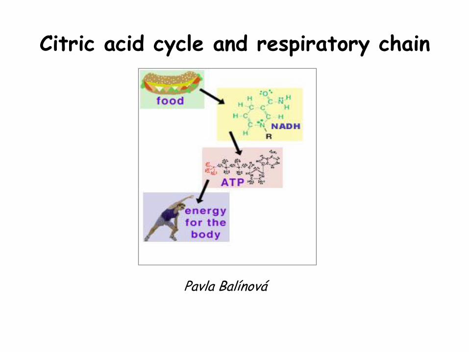
Citric acid cycle and respiratory chain
Pavla Balínová
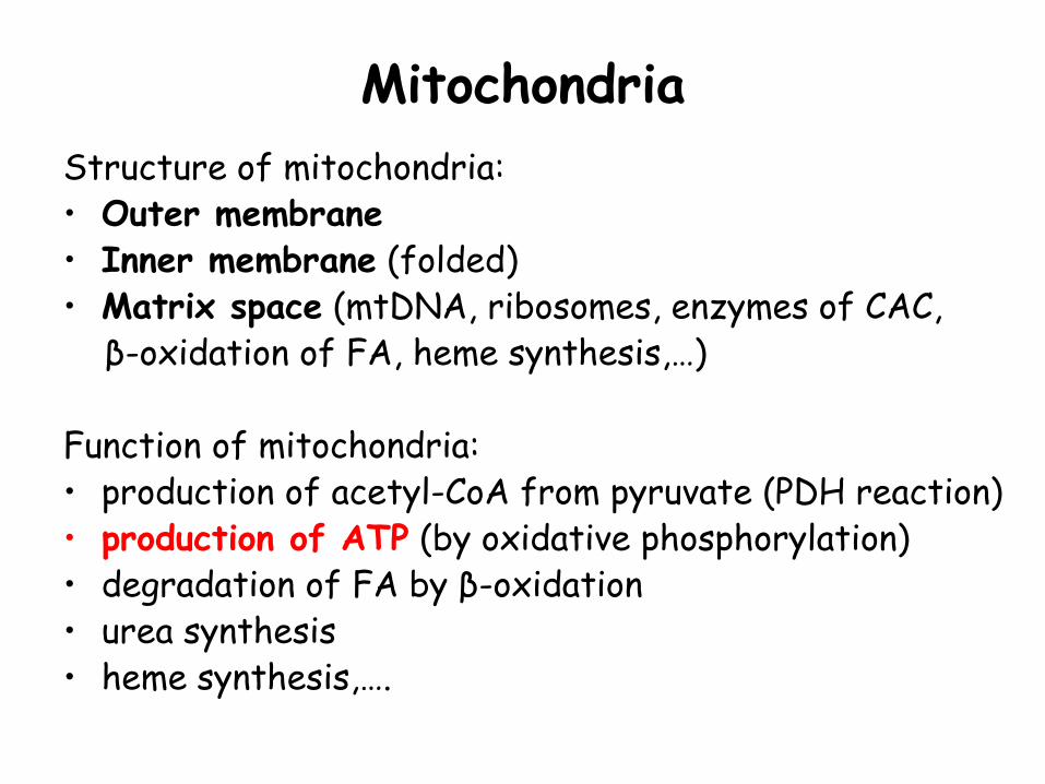
Mitochondria
Structure of mitochondria: • Outer membrane • Inner membrane (folded) • Matrix space (mtDNA, ribosomes, enzymes of CAC, β-oxidation of FA, heme synthesis,…) Function of mitochondria: • production of acetyl-CoA from pyruvate (PDH reaction) • production of ATP (by oxidative phosphorylation) • degradation of FA by β-oxidation • urea synthesis • heme synthesis,….
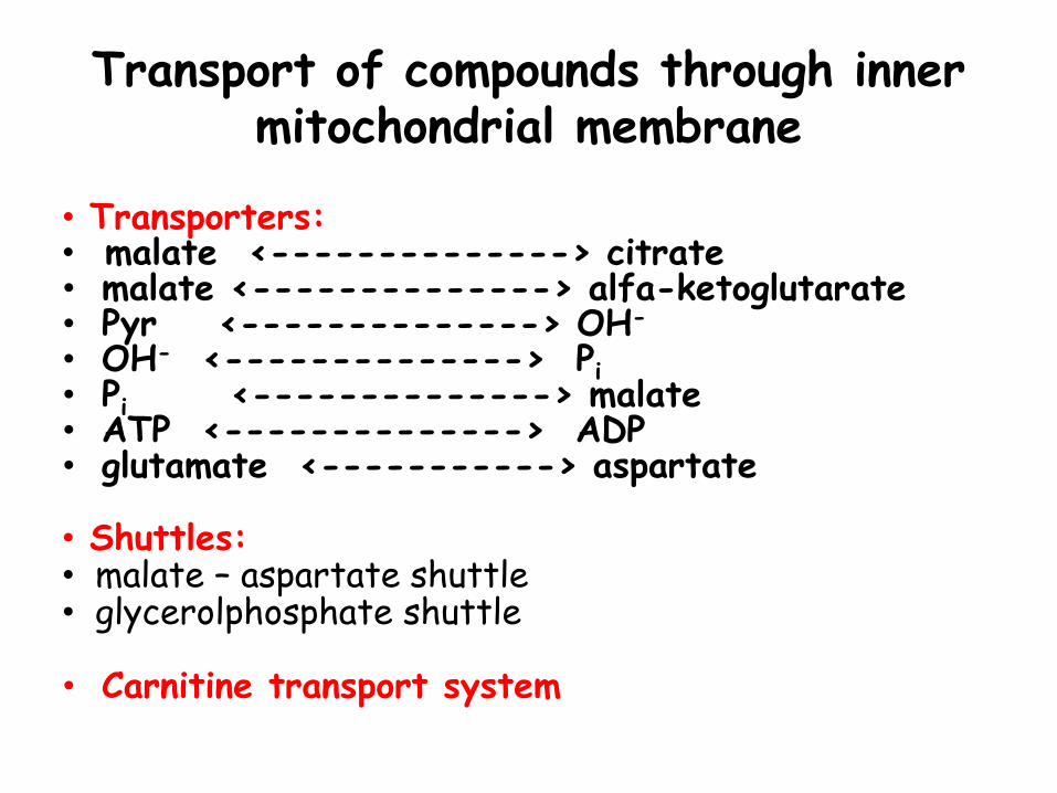
Transport of compounds through inner mitochondrial membrane
• Transporters: • malate <--------------> citrate • malate <--------------> alfa-ketoglutarate • Pyr <--------------> OH- • OH- <--------------> Pi • Pi <--------------> malate • ATP <--------------> ADP • glutamate <-----------> aspartate • Shuttles: • malate – aspartate shuttle • glycerolphosphate shuttle
• Carnitine transport system
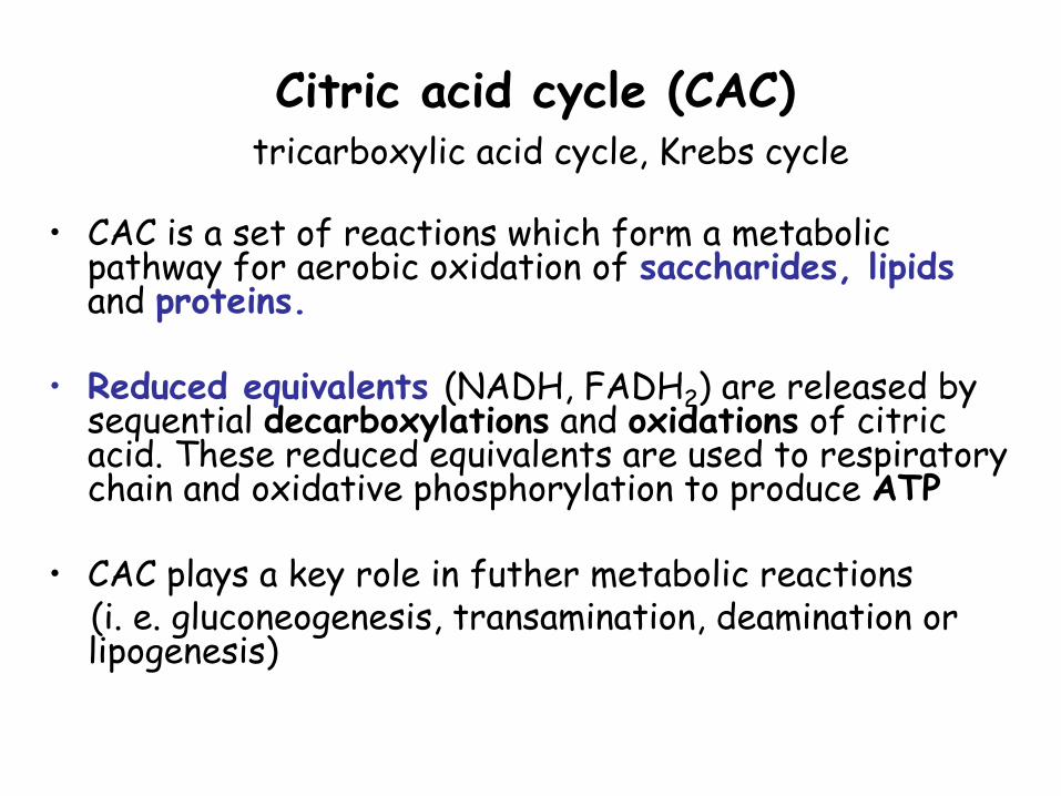
Citric acid cycle (CAC) tricarboxylic acid cycle, Krebs cycle
• CAC is a set of reactions which form a metabolic pathway for aerobic oxidation of saccharides, lipids and proteins.
• Reduced equivalents (NADH, FADH2) are released by
sequential decarboxylations and oxidations of citric acid. These reduced equivalents are used to respiratory chain and oxidative phosphorylation to produce ATP
• CAC plays a key role in futher metabolic reactions (i. e. gluconeogenesis, transamination, deamination or
lipogenesis)
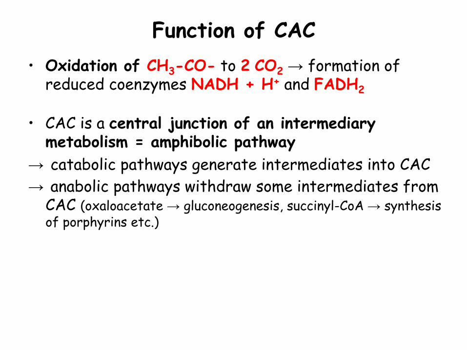
Function of CAC
• Oxidation of CH3-CO- to 2 CO2 → formation of reduced coenzymes NADH + H+ and FADH2
• CAC is a central junction of an intermediary metabolism = amphibolic pathway
→ catabolic pathways generate intermediates into CAC
→ anabolic pathways withdraw some intermediates from CAC (oxaloacetate → gluconeogenesis, succinyl-CoA → synthesis of porphyrins etc.)
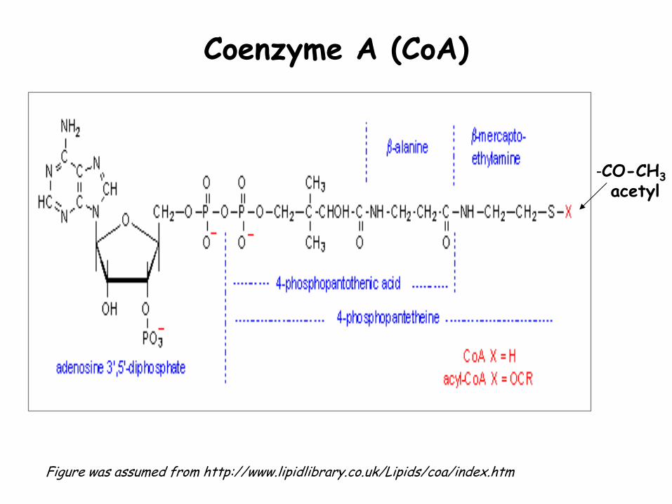
Coenzyme A (CoA)
Figure was assumed from http://www.lipidlibrary.co.uk/Lipids/coa/index.htm
-CO-CH3
acetyl
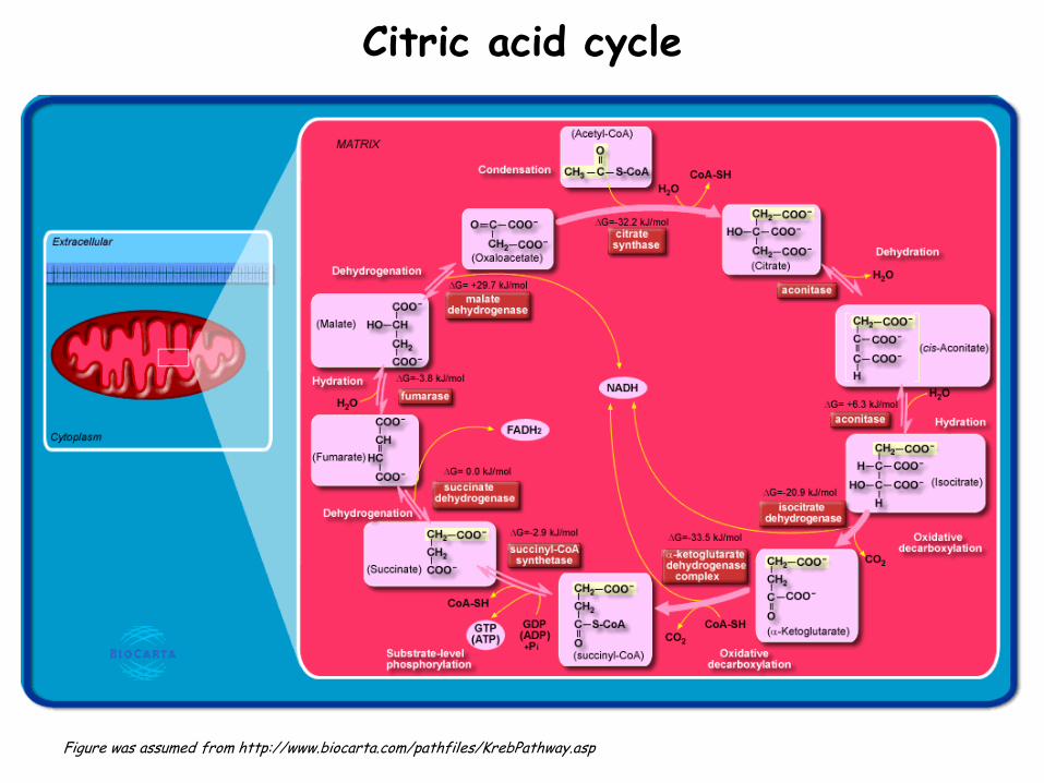
Citric acid cycle
Figure was assumed from http://www.biocarta.com/pathfiles/KrebPathway.asp
One turn of CAC produces: 2 mol CO2
3 mol NADH + H+ 1 mol FADH2
1 mol GTP Anaplerotic (support) reactions:
• Pyr + CO2 + ATP → oxaloacetate + ADP + Pi (pyruvate carboxylase) • degradation of most amino acids gives the following intermediates of CAC: oxaloacetate, α-ketoglutarate, fumarate
• Propionyl-CoA → succinyl-CoA
Regulation of CAC
Regulatory factors of CAC are: • NADH / NAD+ ratio • ATP / AMP ratio • availability of CAC substrates and energy situation within the cell Regulatory enzymes of CAC: Citrate synthase is mainly regulated with availability of acetyl-CoA and oxaloacetate. Isocitrate dehydrogenase and α-ketoglutarate dehydrogenase are inhibited by ↑ NADH / NAD+. On the contrary, these enzymes are activated by AMP and NAD+. The activity of CAC is closely linked to the availability of O2.
Transport of acetyl-CoA within the cell
Mitochondrion:
• acetyl-CoA + oxaloacetate → citrate
(enzyme citrate synthase in CAC)
• citrate is exported from mitochondria to cytoplasm in exchange for malate (antiport)
-----------------------------------------------------------
Cytoplasm:
• citrate is cleaved to acetyl-CoA and oxaloacetate
(enzyme citrate lyase) in the cytoplasm
• reduction of oxaloacetate to malate
(malate dehydrogenase = „malic enzyme“ – NADPH + H+ is produced)
• malate is returned into mitochondria or it is oxidative decarboxylated to pyruvate
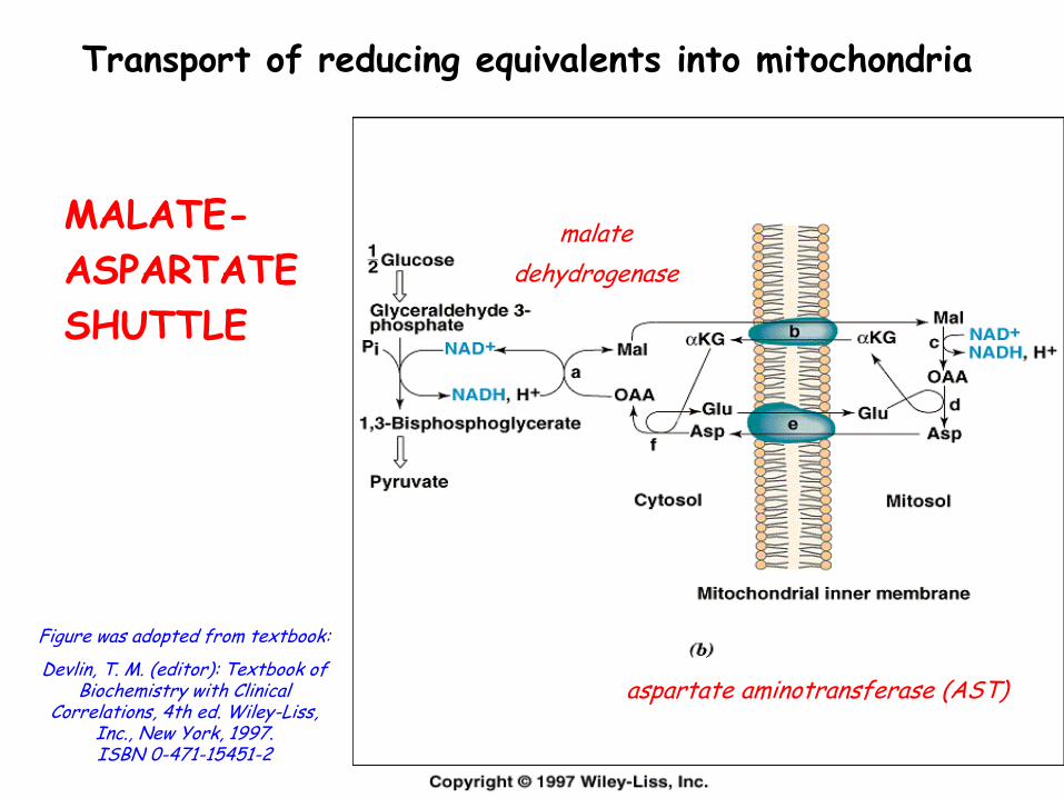
Transport of reducing equivalents into mitochondria
MALATE-
ASPARTATE
SHUTTLE
Figure was adopted from textbook:
Devlin, T. M. (editor): Textbook of Biochemistry with Clinical
Correlations, 4th ed. Wiley-Liss, Inc., New York, 1997. ISBN 0-471-15451-2
malate
dehydrogenase
aspartate aminotransferase (AST)
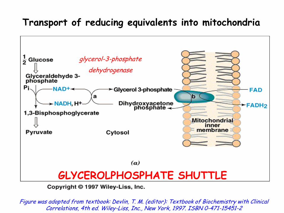
Transport of reducing equivalents into mitochondria
Figure was adopted from textbook: Devlin, T. M. (editor): Textbook of Biochemistry with Clinical Correlations, 4th ed. Wiley-Liss, Inc., New York, 1997. ISBN 0-471-15451-2
glycerol-3-phosphate
dehydrogenase
GLYCEROLPHOSPHATE SHUTTLE
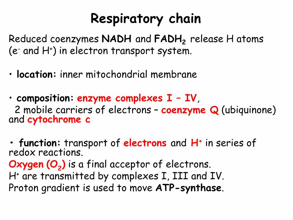
Respiratory chain
Reduced coenzymes NADH and FADH2 release H atoms (e- and H+) in electron transport system. • location: inner mitochondrial membrane • composition: enzyme complexes I – IV, 2 mobile carriers of electrons – coenzyme Q (ubiquinone) and cytochrome c • function: transport of electrons and H+ in series of redox reactions. Oxygen (O2) is a final acceptor of electrons. H+ are transmitted by complexes I, III and IV. Proton gradient is used to move ATP-synthase.
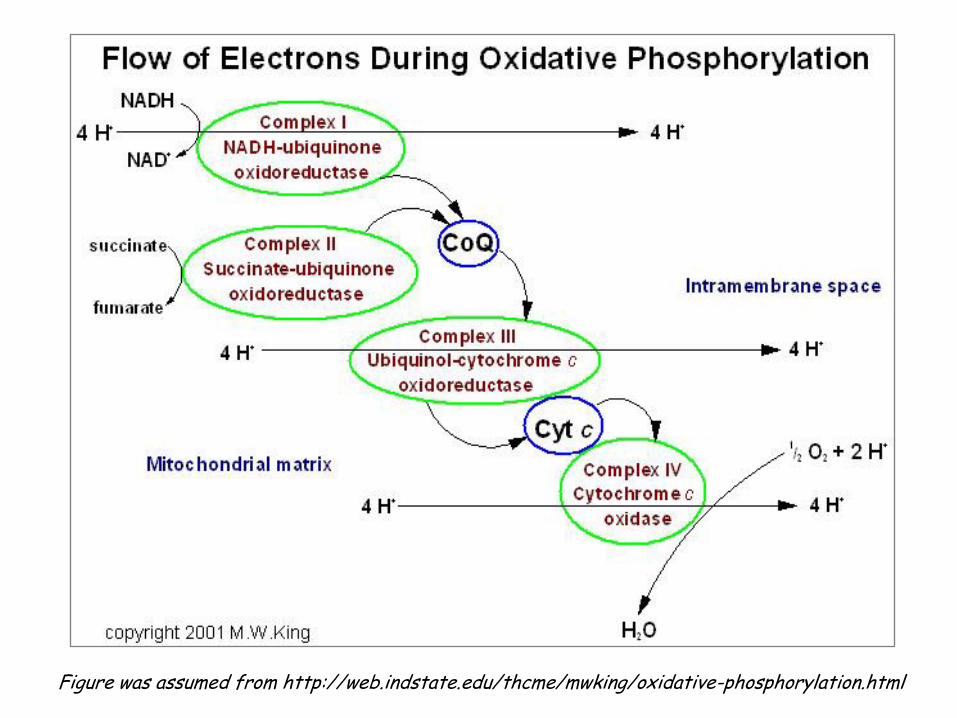
Figure was assumed from http://web.indstate.edu/thcme/mwking/oxidative-phosphorylation.html
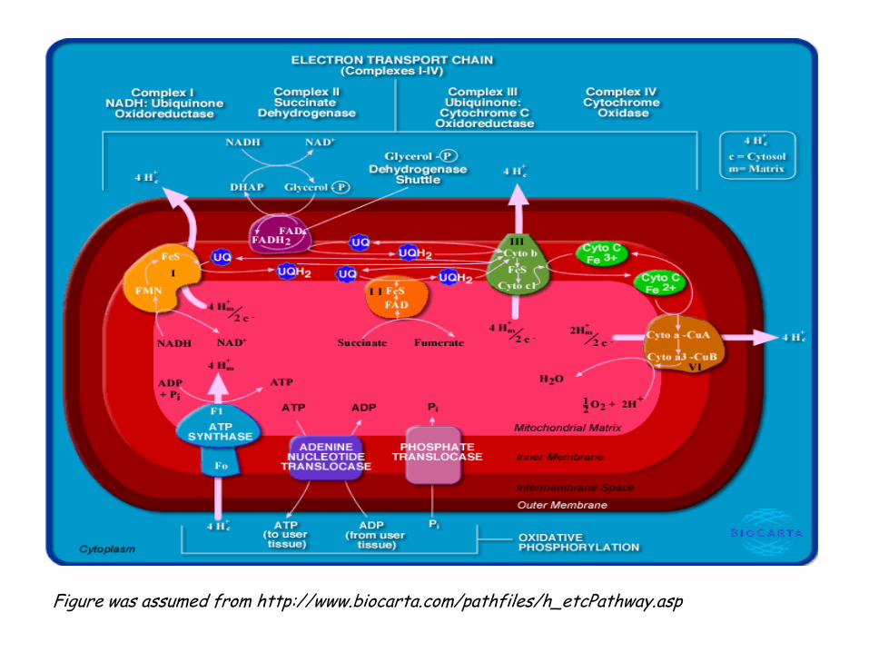
Figure was assumed from http://www.biocarta.com/pathfiles/h_etcPathway.asp
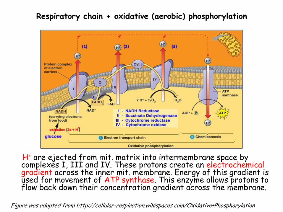
Respiratory chain + oxidative (aerobic) phosphorylation
H+ are ejected from mit. matrix into intermembrane space by complexes I, III and IV. These protons create an electrochemical gradient across the inner mit. membrane. Energy of this gradient is used for movement of ATP synthase. This enzyme allows protons to flow back down their concentration gradient across the membrane.
Figure was adopted from http://cellular-respiration.wikispaces.com/Oxidative+Phosphorylation
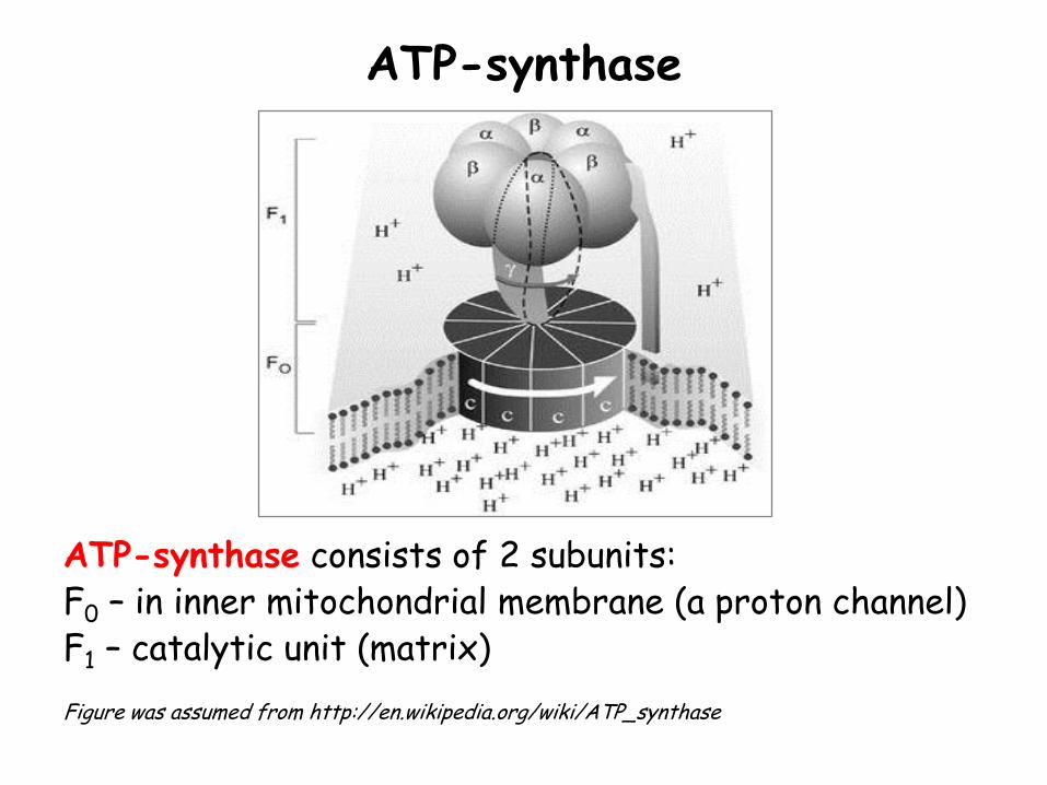
ATP-synthase
ATP-synthase consists of 2 subunits: F0 – in inner mitochondrial membrane (a proton channel) F1 – catalytic unit (matrix) Figure was assumed from http://en.wikipedia.org/wiki/ATP_synthase
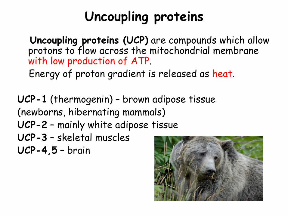
Uncoupling proteins
Uncoupling proteins (UCP) are compounds which allow protons to flow across the mitochondrial membrane with low production of ATP.
Energy of proton gradient is released as heat. UCP-1 (thermogenin) – brown adipose tissue (newborns, hibernating mammals) UCP-2 – mainly white adipose tissue UCP-3 – skeletal muscles UCP-4,5 – brain
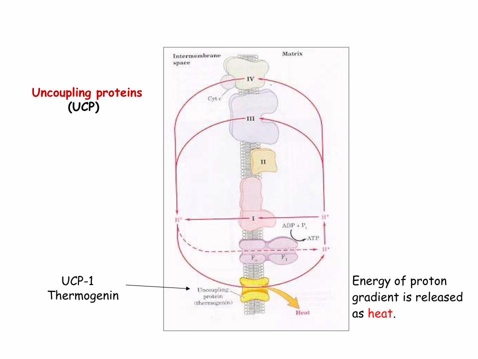
Uncoupling proteins (UCP)
UCP-1 Thermogenin
Energy of proton gradient is released as heat.
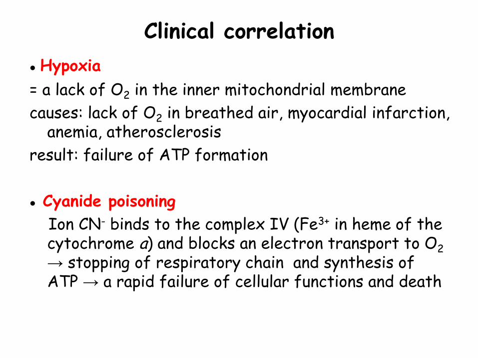
Clinical correlation
● Hypoxia
= a lack of O2 in the inner mitochondrial membrane
causes: lack of O2 in breathed air, myocardial infarction, anemia, atherosclerosis
result: failure of ATP formation
● Cyanide poisoning
Ion CN- binds to the complex IV (Fe3+ in heme of the cytochrome a) and blocks an electron transport to O2 → stopping of respiratory chain and synthesis of ATP → a rapid failure of cellular functions and death


![Respiratory Research BioMed Central · cle [1-3], activation of ion and fluid transport in epithelial cells [4], inhibition of mediator release from mast cells [5], stimulation of](https://static.fdocument.org/doc/165x107/5c8b31f009d3f22c4e8ba411/respiratory-research-biomed-central-cle-1-3-activation-of-ion-and-fluid-transport.jpg)