Chemoenzymatic Probes for Detecting and Imaging...
4
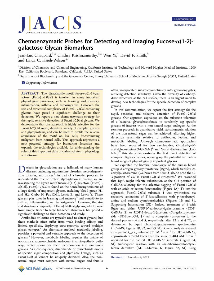
Chemoenzymatic Probes for Detecting and Imaging Fucose-α(1-2)- galactose Glycan Biomarkers Jean-Luc Chaubard, †,‡ Chithra Krishnamurthy, †,‡ Wen Yi, † David F. Smith, § and Linda C. Hsieh-Wilson* ,† † Division of Chemistry and Chemical Engineering, California Institute of Technology and Howard Hughes Medical Institute, 1200 East California Boulevard, Pasadena, California 91125, United States § Department of Biochemistry and the Glycomics Center, Emory University School of Medicine, Atlanta Georgia 30322, United States * S Supporting Information ABSTRACT: The disaccharide motif fucose-α(1-2)-gal- actose (Fucα(1-2)Gal) is involved in many important physiological processes, such as learning and memory, inflammation, asthma, and tumorigenesis. However, the size and structural complexity of Fucα(1-2)Gal-containing glycans have posed a significant challenge to their detection. We report a new chemoenzymatic strategy for the rapid, sensitive detection of Fucα(1-2)Gal glycans. We demonstrate that the approach is highly selective for the Fucα(1-2)Gal motif, detects a variety of complex glycans and glycoproteins, and can be used to profile the relative abundance of the motif on live cells, discriminating malignant from normal cells. This approach represents a new potential strategy for biomarker detection and expands the technologies available for understanding the roles of this important class of carbohydrates in physiology and disease. D efects in glycosylation are a hallmark of many human diseases, including autoimmune disorders, neurodegener- ative diseases, and cancer. 1 As part of a broader program to understand the role of protein glycosylation in disease, we are investigating the glycan motif fucose-α(1-2)-galactose (Fucα(1- 2)Gal). Fucα(1-2)Gal is found on the nonreducing terminus of a large family of important glycans, including blood group H1 and H2, Globo H, Fuc-GM1, Lewis B, and Lewis Y. These glycans play roles in learning and memory 2 and contribute to asthma, inflammation, and tumorigenesis. 3 However, the size and structural complexity of Fucα(1-2)Gal glycans, which range from simple linear to large branched structures, has posed a significant challenge to their detection and study. Antibodies or lectins are typically used to detect glycans, but these methods often suffer from weak binding affinity and limited specificity, displaying cross-reactivity toward multiple glycan epitopes. 4 An alternative method, metabolic labeling, provides a powerful and versatile approach to the detection of glycans. 5 However, metabolic labeling requires the uptake of non-natural monosaccharide analogues into biosynthetic path- ways, which allows for their incorporation into numerous glycans. As a consequence, disaccharide or trisaccharide motifs of specific sugar composition and glycosidic linkage, such as Fucα(1-2)Gal, cannot be uniquely detected. Also, the non- natural sugar must compete with natural sugars and thus is often incorporated substoichiometrically into glycoconjugates, reducing detection sensitivity. Given the diversity of carbohy- drate structures at the cell surface, there is an urgent need to develop new technologies for the specific detection of complex glycans. In this communication, we report the first strategy for the rapid, sensitive, and selective detection of Fucα(1-2)Gal glycans. Our approach capitalizes on the substrate tolerance of a bacterial glycosyltransferase to covalently tag specific glycans of interest with a non-natural sugar analogue. As the reaction proceeds in quantitative yield, stoichiometric addition of the non-natural sugar can be achieved, affording higher detection sensitivity relative to antibodies, lectins, and metabolic labeling. Although chemoenzymatic approaches have been reported for two saccharides, O-linked-β-N- acetylglucosamine(O-GlcNAc) 6 and N-acetyllactosamine (Lac- NAc), 7 this study demonstrates the first direct detection of complex oligosaccharides, opening up the potential to track a broad range of physiologically important glycans. We exploited the bacterial homologue of the human blood group A antigen glycosyltransferase (BgtA), which transfers N- acetylgalactosamine (GalNAc) from UDP-GalNAc onto the C- 3 position of Gal in Fucα(1-2)Gal structures. 8 We reasoned that BgtA might tolerate substitution at the C-2 position of GalNAc, allowing for the selective tagging of Fucα(1-2)Gal with an azido or ketone functionality (Figure 1A). To test the approach, Fucα(1-2)Gal substrate 1 was synthesized via reductive amination of 2′-fucosyllactose with p-nitrobenzyl- amine and sodium cyanoborohydride (Figures 1B and S1, Supporting Information (SI)). Indeed, treatment of 1 with BgtA and either UDP-N-azidoacetylgalactosamine (UDP- GalNAz, 2) or UDP-2-deoxy-2-(acetonyl)-β-D-galactopyrano- side (UDP-ketoGal, 3) led to complete conversion to the desired products 4 and 5, respectively, after 12 h at 4 °C, as determined by liquid chromatography−mass spectrometry (LC−MS; Figures 1B, S2, and S3, SI). Kinetic analysis revealed an apparent k cat /K m value of 5.7 nM −1 min −1 for UDP-GalNAz, approximately 7-fold lower than the value of 40.4 nM −1 min −1 obtained for the natural UDP-GalNAc substrate (Figure S4, SI). Subsequent reaction with an aza-dibenzo-cyclooctyne- biotin derivative (ADIBO-biotin, 6; Figure S2, SI) using Received: December 2, 2011 Communication pubs.acs.org/JACS © XXXX American Chemical Society A dx.doi.org/10.1021/ja211312u | J. Am. Chem. Soc. XXXX, XXX, XXX−XXX
Transcript of Chemoenzymatic Probes for Detecting and Imaging...
Chemoenzymatic Probes for Detecting and Imaging Fucose-α(1-2)-galactose Glycan BiomarkersJean-Luc Chaubard,†,‡ Chithra Krishnamurthy,†,‡ Wen Yi,† David F. Smith,§
and Linda C. Hsieh-Wilson*,†
†Division of Chemistry and Chemical Engineering, California Institute of Technology and Howard Hughes Medical Institute, 1200East California Boulevard, Pasadena, California 91125, United States§Department of Biochemistry and the Glycomics Center, Emory University School of Medicine, Atlanta Georgia 30322, United States
*S Supporting Information
ABSTRACT: The disaccharide motif fucose-α(1-2)-gal-actose (Fucα(1-2)Gal) is involved in many importantphysiological processes, such as learning and memory,inflammation, asthma, and tumorigenesis. However, thesize and structural complexity of Fucα(1-2)Gal-containingglycans have posed a significant challenge to theirdetection. We report a new chemoenzymatic strategy forthe rapid, sensitive detection of Fucα(1-2)Gal glycans. Wedemonstrate that the approach is highly selective for theFucα(1-2)Gal motif, detects a variety of complex glycansand glycoproteins, and can be used to profile the relativeabundance of the motif on live cells, discriminatingmalignant from normal cells. This approach represents anew potential strategy for biomarker detection andexpands the technologies available for understanding theroles of this important class of carbohydrates in physiologyand disease.
Defects in glycosylation are a hallmark of many humandiseases, including autoimmune disorders, neurodegener-
ative diseases, and cancer.1 As part of a broader program tounderstand the role of protein glycosylation in disease, we areinvestigating the glycan motif fucose-α(1-2)-galactose (Fucα(1-2)Gal). Fucα(1-2)Gal is found on the nonreducing terminus ofa large family of important glycans, including blood group H1and H2, Globo H, Fuc-GM1, Lewis B, and Lewis Y. Theseglycans play roles in learning and memory2 and contribute toasthma, inflammation, and tumorigenesis.3 However, the sizeand structural complexity of Fucα(1-2)Gal glycans, which rangefrom simple linear to large branched structures, has posed asignificant challenge to their detection and study.Antibodies or lectins are typically used to detect glycans, but
these methods often suffer from weak binding affinity andlimited specificity, displaying cross-reactivity toward multipleglycan epitopes.4 An alternative method, metabolic labeling,provides a powerful and versatile approach to the detection ofglycans.5 However, metabolic labeling requires the uptake ofnon-natural monosaccharide analogues into biosynthetic path-ways, which allows for their incorporation into numerousglycans. As a consequence, disaccharide or trisaccharide motifsof specific sugar composition and glycosidic linkage, such asFucα(1-2)Gal, cannot be uniquely detected. Also, the non-natural sugar must compete with natural sugars and thus is
often incorporated substoichiometrically into glycoconjugates,reducing detection sensitivity. Given the diversity of carbohy-drate structures at the cell surface, there is an urgent need todevelop new technologies for the specific detection of complexglycans.In this communication, we report the first strategy for the
rapid, sensitive, and selective detection of Fucα(1-2)Galglycans. Our approach capitalizes on the substrate toleranceof a bacterial glycosyltransferase to covalently tag specificglycans of interest with a non-natural sugar analogue. As thereaction proceeds in quantitative yield, stoichiometric additionof the non-natural sugar can be achieved, affording higherdetection sensitivity relative to antibodies, lectins, andmetabolic labeling. Although chemoenzymatic approacheshave been reported for two saccharides, O-linked-β-N-acetylglucosamine(O-GlcNAc)6 and N-acetyllactosamine (Lac-NAc),7 this study demonstrates the first direct detection ofcomplex oligosaccharides, opening up the potential to track abroad range of physiologically important glycans.We exploited the bacterial homologue of the human blood
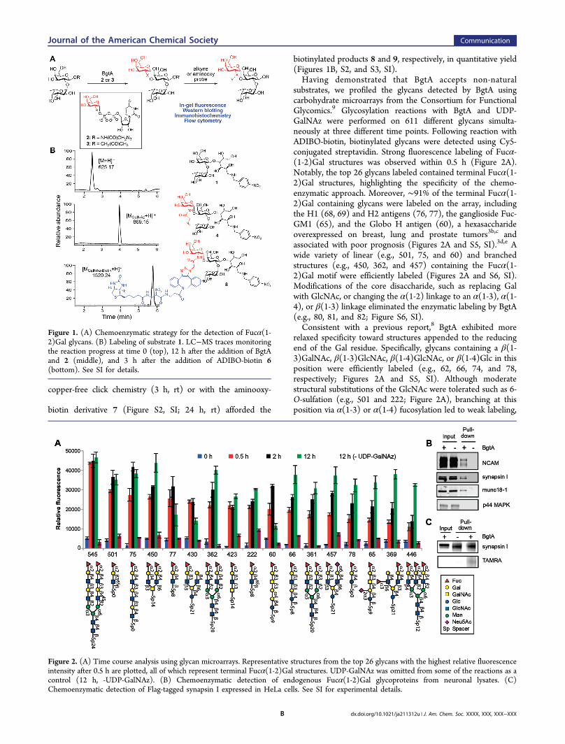
group A antigen glycosyltransferase (BgtA), which transfers N-acetylgalactosamine (GalNAc) from UDP-GalNAc onto the C-3 position of Gal in Fucα(1-2)Gal structures.8 We reasonedthat BgtA might tolerate substitution at the C-2 position ofGalNAc, allowing for the selective tagging of Fucα(1-2)Galwith an azido or ketone functionality (Figure 1A). To test theapproach, Fucα(1-2)Gal substrate 1 was synthesized viareductive amination of 2′-fucosyllactose with p-nitrobenzyl-amine and sodium cyanoborohydride (Figures 1B and S1,Supporting Information (SI)). Indeed, treatment of 1 withBgtA and either UDP-N-azidoacetylgalactosamine (UDP-GalNAz, 2) or UDP-2-deoxy-2-(acetonyl)-β-D-galactopyrano-side (UDP-ketoGal, 3) led to complete conversion to thedesired products 4 and 5, respectively, after 12 h at 4 °C, asdetermined by liquid chromatography−mass spectrometry(LC−MS; Figures 1B, S2, and S3, SI). Kinetic analysis revealedan apparent kcat/Km value of 5.7 nM−1 min−1 for UDP-GalNAz,approximately 7-fold lower than the value of 40.4 nM−1 min−1
obtained for the natural UDP-GalNAc substrate (Figure S4,SI). Subsequent reaction with an aza-dibenzo-cyclooctyne-biotin derivative (ADIBO-biotin, 6; Figure S2, SI) using
Received: December 2, 2011
Communication
pubs.acs.org/JACS
© XXXX American Chemical Society A dx.doi.org/10.1021/ja211312u | J. Am. Chem. Soc. XXXX, XXX, XXX−XXX
copper-free click chemistry (3 h, rt) or with the aminooxy-
biotin derivative 7 (Figure S2, SI; 24 h, rt) afforded the
biotinylated products 8 and 9, respectively, in quantitative yield(Figures 1B, S2, and S3, SI).Having demonstrated that BgtA accepts non-natural
substrates, we profiled the glycans detected by BgtA usingcarbohydrate microarrays from the Consortium for FunctionalGlycomics.9 Glycosylation reactions with BgtA and UDP-GalNAz were performed on 611 different glycans simulta-neously at three different time points. Following reaction withADIBO-biotin, biotinylated glycans were detected using Cy5-conjugated streptavidin. Strong fluorescence labeling of Fucα-(1-2)Gal structures was observed within 0.5 h (Figure 2A).Notably, the top 26 glycans labeled contained terminal Fucα(1-2)Gal structures, highlighting the specificity of the chemo-enzymatic approach. Moreover, ∼91% of the terminal Fucα(1-2)Gal containing glycans were labeled on the array, includingthe H1 (68, 69) and H2 antigens (76, 77), the ganglioside Fuc-GM1 (65), and the Globo H antigen (60), a hexasaccharideoverexpressed on breast, lung and prostate tumors3b,c andassociated with poor prognosis (Figures 2A and S5, SI).3d,e Awide variety of linear (e.g., 501, 75, and 60) and branchedstructures (e.g., 450, 362, and 457) containing the Fucα(1-2)Gal motif were efficiently labeled (Figures 2A and S6, SI).Modifications of the core disaccharide, such as replacing Galwith GlcNAc, or changing the α(1-2) linkage to an α(1-3), α(1-4), or β(1-3) linkage eliminated the enzymatic labeling by BgtA(e.g., 80, 81, and 82; Figure S6, SI).Consistent with a previous report,8 BgtA exhibited more
relaxed specificity toward structures appended to the reducingend of the Gal residue. Specifically, glycans containing a β(1-3)GalNAc, β(1-3)GlcNAc, β(1-4)GlcNAc, or β(1-4)Glc in thisposition were efficiently labeled (e.g., 62, 66, 74, and 78,respectively; Figures 2A and S5, SI). Although moderatestructural substitutions of the GlcNAc were tolerated such as 6-O-sulfation (e.g., 501 and 222; Figure 2A), branching at thisposition via α(1-3) or α(1-4) fucosylation led to weak labeling,
Figure 1. (A) Chemoenzymatic strategy for the detection of Fucα(1-2)Gal glycans. (B) Labeling of substrate 1. LC−MS traces monitoringthe reaction progress at time 0 (top), 12 h after the addition of BgtAand 2 (middle), and 3 h after the addition of ADIBO-biotin 6(bottom). See SI for details.
Figure 2. (A) Time course analysis using glycan microarrays. Representative structures from the top 26 glycans with the highest relative fluorescenceintensity after 0.5 h are plotted, all of which represent terminal Fucα(1-2)Gal structures. UDP-GalNAz was omitted from some of the reactions as acontrol (12 h, -UDP-GalNAz). (B) Chemoenzymatic detection of endogenous Fucα(1-2)Gal glycoproteins from neuronal lysates. (C)Chemoenzymatic detection of Flag-tagged synapsin I expressed in HeLa cells. See SI for experimental details.
Journal of the American Chemical Society Communication
dx.doi.org/10.1021/ja211312u | J. Am. Chem. Soc. XXXX, XXX, XXX−XXXB
as in the case of the Lewis B (61) and Lewis Y (72, 73)antigens, or no appreciable labeling (e.g., 71, and 363; FiguresS5 and S7, SI). Interestingly, we also observed weak labeling ofGalβ(1-4)GlcNAc structures on the glycan array (Figure S7,SI). However, these structures also exhibited high backgroundsignal even in the absence of UDP-GalNAz, and BgtA failed tolabel p-nitrophenyl 2-acetamido-2-deoxy-4-O-(β-D-galactopyr-anosyl)-β-D-glucopyranoside (Galβ(1-4)GlcNAc-pNP) in sol-ution (2 h, 25 °C), suggesting that Galβ(1-4)GlcNAc structuresare not covalently labeled by BgtA. Together, these studiesdemonstrate the strong specificity of BgtA for Fucα(1-2)Galstructures and the power of glycan microarrays to rapidlyprofile the specificities of glycosyltransferases for the develop-ment of chemoenzymatic detection strategies.To determine whether the approach could be used to track
Fucα(1-2)Gal glycoproteins in complex cell lysates, we labeledproteins from rat brain extracts with BgtA and UDP-GalNAz,followed by the Cu(I)-catalyzed azide−alkyne cycloaddition(CuAAC) reaction with tetramethyl-6-carboxyrhodamine dye10 (alkyne-TAMRA; Figure S2, SI). We observed strongfluorescence labeling of Fucα(1-2)Gal glycoproteins, withminimal nonspecific labeling in the absence of BgtA, UDP-GalNAz, or alkyne-TAMRA (Figure S8, SI). To confirm furtherthe specificity of the reaction, we labeled the lysates with thealkyne-biotin derivative 11 (Figure S2, SI), captured thebiotinylated proteins using streptavidin resin, and immuno-blotted for the presence of known Fucα(1-2)Gal glycopro-teins.10 Neural cell adhesion molecule (NCAM), synapsin I,and munc18-1 were all chemoenzymatically labeled anddetected in the presence, but not in the absence, of BgtA(Figure 2B). In contrast, p44 mitogen-associated protein kinase(p44 MAPK), a protein that has not been shown to befucosylated, was not detected. Glycosylated synapsin I was alsoreadily observed following overexpression of Flag-taggedsynapsin I in HeLa cells, chemoenzymatic labeling of thelysates with alkyne-TAMRA, synapsin immunoprecipitation,and visualization using an anti-TAMRA antibody (Figure 2C).Importantly, UEAI lectin affinity chromatography failed to pull-down and detect glycosylated synapsin I when performed onthe same scale (Figure S9, SI). Moreover, previous studies havereported that the Fucα(1-2)Gal-specific antibody A46-B/B10does not immunoprecipitate glycosylated synapsin I from thesame neuronal lysates.10a Thus, our chemoenzymatic approachenables the highly sensitive detection of glycoproteins andprovides a variety of different enrichment strategies andreadouts for the Fucα(1-2)Gal motif.We next investigated whether the chemoenzymatic strategy
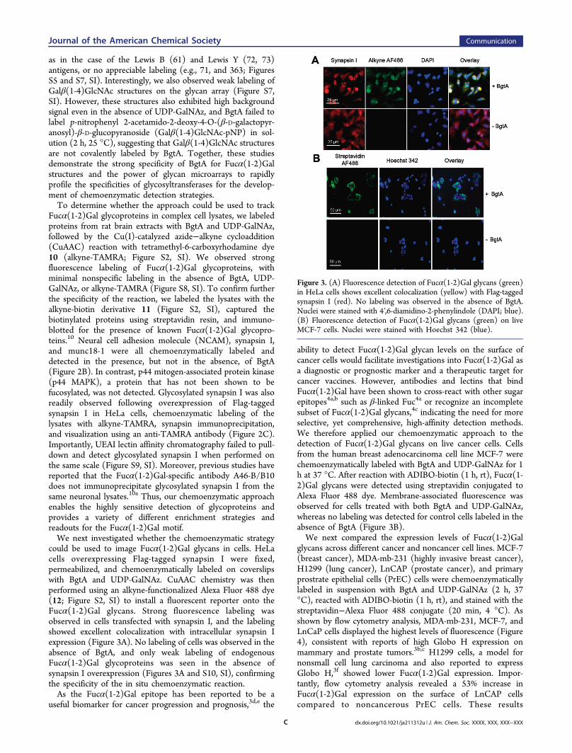
could be used to image Fucα(1-2)Gal glycans in cells. HeLacells overexpressing Flag-tagged synapsin I were fixed,permeabilized, and chemoenzymatically labeled on coverslipswith BgtA and UDP-GalNAz. CuAAC chemistry was thenperformed using an alkyne-functionalized Alexa Fluor 488 dye(12; Figure S2, SI) to install a fluorescent reporter onto theFucα(1-2)Gal glycans. Strong fluorescence labeling wasobserved in cells transfected with synapsin I, and the labelingshowed excellent colocalization with intracellular synapsin Iexpression (Figure 3A). No labeling of cells was observed in theabsence of BgtA, and only weak labeling of endogenousFucα(1-2)Gal glycoproteins was seen in the absence ofsynapsin I overexpression (Figures 3A and S10, SI), confirmingthe specificity of the in situ chemoenzymatic reaction.As the Fucα(1-2)Gal epitope has been reported to be a
useful biomarker for cancer progression and prognosis,3d,e the
ability to detect Fucα(1-2)Gal glycan levels on the surface ofcancer cells would facilitate investigations into Fucα(1-2)Gal asa diagnostic or prognostic marker and a therapeutic target forcancer vaccines. However, antibodies and lectins that bindFucα(1-2)Gal have been shown to cross-react with other sugarepitopes4a,b such as β-linked Fuc4a or recognize an incompletesubset of Fucα(1-2)Gal glycans,4c indicating the need for moreselective, yet comprehensive, high-affinity detection methods.We therefore applied our chemoenzymatic approach to thedetection of Fucα(1-2)Gal glycans on live cancer cells. Cellsfrom the human breast adenocarcinoma cell line MCF-7 werechemoenzymatically labeled with BgtA and UDP-GalNAz for 1h at 37 °C. After reaction with ADIBO-biotin (1 h, rt), Fucα(1-2)Gal glycans were detected using streptavidin conjugated toAlexa Fluor 488 dye. Membrane-associated fluorescence wasobserved for cells treated with both BgtA and UDP-GalNAz,whereas no labeling was detected for control cells labeled in theabsence of BgtA (Figure 3B).We next compared the expression levels of Fucα(1-2)Gal
glycans across different cancer and noncancer cell lines. MCF-7(breast cancer), MDA-mb-231 (highly invasive breast cancer),H1299 (lung cancer), LnCAP (prostate cancer), and primaryprostrate epithelial cells (PrEC) cells were chemoenzymaticallylabeled in suspension with BgtA and UDP-GalNAz (2 h, 37°C), reacted with ADIBO-biotin (1 h, rt), and stained with thestreptavidin−Alexa Fluor 488 conjugate (20 min, 4 °C). Asshown by flow cytometry analysis, MDA-mb-231, MCF-7, andLnCaP cells displayed the highest levels of fluorescence (Figure4), consistent with reports of high Globo H expression onmammary and prostate tumors.3b,c H1299 cells, a model fornonsmall cell lung carcinoma and also reported to expressGlobo H,3f showed lower Fucα(1-2)Gal expression. Impor-tantly, flow cytometry analysis revealed a 53% increase inFucα(1-2)Gal expression on the surface of LnCAP cellscompared to noncancerous PrEC cells. These results
Figure 3. (A) Fluorescence detection of Fucα(1-2)Gal glycans (green)in HeLa cells shows excellent colocalization (yellow) with Flag-taggedsynapsin I (red). No labeling was observed in the absence of BgtA.Nuclei were stained with 4′,6-diamidino-2-phenylindole (DAPI; blue).(B) Fluorescence detection of Fucα(1-2)Gal glycans (green) on liveMCF-7 cells. Nuclei were stained with Hoechst 342 (blue).
Journal of the American Chemical Society Communication
dx.doi.org/10.1021/ja211312u | J. Am. Chem. Soc. XXXX, XXX, XXX−XXXC
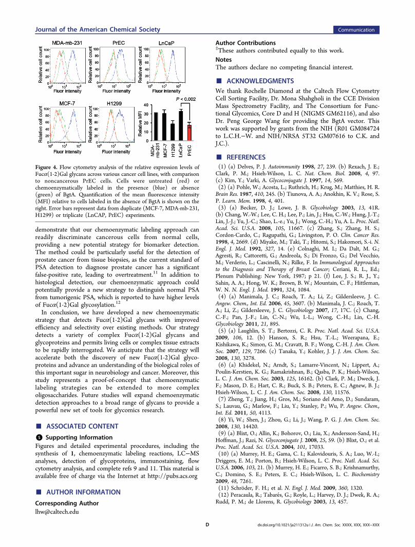
demonstrate that our chemoenzymatic labeling approach canreadily discriminate cancerous cells from normal cells,providing a new potential strategy for biomarker detection.The method could be particularly useful for the detection ofprostate cancer from tissue biopsies, as the current standard ofPSA detection to diagnose prostate cancer has a significantfalse-positive rate, leading to overtreatment.11 In addition tohistological detection, our chemoenzymatic approach couldpotentially provide a new strategy to distinguish normal PSAfrom tumorigenic PSA, which is reported to have higher levelsof Fucα(1-2)Gal glycosylation.12
In conclusion, we have developed a new chemoenzymaticstrategy that detects Fucα(1-2)Gal glycans with improvedefficiency and selectivity over existing methods. Our strategydetects a variety of complex Fucα(1-2)Gal glycans andglycoproteins and permits living cells or complex tissue extractsto be rapidly interrogated. We anticipate that the strategy willaccelerate both the discovery of new Fucα(1-2)Gal glyco-proteins and advance an understanding of the biological roles ofthis important sugar in neurobiology and cancer. Moreover, thisstudy represents a proof-of-concept that chemoenzymaticlabeling strategies can be extended to more complexoligosaccharides. Future studies will expand chemoenzymaticdetection approaches to a broad range of glycans to provide apowerful new set of tools for glycomics research.
■ ASSOCIATED CONTENT
*S Supporting InformationFigures and detailed experimental procedures, including thesynthesis of 1, chemoenzymatic labeling reactions, LC−MSanalyses, detection of glycoproteins, immunostaining, flowcytometry analysis, and complete refs 9 and 11. This material isavailable free of charge via the Internet at http://pubs.acs.org
■ AUTHOR INFORMATION
Corresponding [email protected]
Author Contributions‡These authors contributed equally to this work.NotesThe authors declare no competing financial interest.
■ ACKNOWLEDGMENTSWe thank Rochelle Diamond at the Caltech Flow CytometryCell Sorting Facility, Dr. Mona Shahgholi in the CCE DivisionMass Spectrometry Facility, and The Consortium for Func-tional Glycomics, Core D and H (NIGMS GM62116), and alsoDr. Peng George Wang for providing the BgtA vector. Thiswork was supported by grants from the NIH (R01 GM084724to L.C.H.−W. and NIH/NRSA 5T32 GM07616 to C.K. andJ.C.).
■ REFERENCES(1) (a) Delves, P. J. Autoimmunity 1998, 27, 239. (b) Rexach, J. E.;Clark, P. M.; Hsieh-Wilson, L. C. Nat. Chem. Biol. 2008, 4, 97.(c) Kim, Y.; Varki, A. Glycoconjugate J. 1997, 14, 569.(2) (a) Pohle, W.; Acosta, L.; Ruthrich, H.; Krug, M.; Matthies, H. R.Brain Res. 1987, 410, 245. (b) Tiunova, A. A.; Anokhin, K. V.; Rose, S.P. Learn. Mem. 1998, 4, 401.(3) (a) Becker, D. J.; Lowe, J. B. Glycobiology 2003, 13, 41R.(b) Chang, W.-W.; Lee, C. H.; Lee, P.; Lin, J.; Hsu, C.-W.; Hung, J.-T.;Lin, J.-J.; Yu, J.-C.; Shao, L.-e.; Yu, J.; Wong, C.-H.; Yu, A. L. Proc. Natl.Acad. Sci. U.S.A. 2008, 105, 11667. (c) Zhang, S.; Zhang, H. S.;Cordon-Cardo, C.; Ragupathi, G.; Livingston, P. O. Clin. Cancer Res.1998, 4, 2669. (d) Miyake, M.; Taki, T.; Hitomi, S.; Hakomori, S.-i. N.Engl. J. Med. 1992, 327, 14. (e) Colnaghi, M. I.; Da Dalt, M. G.;Agresti, R.; Cattoretti, G.; Andreola, S.; Di Fronzo, G.; Del Vecchio,M.; Verderio, L.; Cascinelli, N.; Rilke, F. In Immunological Approachesto the Diagnosis and Therapy of Breast Cancer; Ceriani, R. L., Ed.;Plenum Publishing: New York, 1987; p 21. (f) Lee, J. S.; R. J., Y.;Sahin, A. A.; Hong, W. K.; Brown, B. W.; Mountain, C. F.; Hittleman,W. N. N. Engl. J. Med. 1991, 324, 1084.(4) (a) Manimala, J. C.; Roach, T. A.; Li, Z.; Gildersleeve, J. C.Angew. Chem., Int. Ed. 2006, 45, 3607. (b) Manimala, J. C.; Roach, T.A.; Li, Z.; Gildersleeve, J. C. Glycobiology 2007, 17, 17C. (c) Chang,C.-F.; Pan, J.-F.; Lin, C.-N.; Wu, I.-L.; Wong, C.-H.; Lin, C.-H.Glycobiology 2011, 21, 895.(5) (a) Laughlin, S. T.; Bertozzi, C. R. Proc. Natl. Acad. Sci. U.S.A.2009, 106, 12. (b) Hanson, S. R.; Hsu, T.-L.; Weerapana, E.;Kishikawa, K.; Simon, G. M.; Cravatt, B. F.; Wong, C.-H. J. Am. Chem.Soc. 2007, 129, 7266. (c) Tanaka, Y.; Kohler, J. J. J. Am. Chem. Soc.2008, 130, 3278.(6) (a) Khidekel, N.; Arndt, S.; Lamarre-Vincent, N.; Lippert, A.;Poulin-Kerstien, K. G.; Ramakrishnan, B.; Qasba, P. K.; Hsieh-Wilson,L. C. J. Am. Chem. Soc. 2003, 125, 16162. (b) Clark, P. M.; Dweck, J.F.; Mason, D. E.; Hart, C. R.; Buck, S. B.; Peters, E. C.; Agnew, B. J.;Hsieh-Wilson, L. C. J. Am. Chem. Soc. 2008, 130, 11576.(7) Zheng, T.; Jiang, H.; Gros, M.; Soriano del Amo, D.; Sundaram,S.; Lauvau, G.; Marlow, F.; Liu, Y.; Stanley, P.; Wu, P. Angew. Chem.,Int. Ed. 2011, 50, 4113.(8) Yi, W.; Shen, J.; Zhou, G.; Li, J.; Wang, P. G. J. Am. Chem. Soc.2008, 130, 14420.(9) (a) Blixt, O.; Allin, K.; Bohorov, O.; Liu, X.; Andersson-Sand, H.;Hoffman, J.; Razi, N. Glycoconjugate J. 2008, 25, 59. (b) Blixt, O.; et al.Proc. Natl. Acad. Sci. U.S.A. 2004, 101, 17033.(10) (a) Murrey, H. E.; Gama, C. I.; Kalovidouris, S. A.; Luo, W.-I.;Driggers, E. M.; Porton, B.; Hsieh-Wilson, L. C. Proc. Natl. Acad. Sci.U.S.A. 2006, 103, 21. (b) Murrey, H. E.; Ficarro, S. B.; Krishnamurthy,C.; Domino, S. E.; Peters, E. C.; Hsieh-Wilson, L. C. Biochemistry2009, 48, 7261.(11) Schroder, F. H.; et al. N. Engl. J. Med. 2009, 360, 1320.(12) Peracaula, R.; Tabares, G.; Royle, L.; Harvey, D. J.; Dwek, R. A.;Rudd, P. M.; de Llorens, R. Glycobiology 2003, 13, 457.
Figure 4. Flow cytometry analysis of the relative expression levels ofFucα(1-2)Gal glycans across various cancer cell lines, with comparisonto noncancerous PrEC cells. Cells were untreated (red) orchemoenzymatically labeled in the presence (blue) or absence(green) of BgtA. Quantification of the mean fluorescence intensity(MFI) relative to cells labeled in the absence of BgtA is shown on theright. Error bars represent data from duplicate (MCF-7, MDA-mb-231,H1299) or triplicate (LnCAP, PrEC) experiments.
Journal of the American Chemical Society Communication
dx.doi.org/10.1021/ja211312u | J. Am. Chem. Soc. XXXX, XXX, XXX−XXXD