Characteristics of β-wollastonite derived from rice straw ......The chemical composition of the raw...
13
Journal of The Australian Ceramic Society Volume 52[2], 2016, 163 – 174 163 Characteristics of β-wollastonite derived from rice straw ash and limestone Hamisah Ismail 1 , Roslinda Shamsudin 2* , Muhammad Azmi Abdul Hamid 3 and Rozidawati Awang 4 1-4 School of Applied Physics, Faculty of Science & Technology, Universiti Kebangsaan Malaysia, 43600 Bangi, Selangor, Malaysia. E-mail: [email protected] Available online at: www.austceram.com/ACS-Journal Abstract This study aims to characterise β-wollastonite that was derived from rice straw ash and limestone and also its bioactivity by soaking in SBF for 1, 3, 5, 7, 14, and 21 days. Rice straw was fired to obtain SiO 2 and CaO from calcined limestone. The ratio of CaO:SiO 2 was set at 55:45. The precursor mixture was then autoclaved for 8 h at 135 o C and sintered for 3 h at 950 o C. The chemical composition of the raw materials was obtained using XRF, which was approximately 79.0 wt.% SiO 2 and 97.2 wt.% CaO for rice straw ash and limestone respectively. From the phase identification, the rice straw ash has a cristobalite phase, while the limestone has a CaO phase. The β-wollastonite phase was obtained after both autoclaving and sintering were performed. Chemical analysis for soaked β-wollastonite showed that the Ca/P molar ratio was 1.66, which is close to that of “calcium deficient” hydroxyapatite (CDHA). FTIR has indicated the occurrence of rapid adsorption during exposure of β-wollastonite sample to the SBF, which is apparent from the peak of phosphate after one day of soaking. SEM has revealed the formation of hydroxyapatite microstructures on the surface of the immersed β- wollastonite sample. For heavy metal elemental evaluation, metal panel that included As, Cd, Hg and Pb were selected and both precursor and β-wollastonite had fulfilled the requirement of ASTM F1538-03 standard specification. In conclusion, β-wollastonite produced from rice straw ash and limestone has a characteristic of bioactive properties, and is suitable for implant purposes. Keywords: bioceramics; sintering; β-wollastonite; morphology; hydroxyapatite. 1. Introduction Wollastonite (CaSiO 3 ) is largely inert and has a polymorph structure, either α-wollastonite (pseudo- wollastonite), or β-wollastonite [1–3]. Polymorphic materials have an identical chemical composition and stoichiometry, but different crystal structures [4]. Wollastonite changes to form pseudo- wollastonite at 1125 o C, and congruently melts at 1544 o C [5]. Calcium silicate hydrates are transformed into β-wollastonite by annealing in the temperature range of 800 °C to 1150 °C [6]. Various raw materials have been used to synthesise wollastonite, derived from chemical or mineral precursors, to produce an end product with significant purity and good mechanical properties. Previous studies have successfully synthesised wollastonite using chemicals and minerals such as fumed silica, silica commercial, silica sand, and sodium silicate as the precursor for silica [1, 7–11]. Thus, in this study, the silica source was derived from rice straw ash. Rice straw was traditionally removed from the field by the practice of open- field burning [12]; this practice clears the field for new plantings and also cleans the soil of disease- causing agents. Rice straw ash has been widely used as a biomass resource [13-14], animal feed [15], a biosorbent, and as bioethanol [16–18], in the effort to better manage this by-product. However, there are fewer studies concerning rice straw ash relative to those about rice husk ash [19–21] for biomaterial purposes. In recent years, wollastonite has been widely used in cements and ceramics due to its strength, low shrinkage, lack of volatile constituents, and body permeability, as well as its fluxing characteristics [22]. Furthermore, wollastonite is also widely applied in the biomaterials field owing to its bioactivity and degradable materials [23–25]. Calcium silicate has been proven to be an extremely good material for in-vitro bioactivity [26]. The formation rate of hydroxyapatite on its
Transcript of Characteristics of β-wollastonite derived from rice straw ......The chemical composition of the raw...
Journal of The Australian Ceramic Society Volume 52[2], 2016, 163 – 174 163
Characteristics of β-wollastonite derived from rice straw ash and limestone Hamisah Ismail1, Roslinda Shamsudin2*, Muhammad Azmi Abdul Hamid3 and Rozidawati
Awang4 1-4School of Applied Physics, Faculty of Science & Technology, Universiti Kebangsaan Malaysia, 43600 Bangi, Selangor, Malaysia. E-mail: [email protected] Available online at: www.austceram.com/ACS-Journal Abstract This study aims to characterise β-wollastonite that was derived from rice straw ash and limestone and also its bioactivity by soaking in SBF for 1, 3, 5, 7, 14, and 21 days. Rice straw was fired to obtain SiO2 and CaO from calcined limestone. The ratio of CaO:SiO2 was set at 55:45. The precursor mixture was then autoclaved for 8 h at 135oC and sintered for 3 h at 950 oC. The chemical composition of the raw materials was obtained using XRF, which was approximately 79.0 wt.% SiO2 and 97.2 wt.% CaO for rice straw ash and limestone respectively. From the phase identification, the rice straw ash has a cristobalite phase, while the limestone has a CaO phase. The β-wollastonite phase was obtained after both autoclaving and sintering were performed. Chemical analysis for soaked β-wollastonite showed that the Ca/P molar ratio was 1.66, which is close to that of “calcium deficient” hydroxyapatite (CDHA). FTIR has indicated the occurrence of rapid adsorption during exposure of β-wollastonite sample to the SBF, which is apparent from the peak of phosphate after one day of soaking. SEM has revealed the formation of hydroxyapatite microstructures on the surface of the immersed β-wollastonite sample. For heavy metal elemental evaluation, metal panel that included As, Cd, Hg and Pb were selected and both precursor and β-wollastonite had fulfilled the requirement of ASTM F1538-03 standard specification. In conclusion, β-wollastonite produced from rice straw ash and limestone has a characteristic of bioactive properties, and is suitable for implant purposes. Keywords: bioceramics; sintering; β-wollastonite; morphology; hydroxyapatite. 1. Introduction Wollastonite (CaSiO3) is largely inert and has a polymorph structure, either α-wollastonite (pseudo-wollastonite), or β-wollastonite [1–3]. Polymorphic materials have an identical chemical composition and stoichiometry, but different crystal structures [4]. Wollastonite changes to form pseudo-wollastonite at 1125oC, and congruently melts at 1544oC [5]. Calcium silicate hydrates are transformed into β-wollastonite by annealing in the temperature range of 800 °C to 1150 °C [6]. Various raw materials have been used to synthesise wollastonite, derived from chemical or mineral precursors, to produce an end product with significant purity and good mechanical properties. Previous studies have successfully synthesised wollastonite using chemicals and minerals such as fumed silica, silica commercial, silica sand, and sodium silicate as the precursor for silica [1, 7–11]. Thus, in this study, the silica source was derived
from rice straw ash. Rice straw was traditionally removed from the field by the practice of open-field burning [12]; this practice clears the field for new plantings and also cleans the soil of disease-causing agents. Rice straw ash has been widely used as a biomass resource [13-14], animal feed [15], a biosorbent, and as bioethanol [16–18], in the effort to better manage this by-product. However, there are fewer studies concerning rice straw ash relative to those about rice husk ash [19–21] for biomaterial purposes. In recent years, wollastonite has been widely used in cements and ceramics due to its strength, low shrinkage, lack of volatile constituents, and body permeability, as well as its fluxing characteristics [22]. Furthermore, wollastonite is also widely applied in the biomaterials field owing to its bioactivity and degradable materials [23–25]. Calcium silicate has been proven to be an extremely good material for in-vitro bioactivity [26]. The formation rate of hydroxyapatite on its
Ismail et al. 164
surface is quicker when soaked in SBF compared to that of other biocompatible glasses and glass-ceramics. A simulated body fluid (SBF) is a solution with ions concentration nearly to that of human blood plasma, kept under mild conditions of pH and identical body temperature [27]. Composition of the SBF solution as shown in Table 1 is comparable to the human blood plasma [28]. SBF was employed as an in-vitro testing technique to study the formation of an apatite layer on the surface of implants, so as to predict their in-vivo bone bioactivity. This solution was first produced by Kokubo and Takadama [28], where the ion concentrations are comparable to those of human blood plasma. A bioactive material may be roughly defined as a material that has been adopted to encourage a specific activity when soaking in SBF [29]. Examples of bioactive bioceramics include bioactive glasses, bioactive glass-ceramics, bioactive calcium phosphate ceramics and bioactive composites and coatings [30]. In this study, we attempted the production of β-wollastonite using rice straw ash and limestone, as they are abundant, easy to obtain, and are low-cost starting materials. It is an alternative of synthesising β-wollastonite that could be used in the biomedical field sometime in the future. The precursors used in this study did not undergo any purification processes via hydrothermal treatment using autoclaving technique. Autoclaving is used as an alternative technique because it is safe and can eliminate toxic substances from the precursors. A detailed study of the bioactivity of the β-wollastonite derived from different origins of rice straw ash and limestone has not been fully performed. The aim of this study is to evaluate the characteristics of β-wollastonite produced from rice straw and limestone, and also to study its density, heavy metal element content and bioactivity properties after soaking in SBF. Characterisation of β-wollastonite was performed using pycnometer, ICP-AES, FTIR, XRD and FESEM. 2. Materials and methods 2.1 Preparation of the β-wollastonite rice straw ash (β-wRSA) powder Rice straw was collected from a paddy field in Kodiang, Kedah, Malaysia, and the limestone powder was purchased from Holy Mate (M) Sdn. Bhd. in Selangor, Malaysia. Rice straw ash (RSA) was obtained after a firing process at 950 °C for one hour at a heating and cooling rate of 5 °C/min. CaO was obtained after calcined limestone at 1100 °C for 5 h at a heating and cooling rate of 10 °C/min.
β-wollastonite rice straw ash (β-wRSA) powder was prepared via a simple sol-gel process. This method was used by Pei et al. [31] to produce nanowires of calcium silicate; we have devised an alteration to make β-wRSA. The CaO:SiO2 ratio was set at 45:55, which was based on the CaO-SiO2 system diagram [32]. 10 g of the RSA and CaO mixture was soaked in 100 ml of distilled water and stirred manually for 10 minutes. Next, this mixture was autoclaved at 135 °C for 8 h, after which it was left to cool at room temperature. The resulting white precipitate was dried at 90 °C in an oven for one day. Afterwards, the dried white powder was crushed and sintered at 950 °C for 3 h. A chemical element analysis of the RSA and CaO mixture was conducted using an X-ray Fluorescence (XRF, S8 Tiger, Bruker). The sintered powder was also characterised using X-ray diffraction (XRD, D8 Advance, Bruker) equipment with Cu Kα radiation at 40 kV to 20 mA. Particle size analyses and density of the raw materials and after sintered powders were measured using particle sizer Microtrac-X100 and Pycnometer AccuPyc 1340. A Fourier Transform Infrared Spectroscopy (FTIR-ATR, Pelkin Elmer) study was conducted on the β-wRSA powder, containing KBr pellets. The heavy metal element content was completed by Inductively Coupled Plasma Atomic Emission Spectroscopy (ICP-AES, Pelkin Elmer) analysis. Next, the morphologic analysis and elemental evaluation of the β-wollastonite powder were conducted using the Field-Emission Scanning Electron Microscopy (FESEM, Gemini, Zeiss Supra Series, Germany) method coupled with Energy-Dispersive X-ray Spectroscopy (EDS, IncaEnergy, England). The samples were gold sputter coated prior to analysis and the microscope was operated at 3.0 kV. 2.2 In-vitro bioactivity test for β-wRSA samples Approximately 1 g of the powder was manually pressed into Teflon mould using a glass rod into a cylindrical shape of 12 mm height and 6 mm diameter for the purpose of bioactivity testing. The SBF was prepared according to the method described by Kokubo and Takadama [27], with ions concentration nearly equal to that of human blood plasma. Subsequently, separate β-wRSA samples were soaked in the SBF at a pH of 7.4 for 1, 3, 5, 7, 14 and 21 days, at a temperature of 36.5 °C. The cylindrical samples were weighted in the range of 0.4-0.5 g and each sample used around 30 ml of SBF solution. The SBF was periodically refreshed every 3 days. After the soaking period, β-wRSA samples were rinsed in acetone for 2 h, rinsed with deionised water 3 times to remove buffer salts, then dried in an incubator for 24 h. Next, the β-wRSA samples were characterised using FTIR and SEM methods, coupled with EDS. The Ca/P molar ratios
Journal of The Australian Ceramic Society Volume 52[2], 206, 163 – 174 165
of the β-wRSA samples were calculated using the atomic % of Ca and P, which were based on the EDS chemical analysis. 2.3 Degradation study for β-wRSA samples The degradability of the β-wRSA powder was determined from its weight loss percentage after soaking in the SBF, as mentioned in Section 2.2. The cylindrical samples were weighed (mp) before soaking in the SBF. The weight after drying (md) was calculated, and finally the weight loss of β-wRSA was calculated, as shown in Equation (1): Weight loss (%) = 100 x {(mp-md)/(mp)} (1) 3. Results and Discussion 3.1 XRF analysis Results for the elemental analyses of the RSA and CaO mixture using XRF are listed in Table 1. Silica is the most abundant element in the RSA at 79.0 wt.%, which is similar to the figure quoted by Jenkins et al. [33]. Other compounds were also detected in the RSA, including K2O, P2O5, MgO, Al2O3, CaO, MnO, Al2O3, Na2O and Fe2O3. The main factor for selecting RSA in this study is its higher silica content relative to that of other agricultural wastes. Rice straw is also easily obtained in Malaysia but with no in-depth studies. Meanwhile, the most abundant compound in the limestone (post calcination process) was CaO, at
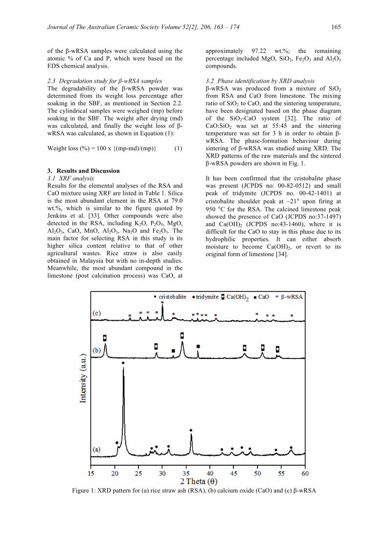
approximately 97.22 wt.%; the remaining percentage included MgO, SiO2, Fe2O3 and Al2O3 compounds. 3.2 Phase identification by XRD analysis β-wRSA was produced from a mixture of SiO2 from RSA and CaO from limestone. The mixing ratio of SiO2 to CaO, and the sintering temperature, have been designated based on the phase diagram of the SiO2-CaO system [32]. The ratio of CaO:SiO2 was set at 55:45 and the sintering temperature was set for 3 h in order to obtain β-wRSA. The phase-formation behaviour during sintering of β-wRSA was studied using XRD. The XRD patterns of the raw materials and the sintered β-wRSA powders are shown in Fig. 1. It has been confirmed that the cristobalite phase was present (JCPDS no: 00-82-0512) and small peak of tridymite (JCPDS no. 00-42-1401) at cristobalite shoulder peak at ~21° upon firing at 950 °C for the RSA. The calcined limestone peak showed the presence of CaO (JCPDS no:37-1497) and Ca(OH)2 (JCPDS no:43-1460), where it is difficult for the CaO to stay in this phase due to its hydrophilic properties. It can either absorb moisture to become Ca(OH)2, or revert to its original form of limestone [34].
Figure 1: XRD pattern for (a) rice straw ash (RSA), (b) calcium oxide (CaO) and (c) β-wRSA
Ismail et al. 166
Table 1: Ion concentrations of SBF and human blood plasma
Ion concentration
Human blood plasma (mM) SBF (mM)
Na+ 142.0 142.0 K+ 5.0 5.0 Mg2+ 1.5 1.5 Ca2+ 2.5 2.5 Cl- 103.0 148.8 HCO3
- 27.0 4.2 HPO4
2- 1.0 1.0 SO4
2- 0.5 0.0
Table 2: Compositions of the raw materials Composition Rice straw ash
(wt%) Calcined calcium carbonate (wt%)
SiO2 79.0 - K2O 9.70 - P2O5 1.47 - MgO 1.21 2.38 Al2O3 0.23 - CaO 2.02 97.22
Others 6.37 0.4
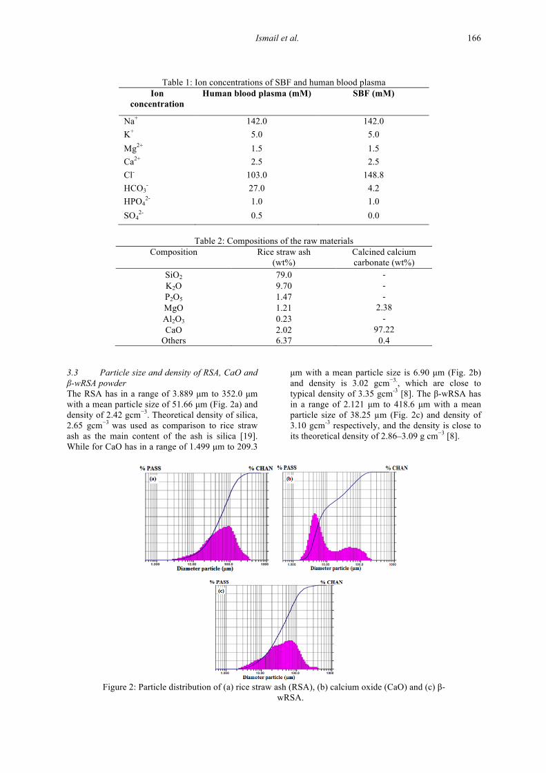
3.3 Particle size and density of RSA, CaO and β-wRSA powder The RSA has in a range of 3.889 µm to 352.0 µm with a mean particle size of 51.66 µm (Fig. 2a) and density of 2.42 gcm−3. Theoretical density of silica, 2.65 gcm−3 was used as comparison to rice straw ash as the main content of the ash is silica [19]. While for CaO has in a range of 1.499 µm to 209.3
µm with a mean particle size is 6.90 µm (Fig. 2b) and density is 3.02 gcm−3., which are close to typical density of 3.35 gcm-3 [8]. The β-wRSA has in a range of 2.121 µm to 418.6 µm with a mean particle size of 38.25 µm (Fig. 2c) and density of 3.10 gcm-3 respectively, and the density is close to its theoretical density of 2.86–3.09 g cm−3 [8].
Figure 2: Particle distribution of (a) rice straw ash (RSA), (b) calcium oxide (CaO) and (c) β-
wRSA.
Journal of The Australian Ceramic Society Volume 52[2], 206, 163 – 174 167
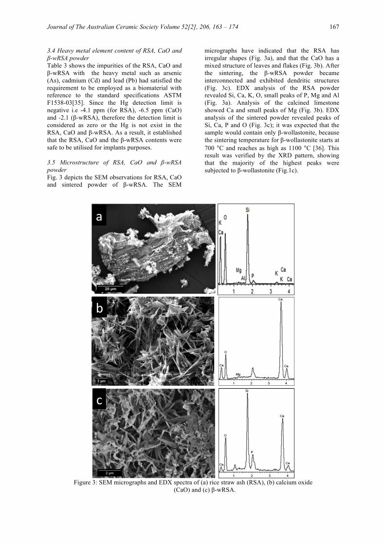
3.4 Heavy metal element content of RSA, CaO and β-wRSA powder Table 3 shows the impurities of the RSA, CaO and β-wRSA with the heavy metal such as arsenic (As), cadmium (Cd) and lead (Pb) had satisfied the requirement to be employed as a biomaterial with reference to the standard specifications ASTM F1538-03[35]. Since the Hg detection limit is negative i.e -4.1 ppm (for RSA), -6.5 ppm (CaO) and -2.1 (β-wRSA), therefore the detection limit is considered as zero or the Hg is not exist in the RSA, CaO and β-wRSA. As a result, it established that the RSA, CaO and the β-wRSA contents were safe to be utilised for implants purposes. 3.5 Microstructure of RSA, CaO and β-wRSA powder Fig. 3 depicts the SEM observations for RSA, CaO and sintered powder of β-wRSA. The SEM
micrographs have indicated that the RSA has irregular shapes (Fig. 3a), and that the CaO has a mixed structure of leaves and flakes (Fig. 3b). After the sintering, the β-wRSA powder became interconnected and exhibited dendritic structures (Fig. 3c). EDX analysis of the RSA powder revealed Si, Ca, K, O, small peaks of P, Mg and Al (Fig. 3a). Analysis of the calcined limestone showed Ca and small peaks of Mg (Fig. 3b). EDX analysis of the sintered powder revealed peaks of Si, Ca, P and O (Fig. 3c); it was expected that the sample would contain only β-wollastonite, because the sintering temperature for β-wollastonite starts at 700 °C and reaches as high as 1100 °C [36]. This result was verified by the XRD pattern, showing that the majority of the highest peaks were subjected to β-wollastonite (Fig.1c).
Figure 3: SEM micrographs and EDX spectra of (a) rice straw ash (RSA), (b) calcium oxide
(CaO) and (c) β-wRSA.
a
b
c
Ismail et al. 168
Table 3: Trace heavy metal element of raw materials and β-wRSA Sample Heavy element content (ppm)
Arsenic (As) Cadmium (Cd) Plumbum (Pb) Mercury (Hg) Rice straw ash (RSA) 0.015 0.001 0.013 0.0 Calcium oxide (CaO) 0.017 0.0000 0.058 0.0 β-wRSA 0.045 0.003 0.014 0.0 ASTM F 1538-03 3 5 30 5
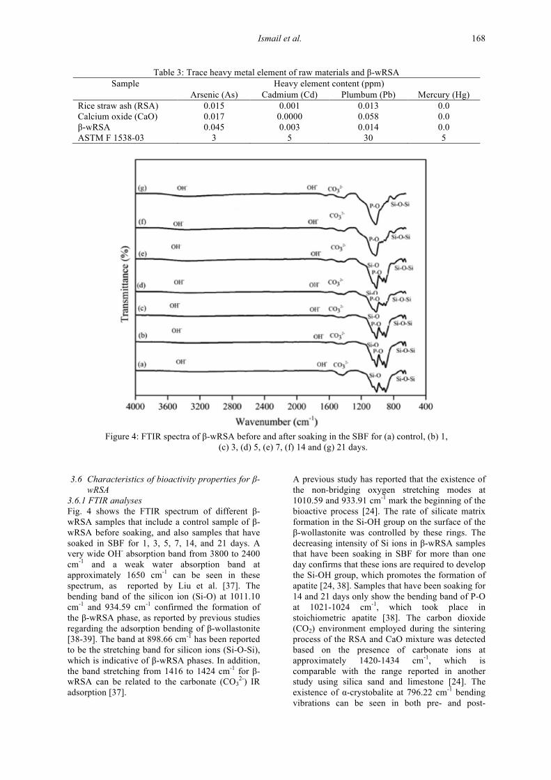
Figure 4: FTIR spectra of β-wRSA before and after soaking in the SBF for (a) control, (b) 1,
(c) 3, (d) 5, (e) 7, (f) 14 and (g) 21 days. 3.6 Characteristics of bioactivity properties for β-
wRSA 3.6.1 FTIR analyses Fig. 4 shows the FTIR spectrum of different β-wRSA samples that include a control sample of β-wRSA before soaking, and also samples that have soaked in SBF for 1, 3, 5, 7, 14, and 21 days. A very wide OH- absorption band from 3800 to 2400 cm-1 and a weak water absorption band at approximately 1650 cm-1 can be seen in these spectrum, as reported by Liu et al. [37]. The bending band of the silicon ion (Si-O) at 1011.10 cm-1 and 934.59 cm-1 confirmed the formation of the β-wRSA phase, as reported by previous studies regarding the adsorption bending of β-wollastonite [38-39]. The band at 898.66 cm-1 has been reported to be the stretching band for silicon ions (Si-O-Si), which is indicative of β-wRSA phases. In addition, the band stretching from 1416 to 1424 cm-1 for β-wRSA can be related to the carbonate (CO3
2-) IR adsorption [37].
A previous study has reported that the existence of the non-bridging oxygen stretching modes at 1010.59 and 933.91 cm-1 mark the beginning of the bioactive process [24]. The rate of silicate matrix formation in the Si-OH group on the surface of the β-wollastonite was controlled by these rings. The decreasing intensity of Si ions in β-wRSA samples that have been soaking in SBF for more than one day confirms that these ions are required to develop the Si-OH group, which promotes the formation of apatite [24, 38]. Samples that have been soaking for 14 and 21 days only show the bending band of P-O at 1021-1024 cm-1, which took place in stoichiometric apatite [38]. The carbon dioxide (CO2) environment employed during the sintering process of the RSA and CaO mixture was detected based on the presence of carbonate ions at approximately 1420-1434 cm-1, which is comparable with the range reported in another study using silica sand and limestone [24]. The existence of α-crystobalite at 796.22 cm-1 bending vibrations can be seen in both pre- and post-
Journal of The Australian Ceramic Society Volume 52[2], 206, 163 – 174 169
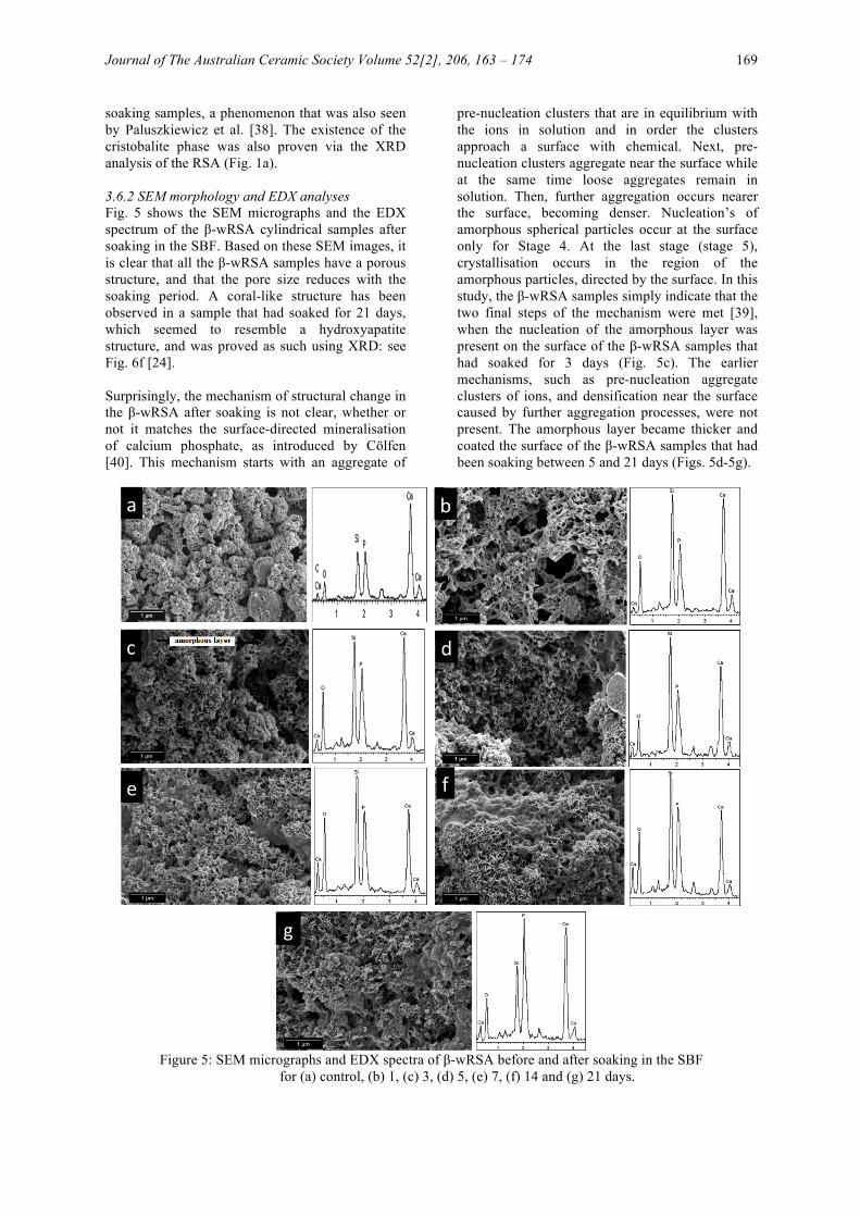
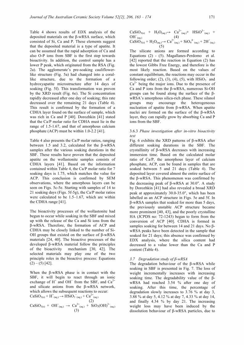
soaking samples, a phenomenon that was also seen by Paluszkiewicz et al. [38]. The existence of the cristobalite phase was also proven via the XRD analysis of the RSA (Fig. 1a). 3.6.2 SEM morphology and EDX analyses Fig. 5 shows the SEM micrographs and the EDX spectrum of the β-wRSA cylindrical samples after soaking in the SBF. Based on these SEM images, it is clear that all the β-wRSA samples have a porous structure, and that the pore size reduces with the soaking period. A coral-like structure has been observed in a sample that had soaked for 21 days, which seemed to resemble a hydroxyapatite structure, and was proved as such using XRD: see Fig. 6f [24]. Surprisingly, the mechanism of structural change in the β-wRSA after soaking is not clear, whether or not it matches the surface-directed mineralisation of calcium phosphate, as introduced by Cölfen [40]. This mechanism starts with an aggregate of
pre-nucleation clusters that are in equilibrium with the ions in solution and in order the clusters approach a surface with chemical. Next, pre-nucleation clusters aggregate near the surface while at the same time loose aggregates remain in solution. Then, further aggregation occurs nearer the surface, becoming denser. Nucleation’s of amorphous spherical particles occur at the surface only for Stage 4. At the last stage (stage 5), crystallisation occurs in the region of the amorphous particles, directed by the surface. In this study, the β-wRSA samples simply indicate that the two final steps of the mechanism were met [39], when the nucleation of the amorphous layer was present on the surface of the β-wRSA samples that had soaked for 3 days (Fig. 5c). The earlier mechanisms, such as pre-nucleation aggregate clusters of ions, and densification near the surface caused by further aggregation processes, were not present. The amorphous layer became thicker and coated the surface of the β-wRSA samples that had been soaking between 5 and 21 days (Figs. 5d-5g).
Figure 5: SEM micrographs and EDX spectra of β-wRSA before and after soaking in the SBF
for (a) control, (b) 1, (c) 3, (d) 5, (e) 7, (f) 14 and (g) 21 days.
a b
c d
e f
g
Ismail et al. 170
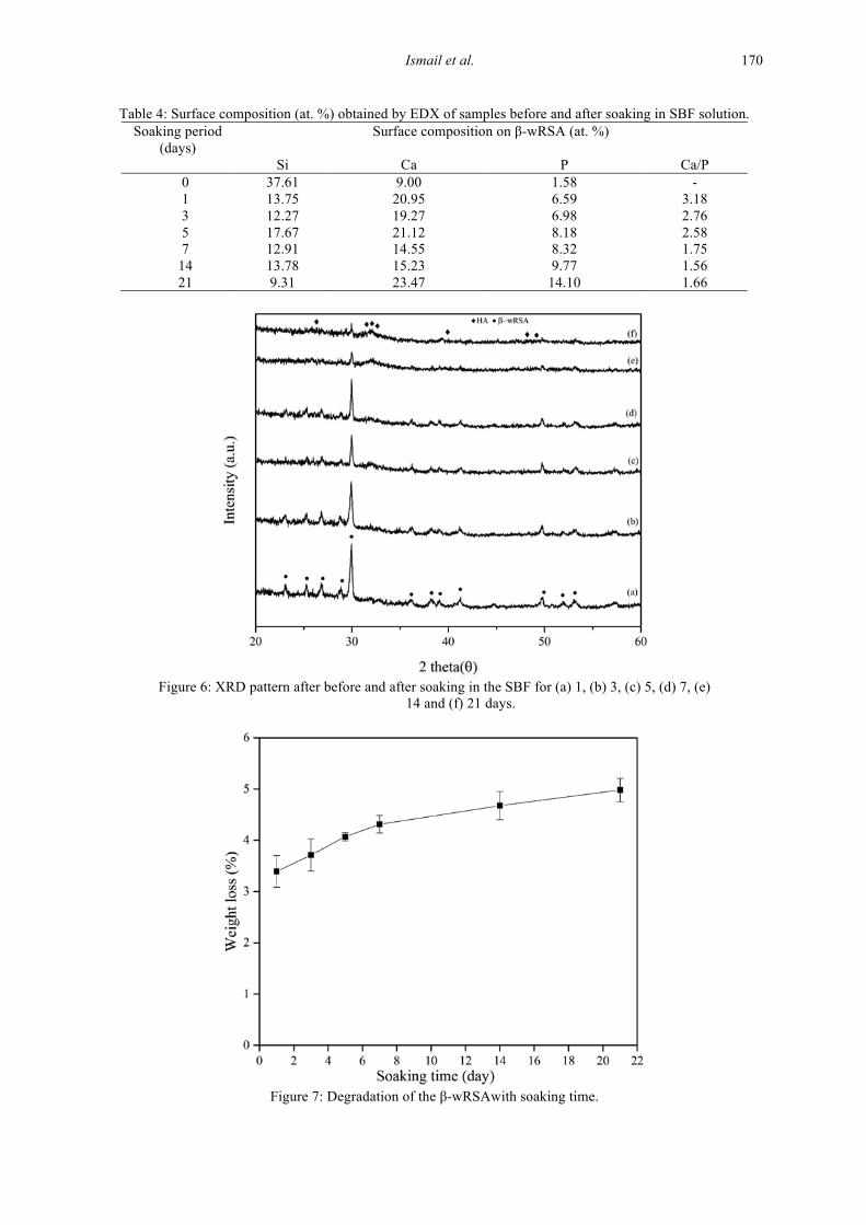
Table 4: Surface composition (at. %) obtained by EDX of samples before and after soaking in SBF solution. Soaking period
(days) Surface composition on β-wRSA (at. %)
Si Ca P Ca/P 0 37.61 9.00 1.58 - 1 13.75 20.95 6.59 3.18 3 12.27 19.27 6.98 2.76 5 17.67 21.12 8.18 2.58 7 12.91 14.55 8.32 1.75
14 13.78 15.23 9.77 1.56 21 9.31 23.47 14.10 1.66
Figure 6: XRD pattern after before and after soaking in the SBF for (a) 1, (b) 3, (c) 5, (d) 7, (e)
14 and (f) 21 days.
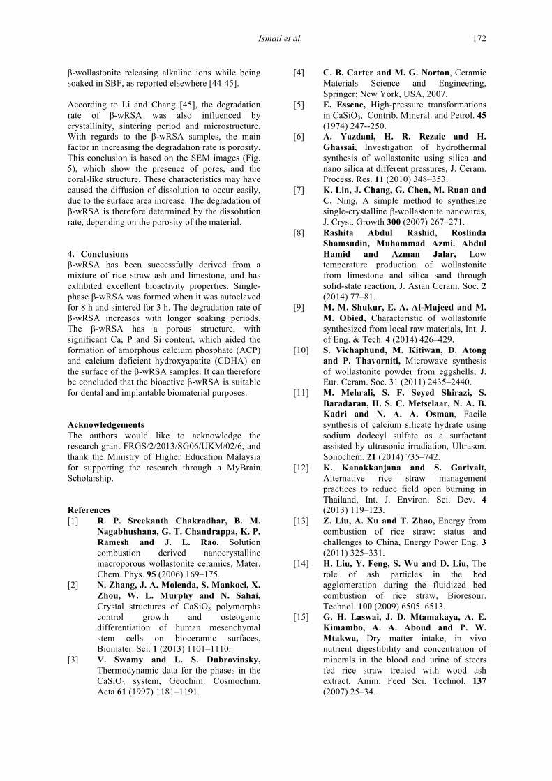
Figure 7: Degradation of the β-wRSAwith soaking time.
Journal of The Australian Ceramic Society Volume 52[2], 206, 163 – 174 171
Table 4 shows results of EDX analysis of the deposited materials on the β-wRSA surface, which consisted of Si, Ca and P. These elements suggest that the deposited material is a type of apatite. It can be assumed that the rapid adsorption of Ca and also O-P ions from SBF is the first step towards bioactivity. In addition, the control sample has a lower P peak, which originated from the RSA (Fig. 2a). The agglomerate’s pre-soaking cauliflower-like structure (Fig. 5a) had changed into a coral-like structure, due to the formation of a hydroxyapatite microstructure after 14 days of soaking (Fig. 5f). This transformation was proven by the XRD result (Fig. 6e). The Si concentration rapidly decreased after one day of soaking; this rate decreased over the remaining 21 days (Table 4). This result is confirmed by the formation of a CDHA layer found on the surface of sample, which was rich in Ca and P [40]. Dorozhkin [41] stated that the Ca/P molar ratio for CDHA must lie in the range of 1.5-1.67, and that of amorphous calcium phosphate (ACP) must be within 1.0-2.2 [41]. Table 4 also presents the Ca/P molar ratios, ranging between 1.5 and 3.2, calculated for the β-wRSA samples after the various soaking durations in the SBF. These results have proven that the deposited apatite on the wollastonite samples consists of CDHA layers [41]. Based on the information contained within Table 4, the Ca/P molar ratio for 7 soaking days is 1.75, which matches the value for ACP. This conclusion is confirmed by SEM observations, where the amorphous layers can be seen on Figs. 5c-5e. Starting with samples of 14 to 21 soaking days (Figs. 5f-5g), the Ca/P molar ratios were calculated to be 1.5–1.67, which are within the CDHA range [41]. The bioactivity processes of the wollastonite had begun to occur while soaking in the SBF and mixed up with the release of the Ca and Si ions from the β-wRSA. Therefore, the formation of ACP and CDHA may be closely linked to the number of Si-OH groups that existed on the surface of β-wRSA materials [24, 40]. The bioactive processes of the developed β-wRSA material follow the principles of the bioactivity mechanism [30, 42]. The selected materials may play one of the two principle roles in the bioactive process: Equations (2) – (5) [42]. When the β-wRSA phase is in contact with the SBF, it will begin to react through an ionic exchange of H+ and OH− from the SBF, and Ca2+ and silicate anions from the β-wRSA network, which allows the subsequent reactions to occur: CaSiO3(s) + H+
(aq.) → HSiO3−
(aq.) + Ca2+(aq.)
(2) CaSiO3(s) + OH−
(aq.) → Ca2+(aq.) + SiO3(OH)3−
(aq.) (3)
CaSiO3(s) + H2O(aq.)→ Ca2+(aq.)+ HSiO3−
(aq.) + OH−
(aq.) (4) CaSiO3(s) + H2O(aq.)→ Ca2+
(aq.) + SiO44−
(aq.) + 2H+(aq.)
(5) The silicate anions are formed according to Equations (2) - (5). Magallanes-Perdomo et al. [42] reported that the reaction in Equation (2) has the lowest Gibbs Free Energy, and therefore is the most likely reaction. Based on the values of constant equilibrium, the reactions may occur in the following order; (2), (3), (4), (5), with HSiO3
− and Ca2+ being the major ions. Due to the presence of Ca and P ions from the β-wRSA, numerous Si-OH groups can be found along the surface of the β-wRSA’s amorphous silica-rich phase. These silanol groups may encourage the heterogeneous nucleation of apatite from β-wRSA. When apatite nuclei are formed on the surface of the β-wRSA layer, they can rapidly grow by absorbing Ca and P ions from the SBF. 3.6.3 Phase investigation after in-vitro bioactivity test Fig. 6 exhibits the XRD patterns of β-wRSA after different soaking durations in the SBF. The crystallinity of β-wRSA decreases with increasing immersion time. Based on the calculated molar ratio of Ca/P, the amorphous layer of calcium phosphate, ACP, can be found in samples that are soaked between 5 and 21 days (Table 4). The deposited layer covered almost the entire surface of the β-wRSA. This phenomenon was confirmed by the decreasing peak of β-wRSA at 30.0°. A study by Dorozhkin [41] had also revealed a broad XRD peak at approximately 30.0-35.0°, which has been labelled as an ACP structure in Figs. 5e and 5f. In β-wRSA samples that soaked for more than 5 days, the previously unstable ACP structure became more prominent [40, 43], and the poorly crystalline HA (JCPDS no: 72-1243) began to form from the conversion of ACP [40]. CDHA is formed in samples soaking for between 14 and 21 days. No β-wRSA peaks have been detected in the sample that soaked for 21 days; this absence was confirmed by EDX analysis, where the silica content had decreased to a value lower than the Ca and P content (Table 4). 3.7 Degradation study of β-wRSA The degradation behaviour of the β-wRSA while soaking in SBF is presented in Fig. 7. The loss of weight incrementally increases with increasing soaking time. The degradability value of the β-wRSA had reached 3.54 % after one day of soaking. After this time, the percentage of degradation slowly increases to 3.76 % at day 3, 3.88 % at day 5, 4.12 % at day 7, 4.33 % at day 14, and finally 4.34 % by day 21. The increasing weight loss may have been induced by the dissolution behaviour of β-wRSA particles, due to
Ismail et al. 172
β-wollastonite releasing alkaline ions while being soaked in SBF, as reported elsewhere [44-45]. According to Li and Chang [45], the degradation rate of β-wRSA was also influenced by crystallinity, sintering period and microstructure. With regards to the β-wRSA samples, the main factor in increasing the degradation rate is porosity. This conclusion is based on the SEM images (Fig. 5), which show the presence of pores, and the coral-like structure. These characteristics may have caused the diffusion of dissolution to occur easily, due to the surface area increase. The degradation of β-wRSA is therefore determined by the dissolution rate, depending on the porosity of the material. 4. Conclusions β-wRSA has been successfully derived from a mixture of rice straw ash and limestone, and has exhibited excellent bioactivity properties. Single-phase β-wRSA was formed when it was autoclaved for 8 h and sintered for 3 h. The degradation rate of β-wRSA increases with longer soaking periods. The β-wRSA has a porous structure, with significant Ca, P and Si content, which aided the formation of amorphous calcium phosphate (ACP) and calcium deficient hydroxyapatite (CDHA) on the surface of the β-wRSA samples. It can therefore be concluded that the bioactive β-wRSA is suitable for dental and implantable biomaterial purposes. Acknowledgements The authors would like to acknowledge the research grant FRGS/2/2013/SG06/UKM/02/6, and thank the Ministry of Higher Education Malaysia for supporting the research through a MyBrain Scholarship. References [1] R. P. Sreekanth Chakradhar, B. M.
Nagabhushana, G. T. Chandrappa, K. P. Ramesh and J. L. Rao, Solution combustion derived nanocrystalline macroporous wollastonite ceramics, Mater. Chem. Phys. 95 (2006) 169–175.
[2] N. Zhang, J. A. Molenda, S. Mankoci, X. Zhou, W. L. Murphy and N. Sahai, Crystal structures of CaSiO3 polymorphs control growth and osteogenic differentiation of human mesenchymal stem cells on bioceramic surfaces, Biomater. Sci. 1 (2013) 1101–1110.
[3] V. Swamy and L. S. Dubrovinsky, Thermodynamic data for the phases in the CaSiO3 system, Geochim. Cosmochim. Acta 61 (1997) 1181–1191.
[4] C. B. Carter and M. G. Norton, Ceramic Materials Science and Engineering, Springer: New York, USA, 2007.
[5] E. Essene, High-pressure transformations in CaSiO3, Contrib. Mineral. and Petrol. 45 (1974) 247--250.
[6] A. Yazdani, H. R. Rezaie and H. Ghassai, Investigation of hydrothermal synthesis of wollastonite using silica and nano silica at different pressures, J. Ceram. Process. Res. 11 (2010) 348–353.
[7] K. Lin, J. Chang, G. Chen, M. Ruan and C. Ning, A simple method to synthesize single-crystalline β-wollastonite nanowires, J. Cryst. Growth 300 (2007) 267–271.
[8] Rashita Abdul Rashid, Roslinda Shamsudin, Muhammad Azmi. Abdul Hamid and Azman Jalar, Low temperature production of wollastonite from limestone and silica sand through solid-state reaction, J. Asian Ceram. Soc. 2 (2014) 77–81.
[9] M. M. Shukur, E. A. Al-Majeed and M. M. Obied, Characteristic of wollastonite synthesized from local raw materials, Int. J. of Eng. & Tech. 4 (2014) 426–429.
[10] S. Vichaphund, M. Kitiwan, D. Atong and P. Thavorniti, Microwave synthesis of wollastonite powder from eggshells, J. Eur. Ceram. Soc. 31 (2011) 2435–2440.
[11] M. Mehrali, S. F. Seyed Shirazi, S. Baradaran, H. S. C. Metselaar, N. A. B. Kadri and N. A. A. Osman, Facile synthesis of calcium silicate hydrate using sodium dodecyl sulfate as a surfactant assisted by ultrasonic irradiation, Ultrason. Sonochem. 21 (2014) 735–742.
[12] K. Kanokkanjana and S. Garivait, Alternative rice straw management practices to reduce field open burning in Thailand, Int. J. Environ. Sci. Dev. 4 (2013) 119–123.
[13] Z. Liu, A. Xu and T. Zhao, Energy from combustion of rice straw: status and challenges to China, Energy Power Eng. 3 (2011) 325–331.
[14] H. Liu, Y. Feng, S. Wu and D. Liu, The role of ash particles in the bed agglomeration during the fluidized bed combustion of rice straw, Bioresour. Technol. 100 (2009) 6505–6513.
[15] G. H. Laswai, J. D. Mtamakaya, A. E. Kimambo, A. A. Aboud and P. W. Mtakwa, Dry matter intake, in vivo nutrient digestibility and concentration of minerals in the blood and urine of steers fed rice straw treated with wood ash extract, Anim. Feed Sci. Technol. 137 (2007) 25–34.
Journal of The Australian Ceramic Society Volume 52[2], 206, 163 – 174 173
[16] C. G. Rocha, D. A. Zaia, R. V. Alfaya and A. A. Alfaya, Use of rice straw as biosorbent for removal of Cu(II), Zn(II), Cd(II) and Hg(II) ions in industrial effluents, J. Hazard. Mater. 166 (2009) 383–388.
[17] P. Binod, R. Sindhu, R. R. Singhania, S. Vikram, L. Devi, S. Nagalakshmi, N. Kurien, R. K. Sukumaran and A. Pandey, Bioethanol production from rice straw: An overview, Bioresour. Technol. 101 (2010) 4767–4774.
[18] J. K. Ko, J. S. Bak, M. W. Jung, H. J. Lee, I.-G. Choi, T. H. Kim and K. H. Kim, Ethanol production from rice straw using optimized aqueous-ammonia soaking pretreatment and simultaneous saccharification and fermentation processes, Bioresour. Technol. 100 (2009) 4374–4380.
[19] J. P. Nayak, S. Kumar and J. Bera, Sol–gel synthesis of bioglass-ceramics using rice husk ash as a source for silica and its characterization, J. Non. Cryst. Solids 356 (2010) 1447–1451.
[20] N. S. C. Zulkifli, I. Ab Rahman, D. Mohamad and A. Husein, A green sol–gel route for the synthesis of structurally controlled silica particles from rice husk for dental composite filler, Ceram. Int. 39 (2013) 4559–4567.
[21] M. Noushad, I. Ab Rahman, N. S. Che Zulkifli, A. Husein and D. Mohamad, Low surface area nanosilica from an agricultural biomass for fabrication of dental nanocomposites, Ceram. Int. 40 (2014) 4163–4171.
[22] Y. H. Yun, S. B. Kim, B. A. Kang, Y. W. Lee, J. S. Oh and K. S. Hwang, β-wollastonite reinforced glass-ceramics prepared from waste fluorescent glass and calcium carbonate, J. Mater. Process. Technol. 178 (2006) 61–66.
[23] H. Li and J. Chang, Preparation and characterization of bioactive and biodegradable wollastonite/poly(D, L-lactic acid) composite scaffolds, J. Mater. Sci. Mater. Med. 15 (2004) 1089–1095.
[24] R. A. Rashid, R. Shamsudin, M. A. A. Hamid and A. Jalar, In-vitro bioactivity of wollastonite materials derived from limestone and silica sand, Ceram. Int. 40 (2014) 6847–6853.
[25] S. K. Padmanabhan, F. Gervaso, M. Carrozzo, F. Scalera, A. Sannino and A. Licciulli, Wollastonite/hydroxyapatite scaffolds with improved mechanical, bioactive and biodegradable properties for bone tissue engineering, Ceram. Int. 39 (2013) 619–627.
[26] X. Wan, C. Chang, D. Mao, L. Jiang and M. Li, Preparation and in vitro bioactivities of calcium silicate nanophase materials, Mater. Sci. Eng. C 25 (2005) 455–461.
[27] T. Kokubo, Bioactive glass ceramics: properties and applications, Biomaterials 12 (1991) 155-163. [28] T. Kokubo and H. Takadama, How
useful is SBF in predicting in vivo bone bioactivity?, Biomaterials 27 (2006) 2907-2915.
[29] L. N. Niu, K. Jiao, T. Wang, W. Zhang, J. Camilleri, B. E. Bergeron, H.-L. Feng, J. Mao, J. H. Chen, D. H. Pashley and F. R. Tay, A review of the bioactivity of hydraulic calcium silicate cements, J. Dent. 42 (2014) 517–533.
[30] L. L. Hench, Bioactive Materials, Ceram. Int. 8842 (1996) 493–507.
[31] L. Z. Pei, L. J. Yang, Y. Yang, C. G. Fan, W. Y. Yin, J. Chen and Q. F. Zhang, A green and facile route to synthesize calcium silicate nanowires, Mater. Charact. 61 (2010) 1281–1285.
[32] U. J. G. Erlend, F. Nordstrand, A. N. Dibbs, A. J. Eraker, E. F. Nordstrand, A. N. Dibbs, A. J. Eraker and U. J. Gibson, Alkaline oxide interface modifiers for silicon fiber production, Opt. Mater. Express 3 (2013) 651-656.
[33] B. M. Jenkins, L. L. Baxter, T. R. M. Jr and T. R. Miles, Combustion properties of biomass, Fuel Process Tech. 54 (1998) 17-46.
[34] T. N. Blanton and C. L. Barnes, Quantitative analysis of calcium oxide desiccant conversion to calcium hydroxide using x-ray diffraction, Adv. X-Ray Anal. 48 (2005) 45–51.
[35] ASTM, Standard Specification for Glass and Glass Ceramic Biomaterials for Implantation, ASTM F1538-03 (2008).
[36] L. H. Long, L. D. Chen and J. Chang, Low temperature fabrication and characterizations of β-CaSiO3 ceramics, Ceram. Int. 32 (2006) 457–460.
[37] X. Liu, C. Ding and Z. Wang, Apatite formed on the surface of plasma-sprayed wollastonite coating immersed in simulated body fluid, Biomaterials 22 (2001) 2007–2012.
[38] C. Paluszkiewicz, M. Blażewicz, J. Podporska and T. Gumuła, Nucleation of hydroxyapatite layer on wollastonite material surface: FTIR studies, Vib. Spectrosc. 48 (2008) 263–268.
[39] S. J. Gadaleta, E. P. Paschalis, F. Betts, R. Mendelsohn and A. L. Boskey, Fourier transform infrared spectroscopy of the solution-mediated conversion of
Ismail et al. 174
amorphous calcium phosphate to hydroxyapatite: new correlations between X-ray diffraction and infrared data, Calcif. Tissue Int. 58 (1996) 9–16.
[40] H. Cölfen, Biomineralization: A crystal-clear view, Nat. Mater. 9 (2010) 960–961.
[41] S. V. Dorozhkin, Amorphous calcium (ortho)phosphates, Acta Biomater. 6 (2010) 4457–4475.
[42] M. Magallanes-Perdomo, Z. B. Luklinska, A. H. De Aza, R. G. Carrodeguas, S. De Aza and P. Pena, Bone-like forming ability of apatite-wollastonite glass ceramic, J. Eur. Ceram. Soc. 31 (2011) 1549–1561
[43] E. D. Eanes, Amorphous calcium phosphate, Monogr. Oral Sci. 18 (2001) 130–147.
[44] F. Zhang, J. Chang, K. Lin and J. Lu, Preparation, mechanical properties and in vitro degradability of wollastonite/tricalcium phosphate macroporous scaffolds from nanocomposite powders, J. Mater. Sci. Mater. Med. 19 (2008) 167–173.
[45] H. Li and J. Chang, Fabrication and characterization of bioactive wollastonite/PHBV composite scaffolds, Biomaterials 25 (2004) 5473–54.

Journal of The Australian Ceramic Society Volume 52[2], 206, 163 – 174 175

![ΕΓΧΕΙΡΙΔΙΟ ΑΝΑΚΥΚΛΩΣΗΣ · 2019. 11. 4. · 6 ΜΠΑΡ: 1. H καμπνια μας ΝΟ STRAW εναι σε ισ ] gει και αποε gγ να βζ πλασ](https://static.fdocument.org/doc/165x107/5fe3deb8d7097c1ed315a967/-2019-11-4-6-oe-1-h-.jpg)