
c2gc36556a 525..534 - UMEXPERT · human hair, finger nails, ... XRD experiment, approximately, ......
10
Green Chemistry PAPER Cite this: Green Chem., 2013, 15, 525 Received 2nd October 2012, Accepted 13th December 2012 DOI: 10.1039/c2gc36556a www.rsc.org/greenchem Dissolution of feather keratin in ionic liquids† Azila Idris,* a,b R. Vijayaraghavan, a Usman Ali Rana,‡ a Dale Fredericks, c A. F. Patti a and D. R. MacFarlane a Keratin from various livestock industries is currently a waste material that has potential as a source of polyamide polymers that could replace fossil fuel derived materials if processing methods can be develo- ped. In this work we have investigated methods for the dissolution and regeneration of keratin. Dissol- ution of keratin (from turkey feather) in ionic liquids was conducted under nitrogen at 130 °C for 10 hours. It was found that [BMIM]Cl, [AMIM]Cl and [choline][thioglycolate] could dissolve turkey feather keratin without addition of solvent or other chemicals. A significant percentage of solubility was obtained, up to 45% by weight. Awater insoluble fraction was recovered by addition of water to the solution (∼50%). The structure and properties of this regenerated, water insoluble fraction were investi- gated. Compared to the starting material, the regenerated keratin shows structural changes rather than chemical changes within the polypeptide chains. The remaining fraction, consisting of water soluble frag- ments, was characterised by gel electrophoresis. 1. Introduction Keratin is a fibrous protein that can be found in feather, wool, human hair, finger nails, animal claws and horn. 1,2 These bio- polymers are plentifully available as a by-product from poultry production and textile industries. 3–5 However this kind of protein is not easy to dissolve or extract because keratin is bound by strong internal interactions that stabilise the protein structure. 4,6 In order to utilise these biopolymers and convert them to usable materials there is a need for alternative sol- vents that are efficient in dissolving keratin and preserving, at least in part, the protein structure. The main objective of the present study therefore is to investigate the use of ionic liquids (ILs) in the dissolution and regeneration of keratin, in particu- lar utilising the unique solvency characteristics and high temp- erature properties of ILs. Feathers are about 90% keratin 7 which contains about 7% of cysteine. 8,9 Feather proteins are insoluble in normal organic solvent due to the tight packing of the α-helix and β-sheet in the polypeptide chain. Additionally, a high degree of crosslinking of the polypeptide chain is affected by cysteine residues which participate in disulfide bridge formation. 6,8,10 In order to extract the keratin, cleaving the disulfide bonds can be achieved by reduction, 11–13 oxidation, 12–14 sulfitolysis 13,15 or oxidative sulfitolysis. 13,16 Even though many reagents have the capability to reduce the disulfide bonds, only a few, such as thiols 17 and ammonium bisulfite, have the required ability and reactivity to preserve and maintain the protein structure. However, these materials are difficult to recycle, often toxic and costly to produce. Ionic liquids (ILs) are generally defined as salts which are liquid below 100 °C and comprised entirely of cations and anions. 18,19 They offer a unique combination of properties as solvents including in some cases low vapour pressure and high thermal stability. 20 The hydrophobicity/hydrophilicity of ionic liquids can be altered by manipulating the structures of the cations and anions. 21 In recent years, a number of ionic liquids have been identified as solvents for the dissolution of biopolymers such as cellulose, starch, wood, lignin, feather and wool. These are generally insoluble in organic solvents but show a reasonable solubility in certain ionic liquids. 4,22–28 Duck feather keratin, for example, was found to be soluble in the ionic liquids (1-butyl-3-methylimidazolium chloride [BMIM]Cl and 1-allyl-3-methylimidazolium chloride [AMIM]- Cl). 29 The same ionic liquids have been found to be useful for wool dissolution by Xie et al. 4 [BMIM]Cl was also found to be useful in the synthesis of wool keratin/cellulose composite materials. The materials displayed a homogeneous structure and did not show a residual fiber structure. 4 Therefore, the dissolution of biopolymers using ionic liquids provide † Electronic supplementary information (ESI) available. See DOI: 10.1039/c2gc36556a ‡ Current address: Sustainable Energy Technologies (SET) center, College of Engineering, King Saud University, PO Box 800, Riyadh 11421, Kingdom of Saudi Arabia. a School of Chemistry, Faculty of Science, Monash University, Clayton, VIC 3800, Australia b Department of Chemistry, Faculty of Science, University of Malaya, 50603 Kuala Lumpur, Malaysia c ARC Special Research Centre for Green Chemistry, Monash University, Clayton, VIC 3800, Australia. E-mail: [email protected], azila̲[email protected] This journal is © The Royal Society of Chemistry 2013 Green Chem., 2013, 15, 525–534 | 525 Downloaded by Monash University on 27 March 2013 Published on 13 December 2012 on http://pubs.rsc.org | doi:10.1039/C2GC36556A View Article Online View Journal | View Issue
Transcript of c2gc36556a 525..534 - UMEXPERT · human hair, finger nails, ... XRD experiment, approximately, ......
Green Chemistry
PAPER
Cite this: Green Chem., 2013, 15, 525
Received 2nd October 2012,Accepted 13th December 2012
DOI: 10.1039/c2gc36556a
www.rsc.org/greenchem
Dissolution of feather keratin in ionic liquids†
Azila Idris,*a,b R. Vijayaraghavan,a Usman Ali Rana,‡a Dale Fredericks,c A. F. Pattia
and D. R. MacFarlanea
Keratin from various livestock industries is currently a waste material that has potential as a source of
polyamide polymers that could replace fossil fuel derived materials if processing methods can be develo-
ped. In this work we have investigated methods for the dissolution and regeneration of keratin. Dissol-
ution of keratin (from turkey feather) in ionic liquids was conducted under nitrogen at 130 °C for
10 hours. It was found that [BMIM]Cl, [AMIM]Cl and [choline][thioglycolate] could dissolve turkey
feather keratin without addition of solvent or other chemicals. A significant percentage of solubility
was obtained, up to 45% by weight. A water insoluble fraction was recovered by addition of water to the
solution (∼50%). The structure and properties of this regenerated, water insoluble fraction were investi-
gated. Compared to the starting material, the regenerated keratin shows structural changes rather than
chemical changes within the polypeptide chains. The remaining fraction, consisting of water soluble frag-
ments, was characterised by gel electrophoresis.
1. Introduction
Keratin is a fibrous protein that can be found in feather, wool,human hair, finger nails, animal claws and horn.1,2 These bio-polymers are plentifully available as a by-product from poultryproduction and textile industries.3–5 However this kind ofprotein is not easy to dissolve or extract because keratin isbound by strong internal interactions that stabilise the proteinstructure.4,6 In order to utilise these biopolymers and convertthem to usable materials there is a need for alternative sol-vents that are efficient in dissolving keratin and preserving, atleast in part, the protein structure. The main objective of thepresent study therefore is to investigate the use of ionic liquids(ILs) in the dissolution and regeneration of keratin, in particu-lar utilising the unique solvency characteristics and high temp-erature properties of ILs. Feathers are about 90% keratin7
which contains about 7% of cysteine.8,9 Feather proteins areinsoluble in normal organic solvent due to the tight packing ofthe α-helix and β-sheet in the polypeptide chain. Additionally,
a high degree of crosslinking of the polypeptide chain isaffected by cysteine residues which participate in disulfidebridge formation.6,8,10 In order to extract the keratin, cleavingthe disulfide bonds can be achieved by reduction,11–13
oxidation,12–14 sulfitolysis13,15 or oxidative sulfitolysis.13,16
Even though many reagents have the capability to reduce thedisulfide bonds, only a few, such as thiols17 and ammoniumbisulfite, have the required ability and reactivity to preserveand maintain the protein structure. However, these materialsare difficult to recycle, often toxic and costly to produce.
Ionic liquids (ILs) are generally defined as salts which areliquid below 100 °C and comprised entirely of cations andanions.18,19 They offer a unique combination of properties assolvents including in some cases low vapour pressure andhigh thermal stability.20 The hydrophobicity/hydrophilicity ofionic liquids can be altered by manipulating the structures ofthe cations and anions.21 In recent years, a number of ionicliquids have been identified as solvents for the dissolution ofbiopolymers such as cellulose, starch, wood, lignin, featherand wool. These are generally insoluble in organic solvents butshow a reasonable solubility in certain ionic liquids.4,22–28
Duck feather keratin, for example, was found to be soluble inthe ionic liquids (1-butyl-3-methylimidazolium chloride[BMIM]Cl and 1-allyl-3-methylimidazolium chloride [AMIM]-Cl).29 The same ionic liquids have been found to be useful forwool dissolution by Xie et al.4 [BMIM]Cl was also found to beuseful in the synthesis of wool keratin/cellulose compositematerials. The materials displayed a homogeneous structureand did not show a residual fiber structure.4 Therefore, thedissolution of biopolymers using ionic liquids provide
†Electronic supplementary information (ESI) available. See DOI:10.1039/c2gc36556a‡Current address: Sustainable Energy Technologies (SET) center, College ofEngineering, King Saud University, PO Box 800, Riyadh 11421, Kingdom ofSaudi Arabia.
aSchool of Chemistry, Faculty of Science, Monash University, Clayton, VIC 3800,
AustraliabDepartment of Chemistry, Faculty of Science, University of Malaya,
50603 Kuala Lumpur, MalaysiacARC Special Research Centre for Green Chemistry, Monash University, Clayton,
VIC 3800, Australia. E-mail: [email protected], azila̲[email protected]
This journal is © The Royal Society of Chemistry 2013 Green Chem., 2013, 15, 525–534 | 525
Dow
nloa
ded
by M
onas
h U
nive
rsity
on
27 M
arch
201
3Pu
blis
hed
on 1
3 D
ecem
ber
2012
on
http
://pu
bs.r
sc.o
rg |
doi:1
0.10
39/C
2GC
3655
6A
View Article OnlineView Journal | View Issue
possibilities for the utilisation and conversion of biopolymersinto useful materials with great potential in industrialapplications.
In the current study we have used [BMIM]Cl, [AMIM]Cl, aswell as novel ionic liquids containing a thiol functional group[choline][thioglycolate] and [bis-(2-ethylhexyl)ammonium]-[thioglycolate] as possible disulfide bridge reducing, orexchange reagents, to dissolve and regenerate the keratin inturkey feather. A combination of analytical techniques includ-ing ATR-FTIR, XRD, solid state NMR, TGA, DSC and gel electro-phoresis has been used to investigate the extent of keratinsolubility in these ionic liquids and the physiochemical prop-erties of the regenerated keratin material. We show that a sig-nificant amount of keratin (approximately 45–50 wt%) can bedissolved in the above mentioned ionic liquids.
2. ExperimentalMaterials preparation
The cleaned turkey feathers used in the experiments were com-mercial materials supplied by Shamrockcraft, purchased fromSpotlight, Clayton, Australia. Only barbs of the turkey featherswere used in the experiments. 1-Butyl-3-methylimidazoliumchloride ([BMIM]Cl, 98% of purity) was purchased from SigmaAldrich. All other ionic liquids were prepared in-house for thisproject as described further below. For the preparation ofthese ionic liquids, allylchloride (98%), N-methylimidazole(99%), choline hydroxide (20 wt% in H2O), thioglycolic acid(98%) and bis-(2-ethylhexyl)amine (99%) were obtained fromSigma Aldrich. For NMR analysis, deuterated chloroform(CDCl3) and deuterated methanol (CD3OD) purchased fromMerck was used. Unless otherwise stated, all other organic sol-vents and reagents were used as received from commercialsuppliers.
Attenuated total reflectance-Fourier transform infraredspectroscopy (ATR-FTIR)
Fourier transform infrared spectra were obtained using aBruker IFS Equinox FTIR system coupled with a Golden Gatesingle bounce diamond micro-Attenuated total reflectancecrystal and a liquid nitrogen cooled Mercury/CadmiumTelluride detector. The FTIR was performed in the wave-number range of 600 cm−1 to 4000 cm−1. The spectra wererecorded with a resolution of 4 cm−1 with 50 scans. Spectrawere baseline corrected.
Powder X-ray diffraction (PXRD)
The powder X-ray diffraction (PXRD) patterns were obtained at22 ± 2 °C using a Sietronics powder diffractometer. For eachXRD experiment, approximately, 1–2 g of the finely groundsample was placed randomly on a locally designed flat brasssample holder fitted with an o-ring sealed covered Mylar sheetproviding an airtight atmosphere. CuKα1 radiation (λ =1.540 Å) was produced at 40 kV and 25 mA. The data were col-lected in the Brag–Brentano (θ/2θ) horizontal geometry using a
2θ-range of 5 to 50.0° (2θ) range with a step size of 0.02° 2θand an accompanying scan rate of 0.5° min−1.
Nuclear magnetic resonance spectroscopy (NMR)1H NMR and 13C NMR spectra were recorded at 400 MHz on aBruker DPX-400 spectrometer. All samples were measured assolutions in deuterated chloroform and methanol. Chemicalshifts are reported in ppm on the δ scale, and the couplingconstant is given in Hz. Chemical shifts were calibrated onthe solvent peak unless otherwise specified. Solid state 1D1H static NMR spectra of neat and regenerated samples wereacquired at a Larmor frequency of 300 MHz on a BrukerAV-300 spectrometer. The 13C CP MAS NMR spectra of thesesamples were acquired using a 4 mm rotor with Kel-f cap at10 kHz spinning rate. The contact time in the CP MAS experi-ments was 2.4 ms with a recycle delay of 1 s and cw decou-pling. The number of scans was ∼90 000 to 100 000. In thecase of 1D 1H static NMR experiments, the pulse length was∼3.6 μs with a recycle delay of 5 s and 16 to 20 scans.
Thermogravimetric analysis (TGA)
The thermal stability of the original and regenerated materialswere investigated by TGA using a Pyris 1 in a flowing dry argonatmosphere between 25 and 700 °C at a heating scan rate of10 °C min−1. The instrument was calibrated using four refer-ence materials, alumel, perkalloy, iron and nickel. Thesamples were first dried under vacuum in an oven at a temp-erature of 70 °C. These samples were then loaded in ceramicpans and equilibrated for 15 minutes at the starting temp-erature of 25 °C before running each experiment.
Differential scanning calorimetry (DSC)
Differential scanning calorimetry (DSC) was conducted on aDSC Q100 series from TA Instruments with 5–10 mg of samplein closed aluminium pans, at a ramp rate of 10 °C per minute.All samples were cooled to −150 °C, held for 5 minutes andheated to 200 °C. Thermal scans below room temperature werecalibrated via the cyclohexane solid–solid transition andmelting point at −87.0 °C and 6.5 °C respectively. Thermalscans above room temperature were calibrated using indium,tin, lead and zinc with melting points at 156.6 °C, 231.9 °Cand 419.5 °C respectively. Transition temperatures are reportedusing the peak maximum of the thermal transition.
Electrospray ionisation mass spectrometry (ESI-MS)
Electrospray ionisation mass spectra were recorded on a Micro-mass Platform II API QMS Electrospray Mass Spectrometer.Samples dissolved in methanol were subjected to a suitablecone voltage, usually 25 V to 35 V. Measurements were madein both the positive and negative modes.
SDS-PAGE electrophoresis
Protein samples were diluted in 4× NuPAGE® loading buffer(Life Technologies) and electrophoresed using the HoeferminiVE vertical electrophoresis system (Amersham Bio-sciences) in 4–12% Bis-Tris NuPAGE® gradient gels (Life
Paper Green Chemistry
526 | Green Chem., 2013, 15, 525–534 This journal is © The Royal Society of Chemistry 2013
Dow
nloa
ded
by M
onas
h U
nive
rsity
on
27 M
arch
201
3Pu
blis
hed
on 1
3 D
ecem
ber
2012
on
http
://pu
bs.r
sc.o
rg |
doi:1
0.10
39/C
2GC
3655
6AView Article Online
Technologies). Proteins were stained and visualised by silverstaining.
Preparation of ionic liquids
1-Allyl-3-methylimidazolium chloride [AMIM]Cl was preparedaccording to the literature30 as described in more detail in theESI.† Structural confirmation of the ionic liquids obtained wascarried out by spectroscopic methods including 1H NMR andmass spectroscopy (electrospray ionisation). As contaminantshave been shown to affect the physical properties of ILs,31,32
water content was determined by Karl Fischer titration.Apart from imidazolium salts, a series of thioglycolate ionic
liquids were also synthesised. The reaction is a simple acid–base neutralisation which involved transferring a proton froma Bronsted acid to a Bronsted base without the use of anysolvent.19 In this investigation, bis-2-ethylhexylammoniumhydroxide and choline hydroxide were combined with oneequivalent of thioglycolic acid, respectively. These chemicalreactions are highly exothermic, therefore an ice bath was usedto control the temperature throughout the chemical reaction.Further details are as follows.
[Choline][thioglycolate]. Choline hydroxide (15.17 ml,0.026 moles) was initially placed in a three-necked flask andequipped with a reflux condenser and a dropping funnel. Anequimolar amount of thioglycolic acid (1.82 ml, 0.026 moles)was then added dropwise to the base while stirring rapidly inan ice bath. A viscous liquid (Scheme 1) was obtained afterevaporation of the water by-product using a rotary evaporator.(4.57 g, 91%); 1H NMR (ppm, 400 MHz, DMSO) δH:
1H NMR(ppm, 400 MHz, DMSO) δH: 4.49 (1H, broad s, OH), 3.84 (2H,q, CH2), 3.44 (4H, m, 2CH2), 3.12 (9H, s, 3CH3), 1.92 (1H, s,SH); ES-MS: ES+ m/z 104.1 [choline]+. ES− m/z 90.9 [thioglyco-late]−. Water content (Karl Fischer): 18 034 ppm.
[Bis-(2-ethylhexyl)ammonium][thioglycolate]. Bis-(2-ethylhexyl)-amine (7.48 g, 30.9 mmoles) was loaded into a three-neckedflask and equipped with a reflux condenser under vigorousstirring with a magnetic stirrer. An equimolar amount of thio-glycolic acid (2.72 g, 29.5 mmoles) was then added dropwise tothe flask. The flask was placed in an ice bath. Evaporation ofwater gave [bis-(2-ethylhexyl)ammonium][thioglycolate], a col-ourless liquid (Scheme 2) (9.8 g, 98%); 1H NMR (ppm,
400 MHz, CDCl3) δH: 7.23 (2H, broad s, +NH2), 3.57 (2H, s,CH2), 2.74 (4H, d, J = 6.4 Hz, 2CH2), 1.73 (2H, m, 2CH), 1.27(17H, m, 8CH2, SH), 0.804 (12H, m, 4CH3); ES-MS: ES+ m/z242.3 [bis-(2-ethylhexyl)ammonium]+. ES− m/z 90.9 [thioglyco-late]−. Water content (Karl Fischer): 8698 ppm.
Keratin solubility
The solubility experiments were conducted in glass vials,which were fitted into a heating block under an inert atmos-phere of N2. To quantify the solubility, small incrementalamounts of feather material (5 wt%) were gradually added tothe ionic liquid (1 g) and mechanically stirred until they werecompletely dissolved in the ionic liquid. Initially it wasobserved that the dissolution of turkey feather occurredrapidly. Then the dissolution rate decreased as the viscosity ofthe turkey feather solution increased. Complete dissolutionwas assumed to have occurred when the feather could not bevisually detected and the ionic liquid solvent remained trans-parent in the glass vial. A laser beam was used to more sensi-tively detect the presence of small particles via light scattering.The dissolution was run at 130 °C for 10 hours.
3. Results and discussionDissolution of keratin in ionic liquids
Keratin was dissolved in two series of ionic liquids which con-sisted of the imidazolium and thioglycolate systems. Thesestudies were undertaken in order to determine the main prop-erties involved in the dissolution process, for example, theeffect of the structure and composition of ionic liquids ontheir dissolving capability. Initially, the dissolution of turkeyfeather was attempted at room temperature and 65 °C. Partialdissolution was observed at 65 °C. At 130 °C, complete dissol-ution was clearly observed. This could be due to the keratinstructures at some level becoming unfolded at the elevatedtemperature,33 hence permitting more interaction betweenkeratin and the ionic liquids. According to Xie et al.,4 tempera-ture has a strong effect on the solubility and at 130 °C, bettersolubility of wool keratin was noted. Therefore, based on thisstudy, the temperature was maintained at 130 °C in order tominimize any potential protein degradation and prevent anydecomposition of the ionic liquids. The keratin that dissolvedcompletely in ionic liquids resulted in a viscous solution.There was no precipitation observed after the dissolutionprocess or after cooling to room temperature.
Table S1 in the ESI† and Fig. 1 show the limiting solubilityin weight percentage of keratin in [BMIM]Cl, [AMIM]Cl,[choline][thioglycolate]. The limiting solubility was found to bearound 50 wt% in all of these cases. This was determinedthrough the sequential addition of the feather material untilthe solution became too viscous for rapid dissolution at130 °C. It is likely that the true solubility is even higher thanindicated here. It is interesting to note that the chloride ionseems to be as effective as the thioglycolate ion in dissolvingthe keratin. In the chloride case, this high solubility is
Scheme 1 Preparation of [choline][thioglycolate].
Scheme 2 Preparation of [bis-(2-ethylhexyl)ammonium][thioglycolate].
Green Chemistry Paper
This journal is © The Royal Society of Chemistry 2013 Green Chem., 2013, 15, 525–534 | 527
Dow
nloa
ded
by M
onas
h U
nive
rsity
on
27 M
arch
201
3Pu
blis
hed
on 1
3 D
ecem
ber
2012
on
http
://pu
bs.r
sc.o
rg |
doi:1
0.10
39/C
2GC
3655
6AView Article Online
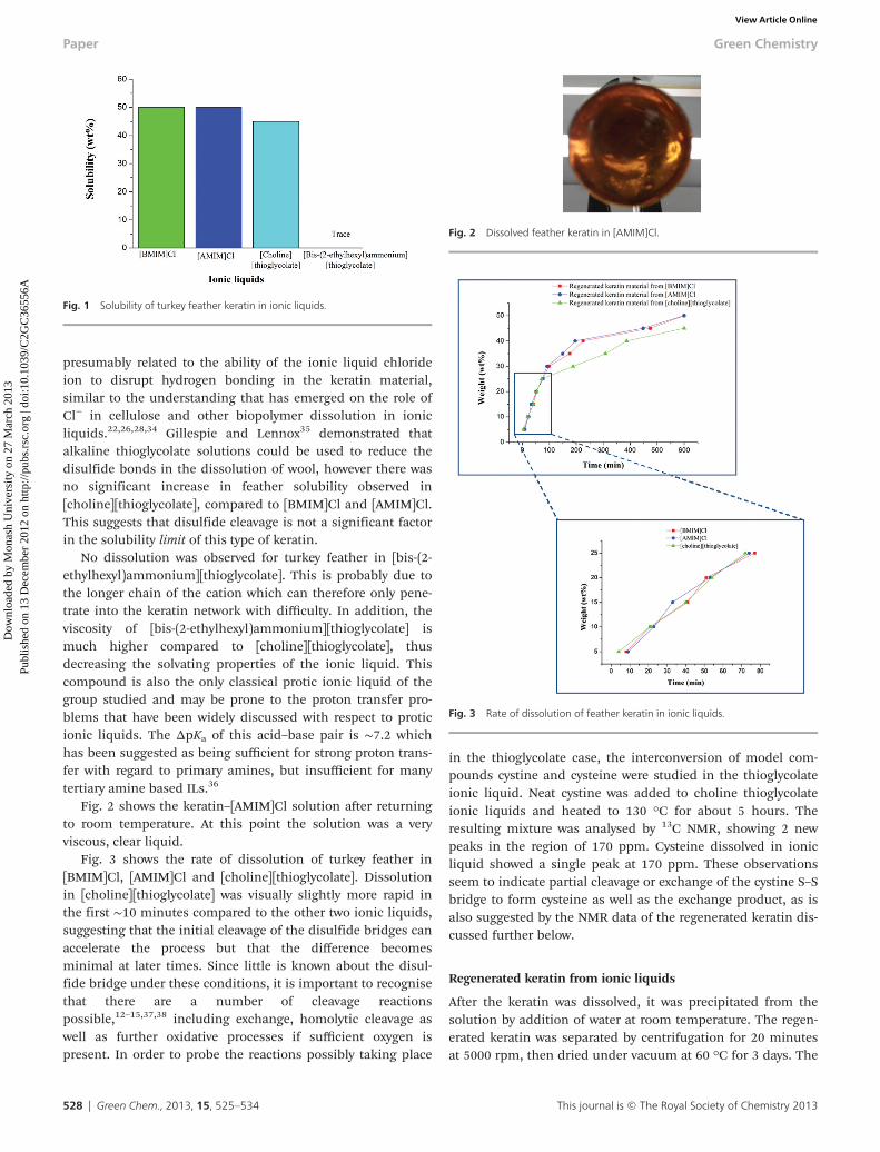
presumably related to the ability of the ionic liquid chlorideion to disrupt hydrogen bonding in the keratin material,similar to the understanding that has emerged on the role ofCl− in cellulose and other biopolymer dissolution in ionicliquids.22,26,28,34 Gillespie and Lennox35 demonstrated thatalkaline thioglycolate solutions could be used to reduce thedisulfide bonds in the dissolution of wool, however there wasno significant increase in feather solubility observed in[choline][thioglycolate], compared to [BMIM]Cl and [AMIM]Cl.This suggests that disulfide cleavage is not a significant factorin the solubility limit of this type of keratin.
No dissolution was observed for turkey feather in [bis-(2-ethylhexyl)ammonium][thioglycolate]. This is probably due tothe longer chain of the cation which can therefore only pene-trate into the keratin network with difficulty. In addition, theviscosity of [bis-(2-ethylhexyl)ammonium][thioglycolate] ismuch higher compared to [choline][thioglycolate], thusdecreasing the solvating properties of the ionic liquid. Thiscompound is also the only classical protic ionic liquid of thegroup studied and may be prone to the proton transfer pro-blems that have been widely discussed with respect to proticionic liquids. The ΔpKa of this acid–base pair is ∼7.2 whichhas been suggested as being sufficient for strong proton trans-fer with regard to primary amines, but insufficient for manytertiary amine based ILs.36
Fig. 2 shows the keratin–[AMIM]Cl solution after returningto room temperature. At this point the solution was a veryviscous, clear liquid.
Fig. 3 shows the rate of dissolution of turkey feather in[BMIM]Cl, [AMIM]Cl and [choline][thioglycolate]. Dissolutionin [choline][thioglycolate] was visually slightly more rapid inthe first ∼10 minutes compared to the other two ionic liquids,suggesting that the initial cleavage of the disulfide bridges canaccelerate the process but that the difference becomesminimal at later times. Since little is known about the disul-fide bridge under these conditions, it is important to recognisethat there are a number of cleavage reactionspossible,12–15,37,38 including exchange, homolytic cleavage aswell as further oxidative processes if sufficient oxygen ispresent. In order to probe the reactions possibly taking place
in the thioglycolate case, the interconversion of model com-pounds cystine and cysteine were studied in the thioglycolateionic liquid. Neat cystine was added to choline thioglycolateionic liquids and heated to 130 °C for about 5 hours. Theresulting mixture was analysed by 13C NMR, showing 2 newpeaks in the region of 170 ppm. Cysteine dissolved in ionicliquid showed a single peak at 170 ppm. These observationsseem to indicate partial cleavage or exchange of the cystine S–Sbridge to form cysteine as well as the exchange product, as isalso suggested by the NMR data of the regenerated keratin dis-cussed further below.
Regenerated keratin from ionic liquids
After the keratin was dissolved, it was precipitated from thesolution by addition of water at room temperature. The regen-erated keratin was separated by centrifugation for 20 minutesat 5000 rpm, then dried under vacuum at 60 °C for 3 days. The
Fig. 1 Solubility of turkey feather keratin in ionic liquids.
Fig. 3 Rate of dissolution of feather keratin in ionic liquids.
Fig. 2 Dissolved feather keratin in [AMIM]Cl.
Paper Green Chemistry
528 | Green Chem., 2013, 15, 525–534 This journal is © The Royal Society of Chemistry 2013
Dow
nloa
ded
by M
onas
h U
nive
rsity
on
27 M
arch
201
3Pu
blis
hed
on 1
3 D
ecem
ber
2012
on
http
://pu
bs.r
sc.o
rg |
doi:1
0.10
39/C
2GC
3655
6AView Article Online

regenerated keratin material was a fawn-coloured hard solid.Table S2 of the ESI† and Fig. 4 show the masses obtained ofthe regenerated aqueous-insoluble fraction. It has beenreported that feather keratin consists of approximately 40%hydrophilic and 60% hydrophobic groups in the amino acidsequence.8 Therefore, it was suggested that some of the dis-solved keratin is insoluble in water and the other part of thedissolved material might be cleaved into smaller constituentpolypeptide subunits which may be water soluble. Gel electro-phoresis analysis was employed to further investigate thesoluble component in the water–ionic liquid mixture producedduring the regeneration step, as discussed further below.
Characterisation of regenerated keratin
ATR studies. In order to further understand the nature ofthe water insoluble keratin material regenerated from the ionicliquids, ATR-FTIR measurements were used. Infrared absorp-tion spectra of the raw material and regenerated fraction(Fig. 5) showed characteristic absorption bands that are typi-cally assigned to the peptide bonds (–CONH). These spectraprovide information as to the orientation of the constituentsand the conformation of the polypeptide chains. The Amide Avibrations of the peptide bond show medium absorptionbands at 3273 cm−1 due to the N–H stretching. A strongabsorption band was also observed at 1627 cm−1 and this wasattributed to CvO (Amide I). A strong absorption peak at1515 cm−1 was attributed to C–N stretching and N–H bendingvibrations (Amide II). A weak band was observed at 1236 cm−1
which was due to the C–N and C–O stretching, N–H andOvC–N bending vibration (Amide III).39,40 It can be seen thatthe spectra are similar to the original starting material andthere are no signatures of new functional groups appearing inthe regenerated material. However, in view of NMR results(below), it appears that there are chemical changes occurringdue to the breaking of disulfide linkages and hydrogen bonds,which cannot be observed clearly in the ATR spectra. Thereforedissolution in these ionic liquids does not strongly affect thepeptide bonds.
XRD studies. Raw material and the regenerated keratinmaterial were examined by X-ray diffraction (XRD). In Fig. 6,the XRD patterns are compared. These figures show crystalli-nity in the feather keratin fibers from the substantial 2θ peakat about 9° (0.98 nm) which has been assigned to both α-helixand β-sheet structures.41,42 The peak at about 17.8° (0.51 nm)corresponds to the diffraction pattern of the α-helix whereasthe peak at about 19° (0.47 nm) is typical of the β-sheet struc-ture.41,42 However, due to the overlapping signals at about17.8° and 19° from the α-helix and β-sheet, both are unable tobe unambiguously assigned. It can be seen from Fig. 6 that theregenerated keratin exhibits similar diffraction patterns, indi-cating regeneration of the crystallinity of the original keratin.However, the peaks at about 9° and 19° are both significantlystronger in the regenerated material, suggesting a greatercontent of the β-sheet structure. These results are consistentwith the 13C CP MAS NMR spectra discussed further below.
Solid state NMR studies. Solid state 13C NMR was con-ducted on the raw material and the regenerated keratinmaterial to investigate the local structure and dynamics of thekeratin molecules. Fig. 7 displays the 13C CP MAS NMR spectra
Fig. 5 ATR-FTIR spectra of (a) raw material and regenerated keratin materialfrom (b) [BMIM]Cl, (c) [AMIM]Cl, and (d) [choline][thioglycolate].
Fig. 6 XRD of (a) raw material and regenerated keratin material from (b)[BMIM]Cl, (c) [AMIM]Cl, and (d) [choline][thioglycolate].
Fig. 4 Recovery of water insoluble fraction of IL dissolved keratin.
Green Chemistry Paper
This journal is © The Royal Society of Chemistry 2013 Green Chem., 2013, 15, 525–534 | 529
Dow
nloa
ded
by M
onas
h U
nive
rsity
on
27 M
arch
201
3Pu
blis
hed
on 1
3 D
ecem
ber
2012
on
http
://pu
bs.r
sc.o
rg |
doi:1
0.10
39/C
2GC
3655
6AView Article Online

of keratin materials. All spectra in Fig. 7 display a distinctasymmetric peak with maxima centred between 172 and174 ppm.
This peak corresponds to the carbonyl carbons present inkeratin. The peak at 130 ppm is ascribed to the aromaticspecies present in the keratin.43 All NMR spectra in Fig. 7display this aromatic peak, which suggests that the aromaticgroup containing amino acid sequences are retained in theregenerated materials. The peak at 54 ppm is related tothe α-carbons, while the one at 40 ppm is ascribed to theβ-carbons present in leucine and cysteine residues.44 Thecarbon peaks at about 30 ppm to 40 ppm are attributed tothe carbon present in the proline, glutamic acid and glutamineresidues. The NMR peaks at low chemical shift centred at20 ppm, 25 ppm and 31 ppm, in addition to the peak as ashoulder at 17 ppm correspond to the alkyl groups of the sidechains.44 A peak at ∼20 ppm is somewhat reduced in the thio-glycolate case – this region is associated with the δ-carbon ofleucine residues.44
Reductive cleavage of disulfide bridges associated withcysteine is expected to reduce the β-carbon signal at 40 ppmand produce a thiol signal around 25–29 ppm.44 The two chlor-ide cases show distinct peaks at 40 ppm that are similar to theraw material, suggesting substantial retention of disulfidebridges in these cases. In the thioglycolate case a peakremains evident, although less distinct. An exchange reaction
in the thioglycolate case could produce a new S–S bridgewhich would also produce resonances near 40 ppm; thereforeit appears likely that some sulfide exchange has taken place.
Since, the alkyl groups and the reduced cystine groups(i.e., cysteine groups) display peaks in the same chemical shiftrange around 25–29 ppm, it is hard to distinguish the reducedcysteine uniquely in these spectra.
The two regenerated materials from [BMIM]Cl and [AMIM]-Cl display NMR spectra that are not very different from theoriginal keratin, while the [choline][thioglycolate] materialshows clear differences in the 50–65 ppm region. This ispossibly because of disruption due to the reductive cleavage ofthe disulfide linkages.
The secondary structure of keratin, which consists ofα-helix and β-sheets, produces a slightly different chemicalshift of the carbonyl group in the NMR spectrum.43,44,46 Thusthe estimated percentage fraction of the α-helix and theβ-sheet can be estimated by the deconvolution of the CvOpeak. Fig. 8 displays the deconvoluted carbonyl NMR peaks ofraw and regenerated materials. The full width at halfmaximum (FWHM) of the peak also provides informationabout the degree of motional freedom possessed by the nuclei.The higher value of full width at half maximum (FWHM)shows that the nuclei possess lower motional freedom. Pre-vious studies showed that such deconvolutions usually resultin two peaks, one at 175.6 ± 0.2 ppm, which is ascribed to the
Fig. 7 The 13C CP MAS NMR spectra of (a) raw material, (b) regenerated keratin material from [BMIM]Cl, (c) regenerated keratin material from [AMIM]Cl, and (d)regenerated keratin material from [choline][thioglycolate].
Paper Green Chemistry
530 | Green Chem., 2013, 15, 525–534 This journal is © The Royal Society of Chemistry 2013
Dow
nloa
ded
by M
onas
h U
nive
rsity
on
27 M
arch
201
3Pu
blis
hed
on 1
3 D
ecem
ber
2012
on
http
://pu
bs.r
sc.o
rg |
doi:1
0.10
39/C
2GC
3655
6AView Article Online
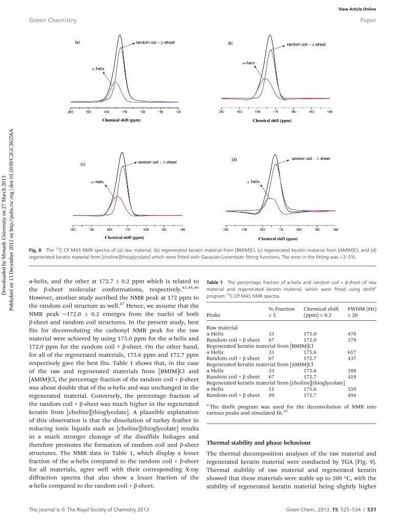
α-helix, and the other at 172.7 ± 0.2 ppm which is related tothe β-sheet molecular conformations, respectively.43,44,46
However, another study ascribed the NMR peak at 172 ppm tothe random coil structure as well.47 Hence, we assume that theNMR peak ∼172.0 ± 0.2 emerges from the nuclei of bothβ-sheet and random coil structures. In the present study, bestfits for deconvoluting the carbonyl NMR peak for the rawmaterial were achieved by using 175.0 ppm for the α-helix and172.0 ppm for the random coil + β-sheet. On the other hand,for all of the regenerated materials, 175.6 ppm and 172.7 ppmrespectively gave the best fits. Table 1 shows that, in the caseof the raw and regenerated materials from [BMIM]Cl and[AMIM]Cl, the percentage fraction of the random coil + β-sheetwas about double that of the α-helix and was unchanged in theregenerated material. Conversely, the percentage fraction ofthe random coil + β-sheet was much higher in the regeneratedkeratin from [choline][thioglycolate]. A plausible explanationof this observation is that the dissolution of turkey feather inreducing ionic liquids such as [choline][thioglycolate] resultsin a much stronger cleavage of the disulfide linkages andtherefore promotes the formation of random coil and β-sheetstructures. The NMR data in Table 1, which display a lesserfraction of the α-helix compared to the random coil + β-sheetfor all materials, agree well with their corresponding X-raydiffraction spectra that also show a lesser fraction of theα-helix compared to the random coil + β-sheet.
Thermal stability and phase behaviour
The thermal decomposition analyses of the raw material andregenerated keratin material were conducted by TGA (Fig. 9).Thermal stability of raw material and regenerated keratinshowed that these materials were stable up to 200 °C, with thestability of regenerated keratin material being slightly higher
Fig. 8 The 13C CP MAS NMR spectra of (a) raw material, (b) regenerated keratin material from [BMIM]Cl, (c) regenerated keratin material from [AMIM]Cl, and (d)regenerated keratin material from [choline][thioglycolate] which were fitted with Gaussian/Lorentzian fitting functions. The error in the fitting was ∼3–5%.
Table 1 The percentage fraction of α-helix and random coil + β-sheet of rawmaterial and regenerated keratin material, which were fitted using dmfita
program 13C CP MAS NMR spectra
Peaks% Fraction± 5
Chemical shift(ppm) ± 0.2
FWHM (Hz)± 20
Raw materialα Helix 33 175.0 470Random coil + β sheet 67 172.0 379Regenerated keratin material from [BMIM]Clα Helix 33 175.6 617Random coil + β sheet 67 172.7 437Regenerated keratin material from [AMIM]Clα Helix 33 175.6 588Random coil + β sheet 67 172.7 419Regenerated keratin material from [choline][thioglycolate]α Helix 11 175.6 559Random coil + β sheet 89 172.7 494
a The dmfit program was used for the deconvolution of NMR intovarious peaks and simulated fit.45
Green Chemistry Paper
This journal is © The Royal Society of Chemistry 2013 Green Chem., 2013, 15, 525–534 | 531
Dow
nloa
ded
by M
onas
h U
nive
rsity
on
27 M
arch
201
3Pu
blis
hed
on 1
3 D
ecem
ber
2012
on
http
://pu
bs.r
sc.o
rg |
doi:1
0.10
39/C
2GC
3655
6AView Article Online
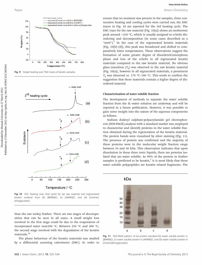
than the raw turkey feather. There are two stages of decompo-sition that can be seen in all cases. A small weight lossinvolved in the first stage could be due to the evaporation ofincorporated water near100 °C. Between 250 °C and 400 °C,the second stage involved with the degradation of the keratinmaterials.10
The phase behaviour of the keratin materials was studiedby a differential scanning calorimeter (DSC). In order to
ensure that no moisture was present in the samples, three con-secutive heating and cooling cycles were carried out; the DSCtraces in Fig. 10 are reported for the 3rd heating cycle. TheDSC trace for the raw material {Fig. 10(a)} shows an exothermicpeak around ∼230 °C, which is usually assigned to α-helix dis-ordering and decomposition (in some cases described as a“melt”).1 In the case of the regenerated keratin materials{Fig. 10(b)–(d)}, this peak was broadened and shifted to com-paratively lower temperatures. These observations suggest theformation of some greater degree of disordered/amorphousphase and loss of the α-helix in all regenerated keratinmaterials compared to the raw keratin material. No obviousglass transition (Tg) was observed in the raw keratin material{Fig. 10(a)}, however in all regenerated materials, a prominentTg was observed ca. 170 °C–180 °C. This tends to confirm thesuggestion that these materials contain a higher degree of dis-ordered material.
Characterisation of water soluble fraction
The development of methods to separate the water solublefraction from the IL–water solution are underway and will bereported in a future publication. However, it was possible togain some insight into the nature of the aqueous componentsas follows.
Sodium dodecyl sulphate-polyacrylamide gel electrophor-esis (SDS-PAGE) analysis with a standard marker was employedto characterise and identify proteins in the water soluble frac-tion obtained during the regeneration of the keratin material.The protein bands were visualised by silver staining (Fig. 11).The presence of protein was confirmed and the majority ofthese proteins were in the molecular weight fraction rangebetween 10 and 40 kDa. This observation indicates that upondissolution in these three ionic liquids, there are proteins iso-lated that are water soluble. As 90% of the protein in feathersamples is predicted to be keratin,7 it is most likely that thesewater soluble polypeptides are keratin related fragments. The
Fig. 9 Single heating scan TGA traces of keratin samples.
Fig. 10 DSC heating scan (3rd cycle) for (a) raw material and regeneratedkeratin material from (b) [BMIM]Cl, (c) [AMIM]Cl, and (d) [choline]-[thioglycolate].
Fig. 11 SDS-PAGE pattern of (a) protein standard (b) water soluble protein in[BMIM]Cl, (c) water soluble protein in [AMIM]Cl, and (d) water soluble protein in[choline][thioglycolate].
Paper Green Chemistry
532 | Green Chem., 2013, 15, 525–534 This journal is © The Royal Society of Chemistry 2013
Dow
nloa
ded
by M
onas
h U
nive
rsity
on
27 M
arch
201
3Pu
blis
hed
on 1
3 D
ecem
ber
2012
on
http
://pu
bs.r
sc.o
rg |
doi:1
0.10
39/C
2GC
3655
6AView Article Online
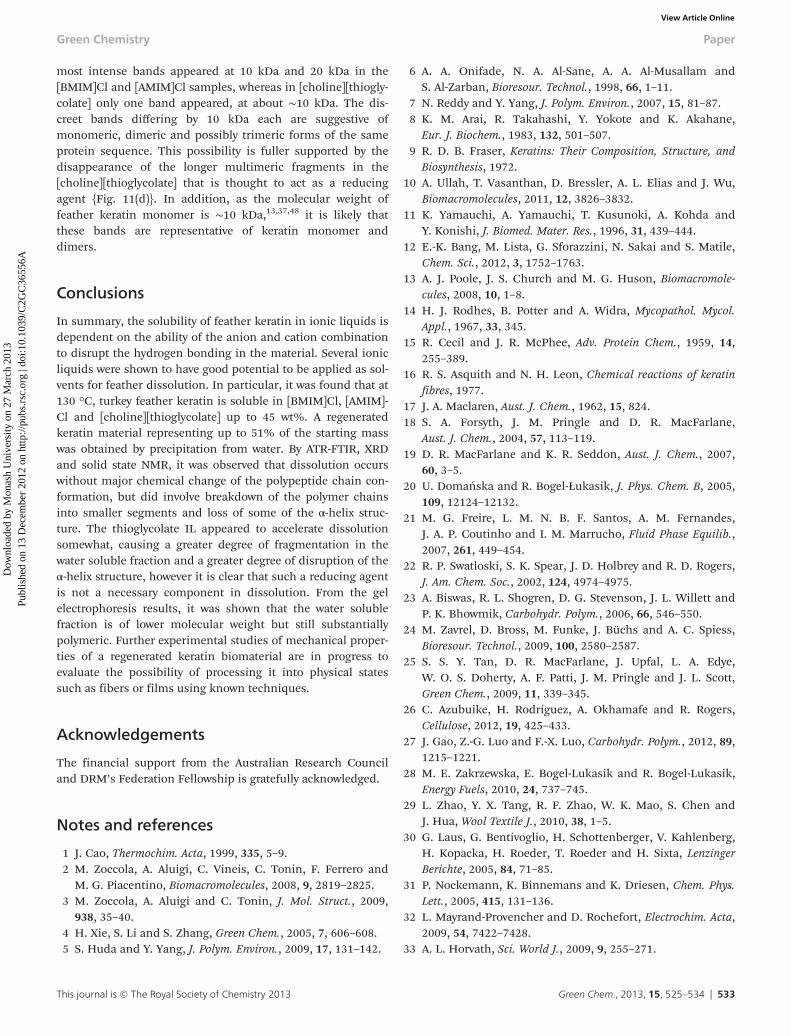
most intense bands appeared at 10 kDa and 20 kDa in the[BMIM]Cl and [AMIM]Cl samples, whereas in [choline][thiogly-colate] only one band appeared, at about ∼10 kDa. The dis-creet bands differing by 10 kDa each are suggestive ofmonomeric, dimeric and possibly trimeric forms of the sameprotein sequence. This possibility is fuller supported by thedisappearance of the longer multimeric fragments in the[choline][thioglycolate] that is thought to act as a reducingagent {Fig. 11(d)}. In addition, as the molecular weight offeather keratin monomer is ∼10 kDa,13,37,48 it is likely thatthese bands are representative of keratin monomer anddimers.
Conclusions
In summary, the solubility of feather keratin in ionic liquids isdependent on the ability of the anion and cation combinationto disrupt the hydrogen bonding in the material. Several ionicliquids were shown to have good potential to be applied as sol-vents for feather dissolution. In particular, it was found that at130 °C, turkey feather keratin is soluble in [BMIM]Cl, [AMIM]-Cl and [choline][thioglycolate] up to 45 wt%. A regeneratedkeratin material representing up to 51% of the starting masswas obtained by precipitation from water. By ATR-FTIR, XRDand solid state NMR, it was observed that dissolution occurswithout major chemical change of the polypeptide chain con-formation, but did involve breakdown of the polymer chainsinto smaller segments and loss of some of the α-helix struc-ture. The thioglycolate IL appeared to accelerate dissolutionsomewhat, causing a greater degree of fragmentation in thewater soluble fraction and a greater degree of disruption of theα-helix structure, however it is clear that such a reducing agentis not a necessary component in dissolution. From the gelelectrophoresis results, it was shown that the water solublefraction is of lower molecular weight but still substantiallypolymeric. Further experimental studies of mechanical proper-ties of a regenerated keratin biomaterial are in progress toevaluate the possibility of processing it into physical statessuch as fibers or films using known techniques.
Acknowledgements
The financial support from the Australian Research Counciland DRM’s Federation Fellowship is gratefully acknowledged.
Notes and references
1 J. Cao, Thermochim. Acta, 1999, 335, 5–9.2 M. Zoccola, A. Aluigi, C. Vineis, C. Tonin, F. Ferrero and
M. G. Piacentino, Biomacromolecules, 2008, 9, 2819–2825.3 M. Zoccola, A. Aluigi and C. Tonin, J. Mol. Struct., 2009,
938, 35–40.4 H. Xie, S. Li and S. Zhang, Green Chem., 2005, 7, 606–608.5 S. Huda and Y. Yang, J. Polym. Environ., 2009, 17, 131–142.
6 A. A. Onifade, N. A. Al-Sane, A. A. Al-Musallam andS. Al-Zarban, Bioresour. Technol., 1998, 66, 1–11.
7 N. Reddy and Y. Yang, J. Polym. Environ., 2007, 15, 81–87.8 K. M. Arai, R. Takahashi, Y. Yokote and K. Akahane,
Eur. J. Biochem., 1983, 132, 501–507.9 R. D. B. Fraser, Keratins: Their Composition, Structure, and
Biosynthesis, 1972.10 A. Ullah, T. Vasanthan, D. Bressler, A. L. Elias and J. Wu,
Biomacromolecules, 2011, 12, 3826–3832.11 K. Yamauchi, A. Yamauchi, T. Kusunoki, A. Kohda and
Y. Konishi, J. Biomed. Mater. Res., 1996, 31, 439–444.12 E.-K. Bang, M. Lista, G. Sforazzini, N. Sakai and S. Matile,
Chem. Sci., 2012, 3, 1752–1763.13 A. J. Poole, J. S. Church and M. G. Huson, Biomacromole-
cules, 2008, 10, 1–8.14 H. J. Rodhes, B. Potter and A. Widra, Mycopathol. Mycol.
Appl., 1967, 33, 345.15 R. Cecil and J. R. McPhee, Adv. Protein Chem., 1959, 14,
255–389.16 R. S. Asquith and N. H. Leon, Chemical reactions of keratin
fibres, 1977.17 J. A. Maclaren, Aust. J. Chem., 1962, 15, 824.18 S. A. Forsyth, J. M. Pringle and D. R. MacFarlane,
Aust. J. Chem., 2004, 57, 113–119.19 D. R. MacFarlane and K. R. Seddon, Aust. J. Chem., 2007,
60, 3–5.20 U. Domańska and R. Bogel-Łukasik, J. Phys. Chem. B, 2005,
109, 12124–12132.21 M. G. Freire, L. M. N. B. F. Santos, A. M. Fernandes,
J. A. P. Coutinho and I. M. Marrucho, Fluid Phase Equilib.,2007, 261, 449–454.
22 R. P. Swatloski, S. K. Spear, J. D. Holbrey and R. D. Rogers,J. Am. Chem. Soc., 2002, 124, 4974–4975.
23 A. Biswas, R. L. Shogren, D. G. Stevenson, J. L. Willett andP. K. Bhowmik, Carbohydr. Polym., 2006, 66, 546–550.
24 M. Zavrel, D. Bross, M. Funke, J. Büchs and A. C. Spiess,Bioresour. Technol., 2009, 100, 2580–2587.
25 S. S. Y. Tan, D. R. MacFarlane, J. Upfal, L. A. Edye,W. O. S. Doherty, A. F. Patti, J. M. Pringle and J. L. Scott,Green Chem., 2009, 11, 339–345.
26 C. Azubuike, H. Rodríguez, A. Okhamafe and R. Rogers,Cellulose, 2012, 19, 425–433.
27 J. Gao, Z.-G. Luo and F.-X. Luo, Carbohydr. Polym., 2012, 89,1215–1221.
28 M. E. Zakrzewska, E. Bogel-Lukasik and R. Bogel-Lukasik,Energy Fuels, 2010, 24, 737–745.
29 L. Zhao, Y. X. Tang, R. F. Zhao, W. K. Mao, S. Chen andJ. Hua, Wool Textile J., 2010, 38, 1–5.
30 G. Laus, G. Bentivoglio, H. Schottenberger, V. Kahlenberg,H. Kopacka, H. Roeder, T. Roeder and H. Sixta, LenzingerBerichte, 2005, 84, 71–85.
31 P. Nockemann, K. Binnemans and K. Driesen, Chem. Phys.Lett., 2005, 415, 131–136.
32 L. Mayrand-Provencher and D. Rochefort, Electrochim. Acta,2009, 54, 7422–7428.
33 A. L. Horvath, Sci. World J., 2009, 9, 255–271.
Green Chemistry Paper
This journal is © The Royal Society of Chemistry 2013 Green Chem., 2013, 15, 525–534 | 533
Dow
nloa
ded
by M
onas
h U
nive
rsity
on
27 M
arch
201
3Pu
blis
hed
on 1
3 D
ecem
ber
2012
on
http
://pu
bs.r
sc.o
rg |
doi:1
0.10
39/C
2GC
3655
6AView Article Online
34 Z. Meng, X. Zheng, K. Tang, J. Liu, Z. Ma and Q. Zhao,Int. J. Biol. Macromol., 2012, 51, 440–448.
35 J. M. Gillespie and F. G. Lennox, Biochim. Biophys. Acta,1953, 12, 481–482.
36 J. Stoimenovski, E. I. Izgorodina and D. R. MacFarlane,Phys. Chem. Chem. Phys., 2010, 12, 10341–10347.
37 P. M. M. Schrooyen, P. J. Dijkstra, R. G. Oberthü, A. Bantjesand J. Feijen, J. Agric. Food Chem., 2000, 48, 4326–4334.
38 C. B. Jones and D. K. Mecham, Arch. Biochem., 1943, 3,193–202.
39 E. Wojciechowska, A. Wlochowicz and A. Weselucha-Birczynska, J. Mol. Struct., 1999, 511–512, 307–318.
40 R. Schor and S. Krimm, Biophys. J., 1961, 1, 467–487.41 R. Meredith, The Mechanical Properties of Textile Fibres,
1956.
42 D. R. Rao and V. B. Gupta, J. Appl. Polym. Sci., 1992, 46,1109–1112.
43 C. M. Carr and W. V. Germasimowicz, Text. Res. J., 1988, 58,418–421.
44 M. J. Duer, N. McDougal and R. C. Murray, Phys. Chem.Chem. Phys., 2003, 5, 2894–2899.
45 D. Massiot, F. Fayon, M. Capron, I. King, S. Le Calvé,B. Alonso, J. O. Durand, B. Bujoli, Z. Gan and G. Hoatson,Magn. Reson. Chem., 2002, 40, 70–76.
46 M. Baias, D. E. Demco, C. Popescu, R. Fechete, C. Melian,B. Blümich and M. Möller, J. Phys. Chem. B, 2009, 113,2184–2192.
47 N. Nishikawa, Y. Tanizawa, S. Tanaka, Y. Horiguchi andT. Asakura, Polymer, 1998, 39, 3835–3840.
48 A. M. Woodin, Biochem. J., 1954, 57, 99–109.
Paper Green Chemistry
534 | Green Chem., 2013, 15, 525–534 This journal is © The Royal Society of Chemistry 2013
Dow
nloa
ded
by M
onas
h U
nive
rsity
on
27 M
arch
201
3Pu
blis
hed
on 1
3 D
ecem
ber
2012
on
http
://pu
bs.r
sc.o
rg |
doi:1
0.10
39/C
2GC
3655
6AView Article Online
![Osteopontin-myeloid zinc finger 1 signaling regulates ... · phosphoprotein of ×298 amino acids in mice and ×314 amino acids in humans [39,43,46,50]. Alternative RNA splicing of](https://static.fdocument.org/doc/165x107/5f0802397e708231d41fdef5/osteopontin-myeloid-zinc-finger-1-signaling-regulates-phosphoprotein-of-298.jpg)


![ΣΙΑΓΩΝΕΣ / BRAKE SHOE S SHOES [517-528].pdf15206005 application ROR alternativereference 15209713 application ROR KATALOGOS 38[517-528]:Layout 1 5/11/11 10:59 AM Page 524 525](https://static.fdocument.org/doc/165x107/613ef0c7c500cf75ab3635c5/-brake-shoe-s-shoes-517-528pdf-15206005-application-ror-alternativereference.jpg)


![Èschilo [Α ῒ σχ ύ λος] fu un tragico ateniese (Eleusi 525 circa - Gela 456-455 a. C.), della cui vita poco sappiamo di sicuro. Sappiamo che prese parte.](https://static.fdocument.org/doc/165x107/5542eb73497959361e8daeaa/eschilo-fu-un-tragico-ateniese-eleusi-525-circa-gela-456-455-a-c-della-cui-vita-poco-sappiamo-di-sicuro-sappiamo-che-prese-parte.jpg)