Bonding of the Polymer Polyetheretherketone (PEEK) to Human
7
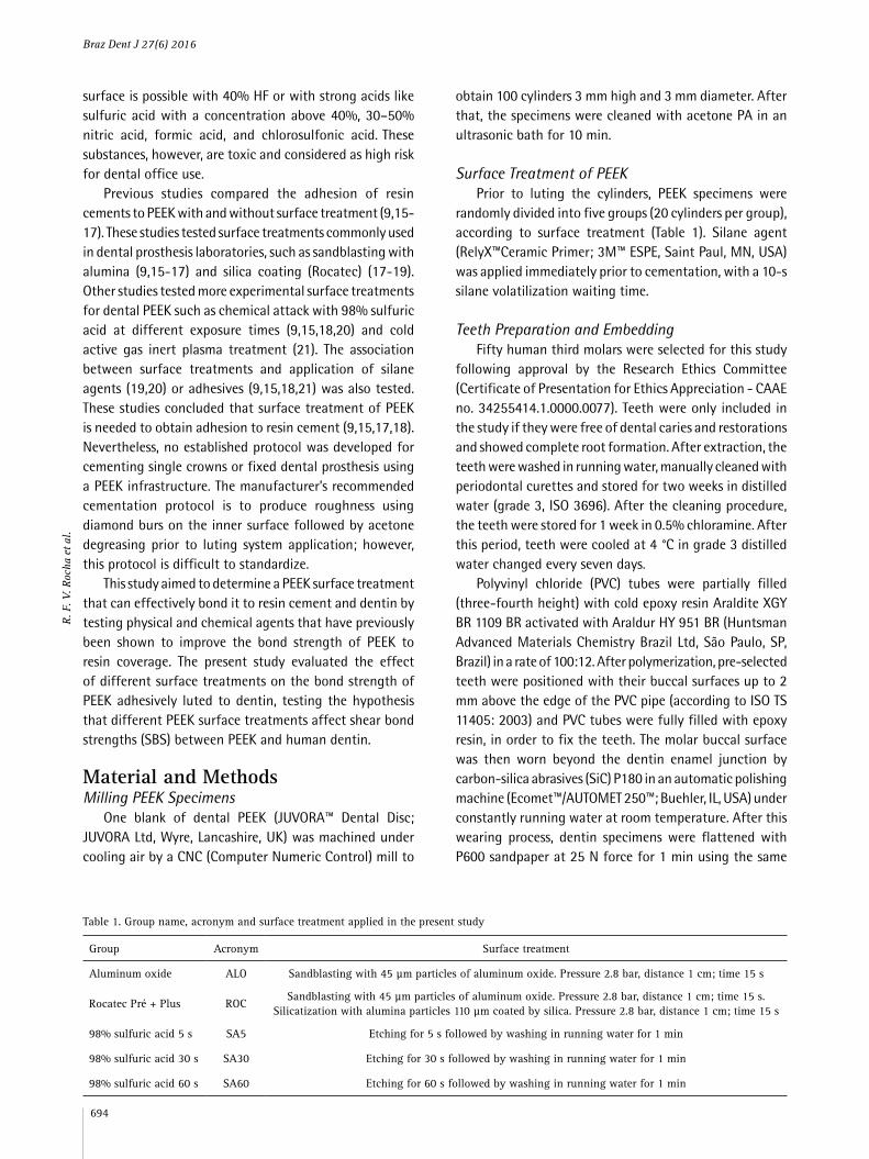
Polyetheretherketone (PEEK) is a material suitable for frameworks of fixed dental prostheses. The effect of different surface treatments on the bond strength of PEEK bonded to human dentin was evaluated. One hundred PEEK cylinders (3 mm×3 mm) were divided into five groups according to surface treatment: silica coating, sandblasting with 45 μm Al 2 O 3 particles, etching with 98% sulfuric acid for 5, 30 and for 60 s. These cylinders were luted with resin cement onto 50 human molars. First, each tooth was embedded in epoxy resin and the buccal dentin surface was exposed. Then, two delimited dentin areas (∅:3 mm) per tooth were etched with 35% phosphoric acid and bonded with a two-step self-priming adhesive system. After the luting procedure the specimens were stored in water (24 h/37 °C). Shear bond strength (SBS) was tested using a universal testing machine (crosshead speed 0.5 mm/min; load cell 50 kgf) and failure types were assessed. Stress data (MPa) were analyzed using the Kruskal-Wallis test. Comparison of the proportions of different failure types was performed using the Bonferroni method (p<0.05). Kruskal- Wallis demonstrated that differences among groups were not significant (p=0.187). Mean SBS were as follows: silica coating, 2.12±1.12 MPa; sandblasting, 2.37±0.86 MPa; sulfuric acid 5 s, 2.28±1.75 MPa; sulfuric acid 30 s, 1.80±0.85 MPa; sulfuric acid 60 s, 1.67±0.94 MPa. Adhesive and mixed failures were predominant in all groups. Both physical and chemical surface treatments produced adhesion between PEEK, resin cement and dentin. Bonding of the Polymer Polyetheretherketone (PEEK) to Human Dentin: Effect of Surface Treatments Regina Furbino Villefort Rocha 1 , Lilian Costa Anami 1 , Tiago Moreira Bastos Campos 2 , Renata Marques de Melo 1 , Rodrigo Othávio de Assunção e Souza 3 , Marco Antonio Bottino 1 1 Prosthodontics Unit, Institute of Science and Technology, UNESP - Universidade Estadual Paulista, São José dos Campos. SP, Brazil 2 Department of Physics, ITA - Instituto Tecnológico da Aeronáutica, São José dos Campos, SP, Brazil 3 Department of Dentistry, Division of Prosthodontics, UFRN - Universidade Federal do Rio Grande do Norte, Natal, RN, Brazil Correspondence: Rodrigo Othávio de Assunção e Souza, Avenida Salgado Filho, 1787, Lagoa Nova, 59056-000 Natal, RN, Brasil. Tel: +55-84-3215-4136. e-mail: [email protected] Key Words: dental prosthesis, polyetheretherketone, polymers, resin cements, shear strength. Introduction Polyetheretherketone (PEEK) is an aromatic, linear, semicrystalline polymer synthesized from aromatic dihalides and biphenolate salts via nucleophilic substitution. It belongs to an important class of high-performance engineering thermoplastics and amorphous PEEK are produced in three viscosity grades (high, medium and low) based on the same basic formula (-C 6 H 4 -O-C 6 H 4 -C 6 H 4 )n) n . The aromatic rings make PEEK resistant to mechanical forces and thermal and oxidative attacks, as well as high temperature (1), which made PEEK an attractive biomaterial for medical use, especially due to its ability to be sterilized by radiation and heat without structural damage (2-4). In dentistry, PEEK has been explored as a material in a number of applications including dental implants, temporary abutments for implant-supported prosthesis, Branemark protocol prosthesis frameworks, removable partial dentures and three-unit fixed dental prostheses (5- 10). Dental PEEK is reinforced by carbon or glass fibers in different percentages and sizes, according to information provided by the manufacturers and it can influence its milling process (2,11-13). Furthermore, different dental PEEK presentations allow the manufacture process of dental prostheses by both milling and lost wax techniques. Despite having desirable mechanical properties for dental prostheses, PEEK does not meet the aesthetic requirements. Thereby the opacity and color of the material require the application of a veneering material, in this case, light-cured composite resins for laboratory use (11). To obtain adhesion to veneering resins and cements, the PEEK surface requires treatment since it has low surface energy. Sandblasting is an efficient method for modifying the surface morphology of materials like metals and polymers. Ourahmoune et al. (14) studied how sandblasting altered surface morphology in several polymeric materials based on PEEK. The authors noted that sandblast-mediated alterations in surface morphology changed wettability and led to an apparent stabilization of roughness parameters after a certain surface treatment time. They also observed that sandblasting resulted in higher levels of roughness in the fiber-reinforced materials than in the non-reinforced materials (14). Other options for surface morphology modification are tribochemical silica-coating and chemical attack. Due to the high resistance of PEEK to chemical agents, 9.5% hydrofluoric acid (HF) is not effective for altering surface morphology (15). Manufacturer’s instructions suggest that chemical attack on the PEEK ISSN 0103-6440 Brazilian Dental Journal (2016) 27(6): 693-699 http://dx.doi.org/10.1590/0103-6440201600796
Transcript of Bonding of the Polymer Polyetheretherketone (PEEK) to Human
Polyetheretherketone (PEEK) is a material suitable for frameworks of fixed dental prostheses. The effect of different surface treatments on the bond strength of PEEK bonded to human dentin was evaluated. One hundred PEEK cylinders (3 mm×3 mm) were divided into five groups according to surface treatment: silica coating, sandblasting with 45 μm Al2O3 particles, etching with 98% sulfuric acid for 5, 30 and for 60 s. These cylinders were luted with resin cement onto 50 human molars. First, each tooth was embedded in epoxy resin and the buccal dentin surface was exposed. Then, two delimited dentin areas (∅:3 mm) per tooth were etched with 35% phosphoric acid and bonded with a two-step self-priming adhesive system. After the luting procedure the specimens were stored in water (24 h/37 °C). Shear bond strength (SBS) was tested using a universal testing machine (crosshead speed 0.5 mm/min; load cell 50 kgf) and failure types were assessed. Stress data (MPa) were analyzed using the Kruskal-Wallis test. Comparison of the proportions of different failure types was performed using the Bonferroni method (p<0.05). Kruskal-Wallis demonstrated that differences among groups were not significant (p=0.187). Mean SBS were as follows: silica coating, 2.12±1.12 MPa; sandblasting, 2.37±0.86 MPa; sulfuric acid 5 s, 2.28±1.75 MPa; sulfuric acid 30 s, 1.80±0.85 MPa; sulfuric acid 60 s, 1.67±0.94 MPa. Adhesive and mixed failures were predominant in all groups. Both physical and chemical surface treatments produced adhesion between PEEK, resin cement and dentin.
B o n d i n g o f t h e P o l y m e r Polyetheretherketone (PEEK) to Human Dentin: Effect of Surface Treatments
Regina Furbino Villefort Rocha1, Lilian Costa Anami1, Tiago Moreira Bastos Campos2, Renata Marques de Melo1, Rodrigo Othávio de Assunção e Souza3, Marco Antonio Bottino1
1Prosthodontics Unit, Institute of Science and Technology, UNESP - Universidade Estadual Paulista, São José dos Campos. SP, Brazil2Department of Physics, ITA - Instituto Tecnológico da Aeronáutica, São José dos Campos, SP, Brazil3Department of Dentistry, Division of Prosthodontics, UFRN - Universidade Federal do Rio Grande do Norte, Natal, RN, Brazil
Correspondence: Rodrigo Othávio de Assunção e Souza, Avenida Salgado Filho, 1787, Lagoa Nova, 59056-000 Natal, RN, Brasil. Tel: +55-84-3215-4136. e-mail: [email protected]
Key Words: dental prosthesis, polyetheretherketone, polymers, resin cements, shear strength.
IntroductionPolyetheretherketone (PEEK) is an aromatic, linear,
semicrystalline polymer synthesized from aromatic dihalides and biphenolate salts via nucleophilic substitution. It belongs to an important class of high-performance engineering thermoplastics and amorphous PEEK are produced in three viscosity grades (high, medium and low) based on the same basic formula (-C6H4-O-C6H4-C6H4)n)
n. The aromatic rings make PEEK resistant to mechanical forces and thermal and oxidative attacks, as well as high temperature (1), which made PEEK an attractive biomaterial for medical use, especially due to its ability to be sterilized by radiation and heat without structural damage (2-4).
In dentistry, PEEK has been explored as a material in a number of applications including dental implants, temporary abutments for implant-supported prosthesis, Branemark protocol prosthesis frameworks, removable partial dentures and three-unit fixed dental prostheses (5-10). Dental PEEK is reinforced by carbon or glass fibers in different percentages and sizes, according to information provided by the manufacturers and it can influence its milling process (2,11-13). Furthermore, different dental PEEK presentations allow the manufacture process of dental prostheses by both milling and lost wax techniques.
Despite having desirable mechanical properties for dental prostheses, PEEK does not meet the aesthetic requirements. Thereby the opacity and color of the material require the application of a veneering material, in this case, light-cured composite resins for laboratory use (11). To obtain adhesion to veneering resins and cements, the PEEK surface requires treatment since it has low surface energy.
Sandblasting is an efficient method for modifying the surface morphology of materials like metals and polymers. Ourahmoune et al. (14) studied how sandblasting altered surface morphology in several polymeric materials based on PEEK. The authors noted that sandblast-mediated alterations in surface morphology changed wettability and led to an apparent stabilization of roughness parameters after a certain surface treatment time. They also observed that sandblasting resulted in higher levels of roughness in the fiber-reinforced materials than in the non-reinforced materials (14). Other options for surface morphology modification are tribochemical silica-coating and chemical attack. Due to the high resistance of PEEK to chemical agents, 9.5% hydrofluoric acid (HF) is not effective for altering surface morphology (15). Manufacturer’s instructions suggest that chemical attack on the PEEK
ISSN 0103-6440Brazilian Dental Journal (2016) 27(6): 693-699http://dx.doi.org/10.1590/0103-6440201600796
Braz Dent J 27(6) 2016
694
R. F
. V. R
ocha
et a
l.
surface is possible with 40% HF or with strong acids like sulfuric acid with a concentration above 40%, 30–50% nitric acid, formic acid, and chlorosulfonic acid. These substances, however, are toxic and considered as high risk for dental office use.
Previous studies compared the adhesion of resin cements to PEEK with and without surface treatment (9,15-17). These studies tested surface treatments commonly used in dental prosthesis laboratories, such as sandblasting with alumina (9,15-17) and silica coating (Rocatec) (17-19). Other studies tested more experimental surface treatments for dental PEEK such as chemical attack with 98% sulfuric acid at different exposure times (9,15,18,20) and cold active gas inert plasma treatment (21). The association between surface treatments and application of silane agents (19,20) or adhesives (9,15,18,21) was also tested. These studies concluded that surface treatment of PEEK is needed to obtain adhesion to resin cement (9,15,17,18). Nevertheless, no established protocol was developed for cementing single crowns or fixed dental prosthesis using a PEEK infrastructure. The manufacturer’s recommended cementation protocol is to produce roughness using diamond burs on the inner surface followed by acetone degreasing prior to luting system application; however, this protocol is difficult to standardize.
This study aimed to determine a PEEK surface treatment that can effectively bond it to resin cement and dentin by testing physical and chemical agents that have previously been shown to improve the bond strength of PEEK to resin coverage. The present study evaluated the effect of different surface treatments on the bond strength of PEEK adhesively luted to dentin, testing the hypothesis that different PEEK surface treatments affect shear bond strengths (SBS) between PEEK and human dentin.
Material and MethodsMilling PEEK Specimens
One blank of dental PEEK (JUVORA™ Dental Disc; JUVORA Ltd, Wyre, Lancashire, UK) was machined under cooling air by a CNC (Computer Numeric Control) mill to
obtain 100 cylinders 3 mm high and 3 mm diameter. After that, the specimens were cleaned with acetone PA in an ultrasonic bath for 10 min.
Surface Treatment of PEEKPrior to luting the cylinders, PEEK specimens were
randomly divided into five groups (20 cylinders per group), according to surface treatment (Table 1). Silane agent (RelyX™Ceramic Primer; 3M™ ESPE, Saint Paul, MN, USA) was applied immediately prior to cementation, with a 10-s silane volatilization waiting time.
Teeth Preparation and Embedding
Fifty human third molars were selected for this study following approval by the Research Ethics Committee (Certificate of Presentation for Ethics Appreciation - CAAE no. 34255414.1.0000.0077). Teeth were only included in the study if they were free of dental caries and restorations and showed complete root formation. After extraction, the teeth were washed in running water, manually cleaned with periodontal curettes and stored for two weeks in distilled water (grade 3, ISO 3696). After the cleaning procedure, the teeth were stored for 1 week in 0.5% chloramine. After this period, teeth were cooled at 4 °C in grade 3 distilled water changed every seven days.
Polyvinyl chloride (PVC) tubes were partially filled (three-fourth height) with cold epoxy resin Araldite XGY BR 1109 BR activated with Araldur HY 951 BR (Huntsman Advanced Materials Chemistry Brazil Ltd, São Paulo, SP, Brazil) in a rate of 100:12. After polymerization, pre-selected teeth were positioned with their buccal surfaces up to 2 mm above the edge of the PVC pipe (according to ISO TS 11405: 2003) and PVC tubes were fully filled with epoxy resin, in order to fix the teeth. The molar buccal surface was then worn beyond the dentin enamel junction by carbon-silica abrasives (SiC) P180 in an automatic polishing machine (Ecomet™/AUTOMET 250™; Buehler, IL, USA) under constantly running water at room temperature. After this wearing process, dentin specimens were flattened with P600 sandpaper at 25 N force for 1 min using the same
Table 1. Group name, acronym and surface treatment applied in the present study
Group Acronym Surface treatment
Aluminum oxide ALO Sandblasting with 45 μm particles of aluminum oxide. Pressure 2.8 bar, distance 1 cm; time 15 s
Rocatec Pré + Plus ROCSandblasting with 45 μm particles of aluminum oxide. Pressure 2.8 bar, distance 1 cm; time 15 s.
Silicatization with alumina particles 110 μm coated by silica. Pressure 2.8 bar, distance 1 cm; time 15 s
98% sulfuric acid 5 s SA5 Etching for 5 s followed by washing in running water for 1 min
98% sulfuric acid 30 s SA30 Etching for 30 s followed by washing in running water for 1 min
98% sulfuric acid 60 s SA60 Etching for 60 s followed by washing in running water for 1 min
Braz Dent J 27(6) 2016
695
Bon
ding
of P
EEK
to h
uman
den
tin
polishing machine. The specimens were then cooled at 4 °C in grade 3 distilled water.
Luting ProcedureThe exposed face of the dentin was covered with an
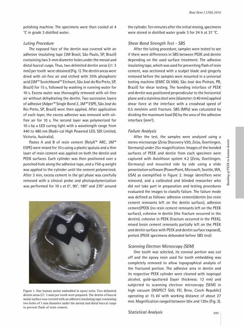
adhesive insulating tape (3M Brasil, São Paulo, SP, Brazil) containing two 3-mm diameter holes under the mesial and distal buccal cusps. Thus, two delimited dentin areas (∅: 3 mm) per tooth were obtained (Fig. 1). The dentin areas were dried with oil-free air and etched with 35% phosphoric acid (3M™ Scotchbond™ Etchant, São José do Rio Preto, SP, Brazil) for 15 s, followed by washing in running water for 10 s. Excess water was thoroughly removed with oil-free air without dehydrating the dentin. Two successive layers of adhesive (Adper™ Single Bond 2, 3M™ ESPE, São José do Rio Preto, SP, Brazil) were then applied. After application of each layer, the excess adhesive was removed with oil-free air for 10 s. The second layer was polymerized for 10 s by a LED curing light with a wavelength range from 440 to 480 nm (Radii-cal High Powered LED, SDI Limited, Victoria, Australia).
Pastes A and B of resin cement (RelyX™ ARC, 3M™ ESPE) were mixed for 10 s using a plastic spatula and a thin layer of resin cement was applied on both the dentin and PEEK surfaces. Each cylinder was then positioned over a punched hole along the adhesive tape, and a 750-g weight was applied to the cylinder until the cement polymerized. After 3 min, excess cement in the gel phase was carefully removed with a clinical probe and photopolymerization was performed for 10 s at 0°, 90°, 180° and 270° around
the cylinder. Ten minutes after the initial mixing, specimens were stored in distilled water grade 3 for 24 h at 37 °C.
Shear Bond Strength Test - SBSAfter the luting procedure, samples were tested to see
if there were differences in SBS between PEEK and dentin depending on the used surface treatment. The adhesive insulating tape, which was used for preventing flash of resin cement, was sectioned with a scalpel blade and gingerly removed before the samples were mounted in a universal testing machine (EMIC DL1000, São José dos Pinhais, PR, Brazil) for shear testing. The bonding interface of PEEK and dentin was positioned perpendicular to the horizontal plane and a stainless steel wire (diameter: 0.40 mm) applied shear force at the interface with a crosshead speed of 0.5 mm/min until fracture. SBS (MPa) was calculated by dividing the maximum load (N) by the area of the adhesive interface (mm2).
Failure AnalysisAfter the test, the samples were analyzed using a
stereo microscope (Zeiss Discovery V20, Zeiss, Goettingen, Germany) under 25× magnification. Images of the bonded surfaces of PEEK and dentin from each specimen were captured with AxioVision system 4.2 (Zeiss, Goettingen, Germany) and mounted side by side using a slide presentation software (PowerPoint, Microsoft, Seattle, WA, USA) as exemplified in Figure 2. Image identifiers were removed, and a calibrated and blinded researcher who did not take part in preparation and testing procedures evaluated the images to classify failure. The failure mode was defined as follows: adhesive cement/dentin (no resin cement remnants left on the dentin surface), adhesive cement/PEEK (no resin cement remnants left on the PEEK surface), cohesive in dentin (the fracture occurred in the dentin), cohesive in PEEK (fracture occurred in the PEEK), mixed (resin cement remnants partially left on the PEEK and dentin surface with PEEK and dentin surface exposed), pretest (PEEK specimens debonded before SBS test)
Scanning Electron Microscopy (SEM)One tooth was selected, its coronal portion was cut
off and the epoxy resin used for tooth embedding was completely removed to allow topographical analysis of the fractured portion. The adhesive area in dentin and its respective PEEK cylinder were cleaned with isopropyl alcohol, gold-sputtered (layer thickness: 12 nm) and subjected to scanning electron microscopy (SEM) in high vacuum (INSPECT S50; FEI, Brno, Czech Republic) operating at 15 kV with working distance of about 27 mm. Magnification ranged between 50× and 120× (Fig. 3).
Statistical Analysis
Figure 1. One human molar embedded in epoxi resin. Two delimited dentin areas (∅: 3 mm) per tooth were prepared. The dentin of buccal molar surface was covered with an adhesive insulating tape containing two holes of 3 mm diameter under the mesial and distal buccal cusps to prevent flash of resin cement.
Braz Dent J 27(6) 2016
696
R. F
. V. R
ocha
et a
l.
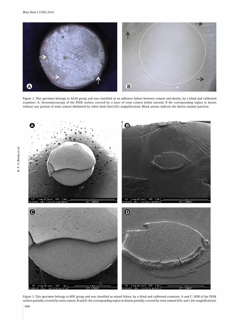
Figure 2. This specimen belongs to SA30 group and was classified as an adhesive failure between cement and dentin, by a blind and calibrated examiner. A: Stereomicroscopy of the PEEK surface covered by a layer of resin cement (white arrows). B the corresponding region in dentin without any portion of resin cement (delimited by white dash line) (25× magnification). Black arrows indicate the dentin enamel junction.
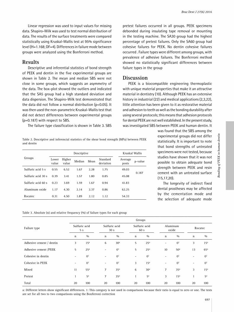
Figure 3. This specimen belongs to ROC group and was classified as mixed failure, by a blind and calibrated examiner. A and C: SEM of the PEEK surface partially covered by resin cement. B and D: the corresponding region in dentin partially covered by resin cement (50× and 120× magnification).
Braz Dent J 27(6) 2016
697
Bon
ding
of P
EEK
to h
uman
den
tin
Linear regression was used to input values for missing data. Shapiro-Wilk was used to test normal distribution of data. The results of the surface treatments were compared statistically using Kruskal-Wallis test at 95% significance level (H=1.168; DF=4). Differences in failure mode between groups were analyzed using the Bonferroni method.
ResultsDescriptive and inferential statistics of bond strength
of PEEK and dentin in the five experimental groups are shown in Table 2. The mean and median SBS were not close in some groups, which suggests an asymmetry of the data. The box-plot showed the outliers and indicated that the SA5 group had a high standard deviation and data dispersion. The Shapiro-Wilk test demonstrated that the data did not follow a normal distribution (p<0.05). It was then used the non-parametric Kruskal-Wallis test that did not detect differences between experimental groups (p=0.187) with respect to SBS.
The failure type classification is shown in Table 3. SBS
pretest failures occurred in all groups. PEEK specimens debonded during insulating tape removal or mounting in the testing machine. The SA30 group had the highest percentage of pretest failures. Only the SA60 group had cohesive failures for PEEK. No dentin cohesive failures occurred . Failure types were different among groups, with prevalence of adhesive failures. The Bonferroni method showed no statistically significant differences between failure types in the group
DiscussionPEEK is a biocompatible engineering thermoplastic
with unique material properties that make it an attractive material in dentistry (10). Although PEEK has an extensive history in industrial (22) and medical applications (2,3,22), little attention has been given to it as restorative material and adhesion to teeth as well as the bonding durability after using several protocols; this means that adhesion protocols for dental PEEK are not well established. In the present study, was investigated SBS between PEEK and human dentin. It
was found that the SBS among the experimental groups did not differ statistically. It is important to note that bond strengths of untreated specimens were not tested, because studies have shown that it was not possible to obtain adequate bond strength between PEEK and resin cement with an untreated surface (15,17,20).
The longevity of indirect fixed dental prostheses may be affected by the cementation mode and the selection of adequate mode
Table 2. Descriptive and inferencial statistics of the shear bond strength (MPa) between PEEK and dentin
Groups
Descriptive Kruskal Wallis
Lowervalue
Highervalue
Median MeanStandard deviation
Average posts
p-value
Sulfuric acid 5 s 0.55 6.52 1.67 2.28 1.75 49.030.187
Sulfuric acid 30 s 0.39 3.41 1.57 1.80 0.85 45.08
Sulfuric acid 60 s 0.23 3.69 1.59 1.67 0.94 41.83
Aluminum oxide 1.17 4.30 2.14 2.37 0.86 62.25
Rocatec 0.31 4.50 1.89 2.12 1.12 54.33
Table 3. Absolute (n) and relative frequency (%) of failure types for each group
Failure type
Groups
Sulfuric acid5 s
Sulfuric acid30 s
Sulfuric acid60 s
Aluminum oxide
Rocatec
n % n % n % n % n %
Adhesive cement / dentin 3 15a 6 30a 5 25a - 01 3 15a
Adhesive cement /PEEK 5 25a - 01 5 25a 10 50a 13 65a
Cohesive in dentin - 01 - 01 - 01 - 01 - 01
Cohesive in PEEK - 01 - 01 3 15a - 01 - 01
Mixed 11 55a 7 35a 6 30a 7 35a 3 15a
Pretest 1 5a 7 35a 1 5a 3 15a 1 5a
Total 20 100 20 100 20 100 20 100 20 100
a: Different letters show significant differences. 1: This category is not used in comparisons because their ratio is equal to zero or one. The tests are set for all two to two comparisons using the Bonferroni correction
Braz Dent J 27(6) 2016
698
R. F
. V. R
ocha
et a
l.
should consider multiple factors including restorative material properties, marginal fit, type of restoration, surface treatment and type of the supporting abutment natural tooth structure: enamel, dentin, cementum (23). PEEK-based restorations face the same challenges as other metal-free restorations. However, the results of this study showed extremely low shear bond strength values (mean value<3MPa in all experimental groups). In recent studies by this research group, about bonding of Y-TZP to dentin, the SBS mean values were higher than 10 MPa in the experimental groups not exposed to aging (24,25).
Previous studies on SBS between PEEK and resin cement tested only one adhesive interface. However, in clinics, multiple interfaces between materials are common. Moreover, these multiple interfaces can change the failure type and may influence resistance values. In this study, two interfaces were tested: cement/dentin and cement/PEEK. The limited literature available on PEEK shows different results from this study’s findings. These differences may be ascribed to different testing methodologies and types of PEEK (especially concerning the reinforcing materials) used in the studies. For example, Uhrenbacher et al. (9) recently reported the effect of different PEEK surface treatments on bonding to human dentin. However, the mechanical tests and resin cement applied were different from those applied in this study, making comparisons between study results difficult, especially due the fact that they used pull-off test and thermal cycling.
The cement/dentin interface was exhaustively studied in dentistry, and the protocols are well established. In the present study, was followed one well documented protocol of cementation that consists in etching the dentin with 35% phosphoric acid and applying a two-step self-priming adhesive system (Adper Single Bond 2), which is the adhesive system recommended for use in association with the resin cement selected for the present study (RelyX ARC). On the other hand, the resin cement/PEEK interface is not enough well known yet. The choice of conventional resin cement for this study was based on the fact that the phosphoric acid groups in the self-adhesive cement formulation were not able to condition the surface of PEEK. In fact, according to the manufacturer, phosphoric acid is not capable of promoting chemical attack on PEEK surface even at 100°C and a concentration of 80%. Other studies have tested the adhesion between PEEK and self-adhesive resin cements commonly used for luting of ceramic and metal prostheses. Rely X Unicem (3M ESPE) was tested several times (15,17,18,21), in addition to Clearfill (15,22) (Kuraray) and Multilink Automix (19) (Ivoclar).
In this study the treatment of the PEEK surface was tested with 98% sulfuric acid at different exposure times aiming to assess its influence on the bond strength of PEEK
to resin cement. Highly corrosive solutions such as 98% sulfuric acid and Piranha solution (sulfuric acid + hydrogen peroxide) have been tested in other in vitro studies for dental PEEK surface treatment (15,17,18). These highly corrosive solutions are needed because PEEK is an apolar and inert polymer with high chemical resistance and low surface energy. Anhydrous H2SO4 is a very polar liquid and a strong oxidizing agent. Besides, concentrated sulfuric acid can dissolve PEEK at room temperature. It causes a swelling process in PEEK that creates porosities on its surface. It was expected that such porosities could act as an anchorage for resin cement, even though Schimidlin et al. (19) observed lack of tag formation for PEEK conditioned with sulfuric acid. The increase of exposure time to sulfuric acid promotes an increase of pore sizes. However, the longer the exposure time, the greater the deterioration of the surface of PEEK, which could lead to cohesive failures. Sproesser et al. (20) related a higher percentage of cohesive failures in PEEK when it was exposed to 90, 120 or 300 s. In the conducted study, the cohesive failure of PEEK was a rare outcome and observed only in specimens exposed to sulfuric acid for 60 s. All the exposure times tested in the present study were able to increase bond strength. Furthermore, despite the high percentage of pretest failures in the SA30 group, no significant differences in the proportion of failure types was found. The SA5 group showed outliers and high standard deviation. But, looking at the SBSs median of SA5, SA30 and SA60 groups, one notes similar values and adhesive failure at the interface cement/PEEK representing 25% or less of the total number of specimens tested in these groups.
It is important to note that sulfuric acid is a strong acid and requires special care in handling, storage and disposal. Its use in prosthetic laboratories or dental office could be justified if significant improvement in bond strength is obtained. No significant differences were found in bond strength between conventional resin cement and PEEK, even when PEEK was exposed to sulfuric acid for 60 s. This contradicts the findings of Sproesser et al. (20) who tested exposure times that ranged from 5 s to 300 s and reported that etching PEEK with 98% sulfuric acid for 60 s was the best exposure time for conventional resin cement (RelyX ARC).
The specimens subjected to air-abrasion (ALO) and tribochemical treatment (ROC) showed similar results of SBSs values and failure mode.
Adhesive failures on the cement/PEEK interface represented 50% in the ALO group and 65% in the ROC group, followed by mixed failures (35% and 15% respectively). It seems that the cement/dentin interface was stronger than the cement/PEEK interface in such groups. Therefore, none of the tested surface treatments was able to improve adhesion between PEEK and resin
Braz Dent J 27(6) 2016
699
Bon
ding
of P
EEK
to h
uman
den
tin
cement, warranting further investigations. Given that traditional methods of surface treatment such as silica coating and sandblasting with aluminum oxide, are safe and well established in prosthetic laboratories and offices, we should opt for using them instead of sulfuric acid.
Both physical and chemical surface treatments were effective in promoting similar bonding among PEEK, cement and dentin.
ResumoO poli-éter-éter-cetona (PEEK) é um material indicado para as estruturas de próteses parciais fixas. O efeito de diferentes tratamentos de superfície na resistência de união entre PEEK e dentina humana foi avaliado. Cem cilindros de PEEK (3 mm×3 mm) foram divididos em cinco grupos de acordo com tratamento de superfície: silicatização, jateamento com partículas de Al2O3 45 µm, condicionamento com ácido sulfúrico 98% por 5, 30 e 60 s. Esses cilindros foram cimentados com cimento resinoso em cinquenta molares humanos. Primeiro, cada dente foi incluído em resina epóxica e a superfície dentinária vestibular foi exposta. Depois, duas áreas (∅:3 mm) em dentina/por dente foram delimitadas, condicionadas com ácido fosfórico a 35% e receberam aplicação de um sistema adesivo de dois passos. Após o procedimento de cimentação, as amostras foram armazenadas em água (24 h/37 °C). A resistência da união ao cisalhamento (SBS) foi testada em uma máquina universal de ensaios (velocidade 0,5 mm/min; célula de carga 50 kgf), e foram avaliados os tipos de falha. Os dados de tensão (MPa) foram analisados pelo teste de Kruskal-Wallis. A comparação das percentagens de diferentes tipos de falha foi realizada utilizando o método de Bonferroni (p<0,05). O teste de Kruskal-Wallis demonstrou que as diferenças entre os grupos não foram significantes (p=0,187). As médias de SBS foram: silicatização, 2,12±1,12 MPa; jateamento, 2,37±0,86 MPa; ácido sulfúrico por 5 s, de 2,28±1,75 MPa; ácido sulfúrico por 30 s, 1,80±0,85 MPa, ácido sulfúrico por 60 s, 1,67±0,94 MPa. Falhas adesivas e mistas foram predominantes em todos os grupos. Ambos tratamentos de superfície, físicos e químicos, promoveram adesão entre PEEK, cimento resinoso e dentina.
AcknowledegmentsThis research was supported by CAPES.
References 1. Yang J, Tyberg CS, Gibson HW. Synthesis of aromatic polyketones via
soluble precursors derived from bis(α-aminonitrile)s. Macromolecules 1999;32:8259-8268. Retrieved online June 25,2014 from: scholar.lib.vt.edu/theses/available/etd-32398–61326/unrestricted/1.pdf
2. Green S, Schlegel J. A polyarylketone biomaterial for use in medical implant applications. Retrieved online June 26, 2014 from: www.medicalpeek.org/pdf/Green_RAPRA_2001.pdf
3. Kurtz SM, Devine JN. PEEK biomaterials in trauma, orthopedic and spinal implants. Biomaterials 2007;28:4845-4869.
4. Toth JM, Wang M, Estes BT, Scifert JL, Seim HB 3rd, Turner AS. Polyetheretherketone as a biomaterial for spinal applications. Biomaterials 2006;27:324-334.
5. Tetelman ED, Babbush CA. A new transitional abutment for immediate aesthetics and function. Implant Dent 2008;17:51-58.
6. Santing HJ, Meijer HJ, Raghoebar GM, Özcan M. Fracture strength and failure mode of maxillary implant-supported provisional single crowns: a comparison of composite resin crowns fabricated directly over PEEK abutments and solid titanium abutments. Clin Implant Dent Relat Res 2012;14:882-889.
7. Bayer S, Komor N, Kramer A, Albrecht D, Mericske-Stern R, Enkling N. Retention force of plastic clips on implant bars: a randomized controlled trial. Clin Oral Implants Res 2012;23:1377-1384.
8. Tannous F, Steiner M, Shahin R, Kern M. Retentive forces and fatigue resistance of thermoplastic resin clasps. Dent Mater 2012;28:273-278.
9. Uhrenbacher J, Schmidlin PR, Keul C, Eichberger M, Roos M, Gernet W, et al.. The effect of surface modification on the retention strength of polyetheretherketone crowns adhesively bonded to dentin abutments. J Prosthet Dent 2014;112:1489-1497.
10. Stawarczyk B, Beuer F, Wimmer T, Jahn D, Sener B, Roos M, et al.. Polyetheretherketone-a suitable material for fixed dental prostheses? J Biomed Mater Res B Appl Biomater 2013;101:1209-1216.
11. Stawarczyk B, Jordan P, Schmidlin PR, Roos M, Eichberger M, Gernet W, et al.. PEEK surface treatment effects on tensile bond strength to veneering resins. J Prosthet Dent 2014;112:1278-1288.
12. Lapa VMC. Optimização da maquinagem do PEEK e seus compósitos PEEK/CF30 e PEEK/GF30. (Tese). Aveiro (Baixo Vouga): Universidade de Aveiro. 2008.
13. Mata F, Gaitonde VN, Karnik SR, Davim JP. Influence of cutting conditions on machinability aspects of PEEK, PEEK CF 30 and PEEK GF 30 composites using PCD tools. J Mater Process Technol 2008;209:1980–1987.
14. Ourahmoune R, Salvia M, Mathia TG, Mesrati N. Surface morphology and wettability of sandblasted PEEK and its composites. Scanning 2014;36:64-75.
15. Zhou L, Qian Y, Zhu Y, Liu H, Gan K, Guo J. The effect of different surface treatments on the bond strength of PEEK composite materials. Dent Mater 2014;30:e209-e215.
16. Kern M, Lehmann F. Influence of surface conditioning on bonding to polyetheretherketon (PEEK). Dent Mater 2012;28:1280-1283.
17. Hallmann L, Mehl A, Sereno N, Hämmerle CHF. The improvement of adhesive properties of PEEK though different pre-treatments. Appl Surf Sci 2012;258:7213–7218.
18. Schmidlin PR, Stawarczyk B, Wieland M, Attin T, Hämmerle CH, Fischer J. Effect of different surface pre-treatments and luting materials on shear bond strength to PEEK. Dent Mater 2010;26:553-559.
19. Fuhrmann G, Steiner M, Freitag-Wolf S, Kern M. Resin bonding to three types of polyaryletherketones (PAEKs)-durability and influence of surface conditioning. Dent Mater 2014;30:357-363.
20. Sproesser O, Schmidlin PR, Uhrenbacher J, Roos M, Gernet W, Stawarczyk B. Effect of sulfuric acid etching of polyetheretherketone on the shear bond strength to resin cements. J Adhes Dent 2014;16:465-472.
21. Stawarczyk B, Bähr N, Beuer F, Wimmer T, Eichberger M, Gernet W, et al.. Influence of plasma pretreatment on shear bond strength of self-adhesive resin cements to polyetheretherketone. Clin Oral Investig 2014;18:163-170.
22. Zhao Y, Wong HM, Wang W, Li P, Xu Z, Chong EY, et al.. Cytocompatibility, osseointegration, and bioactivity of three-dimensional porous and nanostructured network on polyetheretherketone. Biomaterials 2013;34:9264-9277.
23. Edelhoff D, Özcan M. To what extent does the longevity of fixed dental prostheses depend on the function of the cement? Working Group 4 materials: cementation. Clin Oral Impl Res 2007;18(Suppl.3):193-204.
24. Bottino MA, Bergoli C, Lima EG, Marocho SM, Souza RO, Valandro LF. Bonding of Y-TZP to dentin: effects of Y-TZP surface conditioning, resin cement type and aging. Oper Dent 2014;39:291-300.
25. Alves M, Campos F, Bergoli CD, Bottino MA, Özcan M, Souza R. Effect of adhesive cementation strategies on the bonding of Y-TZP to human dentin. Oper Dent 2016 41:276-283.
Received March 11, 2016Accepted May 24, 2016