BMC Microbiology BioMed Central · Janeiro, Rio de Janeiro/RJ, Brazil, 3Instituto de Biologia do...
12
BioMed Central Page 1 of 12 (page number not for citation purposes) BMC Microbiology Open Access Research article Growth inhibition and ultrastructural alterations induced by Δ 24(25) -sterol methyltransferase inhibitors in Candida spp. isolates, including non-albicans organisms Kelly Ishida 1 , Juliany Cola Fernandes Rodrigues 2 , Marcos Dornelas Ribeiro 3,4 , Taíssa Vieira Machado Vila 1 , Wanderley de Souza 2 , Julio A Urbina 5 , Celso Vataru Nakamura 6 and Sonia Rozental* 1 Address: 1 Laboratório de Biologia Celular de Fungos, Instituto de Biofísica Carlos Chagas Filho, Universidade Federal do Rio de Janeiro, Rio de Janeiro/RJ, Brazil, 2 Laboratório de Ultraestrutura Celular Hertha Meyer, Instituto de Biofísica Carlos Chagas Filho, Universidade Federal do Rio de Janeiro, Rio de Janeiro/RJ, Brazil, 3 Instituto de Biologia do Exército – IBEX, Rio de Janeiro, Brazil, 4 Instituto Estadual de Hematologia – Hemorio, Rio de Janeiro, Brazil, 5 Laboratorio de Química Biológica, Instituto Venezolano de Investigaciones Científicas, Caracas 1020, Venezuela and 6 Laboratório de Microbiologia Aplicada a Produtos Naturais e Sintéticos, Universidade Estadual de Maringá/PR, Brazil Email: Kelly Ishida - [email protected]; Juliany Cola Fernandes Rodrigues - [email protected]; Marcos Dornelas Ribeiro - [email protected]; Taíssa Vieira Machado Vila - [email protected]; Wanderley de Souza - [email protected]; Julio A Urbina - [email protected]; Celso Vataru Nakamura - [email protected]; Sonia Rozental* - [email protected] * Corresponding author Abstract Background: Although Candida species are commensal microorganisms, they can cause many invasive fungal infections. In addition, antifungal resistance can contribute to failure of treatment. The purpose of this study was to evaluate the antifungal activity of inhibitors of Δ 24(25) -sterol methyltransferase (24-SMTI), 20-piperidin-2-yl-5α-pregnan-3β-20(R)-diol (AZA), and 24(R,S),25-epiminolanosterol (EIL), against clinical isolates of Candida spp., analysing the ultrastructural changes. Results: AZA and EIL were found to be potent growth inhibitors of Candida spp. isolates. The median MIC 50 was 0.5 μg.ml -1 for AZA and 2 μg.ml -1 for EIL, and the MIC 90 was 2 μg.ml -1 for both compounds. All strains used in this study were susceptible to amphotericin B; however, some isolates were fluconazole- and itraconazole-resistant. Most of the azole-resistant isolates were Candida non-albicans (CNA) species, but several of them, such as C. guilliermondii, C. zeylanoides, and C. lipolytica, were susceptible to 24-SMTI, indicating a lack of cross-resistance. Reference strain C. krusei (ATCC 6258, FLC-resistant) was consistently susceptible to AZA, although not to EIL. The fungicidal activity of 24-SMTI was particularly high against CNA isolates. Treatment with sub-inhibitory concentrations of AZA and EIL induced several ultrastructural alterations, including changes in the cell-wall shape and thickness, a pronounced disconnection between the cell wall and cytoplasm with an electron-lucent zone between them, mitochondrial swelling, and the presence of electron-dense vacuoles. Fluorescence microscopy analyses indicated an accumulation of lipid bodies and alterations in the cell cycle of the yeasts. The selectivity of 24-SMTI for fungal cells versus mammalian cells was assessed by the sulforhodamine B viability assay. Conclusion: Taken together, these results suggest that inhibition of 24-SMT may be a novel approach to control Candida spp. infections, including those caused by azole-resistant strains. Published: 20 April 2009 BMC Microbiology 2009, 9:74 doi:10.1186/1471-2180-9-74 Received: 26 November 2008 Accepted: 20 April 2009 This article is available from: http://www.biomedcentral.com/1471-2180/9/74 © 2009 Ishida et al; licensee BioMed Central Ltd. This is an Open Access article distributed under the terms of the Creative Commons Attribution License (http://creativecommons.org/licenses/by/2.0 ), which permits unrestricted use, distribution, and reproduction in any medium, provided the original work is properly cited.
-
Upload
truongdieu -
Category
Documents
-
view
216 -
download
0
Transcript of BMC Microbiology BioMed Central · Janeiro, Rio de Janeiro/RJ, Brazil, 3Instituto de Biologia do...
BioMed CentralBMC Microbiology
ss
Open AcceResearch articleGrowth inhibition and ultrastructural alterations induced by Δ24(25)-sterol methyltransferase inhibitors in Candida spp. isolates, including non-albicans organismsKelly Ishida1, Juliany Cola Fernandes Rodrigues2, Marcos Dornelas Ribeiro3,4, Taíssa Vieira Machado Vila1, Wanderley de Souza2, Julio A Urbina5, Celso Vataru Nakamura6 and Sonia Rozental*1Address: 1Laboratório de Biologia Celular de Fungos, Instituto de Biofísica Carlos Chagas Filho, Universidade Federal do Rio de Janeiro, Rio de Janeiro/RJ, Brazil, 2Laboratório de Ultraestrutura Celular Hertha Meyer, Instituto de Biofísica Carlos Chagas Filho, Universidade Federal do Rio de Janeiro, Rio de Janeiro/RJ, Brazil, 3Instituto de Biologia do Exército – IBEX, Rio de Janeiro, Brazil, 4Instituto Estadual de Hematologia – Hemorio, Rio de Janeiro, Brazil, 5Laboratorio de Química Biológica, Instituto Venezolano de Investigaciones Científicas, Caracas 1020, Venezuela and 6Laboratório de Microbiologia Aplicada a Produtos Naturais e Sintéticos, Universidade Estadual de Maringá/PR, Brazil
Email: Kelly Ishida - [email protected]; Juliany Cola Fernandes Rodrigues - [email protected]; Marcos Dornelas Ribeiro - [email protected]; Taíssa Vieira Machado Vila - [email protected]; Wanderley de Souza - [email protected]; Julio A Urbina - [email protected]; Celso Vataru Nakamura - [email protected]; Sonia Rozental* - [email protected]
* Corresponding author
AbstractBackground: Although Candida species are commensal microorganisms, they can cause many invasive fungalinfections. In addition, antifungal resistance can contribute to failure of treatment.
The purpose of this study was to evaluate the antifungal activity of inhibitors of Δ24(25)-sterol methyltransferase(24-SMTI), 20-piperidin-2-yl-5α-pregnan-3β-20(R)-diol (AZA), and 24(R,S),25-epiminolanosterol (EIL), againstclinical isolates of Candida spp., analysing the ultrastructural changes.
Results: AZA and EIL were found to be potent growth inhibitors of Candida spp. isolates. The median MIC50 was0.5 μg.ml-1 for AZA and 2 μg.ml-1 for EIL, and the MIC90 was 2 μg.ml-1 for both compounds. All strains used in thisstudy were susceptible to amphotericin B; however, some isolates were fluconazole- and itraconazole-resistant.Most of the azole-resistant isolates were Candida non-albicans (CNA) species, but several of them, such as C.guilliermondii, C. zeylanoides, and C. lipolytica, were susceptible to 24-SMTI, indicating a lack of cross-resistance.Reference strain C. krusei (ATCC 6258, FLC-resistant) was consistently susceptible to AZA, although not to EIL.The fungicidal activity of 24-SMTI was particularly high against CNA isolates. Treatment with sub-inhibitoryconcentrations of AZA and EIL induced several ultrastructural alterations, including changes in the cell-wall shapeand thickness, a pronounced disconnection between the cell wall and cytoplasm with an electron-lucent zonebetween them, mitochondrial swelling, and the presence of electron-dense vacuoles. Fluorescence microscopyanalyses indicated an accumulation of lipid bodies and alterations in the cell cycle of the yeasts. The selectivity of24-SMTI for fungal cells versus mammalian cells was assessed by the sulforhodamine B viability assay.
Conclusion: Taken together, these results suggest that inhibition of 24-SMT may be a novel approach to controlCandida spp. infections, including those caused by azole-resistant strains.
Published: 20 April 2009
BMC Microbiology 2009, 9:74 doi:10.1186/1471-2180-9-74
Received: 26 November 2008Accepted: 20 April 2009
This article is available from: http://www.biomedcentral.com/1471-2180/9/74
© 2009 Ishida et al; licensee BioMed Central Ltd. This is an Open Access article distributed under the terms of the Creative Commons Attribution License (http://creativecommons.org/licenses/by/2.0), which permits unrestricted use, distribution, and reproduction in any medium, provided the original work is properly cited.
Page 1 of 12(page number not for citation purposes)
BMC Microbiology 2009, 9:74 http://www.biomedcentral.com/1471-2180/9/74
BackgroundCandida species are commensal microorganisms of verte-brate hosts that can cause infections ranging from non-life-threatening to invasive illnesses. Although candidae-mia is the most common manifestation of invasive candi-diasis, extensive visceral invasion with Candida can occurin all organs. The eyes, brain, liver, spleen, and kidneys arethe most commonly affected [1]. Candidiasis is the fourthmost common cause of nosocomial bloodstream infec-tions in Brazil and the U.S.A., with a mortality rate ofapproximately 40% [1,2]. A progressive increase in thenumber and severity of candidiasis over the past two dec-ades has been observed worldwide, especially in immuno-compromised patients and also in patients hospitalisedwith serious underlying diseases, during immunosuppres-sive therapy, or parenteral nutrition, as well as amongpatients exposed to invasive medical procedures. Yeasts ofCandida albicans are the most frequently implicated incases of invasive candidiasis infections. However, nowa-days Candida non-albicans (CNA) species such as Candidaglabrata, Candida krusei, and Candida parapsilosis haveincreased in importance and number among fungal infec-tions [1].
Currently, the mainstay of chemotherapy employed forthe treatment of fungal infections comprises drugs thataffect the function or biosynthesis of membrane sterols[3]. The polyenes (such as amphotericin B) were the firstantifungal class used to treat invasive fungal infections.The primary mechanism of amphotericin B is its bindingto the signature 24-alkyl sterols present in fungal cellmembranes, leading to a perturbation of the membraneselective permeability and, consequently, loss of the cellu-lar content. Despite the specific fungicidal effect of pol-yenes, they display significant toxicity to mammalian cells[4]. Another important antifungal class comprises theazoles, such as ketoconazole, fluconazole (FLC), itracona-zole (ITC), posaconazole, and voriconazole, which are thecompounds most frequently used today, and whose spe-cific target is the cytochrome P-450-dependent C14α-demethylase, a key enzyme of the ergosterol biosynthesispathway [4]. Although azoles are one of the main classesof drugs used in the treatment of fungal infections, thesedrugs present several problems such as their fungistaticrather than fungicidal activity, variable drug bioavailabil-ity, lack of intravenous preparations, large number ofdrug-drug interactions, development of resistance, andpotential cross-resistance between different azoles [5].
During the last two decades, some studies have describeda new class of antifungals called azasterols, which areinhibitors of the Δ24(25)-sterol methyltransferase (24-SMT), another key enzyme of the ergosterol biosynthesispathway, which is absent in the mammalian host cells [6-8]. This enzyme catalyses the S-adenosylmethionine-
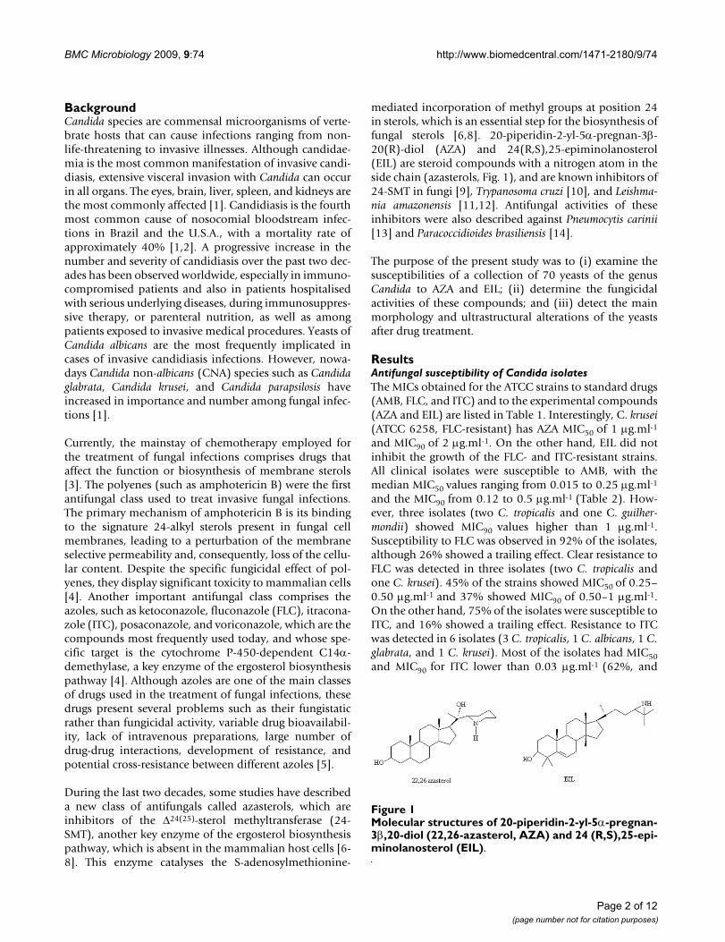
mediated incorporation of methyl groups at position 24in sterols, which is an essential step for the biosynthesis offungal sterols [6,8]. 20-piperidin-2-yl-5α-pregnan-3β-20(R)-diol (AZA) and 24(R,S),25-epiminolanosterol(EIL) are steroid compounds with a nitrogen atom in theside chain (azasterols, Fig. 1), and are known inhibitors of24-SMT in fungi [9], Trypanosoma cruzi [10], and Leishma-nia amazonensis [11,12]. Antifungal activities of theseinhibitors were also described against Pneumocytis carinii[13] and Paracoccidioides brasiliensis [14].
The purpose of the present study was to (i) examine thesusceptibilities of a collection of 70 yeasts of the genusCandida to AZA and EIL; (ii) determine the fungicidalactivities of these compounds; and (iii) detect the mainmorphology and ultrastructural alterations of the yeastsafter drug treatment.
ResultsAntifungal susceptibility of Candida isolatesThe MICs obtained for the ATCC strains to standard drugs(AMB, FLC, and ITC) and to the experimental compounds(AZA and EIL) are listed in Table 1. Interestingly, C. krusei(ATCC 6258, FLC-resistant) has AZA MIC50 of 1 μg.ml-1
and MIC90 of 2 μg.ml-1. On the other hand, EIL did notinhibit the growth of the FLC- and ITC-resistant strains.All clinical isolates were susceptible to AMB, with themedian MIC50 values ranging from 0.015 to 0.25 μg.ml-1
and the MIC90 from 0.12 to 0.5 μg.ml-1 (Table 2). How-ever, three isolates (two C. tropicalis and one C. guilher-mondii) showed MIC90 values higher than 1 μg.ml-1.Susceptibility to FLC was observed in 92% of the isolates,although 26% showed a trailing effect. Clear resistance toFLC was detected in three isolates (two C. tropicalis andone C. krusei). 45% of the strains showed MIC50 of 0.25–0.50 μg.ml-1 and 37% showed MIC90 of 0.50–1 μg.ml-1.On the other hand, 75% of the isolates were susceptible toITC, and 16% showed a trailing effect. Resistance to ITCwas detected in 6 isolates (3 C. tropicalis, 1 C. albicans, 1 C.glabrata, and 1 C. krusei). Most of the isolates had MIC50and MIC90 for ITC lower than 0.03 μg.ml-1 (62%, and
Molecular structures of 20-piperidin-2-yl-5α-pregnan-3β,20-diol (22,26-azasterol, AZA) and 24 (R,S),25-epiminolanos-terol (EIL)Figure 1Molecular structures of 20-piperidin-2-yl-5α-pregnan-3β,20-diol (22,26-azasterol, AZA) and 24 (R,S),25-epi-minolanosterol (EIL).
Page 2 of 12(page number not for citation purposes)

BMC Microbiology 2009, 9:74 http://www.biomedcentral.com/1471-2180/9/74
41%, respectively). Only C. krusei isolates were less sus-ceptible to all standard drugs, showing a MIC90 of 0.5μg.ml-1 for AMB, > 128 μg.ml-1 for FLC, and 2 μg.ml-1 forITC (Table 2).
When the MIC values for 24-SMTI (AZA and EIL) wereanalysed, we observed important antifungal activity foralmost all Candida spp. isolates (Table 3). For AZA, MIC50and MIC90 values lower than 2 μg.ml-1 were observed in86% and 52% of the isolates, respectively. For EIL, MIC50and MIC90 values lower than 2 μg.ml-1 were observed in92% and 63% of the isolates, respectively. C. zeylanoidesand C. lipolytica (a rarely observed CNA) showed MIC50–90values of ≤ 0.03 μg.ml-1 for both inhibitors, whilst C. kru-sei was resistant to EIL, with MIC50–90 values of 8 μg.ml-1
(Table 3). However, both C. krusei and C. lipolytica wereresistant to AZA (MIC50–90 ≥ 16 μg.ml-1) (Table 3). Finally,C. guilliermondii isolates, FLC- and ITC-resistant, were sus-ceptible to AZA, with MIC50–90 values of 0.06 – 0.25μg.ml-1.
Correlations between MIC valuesPositive correlations of the MIC50 values were observedbetween AZA and AMB (r = 0.47), AZA and EIL (r = 0.46),and FLC and ITC (r = 0.79). In addition, positive correla-tions were observed between the MIC90 values of the FLCand ITC (r = 0.71). On the other hand, no significant cor-relations were observed between the MIC values for azolesand 24-SMTI. Some clinical isolates with a trailing effectfor FLC (n = 17) and ITC (n = 11) also showed residualgrowth at higher concentrations of AZA (16 μg.ml-1) of58% (10/17) and 54% (6/11) of the isolates, respectively.Residual growth was not observed in the isolates aftertreatment with EIL.
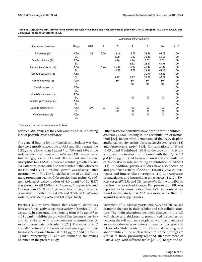
Minimum fungicidal concentration (MFC) of AZA and EILThe MFCs obtained for half of our isolates were higherthan 16 μg.ml-1, revealing a predominant fungistatic activ-ity of the SMTI. Interestingly, 4 CNA isolates (C. glabrata,C. lusitaneae, C. zeylanoides, and C. rugosa) showed MFCslower than 4 μg.ml-1, indicating a remarkable fungicidal
activity, especially for AZA (Table 4). On the other hand,AZA killed a negligible number of the C. albicans isolatesat concentrations lower than 16 μg.ml-1.
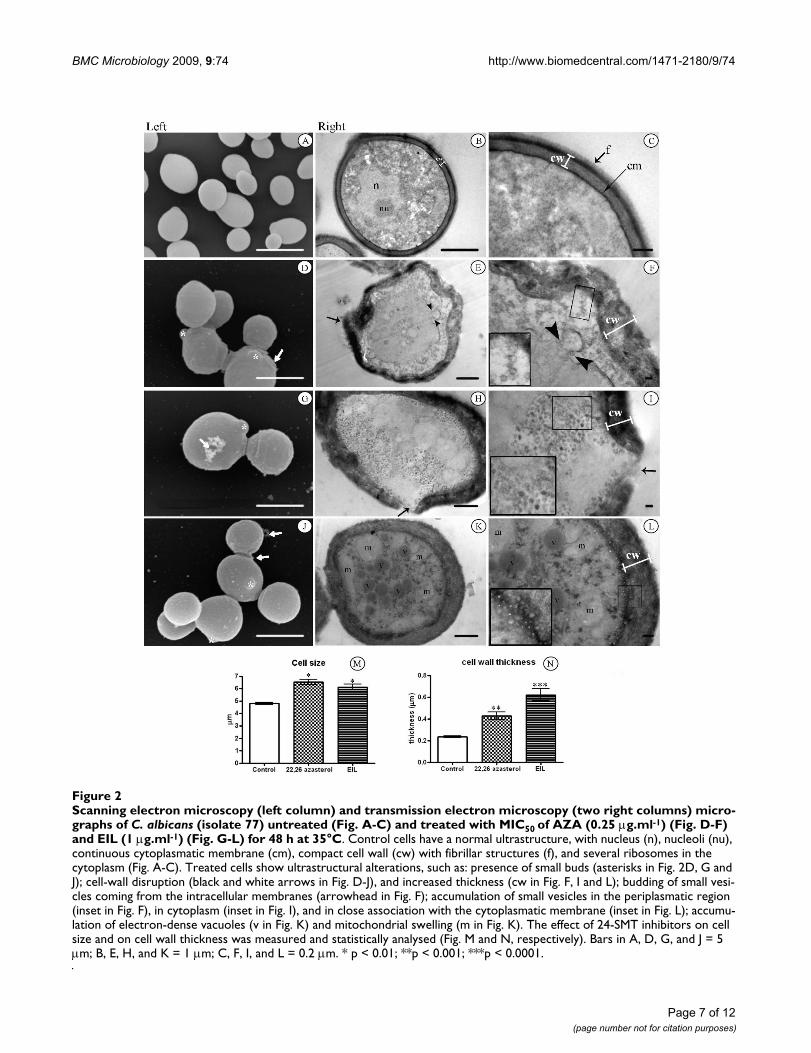
Ultrastructural effectsThe general morphology of untreated C. albicans wasobserved using scanning (Figure 2a) and transmission(Figure 2b–c) electron microscopy. The shape of C. albi-cans varies from spherical (4.90 ± 0.49 μm diameter) tooval cells when viewed by scanning electron microscopy(Figure 2a). Transmission electron microscopy revealedthe presence of normal cell walls with a thickness of 233± 25 nm (Figure 2b–c), including a thin electron-denseouter layer with delicate fibrillar structures clearly visible(f in Figure 2c). A continuous cytoplasmatic membrane(cm)lining a homogeneous and electron-dense cytoplasmcontaining ribosomes, nucleus (n), and nucleoli (nu)could also be observed (Figure 2b–c). Treatment of C. albi-cans with MIC50 of AZA (0.25 μg.ml-1) and EIL (1.00μg.ml-1) induced significant morphological changes,which ranged from discrete alterations to total destructionof the fungal cells. A common alteration observed afterthe treatment with AZA and EIL was a significant increasein cell size, from 5 μm to 7 μm in diameter (Figure 2d, g,j, and 2m). The number of altered cells was counted, andthe morphological alterations appeared in 34.79% and55.17% of the cells after treatment with AZA and EIL,respectively. Among the most frequently observedultrastructural alterations were: (i) presence of small buds(asterisks in Figure 2d, g and 2j); (ii) irregular cell-wallsurfaces (arrows in Fig. 2D and 2E); (iii) loss of cell-wallintegrity, with an apparent shedding of cell components(Fig. 2G–J, white and black arrows); and (iv) a two- tothree-fold increment of the cell wall thickness wasobserved after treatment with AZA and EIL, respectively(Figure 2f, i, l, and 2n). The cytoplasmic membrane oftreated cells also showed several changes, such as the pres-ence of evaginations, discontinuity, detachment from thecell wall (Figure 2e–f); budding of small vesicles thatcould migrate through the periplasmatic region (Figure2e–f, black arrowhead), accumulate in the cytoplasm (Fig-
Table 1: Susceptibility of ATCC strains to Δ24(25) sterol methyl transferase inhibitors, 20-piperidin-2-yl-5α-pregnan-3β, 20-diol (AZA) and 24 (R,S), 25-epiminolanosterol (EIL), and standard antifungals (FLC, ITC, and AMB) by the broth microdilution method.
Strains AZA EIL FLC ITC AMB
MIC50 MIC90 MIC50 MIC90 MIC50 MIC90 MIC50 MIC90 MIC50 MIC90
C. albicans ATCC 10231 > 16 > 16 1 > 16 1 > 128T 0.5 > 16T 0.12 0.25C. parapsilosis ATCC 22019 0.25 4 2 4 2 4 0.03 0.06 0.03 0.06C. tropicalis ATCC 13803 0.25 4 1 2 0.25 2 < 0.03 0.03 0.007 0.25
C. krusei ATCC 6258 0.05 1 > 16 > 16 32 64R 0.12 0.25 0.25 0.25C. glabrata ATCC 2001 1 2 > 16 > 16 4 > 128T 0.12 4T 0.03 0.12
TTrailing Effect, RResistantThe values are expressed in μg.ml-1.
Page 3 of 12(page number not for citation purposes)
BMC Microbiology 2009, 9:74 http://www.biomedcentral.com/1471-2180/9/74
ure 2h–i, details in box), or remain close to the cell mem-brane (Figure 2k–l, details in box). Vesicle sizes rangedfrom 40–80 nm for AZA and 40–220 nm for EIL. Mito-chondrial swelling and electron dense vacuoles accumula-tion was also observed (m, Figure 2k–l). CNA cells treatedwith MIC50 of 24-SMTI showed similar ultrastructuralchanges (data not shown).
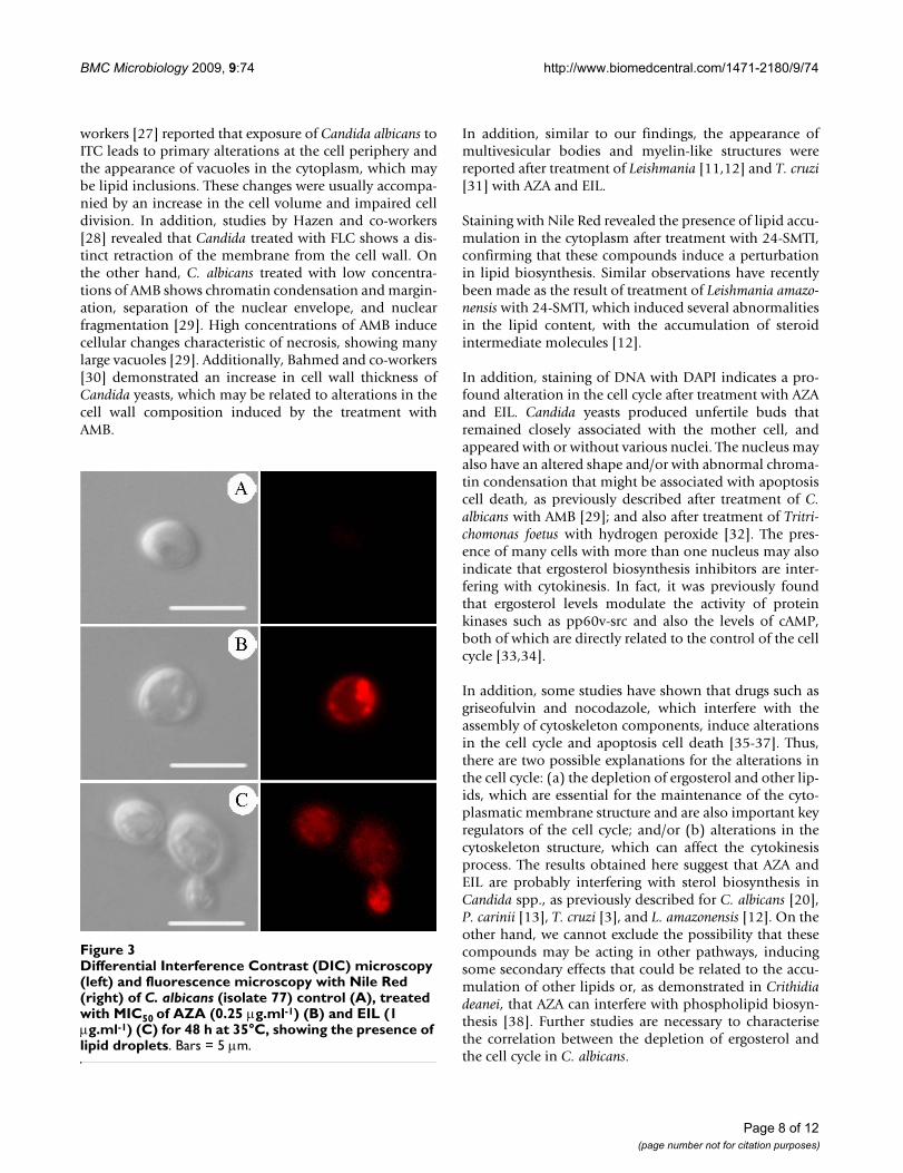
Presence of lipid bodiesTreatment with MIC50 of AZA and EIL induced an accu-mulation of lipid bodies in the cytoplasm, which can becharacterised by the presence of small dots labelled with
Nile Red (Figure 3b–c), which were absent in theuntreated yeasts (Figure 3a). These lipid bodies seen byfluorescence microscopy can be correlated with the small,electron-dense vacuoles seen by transmission electronmicroscopy (see above, ultrastructural effects).
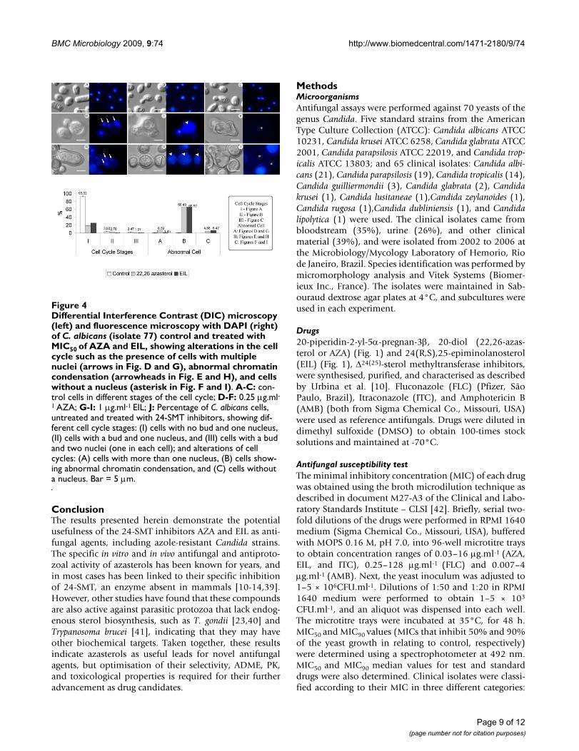
Effect of 24-SMT inhibitors on the cell cycleDAPI staining used to label the DNA revealed that thetreatment of C. albicans with AZA and EIL induced impor-tant alterations in the cell cycle (Figure 4). To quantita-tively assess these alterations, different stages of theuntreated yeasts were considered, such as: (I) cells con-
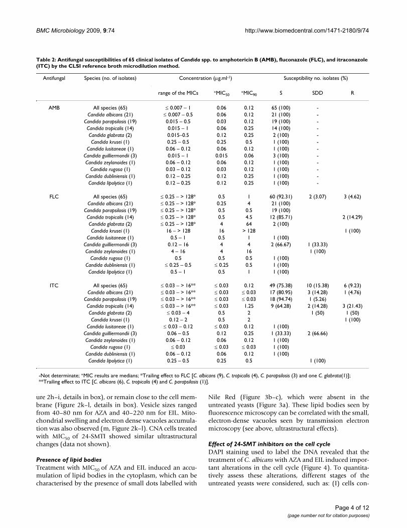
Table 2: Antifungal susceptibilities of 65 clinical isolates of Candida spp. to amphotericin B (AMB), fluconazole (FLC), and itraconazole (ITC) by the CLSI reference broth microdilution method.
Antifungal Species (no. of isolates) Concentration (μg.ml-1) Susceptibility no. isolates (%)
range of the MICs +MIC50+MIC90 S SDD R
AMB All species (65) ≤ 0.007 – 1 0.06 0.12 65 (100) -Candida albicans (21) ≤ 0.007 – 0.5 0.06 0.12 21 (100) -
Candida parapsilosis (19) 0.015 – 0.5 0.03 0.12 19 (100) -Candida tropicalis (14) 0.015 – 1 0.06 0.25 14 (100) -Candida glabrata (2) 0.015–0.5 0.12 0.25 2 (100) -Candida krusei (1) 0.25 – 0.5 0.25 0.5 1 (100) -
Candida lusitaneae (1) 0.06 – 0.12 0.06 0.12 1 (100) -Candida guilliermondii (3) 0.015 – 1 0.015 0.06 3 (100) -Candida zeylanoides (1) 0.06 – 0.12 0.06 0.12 1 (100) -
Candida rugosa (1) 0.03 – 0.12 0.03 0.12 1 (100) -Candida dubliniensis (1) 0.12 – 0.25 0.12 0.25 1 (100) -Candida lipolytica (1) 0.12 – 0.25 0.12 0.25 1 (100) -
FLC All species (65) ≤ 0.25 – > 128* 0.5 1 60 (92.31) 2 (3.07) 3 (4.62)Candida albicans (21) ≤ 0.25 – > 128* 0.25 4 21 (100)
Candida parapsilosis (19) ≤ 0.25 – > 128* 0.5 0.5 19 (100)Candida tropicalis (14) ≤ 0.25 – > 128* 0.5 4.5 12 (85.71) 2 (14.29)Candida glabrata (2) ≤ 0.25 – > 128* 4 64 2 (100)Candida krusei (1) 16 – > 128 16 > 128 1 (100)
Candida lusitaneae (1) 0.5 – 1 0.5 1 1 (100)Candida guilliermondii (3) 0.12 – 16 4 4 2 (66.67) 1 (33.33)Candida zeylanoides (1) 4 – 16 4 16 1 (100)
Candida rugosa (1) 0.5 0.5 0.5 1 (100)Candida dubliniensis (1) ≤ 0.25 – 0.5 ≤ 0.25 0.5 1 (100)Candida lipolytica (1) 0.5 – 1 0.5 1 1 (100)
ITC All species (65) ≤ 0.03 – > 16** ≤ 0.03 0.12 49 (75.38) 10 (15.38) 6 (9.23)Candida albicans (21) ≤ 0.03 – > 16** ≤ 0.03 ≤ 0.03 17 (80.95) 3 (14.28) 1 (4.76)
Candida parapsilosis (19) ≤ 0.03 – > 16** ≤ 0.03 ≤ 0.03 18 (94.74) 1 (5.26)Candida tropicalis (14) ≤ 0.03 – > 16** ≤ 0.03 1.25 9 (64.28) 2 (14.28) 3 (21.43)Candida glabrata (2) ≤ 0.03 – 4 0.5 2 1 (50) 1 (50)Candida krusei (1) 0.12 – 2 0.5 2 1 (100)
Candida lusitaneae (1) ≤ 0.03 – 0.12 ≤ 0.03 0.12 1 (100)Candida guilliermondii (3) 0.06 – 0.5 0.12 0.25 1 (33.33) 2 (66.66)Candida zeylanoides (1) 0.06 – 0.12 0.06 0.12 1 (100)
Candida rugosa (1) ≤ 0.03 ≤ 0.03 ≤ 0.03 1 (100)Candida dubliniensis (1) 0.06 – 0.12 0.06 0.12 1 (100)Candida lipolytica (1) 0.25 – 0.5 0.25 0.5 1 (100)
-Not determinate; +MIC results are medians; *Trailing effect to FLC [C. albicans (9), C. tropicalis (4), C. parapsilosis (3) and one C. glabrata(1)]; **Trailing effect to ITC [C. albicans (6), C. tropicalis (4) and C. parapsilosis (1)].
Page 4 of 12(page number not for citation purposes)
BMC Microbiology 2009, 9:74 http://www.biomedcentral.com/1471-2180/9/74
taining one nucleus, (II) cells with a bud and one nucleus,and (III) cells with a bud and two nuclei (one in eachcell). After treatment with the MIC50s of AZA and EIL, dif-ferent alterations in the nucleus were observed, and thesewere classified as: (A) cells with more than one nucleus,(B) cells showing abnormal chromatin condensation, and(C) cells without a nucleus. Counting the number ofabnormal cells revealed that approximately 66% of theyeasts showed abnormal chromatin condensation,whereas 6.6% of AZA-treated and 1.5% of EIL-treated cellscontained more than one nucleus, and approximately 6%of the cells treated with both compounds had no nucleus(Figure 4).
Cytotoxicity evaluationCytotoxicity of 24-SMTI was evaluated against mamma-lian cells (Vero) using the sulforhodamine B viabilityassay. For both AZA and EIL the CC50 was 40 μg.ml-1,which corresponds to a mean selectivity index of 80 forAZA and 20 for EIL.
DiscussionAlthough C. albicans is the predominant species in candi-diasis, CNA species have increased in frequency in recent
years. The reasons for the emergence of CNA species arenot fully understood, but some medical conditions mayfrequently run the risk of developing candidaemia due tothe CNA species: C. parapsilopsis has been associated withvascular catheters and parenteral nutrition; C. tropicaliswith cancer and neutropenia; and C. krusei and C. glabratawith previous treatments with FLC and ITC [2].
Previous studies have described a high susceptibility of C.albicans isolates to azoles and AMB, whereas CNA isolatesare usually less susceptible and may be intrinsically resist-ant to FLC and ITC [2,15-17]. As reported by other inves-tigators [2,18,19], none of our Candida isolates showedMIC ≥ 2 μg.ml-1 for AMB. MIC values found for ITC andFLU were similar to those previously reported by differentgroups [2,15-17]. However, in the present study, FLC-resistant Candida strains were only observed among CNAspecies (6.8% of the isolates). However, ITC-resistancewas found in C. albicans (1.5%) and CNA isolates (7.7%).
The observation of a high positive correlation betweenMIC values of FLC and ITC (r = 0.79 for MIC50 and r = 0.71for MIC90), in this study, suggests that cross-resistancemay be occurring. However, no correlation was observed
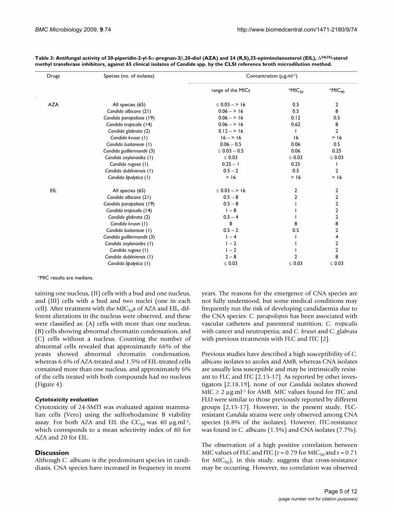
Table 3: Antifungal activity of 20-piperidin-2-yl-5α-pregnan-3β,20-diol (AZA) and 24 (R,S),25-epiminolanosterol (EIL), Δ24(25)-sterol methyl transferase inhibitors, against 65 clinical isolates of Candida spp. by the CLSI reference broth microdilution method.
Drugs Species (no. of isolates) Concentration (μg.ml-1)
range of the MICs +MIC50+MIC90
AZA All species (65) ≤ 0.03 – > 16 0.5 2Candida albicans (21) 0.06 – > 16 0.5 8
Candida parapsilosis (19) 0.06 – > 16 0.12 0.5Candida tropicalis (14) 0.06 – > 16 0.62 8Candida glabrata (2) 0.12 – > 16 1 2Candida krusei (1) 16 – > 16 16 > 16
Candida lusitaneae (1) 0.06 – 0.5 0.06 0.5Candida guilliermondii (3) ≤ 0.03 – 0.5 0.06 0.25Candida zeylanoides (1) ≤ 0.03 ≤ 0.03 ≤ 0.03
Candida rugosa (1) 0.25 – 1 0.25 1Candida dubliniensis (1) 0.5 – 2 0.5 2Candida lipolytica (1) > 16 > 16 > 16
EIL All species (65) ≤ 0.03 – > 16 2 2Candida albicans (21) 0.5 – 8 2 2
Candida parapsilosis (19) 0.5 – 8 1 2Candida tropicalis (14) 1 – 8 1 2Candida glabrata (2) 0.5 – 4 1 2Candida krusei (1) 8 8 8
Candida lusitaneae (1) 0.5 – 2 0.5 2Candida guilliermondii (3) 1 – 4 1 4Candida zeylanoides (1) 1 – 2 1 2
Candida rugosa (1) 1 – 2 1 2Candida dubliniensis (1) 2 – 8 2 8Candida lipolytica (1) ≤ 0.03 ≤ 0.03 ≤ 0.03
+MIC results are medians.
Page 5 of 12(page number not for citation purposes)
BMC Microbiology 2009, 9:74 http://www.biomedcentral.com/1471-2180/9/74
between MIC values of the azoles and 24-SMTI, indicatinglack of possible cross-resistance.
The general finding for our Candida spp. isolates was thatthey were mostly susceptible to AZA and EIL, because theMIC50s were lower than 2 μg.ml-1 for 73% and 88% of theisolates after treatment with AZA and EIL, respectively.Interestingly, some FLC- and ITC-resistant strains weresusceptible to 24-SMTI. However, residual growth of Can-dida after treatment with AZA was similar to that observedfor FLC and ITC. No residual growth was observed aftertreatment with EIL. The fungicidal action of 24-SMTI wasmore prominent against CNA species than against C. albi-cans isolates. A concentration of 4.0 μg.ml-1 of 24-SMTIwas enough to kill 100% of C. lusitanae, C. zeylanoides, andC. rugosa, and 50% of C. glabrata. In contrast, this sameconcentration killed only 4.7% and 9.5% of C. albicansisolates, considering AZA and EIL respectively.
Previous studies have shown that azasterol derivativeshave antifungal activity against a variety of species [7]. 15-azasterol, in concentrations ranging from 0.01 μg.ml-1 to4.08 μg.ml-1, inhibits the growth of Saccharomyces cerevisaeand C. albicans, with a concomitant accumulation ofsterol intermediate molecules [20,21]. The range of MICand MFC values for 15-azasterol analogues against thesefungal species varied from 0.8 to 3.1 μg.ml-1 and 3.1 to 6.3μg.ml-1, respectively [7] and are similar to the valuesobtained in the present study.
Other azasterol derivatives have been shown to inhibit S.cerevisae 24-SMT, leading to the accumulation of zymos-terol [22]. Recent work demonstrated that AZA displaysantifungal activity against Paracoccidioides brasiliensis [14]and Pneumocystis carinii [13]. Concentrations of 5 μM(2.05 μg.ml-1) inhibited 100% of the growth in P. brasil-iensis, and the treatment of P. carinii with the IC50 of 0.3μM (0.12 μg.ml-1) led to growth arrest and accumulationof 24-desakyl sterols, indicating an inhibition of 24-SMT[13]. In addition, previous studies have also shown ananti-protozoan activity of AZA and EIL on T. cruzi epimas-tigotes and intracellular amastigotes [10], L. amazonensispromastigotes and intracellular amastigotes [11,12], Tox-oplasma gondii [23], and Giardia lamblia [24], with MICs inthe low μM to sub-μM range. For protozoans, EIL wasreported to be more active than AZA. In contrast, wefound in this study that AZA was more active than EILagainst Candida spp. isolates.
Treatment of C. albicans yeasts with AZA and EIL causeddramatic changes in their cellular and sub-cellular struc-ture. The main alterations included changes in the cellwall shape and thickness, a pronounced disconnectionbetween the cell wall and cytoplasm, with the presence ofan electron-lucent zone between them, cell collapse andrelease of cellular content, mitochondrial swelling, andabnormalities in the nuclear structure. These findings aresimilar to those previously reported after treatment ofCandida spp. with different azoles [25-28]. Borges and co-
Table 4: Cumulative MFC profile of 65 clinical isolates of Candida spp. treated with 20-piperidin-2-yl-5α-pregnan-3β,20-diol (AZA) and 24(R,S),25-epiminolanosterol (EIL).
Cumulative MFC* (μg.ml-1)
Species (no. isolates) Drugs 0.03 1 2 4 8 16 > 16
All species (65) AZA 1.52 3.04 12.16 16.72 34.96 44.08 100EIL 6.08 15.20 30.40 51.68 100
Candida albicans (21) AZA 4.76 4.76 9.52 9.52 100EIL 9.52 28.57 61.98 100
Candida parapsilosis (19) AZA 5.26 26.31 36.87 68.42 68.42 100EIL 10.52 15.79 26.31 63.15 100
Candida tropicalis (14) AZA 35.71 64.28 100EIL 7.17 7.17 35.71 42.87 100
Candida glabrata (2) AZA 50 50 50 50 100EIL 50 50 50 100
Candida krusei (1) AZA 100EIL 100
Candida lusitaneae (1) AZA 100EIL 100 100 100 100
Candida guilliermondii (3) AZA 100EIL 100
Candida zeylanoides (1) AZA 100 100 100 100 100 100 100EIL 100 100 100 100 100
Candida rugosa (1) AZA 100 100 100 100EIL 100
* data is expressed in percentual of isolates.
Page 6 of 12(page number not for citation purposes)
BMC Microbiology 2009, 9:74 http://www.biomedcentral.com/1471-2180/9/74
Page 7 of 12(page number not for citation purposes)
Scanning electron microscopy (left column) and transmission electron microscopy (two right columns) micrographs of C. albi-cans (isolate 77) untreated (Fig. A-C) and treated with MIC50 of AZA (0.25 μg.ml-1) (Fig. D-F) and EIL (1 μg.ml-1) (Fig. G-L) for 48 h at 35°CFigure 2Scanning electron microscopy (left column) and transmission electron microscopy (two right columns) micro-graphs of C. albicans (isolate 77) untreated (Fig. A-C) and treated with MIC50 of AZA (0.25 μg.ml-1) (Fig. D-F) and EIL (1 μg.ml-1) (Fig. G-L) for 48 h at 35°C. Control cells have a normal ultrastructure, with nucleus (n), nucleoli (nu), continuous cytoplasmatic membrane (cm), compact cell wall (cw) with fibrillar structures (f), and several ribosomes in the cytoplasm (Fig. A-C). Treated cells show ultrastructural alterations, such as: presence of small buds (asterisks in Fig. 2D, G and J); cell-wall disruption (black and white arrows in Fig. D-J), and increased thickness (cw in Fig. F, I and L); budding of small vesi-cles coming from the intracellular membranes (arrowhead in Fig. F); accumulation of small vesicles in the periplasmatic region (inset in Fig. F), in cytoplasm (inset in Fig. I), and in close association with the cytoplasmatic membrane (inset in Fig. L); accumu-lation of electron-dense vacuoles (v in Fig. K) and mitochondrial swelling (m in Fig. K). The effect of 24-SMT inhibitors on cell size and on cell wall thickness was measured and statistically analysed (Fig. M and N, respectively). Bars in A, D, G, and J = 5 μm; B, E, H, and K = 1 μm; C, F, I, and L = 0.2 μm. * p < 0.01; **p < 0.001; ***p < 0.0001.
BMC Microbiology 2009, 9:74 http://www.biomedcentral.com/1471-2180/9/74
workers [27] reported that exposure of Candida albicans toITC leads to primary alterations at the cell periphery andthe appearance of vacuoles in the cytoplasm, which maybe lipid inclusions. These changes were usually accompa-nied by an increase in the cell volume and impaired celldivision. In addition, studies by Hazen and co-workers[28] revealed that Candida treated with FLC shows a dis-tinct retraction of the membrane from the cell wall. Onthe other hand, C. albicans treated with low concentra-tions of AMB shows chromatin condensation and margin-ation, separation of the nuclear envelope, and nuclearfragmentation [29]. High concentrations of AMB inducecellular changes characteristic of necrosis, showing manylarge vacuoles [29]. Additionally, Bahmed and co-workers[30] demonstrated an increase in cell wall thickness ofCandida yeasts, which may be related to alterations in thecell wall composition induced by the treatment withAMB.
In addition, similar to our findings, the appearance ofmultivesicular bodies and myelin-like structures werereported after treatment of Leishmania [11,12] and T. cruzi[31] with AZA and EIL.
Staining with Nile Red revealed the presence of lipid accu-mulation in the cytoplasm after treatment with 24-SMTI,confirming that these compounds induce a perturbationin lipid biosynthesis. Similar observations have recentlybeen made as the result of treatment of Leishmania amazo-nensis with 24-SMTI, which induced several abnormalitiesin the lipid content, with the accumulation of steroidintermediate molecules [12].
In addition, staining of DNA with DAPI indicates a pro-found alteration in the cell cycle after treatment with AZAand EIL. Candida yeasts produced unfertile buds thatremained closely associated with the mother cell, andappeared with or without various nuclei. The nucleus mayalso have an altered shape and/or with abnormal chroma-tin condensation that might be associated with apoptosiscell death, as previously described after treatment of C.albicans with AMB [29]; and also after treatment of Tritri-chomonas foetus with hydrogen peroxide [32]. The pres-ence of many cells with more than one nucleus may alsoindicate that ergosterol biosynthesis inhibitors are inter-fering with cytokinesis. In fact, it was previously foundthat ergosterol levels modulate the activity of proteinkinases such as pp60v-src and also the levels of cAMP,both of which are directly related to the control of the cellcycle [33,34].
In addition, some studies have shown that drugs such asgriseofulvin and nocodazole, which interfere with theassembly of cytoskeleton components, induce alterationsin the cell cycle and apoptosis cell death [35-37]. Thus,there are two possible explanations for the alterations inthe cell cycle: (a) the depletion of ergosterol and other lip-ids, which are essential for the maintenance of the cyto-plasmatic membrane structure and are also important keyregulators of the cell cycle; and/or (b) alterations in thecytoskeleton structure, which can affect the cytokinesisprocess. The results obtained here suggest that AZA andEIL are probably interfering with sterol biosynthesis inCandida spp., as previously described for C. albicans [20],P. carinii [13], T. cruzi [3], and L. amazonensis [12]. On theother hand, we cannot exclude the possibility that thesecompounds may be acting in other pathways, inducingsome secondary effects that could be related to the accu-mulation of other lipids or, as demonstrated in Crithidiadeanei, that AZA can interfere with phospholipid biosyn-thesis [38]. Further studies are necessary to characterisethe correlation between the depletion of ergosterol andthe cell cycle in C. albicans.
Differential Interference Contrast (DIC) microscopy (left) and fluorescence microscopy with Nile Red (right) of C. albi-cans (isolate 77) control (A), treated with MIC50 of AZA (0.25 μg.ml-1) (B) and EIL (1 μg.ml-1) (C) for 48 h at 35°C, showing the presence of lipid dropletsFigure 3Differential Interference Contrast (DIC) microscopy (left) and fluorescence microscopy with Nile Red (right) of C. albicans (isolate 77) control (A), treated with MIC50 of AZA (0.25 μg.ml-1) (B) and EIL (1 μg.ml-1) (C) for 48 h at 35°C, showing the presence of lipid droplets. Bars = 5 μm.
Page 8 of 12(page number not for citation purposes)
BMC Microbiology 2009, 9:74 http://www.biomedcentral.com/1471-2180/9/74
ConclusionThe results presented herein demonstrate the potentialusefulness of the 24-SMT inhibitors AZA and EIL as anti-fungal agents, including azole-resistant Candida strains.The specific in vitro and in vivo antifungal and antiproto-zoal activity of azasterols has been known for years, andin most cases has been linked to their specific inhibitionof 24-SMT, an enzyme absent in mammals [10-14,39].However, other studies have found that these compoundsare also active against parasitic protozoa that lack endog-enous sterol biosynthesis, such as T. gondii [23,40] andTrypanosoma brucei [41], indicating that they may haveother biochemical targets. Taken together, these resultsindicate azasterols as useful leads for novel antifungalagents, but optimisation of their selectivity, ADME, PK,and toxicological properties is required for their furtheradvancement as drug candidates.
MethodsMicroorganismsAntifungal assays were performed against 70 yeasts of thegenus Candida. Five standard strains from the AmericanType Culture Collection (ATCC): Candida albicans ATCC10231, Candida krusei ATCC 6258, Candida glabrata ATCC2001, Candida parapsilosis ATCC 22019, and Candida trop-icalis ATCC 13803; and 65 clinical isolates: Candida albi-cans (21), Candida parapsilosis (19), Candida tropicalis (14),Candida guilliermondii (3), Candida glabrata (2), Candidakrusei (1), Candida lusitaneae (1),Candida zeylanoides (1),Candida rugosa (1),Candida dubliniensis (1), and Candidalipolytica (1) were used. The clinical isolates came frombloodstream (35%), urine (26%), and other clinicalmaterial (39%), and were isolated from 2002 to 2006 atthe Microbiology/Mycology Laboratory of Hemorio, Riode Janeiro, Brazil. Species identification was performed bymicromorphology analysis and Vitek Systems (Biomer-ieux Inc., France). The isolates were maintained in Sab-ouraud dextrose agar plates at 4°C, and subcultures wereused in each experiment.
Drugs20-piperidin-2-yl-5α-pregnan-3β, 20-diol (22,26-azas-terol or AZA) (Fig. 1) and 24(R,S),25-epiminolanosterol(EIL) (Fig. 1), Δ24(25)-sterol methyltransferase inhibitors,were synthesised, purified, and characterised as describedby Urbina et al. [10]. Fluconazole (FLC) (Pfizer, SãoPaulo, Brazil), Itraconazole (ITC), and Amphotericin B(AMB) (both from Sigma Chemical Co., Missouri, USA)were used as reference antifungals. Drugs were diluted indimethyl sulfoxide (DMSO) to obtain 100-times stocksolutions and maintained at -70°C.
Antifungal susceptibility testThe minimal inhibitory concentration (MIC) of each drugwas obtained using the broth microdilution technique asdescribed in document M27-A3 of the Clinical and Labo-ratory Standards Institute – CLSI [42]. Briefly, serial two-fold dilutions of the drugs were performed in RPMI 1640medium (Sigma Chemical Co., Missouri, USA), bufferedwith MOPS 0.16 M, pH 7.0, into 96-well microtitre traysto obtain concentration ranges of 0.03–16 μg.ml-1 (AZA,EIL, and ITC), 0.25–128 μg.ml-1 (FLC) and 0.007–4μg.ml-1 (AMB). Next, the yeast inoculum was adjusted to1–5 × 10⁶CFU.ml-1. Dilutions of 1:50 and 1:20 in RPMI1640 medium were performed to obtain 1–5 × 10³
CFU.ml-1, and an aliquot was dispensed into each well.The microtitre trays were incubated at 35°C, for 48 h.MIC50 and MIC90 values (MICs that inhibit 50% and 90%of the yeast growth in relating to control, respectively)were determined using a spectrophotometer at 492 nm.MIC50 and MIC90 median values for test and standarddrugs were also determined. Clinical isolates were classi-fied according to their MIC in three different categories:
Differential Interference Contrast (DIC) microscopy (left) and fluorescence microscopy with DAPI (right) of C. albicans (isolate 77) control and treated with MIC50 of AZA and EIL, showing alterations in the cell cycle such as the presence of cells with multiple nuclei (arrows in Fig. D and G), abnormal chromatin condensation (arrowheads in Fig. E and H), and cells without a nucleus (asterisk in Fig. F and I)Figure 4Differential Interference Contrast (DIC) microscopy (left) and fluorescence microscopy with DAPI (right) of C. albicans (isolate 77) control and treated with MIC50 of AZA and EIL, showing alterations in the cell cycle such as the presence of cells with multiple nuclei (arrows in Fig. D and G), abnormal chromatin condensation (arrowheads in Fig. E and H), and cells without a nucleus (asterisk in Fig. F and I). A-C: con-trol cells in different stages of the cell cycle; D-F: 0.25 μg.ml-1 AZA; G-I: 1 μg.ml-1 EIL; J: Percentage of C. albicans cells, untreated and treated with 24-SMT inhibitors, showing dif-ferent cell cycle stages: (I) cells with no bud and one nucleus, (II) cells with a bud and one nucleus, and (III) cells with a bud and two nuclei (one in each cell); and alterations of cell cycles: (A) cells with more than one nucleus, (B) cells show-ing abnormal chromatin condensation, and (C) cells without a nucleus. Bar = 5 μm.
Page 9 of 12(page number not for citation purposes)
BMC Microbiology 2009, 9:74 http://www.biomedcentral.com/1471-2180/9/74
susceptible (S), susceptible dose-dependent (SDD), orresistant (R). Interpretative breakpoints proposed by theCLSI [42] for FLC and ITC were used, and concentrationsabove 1 μg.ml-1 were considered resistant for AMB [43].Trailing effect for FLC and ITC was detected at visual read-ing after 24 h of incubation.
The minimum fungicidal concentration (MFC) was deter-mined after 48 h of treatment with the inhibitory concen-trations used in the susceptibility test. An aliquot of eachCandida isolate was transferred onto Sabouraud dextroseagar plates without the presence of drugs. The plates wereincubated at 35°C for 48 h, and the minimum fungicidalconcentration (MFC) was determined. MFC means thelowest concentration that showed no fungal growth [44].
Fluorescence microscopyC. albicans (isolate 77) was treated with MIC50 of AZA andEIL at 35°C for 48 h. Yeasts were washed in PBS, pH 7.2and fixed with 4% paraformaldehyde in PBS for 30 min.Next, the yeasts were adhered to coverslips with poly-L-lysine and incubated with 5 μg.ml-1 Nile Red (Fluka, USA)for 30 min to label the lipid bodies and 1 μg.ml-1 DAPI(Sigma Chemical Co., Missouri, USA) for 10 min to labelthe DNA. Coverslips were mounted in n-propylgallatesolution and observed in a Zeiss Axioplan epifluorescencemicroscope equipped with rhodamine (Nile Red fluores-cence) and DAPI filters, and the images were recordedwith a C5810 Hamamatsu camera. The number of alteredCandida was determined after the counting of at least 300yeast cells. Cell size was measured by means of the SemA-fore 5.0 software (Jeol, Japan).
Transmission electron microscopyC. albicans (isolate 77) was treated with MIC50 of AZA andEIL at 35°C, for 48 h. Yeasts were washed in PBS, pH 7.2and fixed in a solution of 2.5% glutaraldehyde and 4%freshly prepared formaldehyde in 0.1 M cacodylate buffer,pH 7.2, for 2 h at room temperature. After fixation, yeastswere post-fixed for 2 h in 1% osmium tetroxide contain-ing 1.25% potassium ferrocyanide and 5 mM CaCl2 incacodylate buffer, pH 7.2, washed in the same buffer,dehydrated in ethanol, and embedded in Spurr. Ultrathinsections were stained with uranyl acetate and lead citrate,and images were obtained in a Zeiss 900 electron micro-scope equipped with a CCD Camera (Mega view IIImodel, Soft Image System, Germany). Images were proc-essed with iTEM software (Soft Image System, Germany).Cell wall thicknesses and vesicles of untreated and treatedyeasts were measured by means of the SemAfore 5.0 soft-ware (Jeol, Japan).
Scanning electron microscopyC. albicans (isolate 77) treated with MIC50 of AZA and EILat 35°C for 48 h, was fixed as described above for trans-
mission electron microscopy, and subsequently dehy-drated in ethanol, critical-point dried in CO2, coated withgold, and observed in a JEOL JSM-5310 scanning electronmicroscope.
Cytotoxicity tests in mammalian cellsGreen monkey kidney (Vero) cells were maintained inDulbecco's modified Eagle's medium (DMEM, Gibco Inv-itrogen Corporation, New York, USA) supplemented with2 mM L-glutamine, 10% heat-inactivated fetal bovineserum (FBS), and 50 μg.ml-1 gentamicin at 37°C in a 5%CO2/air mixture. In 96-well microtitre trays, 2.5 × 10⁴
cells/well were dispensed and incubated for 24 h. Monol-ayers of Vero cells were treated with different concentra-tions of 24-SMTI for 48 h at 37°C in 5% CO2 and fixed in10% trichloroacetic acid for 1 h at 4°C, stained with sul-forhodamine B for 30 min at 4°C, and the optical densi-ties were obtained in a spectrophotometer at 530 nm [45].The 50% cytotoxic concentration (CC50) and the selectiv-ity index (SI = CC50/MIC50) were calculated.
Statistical analysisStatistical analyses were performed with the Prism 5.0software, and p < 0.05 was considered as significant. Dif-ferences in the cell size and cell-wall thickness ofuntreated and treated Candida spp. were analysed by one-way ANOVA (Dunnett test), and MIC values were ana-lysed by linear regression test.
Authors' contributionsKI, JCFR and SR designed the study and wrote the manu-script. The syntheses of 24-SMT inhibitors were performedby JAU. MDR provided the clinical isolates. KI and TVMVrealized the susceptibility assay, fluorescence and trans-mission electron microscopy. CVN worked on cytotoxicitytests. JAU and WS critically revised the manuscript for itsimportant intellectual content. All authors read andapproved the final manuscript.
AcknowledgementsThis work was supported by the Conselho Nacional de Desenvolvimento Científico e Tecnológico (CNPq) and Fundação Carlos Chagas Filho de Amparo à Pesquisa do Estado do Rio de Janeiro (FAPERJ). J.C.F.R. has a postdoctoral fellowship from the Coordenação de Aperfeiçoamento de Pessoal de Nível Superior (CAPES).
References1. Kauffman CA: Fungal infections. Proc Am Thoracic Soc 2006,
3:35-40.2. Colombo AL, Nucci M, Park BJ, Noue'R SA, Arthington-Skaggs B,
Matta DA, Warnock D, Morgan J: Epidemiology of candidemia inBrazil: a nationwide sentinel surveillance of candidemia ineleven medical centers. J Clin Microbiol 2006, 44:2816-2823.
3. Pappas PG, Rex JH, Sobel JD, Filler SG, Dismukes WE, Walsh TJ,Edwards JE: Guidelines of treatment of candidiasis. Clin InfectDis 2004, 38:161-189.
4. Odds FC, Brown AJ, Gow NA: Antifungal agents: Mechanism ofaction. Trends Microbiol 2003, 11:272-279.
Page 10 of 12(page number not for citation purposes)
BMC Microbiology 2009, 9:74 http://www.biomedcentral.com/1471-2180/9/74
5. Pasqualotto AC, Denning DW: New and emerging treatmentsfor fungal infections. J Antimicrob Chemother 2008, 61(Suppl1):i19-i30.
6. Barret-Bee K, Ryder NS: Biochemical aspects of ergosterol bio-synthesis inhibition. In Emerging targets in antibacterial and antifun-gal chemotherapy Edited by: Sutcliffe J, Georgopapadakou NH. NewYork: Chapman & Hall; 1992:410-436.
7. Burbiel J, Bracher F: Azasteroids as antifungals. Steroids 2003,68:587-594.
8. Oehlschlager AC, Czyzewska E: Rationally designed inhibitors ofsterol biosynthesis. In Emerging targets in antibacterial and antifungalchemotherapy Edited by: Sutcliffe J, Georgopapadakou NH. New York:Chapman & Hall; 1992:437-475.
9. Song Z, Nes WD: Sterol biosynthesis inhibitors: Potential fortransition state analogs and mechanism-based inactivatorstargeted at sterol methyltransferase. Lipids 2007, 42:15-33.
10. Urbina JA, Vivas J, Visbal G, Contreras LM: Modification of thecomposition of Trypanosoma (Schizotrypanum) cruzi epimas-tigotes by Δ24(25) sterol methyltransferase inhibitors andtheir combinations with ketoconazole. Mol Biochem Parasitol1995, 73:199-210.
11. Rodrigues JCF, Bernardes CF, Visbal G, Urbina JA, Vercesi AE, deSouza W: Sterol methenyl transferase inhibitors alter theultrastructure and function of the Leishmania amazonensismitochondrion leading to potent growth inhibition. Protist2007, 158:447-456.
12. Rodrigues JCF, Attias M, Rodriguez C, Urbina JA, de Souza W:Ultrastructural and biochemical alterations induced by22,26-azasterol, a Δ24(25)-sterol methyltransferase inhibitor,on promastigote and amastigote forms of Leishmania amazo-nensis. Antimicrob Agents Chemother 2002, 46:487-499.
13. Urbina JA, Visbal G, Contreras LM, Mclaughlin G, Docampo R: Inhib-itors of D24(25) sterol methyltransferase block sterol syn-thesis and cell proliferation in Pneumocystis carinii. AntimicrobAgents Chemother 1997, 41:1428-1432.
14. Visbal G, Alvarez A, Moreno B, San-Blas G: S-adenosyl-L-methio-nine inhibitors Δ24-sterol methyltransferase and Δ24(28)-sterol methylreductase as possible agents against Paracoc-cidioides brasiliensis. Antimicrob Agents Chemother 2003,47:2966-2970.
15. Borg-von Zepelin M, Kunz L, Rüchel R, Reichard U, Weig M, Groß U:Epidemiology and antifungal susceptibilities of Candida spp.to six antifungal agents: results from a surveillance study onfungaemia in Germany from July to August 2005. J AntimicrobChemother 2007, 60:424-428.
16. Godoy P, Tiraboschi IN, Severo LC, Bustamante B, Calvo B, AlmeidaLP, da Matta DA, Colombo AL: Species distribution and antifun-gal susceptibility profile of Candida spp. bloodstream iso-lates from Latin American hospitals. Mem Inst Oswaldo Cruz2003, 98:401-405.
17. Tortorano AM, Kibbler C, Peman J, Bernhardt H, Klingspor L, GrillotR: Candidaemia in Europe: epidemiology and resistance. IntJ Antimicrob Agents 2006, 27:359-366.
18. Almirante B, Rodriguez D, Park BJ, Cuenca-Estrella M, Planes AM,Almela M, Mensa J, Sanchez F, Ayats J, Gimenez M, Saballs P, FridkinSK, Morgan J, Rodriguez-Tudela JL, Warnock DW, Pahissa A: Epide-miology and predictors of mortality in cases of Candidabloodstream infection: results from population-based sur-veillance, Barcelona, Spain, from 2000 to 2003. J Clin Microbiol2005, 43:1829-1835.
19. Ostrosky-Zeichner L, Rex JH, Pappas PG, Hamill RJ, Larsen RA,Horowitz HW, Powderly WG, Hyslop N, Kauffman CA, Cleary J,Mangino JE, Lee J: Antifungal susceptibility survey of 2,000bloodstream Candida isolates in the United States. AntimicrobAgents Chemother 2003, 47:3149-3154.
20. Georgopapadakou NH, Dix BA, Smith SA, Freudenberger J, Funke PT:Effect of antifungal agents on lipid biosynthesis and mem-brane integrity in Candida albicans. Antimicrob Agents Chemother1987, 31:46-51.
21. Hays PR, Parks LW, Pierce HD, Oehlschlager AC: Accumulation ofergosta-8,14-dien-3beta-ol by Saccharomyces cerevisae cul-tured with an azasterol antimycotic agent. Lipids 1977,12:666-668.
22. Oehlschlager AC, Angus RH, Pierce AM, Srinivasan R: Azasterolinhibition of Δ24-sterol methyltransferase in Saccharomycescerevisae. Biochemistry 1984, 23:3582-3589.
23. Dantas-Leite LRV, Urbina JA, de Souza W, Vommaro RC: Selectiveanti-Toxoplasma gondii activities of azasterols. Int J AntimicrobAgents 2004, 23:620-626.
24. Maia C, Attias M, Urbina JA, Gilbert I, Magaraci F, de Souza W: Aza-sterols impair Giardia lamblia proliferation and inducesencystation. Biochem Biophys Res Commun 2007, 363:310-316.
25. Bellanger P, Nast CC, Fratti R, Sanati H, Ghannoum M: Voricona-zole (UK-109,496) inhibits the growth and alters the mor-phology of fluconazole-susceptible and -resistant Candidaspecies. Antimicrob Agents Chemother 1997, 41:1840-1842.
26. Koul A, Vitullo J, Reyes G, Ghannoum M: Effects of voriconazoleon Candida glabrata in vitro. J Antimicrob Chemother 1999,44:109-112.
27. Borges M, Ven MA Van de: Degenerative changes after itracon-zole treatment. Rev Infect Dis 1987, 9(Suppl 1):S33-42.
28. Hazen KC, Mandell G, Coleman E, Giangqin W: Influence of fluco-nazole at subinhibitory concentrations on cell surface hydro-phobicity and phagocytosis of Candida albicans. FEMSMicrobiology Letters 2000, 183:89-94.
29. Phillips A, Sudbery I, Ramsdale M: Apoptosis induced by environ-mental stresses and amphotericin B in Candida albicans. ProcNatl Acad Sci USA 2003, 100:14327-14332.
30. Bahmed K, Bonaly R, Benallaoua S, Coulon J: Effect of sub-inhibi-tory concentrations of amphotericin B on the yeast surfaceand phagocytic killing activity. Process Biochem 2005, 40:759-765.
31. Vivas JJ, Urbina JA, de Souza W: Ultrastructural alterations inTrypanosoma (Schizotrypanum) cruzi induced by Δ24(25)-sterolmethyltransferase inhibitors and their combinations withketoconazole. Int J Antimicrob Agents 1996, 7:235-240.
32. Mariante RM, Guimarães CA, Linden R, Benchimol M: Hydrogenperoxide induces caspase activation and programmed celldeath in the amitochondrial Tritrichomonas foetus. HistochemCell Biol 2003, 120:129-141.
33. Dahl C, Biemannt HP, Dahl J: A protein kinase antigenicallyrelated to pp6Ov-src possibly involved in yeast cell cycle con-trol: Positive in vivo regulation by sterol. Proc Natl Acad Sci USA1987, 84:4012-4016.
34. Sardari S, Mori Y, Kurosawa T, Daneshtalab M: Modulatory effectof cAMP on fungal ergosterol level and inhibitory activity ofazole drugs. Can J Microbiol 2003, 49:344-349.
35. Pacchierotti F, Bassani B, Marchetti F, Tiveron C: Griseofulvininduces mitotic delay and aneuploidy in bone marrow cells oforally treated mice. Mutagenesis 2002, 17:219-222.
36. Panda D, Rathinasamy K, Santra MK, Wilson L: Kinetic suppressionof microtubule dynamic instability by griseofulvin: Implica-tions for its possible use in the treatment of cancer. Proc NatlAcad Sci USA 2005, 102:9878-9883.
37. Shaw SL, Yeh E, Maddox P, Salmon ED, Bloom K: Astral microtu-bule dynamics in yeast: A microtubule-based searchingmechanism for spindle orientation and nuclear migrationinto the bud. J Cell Biol 1997, 139:985-994.
38. Palmié-Peixoto IV, Rocha M, Urbina JA, de Souza W, Einicker-LamasM, Motta MC: Effects of sterol biosynthesis inhibitors on endo-symbiont-bearing trypanosomatids. FEMS Microbiol Letters2006, 255:33-42.
39. Urbina JA, Vivas J, Lazardi K, Molina J, Payares G, Piras MM, Piras R:Antiproliferative effects of delta 24(25) sterol methyl trans-ferase inhibitors on Trypanosoma (Schizotrypanum) cruzi: invitro and in vivo studies. Chemotherapy 1996, 42(4):294-307.
40. Lorente SO, Rodrigues JC, Jimenez C, Joyce-Menekse M, RodriguesC, Croft SL, Yardley V, de Luca-Fradley K, Ruiz-Perez LM, Urbina J,de Souza W, Gonzalez Pacanowska D, Gilbert IH: Novel azasterolsas potential agents for treatment of leishmaniasis andtrypanosomiasis. Antimicrob Agents Chemother 2004, 48:2937-2950.
41. Gros L, Castillo-Costa VM, Jiménez CJ, Sealey-Cardona M, Vargas S,Estevez AM, Yardley V, Rattray L, Croft SL, Ruiz-Perez LM, Urbina JA,Gilbert IH, González-Pacanowska D: New azasterols againstTrypanosoma brucei: role of 24-sterol methyltransferase ininhibitor action. Antimicrob Agents Chemother 2006, 50:2595-2601.
42. Reference Method for Broth Dilution Antifungal Susceptibil-ity Testing of Yeasts. In Approved Standard Third edition. CLSI,Wayne, PA, USA; Clinical and Laboratory Standards Institute . M27-A3
43. Nguyen MH, Clancy CL, Yu VL, Yu YC, Morris AJ, Snydman DR, Sut-ton DA, Rinaldi MG: Do in vitro susceptibility data predict themicrobiologic response to amphotericin B? Results of a pro-
Page 11 of 12(page number not for citation purposes)
http://www.ncbi.nlm.nih.gov/entrez/query.fcgi?cmd=Retrieve&db=PubMed&dopt=Abstract&list_uids=8577328
http://www.ncbi.nlm.nih.gov/entrez/query.fcgi?cmd=Retrieve&db=PubMed&dopt=Abstract&list_uids=8577328
http://www.ncbi.nlm.nih.gov/entrez/query.fcgi?cmd=Retrieve&db=PubMed&dopt=Abstract&list_uids=9210660
http://www.ncbi.nlm.nih.gov/entrez/query.fcgi?cmd=Retrieve&db=PubMed&dopt=Abstract&list_uids=3551826
http://www.ncbi.nlm.nih.gov/entrez/query.fcgi?cmd=Retrieve&db=PubMed&dopt=Abstract&list_uids=6383468
http://www.ncbi.nlm.nih.gov/entrez/query.fcgi?cmd=Retrieve&db=PubMed&dopt=Abstract&list_uids=9257776
http://www.ncbi.nlm.nih.gov/entrez/query.fcgi?cmd=Retrieve&db=PubMed&dopt=Abstract&list_uids=9257776
http://www.ncbi.nlm.nih.gov/entrez/query.fcgi?cmd=Retrieve&db=PubMed&dopt=Abstract&list_uids=3027844
http://www.ncbi.nlm.nih.gov/entrez/query.fcgi?cmd=Retrieve&db=PubMed&dopt=Abstract&list_uids=3027844
http://www.ncbi.nlm.nih.gov/entrez/query.fcgi?cmd=Retrieve&db=PubMed&dopt=Abstract&list_uids=2438691
http://www.ncbi.nlm.nih.gov/entrez/query.fcgi?cmd=Retrieve&db=PubMed&dopt=Abstract&list_uids=9362516
http://www.ncbi.nlm.nih.gov/entrez/query.fcgi?cmd=Retrieve&db=PubMed&dopt=Abstract&list_uids=9362516
http://www.ncbi.nlm.nih.gov/entrez/query.fcgi?cmd=Retrieve&db=PubMed&dopt=Abstract&list_uids=9362516
BMC Microbiology 2009, 9:74 http://www.biomedcentral.com/1471-2180/9/74
Publish with BioMed Central and every scientist can read your work free of charge
"BioMed Central will be the most significant development for disseminating the results of biomedical research in our lifetime."
Sir Paul Nurse, Cancer Research UK
Your research papers will be:
available free of charge to the entire biomedical community
peer reviewed and published immediately upon acceptance
cited in PubMed and archived on PubMed Central
yours — you keep the copyright
Submit your manuscript here:http://www.biomedcentral.com/info/publishing_adv.asp
BioMedcentral
spective study of patients with Candida fungaemia. J Infect Dis1998, 177:425-30.
44. Ishida K, Mello JCP, Cortez DAG, Dias Filho BP, Ueda-Nakamura T,Nakamura CV: Influence of tannins from Stryphnodendroadstringens on growth and virulence factors of Candida albi-cans. J Antimicrobial Chemother 2006, 58:942-949.
45. Lin Z, Hoult J, Raman A: Sulforhodamine B assay for measuringproliferation of a pigmented melanocyte cell line and itsapplication to the evaluation of crude drugs used in thetreatment of vitiligo. J Ethnopharmacol 1999, 66:141-150.
Page 12 of 12(page number not for citation purposes)


![hydroxypropyl- β- cyclodextrin: Characterization, phase ... · Bioorganic & Medicinal Chemistry . 2007. v.15. p. 5752–5759. [34] ARAÚJO, ... REY, L. Parasitologia . Rio de Janeiro:](https://static.fdocument.org/doc/165x107/5be8dec709d3f2200d8bb604/hydroxypropyl-cyclodextrin-characterization-phase-bioorganic-medicinal.jpg)