Binding of Fluorinated Phenylalanine α-Factor Analogues to Ste2p: Evidence for a Cation−π...
9
pubs.acs.org/Biochemistry Published on Web 04/26/2010 r 2010 American Chemical Society Biochemistry 2010, 49, 5007–5015 5007 DOI: 10.1021/bi100280f Binding of Fluorinated Phenylalanine R-Factor Analogues to Ste2p: Evidence for a Cation-π Binding Interaction between a Peptide Ligand and Its Cognate G Protein-Coupled Receptor † Subramanyam Tantry, ‡ Fa-Xiang Ding, ‡ Mark Dumont, § Jeffrey M. Becker, ) and Fred Naider* ,‡,^ ‡ Department of Chemistry, College of Staten Island of the City University of New York, Staten Island, New York 10314, § University of Rochester School of Medicine and Dentistry, 601 Elmwood Avenue, Box 712, Rochester, New York 14642, and ) Department of Microbiology, University of Tennessee, Knoxville, Tennessee 37996 ^ The Leonard and Esther Kurtz Term Professor at the College of Staten Island Received February 24, 2010; Revised Manuscript Received April 12, 2010 ABSTRACT: Ste2p, a G protein-coupled receptor (GPCR), binds R-factor, WHWLQLKPGQPMY, a tridecapeptide pheromone secreted by yeast cells. Upon R-factor binding, Ste2p undergoes conformational changes activating a signal transduction system through its associated heterotrimeric G protein leading to the arrest of cell growth in the G1 phase to prepare cells for mating. Previous studies have indicated that Tyr at position 13 of R-factor interacts with Arg58 on transmembrane one (TM1) of Ste2p. This observation prompted this investigation to determine whether a cation-π type of interaction occurred between these residues. Tyrosine at position 13 of R-factor was systematically substituted with analogous amino acids with varying cation-π binding energies using solid-phase peptide synthesis, and these analogues were modified by derivatization of their Lys 7 residue with the fluorescent group 7-nitrobenz-2-oxa-1,3-diazole (NBD) to serve as a useful probe for binding determination. Saturation binding of these peptides to Ste2p was assayed using whole yeast cells and a flow cytometer. In parallel the biological activities of the peptides were determined using a growth arrest assay. The data provide evidence for the presence of a cation-π interaction between Arg58 of Ste2p and Tyr 13 of R-factor. Understanding the molecular basis for the interaction of G protein-coupled receptors (GPCRs) with their cognate ligands has long been a thrust of scientific endeavor as these classes of seven-transmembrane (7-TM) receptors are pharmaceutical tar- gets of over 40% of all approved drugs and are centrally involved in a broad spectrum of biological and pathological conditions (1). In the absence of X-ray crystal structures for all but the four GPCRs, rhodopsin, β 1 -adrenergic, β 2 -adrenergic, and adenosine receptors, drug discovery has relied upon ligand diversification and biological assays, receptor mutagenesis, and in silico tools such as computational and homology modeling to judge receptor- ligand interaction (2-4). The R-factor receptor, Ste2p, is one of three GPCR’s present in the yeast Saccharomyces cerevisiae (5). Despite the absence of sequence similarity between mammalian and yeast GPCRs, their mechanism of activation, signal transduction pathways, and 7-TM helical structures appear to be conserved. Evidence for this comes from the fact that Ste2p can activate the mammalian GR olf subunit (6) and that heterologously expressed mammalian receptors such as the β 2 -adrenergic receptor can activate the pheromone response pathway in yeast (7, 8). Two charged cationic residues, Arg58 on TM1 and His94 on TM2, are pre- dicted to reside at sites that are buried inside the hydrophobic membrane environment of Ste2p. A polar pair is highly con- served at these positions in the Ste2ps of various fungi, and molecular modeling experiments suggest that these residues project inward in the transmembrane region of these GPCRs (9). An NMR study of the Ste2p fragment G31-T110 (TM1-TM2) in 1-palmitoyl-2-hydroxy-sn-glycero-3-[phospho-rac-(1-glycerol)] micelles reveals the presence of a highly flexible kink in the G56- VRS-G60 region of TM1 which divides TM1 into two helical sections and exposes the upper end of TM1 to the extracellular domain (10). Tyr 13 of R-factor is believed to penetrate into the hydrophobic region of Ste2p and bind to the receptor near the extracellular face of TM1. The role of Tyr 13 on the carboxy terminal of R-factor in binding to the receptor was previously investigated by our group through alanine scanning and photo- affinity labeling experiments (11, 12). These studies showed that the presence of an aromatic residue at position 13 of R-factor is necessary for strong binding and optimal activity and that the contact point of position 13 of R-factor on the receptor involves residues Phe55-Arg58 on TM1. Recent studies using DOPA (3,4-dihydroxyphenylalanine) oxidative cross-linking with a DOPA 13 -labeled R-factor analogue indicated that the contact point for this tyrosine surrogate was Cys59 on TM1 (13). The cation-π interaction is a noncovalent interaction between a cation and the π-face of an aromatic ring. In biological contexts, it is usually an interaction between the cationic group of lysine or arginine and the π-face of the aromatic rings of phenylalanine, tyrosine, and tryptophan. The average strength of a cation-π interaction is about 3 kcal/mol, and it is now known to be an important contributor to the stabilization of protein secondary structures (reviewed in refs (14-16)). Cation-π interactions play prominent roles in the binding of neurotransmitters such as Ach, † This work was supported by NIH Grants GM22087 (J.M.B. and F.N.) and GM059357 (M.D.). *Corresponding author. Tel: 718-982-3896. Fax: 718-982-3910. E-mail: [email protected].
Transcript of Binding of Fluorinated Phenylalanine α-Factor Analogues to Ste2p: Evidence for a Cation−π...
pubs.acs.org/BiochemistryPublished on Web 04/26/2010r 2010 American Chemical Society
Biochemistry 2010, 49, 5007–5015 5007
DOI: 10.1021/bi100280f
Binding of Fluorinated Phenylalanine R-Factor Analogues to Ste2p:Evidence for a Cation-π Binding Interaction between a Peptide
Ligand and Its Cognate G Protein-Coupled Receptor†
Subramanyam Tantry,‡ Fa-Xiang Ding,‡ Mark Dumont,§ Jeffrey M. Becker, ) and Fred Naider*,‡,^
‡Department of Chemistry, College of Staten Island of the City University of New York, Staten Island, New York 10314,§University of Rochester School of Medicine and Dentistry, 601 Elmwood Avenue, Box 712, Rochester,
New York 14642, and )Department of Microbiology, University of Tennessee, Knoxville, Tennessee 37996^The Leonard and Esther Kurtz Term Professor at the College of Staten Island
Received February 24, 2010; Revised Manuscript Received April 12, 2010
ABSTRACT: Ste2p, a G protein-coupled receptor (GPCR), binds R-factor, WHWLQLKPGQPMY, atridecapeptide pheromone secreted by yeast cells. Upon R-factor binding, Ste2p undergoes conformationalchanges activating a signal transduction system through its associated heterotrimeric G protein leading to thearrest of cell growth in the G1 phase to prepare cells for mating. Previous studies have indicated that Tyr atposition 13 of R-factor interacts with Arg58 on transmembrane one (TM1) of Ste2p. This observationprompted this investigation to determine whether a cation-π type of interaction occurred between theseresidues. Tyrosine at position 13 of R-factor was systematically substituted with analogous amino acids withvarying cation-π binding energies using solid-phase peptide synthesis, and these analogues were modified byderivatization of their Lys7 residue with the fluorescent group 7-nitrobenz-2-oxa-1,3-diazole (NBD) to serveas a useful probe for binding determination. Saturation binding of these peptides to Ste2p was assayed usingwhole yeast cells and a flow cytometer. In parallel the biological activities of the peptides were determinedusing a growth arrest assay. The data provide evidence for the presence of a cation-π interaction betweenArg58 of Ste2p and Tyr13 of R-factor.
Understanding the molecular basis for the interaction ofG protein-coupled receptors (GPCRs) with their cognate ligandshas long been a thrust of scientific endeavor as these classes ofseven-transmembrane (7-TM) receptors are pharmaceutical tar-gets of over 40% of all approved drugs and are centrally involvedin a broad spectrumof biological and pathological conditions (1).In the absence of X-ray crystal structures for all but the fourGPCRs, rhodopsin, β1-adrenergic, β2-adrenergic, and adenosinereceptors, drug discovery has relied upon ligand diversificationandbiological assays, receptormutagenesis, and in silico tools suchas computational and homology modeling to judge receptor-ligand interaction (2-4).
TheR-factor receptor, Ste2p, is one of threeGPCR’s present inthe yeast Saccharomyces cerevisiae (5). Despite the absence ofsequence similarity between mammalian and yeast GPCRs, theirmechanism of activation, signal transduction pathways, and7-TM helical structures appear to be conserved. Evidence forthis comes from the fact that Ste2p can activate the mammalianGRolf subunit (6) and that heterologously expressed mammalianreceptors such as the β2-adrenergic receptor can activate thepheromone response pathway in yeast (7, 8). Two chargedcationic residues, Arg58 on TM1 and His94 on TM2, are pre-dicted to reside at sites that are buried inside the hydrophobicmembrane environment of Ste2p. A polar pair is highly con-served at these positions in the Ste2ps of various fungi, and
molecular modeling experiments suggest that these residuesproject inward in the transmembrane region of these GPCRs (9).
AnNMR study of the Ste2p fragmentG31-T110 (TM1-TM2)in 1-palmitoyl-2-hydroxy-sn-glycero-3-[phospho-rac-(1-glycerol)]micelles reveals the presence of a highly flexible kink in the G56-VRS-G60 region of TM1 which divides TM1 into two helicalsections and exposes the upper end of TM1 to the extracellulardomain (10). Tyr13 of R-factor is believed to penetrate into thehydrophobic region of Ste2p and bind to the receptor near theextracellular face of TM1. The role of Tyr13 on the carboxyterminal of R-factor in binding to the receptor was previouslyinvestigated by our group through alanine scanning and photo-affinity labeling experiments (11, 12). These studies showed thatthe presence of an aromatic residue at position 13 of R-factor isnecessary for strong binding and optimal activity and that thecontact point of position 13 of R-factor on the receptor involvesresidues Phe55-Arg58 on TM1. Recent studies using DOPA(3,4-dihydroxyphenylalanine) oxidative cross-linking with aDOPA13-labeled R-factor analogue indicated that the contactpoint for this tyrosine surrogate was Cys59 on TM1 (13).
The cation-π interaction is a noncovalent interaction betweena cation and the π-face of an aromatic ring. In biological contexts,it is usually an interaction between the cationic group of lysine orarginine and the π-face of the aromatic rings of phenylalanine,tyrosine, and tryptophan. The average strength of a cation-πinteraction is about 3 kcal/mol, and it is now known to be animportant contributor to the stabilization of protein secondarystructures (reviewed in refs (14-16)). Cation-π interactions playprominent roles in the binding of neurotransmitters such as Ach,
†This work was supported by NIH Grants GM22087 (J.M.B. andF.N.) and GM059357 (M.D.).*Corresponding author. Tel: 718-982-3896. Fax: 718-982-3910.
E-mail: [email protected].
5008 Biochemistry, Vol. 49, No. 24, 2010 Tantry et al.
GABA, dopamine,NMDA, and adrenaline to their receptors (17).In the case of the well-studied nicotinic acetylcholine (nAchR)receptor, in which the binding pocket is made up of severaltryptophan residues, progressive fluorination of Trp149 of theR-subunit resulted in a linear correlation between binding and thepredicted cation-π binding energies of various analogues (18, 19).Implementing similar strategies, the evaluation of the agonistbinding site of the GABAc receptor, which has tyrosine at thealigning position, also revealed the presence of a cation-π type ofinteraction (20). Mutational studies of Tyr381 in the bindingdomain of theM1muscarinic Ach receptor, along with evaluationof the affinity and signaling efficacy of three series of ligands, led tothe conclusion that the Tyr381 benzene ring may form a cation-πinteraction with acetylcholine ligand in the activated state but notin the ground state of this GPCR (21).
Given the existing knowledge of the importance of Tyr13 ofR-factor in ligand binding and efficacy, the photoaffinity labelinginvestigations showing that Arg58 in Ste2p contacts Tyr13 ofR-factor, the placement of Arg58 in the hydrophobic core ofTM1, and the prominence of cation-π interactions in receptor-ligand recognition, we decided to explore the possibility that acation-π interaction exists between R-factor and Ste2p. Tyr13
was substituted with various phenylalanine analogues and theiragonist activities and equilibrium binding constants (Kd) weredetermined. Upon progressive fluorination of phenylalaninethere was a linear increase in the Kd of the analogues. Thefluorinated derivatives all retained some agonist activity, andwild-type R-factor efficiently competed with their binding tothe receptor, indicating that the analogues interacted with thepheromone binding site of Ste2p. We conclude that there isevidence for a cation-π interaction between Tyr13 of R-factorand Arg58 of Ste2p.
EXPERIMENTAL PROCEDURES
Peptide Synthesis. All peptides were synthesized on anApplied Biosystems Model 433A automated peptide synthesizerusing Fmoc/OtBu protection and HBTU as the coupling agent.Fastmoc 0.1 mmol chemistry designed by the manufacturer wasemployed for the chain assembly. The carboxyl-terminal Fmocamino acids were coupled to a Wang resin (loading 0.4 mmol/g)on a 0.1mmol scale using triple coupling, and the unreacted resinwas capped with acetic anhydride. Subsequent chain assemblywas carried out using a single coupling followed by aceticanhydride capping. After chain assembly, peptides were cleavedusing trifluoroacetic acid (9.5 mL) and water (0.5 mL) withethanedithiol (0.25mL) as a scavenger. The reactionmixture wasstirred for 2 h at room temperature and filtered. Evaporation ofthe reaction mixture at reduced pressure resulted in a gummyresidue which was precipitated by addition of diethyl ether, andthe crude peptide was isolated by centrifugation. The yield of thecrude peptides ranged from 85% to 92%, and these were usually>80% homogeneous. After isolation, the crude Fmoc-R-factoranalogues were purified using reverse-phase HPLC, and purifi-cation resulted in peptides with >90% homogeneity. Thefluorescent group, 7-nitrobenz-2-oxa-1,3-diazole (NBD), wasincorporated into the side chain of Lys7 of R-factor usingNBD-fluoride and the Fmoc-R-factor analogues following apreviously described procedure (22), the Fmoc protection wasremoved in situ, and the crude reaction mixture was purifiedby reverse-phase preparative HPLC. The isolated yield of theLys7(NBD),Nle12,Tyr13(substituted) R-factors was about 50%
after purification, and all peptides used in binding analyses were>99% pure as judged by analytical HPLC and had the calcu-lated molecular weights as determined by electrospray ionizationmass spectrometry.Saturation Binding Assays. Ligand stock solution was
freshly prepared by dissolving a small amount of the [Lys7-(NBD),Nle12]R-factor analogue in 200 μL of methanol. Waterwas added to give a final volume of 400 μLof themethanol/water(1:1) stock. TheUVabsorbance at 470 nmof the ligand stockwasthen measured, and the concentration of the stock solution wascalculated using a molar extinction coefficient (ε) of the peptideof 23000 M-1 cm-1 at 470 nm (23). Subsequent ligand dilutionswere made using the concentration of this quantified stocksolution as the starting peptide-ligand concentration.
Receptor binding assays were carried out using S. cerevisiaeA3365 which contains a multicopy vector coding for expressionof truncated Ste2p (Met1-K304) as reported previously (23), andS. cerevisiae A454 (ste2-Δ) which does not contain Ste2p wasused as a negative control. Strain A454 contains a multicopyURA3 vector (pMD228) with no insert in host strain A232(ste2-Δ ura3- leu2- bar1). Both pMD228 andA232 are describedearlier (24). Strain A3365 contains pMD1422, which encodes atruncated STE2 allele in A232. pMD1422 was created by ligatinga SacI to SphI fragment from pMD803 (25) containing atruncated STE2 into plasmid pMD1383 also cut with SacI andSphI. pMD1383 was created by using site-directed mutagenesisto eliminate XbaI and HpaI sites in pMD228 (see above) usingoligonucleotides ON622 (AGATGCTTCGTTGACAAAGATAT)and ON623(TCCTATTCTCTGGAAAGTATAGGA).
Strain A3365 was cultured overnight at 30 �C in dropoutSD-ura medium (26) until an OD600 of 0.8-1.2A was reached.A volume of culture corresponding to 1.5� 106 cells (anOD600 of1.0 corresponds to approximately 1.0 � 107 cells/mL) wastransferred to microfuge tubes which were maintained on ice.The cultures were then diluted with ice-cold 20 mM sodiumacetate/acetic acid buffer (pH 4.6) containing the desired con-centration of ligand, and the final volumewas adjusted to 400 μL.The methanol concentration in the final binding experimentnever exceeded 3% by volume. The samples were incubated for60min (in the dark at 4 �C) and analyzed in triplicate over 30minon an Accuri-c6 flow cytometer (Accuri cytometers, Inc., AnnArbor,MI). The samples were excited at 488 nm (blue laser), andthe fluorescence emitted at 530 ( 15 nm on the FL1-A channelwas recorded. The fluorescence data were corrected for auto-fluorescence and were analyzed using Sigma Plot 8.0. Theequilibrium binding constants Kd were obtained by fitting thedata to a single site-specific binding equation, which included anonspecific component N. The Kd values reported herein are theaverage of at least three independent experiments run on separatedays using independently grown yeast cells.Competition Binding Assay. Competition binding assays
were performed on yeast strainA3365 expressing truncated Ste2preceptor (Δ304-431). The cells were streaked out from yeastmaster plates into 4 mL of SD-ura media and were grown at30 �Covernight until they attained anOD600 of 1.0. NBD-labeledand unlabeled R-factor ligand solutions were prepared by dissol-ving a small amount of peptide in 500 μL of 1:1 MeOH:H2O andwere then quantified at 470 and 280 nm, respectively. Afterquantification, appropriate volumes of Lys7(NBD),Nle12,Phe13-(analogue) and unlabeled Lys7,Nle12,Tyr13(WT) R-factor wereplaced in 20 mM sodium acetate buffer (pH 4.6) in a microfugetube. The solution was maintained at 4 �C, and the competition
Article Biochemistry, Vol. 49, No. 24, 2010 5009
binding assay was started by the addition of yeast culturecorresponding to1.5� 106 cells/mL. The total volume of reactionmixture was 400 μL. The concentration of the NBD-labeledanalogue used in the binding assayswere 1.5 times their respectiveKd and was 40 nM for Lys7(NBD),Nle12,Phe13(4-F), 60 nM forLys7(NBD),Nle12,Phe13(3,4-F2), and 300 nM for Lys7(NBD),Nle12,Phe13(2,3,4,5,6-F5). The concentration of unlabeledR-factor, Lys7,Nle12,Tyr13(WT) used was 0, 0.3, 0.5, 1.0, 3.0,5.0, 12, 50, 100, 250, 600, 1000, and 2000 nM.After incubation ofsamples for 60 min on an ice bath they were run on an Accuri-c6flow cytometer in triplicate, and the mean fluorescence on theFL1A channel was measured. The mean fluorescence obtainedwas converted into percent bound and was plotted against theconcentration of unlabeled Lys7,Nle12,Tyr13(WT) on a log scale.GrowthArrestHalo Assay.Yeast strainsA3365 (expressing
truncated Ste2p receptor Δ304-431) and A454 (ste2-Δ) weregrown to OD600 of 1.0 in 4 mL of SD-ura medium at 30 �C. Theconstruction of these strains has been described earlier (23). Avolume (1 mL) of culture corresponding to 6.0 � 106 cells wasgently mixed with 1.0 mL of melted SD-ura medium containing2% Difco agar and quickly spread over plates containing a bedof 2% SD-ura/agar. The plates were incubated for 2 h at 30 �C;then 6 μL portions of R-factor or R-factor analogues at variousconcentrations in 1:1 methanol:H2O were spotted on top of theevenly spread yeast lawn. The plates were then incubated at 30 �Cfor 24-26 h until clearly demarcated halos were observed. Sixmicroliters of 1:1 methanol:H2O spotted as a control did not giverise to any halo formation. The assaywas carried out in triplicate,and the reported values are the mean value of the halo diametersin millimeters. The halo sizes for the triplicate assays were within1.0mm for a given concentration. The concentrations ofR-factorspotted for all of the analogues were 0, 1.56, 3.12, 6.25, 12.5, 25,and 50 μM. [Phe13(2,3,4,5,6-F5)]R-factor which did not give halosusing the lower amount of the above concentrationswas repeatedusing concentrations of 6.25, 12.5, 25, 50, 100, 200, and 400 μM.Thehalo diameters were plotted against the log of the concentration ofR-factor. The data were fit to a linear regression curve using SigmaPlot 8.0. Relative activities were defined as the concentration ofR-factor analogues giving rise to a halo diameter of 18 mm.
RESULTS
The Kd values for the binding of the various fluorophenyl-alanine analogues of R-factor to Ste2p were determined using aflow cytometer and the NBD-labeled pheromones. All analogueswere synthesized by solid-state methods and were purified tohigh homogeneity using reversed-phase HPLC. All peptides weregreater than 99% homogeneous as judged by HPLC and massspectroscopy (data not shown).Fluorescent Binding Analysis of Position 13 Analogues
of R-Factor. In previous investigations we reported the use ofthe flow cytometric method for determining the binding affi-nity of [Lys7(NBD),Nle12]R-factor to various mutant forms ofSte2p (23). In these investigations 5-8-fold higher fluorescencevalues were obtained with truncated Ste2p as compared to full-length Ste2p because the truncated receptor is deficient inendocytosis resulting in higher receptor numbers on the cellmembrane. However, both the truncated and full-length recep-tors exhibit nearly identical binding affinities for R-factor. Wetherefore decided to use the truncated receptor in the presentinvestigation. Using this binding assay we found that theR-factoranalogues with fluorinated phenylalanines at position 13 exhi-
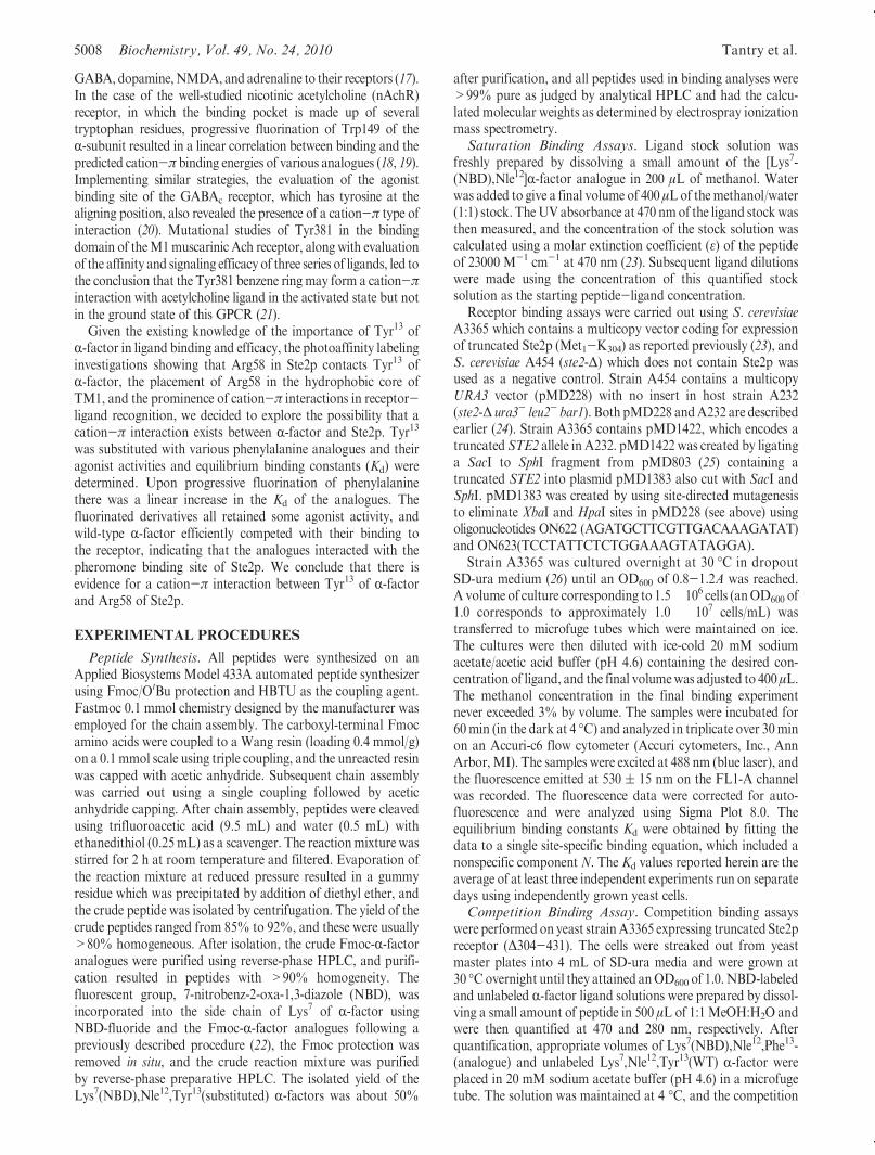
bited saturable binding to truncated Ste2p (A3365 strain) whilethe negative control experiments using a ste2-Δ deficient strain(A454) gave low background fluorescence which did not exhibitsaturation (Figure 1). The results were highly reproducible, andthe data could be fit to a single site binding equation whichallowed us to account for a nonspecific binding component.Using this method we determined the binding affinities forfluorescently labeled wild-type sequence R-factor, [Lys7(NBD),Phe13]R-factor, [Lys7(NBD),Phe13(3-F)]R-factor, [Lys7(NBD),Phe13(4-F)]R-factor, [Lys7(NBD),Phe13(3,4-F2)]R-factor, and[Lys7(NBD),Phe13(2,3,4,5,6-F5)]R-factor (Table 1).
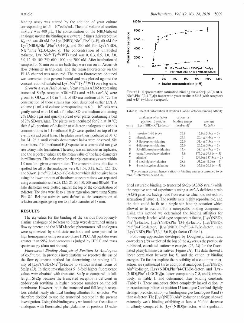
Following approaches developed by Dougherty, Lester, andco-workers (18) we plotted the log of theKd versus the previouslypublished, calculated cation-π energies (27, 28) for the fluori-nated phenylalanine derivatives (Figure 2A). The data showed alinear correlation between log Kd and the cation-π bindingenergies. To further explore the possibility of a cation-π inter-action, we synthesized three additional analogues: [Lys7(NBD),Ala13]R-factor, [Lys7(NBD),Phe13(4-CH3)]R-factor, and [Lys7-(NBD),Phe13(4-OCH3)]R-factor, compounds 7, 8, and 9, respec-tively, in Table 1, and determined their binding constants(Table 1). These analogues either completely lacked cation-πinteraction capabilities at position 13 (analogue 7) or had slightlystronger predicted cation-π binding energies (analogues 8 and 9)than R-factor. The [Lys7(NBD),Ala13]R-factor analogue showedextremely weak binding exhibiting at least a 30-fold decreasein affinity compared to [Lys7(NBD)]R-factor, with significant
Table 1: Effect of Substitution at Position 13 of R-Factor on Binding Affinity
entry
analogues of R-factorposition 13 residue
[Lys7(NBD),X13]R-factor
cation-πbinding energy
(kcal/mol)baverage
Kd (nM)
1 tyrosine (wild type) 26.9 15.9( 3.5 (n=3)
2 phenylalanine 27.1 20.4( 4.4 (n=4)
3 3-fluorophenylalanine 22.0 31.4( 7.3 (n=4)
4 4-fluorophenylalanine 22.0 26.2( 5.9 (n=3)
5 3,4-difluorophenylalanine 17.4 38.1( 4.7 (n=3)
6 pentafluorophenylalanine 3.9 177.3( 59.9 (n=3)
7 alaninea 0 534.0( 137.3 (n=3)
8 4-methylphenylalanine 28.6 53.2( 11.3 (n=3)
9 4-methoxyphenylalanine 28.6 37.8( 20.9 (n=5)
aThe π-ring is absent; hence, cation-π binding energy is assumed to bezero. bReferences 27 and 28.
FIGURE 1: Representative saturation binding curve for [Lys7(NBD),Nle12,Phe13(3,4-F2)]R-factor with yeast strains A3365 (with receptor)and A454 (without receptor).
5010 Biochemistry, Vol. 49, No. 24, 2010 Tantry et al.
variability in the results. The [Lys7(NBD),Phe13(4-CH3)]R-factorshowed a decrease in affinity of approximately 3-fold comparedto normal R-factor whereas the [Lys7(NBD),Phe13(4-OCH3)]-R-factor had an affinity quite similar to the difluorophenyl-alanine analogues accompanied by a large deviation betweenindividual assays. Inclusion of the latter two compounds in thelog Kd versus cation-π binding energy resulted in considerablescattering of the data and a weaker correlation between Kd andbinding energy (Figure 2B).Competition Binding Assay. It was important to ascertain
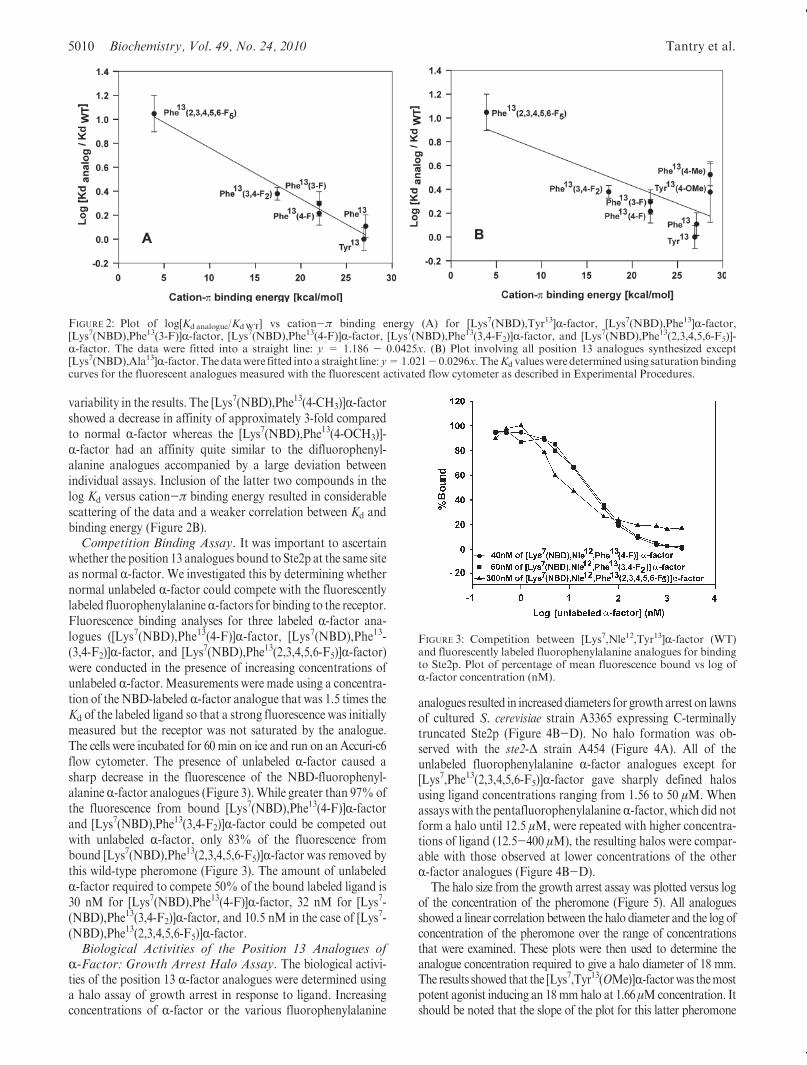
whether the position 13 analogues bound to Ste2p at the same siteas normal R-factor. We investigated this by determining whethernormal unlabeled R-factor could compete with the fluorescentlylabeled fluorophenylalanineR-factors for binding to the receptor.Fluorescence binding analyses for three labeled R-factor ana-logues ([Lys7(NBD),Phe13(4-F)]R-factor, [Lys7(NBD),Phe13-(3,4-F2)]R-factor, and [Lys7(NBD),Phe13(2,3,4,5,6-F5)]R-factor)were conducted in the presence of increasing concentrations ofunlabeled R-factor.Measurements were made using a concentra-tion of the NBD-labeled R-factor analogue that was 1.5 times theKd of the labeled ligand so that a strong fluorescence was initiallymeasured but the receptor was not saturated by the analogue.The cells were incubated for 60 min on ice and run on an Accuri-c6flow cytometer. The presence of unlabeled R-factor caused asharp decrease in the fluorescence of the NBD-fluorophenyl-alanineR-factor analogues (Figure 3).While greater than 97%ofthe fluorescence from bound [Lys7(NBD),Phe13(4-F)]R-factorand [Lys7(NBD),Phe13(3,4-F2)]R-factor could be competed outwith unlabeled R-factor, only 83% of the fluorescence frombound [Lys7(NBD),Phe13(2,3,4,5,6-F5)]R-factor was removed bythis wild-type pheromone (Figure 3). The amount of unlabeledR-factor required to compete 50% of the bound labeled ligand is30 nM for [Lys7(NBD),Phe13(4-F)]R-factor, 32 nM for [Lys7-(NBD),Phe13(3,4-F2)]R-factor, and 10.5 nM in the case of [Lys7-(NBD),Phe13(2,3,4,5,6-F5)]R-factor.Biological Activities of the Position 13 Analogues of
R-Factor: Growth Arrest Halo Assay. The biological activi-ties of the position 13 R-factor analogues were determined usinga halo assay of growth arrest in response to ligand. Increasingconcentrations of R-factor or the various fluorophenylalanine
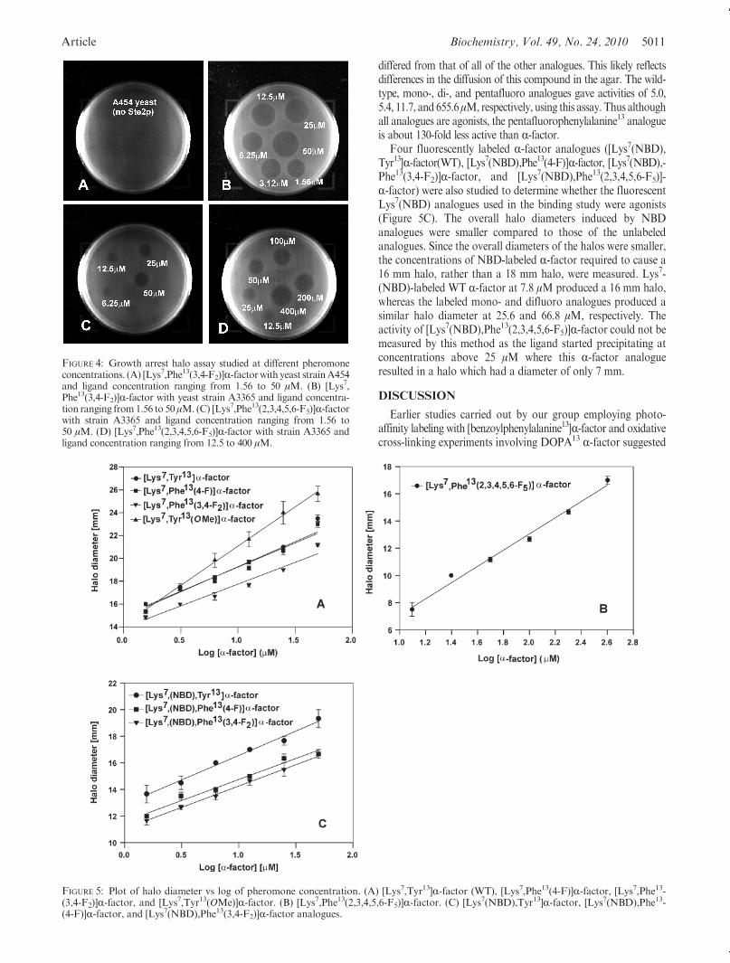
analogues resulted in increaseddiameters for growtharrest on lawnsof cultured S. cerevisiae strain A3365 expressing C-terminallytruncated Ste2p (Figure 4B-D). No halo formation was ob-served with the ste2-Δ strain A454 (Figure 4A). All of theunlabeled fluorophenylalanine R-factor analogues except for[Lys7,Phe13(2,3,4,5,6-F5)]R-factor gave sharply defined halosusing ligand concentrations ranging from 1.56 to 50 μM. Whenassayswith the pentafluorophenylalanineR-factor, which did notform a halo until 12.5 μM, were repeated with higher concentra-tions of ligand (12.5-400 μM), the resulting halos were compar-able with those observed at lower concentrations of the otherR-factor analogues (Figure 4B-D).
The halo size from the growth arrest assay was plotted versus logof the concentration of the pheromone (Figure 5). All analoguesshowed a linear correlation between the halo diameter and the log ofconcentration of the pheromone over the range of concentrationsthat were examined. These plots were then used to determine theanalogue concentration required to give a halo diameter of 18 mm.The results showed that the [Lys7,Tyr13(OMe)]R-factorwas themostpotent agonist inducing an 18mmhalo at 1.66 μMconcentration. Itshould be noted that the slope of the plot for this latter pheromone
FIGURE 2: Plot of log[Kdanalogue/KdWT] vs cation-π binding energy (A) for [Lys7(NBD),Tyr13]R-factor, [Lys7(NBD),Phe13]R-factor,[Lys7(NBD),Phe13(3-F)]R-factor, [Lys7(NBD),Phe13(4-F)]R-factor, [Lys7(NBD),Phe13(3,4-F2)]R-factor, and [Lys7(NBD),Phe13(2,3,4,5,6-F5)]-R-factor. The data were fitted into a straight line: y = 1.186 - 0.0425x. (B) Plot involving all position 13 analogues synthesized except[Lys7(NBD),Ala13]R-factor.Thedatawere fitted intoa straight line: y=1.021- 0.0296x. TheKd valuesweredeterminedusing saturationbindingcurves for the fluorescent analogues measured with the fluorescent activated flow cytometer as described in Experimental Procedures.
FIGURE 3: Competition between [Lys7,Nle12,Tyr13]R-factor (WT)and fluorescently labeled fluorophenylalanine analogues for bindingto Ste2p. Plot of percentage of mean fluorescence bound vs log ofR-factor concentration (nM).
Article Biochemistry, Vol. 49, No. 24, 2010 5011
differed from that of all of the other analogues. This likely reflectsdifferences in the diffusion of this compound in the agar. The wild-type, mono-, di-, and pentafluoro analogues gave activities of 5.0,5.4, 11.7, and 655.6μM, respectively, using this assay. Thus althoughall analogues are agonists, the pentafluorophenylalanine13 analogueis about 130-fold less active than R-factor.
Four fluorescently labeled R-factor analogues ([Lys7(NBD),Tyr13]R-factor(WT), [Lys7(NBD),Phe13(4-F)]R-factor, [Lys7(NBD),-Phe13(3,4-F2)]R-factor, and [Lys7(NBD),Phe13(2,3,4,5,6-F5)]-R-factor) were also studied to determine whether the fluorescentLys7(NBD) analogues used in the binding study were agonists(Figure 5C). The overall halo diameters induced by NBDanalogues were smaller compared to those of the unlabeledanalogues. Since the overall diameters of the halos were smaller,the concentrations of NBD-labeled R-factor required to cause a16 mm halo, rather than a 18 mm halo, were measured. Lys7-(NBD)-labeled WT R-factor at 7.8 μM produced a 16 mm halo,whereas the labeled mono- and difluoro analogues produced asimilar halo diameter at 25.6 and 66.8 μM, respectively. Theactivity of [Lys7(NBD),Phe13(2,3,4,5,6-F5)]R-factor could not bemeasured by this method as the ligand started precipitating atconcentrations above 25 μM where this R-factor analogueresulted in a halo which had a diameter of only 7 mm.
DISCUSSION
Earlier studies carried out by our group employing photo-affinity labeling with [benzoylphenylalanine13]R-factor and oxidativecross-linking experiments involving DOPA13 R-factor suggested
FIGURE 5: Plot of halo diameter vs log of pheromone concentration. (A) [Lys7,Tyr13]R-factor (WT), [Lys7,Phe13(4-F)]R-factor, [Lys7,Phe13-(3,4-F2)]R-factor, and [Lys7,Tyr13(OMe)]R-factor. (B) [Lys7,Phe13(2,3,4,5,6-F5)]R-factor. (C) [Lys7(NBD),Tyr13]R-factor, [Lys7(NBD),Phe13-(4-F)]R-factor, and [Lys7(NBD),Phe13(3,4-F2)]R-factor analogues.
FIGURE 4: Growth arrest halo assay studied at different pheromoneconcentrations. (A) [Lys7,Phe13(3,4-F2)]R-factorwith yeast strainA454and ligand concentration ranging from 1.56 to 50 μM. (B) [Lys7,Phe13(3,4-F2)]R-factor with yeast strain A3365 and ligand concentra-tion ranging from1.56 to 50μM. (C) [Lys7,Phe13(2,3,4,5,6-F5)]R-factorwith strain A3365 and ligand concentration ranging from 1.56 to50 μM. (D) [Lys7,Phe13(2,3,4,5,6-F5)]R-factor with strain A3365 andligand concentration ranging from 12.5 to 400 μM.
5012 Biochemistry, Vol. 49, No. 24, 2010 Tantry et al.
that the Tyr13 position of R-factor interacts with the TM1segment of Ste2p at residuesR58 andCys59, respectively (12, 13),and the presence of an aromatic ring at position 13 of R-factorwas found to be necessary for efficient binding (11, 29). Arg58 onTM1 and His94 on TM2 in Ste2p are believed to be in thehydrophobic core of themembrane. Othermembers of the fungalpheromone GPCR subfamily conserve polar residues at similarpositions (9). The conservation of positively charged residues inthe first and second TM domains of these fungal GPCRs and thecritical nature of Tyr13 of R-factor and of other fungal R-factorssuggested to us that ligand recognition by Ste2p might involve acation-π type of interaction.
Fluorine substitution has been utilized as an important tool totrack cation-π interactions as fluorine is sterically similar tohydrogen but due to its extremely high electronegativity has aremarkable effect on the electronic structure of an aromaticring. Progressive fluorination of the aromatic ring decreases theπ-electron density and thereby decreases the ability of the π-faceto interact with cations. Previously, we studied [Phe13]R-factor,[Phe13(3-F)]R-factor, and [Phe13(4-F)]R-factor using a radio-active binding competition assay and found that these analoguescompeted strongly with R-factor binding and were good ago-nists (29). In the present investigation, using a direct fluorescencesaturation binding assay and multiple fluorinated peptides westudied R-factor analogues with varying predicted cation-πbinding energies and revealed that there is significant influenceof the cation-π binding energies of the phenylalanine ring on theequilibrium binding constant Kd of the receptor. Phenylalaninehas a similar cation-π binding energy as that of tyrosine (27)(Table 1), and ideally phenylalanine replacement should notadversely affect the binding affinity. In our studies, theKd obtainedfor [Phe13]R-factor is similar to that of native [Tyr13]R-factor,which appears to validate this contention.
Phe(3-F) (entry 3, Table 1) and Phe(4-F) (entry 4, Table 1)have similar calculated cation-π binding energies, and R-factoranalogues with these substitutions were found to have similar Kd
values in our binding studies. This indicates that the position ofthe fluorine atom does not have a significant effect on the bindingaffinity, and the trend that we begin to see is mainly due to thedecrease in cation-π binding energy. Progressive fluorination ofthe phenylalanine ring resulted in an increase in Kd, and the datafit well onto a linear correlation plot (Figure 2A). Phe(2,3,4,5,6-F5)has the lowest cation-π binding energy among the fluorinatedR-factor derivatives that were studied, and this analogue exhi-bited approximately a 10-fold decrease in binding affinity com-pared toR-factor. The associated decrease in binding affinity dueto fluorination is not of the magnitude that was reported forligand-gated channels where up to a 2 log change in binding con-stants was observed. This suggests that the associated cation-πcontribution to the R-factor-Ste2p binding may be weaker thanthe contribution to the ligand interactions in ion channels. In fact,we have recently found that position 13 of R-factor also interactswith Cys59 of Ste2p (13) so that contributions of H-bondingbetween the hydroxyl group of Tyr13 to Cys59 and a cation-πinteraction between Tyr13 and Arg58 both contribute to thebinding of R-factor to Ste2p.
An additional way to test for the cation-π interaction wouldbe to carry out a similar binding analysis on ste2 mutantswhere Arg58 is replaced by other residues. We have generatedthree such mutants (R58A, R58D, and R58E). All of these areseriously deficient in binding, with Kd’s for R-factor from 10- to50-fold higher. As shown in the present study the fluorescent
fluorinated R-factor analogues had Kd’s for the wild-type Ste2pranging from 26.2 to 177.3 nM (Table 1). To determine the lattervalue, we needed to use 4 μM peptide, saturation was barelyachieved, and the nonspecific fluorescence was quite high. Sincethe Ste2p containing the substitution R58A binds R-factor withnearly 10-fold lower affinity, saturation binding experimentswould require very high concentrations of [Lys7(NBD),Phe13-(2,3,4,5,6-F5)]R-factor (∼40 μM). As stated above this peptideprecipitates at 25 μM, and the experiment, therefore, could not bedone. However, the marked decrease in affinity noted with thethree ste2 mutants that we generated supports our conclusionthat the Arg58 side chain contributes to the binding energy andwould be consistent with the cation-π interaction.
The use of NBD derivatives of R-factor might lead to a changein the interaction of position 13 of the pheromone and itsreceptor. However, all of the analogues that we tested wereagonists, indicating productive binding to the receptor, and thepotencies of the fluorinated R-factor analogues were within afactor of 3-4 of that of R-factor with the exception of thepentafluorinated analogue. Although the trends in the bindingaffinities were not directly parallel to the trends in the agonistpotencies, we have observed a similar lack of correlation in ourprevious study with R-factor analogues (29). Moreover, theweakest binding fluorinated analogue ([Phe13(2,3,4,5,6-F5)]-R-factor) was by far the weakest agonist. Finally, R-factorefficiently competed with the binding of the Lys7(NBD) analo-gues to Ste2p. On the basis of these observations and previousstudies on R-factor derivatized at the Lys7 side chain (30-33)we conclude that the fluorescent labeling does not result in asignificant perturbation of the interactions of the position 13 sidechain and the receptor. Interestingly, two nonfluorinated ana-logues, [Phe13(4-Me)]R-factor and [Phe13(4-OMe)]R-factor, eachshowed a marked lack of correlation between the Kd values andcation-π binding energies (Figure 2B). This finding contrastswith the correlation observed with fluorinated phenylalanineanalogues (Figure 2A) and indicates that while the insertion offluorine in place of a hydrogen results in primarily an electroniceffect on the benzene ring, other groups can have both electronicand steric effects on binding. These analogues have marginallyhigher cation-π binding energies as compared to phenylalanineand tyrosine (Table 1) and would be predicted to result instronger binding. We observed a 3-fold decrease in the bindingability for [Phe13(4-Me)]R-factor, and the difference reduces to2-fold upon reintroduction of phenolic oxygen, which should be agood electron donor. It is possible that the effect of reintroduc-tion of the donor oxygen atom is at least partially nullified by thesteric bulk of the methoxy group. Despite the fact that it cannotparticipate in a cation-π interaction, the [Lys7(NBD),Ala13]-R-factor does bind weakly to Ste2p. Thus, it is evident thatadditional effects involving steric, electrostatic, and perhapsvan der Waals forces may also play a role in R-factor-Ste2precognition at position 13 of the pheromone.Biological Implications. The results of this paper support
the existence of a cation-π interaction between the carboxyl-terminal Tyr13 residue of the R-factor peptide and the Arg58residue in the first transmembrane domain of Ste2p. Thisinteraction contributes to the favorable energetics of binding ofligand to receptor, since removal of Tyr from the carboxylterminus drastically reduces the peptide affinity and leads toan inactive pheromone (34). However, this interaction involv-ing the C-terminal of R-factor is not, by itself, sufficient forreceptor activation because N-terminally truncated analogues
Article Biochemistry, Vol. 49, No. 24, 2010 5013
of R-factor bind strongly to the receptor but do not lead to signaltransduction (34).
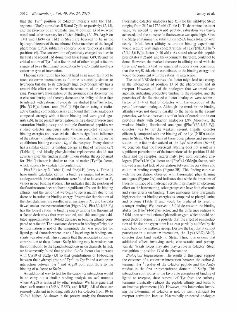
Detection of the interaction of Arg58 of the receptor withTyr13 ofR-factor raises the question of how the positively chargedArg58 is accommodated when there is no ligand bound toreceptor, since the existence of an isolated arginine side chainwithin the hydrophobic transmembrane region of Ste2p isexpected to be energetically unfavorable. One possibility is that,in the absence of ligand, Arg58 participates in an intramolecularcation-π interaction with an aromatic residue elsewhere in thereceptor. Each of the seven phenylalanine and two tryptophanresidues within the predicted transmembrane regions of Ste2pcan be mutated to nonaromatic residues without causing loss offunction (35). Similarly, aromatic residues do not appear to berequired at the positions of two of the three tyrosine residues inthese regions. The remaining tyrosine, Tyr266, can only bemutated to Trp, Phe, or His (35, 36). However, current modelsof Ste2p (9) place Tyr266 too far from Arg58 for any directinteraction.
This suggests that any aromatic side chain involved in anintramolecular cation-π interaction with Arg58 in the absenceof ligand would reside in an extracellular tail or loop. Themost likely site for the interacting side chains would be thefirst extracellular loop EL1, based on its expected proximity toArg58 and on the following: (1) The results of cysteine scanningaccessibility measurements on Ste2p indicate that burial of thisloop in the transmembrane core of the receptor is involved instabilizing the inactive state (37). (2) Mutation of Phe119, alsoin EL1, has been reported to lead to constitutive activity, aswould be expected if EL1 is involved in stabilizing the inactivestate (38). (3) Removal of residues 114-129 from EL1 of Ste2pyields a receptor that signals poorly but is constitutively active(Bhuiyan, Cohen, and Hauser unpublished results). (4) Thesubstitution Tyr111C in EL1 of Ste2p results in a receptor thatis incapable of signaling while retaining high-affinity R-factorbinding, suggesting that Tyr111 is involved in the activationpathway.
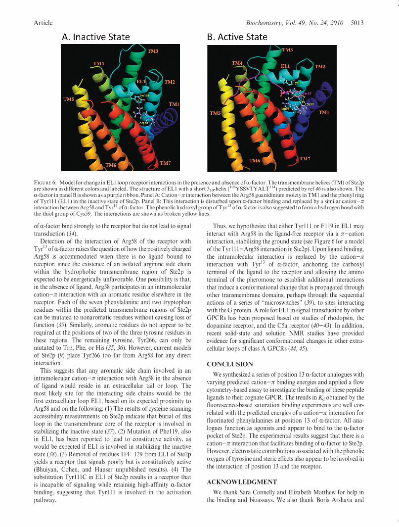
Thus, we hypothesize that either Tyr111 or F119 in EL1 mayinteract with Arg58 in the ligand-free receptor via a π-cationinteraction, stabilizing the ground state (see Figure 6 for a modelof the Tyr111-Arg58 interaction in Ste2p).Upon ligandbinding,the intramolecular interaction is replaced by the cation-πinteraction with Tyr13 of R-factor, anchoring the carboxylterminal of the ligand to the receptor and allowing the aminoterminal of the pheromone to establish additional interactionsthat induce a conformational change that is propagated throughother transmembrane domains, perhaps through the sequentialactions of a series of “microswitches” (39), to sites interactingwith theG protein. A role for EL1 in signal transduction by otherGPCRs has been proposed based on studies of rhodopsin, thedopamine receptor, and the C5a receptor (40-43). In addition,recent solid-state and solution NMR studies have providedevidence for significant conformational changes in other extra-cellular loops of class A GPCRs (44, 45).
CONCLUSION
We synthesized a series of position 13 R-factor analogues withvarying predicted cation-π binding energies and applied a flowcytometry-based assay to investigate the binding of these peptideligands to their cognate GPCR. The trends inKd obtained by thefluorescence-based saturation binding experiments are well cor-related with the predicted energies of a cation-π interaction forfluorinated phenylalanines at position 13 of R-factor. All ana-logues function as agonists and appear to bind to the R-factorpocket of Ste2p. The experimental results suggest that there is acation-π interaction that facilitates binding of R-factor to Ste2p.However, electrostatic contributions associated with the phenolicoxygen of tyrosine and steric effects also appear to be involved inthe interaction of position 13 and the receptor.
ACKNOWLEDGMENT
We thank Sara Connelly and Elizabeth Matthew for help inthe binding and bioassays. We also thank Boris Arshava and
FIGURE 6: Model for change inEL1 loop receptor interactions in the presence and absence ofR-factor. The transmembrane helices (TM) of Ste2pare shown in different colors and labeled. The structure of EL1 with a short 310-helix (
106YSSVTYALT114) predicted by ref 46 is also shown. TheR-factor inpanelB is shownasapurple ribbon. PanelA:Cation-π interactionbetween theArg58 guanidiniummoiety inTM1and thephenyl ringof Tyr111 (EL1) in the inactive state of Ste2p. Panel B: This interaction is disturbed upon R-factor binding and replaced by a similar cation-πinteraction betweenArg58 andTyr13 ofR-factor.The phenolic hydroxyl groupofTyr13 ofR-factor is also suggested to formahydrogenbondwiththe thiol group of Cys59. The interactions are shown as broken yellow lines.
5014 Biochemistry, Vol. 49, No. 24, 2010 Tantry et al.
Leah Cohen for help with the preparation of the manuscript andGeorge Umanah for preparation of Figure 6.
REFERENCES
1. Filmore, D. (2004) It’s a GPCR World. Mod. Drug Discovery 7,24–28.
2. Marshall, G. R. (2001) Peptide interactions with G-protein coupledreceptors. Biopolymers 60, 246–277.
3. Kristiansen, K. (2004) Molecular mechanisms of ligand binding,signaling, and regulationwithin the superfamily ofG-protein-coupledreceptors: molecular modeling and mutagenesis approaches to recep-tor structure and function. Pharmacol. Ther. 103, 21–80.
4. Schyler, S., and Horuk, R. (2006) I want a new drug: G-protein-coupled receptors in drug development. Drug Discovery Today 11,481–493.
5. Fredriksson, R., and Schioth, H. B. (2005) The repertoire of G-pro-tein-coupled receptors in fully sequenced genomes. Mol. Pharmacol.67, 1414–1425.
6. Crowe, M. L., Perry, B. N., and Connerton, I. F. (2000) Golfcomplements a GPA1 null mutation in Saccharomyces cerevisiaeand functionally couples to the STE2 pheromone receptor. J. Recept.Signal Transduction Res. 20, 61–73.
7. King, K., Dohlman,H.G., Thorner, J., Caron,M.G., and Lefkowitz,R. J. (1990) Control of yeast mating signal transduction by amammalian beta 2-adrenergic receptor and Gs alpha subunit. Science(New York) 250, 121–123.
8. Pausch, M. H. (1997) G-protein-coupled receptors in Saccharomycescerevisiae: high-throughput screening assays for drug discovery.Trends Biotechnol. 15, 487–494.
9. Eilers, M., Hornak, V., Smith, S. O., and Konopka, J. B. (2005)Comparison of class A and D G protein-coupled receptors: commonfeatures in structure and activation. Biochemistry 44, 8959–8975.
10. Neumoin, A., Cohen, L. S., Arshava, B., Tantry, S., Becker, J. M.,Zerbe,O., andNaider, F. (2009) Structure of a double transmembranefragment of a G-protein-coupled receptor in micelles. Biophys. J. 96,3187–3196.
11. Abel, M. G., Zhang, Y. L., Lu, H. F., Naider, F., and Becker, J. M.(1998) Structure-function analysis of the Saccharomyces cerevisiaetridecapeptide pheromone using alanine-scanned analogs. J. Pept.Res. 52, 95–106.
12. Son, C. D., Sargsyan, H., Naider, F., and Becker, J. M. (2004)Identification of ligand binding regions of the Saccharomycescerevisiae alpha-factor pheromone receptor by photoaffinity cross-linking. Biochemistry 43, 13193–13203.
13. Umanah, G. K., Son, C., Ding, F., Naider, F., and Becker, J. M.(2009) Cross-linking of a DOPA-containing peptide ligand into itsG protein-coupled receptor. Biochemistry 48, 2033–2044.
14. Ma, J. C., and Dougherty, D. A. (1997) The cation-pi interaction.Chem. Rev. 97, 1303–1324.
15. Gallivan, J. P., and Dougherty, D. A. (1999) Cation-pi interactions instructural biology. Proc. Natl. Acad. Sci. U.S.A. 96, 9459–9464.
16. Crowley, P. B., and Golovin, A. (2005) Cation-pi interactions inprotein-protein interfaces. Proteins 59, 231–239.
17. Zacharias, N., and Dougherty, D. A. (2002) Cation-pi interactions inligand recognition and catalysis. Trends Pharmacol. Sci. 23, 281–287.
18. Zhong, W., Gallivan, J. P., Zhang, Y., Li, L., Lester, H. A., andDougherty, D. A. (1998) From ab initio quantum mechanics tomolecular neurobiology: a cation-pi binding site in the nicotinicreceptor. Proc. Natl. Acad. Sci. U.S.A. 95, 12088–12093.
19. Beene, D. L., Brandt, G. S., Zhong, W., Zacharias, N. M., Lester,H. A., and Dougherty, D. A. (2002) Cation-pi interactions in ligandrecognition by serotonergic (5-HT3A) and nicotinic acetylcholinereceptors: the anomalous binding properties of nicotine. Biochemistry41, 10262–10269.
20. Lummis, S. C., Beene, D. L., Harrison, N. J., Lester, H. A., andDougherty, D. A. (2005) A cation-pi binding interaction with atyrosine in the binding site of the GABAC receptor. Chem. Biol. 12,993–997.
21. Ward, S. D., Curtis, C. A., and Hulme, E. C. (1999) Alanine-scanningmutagenesis of transmembrane domain 6 of the M(1) muscarinicacetylcholine receptor suggests that Tyr381 plays key roles in receptorfunction. Mol. Pharmacol. 56, 1031–1041.
22. Ding, F. X., Lee, B.K., Hauser,M.,Davenport, L., Becker, J.M., andNaider, F. (2001) Probing the binding domain of the Saccharomycescerevisiae alpha-mating factor receptor with rluorescent ligands.Biochemistry 40, 1102–1108.
23. Bajaj, A., Celic, A., Ding, F. X., Naider, F., Becker, J. M., andDumont, M. E. (2004) A fluorescent alpha-factor analogue exhibits
multiple steps on binding to its G protein coupled receptor in yeast.Biochemistry 43, 13564–13578.
24. Leavitt, L. M., Macaluso, C. R., Kim, K. S., Martin, N. P., andDumont, M. E. (1999) Dominant negative mutations in the alpha-factor receptor, a G protein-coupled receptor encoded by theSTE2 gene of the yeast Saccharomyces cerevisiae. Mol. Gen. Genet.261, 917–932.
25. Gehret, A. U., Bajaj, A., Naider, F., and Dumont, M. E. (2006)Oligomerization of the yeast alpha-factor receptor: implications fordominant negative effects of mutant receptors. J. Biol. Chem. 281,20698–20714.
26. Rose, M., Winston, F., and Hieter, P. (1990) Methods in YeastGenetics, Cold Spring Harbor Laboratory, Cold Spring Harbor, NY.
27. Mecozzi, S., West, A. P., Jr., and Dougherty, D. A. (1996) Cation-piinteractions in aromatics of biological and medicinal interest: electro-static potential surfaces as a useful qualitative guide.Proc. Natl. Acad.Sci. U.S.A. 93, 10566–10571.
28. Raines, D. E., Gioia, F., Claycomb, R. J., and Stevens, R. J. (2004)The N-methyl-D-aspartate receptor inhibitory potencies of aromaticinhaled drugs of abuse: evidence for modulation by cation-pi inter-actions. J. Pharmacol. Exp. Ther. 311, 14–21.
29. Liu, S., Henry, L. K., Lee, B. K., Wang, S. H., Arshava, B., Becker,J. M., and Naider, F. (2000) Position 13 analogs of the tridecapeptidemating pheromone from Saccharomyces cerevisiae: design of aniodinatable ligand for receptor binding. J. Pept. Res. 56, 24–34.
30. Shenbagamurthi, P., Baffi, R., Khan, S. A., Lipke, P., Pousman,C., Becker, J. M., and Naider, F. (1983) Structure-activity relation-ships in the dodecapeptide alpha factor of Saccharomyces cerevisiae.Biochemistry 22, 1298–1304.
31. Raths, S. K., Naider, F., and Becker, J. M. (1988) Peptide ana-logues compete with the binding of alpha-factor to its receptor inSaccharomyces cerevisiae. J. Biol. Chem. 263, 17333–17341.
32. Naider, F., Yaron, A., Ewenson, A., Tallon, M., Xue, C. B.,Srinivasan, J. V., Eriotou-Bargiota, E., and Becker, J. M. (1990)Synthetic probes for the alpha-factor receptor. Biopolymers 29,237–245.
33. Naider, F., and Becker, J. M. (2004) The alpha-factor matingpheromone of Saccharomyces cerevisiae: a model for studying theinteraction of peptide hormones and G protein-coupled receptors.Peptides 25, 1441–1463.
34. Eriotou-Bargiota, E., Xue, C. B., Naider, F., and Becker, J. M. (1992)Antagonistic and synergistic peptide analogues of the tridecapeptidemating pheromone of Saccharomyces cerevisiae. Biochemistry 31,551–557.
35. Martin, N. P., Celic, A., and Dumont, M. E. (2002) Mutagenicmapping of helical structures in the transmembrane segments of theyeast alpha-factor receptor. J. Mol. Biol. 317, 765–788.
36. Lee, B. K., Lee, Y. H., Hauser, M., Son, C. D., Khare, S., Naider, F.,and Becker, J. M. (2002) Tyr266 in the sixth transmembrane domainof the yeast alpha-factor receptor plays key roles in receptor activationand ligand specificity. Biochemistry 41, 13681–13689.
37. Hauser, M., Kauffman, S., Lee, B. K., Naider, F., and Becker, J. M.(2007) The first extracellular loop of the Saccharomyces cerevisiae Gprotein-coupled receptor Ste2p undergoes a conformational changeupon ligand binding. J. Biol. Chem. 282, 10387–10397.
38. Parrish, W., Eilers, M., Ying, W., and Konopka, J. B. (2002) Thecytoplasmic end of transmembrane domain 3 regulates the activityof the Saccharomyces cerevisiae G-protein-coupled alpha-factorreceptor. Genetics 160, 429–443.
39. Nygaard, R., Frimurer, T. M., Holst, B., Rosenkilde, M. M., andSchwartz, T. W. (2009) Ligand binding and micro-switches in 7TMreceptor structures. Trends Pharmacol. Sci. 30, 249–259.
40. Lawson, Z., and Wheatley, M. (2004) The third extracellular loop ofG-protein-coupled receptors: more than just a linker between twoimportant transmembrane helices. Biochem. Soc. Trans. 32, 1048–1050.
41. Shi, L., and Javitch, J. A. (2004) The second extracellular loop of thedopamine D2 receptor lines the binding-site crevice. Proc. Natl. Acad.Sci. U.S.A. 101, 440–445.
42. Klco, J. M., Wiegand, C. B., Narzinski, K., and Baranski, T. J. (2005)Essential role for the second extracellular loop in C5a receptoractivation. Nat. Struct. Mol. Biol. 12, 320–326.
43. Klco, J. M., Nikiforovich, G. V., and Baranski, T. J. (2006) Geneticanalysis of the first and third extracellular loops of the C5a receptorreveals an essential WXFGmotif in the first loop. J. Biol. Chem. 281,12010–12019.
44. Ahuja, S., Hornak, V., Yan, E. C., Syrett, N., Goncalves, J. A.,Hirshfeld, A., Ziliox, M., Sakmar, T. P., Sheves, M., Reeves, P. J.,Smith, S. O., and Eilers, M. (2009) Helix movement is coupled to
Article Biochemistry, Vol. 49, No. 24, 2010 5015
displacement of the second extracellular loop in rhodopsin activation.Nat. Struct. Mol. Biol. 16, 168–175.
45. Bokoch, M. P., Zou, Y., Rasmussen, S. G., Liu, C. W., Nygaard,R., Rosenbaum, D.M., Fung, J. J., Choi, H. J., Thian, F. S., Kobilka,T. S., Puglisi, J. D., Weis, W. I., Pardo, L., Prosser, R. S., Mueller,L., and Kobilka, B. K. (2010) Ligand-specific regulation of the
extracellular surface of a G-protein-coupled receptor. Nature 463,108–112.
46. Akal-Strader, A., Khare, S., Xu, D., Naider, F., and Becker, J. M.(2002) Residues in the first extracellular loop of a G protein-coupledreceptor play a role in signal transduction. J. Biol. Chem. 277, 30581–30590.