
Binding Mode of an α Amino Acid-Linked Quinoxaline-2,3 ... · Binding Mode of an α‑Amino...
10
Binding Mode of an α‑Amino Acid-Linked Quinoxaline-2,3-dione Analogue at Glutamate Receptor Subtype GluK1 Charles S. Demmer, † Charlotte Møller, ‡ Patricia M. G. E. Brown, # Liwei Han, ∥ Darryl S. Pickering, ∥ Birgitte Nielsen, § Derek Bowie, # Karla Frydenvang, ‡ Jette S. Kastrup,* ,‡ and Lennart Bunch* ,† † Chemical Neuroscience Group, ‡ Biostructural Research Group, § Medicinal Chemistry Group, ∥ Molecular and Cellular Pharmacology Group, and ⊥ Molecular Pharmacology Group, Department of Drug Design and Pharmacology, Faculty of Health and Medical Sciences, University of Copenhagen, Copenhagen, Denmark # Bowie Lab, Department of Pharmacology & Therapeutics, Faculty of Medicine, McGill University, Montreal, Canada H3G 0B1 * S Supporting Information ABSTRACT: Two α-amino acid-functionalized quinoxalines, 1a (CNG-10301) and 1b (CNG-10300), of a quinoxaline moiety coupled to an amino acid moiety were designed, synthesized, and characterized pharmacologically. While 1a displayed low affinity at native AMPA, KA, and NMDA receptors, and at homomeric GluK1,3 receptors, the affinity for GluK2 was in the midmicromolar range (K i = 136 μM), 1b displayed low to midmicromolar range binding affinity at all the iGluRs (K i =9−126 μM). In functional experiments (outside-out patches excised from transfected HEK293T cells), 100 μM 1a partially blocked GluK1 (33% peak response), while GluK2 was unaffected (96% peak response). Furthermore, 1a was shown not to be an agonist at GluK1 and GluK2 at 100 μM. On the other hand, 100 μM 1b fully antagonized GluK1 (8% peak response) but only partially blocked GluK2 (33% peak response). An X-ray structure at 2.3 Å resolution of 1b in the GluK1-LBD (ligand-binding domain) disclosed an unexpected binding mode compared to the predictions made during the design phase; the quinoxaline moiety remains to act as an amino acid bioisostere, but the amino acid moiety is oriented into a new area within the GluK1 receptor. The structure of the GluK1-LBD with 1b showed a large variation in domain openings of the three molecules from 25° to 49°, demonstrating that the GluK1-LBD is capable of undergoing major domain movements. KEYWORDS: Design, synthesis, radioligand binding, X-ray crystallography, domain opening ■ INTRODUCTION The common α-amino acid (S)-glutamate (Glu) is the major excitatory neurotransmitter of the mammalian central nervous system (CNS). The glutamatergic neurotransmitter system is involved in a vast number of neurological processes such as memory and learning, plasticity, and motor function, 1,2 and thus also in the pathogenesis of many neurological and psychiatric disorders. The class of fast-acting ionotropic Glu receptors (iGluRs) is divided into three groups that again comprise a number of subunits [AMPA receptors GluA1−4, kainic acid (KA) receptors 3−5 GluK1−5, and NMDA receptors 6 GluN1, GluN2A-D, and GluN3A,B]. The iGluRs form ligand-gated ion channels consisting of four subunits, with each subunit having an extracellular region composed of the N- terminal domain and the ligand-binding domain (LBD), a transmembrane region, and a C-terminal region. The LBD forms a clamshell-like domain with the glutamate-binding site located within two lobes designated D1 and D2. A number of competitive antagonists that discriminate among the AMPA, KA, and NMDA receptors have been reported. 7 However, it remains a challenge to discover antagonists, which display selectivity among the subunits within one of these groups. In fact, only GluK1-selective competitive antagonists have been reported to date [e.g., LY466195 8 (Figure 1 and Table 1]. 7 There is therefore an unmet need for the creative design of new chemical scaffolds that eventually can be developed into antagonists with a novel subunit selectivity profile. Such chemical probes are key tools in the elucidation of the role and function of Glu receptors in health and disease. ■ RESULTS AND DISCUSSION Design. Some 20 years ago, a series of substituted quinoxaline-2,3(1H,4H)-diones [DNQX and CNQX (Figure 1 and Table 1)] were identified as potent competitive AMPA/ KA receptor antagonists. 9 Interestingly, the quinoxaline-2,3- (1H,4H)-dione moiety acts as an α-amino acid bioisostere 10 as disclosed by X-ray crystallographic studies. 11,12 A potentially new class of iGluR antagonists was designed with the general molecular formula I (Figure 2). Via the linkage of an α-amino acid to the quinoxaline-2,3(1H,4H)-dione Received: September 23, 2014 Accepted: March 26, 2015 Research Article pubs.acs.org/chemneuro © XXXX American Chemical Society A DOI: 10.1021/acschemneuro.5b00038 ACS Chem. Neurosci. XXXX, XXX, XXX−XXX
Transcript of Binding Mode of an α Amino Acid-Linked Quinoxaline-2,3 ... · Binding Mode of an α‑Amino...
Binding Mode of an α‑Amino Acid-Linked Quinoxaline-2,3-dioneAnalogue at Glutamate Receptor Subtype GluK1Charles S. Demmer,† Charlotte Møller,‡ Patricia M. G. E. Brown,# Liwei Han,∥ Darryl S. Pickering,∥
Birgitte Nielsen,§ Derek Bowie,# Karla Frydenvang,‡ Jette S. Kastrup,*,‡ and Lennart Bunch*,†
†Chemical Neuroscience Group, ‡Biostructural Research Group, §Medicinal Chemistry Group, ∥Molecular and CellularPharmacology Group, and ⊥Molecular Pharmacology Group, Department of Drug Design and Pharmacology, Faculty of Health andMedical Sciences, University of Copenhagen, Copenhagen, Denmark#Bowie Lab, Department of Pharmacology & Therapeutics, Faculty of Medicine, McGill University, Montreal, Canada H3G 0B1
*S Supporting Information
ABSTRACT: Two α-amino acid-functionalized quinoxalines, 1a(CNG-10301) and 1b (CNG-10300), of a quinoxaline moietycoupled to an amino acid moiety were designed, synthesized, andcharacterized pharmacologically. While 1a displayed low affinity atnative AMPA, KA, and NMDA receptors, and at homomeric GluK1,3receptors, the affinity for GluK2 was in the midmicromolar range (Ki= 136 μM), 1b displayed low to midmicromolar range bindingaffinity at all the iGluRs (Ki = 9−126 μM). In functional experiments(outside-out patches excised from transfected HEK293T cells), 100μM 1a partially blocked GluK1 (33% peak response), while GluK2was unaffected (96% peak response). Furthermore, 1a was shown not to be an agonist at GluK1 and GluK2 at 100 μM. On theother hand, 100 μM 1b fully antagonized GluK1 (8% peak response) but only partially blocked GluK2 (33% peak response). AnX-ray structure at 2.3 Å resolution of 1b in the GluK1-LBD (ligand-binding domain) disclosed an unexpected binding modecompared to the predictions made during the design phase; the quinoxaline moiety remains to act as an amino acid bioisostere,but the amino acid moiety is oriented into a new area within the GluK1 receptor. The structure of the GluK1-LBD with 1bshowed a large variation in domain openings of the three molecules from 25° to 49°, demonstrating that the GluK1-LBD iscapable of undergoing major domain movements.
KEYWORDS: Design, synthesis, radioligand binding, X-ray crystallography, domain opening
■ INTRODUCTION
The common α-amino acid (S)-glutamate (Glu) is the majorexcitatory neurotransmitter of the mammalian central nervoussystem (CNS). The glutamatergic neurotransmitter system isinvolved in a vast number of neurological processes such asmemory and learning, plasticity, and motor function,1,2 andthus also in the pathogenesis of many neurological andpsychiatric disorders. The class of fast-acting ionotropic Glureceptors (iGluRs) is divided into three groups that againcomprise a number of subunits [AMPA receptors GluA1−4,kainic acid (KA) receptors3−5 GluK1−5, and NMDAreceptors6 GluN1, GluN2A-D, and GluN3A,B]. The iGluRsform ligand-gated ion channels consisting of four subunits, witheach subunit having an extracellular region composed of the N-terminal domain and the ligand-binding domain (LBD), atransmembrane region, and a C-terminal region. The LBDforms a clamshell-like domain with the glutamate-binding sitelocated within two lobes designated D1 and D2.A number of competitive antagonists that discriminate
among the AMPA, KA, and NMDA receptors have beenreported.7 However, it remains a challenge to discoverantagonists, which display selectivity among the subunits within
one of these groups. In fact, only GluK1-selective competitiveantagonists have been reported to date [e.g., LY4661958
(Figure 1 and Table 1].7 There is therefore an unmet need forthe creative design of new chemical scaffolds that eventually canbe developed into antagonists with a novel subunit selectivityprofile. Such chemical probes are key tools in the elucidation ofthe role and function of Glu receptors in health and disease.
■ RESULTS AND DISCUSSION
Design. Some 20 years ago, a series of substitutedquinoxaline-2,3(1H,4H)-diones [DNQX and CNQX (Figure1 and Table 1)] were identified as potent competitive AMPA/KA receptor antagonists.9 Interestingly, the quinoxaline-2,3-(1H,4H)-dione moiety acts as an α-amino acid bioisostere10 asdisclosed by X-ray crystallographic studies.11,12
A potentially new class of iGluR antagonists was designedwith the general molecular formula I (Figure 2). Via the linkageof an α-amino acid to the quinoxaline-2,3(1H,4H)-dione
Received: September 23, 2014Accepted: March 26, 2015
Research Article
pubs.acs.org/chemneuro
© XXXX American Chemical Society A DOI: 10.1021/acschemneuro.5b00038ACS Chem. Neurosci. XXXX, XXX, XXX−XXX
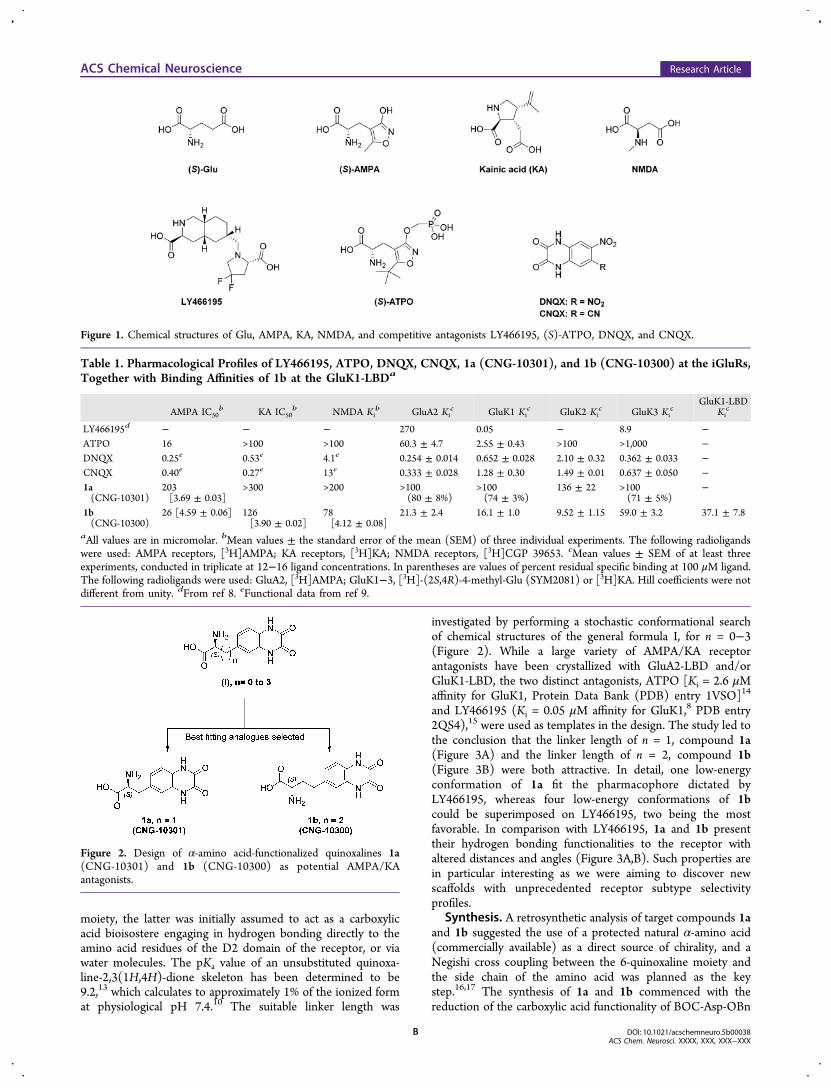
moiety, the latter was initially assumed to act as a carboxylicacid bioisostere engaging in hydrogen bonding directly to theamino acid residues of the D2 domain of the receptor, or viawater molecules. The pKa value of an unsubstituted quinoxa-line-2,3(1H,4H)-dione skeleton has been determined to be9.2,13 which calculates to approximately 1% of the ionized format physiological pH 7.4.10 The suitable linker length was
investigated by performing a stochastic conformational searchof chemical structures of the general formula I, for n = 0−3(Figure 2). While a large variety of AMPA/KA receptorantagonists have been crystallized with GluA2-LBD and/orGluK1-LBD, the two distinct antagonists, ATPO [Ki = 2.6 μMaffinity for GluK1, Protein Data Bank (PDB) entry 1VSO]14
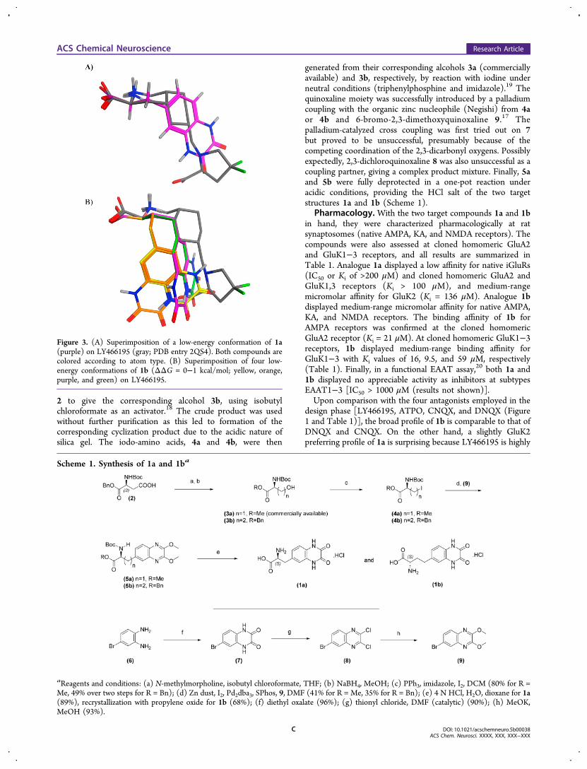
and LY466195 (Ki = 0.05 μM affinity for GluK1,8 PDB entry2QS4),15 were used as templates in the design. The study led tothe conclusion that the linker length of n = 1, compound 1a(Figure 3A) and the linker length of n = 2, compound 1b(Figure 3B) were both attractive. In detail, one low-energyconformation of 1a fit the pharmacophore dictated byLY466195, whereas four low-energy conformations of 1bcould be superimposed on LY466195, two being the mostfavorable. In comparison with LY466195, 1a and 1b presenttheir hydrogen bonding functionalities to the receptor withaltered distances and angles (Figure 3A,B). Such properties arein particular interesting as we were aiming to discover newscaffolds with unprecedented receptor subtype selectivityprofiles.
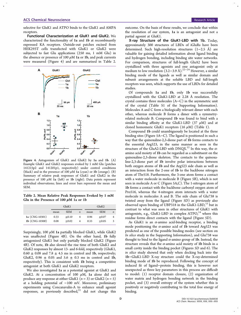
Synthesis. A retrosynthetic analysis of target compounds 1aand 1b suggested the use of a protected natural α-amino acid(commercially available) as a direct source of chirality, and aNegishi cross coupling between the 6-quinoxaline moiety andthe side chain of the amino acid was planned as the keystep.16,17 The synthesis of 1a and 1b commenced with thereduction of the carboxylic acid functionality of BOC-Asp-OBn
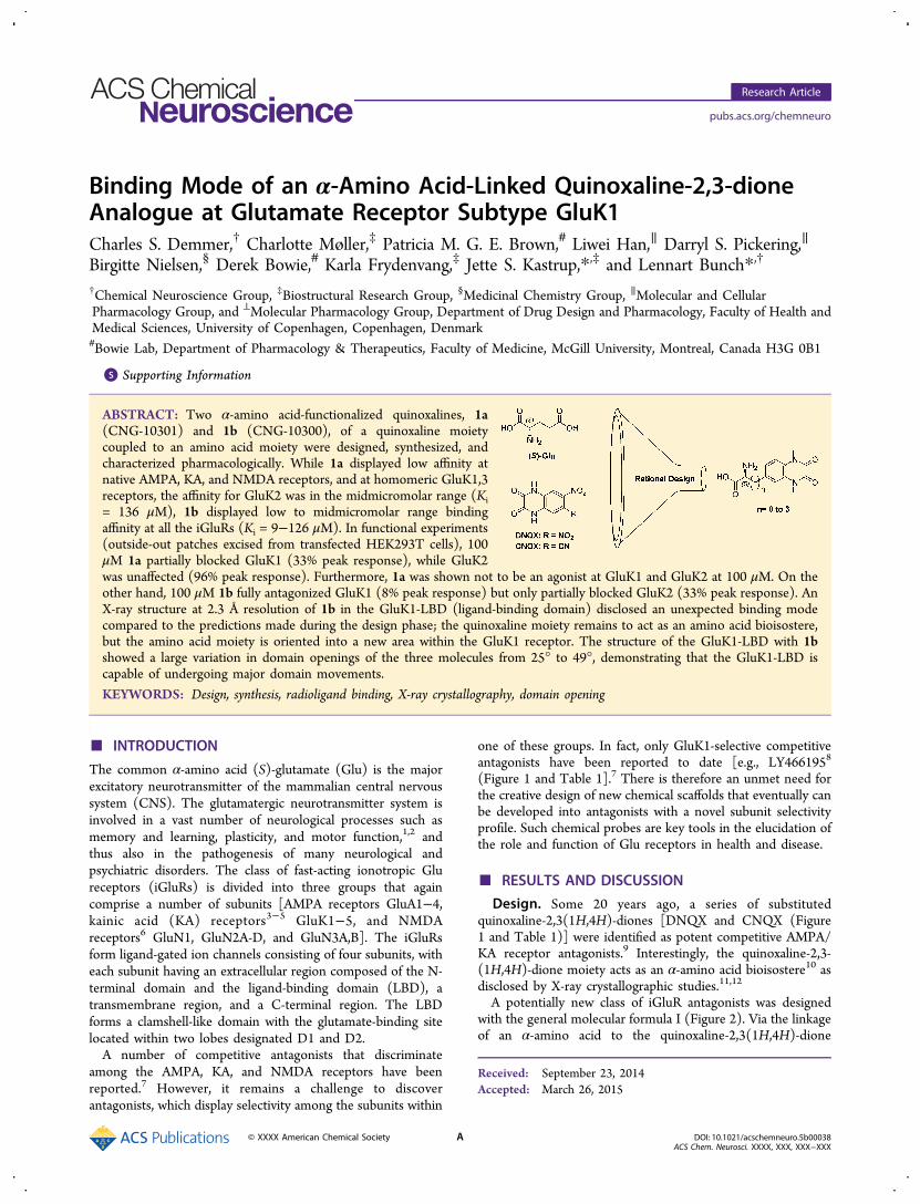
Figure 1. Chemical structures of Glu, AMPA, KA, NMDA, and competitive antagonists LY466195, (S)-ATPO, DNQX, and CNQX.
Table 1. Pharmacological Profiles of LY466195, ATPO, DNQX, CNQX, 1a (CNG-10301), and 1b (CNG-10300) at the iGluRs,Together with Binding Affinities of 1b at the GluK1-LBDa
AMPA IC50b KA IC50
b NMDA Kib GluA2 Ki
c GluK1 Kic GluK2 Ki
c GluK3 Kic
GluK1-LBDKic
LY466195d − − − 270 0.05 − 8.9 −ATPO 16 >100 >100 60.3 ± 4.7 2.55 ± 0.43 >100 >1,000 −DNQX 0.25e 0.53e 4.1e 0.254 ± 0.014 0.652 ± 0.028 2.10 ± 0.32 0.362 ± 0.033 −CNQX 0.40e 0.27e 13e 0.333 ± 0.028 1.28 ± 0.30 1.49 ± 0.01 0.637 ± 0.050 −1a(CNG-10301)
203[3.69 ± 0.03]
>300 >200 >100(80 ± 8%)
>100(74 ± 3%)
136 ± 22 >100(71 ± 5%)
−
1b(CNG-10300)
26 [4.59 ± 0.06] 126[3.90 ± 0.02]
78[4.12 ± 0.08]
21.3 ± 2.4 16.1 ± 1.0 9.52 ± 1.15 59.0 ± 3.2 37.1 ± 7.8
aAll values are in micromolar. bMean values ± the standard error of the mean (SEM) of three individual experiments. The following radioligandswere used: AMPA receptors, [3H]AMPA; KA receptors, [3H]KA; NMDA receptors, [3H]CGP 39653. cMean values ± SEM of at least threeexperiments, conducted in triplicate at 12−16 ligand concentrations. In parentheses are values of percent residual specific binding at 100 μM ligand.The following radioligands were used: GluA2, [3H]AMPA; GluK1−3, [3H]-(2S,4R)-4-methyl-Glu (SYM2081) or [3H]KA. Hill coefficients were notdifferent from unity. dFrom ref 8. eFunctional data from ref 9.
Figure 2. Design of α-amino acid-functionalized quinoxalines 1a(CNG-10301) and 1b (CNG-10300) as potential AMPA/KAantagonists.
ACS Chemical Neuroscience Research Article
DOI: 10.1021/acschemneuro.5b00038ACS Chem. Neurosci. XXXX, XXX, XXX−XXX
B
2 to give the corresponding alcohol 3b, using isobutylchloroformate as an activator.18 The crude product was usedwithout further purification as this led to formation of thecorresponding cyclization product due to the acidic nature ofsilica gel. The iodo-amino acids, 4a and 4b, were then
generated from their corresponding alcohols 3a (commerciallyavailable) and 3b, respectively, by reaction with iodine underneutral conditions (triphenylphosphine and imidazole).19 Thequinoxaline moiety was successfully introduced by a palladiumcoupling with the organic zinc nucleophile (Negishi) from 4aor 4b and 6-bromo-2,3-dimethoxyquinoxaline 9.17 Thepalladium-catalyzed cross coupling was first tried out on 7but proved to be unsuccessful, presumably because of thecompeting coordination of the 2,3-dicarbonyl oxygens. Possiblyexpectedly, 2,3-dichloroquinoxaline 8 was also unsuccessful as acoupling partner, giving a complex product mixture. Finally, 5aand 5b were fully deprotected in a one-pot reaction underacidic conditions, providing the HCl salt of the two targetstructures 1a and 1b (Scheme 1).
Pharmacology. With the two target compounds 1a and 1bin hand, they were characterized pharmacologically at ratsynaptosomes (native AMPA, KA, and NMDA receptors). Thecompounds were also assessed at cloned homomeric GluA2and GluK1−3 receptors, and all results are summarized inTable 1. Analogue 1a displayed a low affinity for native iGluRs(IC50 or Ki of >200 μM) and cloned homomeric GluA2 andGluK1,3 receptors (Ki > 100 μM), and medium-rangemicromolar affinity for GluK2 (Ki = 136 μM). Analogue 1bdisplayed medium-range micromolar affinity for native AMPA,KA, and NMDA receptors. The binding affinity of 1b forAMPA receptors was confirmed at the cloned homomericGluA2 receptor (Ki = 21 μM). At cloned homomeric GluK1−3receptors, 1b displayed medium-range binding affinity forGluK1−3 with Ki values of 16, 9.5, and 59 μM, respectively(Table 1). Finally, in a functional EAAT assay,20 both 1a and1b displayed no appreciable activity as inhibitors at subtypesEAAT1−3 [IC50 > 1000 μM (results not shown)].Upon comparison with the four antagonists employed in the
design phase [LY466195, ATPO, CNQX, and DNQX (Figure1 and Table 1)], the broad profile of 1b is comparable to that ofDNQX and CNQX. On the other hand, a slightly GluK2preferring profile of 1a is surprising because LY466195 is highly
Figure 3. (A) Superimposition of a low-energy conformation of 1a(purple) on LY466195 (gray; PDB entry 2QS4). Both compounds arecolored according to atom type. (B) Superimposition of four low-energy conformations of 1b (ΔΔG = 0−1 kcal/mol; yellow, orange,purple, and green) on LY466195.
Scheme 1. Synthesis of 1a and 1ba
aReagents and conditions: (a) N-methylmorpholine, isobutyl chloroformate, THF; (b) NaBH4, MeOH; (c) PPh3, imidazole, I2, DCM (80% for R =Me, 49% over two steps for R = Bn); (d) Zn dust, I2, Pd2dba3, SPhos, 9, DMF (41% for R = Me, 35% for R = Bn); (e) 4 N HCl, H2O, dioxane for 1a(89%), recrystallization with propylene oxide for 1b (68%); (f) diethyl oxalate (96%); (g) thionyl chloride, DMF (catalytic) (90%); (h) MeOK,MeOH (93%).
ACS Chemical Neuroscience Research Article
DOI: 10.1021/acschemneuro.5b00038ACS Chem. Neurosci. XXXX, XXX, XXX−XXX
C
selective for GluK1 and ATPO binds to the GluK1 and AMPAreceptors.Functional Characterization at GluK1 and GluK2. We
characterized the functionality of 1a and 1b at recombinantlyexpressed KA receptors. Outside-out patches excised fromHEK293T cells transfected with GluK1 or GluK2 weresubjected to fast Glu applications (250 ms, 1 mM Glu) inthe absence or presence of 100 μM 1a or 1b, and peak currentswere measured (Figure 4) and are summarized in Table 2.
Surprisingly, 100 μM 1a partially blocked GluK1, while GluK2was unaffected (Figure 4B). On the other hand, 1b fullyantagonized GluK1 but only partially blocked GluK2 (Figure4B). Of note, 1b also slowed the rise time of both GluK1 andGluK2 responses by almost 13- and 6-fold, respectively (GluK1,0.60 ± 0.08 and 7.8 ± 4.5 ms in control and 1b, respectively;GluK2, 0.96 ± 0.05 and 5.6 ± 0.3 ms in control and 1b,respectively). This is consistent with 1b being a competitiveantagonist at both GluK1 and GluK2 receptors.We also investigated 1a as a potential agonist at GluK1 and
GluK2. At a concentration of 100 μM, 1a alone did notproduce any response at either GluK1 (n = 3) or GluK2 (n = 5)at a holding potential of −100 mV. Moreover, preliminaryexperiments using Concanavalin-A to enhance small agonistresponses, as previously described,21 did not change this
outcome. On the basis of these results, we conclude that withinthe resolution of our system, 1a is an antagonist and not apartial agonist at GluK1.
X-ray Structure of the GluK1-LBD with 1b. Today,approximately 300 structures of LBDs of iGluRs have beendetermined. Such high-resolution structures (1−2.5 Å) arevaluable for gaining detailed information about ligand bindingand hydrogen bonding, including binding site water networks.For comparison, structures of full-length GluA2 have beencrystallized with three agonists and one antagonist only atmedium to low resolution (3.2−3.9 Å).22−24 However, a similarbinding mode of the ligands as well as similar domain andsubunit arrangements at the soluble LBD and full-lengthreceptors was seen, which supports the use of LBDs for detailedstudies.Of compounds 1a and 1b, only 1b was successfully
crystallized with the GluK1-LBD at 2.28 Å resolution. Thecrystal contains three molecules (A−C) in the asymmetric unitof the crystal (Table S1 of the Supporting Information).Molecules A and C form a biologically relevant dimer with eachother, whereas molecule B forms a dimer with a symmetry-related molecule B. Compound 1b was found to bind with asimilar binding affinity at the GluK1-LBD (37 μM) and atcloned homomeric GluK1 receptors (16 μM) (Table 1).Compound 1b could unambiguously be located at the three
binding sites (Figure 5A−C). The ligand is positioned in such away that the quinoxaline-2,3-dione part of 1b forms contacts tothe essential Arg523, in the same manner as seen in thestructure of the GluA2-LBD with DNQX.12 In this way, the α-amino acid moiety of 1b can be regarded as a substituent on thequinoxaline-2,3-dione skeleton. The contacts to the quinoxa-line-2,3-dione part of 1b involve polar interactions betweenboth oxygen atoms of 1b and the Arg523 side chain as well asan interaction from the 2-one of 1b to the backbone nitrogenatom of Thr518. Furthermore, the 3-one atom forms a contactwith a water molecule in molecule B (Figure 5B), which is notseen in molecule A or C (Figure 5A,C). The 1-nitrogen atom of1b forms a contact with the backbone carbonyl oxygen atom ofPro516, whereas the 4-nitrogen atom interacts with a watermolecule in molecules A and B. The side chain of Glu738 istwisted away from the ligand (Figure 5D) as previously alsoobserved upon binding of UBP318 in the GluK1-LBD,15 but incontrast to what was seen in other structures of GluK1 withantagonists, e.g., GluK1-LBD in complex ATPO,14 where thisresidue forms direct contacts with the ligand (Figure 5D).As GluK1 is an α-amino acid-binding receptor, a binding
mode positioning the α-amino acid of 1b toward Arg523 waspredicted as one of the possible binding modes (see section onIn silico study in the Supporting Information), and Glu738 wasthought to bind to the ligand α-amino group of 1b. Instead, thestructure reveals that the α-amino acid moiety of 1b binds in asmall cavity inside the binding pocket (Figures 5D and 6). Thein silico study showed that only when docking back into the1b−GluK1-LBD X-ray structure could the X-ray-determinedbinding mode of 1b be reproduced. Following the concept ofinduced fit of ligand−protein binding, this is however notunexpected as three key parameters in this process are difficultto model: (1) receptor domain closure, (2) organization ofwater matrix and hydrogen bonding network in the bindingpocket, and (3) overall entropy of the system whether this ispositively or negatively contributing to the total free energy ofbinding.
Figure 4. Antagonism of GluK1 and GluK2 by 1a and 1b. (A)Example GluK1 and GluK2 responses evoked by 1 mM Glu (patches141215p2 and 141201p1, respectively) under control conditions(black) and in the presence of 100 μM 1a (cyan) or 1b (orange). (B)Summary of relative peak responses of GluK1 and GluK2 in thepresence of 100 μM 1a (left) or 1b (right). Data points representindividual observations; lines and error bars represent the mean andSEM.
Table 2. Mean Relative Peak Responses Evoked by 1 mMGlu in the Presence of 100 μM 1a or 1b
GluK1 GluK2
mean SEM n mean SEM n
1a (CNG-10301) 0.33 ±0.10 4 0.96 ±0.07 41b (CNG-10300) 0.08 ±0.02 4 0.33 ±0.05 3
ACS Chemical Neuroscience Research Article
DOI: 10.1021/acschemneuro.5b00038ACS Chem. Neurosci. XXXX, XXX, XXX−XXX
D
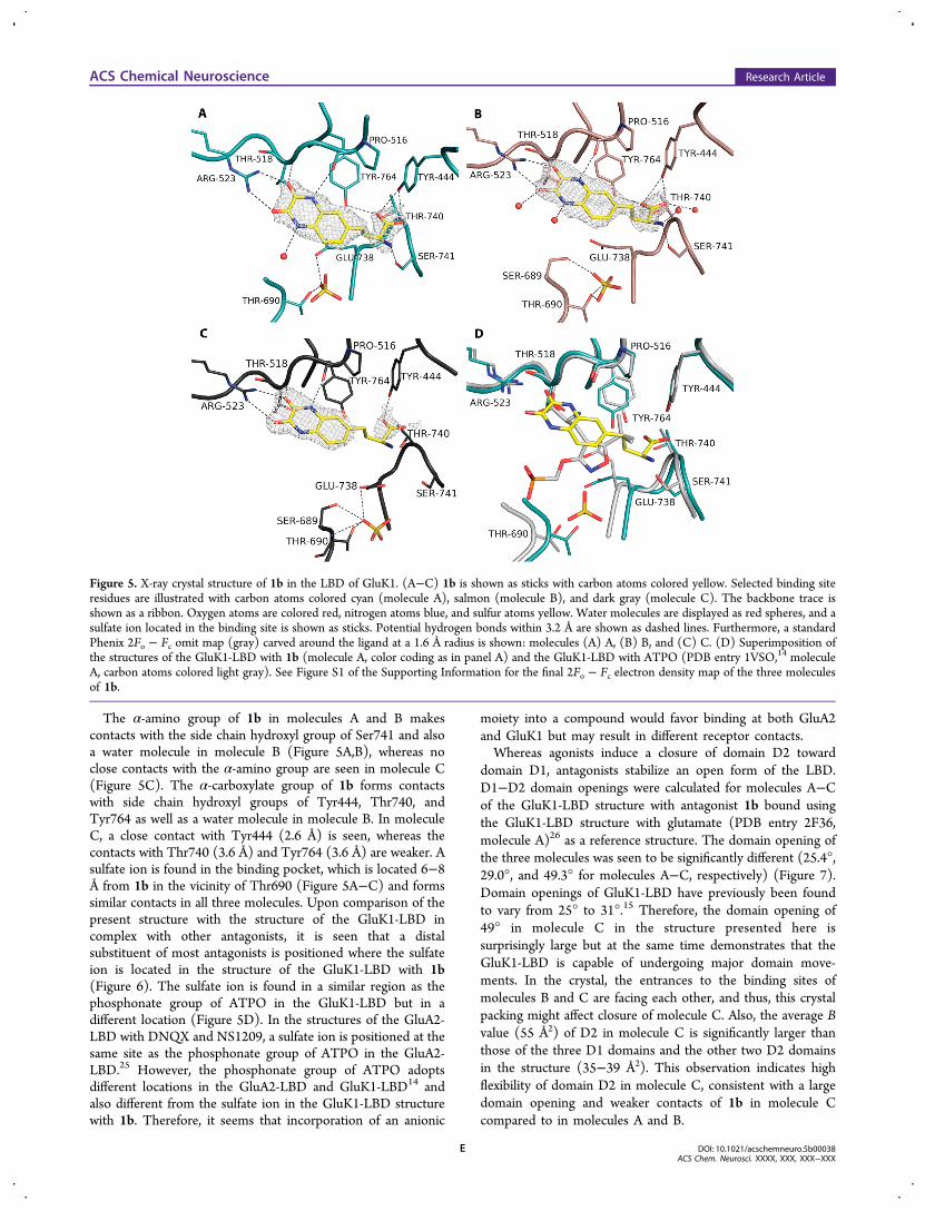
The α-amino group of 1b in molecules A and B makescontacts with the side chain hydroxyl group of Ser741 and alsoa water molecule in molecule B (Figure 5A,B), whereas noclose contacts with the α-amino group are seen in molecule C(Figure 5C). The α-carboxylate group of 1b forms contactswith side chain hydroxyl groups of Tyr444, Thr740, andTyr764 as well as a water molecule in molecule B. In moleculeC, a close contact with Tyr444 (2.6 Å) is seen, whereas thecontacts with Thr740 (3.6 Å) and Tyr764 (3.6 Å) are weaker. Asulfate ion is found in the binding pocket, which is located 6−8Å from 1b in the vicinity of Thr690 (Figure 5A−C) and formssimilar contacts in all three molecules. Upon comparison of thepresent structure with the structure of the GluK1-LBD incomplex with other antagonists, it is seen that a distalsubstituent of most antagonists is positioned where the sulfateion is located in the structure of the GluK1-LBD with 1b(Figure 6). The sulfate ion is found in a similar region as thephosphonate group of ATPO in the GluK1-LBD but in adifferent location (Figure 5D). In the structures of the GluA2-LBD with DNQX and NS1209, a sulfate ion is positioned at thesame site as the phosphonate group of ATPO in the GluA2-LBD.25 However, the phosphonate group of ATPO adoptsdifferent locations in the GluA2-LBD and GluK1-LBD14 andalso different from the sulfate ion in the GluK1-LBD structurewith 1b. Therefore, it seems that incorporation of an anionic
moiety into a compound would favor binding at both GluA2and GluK1 but may result in different receptor contacts.Whereas agonists induce a closure of domain D2 toward
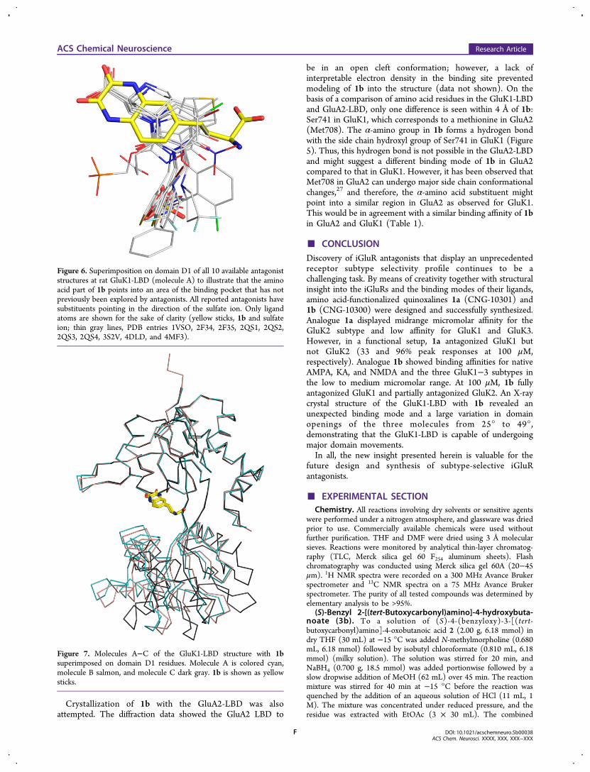
domain D1, antagonists stabilize an open form of the LBD.D1−D2 domain openings were calculated for molecules A−Cof the GluK1-LBD structure with antagonist 1b bound usingthe GluK1-LBD structure with glutamate (PDB entry 2F36,molecule A)26 as a reference structure. The domain opening ofthe three molecules was seen to be significantly different (25.4°,29.0°, and 49.3° for molecules A−C, respectively) (Figure 7).Domain openings of GluK1-LBD have previously been foundto vary from 25° to 31°.15 Therefore, the domain opening of49° in molecule C in the structure presented here issurprisingly large but at the same time demonstrates that theGluK1-LBD is capable of undergoing major domain move-ments. In the crystal, the entrances to the binding sites ofmolecules B and C are facing each other, and thus, this crystalpacking might affect closure of molecule C. Also, the average Bvalue (55 Å2) of D2 in molecule C is significantly larger thanthose of the three D1 domains and the other two D2 domainsin the structure (35−39 Å2). This observation indicates highflexibility of domain D2 in molecule C, consistent with a largedomain opening and weaker contacts of 1b in molecule Ccompared to in molecules A and B.
Figure 5. X-ray crystal structure of 1b in the LBD of GluK1. (A−C) 1b is shown as sticks with carbon atoms colored yellow. Selected binding siteresidues are illustrated with carbon atoms colored cyan (molecule A), salmon (molecule B), and dark gray (molecule C). The backbone trace isshown as a ribbon. Oxygen atoms are colored red, nitrogen atoms blue, and sulfur atoms yellow. Water molecules are displayed as red spheres, and asulfate ion located in the binding site is shown as sticks. Potential hydrogen bonds within 3.2 Å are shown as dashed lines. Furthermore, a standardPhenix 2Fo − Fc omit map (gray) carved around the ligand at a 1.6 Å radius is shown: molecules (A) A, (B) B, and (C) C. (D) Superimposition ofthe structures of the GluK1-LBD with 1b (molecule A, color coding as in panel A) and the GluK1-LBD with ATPO (PDB entry 1VSO,14 moleculeA, carbon atoms colored light gray). See Figure S1 of the Supporting Information for the final 2Fo − Fc electron density map of the three moleculesof 1b.
ACS Chemical Neuroscience Research Article
DOI: 10.1021/acschemneuro.5b00038ACS Chem. Neurosci. XXXX, XXX, XXX−XXX
E
Crystallization of 1b with the GluA2-LBD was alsoattempted. The diffraction data showed the GluA2 LBD to
be in an open cleft conformation; however, a lack ofinterpretable electron density in the binding site preventedmodeling of 1b into the structure (data not shown). On thebasis of a comparison of amino acid residues in the GluK1-LBDand GluA2-LBD, only one difference is seen within 4 Å of 1b:Ser741 in GluK1, which corresponds to a methionine in GluA2(Met708). The α-amino group in 1b forms a hydrogen bondwith the side chain hydroxyl group of Ser741 in GluK1 (Figure5). Thus, this hydrogen bond is not possible in the GluA2-LBDand might suggest a different binding mode of 1b in GluA2compared to that in GluK1. However, it has been observed thatMet708 in GluA2 can undergo major side chain conformationalchanges,27 and therefore, the α-amino acid substituent mightpoint into a similar region in GluA2 as observed for GluK1.This would be in agreement with a similar binding affinity of 1bin GluA2 and GluK1 (Table 1).
■ CONCLUSIONDiscovery of iGluR antagonists that display an unprecedentedreceptor subtype selectivity profile continues to be achallenging task. By means of creativity together with structuralinsight into the iGluRs and the binding modes of their ligands,amino acid-functionalized quinoxalines 1a (CNG-10301) and1b (CNG-10300) were designed and successfully synthesized.Analogue 1a displayed midrange micromolar affinity for theGluK2 subtype and low affinity for GluK1 and GluK3.However, in a functional setup, 1a antagonized GluK1 butnot GluK2 (33 and 96% peak responses at 100 μM,respectively). Analogue 1b showed binding affinities for nativeAMPA, KA, and NMDA and the three GluK1−3 subtypes inthe low to medium micromolar range. At 100 μM, 1b fullyantagonized GluK1 and partially antagonized GluK2. An X-raycrystal structure of the GluK1-LBD with 1b revealed anunexpected binding mode and a large variation in domainopenings of the three molecules from 25° to 49°,demonstrating that the GluK1-LBD is capable of undergoingmajor domain movements.In all, the new insight presented herein is valuable for the
future design and synthesis of subtype-selective iGluRantagonists.
■ EXPERIMENTAL SECTIONChemistry. All reactions involving dry solvents or sensitive agents
were performed under a nitrogen atmosphere, and glassware was driedprior to use. Commercially available chemicals were used withoutfurther purification. THF and DMF were dried using 3 Å molecularsieves. Reactions were monitored by analytical thin-layer chromatog-raphy (TLC, Merck silica gel 60 F254 aluminum sheets). Flashchromatography was conducted using Merck silica gel 60A (20−45μm). 1H NMR spectra were recorded on a 300 MHz Avance Brukerspectrometer and 13C NMR spectra on a 75 MHz Avance Brukerspectrometer. The purity of all tested compounds was determined byelementary analysis to be >95%.
(S)-Benzyl 2-[(tert-Butoxycarbonyl)amino]-4-hydroxybuta-noate (3b). To a solution of (S)-4-(benzyloxy)-3-[(tert-butoxycarbonyl)amino]-4-oxobutanoic acid 2 (2.00 g, 6.18 mmol) indry THF (30 mL) at −15 °C was added N-methylmorpholine (0.680mL, 6.18 mmol) followed by isobutyl chloroformate (0.810 mL, 6.18mmol) (milky solution). The solution was stirred for 20 min, andNaBH4 (0.700 g, 18.5 mmol) was added portionwise followed by aslow dropwise addition of MeOH (62 mL) over 45 min. The reactionmixture was stirred for 40 min at −15 °C before the reaction wasquenched by the addition of an aqueous solution of HCl (11 mL, 1M). The mixture was concentrated under reduced pressure, and theresidue was extracted with EtOAc (3 × 30 mL). The combined
Figure 6. Superimposition on domain D1 of all 10 available antagoniststructures at rat GluK1-LBD (molecule A) to illustrate that the aminoacid part of 1b points into an area of the binding pocket that has notpreviously been explored by antagonists. All reported antagonists havesubstituents pointing in the direction of the sulfate ion. Only ligandatoms are shown for the sake of clarity (yellow sticks, 1b and sulfateion; thin gray lines, PDB entries 1VSO, 2F34, 2F35, 2QS1, 2QS2,2QS3, 2QS4, 3S2V, 4DLD, and 4MF3).
Figure 7. Molecules A−C of the GluK1-LBD structure with 1bsuperimposed on domain D1 residues. Molecule A is colored cyan,molecule B salmon, and molecule C dark gray. 1b is shown as yellowsticks.
ACS Chemical Neuroscience Research Article
DOI: 10.1021/acschemneuro.5b00038ACS Chem. Neurosci. XXXX, XXX, XXX−XXX
F
organic layers were successively washed with an aqueous solution ofHCl (1 × 40 mL, 1 M), H2O (2 × 50 mL), an aqueous solution of 5%NaHCO3 (1 × 50 mL), and finally H2O (3 × 50 mL). It was driedover MgSO4, filtered, and evaporated under reduced pressure, leadingto a yellow oil. The crude was used without purification.General Procedure for the Displacement of the Primary
Hydroxy Group with Iodine. To a solution of PPh3 (2.02 g, 7.70mmol) and imidazole (0.520 g, 7.70 mmol) in DCM (29 mL) at 0 °Cwas added I2 (1.95 g, 7.70 mmol) (orange milky solution). Thereaction mixture was warmed to room temperature and stirred for 10min. The mixture was recooled to 0 °C, and a solution of thecorresponding alcohol (6.16 mmol) in DCM (6 mL) was addeddropwise over 15 min. The solution was stirred at 0 °C for 90 min andthen at room temperature for 2 h. The reaction mixture was filtered offthrough a pad of Celite (2 cm) using a solution of ether and petroleumether (1:1) as the eluent. The organic phase was evaporated underreduced pressure without being heated.(S)-Methyl 2-[(tert-Butoxycarbonyl)amino]-3-iodopropanoate
(4a). Purified by flash column chromatography (ether/petroleumether, 1:4) (Rf = 0.19). White solid (2.85 g, 80%). Mp: 51 °C (Lit. 51°C). 1H NMR (300 MHz, CDCl3): δ 5.34 (d, J = 7.06 Hz, 1H), 4.55−4.47 (m, 1H), 3.79 (s, 3H), 3.62−3.50 (m, 2H), 1.45 (s, 9H).(S)-Benzyl 2-[(tert-Butoxycarbonyl)amino]-4-iodobutanoate (4b).
Purified by flash column chromatography (EtOAc/petroleum ether,1:5) (Rf = 0.5). White solid (1.3 g, 49% over two steps). Mp: 54 °C(Lit. 54 °C). 1H NMR (300 MHz, CDCl3): δ 7.39−7.32 (m, 5H), 5.22(dd, J = 2.91 Hz, 1H), 5.13−5.03 (m, 1H), 4.41−4.35 (m, 1H), 3.21−3.08 (m, 2H), 2.41−2.35 (m, 1H), 2.25−2.17 (m, 1H), 1.45 (s, 9H).General Procedure for the Negishi Cross Coupling Reaction.
Zinc dust (0.61 g, 9.30 mmol) was added to a nitrogen-purged vial.Dry DMF (3.25 mL) was added followed by a catalytic amount of I2(40 mg, 0.15 mmol) (the solution turned yellow and then black again).The corresponding iodine compound, 4a or 4b (3.10 mmol), wasimmediately added followed by another catalytic amount of I2 (40 mg,0.15 mmol) (the solution turned yellow-green and then black again;exothermicity was felt because of zinc insertion). Pd2dba3 (73 mg, 0.08mmol), SPhos (62 mg, 0.150 mmol), and 6-bromo-2,3-dimethox-yquinoxaline 9 (0.290 g, 1.08 mmol) were added to the vial, and thesolution was heated at 50 °C for 4 h. The reaction mixture was filteredoff through a pad of Celite (2 cm), washed with DMF, and evaporatedunder reduced pressure. Water (10 mL) was added, and the solutionwas extracted with Et2O (3 × 20 mL). Combined organic phases werewashed with brine, dried over MgSO4, filtered, and evaporated underreduced pressure.(S)-Methyl 2-[(tert-Butoxycarbonyl)amino]-3-(2,3-dimethoxyqui-
noxalin-6-yl)propanoate (5a). Purified twice by flash columnchromatography [AcOEt/heptane, 3:7 (Rf = 0.30), and thentoluene/AcOEt, 85:15 (Rf = 0.35)]. White solid (0.5 g, 41%). Mp:56.4 °C. [α]D
25 = +67.5 (c = 0.306 g/100 mL, CH2Cl2).1H NMR
(300 MHz, CDCl3): δ 7.69 (d, J = 8.34 Hz, 1H), 7.55 (br s, 1H), 7.26(dd, J = 8.33, 1.98 Hz, 1H), 5.02 (d, J = 7.99 Hz, 1H), 4.66 (dd, J =13.73, 6.05 Hz, 1H), 4.14 (s, 6H), 3.74 (s, 3H), 3.24 (dq, J = 13.97,6.07 Hz, 2H), 1.42 (9H, s). 13C NMR (75 MHz, CDCl3): δ 172.1,154.9, 149.9, 149.7, 136.9, 136.1, 134.6, 127.8, 126.7, 126.3, 79.9, 54.4,54.1, 52.2, 38.1, 28.2.(S)-Benzyl 2-[(tert-Butoxycarbonyl)amino]-4-(2,3-dimethoxyqui-
noxalin-6-yl)butanoate (5b). Purified four times by flash columnchromatography [twice with EtOAc/heptane, 3:7 (Rf = 0.33), thenDCM/MeOH (5%) (Rf = 0.38), and then toluene/EtOAc, 9:1 (Rf =0.30)]. Orange glass solid (1.0 g, 35%). Mp: 67.6 °C. [α]D
25 = +22.0 (c= 0.4 g/100 mL, CH2Cl2).
1H NMR (300 MHz, CDCl3): δ 7.65 (d, J= 8.35 Hz, 1H), 7.52 (br s, 1H), 7.38−7.34 (m, 4H), 7.26 (s, 1H),7.26−7.15 (m, 1H), 5.16 (dd, J = 19.42, 12.28 Hz, 2H), 5.14 (br s,1H), 4.44 (dd, J = 12.99, 7.03 Hz, 1H), 4.14 (s, 6H), 2.88−2.67 (m,2H), 2.34−2.15 (m, 1H), 2.10−1.95 (m, 1H), 1.46 (s, 9H). 13C NMR(75 MHz, CDCl3): δ 172.3, 155.2, 149.8, 149.5, 139.4, 137.0, 135.5,135.2, 128.5, 128.3, 128.2, 127.4, 126.2, 125.4, 79.9, 67.0, 54.0, 53.2,34.2, 31.3, 28.2.(S)-2-Amino-3-(2,3-dioxo-1,2,3,4-tetrahydroquinoxalin-6-yl)-
propanoic Acid Hydrochloride (1a). To a solution of (S)-methyl 2-
[(tert-butoxycarbonyl)amino]-3-(2,3-dimethoxyquinoxalin-6-yl)-propanoate 5a (0.400 g, 1.02 mmol) in dioxane (19 mL) was added anaqueous solution of HCl (19 mL, 4 N), and the reaction mixture washeated under gentle reflux conditions overnight. The reaction mixturewas evaporated, and the residue was triturated with Et2O (3 × 5 mL),leading to a pure white HCl salt (250 mg, 89%). Mp: 278 °C dec.[α]D
25 = +48.0 (c = 0.167 g/100 mL, DMSO). 1H NMR (300 MHz,d6-DMSO): δ 12.00 (s, 1H), 11.95 (s, 1H), 8.33 (br s, 3H), 7.07 (d, J= 8.60 Hz, 1H), 7.01−6.96 (m, 2H), 4.08 (br s, 1H), 3.07 (d, J = 6.42Hz, 2H). 13C NMR (75 MHz, d6-DMSO): δ 170.1, 155.1, 154.9,129.7, 125.5, 124.7, 124.2, 116.1, 115.3, 53.2, 35.2. Anal. Calcd forC12H13N3O4·0.34H2O·1.62HCl): C, 43.88; H, 4.7; N, 12.79. Found:C, 43.58; H, 4.38; N, 13.12.
(S)-2-Amino-4-(2,3-dioxo-1,2,3,4-tetrahydroquinoxalin-6-yl)-butanoic Acid Hydrochloride (1b). To a solution of (S)-benzyl 2-[(tert-butoxycarbonyl)amino]-4-(2,3-dimethoxyquinoxalin-6-yl)-butanoate 5b (0.250 g, 0.52 mmol) in dioxane (12 mL) was added anaqueous solution of HCl (12 mL, 4 N), and the solution was heatedunder gentle reflux conditions overnight. The reaction mixture wasthen evaporated, and the residue was triturated with Et2O (3 × 5 mL).The crude was obtained as a light orange powder (96%).
The crude (150 mg, 0.5 mmol) was dissolved in H2O (8 mL) andthe mixture stirred at room temperature. Propylene oxide (35 μL, 5.0mmol) was slowly added to the reaction mixture, and the solution wasstirred for 30 min. The reaction mixture was left overnight withoutstirring for crystallization and then filtered with a filter paper, andcrystals were washed with H2O (3 × 2 mL). The crystals weredissolved in an aqueous solution of HCl (1.5 mL, 1 M) and evaporatedto dryness. The solid was dissolved in H2O (2 mL) and freeze-dried toafford a light brown solid (106 mg, 68%). Mp: 263 °C dec. [α]D
25 =+35.8 (c = 0.142 g/100 mL, H2O).
1H NMR (300 MHz, d6-DMSO): δ11.95 (s, 1H), 11.91 (s, 1H), 8.48 (br s, 3H), 7.07 (d, J = 7.97 Hz,3H), 6.95 (br s, 1H), 6.94 (br dd, J = 9.13, 1.48 Hz, 1H), 3.87 (signalhidden by the solvent peak, 1H), 2.82−2.67 (m, 1H), 2.67−2.53 (m,1H), 2.10−1.96 (m, 2H). 1H NMR (300 MHz, D2O): δ 7.00 (ddd, J =8.27, 1.68, 1.68 Hz, 1H), 6.94 (dd, J = 8.31, 1.51 Hz, 1H), 6.87 (br d, J= 1.39 Hz, 1H), 3.99 (dd, J = 6.25, 6.25 Hz, 1H), 2.81−2.55 (m, 2H),2.13−1.99 (m, 2H). 13C NMR (75 MHz, d6-DMSO): δ 170.7, 155.3,155.0, 135.4, 125.6, 124.0, 123.1, 115.3, 114.6, 51.6, 32.0, 30.0. Anal.Calcd for C12H14ClN3O4·1.5H2O·0.81HCl): C, 40.84; H, 4.67; N,11.41. Found: C, 40.46; H, 5.04; N, 11.79.
6-Bromoquinoxaline-2,3(1H,4H)-dione (7). A solution of 2-amino-4-bromoaniline 6 (3.0 g, 16.0 mmol) in diethyl oxalate (15 mL) wasrefluxed for 4 h. The reaction mixture was allowed to cool to roomtemperature, after which EtOAc (25 mL) was added. The precipitatewas filtered and washed with EtOAc (3 × 20 mL) and dried undervacuum to give a brown powder (3.75 g, 96%). 1H NMR (300 MHz,d6-DMSO): δ 11.97 (s, 1H), 11.94 (s, 1H), 7.26−7.18 (m, 2H), 7.03(d, J = 9.03 Hz, 1H). Mp: >400 °C dec (Lit. >350 °C).
6-Bromo-2,3-dichloroquinoxaline (8). To a mixture of 6-bromoquinoxaline-2,3(1H,4H)-dione 7 (5.33 g, 22.11 mmol) inthionyl chloride (74 mL) was added a catalytic amount of DMF(three drops). The reaction mixture was heated under reflux for 8 h,cooled to room temperature, and poured very slowly into an ice/waterbath. The solid was filtered, dissolved in EtOAc (500 mL), and driedwith MgSO4. The organic phase was filtered and evaporated to affordthe desired product, 6-bromo-2,3-dichloroquinoxaline 8, as a lightorange-brown solid (1.03 g, 90%). Mp: 131 °C (Lit. 132 °C). 1HNMR (300 MHz, d6-DMSO): δ 8.34 (d, J = 2.01 Hz, 1H), 8.05 (dd, J= 8.91, 2.06 Hz, 1H), 8.00 (d, J = 8.89 Hz, 1H).
6-Bromo-2,3-dimethoxyquinoxaline (9). To a solution of theintermediate 6-bromo-2,3-dichloroquinoxaline 8 (2.25 g, 8.09 mmol)in anhydrous methanol (34 mL) was added a solution of 25% MeOKin MeOH (6.90 mL, 22.6 mmol). The reaction mixture was refluxedfor 3 h and cooled to room temperature. H2O (50 mL) was added, andthe suspension was filtered before being washed with water (3 × 20mL). The solid was dried overnight using a vacuum oven to give thedesired product, 6-bromo-2,3-dimethoxyquinoxaline 9, as a brownpowder (2.02 g, 93%). Mp: 105 °C (Lit. 114−115 °C, solvent of
ACS Chemical Neuroscience Research Article
DOI: 10.1021/acschemneuro.5b00038ACS Chem. Neurosci. XXXX, XXX, XXX−XXX
G
methanol). 1H NMR (300 MHz, d6-DMSO): δ 7.90 (br s, 1H), 7.67(d, J = 9.09 Hz, 1H), 7.63 (d, J = 8.77 Hz, 1H), 4.03 (s, 6H).Pharmacology. Radioligand displacement studies were performed
as previously described at native rat brain AMPA, KA, and NMDAreceptors and at recombinant rat homomeric GluA2(R)o, GluK1(Q)1b,GluK2(VCR)a, and GluK3a receptors as well as the GluK1-LBD.
28 Forrecombinant kainate receptor assays, [3H]SYM2081 was used as theradioligand while [3H]AMPA was employed for the GluA2(R)obinding assay.Cell Culture and Transfection. HEK293T/17 cells (ATCC)
were maintained in minimal essential medium (MEM) containingglutaMAX supplemented with 10% fetal bovine serum (Invitrogen).Cells were plated at a low density (1.6−2.0 × 10⁴ cells/mL) on poly-D-lysine-coated 35 mm plastic dishes and were transiently transfected 24h later using the calcium phosphate technique as previouslydescribed.29 Rat cDNA encoding for GluK1−2a and GluK2a subunitswas cotransfected with cDNA encoding enhanced green fluorescentprotein (eGFP) to identify transfected cells.Electrophysiological Recordings. Experiments were performed
36−48 h after transfection. Agonist solutions were rapidly applied tooutside-out patches excised from transfected cells using a piezoelectricstack (Physik Instrumente). Solution exchange (10−90% rise time of250−350 μs) was determined in a separate experiment by measuringthe liquid junction current. All recordings were performed using anAxopatch 200B (Molecular Devices) using thick-walled borosilicateglass pipettes (3−6 MΩ) coated with dental wax to reduce electricalnoise. Current records were filtered at 5 kHz, digitized at 25 kHz, andseries resistance (3−12 MΩ) compensated by 95%. Recordings wereperformed at a holding potential of −100 mV. All experiments wereperformed at room temperature. All chemicals used for electro-physiology were purchased from Sigma-Aldrich unless otherwiseindicated. The external solution contained 150 mM NaCl, 5 mMHEPES, 0.1 mM MgCl2, and 0.1 mM CaCl2 (pH 7.3−7.4). Theinternal solution contained 115 mM NaCl, 10 mM NaF, 5 mMHEPES, 5 mM Na4BAPTA (Life Technologies), 1 mM MgCl2, 0.5mM CaCl2, and 10 mM Na2ATP (pH 7.3−7.4). The osmotic pressureof all solutions was adjusted to 295−300 mOsm with sucrose.Concentrated (100×) L-Glu stocks were prepared and stored at −20°C; stocks were thawed and diluted on the day of the experiment.Concentrated (3 mM in DMSO) stocks of 1a and 1b were preparedand stored at −20 °C; stocks were thawed on the day of theexperiment and added to the control and agonist external solutions fora final concentration of 100 μM. As a control, an equivalent amount ofDMSO was added to the external solutions not containing 1a or 1b.To test whether 1a was a partial agonist at GluK1 and GluK2receptors, 100 μM 1a was applied to outside-out patches alone. Thepresence of receptors in the patch was confirmed by obtaining aresponse with 1 mM L-Glu. The plant lectin, Concanavalin-A, wasdissolved at ∼1 mg/mL in an external solution and applied to thepatch for 1−2 min. Data were acquired using pClamp10 software(Molecular Devices) and tabulated using Excel (Microsoft Corp.).X-ray Structure Determination. The rat GluK1-LBD (GRIK1_-
RAT, UNP P22756, segment S1 residues 430−544 and segment S2residues 667−805) was expressed and purified in the presence of Gluas previously described.30 Crystallization of GluK1-LBD in complexwith 1b was performed using the hanging drop vapor diffusion methodat 6 °C. A protein solution consisting of 5 mg/mL GluK1-LBD, 5.2mM 1b, 10 mM HEPES, 20 mM NaCl, and 1 mM EDTA (pH 7.0)was prepared and used for crystallization experiments. Crystals usedfor diffraction studies were obtained under the following conditions:24.4% PEG4000, 0.3 M ammonium sulfate, and 0.1 M phosphatecitrate buffer (pH 4.5). The crystals were flash-cooled with liquidnitrogen after immersion in cryo buffer (1 μL of reservoir solutioncontaining 1 μL of UCP). Flash-cooled crystals were stored in liquidnitrogen until data were collected.An X-ray diffraction data set to 2.28 Å resolution was collected on
beamline I911-3 at MAX-lab (Lund, Sweden). The data wereprocessed with XDS31 and scaled using SCALA32 in CCP4i.33 Thestructure was determined by molecular replacement using PHASER34
within CCP4i and the structure of the GluK1-LBD in complex with
(S)-ATPO as a search model (PDB entry 1VSO, molecule A; proteinatoms only).14 The search model was defined as two domains, D1 andD2. Three molecules were found in the asymmetric unit of the crystal.Visual inspection of the structure in COOT35 revealed well-definedelectron density for the structure in most areas and clear densitycorresponding to 1b. The ligand coordinates were created in Maestro(Maestro version 9.2, Schrodinger, LLC, New York, NY) and fit intothe electron density. Topology and parameter files for 1b wereobtained using eLBOW.36 The model used for molecular replacementlacked two loops (residues 493−497 and 712−714), and theseresidues were built in manually. The structure was refined inPHENIX37 with isotropic B factors and validated using tools inCOOT and PHENIX. The final structure was also validated on thePDB ADIT validation server. Statistics of data collection andrefinement can be found in Table S1 of the Supporting Information.D1−D2 domain openings were calculated using the DynDom server.38
In Silico Conformational Search Study. Conformationalsearches and superimposition studies were performed using thesoftware package MOE 2013.08 (Molecular Operating Environment,Chemical Computing Group). The built-in function “stochasticsearch” was used with the standard setup: mmff94x force field,solvation set to GB/SA, and energy cutoff of >7 kcal/mol.
■ ASSOCIATED CONTENT*S Supporting InformationCrystal data, data collection, and refinement statistics of GluK1-LBD in complex with 1b (Table S1), final 2Fo-Fc electrondensity of the three molecules of 1b (Figure S1), and in silicostudy of the binding mode of 1b (Table S2 and Figure S2).This material is available free of charge via the Internet athttp://pubs.acs.org.Accession CodesThe structure of the GluK1-LBD in complex with 1b (CNG-10300) has been deposited in as Protein Data Bank entry4QF9.
■ AUTHOR INFORMATIONCorresponding Authors*Phone: +45 35336244. Fax: +45 35336041. E-mail: [email protected].*E-mail: [email protected] (for X-ray crystallography).Author ContributionsC.S.D. (medicinal chemistry) and C.M. (X-ray crystallography)contributed equally to this work.FundingWe thank the Lundbeck Foundation, the Carlsberg Foundation,The University of Copenhagen Programme of Excellence(GluTarget), the Hørslev Foundation, and Danscatt forfinancial support.NotesThe authors declare no competing financial interest.
■ ACKNOWLEDGMENTSDiffraction data were collected at the MAX-lab synchrotronfacility in Lund, Sweden. Heidi Peterson is thanked fortechnical assistance.
■ ABBREVIATIONSAMPA, α-amino-3-hydroxy-5-methyl-4-isoxazolepropionic acid;CNS, central nervous system; EAATs, excitatory amino acidtransporters; Glu, (S)-glutamate; iGluRs, ionotropic Glureceptors; KA, kainic acid; LBD, ligand-binding domain;mGluRs, metabotropic Glu receptors; NMDA, N-methyl-D-aspartate
ACS Chemical Neuroscience Research Article
DOI: 10.1021/acschemneuro.5b00038ACS Chem. Neurosci. XXXX, XXX, XXX−XXX
H
■ REFERENCES(1) Anggono, V., and Huganir, R. L. (2012) Regulation of AMPAreceptor trafficking and synaptic plasticity. Curr. Opin. Neurobiol. 22,461−469.(2) Lovinger, D. M. (2010) Neurotransmitter roles in synapticmodulation, plasticity and learning in the dorsal striatum. Neuro-pharmacology 58, 951−961.(3) Jane, D. E., Lodge, D., and Collingridge, G. L. (2009) Kainatereceptors: Pharmacology, function and therapeutic potential. Neuro-pharmacology 56, 90−113.(4) Contractor, A., Mulle, C., and Swanson, G. T. (2011) Kainatereceptors coming of age: Milestones of two decades of research. TrendsNeurosci. 34, 154−163.(5) Matute, C. (2011) Therapeutic Potential of Kainate Receptors:Therapeutic Potential of Kainate Receptors. CNS Neurosci. Ther. 17,661−669.(6) Ogden, K. K., and Traynelis, S. F. (2011) New advances inNMDA receptor pharmacology. Trends Pharmacol. Sci. 32, 726−733.(7) Larsen, A. M., and Bunch, L. (2011) Medicinal Chemistry ofCompetitive Kainate Receptor Antagonists. ACS Chem. Neurosci. 2,60−74.(8) Weiss, B., Alt, A., Ogden, A. M., Gates, M., Dieckman, D. K.,Clemens-Smith, A., Ho, K. H., Jarvie, K., Rizkalla, G., and Wright, R. A.(2006) Pharmacological characterization of the competitive GLUK5receptor antagonist decahydroisoquinoline LY466195 in vitro and invivo. J. Pharmacol. Exp. Ther. 318, 772−781.(9) Randle, J. C., Guet, T., Cordi, A., and Lepagnol, J. M. (1992)Competitive inhibition by NBQX of kainate/AMPA receptor currentsand excitatory synaptic potentials: Importance of 6-nitro substitution.Eur. J. Pharmacol. 215, 237−244.(10) Poulie, C. B. M., and Bunch, L. (2013) Heterocycles asnonclassical bioisosteres of α-amino acids. ChemMedChem 8, 205−215.(11) Hogner, A., Greenwood, J. R., Liljefors, T., Lunn, M.-L.,Egebjerg, J., Larsen, I. K., Gouaux, E., and Kastrup, J. S. (2003)Competitive antagonism of AMPA receptors by ligands of differentclasses: Crystal structure of ATPO bound to the GluR2 ligand-bindingcore, in comparison with DNQX. J. Med. Chem. 46, 214−221.(12) Armstrong, N., and Gouaux, E. (2000) Mechanisms foractivation and antagonism of an AMPA-sensitive glutamate receptor:Crystal structures of the GluR2 ligand binding core. Neuron 28, 165−181.(13) Cordi, A. A., Desos, P., Randle, J. C., and Lepagnol, J. (1995)Structure-activity relationships in a series of 3-sulfonylamino-2-(1H)-quinolones, as new AMPA/kainate and glycine antagonists. Bioorg.Med. Chem. 3, 129−141.(14) Hald, H., Naur, P., Pickering, D. S., Sprogøe, D., Madsen, U.,Timmermann, D. B., Ahring, P. K., Liljefors, T., Schousboe, A., andEgebjerg, J. (2007) Partial agonism and antagonism of the ionotropicglutamate receptor iGluR5 structures of the ligand-binding core incomplex with domoic acid and 2-amino-3-[5-tert-butyl-3-(phosphono-methoxy)-4-isoxazolyl]propionic acid. J. Biol. Chem. 282, 25726−25736.(15) Alushin, G. M., Jane, D., and Mayer, M. L. (2011) Binding siteand ligand flexibility revealed by high resolution crystal structures ofGluK1 competitive antagonists. Neuropharmacology 60, 126−134.(16) Jackson, R. F., Moore, R. J., Dexter, C. S., Elliott, J., andMowbray, C. E. (1998) Concise synthesis of enantiomerically purephenylalanine, homophenylalanine, and bishomophenylalanine deriv-atives using organozinc chemistry: NMR studies of amino acid-derivedorganozinc reagents. J. Org. Chem. 63, 7875−7884.(17) Ross, A. J., Lang, H. L., and Jackson, R. F. (2010) Muchimproved conditions for the Negishi cross-coupling of iodoalaninederived zinc reagents with aryl halides. J. Org. Chem. 75, 245−248.(18) Broadrup, R. L., Wang, B., and Malachowski, W. P. (2005) Ageneral strategy for the synthesis of azapeptidomimetic lactams.Tetrahedron 61, 10277−10284.
(19) Adamczyk, M., Johnson, D. D., and Reddy, R. E. (2000) Aconcise synthesis of (S)-(−)-3-(2-carboxy-4-pyrrolyl)-alanine. Tetrahe-dron: Asymmetry 11, 3063−3068.(20) Jensen, A. A., and Brauner-Osborne, H. (2004) Pharmacologicalcharacterization of human excitatory amino acid transporters EAAT1,EAAT2 and EAAT3 in a fluorescence-based membrane potential assay.Biochem. Pharmacol. 67, 2115−2127.(21) Fay, A.-M. L., Corbeil, C. R., Brown, P., Moitessier, N., andBowie, D. (2009) Functional characterization and in silico docking offull and partial GluK2 kainate receptor agonists. Mol. Pharmacol. 75,1096−1107.(22) Sobolevsky, A. I., Rosconi, M. P., and Gouaux, E. (2009) X-raystructure, symmetry and mechanism of an AMPA-subtype glutamatereceptor. Nature 462, 745−756.(23) Yelshanskaya, M. V., Li, M., and Sobolevsky, A. I. (2014)Structure of an agonist-bound ionotropic glutamate receptor. Science345, 1070−1074.(24) Durr, K. L., Chen, L., Stein, R. A., De Zorzi, R., Folea, I. M.,Walz, T., Mchaourab, H. S., and Gouaux, E. (2014) Structure anddynamics of AMPA receptor GluA2 in resting, pre-open, anddesensitized states. Cell 158, 778−792.(25) Kasper, C., Pickering, D. S., Mirza, O., Olsen, L., Kristensen, A.S., Greenwood, J. R., Liljefors, T., Schousboe, A., Watjen, F., Gajhede,M., Sigurskjold, B. W., and Kastrup, J. S. (2006) The structure of amixed GluR2 ligand-binding core dimer in complex with (S)-glutamate and the antagonist (S)-NS1209. J. Mol. Biol. 357, 1184−1201.(26) Mayer, M. L., Ghosal, A., Duolman, N. P., and Jane, D. E.(2006) Crystal structures of the kainate receptor GluR5 ligand bindingcore dimer with novel GluR5-selective antagonists. J. Neurosci. 26,2852−2861.(27) Pøhlsgaard, J., Frydenvang, K., Madsen, U., and Kastrup, J. S.(2011) Lessons from more than 80 structures of the GluA2 ligand-binding domain in complex with agonists, antagonists and allostericmodulators. Neuropharmacology 60, 135−150.(28) Assaf, Z., Larsen, A. P., Venskutonyte, R., Han, L., Abrahamsen,B., Nielsen, B., Gajhede, M., Kastrup, J. S., Jensen, A. A., Pickering, D.S., Frydenvang, K., Gefflaut, T., and Bunch, L. (2013) Chemo-enzymatic Synthesis of New 2,4-syn-Functionalized (S)-GlutamateAnalogues and Structure−Activity Relationship Studies at IonotropicGlutamate Receptors and Excitatory Amino Acid Transporters. J. Med.Chem. 56, 1614−1628.(29) Maclean, D. M., Wong, A. Y. C., Fay, A.-M., and Bowie, D.(2011) Cations but not anions regulate the responsiveness of kainatereceptors. J. Neurosci. 31, 2136−2144.(30) Naur, P., Vestergaard, B., Skov, L. K., Egebjerg, J., Gajhede, M.,and Kastrup, J. S. (2005) Crystal structure of the kainate receptorGluR5 ligand-binding core in complex with (S)-glutamate. FEBS Lett.579, 1154−1160.(31) Kabsch, W. (2010) XDS. Acta Crystallogr. D66, 125−132.(32) Evans, P. (2006) Scaling and assessment of data quality. ActaCrystallogr. D62, 72−82.(33) Winn, M. D., Ballard, C. C., Cowtan, K. D., Dodson, E. J.,Emsley, P., Evans, P. R., Keegan, R. M., Krissinel, E. B., Leslie, A. G.W., McCoy, A., McNicholas, S. J., Murshudov, G. N., Pannu, N. S.,Potterton, E. A., Powell, H. R., Read, R. J., Vagin, A., and Wilson, K. S.(2011) Overview of the CCP4 suite and current developments. ActaCrystallogr. D67, 235−242.(34) McCoy, A. J., Grosse-Kunstleve, R. W., Adams, P. D., Winn, M.D., Storoni, L. C., and Read, R. J. (2007) Phaser crystallographicsoftware. J. Appl. Crystallogr. 40, 658−674.(35) Emsley, P., Lohkamp, B., Scott, W. G., and Cowtan, K. (2010)Features and development of Coot. Acta Crystallogr. D66, 486−501.(36) Moriarty, N. W., Grosse-Kunstleve, R. W., and Adams, P. D.(2009) electronic Ligand Builder and Optimization Workbench(eLBOW): A tool for ligand coordinate and restraint generation.Acta Crystallogr. D65, 1074−1080.
ACS Chemical Neuroscience Research Article
DOI: 10.1021/acschemneuro.5b00038ACS Chem. Neurosci. XXXX, XXX, XXX−XXX
I
(37) Hayward, S., and Lee, R. A. (2002) Improvements in theanalysis of domain motions in proteins from conformational change:DynDom version 1.50. J. Mol. Graphics Modell. 21, 181−183.(38) Hayward, S., and Berendsen, H. J. (1998) Systematic analysis ofdomain motions in proteins from conformational change: New resultson citrate synthase and T 4 lysozyme. Proteins: Struct., Funct., Genet. 30,144−154.
ACS Chemical Neuroscience Research Article
DOI: 10.1021/acschemneuro.5b00038ACS Chem. Neurosci. XXXX, XXX, XXX−XXX
J