![Biochimica et Biophysica Acta - COnnecting REpositories · chronic and acute airway inflammation [2,3]. In this regard, especially terpenoids,likethe monoterpene oxide1,8-cineol](https://static.fdocument.org/doc/165x107/5f0a739a7e708231d42bb33b/biochimica-et-biophysica-acta-connecting-repositories-chronic-and-acute-airway.jpg)
HPLC Determination of α-keto Acids from Human Serum … file/Volume 14 No...
8
ISSN-1996-918X Pak. J. Anal. Environ. Chem. Vol. 14, No. 2 (2013) 8 – 15 HPLC Determination of α-keto Acids from Human Serum Using 2,3-Diamino-2,3-dimethylbutane as Derivatizing Reagent Khalida Parveen Mahar* 1 , Muhammad Yar Khuhawar 2 , Kulsoom Ubedullah Abbasi 2 , Gulam Qadir Shar 1 , Rafia Azmat 3 and Jameel Ahmed Baig 4 1 *Department of Chemistry, Faculty of Science, Shah Abdul Latif University Khairpur, Pakistan 2 Institute of Advanced Research Studies in Chemical Science, University of Sindh, Jamshoro, Pakistan 3 Department of Chemistry, University of Karachi, Pakistan 4 National Centre of Excellence in Analytical Chemistry, University of Sindh, Jamshoro, Pakistan Received 01 April 2012, Revised 28 July 2013, Accepted 26 August 2013 -------------------------------------------------------------------------------------------------------------------------------------------- Abstract Seven α-keto acids, pyruvic acid (PYR), 2-oxobutyric acid (KB), 3-methyl-2-oxobutyric acid (MKBA), 3-methyl-2-oxovaleric acid (K3MVA), 4-methyl-2-oxovaleric acid (K4MVA), 2- oxoglutaric acid (KG) and Phenyl pyruvic acid (PPY) as derivatives of 2,3-diamino-2,3- dimethybutane (DDB) were separated by HPLC column Zorbax C–18 (4.6x150 mm-id). The compounds were eluted with methanol-water-acetonitrile (40:58:2 V/V/V) with flow rate 1 ml/min. UV detection was carried out by photodiode array at 255 nm. Linear calibration plots were obtained with 0.1 to 60 µg/ml with limits of detection (LoD) within 0.04-0.4 µg/ml. The method was applied for the analysis of α-keto acids from serum of diabetic patients with blood glucose level 430-458 mg/dl and healthy volunteers. The amounts of α-keto acids observed 3.24- 9.71 µg/ml with RSD 1.1-1.9% in diabetic patients were higher than healthy volunteer’s 0.11-1.3 µg/ml with RSD 0.9-2.6%. Keywords: α-keto acids; 2,3-diamino-2,3-dimethylbutane (DDB); serum; diabetic patients. -------------------------------------------------------------------------------------------------------------------------------------------- Introduction The interest in the determination of α-keto acids has increased due to their importance in understanding metabolic and nutritional pathophysiological disorders [1-11]. The α-keto acids may also provide additional clinical information concerning mulfuction of vital organ in several diseases (eg burns, sepsis and multiple organ failure) and possible interventions to supplement routine care measures [12-19]. The monitoring the α-keto acids is of particular intrest because these are involved in a number of biochemical reaction, which includes amino acids, glycolysis and carbohydrates metabolism [20-21]. They have a specific function, such as pyruvic acid (PYR) is involved in the biosynthesis of alanine [22-23], while the concentration of phenylpyruvic acid (PPY) is increased in the plasma of patients with hereditary metabolic disease [24-25] and it inhibits renal gluconeogenesis [7]. The branched chain α-keto acids 3-methyl 2-oxovaleric acid (K3MVA) and 4-methyl-2-oxovaleric acid (K4MVA) are involved in protein turn over [9-26]. Different procedures are used for the determination of α-keto acids such as spectrophotometry [27], spectrofluorometry [28] paper [29] and thin layer chromatography [30], high performance liquid chromatography (HPLC) *Corresponding Author Email: [email protected]
Transcript of HPLC Determination of α-keto Acids from Human Serum … file/Volume 14 No...
ISSN-1996-918X
Pak. J. Anal. Environ. Chem. Vol. 14, No. 2 (2013) 8 – 15
HPLC Determination of α-keto Acids from Human Serum Using 2,3-Diamino-2,3-dimethylbutane as
Derivatizing Reagent
Khalida Parveen Mahar*1, Muhammad Yar Khuhawar2, Kulsoom Ubedullah Abbasi2, Gulam Qadir Shar1, Rafia Azmat3 and Jameel Ahmed Baig4
1*Department of Chemistry, Faculty of Science, Shah Abdul Latif University Khairpur, Pakistan 2Institute of Advanced Research Studies in Chemical Science, University of Sindh, Jamshoro, Pakistan
3Department of Chemistry, University of Karachi, Pakistan 4National Centre of Excellence in Analytical Chemistry, University of Sindh, Jamshoro, Pakistan
Received 01 April 2012, Revised 28 July 2013, Accepted 26 August 2013
-------------------------------------------------------------------------------------------------------------------------------------------- Abstract Seven α-keto acids, pyruvic acid (PYR), 2-oxobutyric acid (KB), 3-methyl-2-oxobutyric acid (MKBA), 3-methyl-2-oxovaleric acid (K3MVA), 4-methyl-2-oxovaleric acid (K4MVA), 2-oxoglutaric acid (KG) and Phenyl pyruvic acid (PPY) as derivatives of 2,3-diamino-2,3-dimethybutane (DDB) were separated by HPLC column Zorbax C–18 (4.6x150 mm-id). The compounds were eluted with methanol-water-acetonitrile (40:58:2 V/V/V) with flow rate 1 ml/min. UV detection was carried out by photodiode array at 255 nm. Linear calibration plots were obtained with 0.1 to 60 µg/ml with limits of detection (LoD) within 0.04-0.4 µg/ml. The method was applied for the analysis of α-keto acids from serum of diabetic patients with blood glucose level 430-458 mg/dl and healthy volunteers. The amounts of α-keto acids observed 3.24-9.71 µg/ml with RSD 1.1-1.9% in diabetic patients were higher than healthy volunteer’s 0.11-1.3 µg/ml with RSD 0.9-2.6%. Keywords: α-keto acids; 2,3-diamino-2,3-dimethylbutane (DDB); serum; diabetic patients.
-------------------------------------------------------------------------------------------------------------------------------------------- Introduction The interest in the determination of α-keto acids has increased due to their importance in understanding metabolic and nutritional pathophysiological disorders [1-11]. The α-keto acids may also provide additional clinical information concerning mulfuction of vital organ in several diseases (eg burns, sepsis and multiple organ failure) and possible interventions to supplement routine care measures [12-19]. The monitoring the α-keto acids is of particular intrest because these are involved in a number of biochemical reaction, which includes amino acids, glycolysis and carbohydrates metabolism [20-21]. They have a specific function, such as pyruvic acid
(PYR) is involved in the biosynthesis of alanine [22-23], while the concentration of phenylpyruvic acid (PPY) is increased in the plasma of patients with hereditary metabolic disease [24-25] and it inhibits renal gluconeogenesis [7]. The branched chain α-keto acids 3-methyl 2-oxovaleric acid (K3MVA) and 4-methyl-2-oxovaleric acid (K4MVA) are involved in protein turn over [9-26]. Different procedures are used for the determination of α-keto acids such as spectrophotometry [27], spectrofluorometry [28] paper [29] and thin layer chromatography [30], high performance liquid chromatography (HPLC)
*Corresponding Author Email: [email protected]
Pak. J. Anal. Environ. Chem. Vol. 14, No. 2 (2013)
9
[9, 31-35] and gas chromatography [36-41]. The chromatography of α-keto acids involves derivatization before their detection. The derivatizing reagents for the sensitive determination of α-keto acids using HPLC are mainly 1,2-phenylenediamine [42], 1,2-diamino-45-dimethoxybezene [31], 4,5-diaminophthalhydrazide dihydrochloride [43], 2,4-dinitrophenylhydrazine [44], 4-hydrazino-2-stilbenzole [45] and phenylhydrazine [36] using spectrophotometric, spectrofluorometric or chemiluminiscens detection. The quantitation of α-keto acids by fluorescence HPLC has been reviewed [46]. Simple isocratic HPLC method for the determination of α-keto acids could be of value. The choice of derivatizing reagent which should convert polar keto acids to nonpolar compounds for reproducible elution, separation and determination from reversed phase HPLC. 2,3-diamino-2,3-dimethylbutane (DDB) has been used for the determination of glyoxal and methylglyoxal by GC from serum. The present work examines the reagent DDB for the isocratic HPLC separation and determination of α-keto acids. The analytical conditions are optimized and examined for the repeatability, linearity of response, limit of detection (LoD), limit of quantitation (LoQ) and inter and interday variation. Materials and Methods Chemicals and Reagents Phenylpyruvic acid (PPY) (Sigma, USA), pyruvic acid (PYR), 2-oxobutyric acid (KB), 3-methyl-2-oxobutyric acid (MKBA), 2-oxoglutaric acid (KG), 3-methyl 2-oxovaleric acid (K3MVA), 4-methyl-2-oxovaleric acid (K4MVA), monosodium salt, (Fluka, Switzerland) were used. The standard solutions of α-keto acids (1.0 mg/ml) were prepared in 10% acetic acid (w/v), further solutions were prepared by appropriate dilution. Fisher Scientific, HPLC grade Leicestershire, UK, Methanol, Riedel-dehaen,Germany acetic acid, 37%, hydrochloric acid. (E. Merck, Germany), sodium acetate, potassium chloride, boric acid, ammonium acetate, sodium bicarbonate, sodium tetraborate, sodium
carbonate, ammonium chloride and ammonia (25%) (Merck Germany) were used. Sample Collection and Pretreatment Synthesis of compounds 2,3-diamino-2,3-dimethylbutane (DDB) was prepared from 2-nitropropane by half bromination to 2,3-dimethyl-2,3-dinitrobutane, followed by reduction of nitro groups to amino groups with granules of tin and hydrochloric acid [47], the ether extract of diamine dried over anhydrous sodium sulphate was used.
The Buffer solutions were prepared within pH 1-10 at unit interval as follows: potassium chloride (1 M) and adjusted with (0.1 M) Hydrochloric acid (pH 1-2), sodium acetate (1 M) and acetic acid (1 M) (pH 3-6), ammonium acetate (1 M) (pH-7), sodium tetra borate (1 M) and boric acid (1 M) (pH 8), sodium carbonate (saturated) and sodium bicarbonate (1 M) (pH 9), ammonia solution (1 M) and ammonium chloride (1 M) (pH 10)
Equipment
HPLC was carried out on Agilent model
1100–network HPLC System (Agilent Technology Inc, USA), a syringe sample injector (Rheodyne7725 injector) containing 20 µL loop, two quart pumps G1311A, degasser G1379A, with diode array DAD G1315B detection system. The computer with Chemstation software controlled the HPLC. The column ZORBAX 300 SB-C18 (4.6x150mm id) (Agilent Technology Inc, USA) was used throughout the study. Spectrophotometric studies were carried out with Hitachi 220 Spectrophotometer double-beam (Hitachi (Pvt.) Ltd Tokyo, Japan). pH measurement was carried out with an Orion 420 A pH meter (Orion Research Inc. Boston, U.S.A) with glass electrode and internal reference electrode combined.
HPLC Analytical procedure
An aqueous solution (0.2-1.0 ml) containing PYR, KB, MKBA, K3MVA, KG,
Pak. J. Anal. Environ. Chem. Vol. 14, No. 2 (2013)
10
K4MVA, and PPY (0.1-60 µg) was added 1 mL of DDB reagent solution and 1 mL acetic acid–sodium acetate buffer pH 3.2 in a well-stoppered test tube (Quick fit). The reaction was warmed at 80 0C for 20 min, mixed well and cooled at room temperature (30 0C) for 15 min. The chloroform (1.0 mL) was then added and the contents were mixed well. The layers were allowed to separate and an aliquot (300 µL) from the organic layer was transferred to screw cap vial. The solution (20 µL) was injected on the column Zorbax C-18 (4.6x150 mm-id) and the elution was methanol–water–acetonitrile (40:58:2 v/v/v) with a flow rate 1 ml/min. Detection was carried out by the photodiode array detector at 255 nm.
Determination of α-keto acids from human serum
Blood samples (5 mL) collected from the diabetic patients and healthy volunteers were incubated for 1 hr at 30 0C and centrifuged at 3000 rpm for 30 min. The supernatant layer was collected and methanol added twice the volume (5 mL). The turbid solution was mixed well and again centrifuged at 3000 rpm for 20 min. The supernatant layer was transferred in to a vial and HPLC analytical procedure was followed. The quantitation was by external calibration curve using regression equation Y = ax + b. Determination of α-keto acids from linear calibration from spiked samples
A blood sample (5 mL) collected from diabetic patient was treated for determination of α-keto acids from serum. The serum was divided after deproteinization with methanol in two equal parts. A part was added, PPY (12 µg), PYR (2 µg), KB (5 µg), MKBA (6 µg), KG (10 µg), K3MVA, (10 µg), and K4MVA (20 µg) and both the above parts were processed as HPLC analytical procedure. The quantitation was obtained from linear calibration curve and an increase in the response with added α-keto acids. The blood samples of diabetic patients were obtained with verbal consents from Liaquat University of Medicine and Health Sciences (LUMHS) Hospital, Jamshoro and Hyderabad. The blood samples were collected by vein puncture with hypodermic syringe. The blood glucose level, sex and age of the patients were collected from the record of the
hospital of the patient on the day of sample collection. The data was collected with permission of duty doctor and the patients. The blood samples of healthy volunteers with verbal consents were collected from Research Laboratory of Institute of Advanced Research Studies in Chemical Sciences, who had not taken any medicine at least during last one week. The blood glucose level of healthy volunteers was determined by Micro-lab 300 (Merck, Germany). Results and Discussions Derivatization and separation
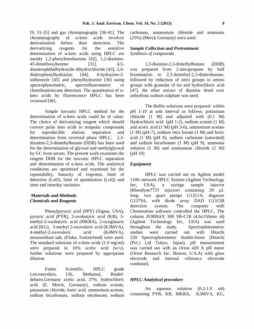
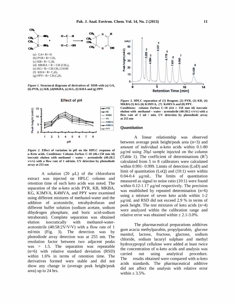
The keto acids contain a carbonyl group on adjacent carbon atom to carboxylic acid in PYR, KB, MKBA, KG, K3MVA, K4MVA, and PPY and reacted with DDB to form cyclic ring structure as substituted tetramethyl dihydro-pyrazinol (Fig. 1). The reaction was slow at room temperature, but was fairly rapid by warming at 70-900C. The derivatives were also extractable in chloroform and the chloroform extract was examined for HPLC elution and separation of α keto acids. The systematic study was initiated by optimization of the derivatization condition and elution from HPLC column. Each of the derivatives formed eluted from HPLC column with methanol-water and indicated single peak and separated from derivatizing reagent. The experimental conditions which gave maximum response (average peak height/peak area) were selected as optimum. The effect of pH, amount of derivatizing reagent (DDB) added and warming time and temperature were examined on derivatization. The pH was optimized within the range 1-10 at unit interval and then at 0.5 unit interval. The derivatization was observed in acidic medium within pH 2-5, with maximum response at 3.5 and acetic acid- sodium acetate buffer was selected (Fig. 2). The addition of the derivatizing reagent was not critical, as long as excess of the reagent was available. The addition of 1ml of the solution was selected. The warming temperature between 50-90 0C at 10 0C of interval and warming time between 5-30 min at 5 min of interval were examined. Warming temperature of 80 0C for 20 min was selected.
Pak. J. Anal. Environ. Chem. Vol. 14, No. 2 (2013)
11
(a) GA= R= H
(b) PYR= R= CH3 (c) KB= R= C2H5 (d) MKBA = R = CH (CH3)2 (e) KG= R= CH.CH2.COOH (f) KHA= R= C4H9 (g) PPY= R= CH2C6H5 Figure 1. Structural diagrams of derivatives of DDB with (a) GA, (b) PYR, (c) KB, (d)MKBA, (e) KG, (f) KHA and (g) PPY
Figure 2. Effect of variation in pH on the HPLC response of -Keto acids. Conditions: Column Zorbax C-18 (46.150 mm id) isocratic elution with methanol – water – acetonitrile (40:28:2 v/v/v) with a flow rate of 1 ml/min. UV detection by photodiode array at 255 nm
A solution (20 µL) of the chloroform
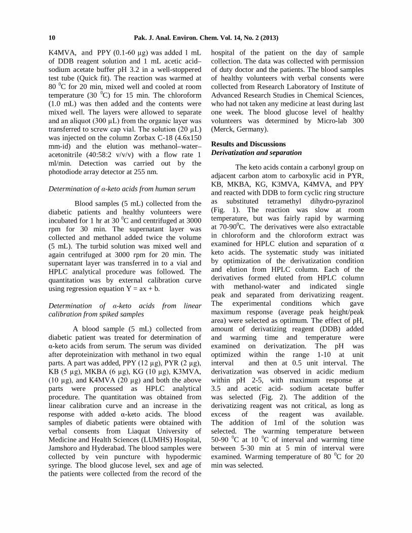
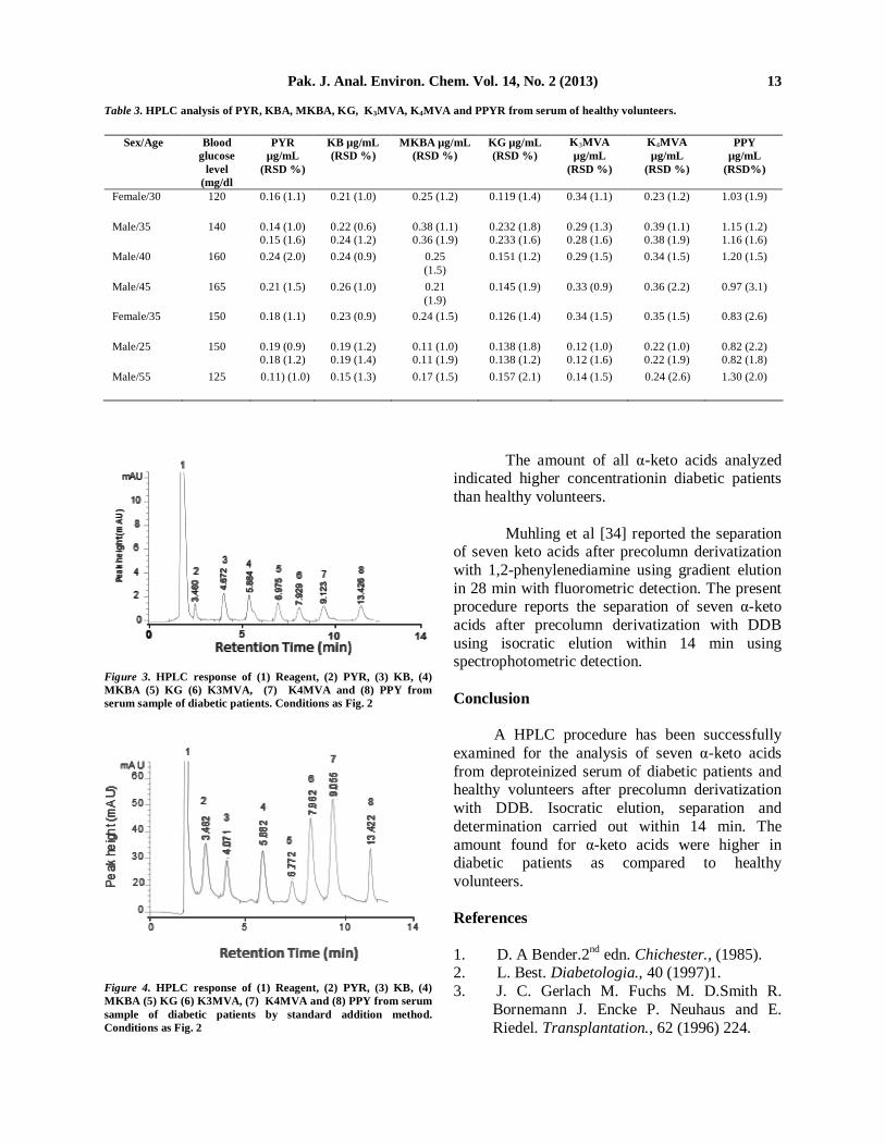
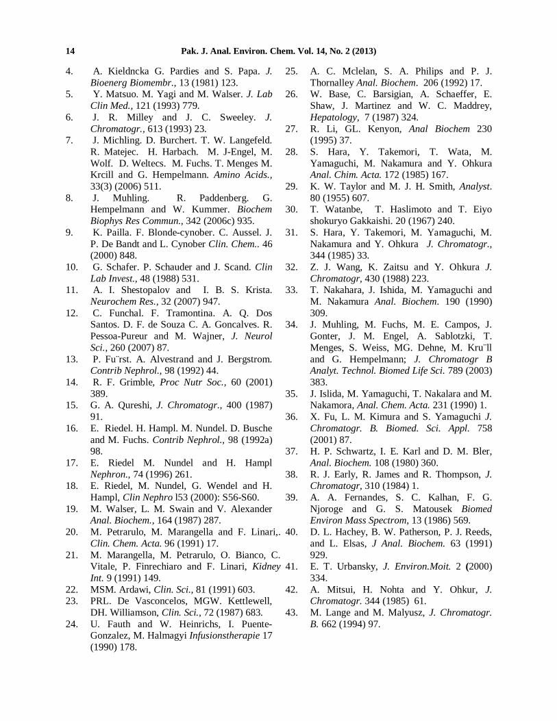
extract was injected on HPLC column and retention time of each keto acids was noted. The separation of the α-keto acids PYR, KB, MKBA, KG, K3MVA, K4MVA, and PPY were examined using different mixtures of methanol-water and the addition of acetonitrile, tetrahydrofuran and different buffer solution (sodium acetate, sodium dihydrogen phosphate, and boric acid-sodium tetraborate). Complete separation was obtained elution isocratically with methanol-water-acetonitrile (40:58:2V/V/V) with a flow rate of 1 ml/min (Fig. 3). The detection was by photodiode array detection was at 255 nm. The resolution factor between two adjacent peaks was > 1.5. The separation was repeatable (n=6) with relative standard deviation (RSD) within 1.6% in terms of retention time. The derivatives formed were stable and did not show any change in (average peak height/peak area) up to 24 hrs.
Figure 3. HPLC separation of (1) Reagent, (2) PYR, (3) KB, (4) MKBA (5) KG (6) K3MVA, (7) K4MVA and (8) PPY. Conditions: column Zorbax C-18 (4.6 150 mm id) isocratic elution with methanol – water - acetonitrile (40:58:2 v/v/v) with a flow rate of 1 ml / min. UV detection by photodiode array at 255 nm Quantitation
A linear relationship was observed between average peak height/peak area (n=3) and amount of individual α-keto acids within 0.1-80 µg/ml using 20µl sample injected on the column (Table 1). The coefficient of determinations (R2) calculated from 5 to 8 calibrators were calculated within 0.991- 0.999. Limits of detection (LoD) and limit of quantitation (LoQ) and (10:1) were within 0.04-0.4 µg/mL. The limits of quantitation measured as signal to noise ratio (10:1) were found within 0.12-1.17 µg/ml respectively. The precision was established by repeated determination (n=6) using a mixture of seven keto acids within 1-2 µg/mL and RSD did not exceed 2.9 % in terms of peak height. The test mixtures of keto acids (n=4) were analyzed within the calibration range and relative error was obtained within ± 2.1-3.0%.
The pharmaceutical preparations additives
gum acacia methylparabin, propylparabin, glucose manitol, lactose, fructose, glactose, sodium chloride, sodium lacuryl sulphate and methyl hydroxypropyl cellulose were added at least twice the concentration of α-keto acids and analysis was carried out using analytical procedure. The results obtained were compared with α-keto acids standards. The pharmaceutical additive did not affect the analysis with relative error within ± 3.5%.
Pak. J. Anal. Environ. Chem. Vol. 14, No. 2 (2013)
12
Table 1. Quantitative HPLC Data of α-keto acids using DDB as derivatizing Reagent.
Name of compound
Calibration
Range
R2 Regression equation
LOD µg/mL
LOQ µg/mL
PYR 0.1-10 0.9912 y=1.811+ 1.0525
0.04 0.12
KB 0.2-21 0.9905 y = 1.0854 + 0.8542
0.06 0.18
MKBA 0.2-15 0.9923 y = 1.239 + 0.3127
0.09 0.27
KG 0.4-60 0.9905 y = 0.286 + 0.9373
0.098 0.294
K3MVA 0.5-60 0.9914 y = 0.3193 + 1.541
0.13 0.39
K4MVA 0.5-60 0.9927 y = 0.2985 + 0.877
0.1 0.3
PPY 0.3-60 0.9912 y = 0.3661 + 1.2033
0.07 0.21
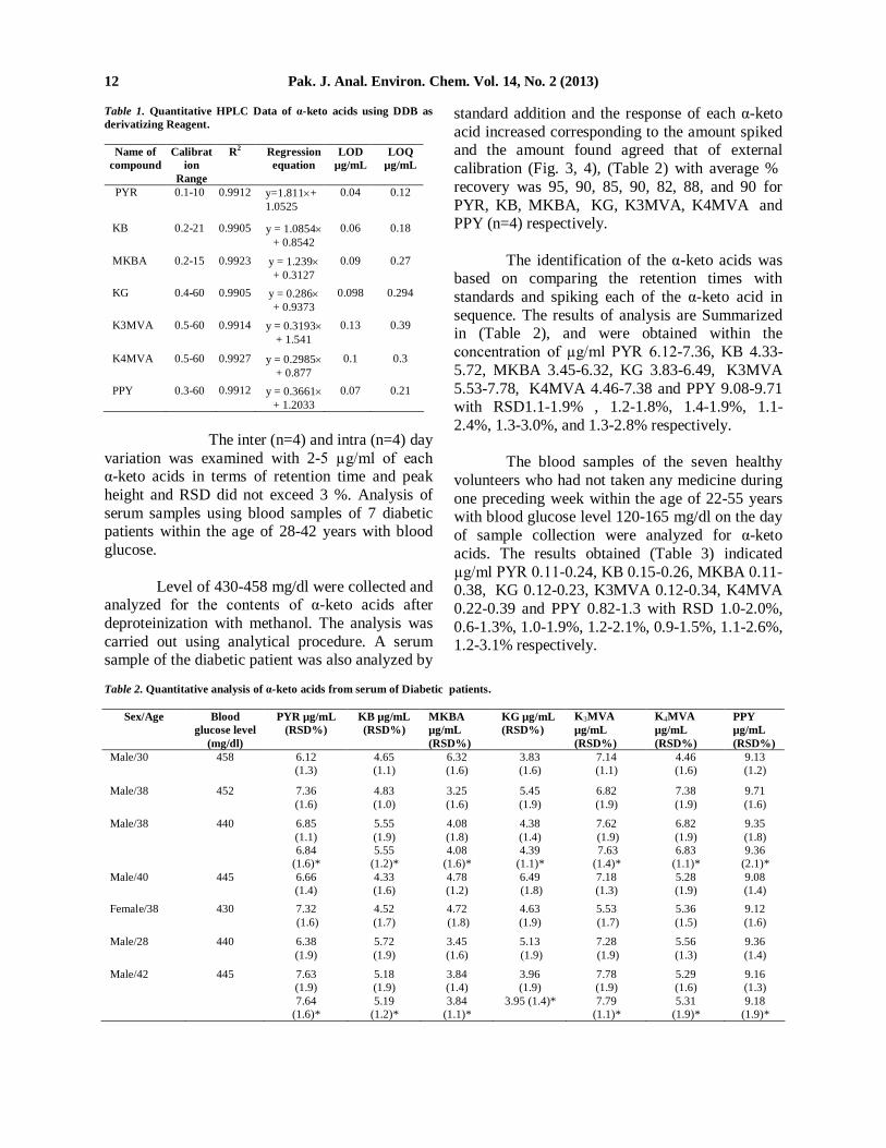
The inter (n=4) and intra (n=4) day variation was examined with 2-5 µg/ml of each α-keto acids in terms of retention time and peak height and RSD did not exceed 3 %. Analysis of serum samples using blood samples of 7 diabetic patients within the age of 28-42 years with blood glucose.
Level of 430-458 mg/dl were collected and
analyzed for the contents of α-keto acids after deproteinization with methanol. The analysis was carried out using analytical procedure. A serum sample of the diabetic patient was also analyzed by
standard addition and the response of each α-keto acid increased corresponding to the amount spiked and the amount found agreed that of external calibration (Fig. 3, 4), (Table 2) with average % recovery was 95, 90, 85, 90, 82, 88, and 90 for PYR, KB, MKBA, KG, K3MVA, K4MVA and PPY (n=4) respectively.
The identification of the α-keto acids was
based on comparing the retention times with standards and spiking each of the α-keto acid in sequence. The results of analysis are Summarized in (Table 2), and were obtained within the concentration of µg/ml PYR 6.12-7.36, KB 4.33-5.72, MKBA 3.45-6.32, KG 3.83-6.49, K3MVA 5.53-7.78, K4MVA 4.46-7.38 and PPY 9.08-9.71 with RSD1.1-1.9% , 1.2-1.8%, 1.4-1.9%, 1.1-2.4%, 1.3-3.0%, and 1.3-2.8% respectively.
The blood samples of the seven healthy volunteers who had not taken any medicine during one preceding week within the age of 22-55 years with blood glucose level 120-165 mg/dl on the day of sample collection were analyzed for α-keto acids. The results obtained (Table 3) indicated µg/ml PYR 0.11-0.24, KB 0.15-0.26, MKBA 0.11-0.38, KG 0.12-0.23, K3MVA 0.12-0.34, K4MVA 0.22-0.39 and PPY 0.82-1.3 with RSD 1.0-2.0%, 0.6-1.3%, 1.0-1.9%, 1.2-2.1%, 0.9-1.5%, 1.1-2.6%, 1.2-3.1% respectively.
Table 2. Quantitative analysis of α-keto acids from serum of Diabetic patients.
Sex/Age Blood glucose level
(mg/dl)
PYR µg/mL (RSD%)
KB µg/mL (RSD%)
MKBA µg/mL (RSD%)
KG µg/mL (RSD%)
K3MVA µg/mL (RSD%)
K4MVA µg/mL (RSD%)
PPY µg/mL (RSD%)
Male/30 458 6.12 (1.3)
4.65 (1.1)
6.32 (1.6)
3.83 (1.6)
7.14 (1.1)
4.46 (1.6)
9.13 (1.2)
Male/38 452 7.36 (1.6)
4.83 (1.0)
3.25 (1.6)
5.45 (1.9)
6.82 (1.9)
7.38 (1.9)
9.71 (1.6)
Male/38 440 6.85 (1.1) 6.84
(1.6)*
5.55 (1.9) 5.55
(1.2)*
4.08 (1.8) 4.08
(1.6)*
4.38 (1.4) 4.39
(1.1)*
7.62 (1.9) 7.63 (1.4)*
6.82 (1.9) 6.83
(1.1)*
9.35 (1.8) 9.36
(2.1)* Male/40 445 6.66
(1.4) 4.33 (1.6)
4.78 (1.2)
6.49 (1.8)
7.18 (1.3)
5.28 (1.9)
9.08 (1.4)
Female/38 430 7.32 (1.6)
4.52 (1.7)
4.72 (1.8)
4.63 (1.9)
5.53 (1.7)
5.36 (1.5)
9.12 (1.6)
Male/28 440 6.38 (1.9)
5.72 (1.9)
3.45 (1.6)
5.13 (1.9)
7.28 (1.9)
5.56 (1.3)
9.36 (1.4)
Male/42 445 7.63 (1.9) 7.64
(1.6)*
5.18 (1.9) 5.19
(1.2)*
3.84 (1.4) 3.84
(1.1)*
3.96 (1.9)
3.95 (1.4)*
7.78 (1.9) 7.79
(1.1)*
5.29 (1.6) 5.31
(1.9)*
9.16 (1.3) 9.18
(1.9)*
Pak. J. Anal. Environ. Chem. Vol. 14, No. 2 (2013)
13
Table 3. HPLC analysis of PYR, KBA, MKBA, KG, K3MVA, K4MVA and PPYR from serum of healthy volunteers.
Sex/Age Blood glucose
level (mg/dl
PYR µg/mL
(RSD %)
KB µg/mL (RSD %)
MKBA µg/mL (RSD %)
KG µg/mL (RSD %)
K3MVA µg/mL
(RSD %)
K4MVA µg/mL
(RSD %)
PPY µg/mL
(RSD%)
Female/30 120 0.16 (1.1) 0.21 (1.0) 0.25 (1.2) 0.119 (1.4) 0.34 (1.1) 0.23 (1.2) 1.03 (1.9)
Male/35 140 0.14 (1.0) 0.15 (1.6)
0.22 (0.6) 0.24 (1.2)
0.38 (1.1) 0.36 (1.9)
0.232 (1.8) 0.233 (1.6)
0.29 (1.3) 0.28 (1.6)
0.39 (1.1) 0.38 (1.9)
1.15 (1.2) 1.16 (1.6)
Male/40 160 0.24 (2.0) 0.24 (0.9) 0.25 (1.5)
0.151 (1.2) 0.29 (1.5) 0.34 (1.5) 1.20 (1.5)
Male/45 165 0.21 (1.5) 0.26 (1.0) 0.21 (1.9)
0.145 (1.9) 0.33 (0.9) 0.36 (2.2) 0.97 (3.1)
Female/35 150 0.18 (1.1) 0.23 (0.9) 0.24 (1.5) 0.126 (1.4) 0.34 (1.5) 0.35 (1.5) 0.83 (2.6)
Male/25 150 0.19 (0.9) 0.18 (1.2)
0.19 (1.2) 0.19 (1.4)
0.11 (1.0) 0.11 (1.9)
0.138 (1.8) 0.138 (1.2)
0.12 (1.0) 0.12 (1.6)
0.22 (1.0) 0.22 (1.9)
0.82 (2.2) 0.82 (1.8)
Male/55 125 0.11) (1.0) 0.15 (1.3) 0.17 (1.5) 0.157 (2.1) 0.14 (1.5) 0.24 (2.6) 1.30 (2.0)
Figure 3. HPLC response of (1) Reagent, (2) PYR, (3) KB, (4) MKBA (5) KG (6) K3MVA, (7) K4MVA and (8) PPY from serum sample of diabetic patients. Conditions as Fig. 2
Figure 4. HPLC response of (1) Reagent, (2) PYR, (3) KB, (4) MKBA (5) KG (6) K3MVA, (7) K4MVA and (8) PPY from serum sample of diabetic patients by standard addition method. Conditions as Fig. 2
The amount of all α-keto acids analyzed
indicated higher concentrationin diabetic patients than healthy volunteers.
Muhling et al [34] reported the separation of seven keto acids after precolumn derivatization with 1,2-phenylenediamine using gradient elution in 28 min with fluorometric detection. The present procedure reports the separation of seven α-keto acids after precolumn derivatization with DDB using isocratic elution within 14 min using spectrophotometric detection. Conclusion A HPLC procedure has been successfully examined for the analysis of seven α-keto acids from deproteinized serum of diabetic patients and healthy volunteers after precolumn derivatization with DDB. Isocratic elution, separation and determination carried out within 14 min. The amount found for α-keto acids were higher in diabetic patients as compared to healthy volunteers. References 1. D. A Bender.2nd edn. Chichester., (1985). 2. L. Best. Diabetologia., 40 (1997)1. 3. J. C. Gerlach M. Fuchs M. D.Smith R.
Bornemann J. Encke P. Neuhaus and E. Riedel. Transplantation., 62 (1996) 224.
Pak. J. Anal. Environ. Chem. Vol. 14, No. 2 (2013)
14
4. A. Kieldncka G. Pardies and S. Papa. J. Bioenerg Biomembr., 13 (1981) 123.
5. Y. Matsuo. M. Yagi and M. Walser. J. Lab Clin Med., 121 (1993) 779.
6. J. R. Milley and J. C. Sweeley. J. Chromatogr., 613 (1993) 23.
7. J. Michling. D. Burchert. T. W. Langefeld. R. Matejec. H. Harbach. M. J-Engel, M. Wolf. D. Weltecs. M. Fuchs. T. Menges M. Krcill and G. Hempelmann. Amino Acids., 33(3) (2006) 511.
8. J. Muhling. R. Paddenberg. G. Hempelmann and W. Kummer. Biochem Biophys Res Commun., 342 (2006c) 935.
9. K. Pailla. F. Blonde-cynober. C. Aussel. J. P. De Bandt and L. Cynober Clin. Chem.. 46 (2000) 848.
10. G. Schafer. P. Schauder and J. Scand. Clin Lab Invest., 48 (1988) 531.
11. A. I. Shestopalov and I. B. S. Krista. Neurochem Res., 32 (2007) 947.
12. C. Funchal. F. Tramontina. A. Q. Dos Santos. D. F. de Souza C. A. Goncalves. R. Pessoa-Pureur and M. Wajner, J. Neurol Sci., 260 (2007) 87.
13. P. Fu¨rst. A. Alvestrand and J. Bergstrom. Contrib Nephrol., 98 (1992) 44.
14. R. F. Grimble, Proc Nutr Soc., 60 (2001) 389.
15. G. A. Qureshi, J. Chromatogr., 400 (1987) 91.
16. E. Riedel. H. Hampl. M. Nundel. D. Busche and M. Fuchs. Contrib Nephrol., 98 (1992a) 98.
17. E. Riedel M. Nundel and H. Hampl Nephron., 74 (1996) 261.
18. E. Riedel, M. Nundel, G. Wendel and H. Hampl, Clin Nephro l53 (2000): S56-S60.
19. M. Walser, L. M. Swain and V. Alexander Anal. Biochem., 164 (1987) 287.
20. M. Petrarulo, M. Marangella and F. Linari,. Clin. Chem. Acta. 96 (1991) 17.
21. M. Marangella, M. Petrarulo, O. Bianco, C. Vitale, P. Finrechiaro and F. Linari, Kidney Int. 9 (1991) 149.
22. MSM. Ardawi, Clin. Sci., 81 (1991) 603. 23. PRL. De Vasconcelos, MGW. Kettlewell,
DH. Williamson, Clin. Sci., 72 (1987) 683. 24. U. Fauth and W. Heinrichs, I. Puente-
Gonzalez, M. Halmagyi Infusionstherapie 17 (1990) 178.
25. A. C. Mclelan, S. A. Philips and P. J. Thornalley Anal. Biochem. 206 (1992) 17.
26. W. Base, C. Barsigian, A. Schaeffer, E. Shaw, J. Martinez and W. C. Maddrey, Hepatology, 7 (1987) 324.
27. R. Li, GL. Kenyon, Anal Biochem 230 (1995) 37.
28. S. Hara, Y. Takemori, T. Wata, M. Yamaguchi, M. Nakamura and Y. Ohkura Anal. Chim. Acta. 172 (1985) 167.
29. K. W. Taylor and M. J. H. Smith, Analyst. 80 (1955) 607.
30. T. Watanbe, T. Haslimoto and T. Eiyo shokuryo Gakkaishi. 20 (1967) 240.
31. S. Hara, Y. Takemori, M. Yamaguchi, M. Nakamura and Y. Ohkura J. Chromatogr., 344 (1985) 33.
32. Z. J. Wang, K. Zaitsu and Y. Ohkura J. Chromatogr, 430 (1988) 223.
33. T. Nakahara, J. Ishida, M. Yamaguchi and M. Nakamura Anal. Biochem. 190 (1990) 309.
34. J. Muhling, M. Fuchs, M. E. Campos, J. Gonter, J. M. Engel, A. Sablotzki, T. Menges, S. Weiss, MG. Dehne, M. Kru¨ll and G. Hempelmann; J. Chromatogr B Analyt. Technol. Biomed Life Sci. 789 (2003) 383.
35. J. Islida, M. Yamaguchi, T. Nakalara and M. Nakamora, Anal. Chem. Acta. 231 (1990) 1.
36. X. Fu, L. M. Kimura and S. Yamaguchi J. Chromatogr. B. Biomed. Sci. Appl. 758 (2001) 87.
37. H. P. Schwartz, I. E. Karl and D. M. Bler, Anal. Biochem. 108 (1980) 360.
38. R. J. Early, R. James and R. Thompson, J. Chromatogr, 310 (1984) 1.
39. A. A. Fernandes, S. C. Kalhan, F. G. Njoroge and G. S. Matousek Biomed Environ Mass Spectrom, 13 (1986) 569.
40. D. L. Hachey, B. W. Patherson, P. J. Reeds, and L. Elsas, J Anal. Biochem. 63 (1991) 929.
41. E. T. Urbansky, J. Environ.Moit. 2 (2000) 334.
42. A. Mitsui, H. Nohta and Y. Ohkur, J. Chromatogr. 344 (1985) 61.
43. M. Lange and M. Malyusz, J. Chromatogr. B. 662 (1994) 97.

Pak. J. Anal. Environ. Chem. Vol. 14, No. 2 (2013)
15
44. G. Garibotto, P. Ancarani, R. Russo, MR. Sala, F. Fiorini and E. Paoletti J. Chromatogr. 572 (1991) 11.
45. T. Hirata, M. Kai, K. Kohashi and Y. Ohkura, 226 (1981) 25.
46. M. Fuchs, J. Engel, M. Campos, R. Matejec, M. Henrich, H. Harbach, M. Wolff, K. Weismu¨ ller, T. Menges, MC. Heidt, ID. Welters, M. Kru¨ ll, G. Hempelmann and J. Muhling , Amino Acids, 36 (2009) 1.


![RESEARCH Open Access Are platinum agents, paclitaxel and ... · Eriko Takatori1†, Tadahiro Shoji1*†, Seisuke Kumagai1†, Takashi Sawai2†, Akira Kurose3 ... [2,3], is effective](https://static.fdocument.org/doc/165x107/5beade0509d3f2cb5e8b7877/research-open-access-are-platinum-agents-paclitaxel-and-eriko-takatori1.jpg)
![Badanie wysokospinowych stanów z seniority ν > 2,3 w ... · izomerów 10+ w izotopach Sn [Fot11, Pie11] oraz bardziej kompletne struktury obejmujące również izotopy nieparzyste](https://static.fdocument.org/doc/165x107/5f3f384ed8c10e3fab6a7f2d/badanie-wysokospinowych-stanw-z-seniority-23-w-izomerw-10-w-izotopach.jpg)

![élyi[1])toBracewell)[2,3] · Technical)Notes) Converting)Erdélyi[1])toBracewell)[2,3]! Erdélyidefinedhis!Hankel!transform!of!order!v!as![1]:! ( )( )1 2 0 gy f xJ xy xy dx(;) ()υ](https://static.fdocument.org/doc/165x107/5edc9e48ad6a402d66675c69/lyi1tobracewell23-technicalnotes-convertingerdlyi1tobracewell23.jpg)